Embed Size (px)
Citation preview
ANTENOR LUIZ LIMA RODRIGUES
TESTE DE CAMINHADA DE SEIS MINUTOS COMO RECURSO PARA PRESCRIÇÃO DE TREINAMENTO FÍSICO DE ALTA INTENSIDADE PARA PACIENTES COM DOENÇA
PULMONAR OBSTRUTIVA CRÔNICA
Londrina
2016
ANTENOR LUIZ LIMA RODRIGUES
TESTE DE CAMINHADA DE SEIS MINUTOS COMO RECURSO PARA PRESCRIÇÃO DE TREINAMENTO FÍSICO DE ALTA INTENSIDADE PARA PACIENTES COM DOENÇA
PULMONAR OBSTRUTIVA CRÔNICA
Dissertação apresentada ao Programa de Pós-Graduação em Ciências da Reabilitação (Programa Associado entre Universidade Estadual de Londrina [UEL] e Universidade Norte do Paraná [UNOPAR]), como requisito parcial à obtenção do título de Mestre em Ciências da Reabilitação. Orientador: Prof. Dr. Fabio Pitta
Londrina 2016
ANTENOR LUIZ LIMA RODRIGUES
TESTE DE CAMINHADA DE SEIS MINUTOS COMO RECURSO PARA PRESCRIÇÃO DE TREINAMENTO FÍSICO DE ALTA INTENSIDADE PARA PACIENTES COM DOENÇA
PULMONAR OBSTRUTIVA CRÔNICA
Dissertação apresentada ao Programa de Pós-Graduação em Ciências da Reabilitação (Programa Associado entre Universidade Estadual de Londrina [UEL] e Universidade Norte do Paraná [UNOPAR]), como requisito parcial à obtenção do título de Mestre em Ciências da Reabilitação.
BANCA EXAMINADORA
____________________________________ Prof. Dr. Fabio Pitta
Universidade Estadual de Londrina, UEL
____________________________________ Prof. Dr. Dartagnan Pinto Guedes
Universidade Norte do Paraná, UNOPAR
____________________________________ Prof. Dr. Wellington Pereira dos Santos
Yamaguti Departamento de Reabilitação,
Hospital Sírio-Libanês, HSL
Londrina, 04 de Fevereiro de 2016.

Dedico este trabalho à minha familia
AGRADECIMENTOS
Agradeço primeiramente à Deus, por ter me colocado em uma família que
sempre fez o possível, e o que parecia impossível, por mim. Depois, por ter colocado
em meu caminho tantas pessoas dispostas a me ajudar (amigos, professores,
colegas e etc.). Agradeço a Deus também por sempre ter me acompanhado em
todos os meus passos, mesmo que em alguns momentos eu não tenha me
lembrado. Por fim, agradeço a Deus por ter me capacitado a chegar até aqui.
Mesmo sem entender eu sei que seus planos são maiores que os meus.
Agradeço também ao meu orientador, Prof. Fabio Pitta, pela confiança, pela
oportunidade, pela paciência, por todo o conhecimento compartilhado e por ter
apostado em mim desde o principio. Meu agradecimento é imensurável! Graças a
você eu tenho a oportunidade de fazer hoje o que eu gosto! Muito obrigado!
Agradeço também por toda a paciência, companheirismo, risadas, e
momentos que não podem ser descritos, nem compartilhados aqui, aos meus
amigos, amigos que trabalham comigo, mas que não são amigos de trabalho:
Fernanda Moraka; Andrea Morita (Japa); Thaís Rebeca; Igor Brito; e também às
mais recentes Larissa, e Debora. Além do Wesley (negão), e do Fernando Vieira.
A todos os colegas do LFIP meu muito obrigado por terem me
recebido e compartilhado todo o conhecimento, que nunca foi pouco. Muito obrigado
por todas as risadas, todos os momentos de trabalho que foram transformados em
diversão, e também obrigado pelos jogos- o Game of Trolls nunca será esquecido!
Todos esses momentos e todas as experiencias (boas e ruins) que passei durante
esses quatro anos de LFIP construiram a pessoa que eu sou hoje.
A todos meu muito obrigado!

“If you close your eyes
Because the house is on fire
And think you couldn't move
Until the fire dies
The things you never did
Cause you might die trying...
...You will be as good as a dead”
Dave Matthews Band / Mark Batson
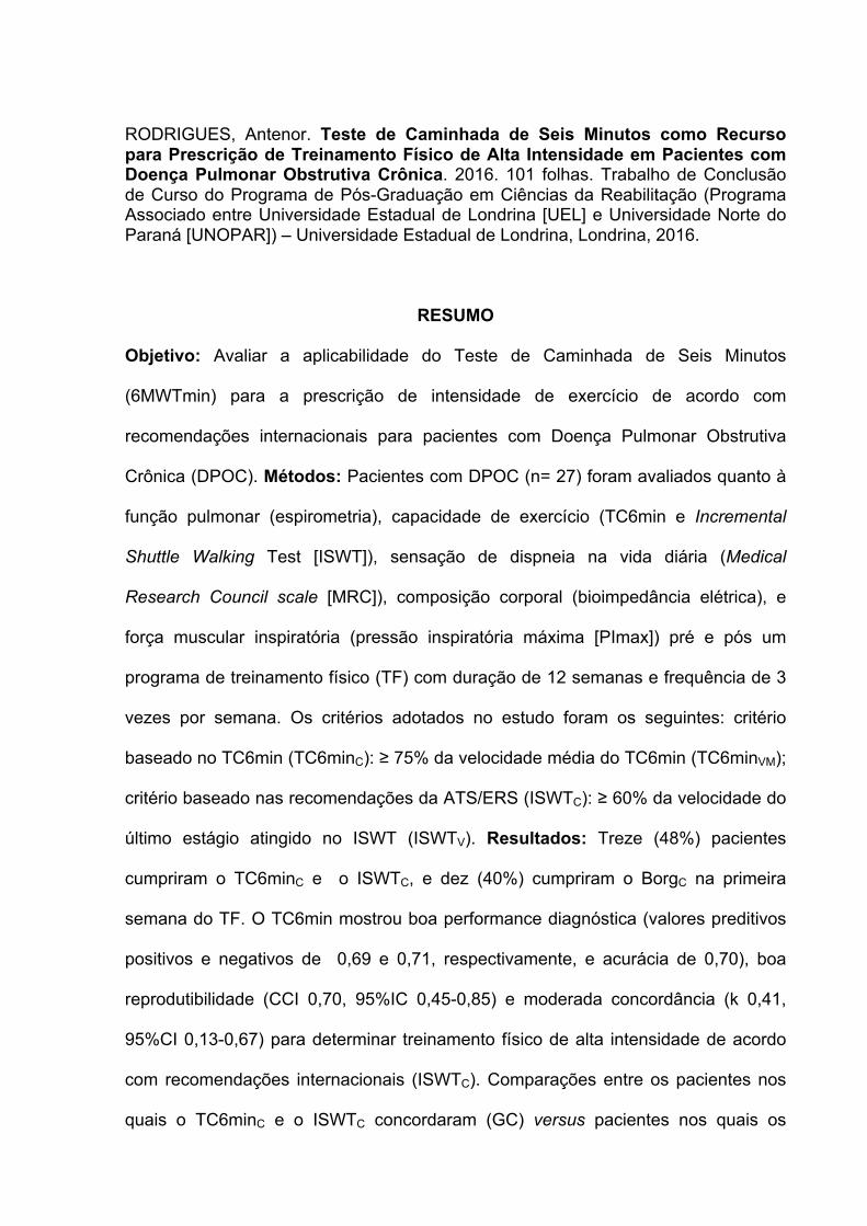
RODRIGUES, Antenor. Teste de Caminhada de Seis Minutos como Recurso para Prescrição de Treinamento Físico de Alta Intensidade em Pacientes com Doença Pulmonar Obstrutiva Crônica. 2016. 101 folhas. Trabalho de Conclusão de Curso do Programa de Pós-Graduação em Ciências da Reabilitação (Programa Associado entre Universidade Estadual de Londrina [UEL] e Universidade Norte do Paraná [UNOPAR]) – Universidade Estadual de Londrina, Londrina, 2016.
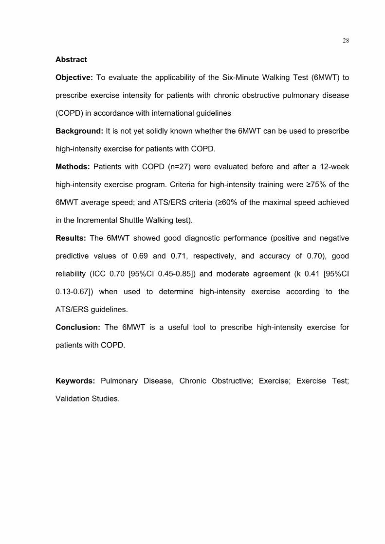
RESUMO
Objetivo: Avaliar a aplicabilidade do Teste de Caminhada de Seis Minutos
(6MWTmin) para a prescrição de intensidade de exercício de acordo com
recomendações internacionais para pacientes com Doença Pulmonar Obstrutiva
Crônica (DPOC). Métodos: Pacientes com DPOC (n= 27) foram avaliados quanto à
função pulmonar (espirometria), capacidade de exercício (TC6min e Incremental
Shuttle Walking Test [ISWT]), sensação de dispneia na vida diária (Medical
Research Council scale [MRC]), composição corporal (bioimpedância elétrica), e
força muscular inspiratória (pressão inspiratória máxima [PImax]) pré e pós um
programa de treinamento físico (TF) com duração de 12 semanas e frequência de 3
vezes por semana. Os critérios adotados no estudo foram os seguintes: critério
baseado no TC6min (TC6minC): ≥ 75% da velocidade média do TC6min (TC6minVM);
critério baseado nas recomendações da ATS/ERS (ISWTC): ≥ 60% da velocidade do
último estágio atingido no ISWT (ISWTV). Resultados: Treze (48%) pacientes
cumpriram o TC6minC e o ISWTC, e dez (40%) cumpriram o BorgC na primeira
semana do TF. O TC6min mostrou boa performance diagnóstica (valores preditivos
positivos e negativos de 0,69 e 0,71, respectivamente, e acurácia de 0,70), boa
reprodutibilidade (CCI 0,70, 95%IC 0,45-0,85) e moderada concordância (k 0,41,
95%CI 0,13-0,67) para determinar treinamento físico de alta intensidade de acordo
com recomendações internacionais (ISWTC). Comparações entre os pacientes nos
quais o TC6minC e o ISWTC concordaram (GC) versus pacientes nos quais os

critérios não concordaram (GNC) na primeira semana de TF revelaram que o GC
apresentou maior velocidade (3,9 ± 2,1 vs. 3,0 ± 0,74 km/h; P= 0,01), maior
%TC6minVM (77 [76 - 86] vs. 74 [62 - 75]; P< 0,001), maior %ISWTV (70 ± 4 vs. 52 ±
10; P< 0,001) e maior relação 6MWTVM/ISWTV (0,87 ± 0,08 vs. 0,70 ± 0,09; P=0,04).
Adicionalmente, pacientes nos quais 75% da TC6minvm era maior ou igual que 60%
da ISWTv foram comparados com os casos que 75% da TC6minVM era menor que
60% da ISWTV. Esses resultados mostraram pior função pulmonar (CVF 65 ± 13 vs.
82 ± 20; VEF1 45 [31 - 55] vs. 61 [44 - 69] %predito, P< 0.05 para todos), maior
sensação de dispneia na vida diária (4 [3-4] vs. 2,5 [2-4], pontos na escala MRC; P=
0,04), menor ISWT (379 ± 100 vs. 516 ± 207, m, P= 0.03) e maior relação
6MWTVM/ISWTV (0.89 ± 0.6 vs. 0.72 ± 0.05 P< 0,001) para o grupo no qual 75% da
TC6minVM era igual ou maior que 60% da ISWTV. Após TF os pacientes
apresentaram aumento da capacidade de exercício (6MWT pré vs pós TF: 464 ±
70,4 vs. 506 ± 85 m; P= 0,003; e 86 ± 14 vs. 94 ± 17 %predito; P= 0.003). Após TF,
o grupo GNC teve maior aumento na velocidade em km/h do que o GC (66 [46 - 79]
vs. 28 [21 - 40] %∆, P= 0.02), e em %ISWTV pós TF (63 [46 - 67] vs. 27 [14 - 30] %∆;
P= 0,01). Conclusão: O TC6min se mostrou uma ferramenta útil para a prescrição
de treinamento de alta intensidade em pacientes com DPOC.
Palavras-chave: Doença Pulmonar Obstrutiva Crônica; Exercício; Tolerância ao Exercício; Estudos de Validação.

RODRIGUES, Antenor. Is the Six-Minute Walking test a tool to prescribe high-intensity exercise in Chronic Obstructive Pulmonary Disease?2016. 101 folhas. Trabalho de Conclusão de Curso do Programa de Pós-Graduação em Ciências da Reabilitação (Programa Associado entre Universidade Estadual de Londrina [UEL] e Universidade Norte do Paraná [UNOPAR]) – Universidade Estadual de Londrina, Londrina, 2016.
ABSTRACT
Objective: To evaluate the applicability of the Six-Minute Walking Test (6MWT) to
prescribe exercise intensity in accordance with international guidelines in patients
with chronic obstructive pulmonary disease (COPD). Methods: Patients with COPD
(n=27) were evaluated concerning lung function (spirometry), exercise capacity
(6MWT and Incremental Shuttle Walking Test [ISWT]), dyspnea sensation in daily life
(Medical Research Council Scale [MRC]), body composition (bioelectrical
impedance), and inspiratory muscle strength (maximal inspiratory pressure [MIP])
before and after a 12-week, 3 times/week, high-intensity exercise training program
(ET). High-intensity criteria were: 6MWT criteria (6MWTC): ≥75% of the 6MWT
average speed (6MWTAS); ATS/ERS criteria (ISWTC): ≥60% of the speed achieved in
the ISWT last stage (ISWTS). Results: According to 6MWTC and ISWTC, 13 (48%)
patients achieved the desired training intensity in the first week of the ET whereas ten
(40%) patients achieved BorgC. The 6MWT showed good diagnostic performance
(positive and negative predictive values of 0.69 and 0.71, respectively, and accuracy
of 0.70), good reliability (ICC 0.703, 95%CI 0.447-0.853) and moderate agreement (k
0.41, 95%CI 0.13-0.67) when used to determine high-intensity exercise according to
the ATS/ERS guidelines. When comparing patients in which the 6MWTC and ISWTC
agreed (GAweek1) versus the cases in which they did not agree (GNAweek1) in the first
week of ET, results revealed that GAweek1 had higher speed (3.9 ± 2.1 vs. 3 ± 0.74,
km/h, P= 0.01), %6MWTAS (77 [76 - 86] vs. 74 [62 – 75], P< 0.001), %ISWTS (70.4 ±
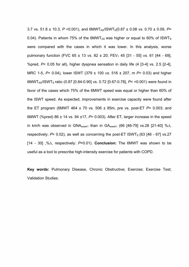
3.7 vs. 51.8 ± 10.3, P <0.001), and 6MWTAS/ISWTS(0.87 ± 0.08 vs. 0.70 ± 0.09, P=
0.04). Patients in whom 75% of the 6MWTAS was higher or equal to 60% of ISWTS
were compared with the cases in which it was lower. In this analysis, worse
pulmonary function (FVC 65 ± 13 vs. 82 ± 20; FEV1 45 [31 - 55] vs. 61 [44 - 69],
%pred, P< 0.05 for all), higher dyspnea sensation in daily life (4 [3-4] vs. 2.5 [2-4],
MRC 1-5, P= 0.04), lower ISWT (379 ± 100 vs. 516 ± 207, m P= 0.03) and higher
6MWTAS/ISWTS ratio (0.87 [0.84-0.90] vs. 0.72 [0.67-0.78], P= <0.001) were found in
favor of the cases which 75% of the 6MWT speed was equal or higher than 60% of
the ISWT speed. As expected, improvements in exercise capacity were found after
the ET program (6MWT 464 ± 70 vs. 506 ± 85m, pre vs. post-ET P= 0.003; and
6MWT (%pred) 86 ± 14 vs. 94 ±17, P= 0.003). After ET, larger increase in the speed
in km/h was observed in GNAweek1 than in GAweek1 (66 [46-79] vs.28 [21-40] %∆,
respectively; P= 0.02), as well as concerning the post-ET ISWTS (63 [46 - 67] vs.27
[14 - 30] ,%∆, respectively: P=0.01). Conclusion: The 6MWT was shown to be
useful as a tool to prescribe high-intensity exercise for patients with COPD.
Key words: Pulmonary Disease, Chronic Obstructive; Exercise; Exercise Test;
Validation Studies.

LISTA DE ILUSTRAÇÕES
Figura 1 – Intensity of training at the first and 12th week of the exercise training
program. Left: % of the 6MWTAS. Rigth: % ISWTS. Lines are the cutoff for high-
intensity exercise training according to each criteria ................................................ 56
Figura 2 – Dyspnea (left) and fatigue (rigth) sensation during the first and 12th week of
the exercise training program. Lines are the cutoff for high-intensity exercise training
according to each criteria ........................................................................................... 57
Figura 3 – Bland-Altman plots of 60%ISWT – 75%6MWT at baseline (upper plot) and
post-ET (lower plot) .................................................................................................. 58
LISTA DE TABELAS
Tabela 1 – General characteristics of the included patients with COPD before
exercise training. ....................................................................................................... 49
Tabela 2 – Comparison between patients whose 6MWTC and ISWTC agree vs.
patients in whom these criteria did not agree at the first week of the exercise training
program. ................................................................................................................... 51
Tabela 3 – Differences between patients in whom 75% 6MWTAS was equal to or
higher than 60% ISWTS ............................................................................................ 53
Tabela 4 – Pre and post-exercise training characteristics of patients with COPD who
finished the program. ................................................................................................ 54
12
LISTA DE ABREVIATURAS E SIGLAS
[LA]: concentração de lactato
ACSM: American College of Sports Medicine
AFVD: nível de atividade física na vidadiaria
ATS: American Thoracic Society
AVD: atividades de vida diária
CO2: dióxido de carbono
CPT: capacidade pulmonar total
CS: critical speed
CVF: capacidade vital forçada
DPOC: Doença Pulmonar Obstrutiva Crônica
EADPOC: exacerbação aguda da DPOC
EENM: eletro-estimulação neuromuscular
ERS: European Respiratory Society
GOLD: Global Initiative for Chronic Obstructive Pulmonary Disease
H+: hidrogênio
ISO-VE: volume minuto no isotempo
ISO-VO2: consumo de oxigênio no isotempo
LA: limiar de lactato
MDCI: minima diferença clinicamente importante
O2: oxigênio
PaCO2: pressão arterial de monóxido de carbono
RER: quociente respiratório
RP: reabilitação pulmonar

13
sRaw: Resistência especíifica das vias aéreas
TC6min: Teste de caminhada de seis minutos
TC6minVM: velocidade média do TC6min
TCPE: Teste cardiopulmonar de esforço
TI/TTOT: relação entre tempo inspiratório e tempo total do ciclo respiratório
TLCO: fator de transferência do monóxido de carbono
VA: ventilação alveolar
VCO2: volume expirado de dióxido de carbono
VD: espaço morto
VE: volume minuto
VEF1: volume expiratório forçado no primeiro segundo
VEF1/CVF: relação entre volume expiratório forçado no primeiro segundo e capacidade
vital forçada
VO2: consumo de oxigênio
VO2max-VO2LA: diferença entre consumo máximo de oxigênio e consumo de oxigênio no
limiar de lactato
VO2max: Consumo máximo de oxigênio
VO2TC6min: consumo de oxigênio do TC6min
VO2TCPE: consumo máximo de oxigênio durante um TCPE
VR: volume residual
VRE: volume de reserve expiratório
VT: volume corrente
14
Sumário
1. INTRODUÇÃO ...................................................................................................... 15
2. REVISÃO DE LITERATURA ................................................................................. 17
2.1 Doença Pulmonar Obstrutiva Crônica (DPOC) ....................................................... 17
2.2 Limitação ao Exercício em Pacientes com DPOC .................................................. 18
2.2.1 Limitação ventilatória ............................................................................................ 18
2.2.2 Limitação muscular ............................................................................................... 20
2.2.3 Limitação Cardíaca. .............................................................................................. 21
2.2.4 Em resumo ............................................................................................................ 21
2.2 Efeitos e Prescrição de Programas de Treinamento Físico de Alta Intensidade
em Pacientes com DPOC ................................................................................................ 22
2.2.1 Efeitos dos programas de treinamento físico em pacientes com DPOC .............. 22
2.2.2Prescrição de treinamento físico de alta intensidade em pacientes com DPOC ... 23
2.2.3 O Teste de caminhada de seis minutos (TC6min) e seu uso como recurso para
prescrição de treinamento físico de alta intensidade em DPOC ................................... 24
3. ARTIGO ................................................................................................................. 27
4. Conclusão GERAL ............................................................................................... 59
5. REFERÊNCIAS ..................................................................................................... 60
6. APÊNDICES .......................................................................................................... 72
Apêndice A- Termo de cosentimento livre e esclarecido ................................................. 72
7. ANEXOS ................................................................................................................ 76
Anexo A- Normas de formatação da revista Heart and Lung. .......................................... 76
Anexo B- Parecer de aprovação do comitê de ética em pesquisa ................................. 101
15
1. INTRODUÇÃO
O Teste de Caminhada de Seis minutos (TC6min) é um teste
reprodutível, confiável e responsivo à intervenções em pacientes com Doença
Pulmonar Obstrutiva Crônica (DPOC). Trata-se de um dos testes de campo mais
utilizados para essa população1. No entanto, o uso de TC6min como recurso para
prescrição da intensidade de treinamento físico (TF) em alguns estudos2-5 tem
recentemente aumentado o interesse sobre o estudo deste tópico6.
A prescrição da intensidade de exercício de programas de TF deve
ser realizada de maneira personalizada e baseada na capacidade máxima de
exercício (Wmax) de cada indivíduo, indiferentemente se no caso de estados de
saúde ou de doença7,8. Apesar do fato do Teste Cardiopulmonar de Esforço (TCPE)
ser considerado o teste padrão ouro para avaliação da Wmax9, o uso do TC6min
para prescrição da intensidade de exercício baseia-se em respostas similares de
consumo máximo de oxigênio (VO2max) durante o TC6min e o TCPE em pacientes
com DPOC10. No entanto, vieses metodológicos (e.g., TCPE realizado em bicicleta e
pista não-padronizada para realização do TC6min) podem ter induzido resultados
cientificamente questionáveis6.
Paradoxalmente, alguns estudos tem encontrado que a velocidade
média do TC6min (TC6minVM) é similar ou menor que a velocidade máxima que um
individuo é capaz de sustentar enquanto mantem respostas integradas dos sistemas
envolvidos na oferta e no consumo de oxigênio, ou critical speed (CS)11,12. Em
pacientes com DPOC a CS foi encontrada como sendo abaixo de 85% da Wmax,
indicando o perfil do TC6min como um teste de avaliação da capacidade
funcional13,14 (ou sub–máxima) de exercício que é o perfil de resposta para o qual o
teste foi desenvolvido1.
Apesar dos resultados contraditórios na literatura sobre o TC6min
ser um teste que avalia a capacidade máxima ou sub máxima de exercício em
pacientes com DPOC6,10-12,15,16, diferentes porcentagens da TC6minVM tem sido
utilizadas para prescrição da intensidade de programas de TF em pacientes com
DPOC2-5. Estes programas se mostraram comprovadamente eficazes na promoção
do aumento da capacidade de exercício e da qualidade de vida, ultrapassando os
valores de mínima diferença clinicamente importante (MDCI) para esta população.
16
Com a finalidade de testar se o TC6min poderia ser utilizado como
recurso para prescrição de TF de alta intensidade em paciente com DPOC, um
estudo recente demostrou uma ampla variabilidade na resposta do consumo de
oxigênio (VO2) quando pacientes caminharam por dez minutos em velocidade
correspondente a 80% da TC6minVM6. Sendo assim, apesar do TC6min ser
atualmente utilizado para prescrição de TF, algumas questões permanecem sem
respostas, como por exemplo: 1) o TC6min é um recurso válido para a prescrição de
TF em DPOC?; 2) caso ele seja válido, qual é o melhor método para estimar
intensidade do TC6min que deveria ser utilizada para prescrição de TF?; 3) existe
concordância entre a prescrição de TF realizada por meio do TC6min e aquela
realizada de acordo com as recomendações internacionais?; 4) programas que
prescrevem exercício por meio do TC6min podem ser comparados a programas com
prescrição baseada em um TCPE, em estudos de revisão sistemática e/ou meta-
analise?
De tal modo, ainda não se sabe se o TC6min pode ou não ser
utilizado como recurso para prescrição da intensidade de TF para pacientes com
DPOC, e essa questão continua um campo aberto a discussões. Portanto, o objetivo
principal deste estudo foi o de avaliar a aplicabilidade do TC6min como recurso para
prescrição de TF de alta intensidade, de acordo com critérios internacionalmente
recomendados para pacientes com DPOC.

17
2. REVISÃO DE LITERATURA – CONTEXTUALIZAÇÃO
2.1 DOENÇA PULMONAR OBSTRUTIVA CRÔNICA (DPOC)
Apesar de ter os primeiros relatos de alterações anatômicas
compatíveis com a doença (i.e. volume pulmonar aumentado e alvéolos dilatados)
datados do ano de 1793, a doença que viria a se tornar, aproximadamente 200 anos
depois reconhecida mundialmente pela sigla DPOC foi descrita pela primeira vez
como enfisema (do grego, en= dentro; physan= soprar) por Laennec no ano de
1824. O emprego deste termo foi utilizado para descrever um pulmão com
quantidades excessivas de ar17. No entanto, o termo doença pulmonar obstrutiva
crônica (DPOC) foi uniformizado em todo o mundo somente no ano de 2000, quando
a Organização Mundial de Saúde organizou a iniciativa GOLD, do inglês Global
Initiative for Chronic Obstructive Pulmonary Disease17.
Após a criação da iniciativa GOLD, o termo DPOC englobou o
enfisema e a bronquite crônica, doenças que eram anteriormente tratadas como
patologias distintas. Desde então, achados característicos de enfisema e de
bronquite crônica tornaram-se fenótipos dentro de uma mesma doença, a DPOC18.
De acordo com o ultimo documento publicado pela GOLD19, a DPOC
é definida como uma doença prevenível e tratável, caracterizada por obstrução
persistente, geralmente progressiva e associada a uma resposta inflamatória crônica
nas vias aéreas e no pulmão, causada por partículas e gases nocivos.
Exacerbações e comorbidades contribuem para a piora do estado geral do portador
de DPOC de maneira particular à cada individuo.
O diagnóstico da DPOC é feito por meio de prova de função
pulmonar, baseando-se na relação VEF1/CVF (volume expiratório forçado no
primeiro segundo [VEF1]/Capacidade vital forçada [CVF]) após o uso de
broncodilatador19. No caso de presença de obstrução ao fluxo aéreo (VEF1/CVF
<0.70) o paciente é diagnosticado como portador de DPOC, e então classificado em
um dos quatro estágios da doença (GOLD I: VEF1 ≥80% predito; GOLD II: VEF1 ≥50
e <80% predito; GOLD III: VEF1 ≥30 e <50% predito; GOLD IV: VEF1<30%
predito)19.
Apesar de ter o diagnóstico baseado em índices de função pulmonar
18
(relação VEF1/CVF e VEF1 % predito), outras alterações como inatividade física20,
inflamação sistêmica, frequência de exacerbações19, hiperinsuflação21, maior
estresse oxidativo22, fraqueza muscular respiratória23 e periférica24, e sintomas de
ansiedade e depressão25, por exemplo, também tem papel importante no impacto
da doença sobre seus portadores. Dentre estes, fatores que tem relação importante
com o tema desta dissertação serão discutidos nos próximos capítulos.
2.2 LIMITAÇÃO AO EXERCÍCIO EM PACIENTES COM DPOC
Apesar de ser indiscutivelmente multifatorial8, a limitação ao
exercício em pacientes com DPOC tem fomentado discussões científicas
importantes26-30. Nesta sessão, serão discutidos os focos muscular, ventilatório e
cardíaco e seus papéis na limitação ao exercício em pacientes com DPOC. Isso tem
o intuito de esclarecer como e por que os mecanismos responsáveis pela
manutenção da homeostase corporal durante o exercício falham na execução desta
tarefa em pacientes com DPOC, levando-os ao desenvolvimento de maior
sintomatologia (sensação de dispneia e fadiga) e interrupção precoce de atividades
e/ou exercício físico.
2.2.1 LIMITAÇÃO VENTILATÓRIA
A limitação ao fluxo expiratório (VEF1/CVF <0,7) presente em
pacientes com DPOC é a expressão final de varias alterações da mecânica
respiratória destes indivíduos. No entanto, ela não é completamente adequada para
representar toda a heterogeneidade das anormalidades fisiológicas presentes
nesses pacientes31. Estudos tem mostrado uma ampla variabilidade no
comprometimento da capacidade de difusão pulmonar (TLCO), da resistência
especifica das vias aéreas (sRaw), e do grau de aprisionamento aéreo ao repouso
(maior volume residual [VR] em % do predito) em grupos de pacientes com valores
semelhantes de VEF1%predito32. Contudo, quando esses pacientes são submetidos
a atividades com maior carga metabólica, o aumento da exigência ventilatória que
visa suprir as demandas impostas por esta atividade faz com que fatores
imperceptíveis ao repouso se manifestem (ou se ampliem), permitindo assim uma
avaliação mais completa dos mecanismos responsáveis pela limitação ventilatória

19
durante o exercício em pacientes com DPOC33.
O aumento da ventilação alveolar (VA) durante o exercício (na
ausência de acidose metabólica) é diretamente proporcional ao aumento dos níveis
de dióxido de carbono (CO2) ventilado pelos pulmões, e pela quantidade de ar
ventilada através do espaço morto do indivíduo (VE= 863 VCO2/PaCO2[1-VD/Vt])34.
Sendo assim, durante atividades com maior carga metabólica (i.e. exercício físico ou
atividades de vida diária [AVD]) a maior liberação de CO2 pelos grupos musculares
responsáveis pela realização de uma determinada atividade atinge os pulmões e
desencadeia uma maior demanda ventilatória com o intuito de eliminar o CO2 dos
pulmões. A maior relação VD/VT (maior espaço morto, tanto anatômico
[aprisionamento aéreo e hiperinsuflação dinâmica] quanto fisiológico) que pode estar
presente ao repouso e/ou se manifestar/ampliar durante atividades físicas também
contribui para o aumento da demanda ventilatória (VE) dessa população31,33. O VE
aumentado também proporciona maior capacidade pulmonar de captação de
oxigênio (O2), aumentando também a oferta de O2 aos músculos responsáveis por
realizar trabalho. No entanto, limitações mecânicas (i.e. hiperinsulflação dinâmica)35
e fisiológicas (i.e. menor fluxo sanguíneo pulmonar)36-38 interferem de maneira
prejudicial no ajuste fino entre a produção de CO2 e o aumento da ventilação
([in]eficiência ventilatória) durante o exercício em pacientes com DPOC39-41
Durante o exercício o aumento da ventilação (VE) é obtido às custas
de um aumento da frequência respiratória33, fato esse que cursa com uma alteração
da relação TI/TTOT, causando uma diminuição do tempo expiratório. Apesar de ser
uma alteração necessária para atingir as demandas metabólicas impostas por
determinada atividade, o aumento da frequência respiratória nos pacientes com
DPOC (i.e. com obstrução ao fluxo expiratório) faz com que eles tenham menos
tempo para eliminar todo o ar inspirado previamente. Consequentemente, isso
ocasiona o aumento do aprisionamento aéreo durante a execução de determinada
atividade, conhecido como hiperinsuflação dinâmica, que somado à hiperinsuflação
estática pulmonar já existente ao repouso, aumenta ainda mais a demanda
ventilatória31,33,42,43.
Devido ao aumento da hiperinsuflação pulmonar destes pacientes,
de uma maneira tempo-dependente, eles são colocados em desvantagem mecânica,
por estarem respirando cada vez mais próximos à capacidade pulmonar total (CPT).
Isso aumenta ainda mais o já aumentado trabalho respiratório desses indivíduos42.
20
Ainda como efeito deletério da hiperinsuflação dinâmica e da necessidade de
respirar em volumes próximos a CPT, ocorre uma redução do volume de reserva
inspiratório (VRI) a níveis críticos (O’Donnell threshold44), o que eleva o nível de
sensação de dispneia desses indivíduos, fazendo com que o exercício e/ou atividade
seja interrompido31,33,35,41,42,44.
2.2.2 LIMITAÇÃO MUSCULAR
A importância da disfunção muscular periférica (perda de uma ou
das duas principais propriedades do tecido muscular, força e endurance43) na
limitação ao exercício em pacientes com DPOC fica clara quando mesmo um ano
após a restauração da capacidade ventilatória (transplante pulmonar) de pacientes
com DPOC, estes ainda apresentam baixa tolerância ao exercício. Isso indica que
outros fatores, além da função pulmonar, influenciam a tolerância ao exercício nessa
população45.
Atualmente, é sabido que um terço dos pacientes com DPOC
apresentam limitação funcional da musculatura periférica46. Além disso, o
decréscimo de força e/ou endurance muscular pode ser até quatro vezes mais
rápido nesses pacientes quando comparados a indivíduos saudáveis pareados47.
Alterações musculares como a mudança de fibras do tipo oxidativas (tipo I) para
fibras com maior atividade glicolitica (tipo II)48, diminuição da densidade capilar49,
diminuição da eficiência e do volume mitocondrial50,51 e aumento da inflamação
local52,53 são achados comuns em pacientes com DPOC.
Recentemente, o desequilibrio entre oferta e consumo de oxigênio
aos músculos envolvidos na realização de trabalho durante uma determinada
atividade tem se mostrado como outro fator importante relacionado à disfunção
muscular na limitação ao exercício em pacientes com DPOC39,40,54-58. Além disso,
intervenções que proporcionam um melhor equilibrio entre a oferta e o consumo de
oxigênio muscular durante o exercício tem refletido também em aumento da
capacidade de exercício de maneira aguda39,54-56,58. Os efeitos da maior oferta de
oxigênio aos músculos durante o exercício a longo prazo ainda não foram descritos.
Aparentemente, a menor oferta de oxigênio aos músculos envolvidos
na realização do trabalho, somado a menor eficiência muscular e as alterações

21
histopatológicas descritas anteriormente causam liberação precoce de metabólitos
derivados do metabolismo energético anaeróbio (e.g. H+ e Lactato) alterando o pH e
perturbando a homeostase corporal durante o exercício, levando ao aumento da
sensação de fadiga muscular local e ao término do exercício/atividade24,30,46.
2.2.3 LIMITAÇÃO CARDÍACA.
Disfunções cardíacas crônicas estão comumente presentes em
pacientes com DPOC devido ao aumento da sobrecarga imposta ao ventrículo
direito, que pode ser desencadeada pelo aumento da resistência vascular
pulmonar59, da vasoconstrição hipóxia60 ou pelo acúmulo de eritrócitos61 decorrentes
da patofisiologia da doença. Devido a mudanças estruturais relacionadas às
alterações do ventrículo direito (i.e. deslocamento do septo cardíaco), o ventrículo
esquerdo também pode ter sua função comprometida, limitando sua capacidade de
suprir as demandas impostas por atividades com maior carga metabólica (i.e.
exercício/atividade física e/ou AVD)62. De maneira aguda, o aparecimento da
hiperinsuflação dinâmica durante o exercício também pode limitar a função cardíaca
devido à restrições mecânicas63,64.
2.2.4 EM RESUMO
De maneira geral, pacientes com DPOC apresentam uma
captação prejudicada de O2 e menor eficiência na eliminação de CO2 pelos pulmões,
entrega deteriorada de O2 aos músculos devido à função cardíaca prejudicada, e
menor eficiência muscular periférica. Em conjunto, essas alterações
desencadeariam uma perturbação da homeostase corporal de magnitude maior que
a capacidade de respostas destes sistemas, causando o termino do
exercício/atividade realizada devido a manifestações clínicas como aumento da
sensação de dispneia e fadiga.
Devido à sintomatologia desencadeada pelo conjunto de
limitações presente nesses pacientes durante a realização de AVD com maior
demanda metabólica, os mesmos passam a adotar um estilo de vida menos ativo
(i.e. evitar realização de atividades que desencadeiam sintomas). O menor nível de
atividades física, por sua vez, acarreta uma piora da capacidade de exercício, e por

22
conseguinte, maior sintomatologia durante a realização das mesmas AVD,
caracterizando o ciclo vicioso ou espiral negativo da DPOC19,65.
2.3 EFEITOS E PRESCRIÇÃO DE PROGRAMAS DE TREINAMENTO FÍSICO DE ALTA INTENSIDADE EM PACIENTES COM DPOC
“The Cochrane Airways editorial board made the unusual decision
that this review is now closed. Therefore, it will no longer be updated”66.
A frase acima citada, de autoria do grupo Cochrane, foi publicada
recentemente após a última atualização de revisão sistemática sobre Reabilitação
Pulmonar (RP) desenvolvida pelo mesmo grupo67. Ela elimina qualquer dúvida
restante sobre a eficácia dos programas de RP na melhora da qualidade de vida e
capacidade de exercício de pacientes com DPOC, e indica que novas revisões
sistemáticas sobre esse tema não são mais necessárias. No entanto, o TF é o
componente principal dos programas de RP, e portanto a prescrição da intensidade
de exercício tem papel fundamental nos comprovados efeitos benéficos da RP. Visto
que este é o tema principal deste trabalho, uma breve revisão sobre esses efeitos
será realizada. Além disso, recomendações internacionais para prescrição da
intensidade dos programas de TF e estudos que fundamentam o uso do TC6min na
prescrição da intensidade de TF também serão expostos e confrontados de forma a
elucidar a importância da questão sob investigação nesta dissertação.
2.3.1 EFEITOS DOS PROGRAMAS DE TREINAMENTO FÍSICO EM PACIENTES COM DPOC
Após alguns resultados iniciais controversos68 os efeitos do exercício
de endurance em pacientes com DPOC foram comprovados em um estudo realizado
por Casaburi et al. no ano de 1991. Neste estudo, um programa de TF com
exercícios de alta intensidade foi altamente eficiente na promoção da melhora da
capacidade submáxima de exercício de pacientes com DPOC, e sua superioridade
em comparação a um programa realizado com intensidade moderada foi
comprovada69. A partir de então, diversos outros estudos corroboraram esses
23
achados, além de expandirem a eficácia desses programas para desfechos como o
aumento do número e da proporção das fibras tipo I em relação à fibras tipo II, a
melhora da função cardíaca, a diminuição da sensação de dispneia na vida diária, o
aumento da capacidade funcional e máxima de exercício, aumento da força
muscular respiratória e periférica, melhora dos sintomas de ansiedade e depressão e
melhora da qualidade de vida em pacientes com DPOC, independentemente da
gravidade da doença2-5,8,11,13,24,66,67,70-81.
Dentre os mecanismos responsáveis pelos efeitos dos programas de
TF, a melhora da eficiência muscular, o aumento da capacidade muscular oxidativa,
e a diminuição da hiperinsuflação dinâmica são responsáveis por promover a
diminuição da demanda ventilatória (VE), e por conseguinte a diminuição da
sensação de dispneia durante a realização de uma atividade com mesma demanda
ventilatória (Iso-VE) e/ou metabólica (Iso-VO2) após um programa de
treinamento24,31,49,66,67,69,75,76,80.
Os exercícios de fortalecimento muscular periférico também tem sido
aplicados em pacientes com DPOC com o intuito de reverter ou amenizar a
disfunção muscular presente nesses pacientes4,82-87. O treinamento de força
muscular realizado isoladamente é eficaz para promover a melhora da força
muscular, capacidade de exercício e qualidade de vida em pacientes com DPOC. No
entanto, a associação do treinamento de força com exercício de endurance é
considerada indispensável, pois promove resultados mais completos quando
comparada a qualquer um dos dois protocolos de treinamento aplicados de forma
isolada4,83,84,88.
2.3.2 PRESCRIÇÃO DE TREINAMENTO FÍSICO DE ALTA INTENSIDADE EM PACIENTES COM DPOC
Atualmente diferentes associações (e.g. American Thoracic
Society [ATS], European Respiratory Society [ERS], American Colege of Sports
Medicine [ACSM]) possuem recomendações específicas para a prescrição de
intensidade de exercício. No entanto, cada associação propõe limites específicos
para determinar a faixa de intensidade que compreende cada domínio de
intensidade de exercício (e.g. baixa, modera e alta intensidade)7,8. Porém, todas as
24
associações concordam que cada domínio deve ser delimitado sempre baseando-se
na capacidade máxima de exercício do individuo7,8. Além disso, é proposto por
alguns autores que a intensidade de exercício deveria ser determinada de acordo
com respostas fisiológicas intrínsecas individuais (e.g. exercício de alta intensidade
seriam os exercícios realizados entre o limiar de lactato e a CP [do inglês critical
power, ou trabalho critico], que corresponde à velocidade máxima que um individuo
é capaz de sustentar enquanto mantem respostas integradas dos sistemas
envolvidos na oferta e no consumo de oxigênio)89.
Além de recomendações pautadas em respostas fisiológicas, que
necessitam de testes máximos (e.g. TCPE e ISWT) para prescrição da intensidade
do exercício7,8,89 o exercício também pode ser prescrito com base na sensação de
esforço percebida pelo paciente7,8. Nesse caso a mensuração pode ser realizada por
meio da escala de Borg modificada (0-10), objetivando uma sensação de esforço
entre 4-6,8,90,91 ou entre 14-17 para a escala de Borg original (6-20)7.
No entanto, recomendações especificas para pacientes com
doenças pulmonares crônicas são fornecidas por duas das mais importantes
associações de medicina respiratória do mundo, ATS e ERS8. Devido à grande
aceitabilidade das recomendações relacionadas a doenças pulmonares conforme
proposto por essas duas entidades no campo clínico e científico, o critério sugerido
pelas mesmas foi adotado neste estudo. Sendo assim, exercício de alta intensidade
foi considerado como todo exercício realizado acima de 60% da capacidade máxima
de exercício individual8.
2.3.3 O TESTE DE CAMINHADA DE SEIS MINUTOS (TC6MIN) E SEU USO COMO RECURSO PARA PRESCRIÇÃO DE TREINAMENTO FÍSICO DE ALTA INTENSIDADE EM DPOC
O TC6min é um teste reprodutível, confiável e responsivo a
intervenções em pacientes DPOC, e um dos testes de campo mais utilizados para
essa população1. Trata-se de um teste de velocidade auto-ditada pelo paciente, no
qual os pacientes são orientados a caminhar a maior distância possível em seis
minutos, em um corredor de exatos 30 metros. O principal desfecho do teste é a
distância percorrida pelos indivíduos em metros. Como o incentivo verbal pode
influenciar no resultado do teste, instruções padronizadas de incentivo devem ser
25
oferecidas a cada minuto durante o teste1.
O uso do TC6min para prescrição da intensidade de exercício teve
inicio após um estudo no qual pacientes com DPOC foram submetidos a realização
de um TC6min e um TCPE em ciclo ergômetro de membros inferiores. Os resultados
demonstraram a comparação dos valores de VO2, VCO2, RER (quociente
respiratório), VE, e [LA] (concentração de lactato) ao final do TC6min (VO2TC6min)
com os valores obtidos ao final do TCPE (VO2TCPE)10. Apesar da ausência de
diferença estatística entre o VO2TC6min e o VO2maxTCPE, o TCPE apresentou
valores estatisticamente maiores de VCO2, RER, VE, e [LA].
De fato, o uso do TC6min para prescrição da intensidade de
exercício não seria equivocado, já que algumas recomendações são baseadas
apenas em valores de porcentagem do VO2max para prescrição de intensidade de
exercício7. Contudo, limitações metodológicas do estudo, como o uso do ciclo
ergômetro de membros inferiores para a realização do TCPE e a pista não-
padronizada para realização do TC6min, podem ter gerado resultados
questionáveis6.
O ciclo ergômetro de membros inferiores é o ergômetro mais
comumente utilizado para a realização do TCPE devido à menor complexidade na
realização de procedimentos intra-teste9,92. No entanto, esta preferência pela
realização do TCPE no ciclo ergômetro de membros inferiores não considera
respostas fisiológicas e perceptuais específicas a cada atividade (andar e
pedalar)9,92-96.
Já é consenso que, quando realizado em esteira, maiores valores de
VO2max são atingidos em comparação ao ciclo ergômetro de membros inferiores,
tanto para indivíduos saudáveis como para pacientes com DPOC9,92-96. Isso ocorre
devido à maior massa muscular envolvida na realização de trabalho e maior trabalho
muscular realizado contra a ação da gravidade (e.g. aumento da inclinação da
esteira). Sendo assim, o achado de que o VO2TC6min é similar ao VO2max de um
TCPE em ciclo ergômetro de membros inferiores10 não significa que o VO2TC6min é
igual ao VO2max do individuo, mas sim que o VO2TC6min pode ser menor que o
VO2max de um teste realizado em esteira. De acordo com estas informações parece
que uma adaptação dos resultados advindos do TC6min seria necessária para a
correta prescrição de exercício baseada na capacidade máxima do individuo (i.e.
VO2max).
26
Após estudos se basearem nos resultados do TC6min para
prescrição de exercício2-5, outros estudos tem procurado responder à mesma
questão abordada neste documento, ou seja, a aplicabilidade do TC6min para a
prescrição de exercício. No entanto, hiatos metodológicos também tem impactado
de forma negativa na validade desses resultados.
Com o intuito de testar a eficiência de 80% da velocidade média do
TC6min (TC6minVM) para a prescrição de exercício, Zainuidin et al.6 submeteram
pacientes com DPOC à realização de três atividades: 1) TC6min; 2) TCPE em ciclo
ergômetro de membros inferiores; e 3) caminhada de dez minutos a 80% da
TC6minVM. Como resultado, uma grande variabilidade na resposta do VO2 durante a
caminhada a 80% da TC6minVM foi encontrada, com valores de VO2 entre 52% e
100% do VO2max. Além da grande variabilidade na resposta do VO2, a realização do
TC6min fora das recomendações internacionais (i.e. corredor de 30 metros) e a
realização do TCPE em ciclo ergômetro de membros inferiores comprometeram a
validade externa dos resultados. Em outro estudo realizado por Ciftci et al.15
novamente as já mencionadas falhas metodológicas podem ser observadas (i.e.
TCPE em cicloergômetro de membros inferiores e TC6min realizado em corredor de
20 metros). Sendo assim, a aplicabilidade do TC6min como ferramenta para a
prescrição de exercício continua um campo aberto de investigação.
Por fim, como o trabalho muscular se mostrou importante para a
magnitude de melhora após treinamento físico97,98, o uso do TC6min para prescrição
de TF em relação à capacidade de máxima de trabalho seria de suma importância
para esta população. Ao menos ao conhecimento dos autores, isso ainda não foi
estudado. Sendo assim, este estudo extende os achados da literatura atual sobre a
aplicabilidade do TC6min na prescrição de exercício em relação à porcentagem do
trabalho máximo do paciente. Com isso, evita-se repetir o objetivo dos estudos
citados anteriormente, que foi avaliar o uso do TC6min na prescrição de exercício
baseado no VO2max do indivíduo.
27
3. ARTIGO
Submetido ao periódico Heart and Lung
Title: Is the Six-Minute Walking test a useful tool to prescribe high-intensity
exercise in patients with Chronic Obstructive Pulmonary Disease?
Authors: Antenor Rodrigues1; Marianna Di Martino1; Aline G. Nellessen1; Nidia A.
Hernandes1; J. Alberto Neder2; Fabio Pitta1*.
1Laboratório de Pesquisa em Fisioterapia Pulmonar (LFIP), Departamento de
Fisioterapia, Universidade Estadual de Londrina (UEL), Londrina, Paraná, Brazil.
2Laboratory of Clinical Exercise Physiology (LACEP), Division of Respiratory and
Critical Care Medicine, Department of Medicine, Queen’s University, Kingston,
Canada.
*Corresponding Author:
The correspondence address is:
Prof. Fabio Pitta
Laboratório de Pesquisa em Fisioterapia Pulmonar (LFIP), Departamento de
Fisioterapia, Universidade Estadual de Londrina (UEL), Londrina, Paraná, Brazil.
Avenida Robert Koch, 60 – Vila Operária, 86038-350, Londrina – Paraná, Brazil.
Telephone: +55 43 3371-2477
E-mail: [email protected]
28
Abstract
Objective: To evaluate the applicability of the Six-Minute Walking Test (6MWT) to
prescribe exercise intensity for patients with chronic obstructive pulmonary disease
(COPD) in accordance with international guidelines
Background: It is not yet solidly known whether the 6MWT can be used to prescribe
high-intensity exercise for patients with COPD.
Methods: Patients with COPD (n=27) were evaluated before and after a 12-week
high-intensity exercise program. Criteria for high-intensity training were ≥75% of the
6MWT average speed; and ATS/ERS criteria (≥60% of the maximal speed achieved
in the Incremental Shuttle Walking test).
Results: The 6MWT showed good diagnostic performance (positive and negative
predictive values of 0.69 and 0.71, respectively, and accuracy of 0.70), good
reliability (ICC 0.70 [95%CI 0.45-0.85]) and moderate agreement (k 0.41 [95%CI
0.13-0.67]) when used to determine high-intensity exercise according to the
ATS/ERS guidelines.
Conclusion: The 6MWT is a useful tool to prescribe high-intensity exercise for
patients with COPD.
Keywords: Pulmonary Disease, Chronic Obstructive; Exercise; Exercise Test;
Validation Studies.

29
Abbreviations:
6MWT: Six-minute walking test;
6MWTAS/ISWTS: ratio between average speed during the 6MWT and speed achieved
in the last stage of the ISWT;
6MWTC: 6MWT criteria;
6MWTAS: average speed during the 6MWT;
BorgC: Borg criteria;
COPD: Chronic Obstructive Pulmonary Disease;
CPET: cardiopulmonary exercise test;
ET: Exercise training;
GOLD: Global initiative for Chronic Obstructive Pulmonary Disease;
ICC: intra class correlation coefficient;
ISWTC: ISWT criteria;
ISWTS: speed achieved in the last stage of the ISWT;
K: Cohen’s kappa value;
NPV: negative predictive value;
PPV: positive predictive value;
UEL: Universidade Estadual de Londrina;
VO2: oxygen consumption.
VO2max: maximum oxygen consumption;
30
Introduction
The six-minute walking test (6MWT) has proved to be reproducible, reliable,
and responsive to interventions in patients with Chronic Obstructive Pulmonary
Disease (COPD) and is possibly the most used field test in this population1.
Guidelines2 do not specifically recommend the 6MWT as a tool to prescribe high-
intensity exercise training (ET). However, this test has been used by many in order to
prescribe exercise intensity during ET programs3-6, and this has generated recent
interest in this topic7.
The prescription of exercise intensity in ET programs must be individually
tailored and based on the maximal exercise capacity both in healthy subjects and
patients2,8. The cardiopulmonary exercise test (CPET) is considered the gold
standard method to measure maximal exercise capacity9. At the same time, the use
of the 6MWT to prescribe ET intensity for patients with COPD has been based on the
similarity of responses concerning maximal oxygen consumption (VO2max) during the
6MWT and during the CPET in COPD10. However, methodological gaps (e.g., CPET
performed on cycle-ergometer [instead of treadmill/walking] and non-standardized
track for the 6MWT) could have led the authors to misleading conclusions on this
matter7. Some studies have found that the 6MWT average speed (6MWTAS) is lower
or similar to the maximal sustainable speed that a person is able to deal while
maintaining an integrated response of the systems involved in O2 transport and
utilization, the critical speed11,12. In patients with COPD, the critical speed has been
indicated to be at intensities bellow 85% of maximal exercise capacity13,14, supporting
the sub-maximal (or functional) profile in which the 6MWT has been commonly
suggested.

31
Despite the conflicting results in the literature, different percentages of the
6MWTAS have been used to prescribe exercise intensity for patients with COPD3-6,
and indeed these programs have improved exercise capacity and quality of life
exceeding the minimal important difference for this population. However, a recent
study has investigated whether or not 80% 6MWTAS could be adequately used for
exercise prescription7, and in this case a wide variability in the VO2max response was
found.
Therefore, the issue of whether or not the performance in the 6MWT can be
adequately applied to prescribe ET intensity for patients with COPD remains
unsolved. The aim of this study was to evaluate the applicability of the 6MWT to
prescribe exercise intensity for patients with chronic obstructive pulmonary disease
(COPD) in accordance with international guidelines. This will be done by investigating
the agreement between high-intensity exercise prescribed based on the 6MWT and
based on international recommendations2.
Material and Methods
The study was approved by the Research Ethics Committee of the University
Hospital, State University of Londrina, Londrina, Brazil (N 123/09).
Study Design
In this longitudinal study, anthropometric data, lung function (spirometry),
functional and maximal exercise capacity (6MWT and Incremental Shuttle Walking
Test [ISWT], respectively), dyspnea sensation in daily life (Medical Research Council

32
Scale, MRC), body composition (bioelectrical impedance) and inspiratory muscle
strength (maximal inspiratory pressure, MIP) were assessed before and after a 12-
week high-intensity ET program. At the first and the 12th week of the ET program,
data concerning dyspnea and fatigue sensation (modified Borg scale), speed (km/h),
and intensity relative to the 6MWTAS and speed achieved in the last stage of the
ISWT (ISWTS) were also collected.
Subjects
Patients with diagnosis of COPD according to GOLD15 who satisfied the
following inclusion criteria took part in the study: 1) Stable condition for at least three
months before entering the program; 2) absence of severe and/or unstable
cardiovascular disease; 3) ability to perform the proposed assessments and
activities; 4) not having attended to any formal ET program in the preceding year.
Exclusion criteria were: physical or cognitive inability to perform the proposed
activities; development or diagnose of other conditions/diseases that could influence
the proposed activities and/or the results of the study. Patients who dropped out of
the ET program due to severe acute exacerbation of COPD, hospitalization for any
cause, severe unrelated health problems, lack of motivation and/or adherence,
transportation difficulties or any personal reason were excluded from the post-ET
statistical analysis.
Assessments
33
Patients were evaluated concerning their lung function by spirometry16
(Spirobank spirometer, version 3.6 MIR, Rome, Italy), inspiratory muscle strength by
MIP17 (Makil®, Brasil), functional exercise capacity by the 6MWT and maximum
exercise capacity by the ISWT, both tests performed according to international
guidelines1. National reference values were used for calculating the predicted values
of these tests18-21.
Dyspnea sensation in daily life was measured by the MRC scale22 and body
composition by bioelectrical impedance23 (Biodynamics, EUA). Furthermore, in the
first and last week of the ET program the following measurements were also
performed at the end of the exercise on treadmill: dyspnea and lower limb fatigue
sensation by the modified 10-point Borg scale24; and speed performed by the patient
during the ET (in km/h).
Details regarding the ET program have been previously described3, including
the fact that this program was shown to be beneficial for improving exercise capacity
in patients with COPD3. In brief, it was a high-intensity ET program composed by
whole-body endurance exercise performed on cycle-ergometer and treadmill plus
upper and lower-limbs strengthening exercises. For the cycle ergometry training the
intensity was initially set at 60% of the maximal estimated work rate25, whereas for
the treadmill walking it was initially set at 75% of the average walking speed during
the baseline 6MWT. The strength training intensity was initially set at 70% of the
baseline one-repetition maximum test. There was increase in training intensity every
week, guided by a pre-determined schedule and also driven by the patient’s
perception of symptoms (modified Borg score between four and six as target).
Criteria for high-intensity exercise
34
According to international pulmonary rehabilitation guidelines2 the threshold for
high-intensity exercise for patients with pulmonary diseases is 60% of the maximal
exercise capacity. Despite the fact that other criteria are described in the literature8,26,
this criterion was followed in this study based on its high acceptance in the COPD
research field2,4,27,28. Exercise above 60% of the ISWTS was therefore used as cut off
value for the ISWT criterion (ISWTC).
The cutoff value of 75% 6MWTAS, as applied in previous studies3-6, was used
as the 6MWT criterion (6MWTC) in the present analysis. The agreement and
reliability of this criterion in comparison to ISWTC were the main focus of this study.
Since another recommendation to guide high-intensity ET prescription is a
value in the Borg scale between four and six2 (BorgC), the agreement of this criterion
was also compared with the ISWTC and the 6MWTC as secondary results. BorgC had
two variants: BorgC D “OR” F concerned achieving one of the two criteria (dyspnea or
fatigue, any of them), whereas BorgC D “AND” F concerned achieving both criteria.
Statistical analysis
Statistical analysis was performed with the Statistical Package of Social
Science (SPSS) 17.0 (SPSS Inc., Chicago, IL, USA). Normality in data distribution
was checked with the Shapiro-Wilk test. Accordingly, continuous variables normally
distributed were expressed as mean ± standard deviation (SD), non-normally
distributed continuous variables as median (interquartile range, IQR), and categorical
variables as number of cases and percentages (n, [%]).
35
Comparisons between changes due to intervention were done using the
paired T-test. Since 12 patients dropped out during the ET program, they were not
considered for the post-ET statistical analysis. The unpaired T-test or Mann-Whitney
test were used for comparisons between patients in which the 6MWTC and ISWTC
agreed (GAweek1) versus the cases in which they did not agree (GNAweek1) and
between patients in whom 75% of the 6MWTAS was higher or equal to 60% of ISWTS
with the cases in which it was lower. The Pearson product-moment correlation
coefficient was used to analyze correlations between two continuous, normally
distributed variables. Correlations involving one categorical and one continuous
variable were investigated with the point-biserial correlation coefficient.
Positive predictive values (PPV), negative predictive values (NPV) and
accuracy were calculated for each of the criteria according to recommended
equations29. The Cohen’s kappa (k) test was used to determine whether there was
agreement between each criterion at the first and last weeks of the ET program and
how strong this agreement eventually was. For the k statistics patients who exercised
above the criteria (e.g. >60% ISWTS) were represented by the number one and those
who exercised below the criteria (e.g. ≤60% ISWTS) by the number zero.
Classification of the k value was done according to Landis et al.30 as poor agreement
(k lower than zero); slight agreement (k= 0-20); fair agreement (k= 0.21-0.40);
moderate agreement (k= 0.41-0.60); substantial agreement (k= 0.61-0.80); and
almost perfect agreement (0.81-1). Finally, reliability between 75% 6MWTAS and 60%
ISWTS was analyzed with the intra-class correlation coefficient (ICC) at the first and
last week of the program.
Results derived from analyses of week one and week 12 were considered as
evaluation of the clinical applicability of the test (e.g. agreement between criterias).
36
The psychometric propretie of the test to prescribe ET was evaluated by the ICC and
Bland-Altman plots.
Results
Baseline sample characteristics
Twenty-seven patients were included in the high-intensity ET program and had
full available data on the speed performed in the first and last week of the program.
Two out of the 27 patients did not have Borg scale data, and baseline data of the
MRC was not obtained in one patient due to technical issues.
The sample was composed by patients with moderate to very-severe airflow
obstruction, mean age of 67 years and classified as ranging from underweight to
obese according to their BMI (table 1). In general, they exhibited a relatively
preserved functional exercise capacity as measured by the 6MWT and impaired
maximal exercise capacity according to the ISWT (table 1). Most patients were
symptomatic in terms of breathlessness and women had decreased average fat-free
mass index (14 ± 1.7 Kg/m2), which was better preserved in men (17.4 ± 1.4
Kg/m2)31. The average MIP was above the absolute cutoff values considered for the
diagnosis of inspiratory muscle weakness32 (table 1).
During the first week of ET more than a half of the patients failed to achieve
the cutoff for high-intensity exercise regardless of the criterion (figures 1 and 2).
Baseline predictive values and accuracy

37
The 6MWTC demonstrated a PPV of 0.69, a NPV of 0.71, and an accuracy of
0.70 to identify patients who achieve the ISWTC. 6MWTC also showed a PPV of 0.38,
a NPV of 0.60, and an accuracy of 0.50 to identify patients who achieve the BorgC.
Between ISWTC and BorgC, values were 0.60 for PPV, 0.59 for NPV, and 0.59 for
accuracy.
Baseline agreement between criteria
According to 6MWTC and ISWTC, 13 (48%) patients achieved the desired
training intensity in the first week of the ET (figure 1); whereas ten (40%) patients
achieved BorgC (figure 2).
As analyzed by the Cohen’s kappa test, agreement of 6MWTC with ISWTC,
BorgC D “OR” F and BorgC D “AND” F was 0.41 ± 0.18, -0.02 ± 0.19 and 0.10 ± 0.15,
respectively. Agreement of ISWTC with BorgC D “OR” F and BorgC D “AND” F was
0.18 ± 0.19 and 0.09 ± 0.15, respectively. Finally, agreement between BorgC D “OR”
F with BorgC D “AND” F was 0.50 ± 0.16.
Baseline reliability of the 75% 6MWTC
An ICC of 0.70 (95% CI 0.45-0.85) was found between 75% 6MWTAS and the
60% ISWTS, and this respective Bland & Altman analysis is depicted in figure 3,
upper plot.
The 6MWT 75% cutoff point.

38
In the first week, comparisons between patients in which the 6MWTC and
ISWTC agreed (GAweek1) versus the cases in which they did not agree (GNAweek1)
revealed higher speed, %6MWTAS, %ISWTS and 6MWTAS/ISWTS during the first
week of ET in favor of the GAweek1 (table 2).
Patients in whom 75% of the 6MWTAS was higher or equal to 60% of ISWTS
were compared with the cases in which it was lower. In this analysis, lower
pulmonary function and maximal exercise capacity, higher dyspnea sensation in daily
life, and higher 6MWTAS/ISWTS ratio were found in those cases which 75% of 6MWT
speed was equal or higher than 60% of ISWT speed (table 3). A large variability in
the percentage of 6MWTAS representing 60% of ISWTS occurred, with values ranging
from 58% to 92% of the 6MWTAS found to be equal to 60% of ISWTS.
A strong correlation was found between the intensity of the 6MWT
representing 60% of ISWTS and the 6MWTAS/ISWTS ratio (r= -0.991, P<0.001)
Improvements post-ET program
Fifteen out of 27 patients finished the program. The reasons for dropping out
of the program were: severe acute exacerbation of COPD (n=1); severe unrelated
health problems (n= 4); lack of adherence (n=3) and personal reasons (n= 4).
At the last week of the ET program patients increased the speed in km/h and
also relatively to the 6MWT and ISWT. As expected, improvements in exercise
capacity were also found after the ET program (table 4).
Additionally, larger increase in the speed in km/h was observed in GNAweek1
when compared to GAweek1 (66 [46 - 79] vs. 28 [21 - 40] %∆, respectively; P= 0.02),
39
as well as concerning the increase in the intensity of exercise in %ISWTS (63 [46 -
67] vs. 27 [14 - 30], %∆, respectively: P= 0.01).
Despite the fact that not all patients achieved the high-intensity criteria at the
first week of ET, all patients were exercising above 6MWTC and ISWTC at the last
week of ET (figure 1).
Post-ET predictive values, agreement and accuracy
Due to the fact that all patients were exercising above the 6MWTC and ISWTC
at the end of the ET program (figure 1), calculation of PPV, NPV and accuracy, as
well as the Cohen’s k value, were mathematically limited.
Post-ET reliability of the 75% 6MWTC
After the ET program, ICC between 75% 6MWTMS and 60% ISWTMS in the 15
completers was 0.68 (95% CI 0.28-0.88), and this respective Bland & Altman
analysis is depicted in figure 3, lower plot.
Discussion
The main findings of this study were the good performance of the 6MWTC to
detect high-intensity exercise in patients with COPD according to international
recommendations2, represented in the present study by the ISWTC. This was
indicated by its good PPV, NPV, accuracy and reliability, as well as its moderate
agreement. Secondary findings included 1) the lower exercise tolerance (i.e., lower
40
training intensity) in those patients in which 6MWTC and ISWTC did not agree in the
beginning of the program (GNAweek1 vs. GAweek1); 2) the higher increase in exercise
tolerance (i.e., higher increase in training intensity from week one to week 12) in the
GNAweek1; and 3) similar exercise training intensities at the last week of exercise
training in both groups.
The use of the 6MWT to prescribe ET intensity seemed justified due to
previous research showing no statistical difference when the VO2 of the 6MWT was
compared with the VO2max achieved in a CPET in COPD10. More recently, increasing
interest in this theme has risen7,33. Noteworthy, methodological gaps are present in
the current literature such as the CPET performed on cycle-ergometer7,10, the use of
only one variable to guide conclusions10, the use of a non-standardized track to
perform the 6MWT7, and even the comparison with expected results33. In fact, these
limitations could have contributed to the misconception of the 6MWT being
considered by some as a maximal test for patients with COPD.
A number of studies have used the 6MWT as a tool to prescribe high-intensity
ET, and beyond doubt, they had achieved improvements in exercise capacity and
quality of life above the minimal clinical important difference in patients with COPD3-6.
The present results indicated a good performance of the 6MWTC in reflecting the
ISWTC. All things considered, it seems reasonable to recommend the 6MWT as a
tool to prescribe ET intensity, in the absence of a CPET, since it showed a good ICC,
PPV, NPV and accuracy, besides proving to generate improvements in exercise
capacity and quality of life3-6. However, further studies are needed to determine the
exact range of the 6MWTAS which is able to represent 60% of maximal exercise
capacity in patients with COPD.

41
Despite GNAweek1 having lower exercise tolerance in the first week of ET, there
were no baseline differences in resting pulmonary function and exercise capacity
between the two groups (table 2). This indicates that other factors not evaluated in
this study, such as muscle dysfunction and fatigue34, dynamic ventilatory and lung
mechanics constraints35, and metabolic factors36 may be responsible for the lower
exercise tolerance in GNAweek1.
In spite of the larger increase in exercise intensity from week one to week 12
in GNAweek1, improvements in functional and maximal exercise capacity were similar
between the groups. The larger increase in ET intensity in favor of the GNAweek1 is in
accordance with previous literature findings37,38, showing larger improvements in
patients with worse exercise tolerance at the beginning of ET programs.
The moderate (or relatively modest) agreement between 6MWTC and ISWTC
is the statistical portrait of the high variability (from 58% to 92%) of %6MWTAS
representing 60% of ISWTS revealed in the present results. This variability is
represented by the ratio between 6MWTAS/ISWTS, which represents how much of the
maximal exercise capacity the patient is able to use in his/her functionality1. As this
ratio showed a strong correlation with the intensity of the 6MWT representing 60% of
ISWTS (see results), this points out to this variability as the main responsible factor
for this modest agreement between 6MWTC and ISWTC. Moreover, a higher
6MWTAS/ISWTS ratio was found in patients whose 6MWTC agrees with the ISWTC
(table 3), confirming the relevance of this relationship.
In accordance with the variability of the %6MWTAS representing 60% ISWTS
and the higher variability in 6MWTAS/ISWTS ratio found in this study (mean 0.81,
range 0.65-1.03), a higher variability in the %VO2 has also been previously shown
when patients were asked to walk at 80% 6MWTAS (mean 77, range 52-100
42
%VO2max)7. Altogether these results indicate that some, but not all patients use their
maximal exercise capacity in functional activities, and in this subgroup of patients
maximal and submaximal exercise capacity are comparable. However, the clinical
importance of this variability, represented by the 6MWTAS/ISWTS ratio is not clear.
Even though recommended as a tool to guide ET intensity for patients with
chronic lung diseases, including COPD, BorgC showed slightly (lower) agreement
with international recommendations2. A long time has passed since studies set out
the use of dyspnea and fatigue sensation (Borg scale) to determine high-intensity
exercise for patients with COPD, firstly based on percentage of maximal heart rate24,
and afterwards based on oxygen consumption39. However, a key point must be
accounted for to justify the more modest agreement between ISWTC and BorgC:
studies found a Borg scale value between 4-6 (out of Borg scale 0-10) to represent
an intensity of 80% of maximal exercise capacity24,39, a divergent intensity from the
recommended in the same guideline (i.e. 60% maximal exercise capacity)2. On the
other hand, BorgC showed an acceptable diagnostic performance (PPV, NPV and
AC), which provides some basis for its use in the absence of a CPET and a 6MWT.
Limitations
The fact all patients were exercising above the 6MWTC and ISWTC at the last
week of training made unfeasible the calculation of agreement, PPV, NPV, and
accuracy at week 12 due to limitations in the test equations. This may limit the
present results, affecting the decision to use or not the 6MWTC to prescribe exercise
intensity for patients who have had already finished an ET program. However, as
shown in this study, patients were exercising at an average of 101.7% (range, 89.1 -

43
114.4) of 6MWTAS after three months of ET; therefore, the 6MWTAS could be used
accordingly as target intensity in programs in which the intensity is adjusted from time
to time.
Conclusion
In conclusion, the 6MWT can be used as a tool to prescribe ET at high
intensity for patients with COPD, since it showed good PPV, NPV, accuracy,
reliability and moderate agreement in prescribing high-intensity exercise according to
international guidelines2 (i.e., 60% of the maximal speed achieved in a maximal
exercise test, the ISWT). Moreover, patients who exercised according to one or both
of these criteria improved exercise capacity in the same magnitude, and most
importantly, exceeding the values of minimal important difference for this population.

44
References
1. Holland AE, Spruit MA, Troosters T, et al. An official European Respiratory
Society/American Thoracic Society technical standard: field walking tests in
chronic respiratory disease. Eur Respir J. 2014;44(6):1428-1446.
2. Spruit MA, Singh SJ, Garvey C, et al. An official American Thoracic
Society/European Respiratory Society statement: key concepts and advances
in pulmonary rehabilitation. Am J Respir Crit Care Med. 2013;188(8):e13-64.
3. Probst VS, Kovelis D, Hernandes NA, Camillo CA, Cavalheri V, Pitta F. Effects
of 2 exercise training programs on physical activity in daily life in patients with
COPD. Respir Care. 2011;56(11):1799-1807.
4. Pitta F, Troosters T, Probst VS, Langer D, Decramer M, Gosselink R. Are
patients with COPD more active after pulmonary rehabilitation? Chest.
2008;134(2):273-280.
5. Troosters T, Gosselink R, Decramer M. Short- and long-term effects of
outpatient rehabilitation in patients with chronic obstructive pulmonary disease:
a randomized trial. Am J Medicine. 2000;109(3):207-212.
6. Spruit MA, Gosselink R, Troosters T, De Paepe K, Decramer M. Resistance
versus endurance training in patients with COPD and peripheral muscle
weakness. Eur Respir J. 2002;19(6):1072-1078.
7. Zainuldin R, Mackey MG, Alison JA. Prescription of walking exercise intensity
from the 6-minute walk test in people with chronic obstructive pulmonary
disease. J Cardiopulm Rehabil Prev. 2015;35(1):65-69.
8. Garber CE, Blissmer B, Deschenes MR, et al. American College of Sports
Medicine position stand. Quantity and quality of exercise for developing and
maintaining cardiorespiratory, musculoskeletal, and neuromotor fitness in
45
apparently healthy adults: guidance for prescribing exercise. Med Sci Sports
Exerc. 2011;43(7):1334-1359.
9. American Thoracic S, American College of Chest P. ATS/ACCP Statement on
cardiopulmonary exercise testing. Am J Respir Crit Care Med.
2003;167(2):211-277.
10. Troosters T, Vilaro J, Rabinovich R, et al. Physiological responses to the 6-min
walk test in patients with chronic obstructive pulmonary disease. Eur Respir J.
2002;20(3):564-569.
11. Dolmage TE, Evans RA, Hill K, Blouin M, Brooks D, Goldstein RS. The effect
of pulmonary rehabilitation on critical walk speed in patients with COPD: a
comparison with self-paced walks. Chest. 2012;141(2):413-419.
12. Casas A, Vilaro J, Rabinovich R, et al. Encouraged 6-min walking test
indicates maximum sustainable exercise in COPD patients. Chest.
2005;128(1):55-61.
13. Puente-Maestu L, Sanz ML, Sanz P, Ruiz de Ona JM, Rodriguez-Hermosa JL,
Whipp BJ. Effects of two types of training on pulmonary and cardiac
responses to moderate exercise in patients with COPD. Eur Respir J.
2000;15(6):1026-1032.
14. Neder JA, Jones PW, Nery LE, Whipp BJ. Determinants of the exercise
endurance capacity in patients with chronic obstructive pulmonary disease.
The power-duration relationship. Am J Respir Crit Care Med. 2000;162(2 Pt
1):497-504.
15. Vestbo J, Hurd SS, Agusti AG, et al. Global strategy for the diagnosis,
management, and prevention of chronic obstructive pulmonary disease: GOLD
executive summary. Am J Respir Crit Care Med. 2013;187(4):347-365.
46
16. Miller MR, Hankinson J, Brusasco V, et al. Standardisation of spirometry. Eur
Respir J. 2005;26(2):319-338.
17. Black LF, Hyatt RE. Maximal respiratory pressures: normal values and
relationship to age and sex. Am Rev Respir Dis. 1969;99(5):696-702.
18. Pereira CA, Sato T, Rodrigues SC. New reference values for forced
spirometry in white adults in Brazil. Braz J Pneumol. 2007;33(4):397-406.
19. Neder JA, Andreoni S, Lerario MC, Nery LE. Reference values for lung
function tests. II. Maximal respiratory pressures and voluntary ventilation. Braz
J Med Biol Res. 1999;32(6):719-727.
20. Britto RR, Probst VS, de Andrade AF, et al. Reference equations for the six-
minute walk distance based on a Brazilian multicenter study. Braz J Phys
Ther. 2013;17(6):556-563.
21. Probst VS, Hernandes NA, Teixeira DC, et al. Reference values for the
incremental shuttle walking test. Respir Med. 2012;106(2):243-248.
22. Kovelis D, Segretti NO, Probst VS, Lareau SC, Brunetto AF, Pitta F. Validation
of the Modified Pulmonary Functional Status and Dyspnea Questionnaire and
the Medical Research Council scale for use in Brazilian patients with chronic
obstructive pulmonary disease. J Bras Pneumol. 2008;34(12):1008-1018.
23. Kyle UG, Pichard C, Rochat T, Slosman DO, Fitting JW, Thiebaud D. New
bioelectrical impedance formula for patients with respiratory insufficiency:
comparison to dual-energy X-ray absorptiometry. Eur Respir J.
1998;12(4):960-966.
24. Borg GA. Psychophysical bases of perceived exertion. Med Sci Sports Exerc.
1982;14(5):377-381.

47
25. Cavalheri V, Hernandes NA, Camillo CA, Probst VS, Ramos D, Pitta F.
Estimation of maximal work rate based on the 6-minute walk test and fat-free
mass in chronic obstructive pulmonary disease. Arch Phys Med Rehabil.
2010;91(10):1626-1628.
26. Mark Burnley and Andrew M. Jones. Oxygen uptake kinetics as a determinant
of sports performance. Eur J Sport Sci. 2007;7(2):63-79.
27. Gloeckl R, Marinov B, Pitta F. Practical recommendations for exercise training
in patients with COPD. Eur Respir Rev. 2013;22(128):178-186.
28. Maltais F, LeBlanc P, Jobin J, et al. Intensity of training and physiologic
adaptation in patients with chronic obstructive pulmonary disease. Am J
Respir Crit Care Med. 1997;155(2):555-561.
29. Altman DG, Bland JM. Diagnostic tests 2: Predictive values. BMJ.
1994;309(6947):102.
30. Landis JR, Koch GG. The measurement of observer agreement for categorical
data. Biometrics. 1977;33(1):159-174.
31. Schols AM, Ferreira IM, Franssen FM, et al. Nutritional assessment and
therapy in COPD: a European Respiratory Society statement. Eur Respir J.
2014;44(6):1504-1520.
32. Lotters F, van Tol B, Kwakkel G, Gosselink R. Effects of controlled inspiratory
muscle training in patients with COPD: a meta-analysis. Eur Respir J.
2002;20(3):570-576.
33. van Gestel AJ, Baty F, Rausch-Osthof AK, Brutsche MH. Cardiopulmonary
and gas-exchange responses during the six-minute walk test in patients with
chronic obstructive pulmonary disease. Respiration. 2014;88(4):307-314.
48
34. Maltais F, Decramer M, Casaburi R, et al. An official American Thoracic
Society/European Respiratory Society statement: update on limb muscle
dysfunction in chronic obstructive pulmonary disease. Am J Respir Crit Care
Med. 2014;189(9):e15-62.
35. O'Donnell DE, Neder JA, Elbehairy AF. Physiological impairment in mild
COPD. Respirology. 2015.
36. Vogiatzis I, Zakynthinos S. The physiological basis of rehabilitation in chronic
heart and lung disease. J Appl Physiol. 2013;115(1):16-21.
37. Troosters T, Gosselink R, Decramer M. Exercise training in COPD: how to
distinguish responders from nonresponders. J Cardiopul Rehabil.
2001;21(1):10-17.
38. Spruit MA, Augustin IM, Vanfleteren L, et al. Differential response to
pulmonary rehabilitation in COPD: multidimensional profiling. Eur Respir J.
2015.
39. Horowitz MB, Littenberg B, Mahler DA. Dyspnea ratings for prescribing
exercise intensity in patients with COPD. Chest. 1996;109(5):1169-1175.

49
Table 1. General characteristics of the included patients
with COPD before exercise training.
Baseline
Anthropometrics
Male/Female, no 17/10
Age, yrs 67 ± 8.5
Height, cm 162 ± 8
BMI, kg/cm2 25.8 ± 5
Função Pulmonar
FVC, l 2.5 ± 0.75
FVC, %pred 73 ± 18
FEV1, l/min 1.3 ± 0.53
FEV1, %pred 52 [32 - 62]
FEV1/FVC, % 51 ± 13
FEV1/FVC, %pred 65 ± 18
Exercise capacity
6MWT, m 464 ± 70
6MWT, %pred 86 ± 14
ISWT, m 440 ± 168
ISWT, %pred 65 ± 22
6MWTAS/ISWTS 0.81 ± 0.10
Dyspnea in daily life
MRC, 1-5 4 [2 - 4]
Body composition
FFMI, kg/m2 16.5 ± 1.9
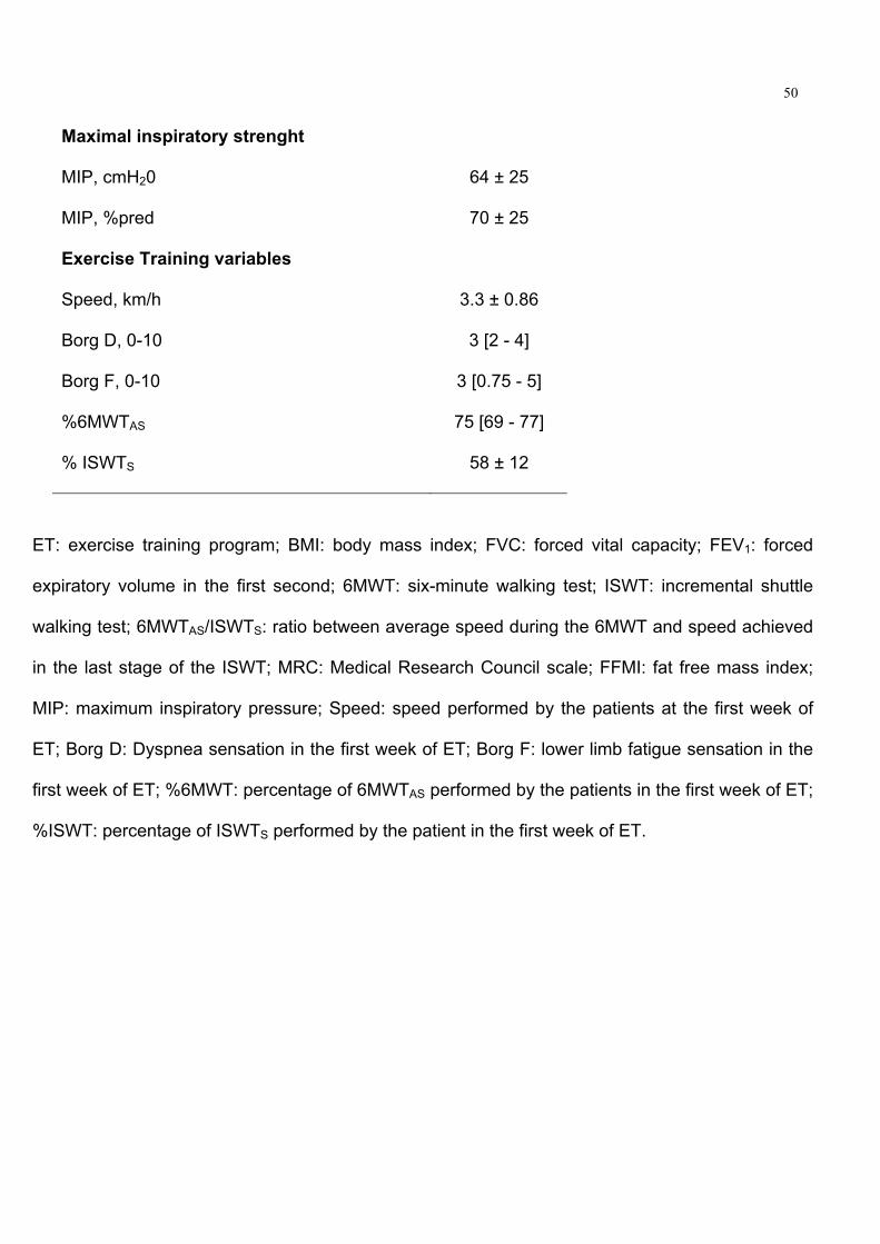
50
Maximal inspiratory strenght
MIP, cmH20 64 ± 25
MIP, %pred 70 ± 25
Exercise Training variables
Speed, km/h 3.3 ± 0.86
Borg D, 0-10 3 [2 - 4]
Borg F, 0-10 3 [0.75 - 5]
%6MWTAS 75 [69 - 77]
% ISWTS 58 ± 12
ET: exercise training program; BMI: body mass index; FVC: forced vital capacity; FEV1: forced
expiratory volume in the first second; 6MWT: six-minute walking test; ISWT: incremental shuttle
walking test; 6MWTAS/ISWTS: ratio between average speed during the 6MWT and speed achieved
in the last stage of the ISWT; MRC: Medical Research Council scale; FFMI: fat free mass index;
MIP: maximum inspiratory pressure; Speed: speed performed by the patients at the first week of
ET; Borg D: Dyspnea sensation in the first week of ET; Borg F: lower limb fatigue sensation in the
first week of ET; %6MWT: percentage of 6MWTAS performed by the patients in the first week of ET;
%ISWT: percentage of ISWTS performed by the patient in the first week of ET.
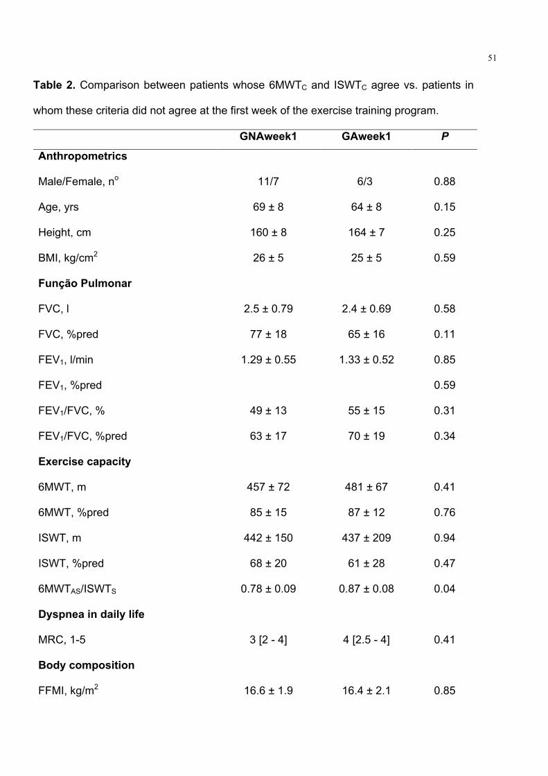
51
Table 2. Comparison between patients whose 6MWTC and ISWTC agree vs. patients in
whom these criteria did not agree at the first week of the exercise training program.
GNAweek1 GAweek1 P
Anthropometrics
Male/Female, no 11/7 6/3 0.88
Age, yrs 69 ± 8 64 ± 8 0.15
Height, cm 160 ± 8 164 ± 7 0.25
BMI, kg/cm2 26 ± 5 25 ± 5 0.59
Função Pulmonar
FVC, l 2.5 ± 0.79 2.4 ± 0.69 0.58
FVC, %pred 77 ± 18 65 ± 16 0.11
FEV1, l/min 1.29 ± 0.55 1.33 ± 0.52 0.85
FEV1, %pred
0.59
FEV1/FVC, % 49 ± 13 55 ± 15 0.31
FEV1/FVC, %pred 63 ± 17 70 ± 19 0.34
Exercise capacity
6MWT, m 457 ± 72 481 ± 67 0.41
6MWT, %pred 85 ± 15 87 ± 12 0.76
ISWT, m 442 ± 150 437 ± 209 0.94
ISWT, %pred 68 ± 20 61 ± 28 0.47
6MWTAS/ISWTS 0.78 ± 0.09 0.87 ± 0.08 0.04
Dyspnea in daily life
MRC, 1-5 3 [2 - 4] 4 [2.5 - 4] 0.41
Body composition
FFMI, kg/m2 16.6 ± 1.9 16.4 ± 2.1 0.85
52
Maximal inspiratory strenght
MIP, cmH20 65 ± 25 62 ± 26 0.75
MIP, %pred 72 ± 25 65 ± 26 0.48
Exercise Training variables
Speed, km/h 3 ± 0.74 3.9 ± 0.79 0.007
Borg D, 0-10 2 [1.25 - 3.75] 3 [2.5 - 6] 0.12
Borg F, 0-10 2 [0.25 - 4.75] 4 [0.25 - 4.75] 0.27
%6MWTAS 74 [62 - 75] 77 [76 - 86] <0.001
% ISWTS 52 ± 10 70 ± 4 <0.001
GAweek1: patients whose 6MWTC and ISWTC agreed in the first week of ET; GNAweek1:
patients whose 6MWTC and ISWTC did not agree in the first week of ET. BMI: body mass
index; FVC: forced vital capacity; FEV1: forced expiratory volume in the first second;
6MWT: six-minute walking test; ISWT: incremental shuttle walking test; 6MWTAS/ISWTS:
ratio between average speed during the 6MWT and speed achieved in the last stage of
the ISWT; MRC: Medical Research Council scale; FFMI: fat free mass index; MIP:
maximum inspiratory pressure; Speed: speed performed by the patients at the first week
of ET; Borg D: Dyspnea sensation in the first week of ET; Borg F: lower limb fatigue
sensation in the first week of ET; %6MWT: percentage of 6MWTAS performed by the
patients in the first week of ET; %ISWT: percentage of ISWTS performed by the patient in
the first week of ET
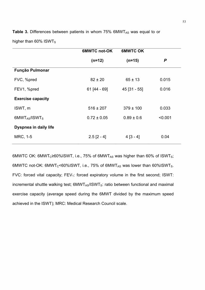
53
Table 3. Differences between patients in whom 75% 6MWTAS was equal to or
higher than 60% ISWTS
6MWTC not-OK
(n=12)
6MWTC OK
(n=15) P
Função Pulmonar
FVC, %pred 82 ± 20 65 ± 13 0.015
FEV1, %pred 61 [44 - 69] 45 [31 - 55] 0.016
Exercise capacity
ISWT, m 516 ± 207 379 ± 100 0.033
6MWTAS/ISWTS 0.72 ± 0.05 0.89 ± 0.6 <0.001
Dyspnea in daily life
MRC, 1-5 2.5 [2 - 4] 4 [3 - 4] 0.04
6MWTC OK: 6MWTC≥60%ISWT, i.e., 75% of 6MWTAS was higher than 60% of ISWTS;
6MWTC not-OK: 6MWTC<60%ISWT, i.e., 75% of 6MWTAS was lower than 60%ISWTS.
FVC: forced vital capacity; FEV1: forced expiratory volume in the first second; ISWT:
incremental shuttle walking test; 6MWTAS/ISWTS: ratio between functional and maximal
exercise capacity (average speed during the 6MWT divided by the maximum speed
achieved in the ISWT); MRC: Medical Research Council scale.

54
Table 4. Baseline and post-exercise training characteristics of patients with COPD who
finished the program.
Variavel Baseline Post-ET P
Anthropometrics
Male/Female, no 10/5
Age, yrs 64 [60 - 75]
Height, cm 162 [157 - 167]
BMI, kg/cm2 28 ± 5 28 ± 5.3 0.65
Função Pulmonar
FVC, l 2.6 ± 0.83 2.6 ± 0.87 0.69
FVC, %pred 74 ± 19 73 ± 22 0.9
FEV1, l/min 1.4 ± 0.61 1.48 ± 0.74 0.37
FEV1, %pred 57 [31 - 68] 52 [30 - 64] 0.73
FEV1/FVC, % 59 [38 - 63] 60 [36 - 64] 0.02
FEV1/FVC, %pred 65 ± 19 69 ± 21 0.16
Exercise capacity
6MWT, m 466 ± 71 506 ± 85 0.003
6MWT, %pred 87 ± 15 94 ± 17 0.003
ISWT, m 483 ± 179 509 ± 208 0.21
ISWT, %pred 70 ± 22 76 ± 0.14
6MWTAS/ISWTS 0.78 ± 0.10 0.82 ± 0.06 0.95
Dyspnea in daily life
MRC, 1-5 3.5 [2 - 4] 4 [2 - 4] 0.5
Body composition
FFMI, kg/m2 17.2 ± 1.8 17.4 ± 1.9 0.5
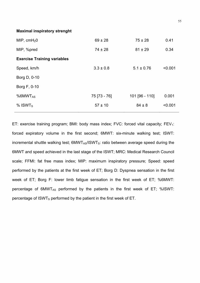
55
Maximal inspiratory strenght
MIP, cmH20 69 ± 28 75 ± 28 0.41
MIP, %pred 74 ± 28 81 ± 29 0.34
Exercise Training variables
Speed, km/h 3.3 ± 0.8 5.1 ± 0.76 <0.001
Borg D, 0-10
Borg F, 0-10
%6MWTAS 75 [73 - 76] 101 [96 - 110] 0.001
% ISWTS 57 ± 10 84 ± 8 <0.001
ET: exercise training program; BMI: body mass index; FVC: forced vital capacity; FEV1:
forced expiratory volume in the first second; 6MWT: six-minute walking test; ISWT:
incremental shuttle walking test; 6MWTAS/ISWTS: ratio between average speed during the
6MWT and speed achieved in the last stage of the ISWT; MRC: Medical Research Council
scale; FFMI: fat free mass index; MIP: maximum inspiratory pressure; Speed: speed
performed by the patients at the first week of ET; Borg D: Dyspnea sensation in the first
week of ET; Borg F: lower limb fatigue sensation in the first week of ET; %6MWT:
percentage of 6MWTAS performed by the patients in the first week of ET; %ISWT:
percentage of ISWTS performed by the patient in the first week of ET.

56

57
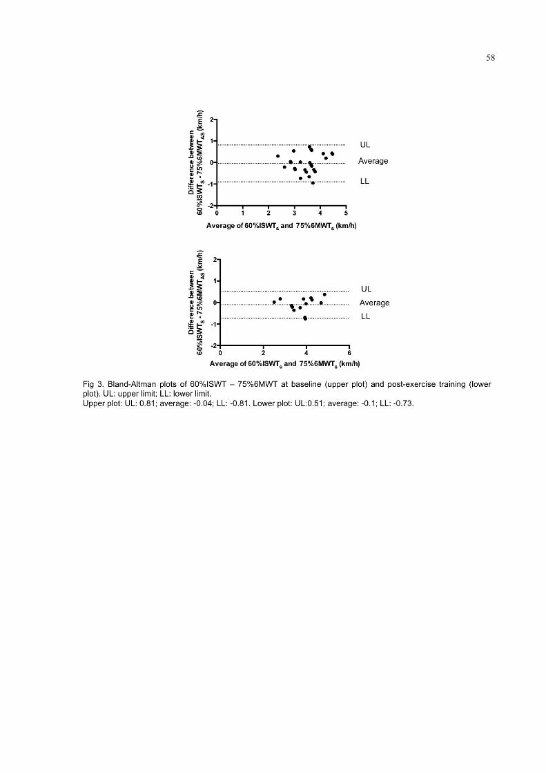
58

59
4. CONCLUSÃO GERAL
Com base nos resultados deste estudo e dos achados de literatura
descritos na revisão realizada nesta dissertação, pode-se concluir que o TC6min é
um teste útil para a prescrição de TF para pacientes com DPOC na ausência de
TCPE. Os resultados indicaram sua boa performance diagnóstica, indicada pelos
valores preditivos positivos e negativos e acurácia, boa confiabilidade e aceitável
concordância na prescrição de TF de alta intensidade de acordo com critérios
internacionais para esa população. Somado a isso, pacientes com DPOC
submetidos a um protocolo de TF prescrito com base no TC6min experienciam
melhoras após TF que ultrapassam a mínima melhora clinicamente importante para
essa população. No entanto, estudos desenhados com o intuito de desenvolver uma
adaptação dos resultados do TC6min para prescrição de exercício são necessários.
Isso poderia diminuir a variabilidade (em relação à intensidade máxima) da
intensidade de exercício que acontece quando a intensidade é prescrita com base
no TC6min.
60
5. REFERENCIAS
1. Holland AE, Spruit MA, Troosters T, et al. An official European Respiratory
Society/American Thoracic Society technical standard: field walking tests in
chronic respiratory disease. Eur Respir J. 2014;44(6):1428-1446.
2. Probst VS, Kovelis D, Hernandes NA, Camillo CA, Cavalheri V, Pitta F. Effects
of 2 exercise training programs on physical activity in daily life in patients with
COPD. Respir Care. 2011;56(11):1799-1807.
3. Troosters T, Gosselink R, Decramer M. Short- and long-term effects of
outpatient rehabilitation in patients with chronic obstructive pulmonary disease:
a randomized trial. Am J Respir Crit Care Med. 2000;109(3):207-212.
4. Spruit MA, Gosselink R, Troosters T, De Paepe K, Decramer M. Resistance
versus endurance training in patients with COPD and peripheral muscle
weakness. Eur Respir J. 2002;19(6):1072-1078.
5. Pitta F, Troosters T, Probst VS, Langer D, Decramer M, Gosselink R. Are
patients with COPD more active after pulmonary rehabilitation? Chest.
2008;134(2):273-280.
6. Zainuldin R, Mackey MG, Alison JA. Prescription of walking exercise intensity
from the 6-minute walk test in people with chronic obstructive pulmonary
disease. J Cardiopul Rehabil Prev. 2015;35(1):65-69.
7. Garber CE, Blissmer B, Deschenes MR, et al. American College of Sports
Medicine position stand. Quantity and quality of exercise for developing and
maintaining cardiorespiratory, musculoskeletal, and neuromotor fitness in
apparently healthy adults: guidance for prescribing exercise. Med Sci Sports
Exer. 2011;43(7):1334-1359.
61
8. Spruit MA, Singh SJ, Garvey C, et al. An official American Thoracic
Society/European Respiratory Society statement: key concepts and advances
in pulmonary rehabilitation. Am J Respir Crit Care Med. 2013;188(8):e13-64.
9. American Thoracic S, American College of Chest P. ATS/ACCP Statement on
cardiopulmonary exercise testing. Am J Respir Crit Care Med.
2003;167(2):211-277.
10. Troosters T, Vilaro J, Rabinovich R, et al. Physiological responses to the 6-min
walk test in patients with chronic obstructive pulmonary disease. Eur Respir J.
2002;20(3):564-569.
11. Dolmage TE, Evans RA, Hill K, Blouin M, Brooks D, Goldstein RS. The effect
of pulmonary rehabilitation on critical walk speed in patients with COPD: a
comparison with self-paced walks. Chest. 2012;141(2):413-419.
12. Casas A, Vilaro J, Rabinovich R, et al. Encouraged 6-min walking test
indicates maximum sustainable exercise in COPD patients. Chest.
2005;128(1):55-61.
13. Puente-Maestu L, Sanz ML, Sanz P, Ruiz de Ona JM, Rodriguez-Hermosa JL,
Whipp BJ. Effects of two types of training on pulmonary and cardiac
responses to moderate exercise in patients with COPD. Eur Respir J.
2000;15(6):1026-1032.
14. Neder JA, Jones PW, Nery LE, Whipp BJ. Determinants of the exercise
endurance capacity in patients with chronic obstructive pulmonary disease.
The power-duration relationship. Am J Respir Crit Care Med. 2000;162(2 Pt
1):497-504.

62
15. Ciftci F, Sen E, Akkoca Yildiz O, Saryal S. A comparison of cardiopulmonary
exercise test and 6 minute walking test in determination of exercise capacity in
chronic obstructive pulmonary disease. Tuberk Toraks. 2014;62(4):259-266.
16. van Gestel AJ, Baty F, Rausch-Osthof AK, Brutsche MH. Cardiopulmonary
and gas-exchange responses during the six-minute walk test in patients with
chronic obstructive pulmonary disease. Respiration. 2014;88(4):307-314.
17. Tarantino AB. Doenças Pulmonares. Vol. Sixth ed: Guanabara Koogan; 2007.
18. Marsh SE, Travers J, Weatherall M, et al. Proportional classifications of COPD
phenotypes. Thorax. 2008;63(9):761-767.
19. Vestbo J, Hurd SS, Agusti AG, et al. Global strategy for the diagnosis,
management, and prevention of chronic obstructive pulmonary disease: GOLD
executive summary. Am J Respir Crit Care Med. 2013;187(4):347-365.
20. Watz H, Pitta F, Rochester CL, et al. An official European Respiratory Society
statement on physical activity in COPD. Eur Respir J. 2014;44(6):1521-1537.
21. Ramon MA, Ferrer J, Gimeno-Santos E, et al. Inspiratory capacity-to-total lung
capacity ratio and dyspnoea predict exercise capacity decline in COPD.
Respirology. 2015.
22. Kirkham PA, Barnes PJ. Oxidative stress in COPD. Chest. 2013;144(1):266-
273.
23. Barreiro E, Gea J. Respiratory and Limb Muscle Dysfunction in COPD. COPD.
2015;12(4):413-426.
24. Maltais F, Decramer M, Casaburi R, et al. An official American Thoracic
Society/European Respiratory Society statement: update on limb muscle
dysfunction in chronic obstructive pulmonary disease. Am J Respir Crit Care
Med. 2014;189(9):e15-62.

63
25. Panagioti M, Scott C, Blakemore A, Coventry PA. Overview of the prevalence,
impact, and management of depression and anxiety in chronic obstructive
pulmonary disease. Int J Chron Obstruct Pulmon Dis. 2014;9:1289-1306.
26. O'Donnell DE, Webb KA. The major limitation to exercise performance in
COPD is dynamic hyperinflation. J Appl Physiol. 2008;105(2):753-755;
discussion 755-757.
27. Nici L. The major limitation to exercise performance in COPD is inadequate
energy supply to the respiratory and locomotor muscles vs. lower limb muscle
dysfunction vs. dynamic hyperinflation. Difficulties in determining the primary
physiological abnormality that limits exercise performance in COPD. J Appl
Physiol. 2008;105(2):760-761.
28. Neder JA. The major limitation to exercise performance in COPD is
inadequate energy supply to the respiratory and locomotor muscles vs. lower
limb muscle dysfunction vs. dynamic hyperinflation. Interpretation of exercise
intolerance in COPD requires an integrated, multisystemic approach. J Appl
Physiol. 2008;105(2):758-759.
29. Debigare R, Maltais F. The major limitation to exercise performance in COPD
is lower limb muscle dysfunction. J Appl Physiol. 2008;105(2):751-753;
discussion 755-757.
30. Aliverti A, Macklem PT. The major limitation to exercise performance in COPD
is inadequate energy supply to the respiratory and locomotor muscles. J Appl
Physiol. 2008;105(2):749-751; discussion 755-747.
31. O'Donnell DE, Laveneziana P, Webb K, Neder JA. Chronic obstructive
pulmonary disease: clinical integrative physiology. Clin Chest Med.
2014;35(1):51-69.
64
32. Deesomchok A, Webb KA, Forkert L, et al. Lung hyperinflation and its
reversibility in patients with airway obstruction of varying severity. COPD.
2010;7(6):428-437.
33. Guenette JA, Chin RC, Cheng S, et al. Mechanisms of exercise intolerance in
global initiative for chronic obstructive lung disease grade 1 COPD. Eur Respir
J. 2014;44(5):1177-1187.
34. Whipp BJ. Ventilatory control during exercise in humans. Ann Rev Physiol.
1983;45:393-413.
35. O'Donnell DE, Revill SM, Webb KA. Dynamic hyperinflation and exercise
intolerance in chronic obstructive pulmonary disease. Am J Respir Crit Care
Med. 2001;164(5):770-777.
36. Rodriguez-Roisin R, Drakulovic M, Rodriguez DA, Roca J, Barbera JA,
Wagner PD. Ventilation-perfusion imbalance and chronic obstructive
pulmonary disease staging severity. J Appl Physiol. 2009;106(6):1902-1908.
37. O'Donnell DE, Neder JA, Webb KA, Elbehairy AF. Reply: Effects of Mild
Chronic Obstructive Pulmonary Disease on Gas Exchange during Cycling and
Walking. Am J Respir Crit Care Med. 2015;192(9):1138-1139.
38. Neder JA, O'Donnell CD, Cory J, et al. Ventilation Distribution Heterogeneity
at Rest as a Marker of Exercise Impairment in Mild-to-Advanced COPD.
COPD. 2015;12(3):249-256.
39. Chiappa GR, Queiroga F, Jr., Meda E, et al. Heliox improves oxygen delivery
and utilization during dynamic exercise in patients with chronic obstructive
pulmonary disease. Am J Respir Crit Care Med. 2009;179(11):1004-1010.
40. Chiappa GR, Borghi-Silva A, Ferreira LF, et al. Kinetics of muscle
deoxygenation are accelerated at the onset of heavy-intensity exercise in

65
patients with COPD: relationship to central cardiovascular dynamics. J Appl
Physiol. 2008;104(5):1341-1350.
41. Neder JA, Arbex FF, Alencar MC, et al. Exercise ventilatory inefficiency in mild
to end-stage COPD. Eur Respir J. 2015;45(2):377-387.
42. Faisal A, Alghamdi BJ, Ciavaglia CE, et al. Common Mechanisms of Dyspnea
in Chronic Interstitial and Obstructive Lung Disorders. Am J Respir Crit Care
Med. 2015.
43. Gea J, Orozco-Levi M, Barreiro E, Ferrer A, Broquetas J. Structural and
functional changes in the skeletal muscles of COPD patients: the
"compartments" theory. Monaldi Arch Chest Dis. 2001;56(3):214-224.
44. Casaburi R, Rennard SI. Exercise limitation in chronic obstructive pulmonary
disease. The O'Donnell threshold. Am J Respir Crit Care Med.
2015;191(8):873-875.
45. Williams TJ, Patterson GA, McClean PA, Zamel N, Maurer JR. Maximal
exercise testing in single and double lung transplant recipients. Am Rev Respir
Dis. 1992;145(1):101-105.
46. Gea J, Agusti A, Roca J. Pathophysiology of muscle dysfunction in COPD. J
Appl Physiol. 2013;114(9):1222-1234.
47. Hopkinson NS, Tennant RC, Dayer MJ, et al. A prospective study of decline in
fat free mass and skeletal muscle strength in chronic obstructive pulmonary
disease. Respir Res. 2007;8:25.
48. Gosker HR, van Mameren H, van Dijk PJ, et al. Skeletal muscle fibre-type
shifting and metabolic profile in patients with chronic obstructive pulmonary
disease. Eur Respir J. 2002;19(4):617-625.
66
49. Jobin J, Maltais F, Doyon JF, et al. Chronic obstructive pulmonary disease:
capillarity and fiber-type characteristics of skeletal muscle. J Cardiopul
Rehabil. 1998;18(6):432-437.
50. Jakobsson P, Jorfeldt L, Henriksson J. Metabolic enzyme activity in the
quadriceps femoris muscle in patients with severe chronic obstructive
pulmonary disease. Am J Respir Crit Care Med. 1995;151(2 Pt 1):374-377.
51. Gosker HR, Hesselink MK, Duimel H, Ward KA, Schols AM. Reduced
mitochondrial density in the vastus lateralis muscle of patients with COPD. Eur
Respir J. 2007;30(1):73-79.
52. Wouters EF. Local and systemic inflammation in chronic obstructive
pulmonary disease. Proc Am Thorac Soc. 2005;2(1):26-33.
53. Montes de Oca M, Torres SH, De Sanctis J, Mata A, Hernandez N, Talamo C.
Skeletal muscle inflammation and nitric oxide in patients with COPD. Eur
Respir J. 2005;26(3):390-397.
54. Sperandio PA, Oliveira MF, Rodrigues MK, et al. Sildenafil improves
microvascular O2 delivery-to-utilization matching and accelerates exercise O2
uptake kinetics in chronic heart failure. Am J Physiol Heart Circ Physiol.
2012;303(12):H1474-1480.
55. Siqueira AC, Borghi-Silva A, Bravo DM, Ferreira EM, Chiappa GR, Neder JA.
Effects of hyperoxia on the dynamics of skeletal muscle oxygenation at the
onset of heavy-intensity exercise in patients with COPD. Respir Physiol
Neurobiol. 2010;172(1-2):8-14.
56. Roseguini BT, Hirai DM, Alencar MC, et al. Sildenafil improves skeletal muscle
oxygenation during exercise in men with intermittent claudication. Am J
Physiol Regul Integr Comp Physiol. 2014;307(4):R396-404.

67
57. Medeiros WM, Fernandes MC, Azevedo DP, et al. Oxygen delivery-utilization
mismatch in contracting locomotor muscle in COPD: peripheral factors. Am J
Physiol Regul Integr Comp Physiol.. 2015;308(2):R105-111.
58. Berton DC, Barbosa PB, Takara LS, et al. Bronchodilators accelerate the
dynamics of muscle O2 delivery and utilisation during exercise in COPD.
Thorax. 2010;65(7):588-593.
59. Santos S, Peinado VI, Ramirez J, et al. Characterization of pulmonary
vascular remodelling in smokers and patients with mild COPD. Eur Respir J.
2002;19(4):632-638.
60. Voelkel NF, Tuder RM. Hypoxia-induced pulmonary vascular remodeling: a
model for what human disease? J Clin Invest. 2000;106(6):733-738.
61. Chetty KG, Brown SE, Light RW. Improved exercise tolerance of the
polycythemic lung patient following phlebotomy. Am J Med. 1983;74(3):415-
420.
62. MacNee W. Pathophysiology of cor pulmonale in chronic obstructive
pulmonary disease. Part One. Am J Respir Crit Care Med. 1994;150(3):833-
852.
63. Visca D, Aiello M, Chetta A. Cardiovascular function in pulmonary
emphysema. Biomed Res Int. 2013;2013:184678.
64. Tzani P, Aiello M, Elia D, et al. Dynamic hyperinflation is associated with a
poor cardiovascular response to exercise in COPD patients. Respir Res.
2011;12:150.
65. Folgering H, von Herwaarden C. Exercise limitations in patients with
pulmonary diseases. Int J Sports Med. 1994;15(3):107-111.
68
66. Lacasse Y, Cates CJ, McCarthy B, Welsh EJ. This Cochrane Review is
closed: deciding what constitutes enough research and where next for
pulmonary rehabilitation in COPD. Cochrane Database Syst Rev.
2015;11:ED000107.
67. McCarthy B, Casey D, Devane D, Murphy K, Murphy E, Lacasse Y.
Pulmonary rehabilitation for chronic obstructive pulmonary disease. Cochrane
Database Syst Rev.. 2015;2:CD003793.
68. Belman MJ, Kendregan BA. Exercise training fails to increase skeletal muscle
enzymes in patients with chronic obstructive pulmonary disease. Am Rev
Respir Dis. 1981;123(3):256-261.
69. Casaburi R, Patessio A, Ioli F, Zanaboni S, Donner CF, Wasserman K.
Reductions in exercise lactic acidosis and ventilation as a result of exercise
training in patients with obstructive lung disease. Am Rev Respir Dis.
1991;143(1):9-18.
70. Rodriguez DA, Arbillaga A, Barberan-Garcia A, et al. Effects of interval and
continuous exercise training on autonomic cardiac function in COPD patients.
Clin Respir J. 2014.
71. Nasis I, Kortianou E, Vasilopoulou M, et al. Hemodynamic effects of high
intensity interval training in COPD patients exhibiting exercise-induced
dynamic hyperinflation. Respir Physiol Neurobiol. 2015;217:8-16.
72. Gimeno-Santos E, Rodriguez DA, Barberan-Garcia A, et al. Endurance
exercise training improves heart rate recovery in patients with COPD. COPD.
2014;11(2):190-196.
69
73. Georgiopoulou VV, Dimopoulos S, Sakellariou D, et al. Cardiopulmonary
rehabilitation enhances heart rate recovery in patients with COPD. Respir
Care. 2012;57(12):2095-2103.
74. Bronstad E, Tjonna AE, Rognmo O, et al. Aerobic exercise training improves
right- and left ventricular systolic function in patients with COPD. COPD.
2013;10(3):300-306.
75. Whittom F, Jobin J, Simard PM, et al. Histochemical and morphological
characteristics of the vastus lateralis muscle in patients with chronic
obstructive pulmonary disease. Med Sci Sports Exer. 1998;30(10):1467-1474.
76. Vogiatzis I, Terzis G, Stratakos G, et al. Effect of pulmonary rehabilitation on
peripheral muscle fiber remodeling in patients with COPD in GOLD stages II to
IV. Chest. 2011;140(3):744-752.
77. Vogiatzis I, Terzis G, Nanas S, et al. Skeletal muscle adaptations to interval
training in patients with advanced COPD. Chest. 2005;128(6):3838-3845.
78. Troosters T, Gosselink R, Decramer M. Exercise training in COPD: how to
distinguish responders from nonresponders. J Cardiopul Rehabil.
2001;21(1):10-17.
79. Spruit MA, Augustin IM, Vanfleteren L, et al. Differential response to
pulmonary rehabilitation in COPD: multidimensional profiling. Eur Respir J.
2015.
80. Vogiatzis I, Zakynthinos S. The physiological basis of rehabilitation in chronic
heart and lung disease. J Appl Physiol. 2013;115(1):16-21.
81. Maltais F, LeBlanc P, Jobin J, et al. Intensity of training and physiologic
adaptation in patients with chronic obstructive pulmonary disease. Am J
Respir Crit Care Med. 1997;155(2):555-561.
70
82. Panton LB, Golden J, Broeder CE, Browder KD, Cestaro-Seifer DJ, Seifer FD.
The effects of resistance training on functional outcomes in patients with
chronic obstructive pulmonary disease. Eur J Appl Physiol. 2004;91(4):443-
449.
83. Ortega F, Toral J, Cejudo P, et al. Comparison of effects of strength and
endurance training in patients with chronic obstructive pulmonary disease. Am
J Respir Crit Care Med. 2002;166(5):669-674.
84. Mador MJ, Bozkanat E, Aggarwal A, Shaffer M, Kufel TJ. Endurance and
strength training in patients with COPD. Chest. 2004;125(6):2036-2045.
85. Kongsgaard M, Backer V, Jorgensen K, Kjaer M, Beyer N. Heavy resistance
training increases muscle size, strength and physical function in elderly male
COPD-patients--a pilot study. Respir Med. 2004;98(10):1000-1007.
86. Hoff J, Tjonna AE, Steinshamn S, Hoydal M, Richardson RS, Helgerud J.
Maximal strength training of the legs in COPD: a therapy for mechanical
inefficiency. Med Sci Sports Exerc. 2007;39(2):220-226.
87. Clark CJ, Cochrane LM, Mackay E, Paton B. Skeletal muscle strength and
endurance in patients with mild COPD and the effects of weight training. Eur
Respir J. 2000;15(1):92-97.
88. Bernard S, Whittom F, Leblanc P, et al. Aerobic and strength training in
patients with chronic obstructive pulmonary disease. Am J Respir Crit Care
Med. 1999;159(3):896-901.
89. Mark Brunley and Andew M. Jones. Oxygen uptake kinetics as a determinant
of sports performance. Eur J Sport Sci. 2007;7(2):63-79.
90. Borg GA. Psychophysical bases of perceived exertion. Med Sci Sports Exerc.
1982;14(5):377-381.

71
91. Horowitz MB, Littenberg B, Mahler DA. Dyspnea ratings for prescribing
exercise intensity in patients with COPD. Chest. 1996;109(5):1169-1175.
92. Hsia D, Casaburi R, Pradhan A, Torres E, Porszasz J. Physiological
responses to linear treadmill and cycle ergometer exercise in COPD. Eur
Respir J. 2009;34(3):605-615.
93. Palange P, Forte S, Onorati P, Manfredi F, Serra P, Carlone S. Ventilatory and
metabolic adaptations to walking and cycling in patients with COPD. J Appl
Physiol. 2000;88(5):1715-1720.
94. Murray JA, Waterman LA, Ward J, Baird JC, Mahler DA. Perceptual and
physiologic responses during treadmill and cycle exercise in patients with
COPD. Chest. 2009;135(2):384-390.
95. Mathur RS, Revill SM, Vara DD, Walton R, Morgan MD. Comparison of peak
oxygen consumption during cycle and treadmill exercise in severe chronic
obstructive pulmonary disease. Thorax. 1995;50(8):829-833.
96. Mahler DA, Gifford AH, Waterman LA, Ward J, Machala S, Baird JC.
Mechanism of greater oxygen desaturation during walking compared with
cycling in patients with COPD. Chest. 2011;140(2):351-358.
97. Mador MJ. Exercise training in patients with COPD: one leg is better than two?
Chest. 2008;133(2):337-339.
98. Bjorgen S, Hoff J, Husby VS, et al. Aerobic high intensity one and two legs
interval cycling in chronic obstructive pulmonary disease: the sum of the parts
is greater than the whole. Eur J Appl Physiol. 2009;106(4):501-507.

72
6. APÊNDICES
APÊNDICE A- Termo de cosentimento livre e esclarecido
Prezado(a) Senhor(a):
O(A) Sr(a) está sendo convidado para participar de um projeto de pesquisa chamado
“Efeitos de um programa de exercício físico de longa duração sobre aspectos
pulmonares e sistêmicos em pacientes com Doença Pulmonar Obstrutiva Crônica
(DPOC)”, cujos pesquisadores responsáveis são Prof. Dr. Fábio de Oliveira Pitta e
Nidia A. Hernandes, do Departamento de Fisioterapia da Universidade Estadual de
Londrina (UEL). O estudo analisará principalmente as melhoras obtidas após 6
meses de treinamento utilizando-se dois tipos diferentes de exercício físico.
Justificativa: O presente estudo contribuirá para solucionar uma questão não
resolvida e que tem sido alvo de grande debate: levando em conta que a longa
duração do programa de treinamento físico é fundamental no processo de
conscientizar pacientes com DPOC a aumentar sua atividade física diária, será
necessário que o treinamento físico realizado durante esse programa de longa
duração seja feito em alta intensidade, ou um programa de baixa intensidade de
treinamento já atingiria o objetivo? Se programas de longa duração, porém com
baixa intensidade de treinamento, trouxerem benefícios importantes no aumento da
atividade física diária, isso poderá trazer mudança no entendimento atual sobre
protocolos de exercício em pacientes com DPOC, que hoje são realizados sob alta
intensidade de treinamento.

73
Objetivo: Comparar os efeitos de dois protocolos de treinamento físico em um
programa de reabilitação de longa duração (6 meses) em pacientes com DPOC: um
protocolo de alta intensidade (baseado em treinamento de endurance e força) e um
protocolo de baixa intensidade (baseado em exercícios respiratórios e de
readequação do complexo tóraco-pulmonar).
Procedimentos: Os pacientes incluídos realizarão uma série de testes que incluirá
avaliação da função pulmonar, capacidade máxima e funcional de exercício, força
muscular periférica e respiratória, atividade física na vida diária, composição
corporal, função autonômica cardíaca, qualidade de vida, estado funcional e
sensação de dispnéia. A realização dos testes requer uma visita de
aproximadamente 2 horas ao Hospital Universitário Regional Norte do Paraná, em
Londrina, além do uso do pequeno aparelho na cintura durante dois dias (12 horas
por dia, apenas durante o dia e não de noite).Após a avaliação inicial, os pacientes
serão divididos em dois grupos: um grupo realizará um programa de exercícios
físicos direcionado ao aumento da mobilidade torácica realizado 3 vezes por
semana, durante 24 semanas ; e outro grupo realizará um programa de treinamento
de endurance e força de membros superiores e inferiores realizado 3 vezes por
semana, durante 24 semanas. Ao final do programa de treinamento, os participantes
serão reavaliados seguindo os mesmos testes realizados na avaliação inicial.
Custos: A pesquisa é gratuita e portanto não envolve qualquer custo por parte dos
indivíduos. Não haverá qualquer gratificação financeira pela participação. No
entanto, em caso de eventuais danos ocorridos exclusivamente por causa deste
estudo, o Sr(a) terá direito a tratamento médico completo oferecido pela instituição.

74
Riscos: Nenhum dos procedimentos utilizados constitui risco direto para a
integridade física ou moral dos participantes. Além disso, os participantes poderão
abandonar o estudo a qualquer momento que se achar conveniente, sem qualquer
prejuízo em nenhum sentido.
Sigilo: Embora os resultados da pesquisa possam ser divulgados em publicações e
eventos científicos, a identidade dos participantes será sempre preservada de
maneira sigilosa, ou seja, em segredo.
Caso o(a) Sr(a) aceite esse convite e concorde voluntariamente em participar do
estudo assinando este termo de consentimento, consideramos que o Sr(a) acredita
que foi suficientemente informada pela pesquisadora Nidia Aparecida Hernandes
sobre a pesquisa, os procedimentos envolvidos nela, assim como os possíveis riscos
e benefícios decorrentes dessa participação. Ressaltamos novamente que o Sr(a)
pode retirar seu consentimento a qualquer momento, sem que isto leve a qualquer
prejuízo em nenhum sentido.
Local e data: ______________________________________________________
Nome do participante: _______________________________________________
Assinatura do participante ou responsável:_______________________________
Assinatura do pesquisador: ___________________________________________
Colocamo-nos à disposição para qualquer esclarecimento que se fizer necessário
nos telefones (43) 3371-2477 ou pessoalmente no Ambulatório de Fisioterapia
Respiratória do Hospital Universitário Regional Norte do Paraná: Av. Robert Koch,

75
60 – Vila Operária – Londrina – PR (perguntar pelo Professor Fábio de Oliveira
Pitta).
Atenciosamente,
Prof. Fábio de Oliveira Pitta
Coordenador do Projeto
76
7. ANEXOS
ANEXO A- Normas de formatação da revista Heart and Lung.
Heart and Lung: The Journal of Acute and Critical Care, the official publication of The
American Association of Heart Failure Nurses, presents original, peer-reviewed
articles on techniques, advances, investigations, and observations related to the care
of patients with acute and critical illness and patients with chronic cardiac or
pulmonary disorders.
The Journal's acute care articles focus on the care of hospitalized patients, including
those in the critical and acute care settings. Because most patients who are
hospitalized in acute and critical care settings have chronic conditions, we are also
interested in the chronically critically ill, the care of patients with chronic
cardiopulmonary disorders, their rehabilitation, and disease prevention. The
Journal's heart failure articles focus on all aspects of the care of patients with this
condition. Manuscripts that are relevant to populations across the human lifespan are
welcome.
We are interested in publishing articles representing a broad range of science and
clinical practice in a variety of settings as it pertains to our target population. Because
health care and the health sciences are global, interdisciplinary, multidisciplinary, and
transdisciplinary, we encourage authors to submit manuscripts that reflect these
perspectives. Many articles also provide nurses with a framework for applying
research results in clinical practice.

77
Editor:
Nancy S. Redeker, PhD, RN, FAHA, FAAN Yale University, New Haven, CT
Managing Editor:
Ardis O'Meara Towson, MD
Publisher:
Mercedes Simoncelli Elsevier Inc. 1600 JFK Blvd, Suite 1800 Philadelphia, PA
19103-2899
Journal Manager:
Types of paper
We publish original research, case reports, reviews, and observations that are on the
cutting edge of science and clinical practice. Reports of well-designed clinical trials
and systematic reviews are especially welcome. Discussions of translational research
and implementation and improvement science, as well as costs of care, patient
education, and health policy are of high relevance to our focus.
ARTICLE CATEGORIES
Letters to the Editor
Letters to the Editor raising some point of current interest or commenting on an article
that appeared in the Journal will be considered for publication. The Editor reserves

78
the right to accept, reject, or excerpt letters without changing the views expressed by
the writer. The author of an original article will have an opportunity to reply to
published comments. All such letters should be sent to Dr. Redeker.
Reports of Original Research
Systematic Reviews
Reports of analyses of healthy policy, education, economic or organizational issues
pertaining to the care of patients with acute and critical illness and patients with
chronic cardiac or pulmonary disorders.
Ethics in publishing
For information on Ethics in publishing and Ethical guidelines for journal publication
see http://www.elsevier.com/publishingethicsand http://www.elsevier.com/journal-
authors/ethics.
Protection of Human Subjects and Animals in Research
All manuscripts that involve human subjects or animals in research must conform to
the principles of the International Committee of Medical Journal Editors, available at
http://www.icmje.org/ethical_6protection.html.
Conflict of interest
All authors must disclose any financial and personal relationships with other people
or organizations that could inappropriately influence (bias) their work. Examples of
potential conflicts of interest include employment, consultancies, stock ownership,
honoraria, paid expert testimony, patent applications/registrations, and grants or

79
other funding. If there are no conflicts of interest then please state this: 'Conflicts of
interest: none'. See also http://www.elsevier.com/conflictsofinterest. Further
information and an example of a Conflict of Interest form can be found at:
http://service.elsevier.com/app/answers/detail/a_id/286/supporthub/publishing.
Submission declaration
Submission of an article implies that the work described has not been published
previously (except in the form of an abstract or as part of a published lecture or
academic thesis or as an electronic preprint, see
http://www.elsevier.com/sharingpolicy), that it is not under consideration for
publication elsewhere, that its publication is approved by all authors and tacitly or
explicitly by the responsible authorities where the work was carried out, and that, if
accepted, it will not be published elsewhere including electronically in the same form,
in English or in any other language, without the written consent of the copyright-
holder.
Authorship
All authors should have made substantial contributions to all of the following: (1) the
conception and design of the study, or acquisition of data, or analysis and
interpretation of data, (2) drafting the article or revising it critically for important
intellectual content, (3) final approval of the version to be submitted.
Changes to authorship
Authors are expected to consider carefully the list and order of authors before
submitting their manuscript and provide the definitive list of authors at the time of the
original submission. Any addition, deletion or rearrangement of author names in the

80
authorship list should be made only before the manuscript has been accepted and
only if approved by the journal Editor. To request such a change, the Editor must
receive the following from the corresponding author: (a) the reason for the change
in author list and (b) written confirmation (e-mail, letter) from all authors that they
agree with the addition, removal or rearrangement. In the case of addition or removal
of authors, this includes confirmation from the author being added or removed.
Only in exceptional circumstances will the Editor consider the addition, deletion or
rearrangement of authors after the manuscript has been accepted. While the Editor
considers the request, publication of the manuscript will be suspended. If the
manuscript has already been published in an online issue, any requests approved by
the Editor will result in a corrigendum.
Copyright
Upon acceptance of an article, authors will be asked to complete a 'Journal
Publishing Agreement' (for more information on this and copyright, see
http://www.elsevier.com/copyright). An e-mail will be sent to the corresponding author
confirming receipt of the manuscript together with a 'Journal Publishing Agreement'
form or a link to the online version of this agreement.
Subscribers may reproduce tables of contents or prepare lists of articles including
abstracts for internal circulation within their institutions. Permission of the Publisher is
required for resale or distribution outside the institution and for all other derivative
works, including compilations and translations (please consult
http://www.elsevier.com/permissions). If excerpts from other copyrighted works are
included, the author(s) must obtain written permission from the copyright owners and
credit the source(s) in the article. Elsevier has preprinted forms for use by authors in

81
these cases: please consult http://www.elsevier.com/permissions.
For open access articles: Upon acceptance of an article, authors will be asked to
complete an 'Exclusive License Agreement' (for more information see
http://www.elsevier.com/OAauthoragreement). Permitted third party reuse of open
access articles is determined by the author's choice of user license (see
http://www.elsevier.com/openaccesslicenses).
Author rights
As an author you (or your employer or institution) have certain rights to reuse your
work. For more information see http://www.elsevier.com/copyright.
Role of the funding source
You are requested to identify who provided financial support for the conduct of the
research and/or preparation of the article and to
briefly describe the role of the sponsor(s), if any, in study design; in the collection,
analysis and interpretation of data; in the writing of the report; and in the decision to
submit the article for publication. If the funding source(s) had no such involvement
then this should be stated.
Funding body agreements and policies
Elsevier has established a number of agreements with funding bodies which allow
authors to comply with their funder's open access policies. Some authors may also
be reimbursed for associated publication fees. To learn more about existing
agreements please visit

82
http://www.elsevier.com/fundingbodies. After acceptance, open access papers will be
published under a noncommercial license. For authors requiring a commercial CC BY
license, you can apply after your manuscript is accepted for publication.
Open access
This journal offers authors a choice in publishing their research:
Open access
• Articles are freely available to both subscribers and the wider public with permitted
reuse • An open access publication fee is payable by authors or on their behalf e.g.
by their research funder or institution Subscription • Articles are made available to
subscribers as well as developing countries and patient groups through our universal
access programs ( http://www.elsevier.com/access). • No open access publication
fee payable by authors.
Regardless of how you choose to publish your article, the journal will apply the same
peer review criteria and acceptance standards.
For open access articles, permitted third party (re)use is defined by the following
Creative Commons user licenses:
Creative Commons Attribution-NonCommercial-NoDerivs (CC BY-NC-ND)
For non-commercial purposes, lets others distribute and copy the article, and to
include in a collective work (such as an anthology), as long as they credit the
author(s) and provided they do not alter or modify the article.
The open access publication fee for this journal is USD 2500, excluding taxes. Learn

83
more about Elsevier's pricing policy: http://www.elsevier.com/openaccesspricing.
Green open access
Authors can share their research in a variety of different ways and Elsevier has a
number of green open access options available. We recommend authors see our
green open access page for further information (
http://elsevier.com/greenopenaccess). Authors can also self-archive their
manuscripts immediately and enable public access from their institution's repository
after an embargo period. This is the version that has been accepted for publication
and which typically includes author-incorporated changes suggested during
submission, peer review and in editor-author communications. Embargo period: For
subscription articles, an appropriate amount of time is needed for journals to deliver
value to subscribing customers before an article becomes freely available to the
public. This is the embargo period and it begins from the date the article is formally
published online in its final and fully citable form.
This journal has an embargo period of 12 months.
Language (usage and editing services)
Please write your text in good English (American or British usage is accepted, but not
a mixture of these). Authors who feel their English language manuscript may require
editing to eliminate possible grammatical or spelling errors and to conform to correct
scientific English may wish to use the English Language Editing service available
from Elsevier's WebShop( http://webshop.elsevier.com/languageediting/) or visit our
customer support site ( http://support.elsevier.com) for more information.
Informed consent and patient details
84
Studies on patients or volunteers require ethics committee approval and informed
consent, which should be documented in the paper. Appropriate consents,
permissions and releases must be obtained where an author wishes to include case
details or other personal information or images of patients and any other individuals
in an Elsevier publication. Written consents must be retained by the author and
copies of the consents or evidence that such consents have been obtained must be
provided to Elsevier on request. For more information, please review the Elsevier
Policy on the Use of Images or Personal Information of Patients or other Individuals,
http://www.elsevier.com/patient-consent-policy. Unless you have written permission
from the patient (or, where applicable, the next of kin), the personal details of any
patient included in any part of the article and in any supplementary materials
(including all illustrations and videos) must be removed before submission.
Submission
Our online submission system guides you stepwise through the process of entering
your article details and uploading your files. The system converts your article files to
a single PDF file used in the peer-review process. Editable files (e.g., Word, LaTeX)
are required to typeset your article for final publication. All correspondence, including
notification of the Editor's decision and requests for revision, is sent by e-mail.
Submit your article
Please submit your article via http://ees.elsevier.com/hl/.
Manuscript submissions should conform to the guidelines set forth in the "Uniform
Requirements for Manuscripts Submitted to Biomedical Journals: Writing and Editing
for Biomedical Publication," available from http://www.ICMJE.org. Please refer to
85
http://www.consort-statement.org/consort-statement/ for CONSORT guidelines for
clinical trials. The CONSORT Statement is intended to improve the reporting of a
randomized controlled trial (RCT), enabling readers to understand a trial's design,
conduct, analysis and interpretation, and to assess the validity of its results.
Manuscript Components
The manuscript should be arranged as follows: 1) title; 2) structured abstract and key
words; 3) abbreviations list; 4) text; 5) references; 6) figure titles and legends; and 7)
tables. Page numbering should begin with the first page.
Manuscript style should follow the AMA Style, 10th edition. Articles should be no
more than 15 pages in length, not including title page, figures, tables, and references.
Author(s) name and credentials should be listed according to the AMA-10
recommendations.
Text
Manuscript text should be double-spaced, left-aligned (unjustified), and in a 12-point
font. The body of text should be structured with the following headings: Introduction,
Methods, Results, and Discussion. Sub-headings may be used as appropriate. Each
reference, figure and table should be cited in the text in numerical order according to
order of first mention.
Double-blind review
This journal uses double-blind review, which means that both the reviewer and author
name(s) are not allowed to be revealed to one another for a manuscript under
review. The identities of the authors are concealed from the reviewers, and vice

86
versa. For more information please refer to http://www.elsevier.com/reviewers/peer-
review. To facilitate this, please include the following separately: Title page (with
author details): This should include the title, authors' names and affiliations, and a
complete address for the corresponding author including an e-mail address.
Blinded manuscript (no author details): The main body of the paper (including the
references, figures, tables and any Acknowledgements) should not include any
identifying information, such as the authors' names or affiliations.
Use of word processing software
It is important that the file be saved in the native format of the word processor used.
The text should be in single-column format. Keep the layout of the text as simple as
possible. Most formatting codes will be removed and replaced on processing the
article. In particular, do not use the word processor's options to justify text or to
hyphenate words. However, do use bold face, italics, subscripts, superscripts etc.
When preparing tables, if you are using a table grid, use only one grid for each
individual table and not a grid for each row. If no grid is used, use tabs, not spaces,
to align columns. The electronic text should be prepared in a way very similar to that
of conventional manuscripts (see also the Guide to Publishing with Elsevier:
http://www.elsevier.com/guidepublication). Note that source files of figures, tables
and text graphics will be required whether or not you embed your figures in the text.
See also the section on Electronic artwork.
To avoid unnecessary errors you are strongly advised to use the 'spell-check' and
'grammar-check' functions of your word processor.
Article structure

87
Subdivision - unnumbered sections
Divide your article into clearly defined sections. Each subsection is given a brief
heading. Each heading should appear on its own separate line. Subsections should
be used as much as possible when cross-referencing text: refer to the subsection by
heading as opposed to simply 'the text'.
Introduction
State the objectives of the work and provide an adequate background, avoiding a
detailed literature survey or a summary of the results.
Material and methods
Provide sufficient detail to allow the work to be reproduced. Methods already
published should be indicated by a reference: only relevant modifications should be
described.
Theory/calculation
A Theory section should extend, not repeat, the background to the article already
dealt with in the Introduction and lay the foundation for further work. In contrast, a
Calculation section represents a practical development from a theoretical basis.
Results
Results should be clear and concise.
Discussion
This should explore the significance of the results of the work, not repeat them. A
combined Results and Discussion section is often appropriate. Avoid extensive
88
citations and discussion of published literature.
Conclusions
The main conclusions of the study may be presented in a short Conclusions section,
which may stand alone or form a subsection of a Discussion or Results and
Discussion section.
Appendices
If there is more than one appendix, they should be identified as A, B, etc. Formulae
and equations in appendices should be given separate numbering: Eq. (A.1), Eq.
(A.2), etc.; in a subsequent appendix, Eq. (B.1) and so on. Similarly for tables and
figures: Table A.1; Fig. A.1, etc.
Essential title page information
• Title. Concise and informative. Titles are often used in information-retrieval
systems. Avoid abbreviations and formulae where possible. • Author names and
affiliations. Please clearly indicate the given name(s) and family name(s) of each
author and check that all names are accurately spelled. Present the authors'
affiliation addresses (where the actual work was done) below the names. Indicate all
affiliations with a lower-case superscript letter immediately after the author's name
and in front of the appropriate address. Provide the full postal address of each
affiliation, including the country name and, if available, the e-mail address of each
author. • Corresponding author. Clearly indicate who will handle correspondence at
all stages of refereeing and publication, also post-publication. Ensure that the e-mail
address is given and that contact details are kept up to date by the
corresponding author.
89
• Present/permanent address.If an author has moved since the work described in
the article was done, or was visiting at the time, a 'Present address' (or 'Permanent
address') may be indicated as a footnote to that author's name. The address at which
the author actually did the work must be retained as the main, affiliation address.
Superscript Arabic numerals are used for such footnotes.
Abstract
Provide a structured abstract of no more than 150 words, essential data should be
presented in 5 paragraphs under separate headings in the following order:
Objectives, Background, Methods, Results, and Conclusions. A non-structured
abstract is appropriate for review articles.
Highlights
Highlights are a short collection of bullet points that convey the core findings of the
article. Highlights are optional and should be submitted in a separate editable file in
the online submission system. Please use 'Highlights' in the file name and include 3
to 5 bullet points (maximum 85 characters, including spaces, per bullet point). See
http://www.elsevier.com/highlights for examples.
Keywords
Immediately after the abstract, provide a maximum of 6 keywords, using American
spelling and avoiding general and plural terms and multiple concepts (avoid, for
example, 'and', 'of'). Be sparing with abbreviations: only abbreviations firmly
established in the field may be eligible. These keywords will be used for indexing
purposes.

90
Abbreviations
Define abbreviations that are not standard in this field in a footnote to be placed on
the first page of the article. Such abbreviations that are unavoidable in the abstract
must be defined at their first mention there, as well as in the footnote. Ensure
consistency of abbreviations throughout the article.
Acknowledgments
Include acknowledgments on the title page. List here those individuals who provided
help during the research (e.g., providing language help, writing assistance or proof
reading the article, etc.).
Units
Follow internationally accepted rules and conventions: use the international system
of units (SI). If other units are mentioned, please give their equivalent in SI.
Math formulae
Please submit math equations as editable text and not as images. Present simple
formulae in line with normal text where possible and use the solidus (/) instead of a
horizontal line for small fractional terms, e.g., X/Y. In principle, variables are to be
presented in italics. Powers of e are often more conveniently denoted by exp.
Number consecutively any equations that have to be displayed separately from the
text (if referred to explicitly in the text).
Footnotes
Footnotes should be used sparingly. Number them consecutively throughout the
91
article. Many word processors can build footnotes into the text, and this feature may
be used. Otherwise, please indicate the position of footnotes in the text and list the
footnotes themselves separately at the end of the article. Do not include footnotes in
the Reference list.
Artwork
Electronic artwork
General points
• Make sure you use uniform lettering and sizing of your original artwork. • Embed the
used fonts if the application provides that option. • Aim to use the following fonts in
your illustrations: Arial, Courier, Times New Roman, Symbol, or use fonts that look
similar. • Number the illustrations according to their sequence in the text. • Use a
logical naming convention for your artwork files. • Provide captions to illustrations
separately. • Size the illustrations close to the desired dimensions of the published
version. • Submit each illustration as a separate file. A detailed guide on electronic
artwork is available on our website:
http://www.elsevier.com/artworkinstructions. You are urged to visit this site; some
excerpts from the detailed information are given here. Formats If your electronic
artwork is created in a Microsoft Office application (Word, PowerPoint, Excel) then
please supply 'as is' in the native document format. Regardless of the application
used other than Microsoft Office, when your electronic artwork is finalized, please
'Save as' or convert the images to one of the following formats (note the resolution
requirements for line drawings, halftones, and line/halftone combinations given
below): EPS (or PDF): Vector drawings, embed all used fonts. TIFF (or JPEG): Color
92
or grayscale photographs (halftones), keep to a minimum of 300 dpi. TIFF (or JPEG):
Bitmapped (pure black & white pixels) line drawings, keep to a minimum of 1000
dpi. TIFF (or JPEG): Combinations bitmapped line/half-tone (color or grayscale),
keep to a minimum of 500 dpi. Please do not: • Supply files that are optimized for
screen use (e.g., GIF, BMP, PICT, WPG); these typically have a low number of pixels
and limited set of colors; • Supply files that are too low in resolution; • Submit
graphics that are disproportionately large for the content.
Color artwork
Please make sure that artwork files are in an acceptable format (TIFF (or JPEG),
EPS (or PDF), or MS Office files) and with the correct resolution. If, together with
your accepted article, you submit usable color figures then Elsevier will ensure, at no
additional charge, that
these figures will appear in color online (e.g., ScienceDirect and other sites)
regardless of whether or not these illustrations are reproduced in color in the printed
version. For color reproduction in print, you will receive information regarding
the costs from Elsevier after receipt of your accepted article. Please indicate
your preference for color: in print or online only. For further information on the
preparation of electronic artwork, please see
http://www.elsevier.com/artworkinstructions.
Illustration services
Elsevier's WebShop( http://webshop.elsevier.com/illustrationservices) offers
Illustration Services to authors preparing to submit a manuscript but concerned about
the quality of the images accompanying their article. Elsevier's expert illustrators can
93
produce scientific, technical and medical-style images, as well as a full range of
charts, tables and graphs. Image 'polishing' is also available, where our illustrators
take your image(s) and improve them to a professional standard. Please visit the
website to find out more.
Figure captions
Ensure that each illustration has a caption. Supply captions separately, not attached
to the figure. A caption should comprise a brief title (not on the figure itself) and a
description of the illustration. Keep text in the illustrations themselves to a minimum
but explain all symbols and abbreviations used.
Tables
Please submit tables as editable text and not as images. Tables can be placed either
next to the relevant text in the article, or on separate page(s) at the end. Number
tables consecutively in accordance with their appearance in the text and place any
table notes below the table body. Be sparing in the use of tables and ensure that the
data presented in them do not duplicate results described elsewhere in the article.
Please avoid using vertical rules.
References
Citation in text
Please ensure that every reference cited in the text is also present in the reference
list (and vice versa). Any references cited in the abstract must be given in full.
Unpublished results and personal communications are not recommended in the
reference list, but may be mentioned in the text. If these references are included in
94
the reference list they should follow the standard reference style of the journal and
should include a substitution of the publication date with either 'Unpublished results'
or 'Personal communication'. Citation of a reference as 'in press' implies that the item
has been accepted for publication.
Reference links
Increased discoverability of research and high quality peer review are ensured by
online links to the sources cited. In order to allow us to create links to abstracting and
indexing services, such as Scopus, CrossRef and PubMed, please ensure that data
provided in the references are correct. Please note that incorrect surnames,
journal/book titles, publication year and pagination may prevent link creation. When
copying references, please be careful as they may already contain errors. Use of the
DOI is encouraged.
Web references
As a minimum, the full URL should be given and the date when the reference was
last accessed. Any further information, if known (DOI, author names, dates, reference
to a source publication, etc.), should also be given. Web references can be listed
separately (e.g., after the reference list) under a different heading if desired, or can
be included in the reference list.
References in a special issue
Please ensure that the words 'this issue' are added to any references in the list (and
any citations in the text) to other articles in the same Special Issue.
Reference management software

95
Most Elsevier journals have their reference template available in many of the most
popular reference management software products. These include all products that
support Citation Style Language styles ( http://citationstyles.org), such as Mendeley (
http://www.mendeley.com/features/reference-manager) and Zotero (
https://www.zotero.org/), as well as EndNote ( http://endnote.com/downloads/styles).
Using the word processor plug-ins from these products, authors only need to select
the appropriate journal template when preparing their article, after which citations and
bibliographies will be automatically formatted in the journal's style. If no template is
yet available for this journal, please follow the format of the sample references and
citations as shown in this Guide.
Users of Mendeley Desktop can easily install the reference style for this journal by
clicking the following link:
http://open.mendeley.com/use-citation-style/heart-and-lung-the-journal-of-acute-and-
critical-care
When preparing your manuscript, you will then be able to select this style using the
Mendeley plug-ins for Microsoft Word or LibreOffice.
Reference formatting
There are no strict requirements on reference formatting at submission. References
can be in any style or format as long as the style is consistent. Where applicable,
author(s) name(s), journal title/book title, chapter title/article title, year of publication,
volume number/book chapter and the pagination must be present. Use of DOI is
highly encouraged. The reference style used by the journal will be applied to the
accepted article by Elsevier at the proof stage. Note that missing data will be
96
highlighted at proof stage for the author to correct. If you do wish to format the
references yourself they should be arranged according to the following examples:
Reference style
Text: Indicate references by (consecutive) superscript arabic numerals in the order in
which they appear in the text. The numerals are to be used outside periods and
commas, inside colons and semicolons. For further detail and examples you are
referred to the AMA Manual of Style, A Guide for Authors and Editors, Tenth Edition,
ISBN 0-978-0-19-517633-9 (see http://www.amamanualofstyle.com).
List: Number the references in the list in the order in which they appear in the
text. Examples: Reference to a journal publication: 1. Van der Geer J, Hanraads JAJ,
Lupton RA. The art of writing a scientific article. J SciCommun. 2010;163:51–59.
Reference to a book:
2. Strunk W Jr, White EB. The Elements of Style. 4th ed. New York, NY: Longman;
2000. Reference to a chapter in an edited book: 3. Mettam GR, Adams LB. How to
prepare an electronic version of your article. In: Jones BS, Smith RZ, eds.
Introduction to the Electronic Age. New York, NY: E-Publishing Inc; 2009:281–304.
Journal abbreviations source
Journal names should be abbreviated according to the List of Title Word
Abbreviations: http://www.issn.org/services/online- services/access-to-the-ltwa/.
Video data
Elsevier accepts video material and animation sequences to support and enhance
your scientific research. Authors who have video or animation files that they wish to
97
submit with their article are strongly encouraged to include links to these within the
body of the article. This can be done in the same way as a figure or table by referring
to the video or animation content and noting in the body text where it should be
placed. All submitted files should be properly labeled so that they directly relate to the
video file's content. In order to ensure that your video or animation material is directly
usable, please provide the files in one of our recommended file formats with a
preferred maximum size of 150 MB. Video and animation files supplied will be
published online in the electronic version of your article in Elsevier Web products,
including ScienceDirect: http://www.sciencedirect.com. Please supply 'stills' with your
files: you can choose any frame from the video or animation or make a separate
image. These will be used instead of standard icons and will personalize the link to
your video data. For more detailed instructions please visit our video instruction
pages at http://www.elsevier.com/artworkinstructions. Note: since video and
animation cannot be embedded in the print version of the journal, please provide text
for both the electronic and the print version for the portions of the article that refer to
this content.
AudioSlides
The journal encourages authors to create an AudioSlides presentation with their
published article. AudioSlides are brief, webinar-style presentations that are shown
next to the online article on ScienceDirect. This gives authors the opportunity to
summarize their research in their own words and to help readers understand what
the paper is about. More information and examples are available at
http://www.elsevier.com/audioslides. Authors of this journal will automatically receive
an invitation e-mail to create an AudioSlides presentation after acceptance of their
98
paper.
Supplementary material
Supplementary material can support and enhance your scientific research.
Supplementary files offer the author additional possibilities to publish supporting
applications, high-resolution images, background datasets, sound clips and more.
Please note that such items are published online exactly as they are submitted; there
is no typesetting involved (supplementary data supplied as an Excel file or as a
PowerPoint slide will appear as such online). Please submit the material together
with the article and supply a concise and descriptive caption for each file. If you wish
to make any changes to supplementary data during any stage of the process, then
please make sure to provide an updated file, and do not annotate any corrections on
a previous version. Please also make sure to switch off the 'Track Changes' option in
any Microsoft Office files as these will appear in the published supplementary file(s).
For more detailed instructions please visit our artwork instruction pages at
http://www.elsevier.com/artworkinstructions.
Submission checklist
The following list will be useful during the final checking of an article prior to sending
it to the journal for review. Please consult this Guide for Authors for further details of
any item. Ensure that the following items are present: One author has been
designated as the corresponding author with contact details:
• E-mail address • Full postal address All necessary files have been uploaded, and
contain: • Keywords • All figure captions • All tables (including title, description,
footnotes) Further considerations • Manuscript has been 'spell-checked' and
99
'grammar-checked' • References are in the correct format for this journal • All
references mentioned in the Reference list are cited in the text, and vice versa •
Permission has been obtained for use of copyrighted material from other sources
(including the Internet) Printed version of figures (if applicable) in color or black-and-
white • Indicate clearly whether or not color or black-and-white in print is required. For
any further information please visit our customer support site at
http://support.elsevier.com.
Use of the Digital Object Identifier
The Digital Object Identifier (DOI) may be used to cite and link to electronic
documents. The DOI consists of a unique alpha-numeric character string which is
assigned to a document by the publisher upon the initial electronic publication. The
assigned DOI never changes. Therefore, it is an ideal medium for citing a document,
particularly 'Articles in press' because they have not yet received their full
bibliographic information. Example of a correctly given DOI (in URL format; here an
article in the journal Physics Letters B):
http://dx.doi.org/10.1016/j.physletb.2010.09.059
When you use a DOI to create links to documents on the web, the DOIs are
guaranteed never to change.
Online proof correction
Corresponding authors will receive an e-mail with a link to our online proofing system,
allowing annotation and correction of proofs online. The environment is similar to MS
Word: in addition to editing text, you can also comment on figures/tables and answer
questions from the Copy Editor. Web-based proofing provides a faster and less error-
100
prone process by allowing you to directly type your corrections, eliminating the
potential introduction of errors.
If preferred, you can still choose to annotate and upload your edits on the PDF
version. All instructions for proofing will be given in the e-mail we send to authors,
including alternative methods to the online version and PDF. We will do everything
possible to get your article published quickly and accurately. Please use this proof
only for checking the typesetting, editing, completeness and correctness of the text,
tables and figures. Significant changes to the article as accepted for publication will
only be considered at this stage with permission from the Editor. It is important to
ensure that all corrections are sent
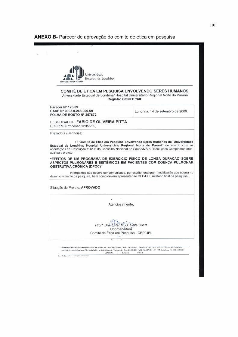
101
ANEXO B- Parecer de aprovação do comite de etica em pesquisa
![[COMPLEMENTAR] Capítulo 4 - GDUCC - Seis Anos - Avaliando a Caminhada e Construindo o Futuro](https://img.document.onl/doc/110x75/55cf8f73550346703b9c7e7f/complementar-capitulo-4-gducc-seis-anos-avaliando-a-caminhada-e-construindo.jpg)