Embed Size (px)
Citation preview
REVIEW ARTICLEpublished: 30 August 2013
doi: 10.3389/fnsys.2013.00045
The anterior thalamus provides a subcortical circuitsupporting memory and spatial navigationMaciej M. Jankowski1, Kim C. Ronnqvist1, Marian Tsanov1, Seralynne D. Vann2, Nicholas F. Wright2,
Jonathan T. Erichsen3, John P. Aggleton2 and Shane M. O’Mara1*
1 Trinity College Institute of Neuroscience, Trinity College Dublin, Dublin 2, Ireland2 School of Psychology, Cardiff University, Cardiff, UK3 School of Optometry and Vision Sciences, Cardiff University, Cardiff, UK
Edited by:
Yuri B. Saalmann, PrincetonUniversity, USA
Reviewed by:
Natasha Sigala, University ofSussex, UKJorge Medina, Universidad deBuenos Aires, Argentina
*Correspondence:
Shane M. O’Mara, Trinity CollegeInstitute of Neuroscience, TrinityCollege Dublin, College Green,Dublin 2, Irelande-mail: [email protected]
The anterior thalamic nuclei (ATN), a central component of Papez’ circuit, are generallyassumed to be key constituents of the neural circuits responsible for certain categories oflearning and memory. Supporting evidence for this contention is that damage to eitherof two brain regions, the medial temporal lobe and the medial diencephalon, is mostconsistently associated with anterograde amnesia. Within these respective regions, thehippocampal formation and the ATN (anteromedial, anteroventral, and anterodorsal) arethe particular structures of interest. The extensive direct and indirect hippocampal-anteriorthalamic interconnections and the presence of theta-modulated cells in both sites furthersupport the hypothesis that these structures constitute a neuronal network crucialfor memory and cognition. The major tool in understanding how the brain processesinformation is the analysis of neuronal output at each hierarchical level along the pathwayof signal propagation coupled with neuroanatomical studies. Here, we discuss theelectrophysiological properties of cells in the ATN with an emphasis on their role in spatialnavigation. In addition, we describe neuroanatomical and functional relationships betweenthe ATN and hippocampal formation.
Keywords: anterior thalamus, memory, spatial navigation, theta rhythm, head direction cells
INTRODUCTIONThat the hippocampal formation is vital for memory is unde-niable. For this reason, understanding hippocampal learningmechanisms remains one of the principal objectives in neu-roscience. However, this problem must be addressed from abroad perspective, i.e., one that includes the many connectionsof the hippocampal formation, some of which are now knownto be critical for hippocampal mnemonic functions. The medialdiencephalon is extensively connected with the hippocampal for-mation, damage to this area being frequently associated withanterograde amnesia (Aggleton and Sahgal, 1993; Aggleton andBrown, 1999). Within the medial diencephalon, the anterior tha-lamic nuclei (ATN) are an important part of the neuronal systemsinvolved in spatial navigation (Clark and Taube, 2012), comple-menting their role in mnemonic functions (Buzsáki and Moser,2013). This review will summarize some recent data concerningthe anatomical and physiological properties of the anterior thala-mic neurons, their role in spatial navigation, and their relevanceto pathophysiological conditions associated with the ATN.
GENERAL ANATOMY OF THALAMUSThe thalamus is a bilateral, symmetrical structure comprising themajority of the diencephalon, with the medial thalamus beingbordered, and in places split, by the third ventricle. The thala-mus is classically divided into several groups of nuclei, describedby their anatomical location: medial, lateral, ventral, and anterior,as well as the posterior (pulvinar) nuclei. This review focuses on
the ATN, which is divided into the anterodorsal, anteroventral,and anteromedial nuclei, all located in the rostral part of the dor-somedial thalamus (Figure 1) (Morel et al., 1997; Wiegell et al.,2003). There is some uncertainty about the nuclei that comprisethe ATN, with many authors regarding the lateral dorsal thalamicnucleus as part of the ATN due to its limbic associations (Morelet al., 1997). Some researchers have argued that the anterome-dial nucleus is actually a part of the anteroventral nucleus (seeAlelú-Paz and Giménez-Amaya, 2007), although in rodents andmonkeys these two nuclei have clearly visible differences whenexamined in histological and immunochemical preparations. Thethree major nuclei within the ATN also have distinct patterns ofconnectivity.
HISTOLOGYWhile the human anteroventral thalamic nucleus is distinguishedby its homogenous and dense cell population (Morel et al., 1997),the neurons in anteromedial nucleus are larger and more widelydispersed. In contrast, the anterodorsal nucleus contains densely-packed small cells (Morel et al., 1997). All areas of the ATN showvarying degrees of immunoreactivity to acetylcholinesterase, aswell as the calcium-binding proteins calretinin, calbindin-D28Kand parvalbumin (Morel et al., 1997; Fortin et al., 1998; Munkleet al., 2000; Alelú-Paz and Giménez-Amaya, 2007). In humans,the neuropil in ATN also stains variably in different areas for neu-ropeptides. These neuropeptide analyses reveal numerous sub-stance P positive varicose fibers scattered throughout the ATN,
Frontiers in Systems Neuroscience www.frontiersin.org August 2013 | Volume 7 | Article 45 | 1
SYSTEMS NEUROSCIENCE
Jankowski et al. Anterior thalamus in memory and navigation
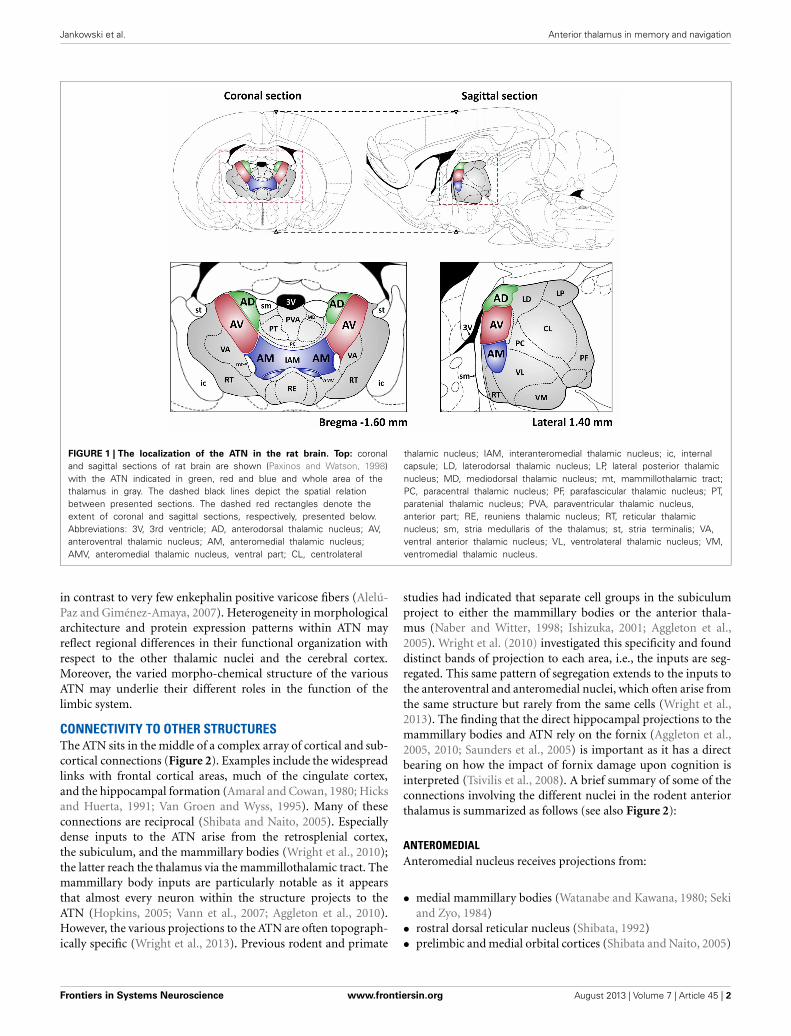
FIGURE 1 | The localization of the ATN in the rat brain. Top: coronaland sagittal sections of rat brain are shown (Paxinos and Watson, 1998)with the ATN indicated in green, red and blue and whole area of thethalamus in gray. The dashed black lines depict the spatial relationbetween presented sections. The dashed red rectangles denote theextent of coronal and sagittal sections, respectively, presented below.Abbreviations: 3V, 3rd ventricle; AD, anterodorsal thalamic nucleus; AV,anteroventral thalamic nucleus; AM, anteromedial thalamic nucleus;AMV, anteromedial thalamic nucleus, ventral part; CL, centrolateral
thalamic nucleus; IAM, interanteromedial thalamic nucleus; ic, internalcapsule; LD, laterodorsal thalamic nucleus; LP, lateral posterior thalamicnucleus; MD, mediodorsal thalamic nucleus; mt, mammillothalamic tract;PC, paracentral thalamic nucleus; PF, parafascicular thalamic nucleus; PT,paratenial thalamic nucleus; PVA, paraventricular thalamic nucleus,anterior part; RE, reuniens thalamic nucleus; RT, reticular thalamicnucleus; sm, stria medullaris of the thalamus; st, stria terminalis; VA,ventral anterior thalamic nucleus; VL, ventrolateral thalamic nucleus; VM,ventromedial thalamic nucleus.
in contrast to very few enkephalin positive varicose fibers (Alelú-Paz and Giménez-Amaya, 2007). Heterogeneity in morphologicalarchitecture and protein expression patterns within ATN mayreflect regional differences in their functional organization withrespect to the other thalamic nuclei and the cerebral cortex.Moreover, the varied morpho-chemical structure of the variousATN may underlie their different roles in the function of thelimbic system.
CONNECTIVITY TO OTHER STRUCTURESThe ATN sits in the middle of a complex array of cortical and sub-cortical connections (Figure 2). Examples include the widespreadlinks with frontal cortical areas, much of the cingulate cortex,and the hippocampal formation (Amaral and Cowan, 1980; Hicksand Huerta, 1991; Van Groen and Wyss, 1995). Many of theseconnections are reciprocal (Shibata and Naito, 2005). Especiallydense inputs to the ATN arise from the retrosplenial cortex,the subiculum, and the mammillary bodies (Wright et al., 2010);the latter reach the thalamus via the mammillothalamic tract. Themammillary body inputs are particularly notable as it appearsthat almost every neuron within the structure projects to theATN (Hopkins, 2005; Vann et al., 2007; Aggleton et al., 2010).However, the various projections to the ATN are often topograph-ically specific (Wright et al., 2013). Previous rodent and primate
studies had indicated that separate cell groups in the subiculumproject to either the mammillary bodies or the anterior thala-mus (Naber and Witter, 1998; Ishizuka, 2001; Aggleton et al.,2005). Wright et al. (2010) investigated this specificity and founddistinct bands of projection to each area, i.e., the inputs are seg-regated. This same pattern of segregation extends to the inputs tothe anteroventral and anteromedial nuclei, which often arise fromthe same structure but rarely from the same cells (Wright et al.,2013). The finding that the direct hippocampal projections to themammillary bodies and ATN rely on the fornix (Aggleton et al.,2005, 2010; Saunders et al., 2005) is important as it has a directbearing on how the impact of fornix damage upon cognition isinterpreted (Tsivilis et al., 2008). A brief summary of some of theconnections involving the different nuclei in the rodent anteriorthalamus is summarized as follows (see also Figure 2):
ANTEROMEDIALAnteromedial nucleus receives projections from:
• medial mammillary bodies (Watanabe and Kawana, 1980; Sekiand Zyo, 1984)
• rostral dorsal reticular nucleus (Shibata, 1992)• prelimbic and medial orbital cortices (Shibata and Naito, 2005)
Frontiers in Systems Neuroscience www.frontiersin.org August 2013 | Volume 7 | Article 45 | 2

Jankowski et al. Anterior thalamus in memory and navigation
FIGURE 2 | The color-coded diagram presents the main direct connections
of the anterodorsal (AD), anteroventral (AV), and anteromedial (AM)
thalamic nuclei in the rat brain. Black arrows depict reciprocal connections,green efferents, and red afferents of the three anterior thalamic nuclei (ATN).
Structures in blue contain head direction cells, and so constitute a part of thehierarchically organized head direction system (Clark and Taube, 2012). Thevarious indirect connections of the ATN, along with the connections betweenother highlighted structures, are not included in this scheme.
• anterior cingulate and dysgranular retrosplenial cortex(Shibata, 1993b; Shibata and Naito, 2005; Wright et al., 2010)
• secondary motor cortices (Shibata and Naito, 2005)• entorhinal cortex (Wright et al., 2010)• subiculum (Wright et al., 2010)
Anteromedial nucleus projects to:
• frontal area 2 (Shibata and Kato, 1993), frontal polar andmedial orbital cortex (Van Groen et al., 1999)
• anterior cingulate (Shibata and Kato, 1993) and dysgranularretrosplenial cortex (Shibata and Kato, 1993; Van Groen et al.,1999)
• entorhinal cortex (Shibata, 1993a; Shibata and Kato, 1993; VanGroen et al., 1999);
• perirhinal cortex (Shibata, 1993a; Van Groen et al., 1999)• presubiculum, subiculum (Shibata, 1993a; Van Groen et al.,
1999)• visual cortex area 18 b (Van Groen et al., 1999)• temporal area 2, occipital area 1 and 2 (Shibata, 1993a)• (medial) secondary motor cortices (Shibata and Naito, 2005)
ANTERODORSALAnterodorsal nucleus receives projections from:
• lateral mammillary bodies (Watanabe and Kawana, 1980;Shibata, 1992)
• subiculum, para-and postsubiculum (Seki and Zyo, 1984; VanGroen and Wyss, 1990a,c; Wright et al., 2010)
• retina (Conrad and Stumpf, 1975; Itaya et al., 1981, 1986)
• anterior cingulate cortex (Shibata and Naito, 2005)• granular retrosplenial cortex (Wright et al., 2010)• caudal dorsal reticular nucleus (Shibata, 1992)
Anterodorsal nucleus projects to:
• pre-, para-, and postsubiculum (Van Groen and Wyss, 1990a,c,1995)
• hippocampus (Wyss et al., 1979; Amaral and Cowan, 1980)• granular retrosplenial cortex (Van Groen and Wyss, 1990b;
Shibata, 1993b; Van Groen and Wyss, 2003)
ANTEROVENTRALAnteroventral nucleus receives projections from:
• medial mammillary bodies (Watanabe and Kawana, 1980)• caudal dorsal reticular nucleus and laterodorsal tegmental
nucleus (Shibata, 1992)• subiculum and postsubiculum (Van Groen and Wyss, 1990c;
Wright et al., 2010)• anterior cingulate cortex, granular and dysgranular retrosple-
nial cortex (Van Groen and Wyss, 1990b, 2003; Shibata andNaito, 2005; Wright et al., 2010)
• secondary motor cortex (Shibata and Naito, 2005)
Anteroventral nucleus projects to:
• pre-, para-, and postsubiculum (Van Groen and Wyss, 1990c;Shibata, 1993a; Van Groen and Wyss, 1995)
Frontiers in Systems Neuroscience www.frontiersin.org August 2013 | Volume 7 | Article 45 | 3
Jankowski et al. Anterior thalamus in memory and navigation
• entorhinal cortex (Shibata, 1993a)• anterior cingulate, granular and dysgranular retrosplenial cor-
tex (Shibata, 1993b; Van Groen and Wyss, 2003)• secondary motor cortex (Shibata and Naito, 2005)
FUNCTIONAL CONSIDERATIONSThe circuit outlined by Papez (1937) is still highly relevantwhen considering the functional and cognitive aspects of sys-tems involving the anterior thalamus. This circuit highlightedthe following projections: hippocampal formation > mammillarybodies > anterior thalamus > cingulate cortex > parahippocam-pal gyrus > hippocampal formation. Since then, the anatomicaldefinition of Papez’ circuit has been further refined (Shah et al.,2012). The ATN still occupy an important position as estab-lished by studies using traditional fiber dissection techniques(Shah et al., 2012), as well as in vivo diffusion spectrum imaging(Granziera et al., 2011) of Papez’ circuit. Although Papez origi-nally suggested that this pathway underlay emotional processingby the brain, our current understanding of Papez’ circuit suggeststhat it has a particular and special role in supporting the neuralsubstrates of explicit learning and memory (Vertes et al., 2001;Shah et al., 2012). Aggleton and Brown (1999) developed theidea of an extended hippocampal-diencephalic network for the
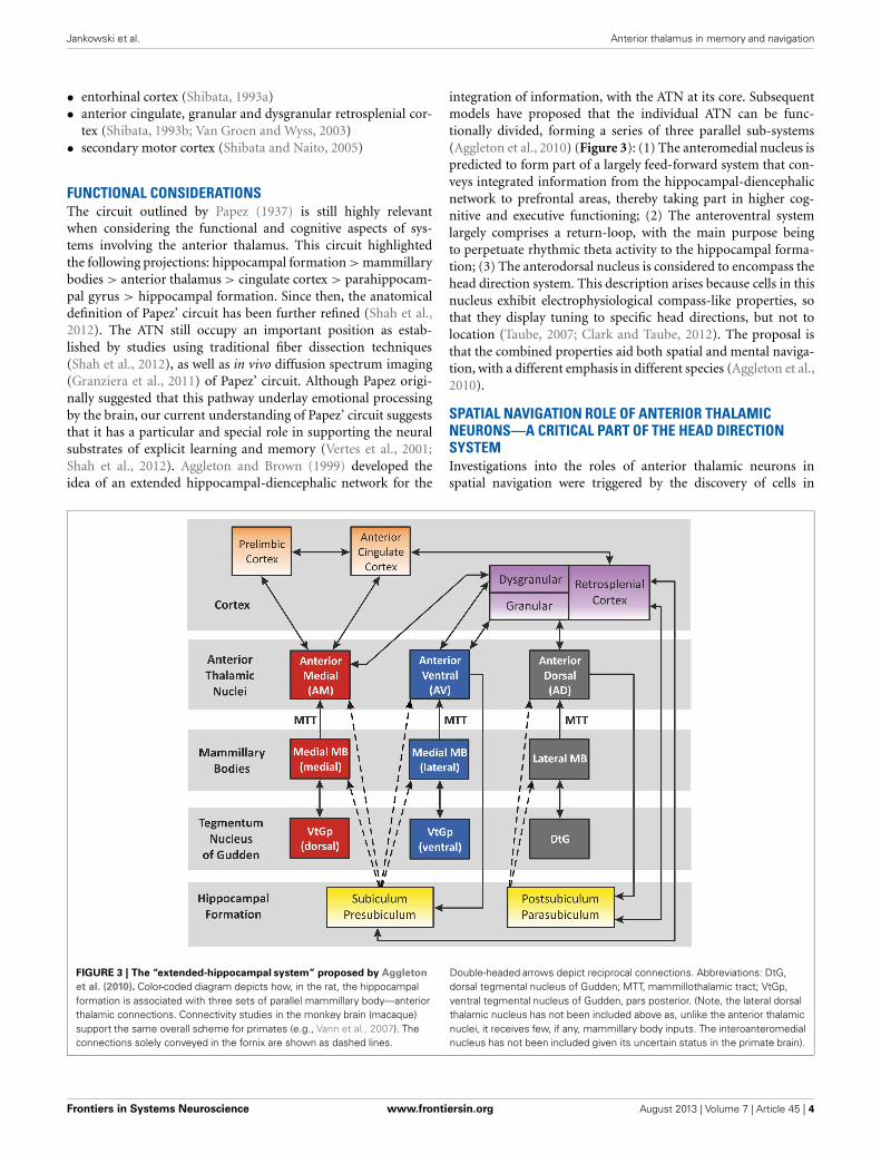
integration of information, with the ATN at its core. Subsequentmodels have proposed that the individual ATN can be func-tionally divided, forming a series of three parallel sub-systems(Aggleton et al., 2010) (Figure 3): (1) The anteromedial nucleus ispredicted to form part of a largely feed-forward system that con-veys integrated information from the hippocampal-diencephalicnetwork to prefrontal areas, thereby taking part in higher cog-nitive and executive functioning; (2) The anteroventral systemlargely comprises a return-loop, with the main purpose beingto perpetuate rhythmic theta activity to the hippocampal forma-tion; (3) The anterodorsal nucleus is considered to encompass thehead direction system. This description arises because cells in thisnucleus exhibit electrophysiological compass-like properties, sothat they display tuning to specific head directions, but not tolocation (Taube, 2007; Clark and Taube, 2012). The proposal isthat the combined properties aid both spatial and mental naviga-tion, with a different emphasis in different species (Aggleton et al.,2010).
SPATIAL NAVIGATION ROLE OF ANTERIOR THALAMICNEURONS—A CRITICAL PART OF THE HEAD DIRECTIONSYSTEMInvestigations into the roles of anterior thalamic neurons inspatial navigation were triggered by the discovery of cells in
FIGURE 3 | The “extended-hippocampal system” proposed by Aggleton
et al. (2010). Color-coded diagram depicts how, in the rat, the hippocampalformation is associated with three sets of parallel mammillary body—anteriorthalamic connections. Connectivity studies in the monkey brain (macaque)support the same overall scheme for primates (e.g., Vann et al., 2007). Theconnections solely conveyed in the fornix are shown as dashed lines.
Double-headed arrows depict reciprocal connections. Abbreviations: DtG,dorsal tegmental nucleus of Gudden; MTT, mammillothalamic tract; VtGp,ventral tegmental nucleus of Gudden, pars posterior. (Note, the lateral dorsalthalamic nucleus has not been included above as, unlike the anterior thalamicnuclei, it receives few, if any, mammillary body inputs. The interoanteromedialnucleus has not been included given its uncertain status in the primate brain).
Frontiers in Systems Neuroscience www.frontiersin.org August 2013 | Volume 7 | Article 45 | 4
Jankowski et al. Anterior thalamus in memory and navigation
the postsubiculum that discharge as a function of the animal’shead direction in the horizontal plane, but independent of itsbehavior and location in the environment (Ranck, 1984; Taubeet al., 1990). Knowing that the postsubiculum contains recip-rocal connections with the ATN (anterodorsal nucleus in par-ticular) led to the suspicion that the anterior thalamus mightalso possess head direction cells. In 1995, Taube reported thatsuch cells, referred to as head direction cells (because they onlydischarge whenever the animal points its head in a particu-lar direction), were indeed present in the ATN (Taube, 1995).Head direction cells are believed to encode primary informationfor spatial orientation in the environment, namely an animal’sperceived directional heading with respect to its environment(for review see Taube, 2007; Clark and Taube, 2012). So far,the largest proportions of head direction cells in the thalamushave been found in the anterodorsal and lateral dorsal thalamicnuclei, with additional head direction cells in the anteroventralnucleus (Taube, 2007; Tsanov et al., 2011a; Clark and Taube,2012). Moreover, head direction cells are also found in corticalstructures such as the postsubiculum, parasubiculum, retros-plenial, and medial entorhinal cortex, as well as in subcorticalbrain regions like the lateral mammillary nucleus (LMN) anddorsal tegmental nucleus of Gudden (DTG) (Clark and Taube,2012).
There is now considerable evidence that the ATN are part ofan interconnected circuit, which is organized hierarchically andis responsible for the propagation of head directional signals inthe central nervous system (Taube, 2007; Clark and Taube, 2012).Such a notion is supported by experiments in which lesions ofthe lower structures of this circuitry (e.g., anterodorsal thala-mus) completely abolished head direction cell activity in highercomponents (e.g., postsubiculum, parasubiculum or superficiallayers of medial entorhinal cortex), whereas destruction of post-subiculum did not disrupt head direction signals in subcorticalstructures (Goodridge and Taube, 1997; Clark and Taube, 2011,2012). Moreover, damage to the postsubiculum or retrosplenialcortex disrupted anterodorsal nucleus head direction cell tuningto visual landmark cues, suggesting that these cortical structuresare important for the visual regulation of head direction cellsactivity in the anterior thalamus (Goodridge and Taube, 1997;Clark et al., 2010; Yoder et al., 2011b). The medial entorhinalcortex seems to be at the top of this hierarchical head direc-tion system because, after lesion, the discharge characteristicsof anterodorsal head direction cells were only mildly affected.Furthermore, entorhinal cortex lesions did not cause clear deficitsin landmark processing or angular path integration (neural inte-gration of head angular velocity signals) by anterodorsal headdirection cells (Clark and Taube, 2011). Further evidence for thehierarchical organization of head direction cell circuitry comesfrom experiments in which lesions in “lower” structures, e.g.,bilateral damage of the dorsal tegmental nucleus of Gudden or thelateral mammillary nucleus, abolished head direction cell activ-ity in the anterodorsal thalamic nucleus (Blair et al., 1998, 1999;Bassett et al., 2007). In contrast to this general pattern of hierar-chical organization, lesions in lateral dorsal thalamic nucleus hadlittle effect on the firing properties of head direction cells in post-subiculum (Golob et al., 1998), whereas an intact anterodorsal
thalamic nucleus is necessary for the presence of head direc-tion cell activity in the postsubiculum (Goodridge and Taube,1997).
Thalamic head direction cells are influenced by both externaland internal sources of information (Taube, 2007; Yoder et al.,2011a). Although external cues exert strong influences on anteriorthalamic head direction cells, these cells can maintain direc-tional firing preferences in the dark and in new environments(Taube and Burton, 1995; Goodridge et al., 1998). This obser-vation suggests that head direction cells are strongly influencedby internal sources of information, i.e., vestibular, propriorecep-tion, or motor efference. One implication is that the vestibularsystem may be particularly important for this aspect of spatialnavigation (Potegal, 1982). This hypothesis was verified exper-imentally by (Stackman and Taube, 1997), who recorded fromhead direction cells in the anterodorsal thalamus before and afterneurotoxic lesions that destroyed the hair cells in the vestibu-lar labyrinth. As a result, head direction cells in the anteriorthalamus lost their directional specificity. Moreover, in lesionedanimals, a new subset of neurons, characterized by intermit-tent firing bursts without specified directionality, was observed.The appearance of a new subset of cells in lesioned animals thatwere not recorded in intact animals suggests that head direc-tion cells may alter their physiology in the absence of indirectvestibular input, and that other sensory systems (e.g., visual,somatosensory/tactile or olfactory) are unable to compensatefor the loss of vestibular information in order to retain direc-tion. The absence of head direction cell activity in animals withvestibular lesions persisted for up to 3 months post-surgery, indi-cating that indirect vestibular inputs remain crucial for anteriorthalamic head direction cell function (Clark and Taube, 2012).However, in the anterodorsal thalamic nucleus of transgenic oto-conia deficient tilted mice, which exhibit an impaired sense oflinear acceleration and head tilt, directionally tuned cells wererecorded (Yoder and Taube, 2009). Nevertheless, the head direc-tion cells recorded in tilt mice often appeared to be unstable.These cells retained directional information for the duration ofa single recording session, but often lost directionality across sub-sequent recording sessions. These experiments (Yoder and Taube,2009) provided the first conclusive evidence that the otolithorgans are important for maintenance of a robust head directionsignal.
One of the main questions that emerged after the discov-ery that the anterodorsal head direction signal is dependent onindirect vestibular inputs was: Are anterodorsal head directioncells activated in the same manner during active and passivemovement? Initially, (Knierim et al., 1995) and Taube (1995)both reported substantial reductions in firing rates during pas-sive rotation, producing near or complete suppression of theanterodorsal head direction response when the animal’s bodywas tightly restrained except its head. Reductions in firing ratesduring passive rotation were also observed in the postsubicu-lum and retrosplenial cortex (Chen et al., 1994; Golob et al.,1998). In contrast to these observations, (Zugaro et al., 2001)found only mild inhibition of anterodorsal head direction cellfiring, with peak firing rates reduced by only 27% and no lossof directional responding during unrestrained passive movement.
Frontiers in Systems Neuroscience www.frontiersin.org August 2013 | Volume 7 | Article 45 | 5
Jankowski et al. Anterior thalamus in memory and navigation
(Bassett et al., 2005) found only a 23% reduction in the peak fir-ing rates of anterodorsal head direction cells when the animalswere passively moved while loosely restrained. The above obser-vations suggest that the tight restraint of the animal may, in itself,be a factor which decreases firing rate of anterodorsal head direc-tion cells. However, in the studies by Knierim et al. (1995) andTaube (1995), the head of the animal was not fully immobilizedwhile the trunk was tightly restrained. Therefore, Shinder andTaube (2011) prepared a rotatable, horizontal plane platform thatwas equipped with an immobilizing tube for the trunk and holderfor the head. Before the recording session, the rat was immobi-lized and its head fixed to the platform by a bar connected to therestraint bolt, which had been previously mounted to the skull.Experiments revealed that passive movement during head-fixedrestraint did not reduce anterodorsal head direction cell firing,relative to active movement (Shinder and Taube, 2011). Moreover,anterodorsal head direction cell responses were also maintainedduring passive movement in the dark, suggesting that visual,motor, and proprioceptive inputs are not necessary to generatedirection-specific responses in head direction cells. This exper-iment further supports the hypothesis that indirect vestibularinput is crucial for head direction cell activity in the anterodorsalthalamus.
Another cell type relevant for spatial navigation is the “placecell.” Place cells discharge when an animal is in a particular loca-tion in the environment (O’Keefe and Dostrovsky, 1971). So far,“true” place cells have only been recorded in the hippocampus(Clark and Taube, 2012). However, the anterior thalamus, as partof the limbic system and head direction system, may contribute tothe function of hippocampal place cells. Several theories have sug-gested that place cells use the signal from the head direction sys-tem to establish and maintain place-field activity (McNaughtonet al., 1996; Touretzky and Redish, 1996; Sharp, 1999). Caltonet al. (2003) reported that, after lesions of the anterodorsal tha-lamic nucleus, place cells continued to exhibit location specificactivity, but the place fields were somewhat degraded and cellswere more directionally-sensitive. These observations suggest thatinput from anterodorsal head direction cells may be importantfor processing and integrating spatial information within thehippocampal circuits containing place cells.
THETA RHYTHM IN THE ANTERIOR THALAMIC NUCLEIThe nuclei within Papez’ pathway that mediate head directionsignals are closely paralleled by those adjacent nuclei mediatingtheta rhythm (a sinusoidal oscillation of 6–12 Hz). Theta rhythmis considered to play a critical role in spatial and non-spatialmnemonic functions of the limbic system (Burgess et al., 2002;Buzsaki, 2005). Both circuits (HD vs. theta) include the tegmentalnuclei of Gudden (dorsal vs. ventral), the mammillary bodies (lat-eral vs. medial), the ATN (anterodorsal vs. anteroventral) and thesubicular/entorhinal cortices (Swanson and Cowan, 1977; Witteret al., 1990; Shibata, 1993b; Van Groen and Wyss, 1995; Gonzalo-Ruiz et al., 1997; Van Groen et al., 1999). Electrophysiologicalstudies in rats support this idea, because plasticity betweensequentially-activated hippocampal place cells occurs duringtheta epochs (Mehta et al., 2000; Ekstrom et al., 2001), implicat-ing the theta cycle as an information quantum (Skaggs et al., 1996;
Buzsaki, 2002). Theta rhythm commonly modulates the spiketrains of spatially-tuned neurons such as hippocampal place cells(O’Keefe and Dostrovsky, 1971), entorhinal grid cells (Haftinget al., 2005), and border cells (Savelli et al., 2008; Solstad et al.,2008). These neurons, together with HD cells, are believed to par-ticipate in computing the animal’s location in the environment byintegrating its movement velocity over time, the process referredto as path integration (McNaughton et al., 1996; Etienne andJeffery, 2004).
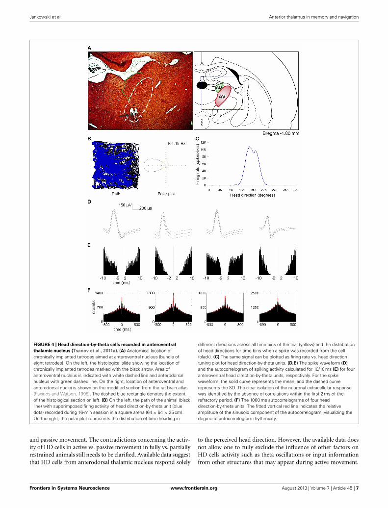
So far, the anterodorsal thalamic nucleus is the best-describedthalamic nucleus with respect to the electrophysiological proper-ties of its neurons in freely moving animals. A particular focus onthis nucleus stems from the fact that it contains high numbers ofhead direction cells (Taube, 2007; Clark and Taube, 2012). Single-unit recordings in other ATN (anteroventral and anteromedial)in urethane-anesthetized rats reveal that some anteroventral neu-rons tend to fire in theta-rhythmic manner (Vertes et al., 2001).This observation was confirmed by single-unit recordings both infreely moving rats foraging for food pellets and during naturallyoccurring sleep (Tsanov et al., 2011b). An identified subgroupof anteroventral neurons was strongly entrained by theta oscil-lations and synchronized their bursting activity in theta range.Moreover, theta and spindle oscillations differed in their spa-tial distribution within the anteroventral nucleus, suggesting thatseparate cellular sources are responsible for these oscillations.Approximately 23% of anteroventral neurons were assigned tothe slow- and fast-spiking bursting units that are selectivelyentrained to theta rhythm (Tsanov et al., 2011b). Importantly,Tsanov et al. (2011a) also reported large subpopulation of headdirection cells (39%) in the anteroventral thalamic nucleus thatexhibit rhythmic spiking in the theta range. This class of units istermed head direction-by-theta cells, which discharge predom-inantly in spike trains at theta frequency whenever the animalis heading/facing in the preferred direction (Figure 4). Neuronspossessing both theta and head-directional properties have beendescribed earlier at the higher level of this circuitry, namely thepresubicular/parasubicular region (Cacucci et al., 2004; Boccaraet al., 2010). Tsanov et al. (2011a) showed for the first time thatthe integration of head-directional and theta information takesplace at the level of the anteroventral thalamic nucleus. It is likelythat this integrated information is sent in an ascending projec-tion within Papez’ circuit and so contributes to the complex firingproperties of the presubiculum and parasubiculum as well asother parts of the extended hippocampal formation. Moreover,it is possible that non-directional theta cells from anteroventralthalamic nucleus may contribute to the priming of retrosple-nial cells, thus magnifying the influence of anterodorsal headdirection cells on neurons in retrosplenial cortex (Albo et al.,2003). Directional information may also be particularly impor-tant for animals engaged in locomotor/exploratory behaviors(theta states) and less during non-locomotor activities (non-thetastates). This notion is supported by work of Zugaro et al. (2001),who reported that anterodorsal head direction cells fire at signif-icantly higher rates during active, compared to passive, motionof rats. However, Shinder and Taube (2011), using their plat-form for full immobilization of the rat, found that the firing ofanterodorsal head direction cells does not differ between active
Frontiers in Systems Neuroscience www.frontiersin.org August 2013 | Volume 7 | Article 45 | 6
Jankowski et al. Anterior thalamus in memory and navigation
FIGURE 4 | Head direction-by-theta cells recorded in anteroventral
thalamic nucleus (Tsanov et al., 2011a). (A) Anatomical location ofchronically implanted tetrodes aimed at anteroventral nucleus (bundle ofeight tetrodes). On the left, the histological slide showing the location ofchronically implanted tetrodes marked with the black arrow. Area ofanteroventral nucleus is indicated with white dashed line and anterodorsalnucleus with green dashed line. On the right, location of anteroventral andanterodorsal nuclei is shown on the modified section from the rat brain atlas(Paxinos and Watson, 1998). The dashed blue rectangle denotes the extentof the histological section on left. (B) On the left, the path of the animal (blackline) with superimposed firing activity of head direction-by-theta unit (bluedots) recorded during 16-min session in a square arena (64 × 64 × 25 cm).On the right, the polar plot represents the distribution of time heading in
different directions across all time bins of the trial (yellow) and the distributionof head directions for time bins when a spike was recorded from the cell(black). (C) The same signal can be plotted as firing rate vs. head directiontuning plot for head direction-by-theta units. (D,E) The spike waveform (D)
and the autocorrelogram of spiking activity calculated for 10/10 ms (E) for fouranteroventral head direction-by-theta units, respectively. For the spikewaveform, the solid curve represents the mean, and the dashed curverepresents the SD. The clear isolation of the neuronal extracellular responsewas identified by the absence of correlations within the first 2 ms of therefractory period. (F) The 1000 ms autocorrelograms of four headdirection-by-theta units. The fitted vertical red line indicates the relativeamplitude of the sinusoid component of the autocorrelogram, visualizing thedegree of autocorrelogram rhythmicity.
and passive movement. The contradictions concerning the activ-ity of HD cells in active vs. passive movement in fully vs. partiallyrestrained animals still needs to be clarified. Available data suggestthat HD cells from anterodorsal thalamic nucleus respond solely
to the perceived head direction. However, the available data doesnot allow one to fully exclude the influence of other factors onHD cells activity such as theta oscillations or input informationfrom other structures that may appear during active movement.
Frontiers in Systems Neuroscience www.frontiersin.org August 2013 | Volume 7 | Article 45 | 7
Jankowski et al. Anterior thalamus in memory and navigation
Clearly, the role of theta oscillations in the ATN on the function ofhead direction cells at all levels of the hierarchical head directioncircuitry remains to be fully elucidated.
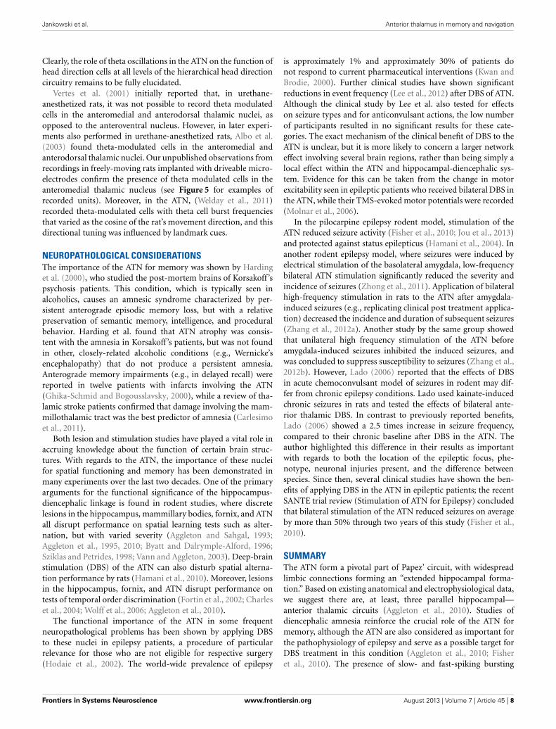
Vertes et al. (2001) initially reported that, in urethane-anesthetized rats, it was not possible to record theta modulatedcells in the anteromedial and anterodorsal thalamic nuclei, asopposed to the anteroventral nucleus. However, in later experi-ments also performed in urethane-anesthetized rats, Albo et al.(2003) found theta-modulated cells in the anteromedial andanterodorsal thalamic nuclei. Our unpublished observations fromrecordings in freely-moving rats implanted with driveable micro-electrodes confirm the presence of theta modulated cells in theanteromedial thalamic nucleus (see Figure 5 for examples ofrecorded units). Moreover, in the ATN, (Welday et al., 2011)recorded theta-modulated cells with theta cell burst frequenciesthat varied as the cosine of the rat’s movement direction, and thisdirectional tuning was influenced by landmark cues.
NEUROPATHOLOGICAL CONSIDERATIONSThe importance of the ATN for memory was shown by Hardinget al. (2000), who studied the post-mortem brains of Korsakoff ’spsychosis patients. This condition, which is typically seen inalcoholics, causes an amnesic syndrome characterized by per-sistent anterograde episodic memory loss, but with a relativepreservation of semantic memory, intelligence, and proceduralbehavior. Harding et al. found that ATN atrophy was consis-tent with the amnesia in Korsakoff ’s patients, but was not foundin other, closely-related alcoholic conditions (e.g., Wernicke’sencephalopathy) that do not produce a persistent amnesia.Anterograde memory impairments (e.g., in delayed recall) werereported in twelve patients with infarcts involving the ATN(Ghika-Schmid and Bogousslavsky, 2000), while a review of tha-lamic stroke patients confirmed that damage involving the mam-millothalamic tract was the best predictor of amnesia (Carlesimoet al., 2011).
Both lesion and stimulation studies have played a vital role inaccruing knowledge about the function of certain brain struc-tures. With regards to the ATN, the importance of these nucleifor spatial functioning and memory has been demonstrated inmany experiments over the last two decades. One of the primaryarguments for the functional significance of the hippocampus-diencephalic linkage is found in rodent studies, where discretelesions in the hippocampus, mammillary bodies, fornix, and ATNall disrupt performance on spatial learning tests such as alter-nation, but with varied severity (Aggleton and Sahgal, 1993;Aggleton et al., 1995, 2010; Byatt and Dalrymple-Alford, 1996;Sziklas and Petrides, 1998; Vann and Aggleton, 2003). Deep-brainstimulation (DBS) of the ATN can also disturb spatial alterna-tion performance by rats (Hamani et al., 2010). Moreover, lesionsin the hippocampus, fornix, and ATN disrupt performance ontests of temporal order discrimination (Fortin et al., 2002; Charleset al., 2004; Wolff et al., 2006; Aggleton et al., 2010).
The functional importance of the ATN in some frequentneuropathological problems has been shown by applying DBSto these nuclei in epilepsy patients, a procedure of particularrelevance for those who are not eligible for respective surgery(Hodaie et al., 2002). The world-wide prevalence of epilepsy
is approximately 1% and approximately 30% of patients donot respond to current pharmaceutical interventions (Kwan andBrodie, 2000). Further clinical studies have shown significantreductions in event frequency (Lee et al., 2012) after DBS of ATN.Although the clinical study by Lee et al. also tested for effectson seizure types and for anticonvulsant actions, the low numberof participants resulted in no significant results for these cate-gories. The exact mechanism of the clinical benefit of DBS to theATN is unclear, but it is more likely to concern a larger networkeffect involving several brain regions, rather than being simply alocal effect within the ATN and hippocampal-diencephalic sys-tem. Evidence for this can be taken from the change in motorexcitability seen in epileptic patients who received bilateral DBS inthe ATN, while their TMS-evoked motor potentials were recorded(Molnar et al., 2006).
In the pilocarpine epilepsy rodent model, stimulation of theATN reduced seizure activity (Fisher et al., 2010; Jou et al., 2013)and protected against status epilepticus (Hamani et al., 2004). Inanother rodent epilepsy model, where seizures were induced byelectrical stimulation of the basolateral amygdala, low-frequencybilateral ATN stimulation significantly reduced the severity andincidence of seizures (Zhong et al., 2011). Application of bilateralhigh-frequency stimulation in rats to the ATN after amygdala-induced seizures (e.g., replicating clinical post treatment applica-tion) decreased the incidence and duration of subsequent seizures(Zhang et al., 2012a). Another study by the same group showedthat unilateral high frequency stimulation of the ATN beforeamygdala-induced seizures inhibited the induced seizures, andwas concluded to suppress susceptibility to seizures (Zhang et al.,2012b). However, Lado (2006) reported that the effects of DBSin acute chemoconvulsant model of seizures in rodent may dif-fer from chronic epilepsy conditions. Lado used kainate-inducedchronic seizures in rats and tested the effects of bilateral ante-rior thalamic DBS. In contrast to previously reported benefits,Lado (2006) showed a 2.5 times increase in seizure frequency,compared to their chronic baseline after DBS in the ATN. Theauthor highlighted this difference in their results as importantwith regards to both the location of the epileptic focus, phe-notype, neuronal injuries present, and the difference betweenspecies. Since then, several clinical studies have shown the ben-efits of applying DBS in the ATN in epileptic patients; the recentSANTE trial review (Stimulation of ATN for Epilepsy) concludedthat bilateral stimulation of the ATN reduced seizures on averageby more than 50% through two years of this study (Fisher et al.,2010).
SUMMARYThe ATN form a pivotal part of Papez’ circuit, with widespreadlimbic connections forming an “extended hippocampal forma-tion.” Based on existing anatomical and electrophysiological data,we suggest there are, at least, three parallel hippocampal—anterior thalamic circuits (Aggleton et al., 2010). Studies ofdiencephalic amnesia reinforce the crucial role of the ATN formemory, although the ATN are also considered as important forthe pathophysiology of epilepsy and serve as a possible target forDBS treatment in this condition (Aggleton et al., 2010; Fisheret al., 2010). The presence of slow- and fast-spiking bursting
Frontiers in Systems Neuroscience www.frontiersin.org August 2013 | Volume 7 | Article 45 | 8
Jankowski et al. Anterior thalamus in memory and navigation
FIGURE 5 | Theta modulated cells from the anteromedial thalamic
nucleus. (A) Recording sites and three examples of theta modulated cellsrecorded in the superficial part of anteromedial nucleus. On the left, thehistological slide showing the location of chronically implanted tetrodesmarked with the black arrow. In this case, three theta modulated cells wererecorded in the superficial part of anteromedial nucleus. Estimated location ofrecorded cells is marked below by green dots on the section from rat brainatlas (Paxinos and Watson, 2006). On the right, parameters of three thetamodulated cells recorded in this rat are presented. From the top for each cell,the waveform, autocorrelation for 10 ms, autocorrelation for 1000 ms andinterspike interval histogram (ISIH) are presented. All three cells exhibitdifferent firing rate and waveforms, but all are modulated in the theta rhythmfrequency, which is visible as 6–10 peaks on 1000 ms autocorrelogram. TheISIH indicates that recorded cells were not bursting neurons (there is no peakof firing before 5 ms). (B) Recording site and example of theta modulated cell
recorded in the bottom part of anteromedial nucleus. In the top, thehistological slide shows the location of chronically implanted tetrodes,marked with the black arrow. In this case, a theta modulated cell wasrecorded in the bottom part of anteromedial nucleus (see also estimatedposition of the cell on the right). Below, the waveform, autocorrelation for10 ms, autocorrelation for 1000 ms, and ISIH are presented. The 1000 msautocorrelogram indicates that this cell was modulated in the frequency oftheta rhythm and ISIH clearly shows that this cell is a bursting neuron.Recordings were performed in rats chronically implanted with driveable32-channel microelectrodes organized in tetrodes. Each recording sessionlasted 20 min and was performed in freely moving rats foraging for foodpellets in a circular arena (96 cm diameter). Abbreviations: AM, anteromedialthalamic nucleus; 3 V, third ventricle; AMV, anteromedial thalamic nucleus,ventral part; f, fornix; mt, mammillothalamic tract; sm, stria medullaris of thethalamus.
anterior thalamic units, which discharge within the theta fre-quency, suggest that the anterior thalamus is involved in thepropagation of theta signals through Papez’ pathway (Verteset al., 2001; Tsanov et al., 2011b). Such theta propagation could
have resulting mnemonic functions. The large populations ofhead direction cells recorded in the anterodorsal and anteroven-tral thalamic nuclei indicate that the anterior thalamus plays animportant role in spatial navigation. Furthermore, the central
Frontiers in Systems Neuroscience www.frontiersin.org August 2013 | Volume 7 | Article 45 | 9
Jankowski et al. Anterior thalamus in memory and navigation
position of the anterodorsal and anteroventral thalamic nucleiin the hierarchically-organized head direction circuitry (Clarkand Taube, 2012) and the apparent integration of theta andhead direction information at the level of anteroventral tha-lamic nucleus (Tsanov et al., 2011a) underline the impor-tance of this region for spatial orientation. Evidence to datesuggests that the ATN serve as a subcortical gate for infor-mation used in path integration processes by cortical struc-tures. A final point is that, by framing the contributions ofthe ATN within Papez’ circuit, there is the strong implica-tion that the functions of these nuclei are principally driven
by the hippocampus. In fact, actions in the opposite direc-tion may prove to be equally crucial. Just as the head directionsystem relies on inputs from “lower” sites within the tegmen-tum, i.e., inputs independent of the hippocampus, so there isreason to believe that other tegmental inputs (e.g., from theventral tegmental nucleus of Gudden and from the lateral dor-sal tegmental nucleus) will prove vital in understanding thebroader role of these diencephalic nuclei in supporting mem-ory (Vann, 2009). Consequently, these tegmental inputs may alsoprove to be of considerable importance for medial temporal lobeactivity.
REFERENCESAggleton, J. P., and Brown, M. W.
(1999). Episodic memory, amne-sia, and the hippocampal-anteriorthalamic axis. Behav. Brain Sci. 22,425–444. discussion: 444–489. doi:10.1017/S0140525X99002034
Aggleton, J. P., Neave, N., Nagle, S.,and Hunt, P. R. (1995). A compar-ison of the effects of anterior tha-lamic, mamillary body and fornixlesions on reinforced spatial alterna-tion. Behav. Brain Res. 68, 91–101.doi: 10.1016/0166-432800163-A
Aggleton, J. P., O’Mara, S. M., Vann,S. D., Wright, N. F., Tsanov,M., and Erichsen, J. T. (2010).Hippocampal-anterior thala-mic pathways for memory:uncovering a network of directand indirect actions. Eur. J.Neurosci. 31, 2292–2307. doi:10.1111/j.1460-9568.2010.07251.x
Aggleton, J. P., and Sahgal, A.(1993). The contribution ofthe anterior thalamic nucleito anterograde amnesia.Neuropsychologia 31, 1001–1019.doi: 10.1016/0028-393290029-Y
Aggleton, J. P., Vann, S. D., andSaunders, R. C. (2005). Projectionsfrom the hippocampal regionto the mammillary bodiesin macaque monkeys. Eur. J.Neurosci. 22, 2519–2530. doi:10.1111/j.1460-9568.2005.04450.x
Albo, Z., Di Prisco, G. V., and Vertes,R. P. (2003). Anterior thalamic unitdischarge profiles and coherencewith hippocampal theta rhythm.Thalamus Relat. Syst. 2, 133–144.
Alelú-Paz, R., and Giménez-Amaya, J.M. (2007). Chemical parcellation ofthe anterior thalamic nuclei in thehuman brain. J. Neural Transm. 114,969–981. doi: 10.1007/s00702-007-0633-8
Amaral, D. G., and Cowan, W. M.(1980). Subcortical afferentsto the hippocampal forma-tion in the monkey. J. Comp.Neurol. 189, 573–591. doi:10.1002/cne.901890402
Bassett, J. P., Tullman, M. L., andTaube, J. S. (2007). Lesions ofthe tegmentomammillary cir-cuit in the head direction systemdisrupt the head direction sig-nal in the anterior thalamus.J. Neurosci. 27, 7564–7577. doi:10.1523/JNEUROSCI.0268-07.2007
Bassett, J. P., Zugaro, M. B., Muir,G. M., Golob, E. J., Muller, R. U.,and Taube, J. S. (2005). Passivemovements of the head do notabolish anticipatory firing prop-erties of head direction cells.J. Neurophysiol. 93, 1304–1316. doi:10.1152/jn.00490.2004
Blair, H. T., Cho, J., and Sharp, P. E.(1998). Role of the lateral mammil-lary nucleus in the rat head direc-tion circuit: a combined single unitrecording and lesion study. Neuron21, 1387–1397. doi: 10.1016/S0896-627380657-1
Blair, H. T., Cho, J., and Sharp, P.E. (1999). The anterior thalamichead-direction signal is abolished bybilateral but not unilateral lesionsof the lateral mammillary nucleus.J. Neurosci. 19, 6673–6683.
Boccara, C. N., Sargolini, F., Thoresen,V. H., Solstad, T., Witter, M. P.,Moser, E. I., et al. (2010). Gridcells in pre- and parasubiculum.Nat. Neurosci. 13, 987–994. doi:10.1038/nn.2602
Burgess, N., Maguire, E. A., andO’Keefe, J. (2002). The human hip-pocampus and spatial and episodicmemory. Neuron 35, 625–641. doi:10.1016/S0896-627300830-9
Buzsaki, G. (2002). Theta oscil-lations in the hippocampus.Neuron 33, 325–340. doi:10.1016/S0896-627300586-X
Buzsaki, G. (2005). Theta rhythmof navigation: link between pathintegration and landmark naviga-tion, episodic and semantic mem-ory. Hippocampus 15, 827–840. doi:10.1002/hipo.20113
Buzsáki, G., and Moser, E. I.(2013). Memory, naviga-tion and theta rhythm in the
hippocampal-entorhinal system.Nat. Neurosci. 16, 130–138. doi:10.1038/nn.3304
Byatt, G., and Dalrymple-Alford, J.C. (1996). Both anteromedial andanteroventral thalamic lesionsimpair radial-maze learning in rats.Behav. Neurosci. 110, 1335–1348.doi: 10.1037/0735-7044.110.6.1335
Cacucci, F., Lever, C., Wills, T.J., Burgess, N., and O’Keefe,J. (2004). Theta-modulatedplace-by-direction cells in thehippocampal formation in the rat.J. Neurosci. 24, 8265–8277. doi:10.1523/JNEUROSCI.2635-04.2004
Calton, J. L., Stackman, R. W.,Goodridge, J. P., Archey, W. B.,Dudchenko, P. A., and Taube, J.S. (2003). Hippocampal place cellinstability after lesions of the headdirection cell network. J. Neurosci.23, 9719–9731.
Carlesimo, G. A., Lombardi, M.G., and Caltagirone, C. (2011).Vascular thalamic amnesia: a reap-praisal. Neuropsychologia 49,777–789. doi: 10.1016/j.neuropsychologia.2011.01.026
Charles, D. P., Gaffan, D., and Buckley,M. J. (2004). Impaired recency judg-ments and intact novelty judgmentsafter fornix transection in mon-keys. J. Neurosci. 24, 2037–2044. doi:10.1523/JNEUROSCI.3796-03.2004
Chen, L. L., Lin, L. H., Green, E. J.,Barnes, C. A., and Mcnaughton,B. L. (1994). Head-direction cellsin the rat posterior cortex. I.Anatomical distribution and behav-ioral modulation. Exp. Brain Res.101, 8–23. doi: 10.1007/BF00243212
Clark, B. J., Bassett, J. P., Wang, S. S.,and Taube, J. S. (2010). Impairedhead direction cell representationin the anterodorsal thalamus afterlesions of the retrosplenial cortex.J. Neurosci. 30, 5289–5302. doi:10.1523/JNEUROSCI.3380-09.2010
Clark, B. J., and Taube, J. S. (2011).Intact landmark control andangular path integration by headdirection cells in the anterodorsal
thalamus after lesions of the medialentorhinal cortex. Hippocampus 21,767–782. doi: 10.1002/hipo.20874
Clark, B. J., and Taube, J. S. (2012).Vestibular and attractor networkbasis of the head direction cellsignal in subcortical circuits.Front. Neural Circuits 6:7. doi:10.3389/fncir.2012.00007
Conrad, C. D., and Stumpf, W.E. (1975). Direct visual inputto the limbic system: crossedretinal projections to the nucleusanterodorsalis thalami in the treeshrew. Exp. Brain Res. 23, 141–149.doi: 10.1007/BF00235456
Ekstrom, A. D., Meltzer, J.,McNaughton, B. L., and Barnes, C.A. (2001). NMDA receptor antago-nism blocks experience-dependentexpansion of hippocampal “placefields.” Neuron 31, 631–638. doi:10.1016/S0896-627300401-9
Etienne, A. S., and Jeffery, K. J.(2004). Path integration in mam-mals. Hippocampus 14, 180–192.doi: 10.1002/hipo.10173
Fisher, R., Salanova, V., Witt, T.,Worth, R., Henry, T., Gross, R.,et al. (2010). Electrical stimula-tion of the anterior nucleus of tha-lamus for treatment of refractoryepilepsy. Epilepsia 51, 899–908. doi:10.1111/j.1528-1167.2010.02536.x
Fortin, M., Asselin, M. C., Gould,P. V., and Parent, A. (1998).Calretinin-immunoreactive neu-rons in the human thalamus.Neuroscience 84, 537–548. doi:10.1016/S0306-452200486-7
Fortin, N. J., Agster, K. L., andEichenbaum, H. B. (2002). Criticalrole of the hippocampus in mem-ory for sequences of events. Nat.Neurosci. 5, 458–462.
Ghika-Schmid, F., and Bogousslavsky,J. (2000). The acute behavioral syn-drome of anterior thalamic infarc-tion: a prospective study of 12 cases.Ann. Neurol. 48, 220–227.
Golob, E. J., Wolk, D. A., and Taube, J.S. (1998). Recordings of postsubicu-lum head direction cells following
Frontiers in Systems Neuroscience www.frontiersin.org August 2013 | Volume 7 | Article 45 | 10

Jankowski et al. Anterior thalamus in memory and navigation
lesions of the laterodorsal thalamicnucleus. Brain Res. 780, 9–19. doi:10.1016/S0006-899301076-7
Gonzalo-Ruiz, A., Morte, L., andLieberman, A. R. (1997). Evidencefor collateral projections to theretrosplenial granular cortex andthalamic reticular nucleus from glu-tamate and/or aspartate-containingneurons of the anterior thalamicnuclei in the rat. Exp. Brain Res. 116,63–72. doi: 10.1007/PL00005745
Goodridge, J. P., Dudchenko, P. A.,Worboys, K. A., Golob, E. J., andTaube, J. S. (1998). Cue control andhead direction cells. Behav. Neurosci.112, 749–761. doi: 10.1037/0735-7044.112.4.749
Goodridge, J. P., and Taube, J. S. (1997).Interaction between the postsubicu-lum and anterior thalamus in thegeneration of head direction cellactivity. J. Neurosci. 17, 9315–9330.
Granziera, C., Hadjikhani, N., Arzy, S.,Seeck, M., Meuli, R., and Krueger,G. (2011). In-vivo magnetic res-onance imaging of the structuralcore of the Papez circuit in humans.Neuroreport 22, 227–231. doi:10.1097/WNR.0b013e328344f75f
Hafting, T., Fyhn, M., Molden, S.,Moser, M. B., and Moser, E.I. (2005). Microstructure of aspatial map in the entorhinalcortex. Nature 436, 801–806. doi:10.1038/nature03721
Hamani, C., Dubiela, F. P., Soares, J. C.,Shin, D., Bittencourt, S., Covolan,L., et al. (2010). Anterior thala-mus deep brain stimulation at highcurrent impairs memory in rats.Exp. Neurol. 225, 154–162. doi:10.1016/j.expneurol.2010.06.007
Hamani, C., Ewerton, F. I., Bonilha,S. M., Ballester, G., Mello, L.E., and Lozano, A. M. (2004).Bilateral anterior thalamic nucleuslesions and high-frequency stim-ulation are protective againstpilocarpine-induced seizures andstatus epilepticus. Neurosurgery54, 191–195. discussion: 195–197.doi: 10.1227/01.NEU.0000097552.31763.AE
Harding, A., Halliday, G., Caine, D.,and Kril, J. (2000). Degenerationof anterior thalamic nucleidifferentiates alcoholics withamnesia. Brain 123, 141–154. doi:10.1093/brain/123.1.141
Hicks, R. R., and Huerta, M. F. (1991).Differential thalamic connectivity ofrostral and caudal parts of cor-tical area Fr2 in rats. Brain Res.568, 325–329. doi: 10.1016/0006-899391420-6
Hodaie, M., Wennberg, R. A.,Dostrovsky, J. O., and Lozano, A. M.(2002). Chronic anterior thalamus
stimulation for intractable epilepsy.Epilepsia 43, 603–608. doi: 10.1046/j.1528-1157.2002.26001.x
Hopkins, D. A. (2005). “Neuroanatomyof head direction cell circuits,” inHead Direction Cells and the NeuralMechanisms of Spatial Orientation,eds S. I. Wiener and J. S. Taube(Cambridge, MA: MIT Press),17–44.
Ishizuka, N. (2001). Laminar orga-nization of the pyramidal celllayer of the subiculum in the rat.J. Comp. Neurol. 435, 89–110. doi:10.1002/cne.1195
Itaya, S. K., Van Hoesen, G. W., andBenevento, L. A. (1986). Direct reti-nal pathways to the limbic thalamusof the monkey. Exp. Brain Res. 61,607–613. doi: 10.1007/BF00237587
Itaya, S. K., Van Hoesen, G. W., andJenq, C. B. (1981). Direct retinalinput to the limbic system of therat. Brain Res. 226, 33–42. doi:10.1016/0006-899391081-7
Jou, S. B., Kao, I. F., Yi, P. L., andChang, F. C. (2013). Electrical stim-ulation of left anterior thalamicnucleus with high-frequency andlow-intensity currents reduces therate of pilocarpine-induced epilepsyin rats. Seizure. 22, 221–229. doi:10.1016/j.seizure.2012.12.015
Knierim, J. J., Kudrimoti, H. S., andMcNaughton, B. L. (1995). Placecells, head direction cells, and thelearning of landmark stability.J. Neurosci. 15, 1648–1659.
Kwan, P., and Brodie, M. J. (2000). Earlyidentification of refractory epilepsy.N. Engl. J. Med. 342, 314–319. doi:10.1056/NEJM200002033420503
Lado, F. A. (2006). Chronic bilat-eral stimulation of the anteriorthalamus of kainate-treatedrats increases seizure fre-quency. Epilepsia 47, 27–32. doi:10.1111/j.1528-1167.2006.00366.x
Lee, K. J., Shon, Y. M., and Cho, C. B.(2012). Long-term outcome of ante-rior thalamic nucleus stimulationfor intractable epilepsy. Stereotact.Funct. Neurosurg. 90, 379–385. doi:10.1159/000339991
McNaughton, B. L., Barnes, C. A.,Gerrard, J. L., Gothard, K., Jung,M. W., Knierim, J. J., et al. (1996).Deciphering the hippocampal poly-glot: the hippocampus as a pathintegration system. J. Exp. Biol. 199,173–185.
Mehta, M. R., Quirk, M. C., andWilson, M. A. (2000). Experience-dependent asymmetric shapeof hippocampal receptivefields. Neuron 25, 707–715. doi:10.1016/S0896-627381072-7
Molnar, G. F., Sailer, A., Gunraj, C.A., Cunic, D. I., Wennberg, R.
A., Lozano, A. M., et al. (2006).Changes in motor cortex excitabil-ity with stimulation of anteriorthalamus in epilepsy. Neurology66, 566–571. doi: 10.1212/01.wnl.0000198254.08581.6b
Morel, A., Magnin, M., andJeanmonod, D. (1997).Multiarchitectonic and stereo-tactic atlas of the human thalamus.J. Comp. Neurol. 387, 588–630.
Munkle, M. C., Waldvogel, H. J., andFaull, R. L. (2000). The distribu-tion of calbindin, calretinin andparvalbumin immunoreactivityin the human thalamus. J. Chem.Neuroanat. 19, 155–173. doi:10.1016/S0891-061800060-0
Naber, P. A., and Witter, M. P. (1998).Subicular efferents are organizedmostly as parallel projections: adouble-labeling, retrograde-tracingstudy in the rat. J. Comp. Neurol.393, 284–297.
O’Keefe, J., and Dostrovsky, J. (1971).The hippocampus as a spatialmap. Preliminary evidence fromunit activity in the freely-movingrat. Brain Res. 34, 171–175. doi:10.1016/0006-899390358-1
Papez, J. W. (1937). A proposed mech-anism of emotion. Arch. NeurPsych.38, 725–743. doi: 10.1001/arch-neurpsyc.1937.02260220069003
Paxinos, G., and Watson, C. (1998). TheRat Brain in Stereotaxic Coordinates,San Diego: Academic Press.
Paxinos, G., and Watson, C. (2006).The Rat Brain in StereotaxicCoordinates 6th edn. Amsterdam:Elsevier.
Potegal, M. (1982). “Vestibular andneostriatal contributions to spatialorientation,” in Spatial AbilitiesDevelopment and PhysiologicalFoundations, ed M. Potegal(New York, NY: Academic Press),361–387.
Ranck, J. B. J. (1984). Head-directioncells in the deep layer of dorsal pre-subiculum in freely moving rats.Soc. Neurosci. Abstr. 10, 599.
Saunders, R. C., Mishkin, M., andAggleton, J. P. (2005). Projectionsfrom the entorhinal cortex, perirhi-nal cortex, presubiculum, and para-subiculum to the medial thalamusin macaque monkeys: identifyingdifferent pathways using disconnec-tion techniques. Exp. Brain Res.167, 1–16. doi: 10.1007/s00221-005-2361-3
Savelli, F., Yoganarasimha, D., andKnierim, J. J. (2008). Influenceof boundary removal on the spa-tial representations of the medialentorhinal cortex. Hippocampus18, 1270–1282. doi: 10.1002/hipo.20511
Seki, M., and Zyo, K. (1984).Anterior thalamic afferentsfrom the mamillary body andthe limbic cortex in the rat.J. Comp. Neurol. 229, 242–256. doi:10.1002/cne.902290209
Shah, A., Jhawar, S. S., and Goel,A. (2012). Analysis of theanatomy of the Papez circuitand adjoining limbic systemby fiber dissection techniques.J. Clin. Neurosci. 19, 289–298. doi:10.1016/j.jocn.2011.04.039
Sharp, P. E. (1999). Complimentaryroles for hippocampal versussubicular/entorhinal place cells incoding place, context, and events.Hippocampus 9, 432–443.
Shibata, H. (1992). Topographic orga-nization of subcortical projectionsto the anterior thalamic nuclei in therat. J. Comp. Neurol. 323, 117–127.doi: 10.1002/cne.903230110
Shibata, H. (1993a). Direct projectionsfrom the anterior thalamic nuclei tothe retrohippocampal region in therat. J. Comp. Neurol. 337, 431–445.doi: 10.1002/cne.903370307
Shibata, H. (1993b). Efferent projec-tions from the anterior thalamicnuclei to the cingulate cortex in therat. J. Comp. Neurol. 330, 533–542.doi: 10.1002/cne.903300409
Shibata, H., and Kato, A. (1993).Topographic relationshipbetween anteromedial thalamicnucleus neurons and their cor-tical terminal fields in the rat.Neurosci. Res. 17, 63–69. doi:10.1016/0168-010290030-T
Shibata, H., and Naito, J. (2005).Organization of anterior cingulateand frontal cortical projectionsto the anterior and laterodor-sal thalamic nuclei in the rat.Brain Res. 1059, 93–103. doi:10.1016/j.brainres.2005.08.025
Shinder, M. E., and Taube, J. S. (2011).Active and passive movement areencoded equally by head directioncells in the anterodorsal thalamus.J. Neurophysiol. 106, 788–800. doi:10.1152/jn.01098.2010
Skaggs, W. E., Mcnaughton, B. L.,Wilson, M. A., and Barnes, C.A. (1996). Theta phase precessionin hippocampal neuronal popula-tions and the compression of tem-poral sequences. Hippocampus 6,149–172.
Solstad, T., Boccara, C. N., Kropff,E., Moser, M. B., and Moser, E. I.(2008). Representation of geomet-ric borders in the entorhinal cor-tex. Science 322, 1865–1868. doi:10.1126/science.1166466
Stackman, R. W., and Taube, J. S.(1997). Firing properties of headdirection cells in the rat anterior
Frontiers in Systems Neuroscience www.frontiersin.org August 2013 | Volume 7 | Article 45 | 11

Jankowski et al. Anterior thalamus in memory and navigation
thalamic nucleus: dependence onvestibular input. J. Neurosci. 17,4349–4358.
Swanson, L. W., and Cowan, W.M. (1977). An autoradiographicstudy of the organization of theefferent connections of the hip-pocampal formation in the rat.J. Comp. Neurol. 172, 49–84. doi:10.1002/cne.901720104
Sziklas, V., and Petrides, M. (1998).Memory and the region ofthe mammillary bodies. Prog.Neurobiol. 54, 55–70. doi:10.1016/S0301-008200064-6
Taube, J. S. (1995). Head directioncells recorded in the anterior tha-lamic nuclei of freely moving rats.J. Neurosci. 15, 70–86.
Taube, J. S. (2007). The head directionsignal: origins and sensory-motorintegration. Annu. Rev. Neurosci.30, 181–207. doi: 10.1146/annurev.neuro.29.051605.112854
Taube, J. S., and Burton, H. L. (1995).Head direction cell activity moni-tored in a novel environment andduring a cue conflict situation.J. Neurophysiol. 74, 1953–1971.
Taube, J. S., Muller, R. U., and Ranck,J. B. Jr. (1990). Head-directioncells recorded from the post-subiculum in freely moving rats.I. Description and quantitativeanalysis. J. Neurosci. 10, 420–435.
Touretzky, D. S., and Redish, A. D.(1996). Theory of rodent navigationbased on interacting representationsof space. Hippocampus 6, 247–270.
Tsanov, M., Chah, E., Vann, S. D.,Reilly, R. B., Erichsen, J. T.,Aggleton, J. P., et al. (2011a).Theta-modulated head directioncells in the rat anterior thalamus.J. Neurosci. 31, 9489–9502. doi:10.1523/JNEUROSCI.0353-11.2011
Tsanov, M., Chah, E., Wright, N.,Vann, S. D., Reilly, R., Erichsen,J. T., et al. (2011b). Oscillatoryentrainment of thalamic neuronsby theta rhythm in freely movingrats. J. Neurophysiol. 105, 4–17. doi:10.1152/jn.00771.2010
Tsivilis, D., Vann, S. D., Denby, C.,Roberts, N., Mayes, A. R., Montaldi,D., et al. (2008). A disproportionaterole for the fornix and mammil-lary bodies in recall versus recog-nition memory. Nat. Neurosci. 11,834–842. doi: 10.1038/nn.2149
Van Groen, T., Kadish, I., and Wyss,J. M. (1999). Efferent connectionsof the anteromedial nucleus ofthe thalamus of the rat. BrainRes. Brain Res. Rev. 30, 1–26. doi:10.1016/S0165-017300006-5
Van Groen, T., and Wyss, J. M.(1990a). The connections of pre-subiculum and parasubiculum inthe rat. Brain Res. 518, 227–243. doi:10.1016/0006-899390976-I
Van Groen, T., and Wyss, J. M.(1990b). Connections of the retro-splenial granular a cortex in the rat.J. Comp. Neurol. 300, 593–606. doi:10.1002/cne.903000412
Van Groen, T., and Wyss, J. M.(1990c). The postsubicular cortexin the rat: characterization of thefourth region of the subicular cor-tex and its connections. Brain Res.529, 165–177. doi: 10.1016/0006-899390824-U
Van Groen, T., and Wyss, J. M. (1995).Projections from the anterodorsaland anteroventral nucleus of thethalamus to the limbic cortex in therat. J. Comp. Neurol. 358, 584–604.doi: 10.1002/cne.903580411
Van Groen, T., and Wyss, J. M.(2003). Connections of the retros-plenial granular b cortex in the rat.J. Comp. Neurol. 463, 249–263. doi:10.1002/cne.10757
Vann, S. D. (2009). Gudden’s ven-tral tegmental nucleus is vitalfor memory: re-evaluatingdiencephalic inputs for amne-sia. Brain 132, 2372–2384. doi:10.1093/brain/awp175
Vann, S. D., and Aggleton, J. P.(2003). Evidence of a spatial encod-ing deficit in rats with lesions ofthe mammillary bodies or mam-millothalamic tract. J. Neurosci. 23,3506–3514.
Vann, S. D., Saunders, R. C., andAggleton, J. P. (2007). Distinct, par-allel pathways link the medial mam-millary bodies to the anterior tha-lamus in macaque monkeys. Eur.J. Neurosci. 26, 1575–1586. doi:10.1111/j.1460-9568.2007.05773.x
Vertes, R. P., Albo, Z., and VianaDi Prisco, G. (2001). Theta-rhythmically firing neurons inthe anterior thalamus: implica-tions for mnemonic functions ofPapez’s circuit. Neuroscience 104,619–625. doi: 10.1016/S0306-452200131-2
Watanabe, K., and Kawana, E. (1980). Ahorseradish peroxidase study on themammillothalamic tract in the rat.Acta Anat. (Basel.) 108, 394–401.doi: 10.1159/000145322
Welday, A. C., Shlifer, I. G., Bloom,M. L., Zhang, K., and Blair, H.T. (2011). Cosine directionaltuning of theta cell burst fre-quencies: evidence for spatialcoding by oscillatory interference.
J. Neurosci. 31, 16157–16176.doi: 10.1523/JNEUROSCI.0712-11.2011
Wiegell, M. R., Tuch, D. S., Larsson,H. B., and Wedeen, V. J. (2003).Automatic segmentation of thala-mic nuclei from diffusion tensormagnetic resonance imaging.Neuroimage 19, 391–401. doi:10.1016/S1053-811900044-2
Witter, M. P., Ostendorf, R. H.,and Groenewegen, H. J. (1990).Heterogeneity in the dorsalsubiculum of the rat. distinctneuronal zones project to dif-ferent cortical and subcorticaltargets. Eur. J. Neurosci. 2,718–725. doi: 10.1111/j.1460-9568.1990.tb00462.x
Wolff, M., Gibb, S. J., and Dalrymple-Alford, J. C. (2006). Beyond spatialmemory: the anterior thalamusand memory for the temporalorder of a sequence of odor cues.J. Neurosci. 26, 2907–2913. doi:10.1523/JNEUROSCI.5481-05.2006
Wright, N. F., Erichsen, J. T., Vann, S.D., O’Mara, S. M., and Aggleton,J. P. (2010). Parallel but sepa-rate inputs from limbic cortices tothe mammillary bodies and ante-rior thalamic nuclei in the rat.J. Comp. Neurol. 518, 2334–2354.doi: 10.1002/cne.22336
Wright, N. F., Vann, S. D., Erichsen,J. T., O’Mara, S. M., and Aggleton,J. P. (2013). Segregation of par-allel inputs to the anteromedialand anteroventral thalamic nucleiof the rat. J. Comp. Neurol. 521,2966–2986. doi: 10.1002/cne.23325
Wyss, J. M., Swanson, L. W., andCowan, W. M. (1979). A studyof subcortical afferents to thehippocampal formation in therat. Neuroscience 4, 463–476. doi:10.1016/0306-452290124-6
Yoder, R. M., Clark, B. J., Brown, J. E.,Lamia, M. V., Valerio, S., Shinder,M. E., et al. (2011a). Both visualand idiothetic cues contribute tohead direction cell stability duringnavigation along complex routes.J. Neurophysiol. 105, 2989–3001.doi: 10.1152/jn.01041.2010
Yoder, R. M., Clark, B. J., and Taube,J. S. (2011b). Origins of land-mark encoding in the brain.Trends Neurosci. 34, 561–571. doi:10.1016/j.tins.2011.08.004
Yoder, R. M., and Taube, J. S. (2009).Head direction cell activity inmice: robust directional signaldepends on intact otolith organs.J. Neurosci. 29, 1061–1076. doi:10.1523/JNEUROSCI.1679-08.2009
Zhang, Q., Wu, Z. C., Yu, J. T., Yu,N. N., Zhong, X. L., and Tan, L.(2012a). Mode-dependent effect ofhigh-frequency electrical stimula-tion of the anterior thalamic nucleuson amygdala-kindled seizures inrats. Neuroscience 217, 113–122. doi:10.1016/j.neuroscience.2012.05.009
Zhang, Q., Wu, Z. C., Yu, J. T., Zhong,X. L., Xing, Y. Y., Tian, Y., et al.(2012b). Anticonvulsant effect ofunilateral anterior thalamic highfrequency electrical stimulation onamygdala-kindled seizures in rat.Brain Res. Bull. 87, 221–226. doi:10.1016/j.brainresbull.2011.11.023
Zhong, X. L., Lv, K. R., Zhang, Q.,Yu, J. T., Xing, Y. Y., Wang, N.D., et al. (2011). Low-frequencystimulation of bilateral anteriornucleus of thalamus inhibitsamygdale-kindled seizures in rats.Brain Res. Bull. 86, 422–427. doi:10.1016/j.brainresbull.2011.08.014
Zugaro, M. B., Tabuchi, E., Fouquier,C., Berthoz, A., and Wiener, S. I.(2001). Active locomotion increasespeak firing rates of anterodorsalthalamic head direction cells.J. Neurophysiol. 86, 692–702.
Conflict of Interest Statement: Theauthors declare that the researchwas conducted in the absence of anycommercial or financial relationshipsthat could be construed as a potentialconflict of interest.
Received: 19 April 2013; accepted: 08August 2013; published online: 30 August2013.Citation: Jankowski MM, RonnqvistKC, Tsanov M, Vann SD, Wright NF,Erichsen JT, Aggleton JP, and O’MaraSM (2013) The anterior thalamus pro-vides a subcortical circuit supportingmemory and spatial navigation. Front.Syst. Neurosci. 7:45. doi: 10.3389/fnsys.2013.00045This article was submitted to the journalFrontiers in Systems Neuroscience.Copyright © 2013 Jankowski,Ronnqvist, Tsanov, Vann, Wright,Erichsen, Aggleton, and O’Mara. This isan open-access article distributed underthe terms of the Creative CommonsAttribution License (CC BY). The use,distribution or reproduction in otherforums is permitted, provided the orig-inal author(s) or licensor are creditedand that the original publication inthis journal is cited, in accordancewith accepted academic practice. Nouse, distribution or reproduction ispermitted which does not comply withthese terms.
Frontiers in Systems Neuroscience www.frontiersin.org August 2013 | Volume 7 | Article 45 | 12


![Orchestrating Virtualized Network Functionsperformance [11], [12]. NFV provides ample opportunities for network optimization and cost reduction. Previously, middle-boxes were hardware](https://img.document.onl/doc/110x75/5ed155afefd7b2537304c8d8/orchestrating-virtualized-network-functions-performance-11-12-nfv-provides.jpg)


![Supporting information revised · -13 ≤ k ≤ 15 -17 ≤ l ≤ 17 Reflections collected 26553 Reflections unique [R (int)] 7884 [0.0230] Completeness to theta = 22.61 99.6 % Absorption](https://img.document.onl/doc/110x75/5f81254824efd14af55a89a3/supporting-information-13-a-k-a-15-17-a-l-a-17-reflections-collected-26553.jpg)