Embed Size (px)
Citation preview

Universidade do Estado do Rio
de Janeiro
Centro Biomédico
Faculdade de Ciências Médicas
Helce Ribeiro Julio Junior
Estudo estrutural da bexiga em fetos humanos normais e com síndrome de
Prune Belly
Rio de Janeiro 2018

Helce Ribeiro Julio Junior
Estudo estrutural da bexiga em fetos humanos normais e com síndrome de
Prune Belly
Tese de Doutorado submetida ao
Programa de Pós-Graduação em
Fisiopatologia e Ciências Cirúrgicas, PG-
Fisiocirurgia, UERJ, como parte dos
requisitos para obtenção do Título de
Doutor.
Orientador: Prof. Dr. Luciano Alves Favorito
Rio de Janeiro 2018
CATALOGAÇÃO NA FONTE
UERJ/REDE SIRIUS/BIBLIOTECA CB-A
Autorizo, apenas para fins acadêmicos e científicos, a reprodução total ou parcial desta tese,
desde que citada a fonte.
__________________________________________________ _______________
Assinatura Data
Ribeiro Julio Junior, HelceRH474 Estudo estrutural da bexiga em fetos humanos normais e
com síndrome de Prune Belly / Helce Ribeiro Julio Junior;orientador Luciano Alves Favorito. -- Rio de Janeiro, 2018.56 p.
Tese (Doutorado em Fisiopatologia e Ciências Cirúrgicas) -- Universidade do Estado do Rio de Janeiro, Faculdade de Ciências Médicas. Pós-Graduação em Fisiopatologia e Ciências Cirúrgicas PG - Fisiocirurgia.
Bibliografia: f. 41-44.
1. Bexiga. 2. Síndrome de Prune Belly. 3. Histologia. I. AlvesFavorito, Luciano, orient. II. Título.

Helce Ribeiro Julio Junior
Estudo estrutural da bexiga em fetos humanos normais e com síndrome de Prune Belly
Tese de Doutorado submetida ao Programa de
Pós-Graduação em Fisiopatologia e Ciências
Cirúrgicas, PG-Fisiocirurgia, UERJ, como parte
dos requisitos para obtenção do Título de Doutor.
Orientador: _____________________________________________
Prof. Dr. Luciano Alves Favorito Universidade do Estado do Rio de Janeiro - Uerj
Banca Examinadora:
_____________________________________________ Prof. Dr. Francisco J. B. Sampaio Universidade do Estado do Rio de Janeiro - Uerj
____________________________________________ Prof. Dr. Waldemar Silva Costa Universidade do Estado do Rio de Janeiro - Uerj
_____________________________________________ Prof. Dr. Valter Javaroni Hospital Federal do Andaraí
_____________________________________________ Prof. Dr. João Paulo M. de Carvalho Hospital Federal Cardoso Fontes
_____________________________________________ Prof. Dr. Nicolino Cesar Rosito Universidade Federal do Rio Grande do Sul
Rio de Janeiro 2018
Dedico esta tese à minha família, em especial minha esposa Gisele que sempre me apoiou. Também não poderia esquecer dos meus colegas de laboratório e em especial meu orientador
professor dr. Luciano Alves Favorito e ao professor dr. Francisco José Barcellos Sampaio, que me recebeu de portas abertas.
DEDICATÓRIA

AGRADECIMENTOS
Gostaria de agradecer a minha família e amigos, bem como colegas de laboratório que
me apoiaram neste período e, em especial, ao meu orientador professor dr. Luciano Alves
Favorito.
O presente trabalho foi realizado na Unidade de Pesquisa Urogenital, Centro
Biomédico, Universidade Estadual do Rio de Janeiro. Recebeu apoio financeiro, direta ou
indiretamente, de CNPq, FAPERJ e CAPES.
CONFLITO DE INTERESSES
Não há conflito de interesses.

RESUMO
JUNIOR, Helce Ribeiro Julio. Estudo estrutural da bexiga em fetos humanos normais e com síndrome de Prune Belly. 2018. 58 f. Tese (Doutorado em Fisiopatologia e Ciências Cirúrgicas) – Centro Biomédico - Faculdade de Ciências Médicas, Universidade do estado do Rio de Janeiro. Rio de Janeiro, 2018.
A síndrome de Prune Belly é uma desordem caracterizada pela deficiência dos músculos da parede abdominal, malformações do trato urinário e criptorquia bilateral. O objetivo deste trabalho é a avaliação estrutural da bexiga de fetos com síndrome de Prune Belly (SPB). Foram estudadas três bexigas de fetos masculinos com esta síndrome e sete bexigas de fetos sem esta anomalia. A idade gestacional dos fetos foi determinada em semanas pós-concepção (SPC), de acordo com o critério do comprimento do maior pé. Após estas medidas, os fetos foram cuidadosamente dissecados com magnificação de 16/25x. O abdome e a pelve fetal foram dissecados para a identificação do trato urogenital. A bexiga e a próstata foram separados das outras estruturas e fixados em formalina tamponada 10 %. Realizou-se processamento histológico de rotina para inclusão em parafina e, em seguida, foram feitos cortes de 5 µm de espessura com intervalo de 200 µm entre cada um deles. Os cortes foram corados com hematoxilina-eosina para verificar a integridade do material. Foi utilizado o Tricrômico de Masson para quantificar o tecido conjuntivo e o tecido muscular; Resorcina Fucsina de Weigert com prévia oxidação para observação das fibras do sistema elástico; e Vermelho de Picro-Sirius com polarização para observação dos diferentes tipos de colágeno. O tecido muscular, o tecido conjuntivo e as fibras do sistema elástico foram quantificadas por método estereológico. Imunostoquímica com tubulina (tubulina beta lll, anticorpo monoclonal de rato) foi realizada para avaliação dos nervos da bexiga. As imagens foram capturadas com a utilização do microscópio Olympus BX51 e da câmera Olympus DP70. As imagens, em seguida, eram transferidas para o software Image Pro. As fibras foram quantificadas com a utilização do software Image J (versão 1.34s; National Institute of Health, Bethesda, MD) para a determinação da densidade volumétrica (Vv) de cada componente. As médias foram estatisticamente comparadas utilizados os testes T não pareado e o Mann-Whitney test. O teste de Wilcoxon foi usado para as variáveis continuas. O valor de p<0.05 foi considerado estatisticamente significativo. Os fetos com síndrome de Prune Belly tinham idade entre 17 e 31 SPC, pesavam entre 240 e 2150 g e tinham o CVC entre 18 e 43 cm. Os fetos do grupo controle tinham idade entre 12 e 35 SPC, pesavam entre 210 e 2860 g e tinham o CVC entre 18 e 34 cm. A análise quantitativa demonstrou que o tecido muscular era significativamente menor (p=0.04) nos fetos com síndrome de Prune Belly (9.67% a 17.75%, média=13.2%) quando comparado com o grupo controle (13.33% a 26.56%, média=17.43%). A análise qualitativa do colágeno demonstrou a predominância da coloração verde no grupo controle, sugerindo a presença de colágeno tipo 3 e a predominância de vermelha nos fetos com síndrome de Prune Belly, sugerindo a predominância de colágeno tipo 1 neste grupo. A análise qualitativa dos nervos com imunohistoquímica com tubulina demonstraram a predominância de nervos no grupo controle quando comparado com os fetos com síndrome de Prune Belly. A bexiga dos fetos com síndrome de Prune Belly tem uma concentração menor de fibras musculares lisas, colágeno tipo 3 e nervos. A alteração estrutural pode ser um dos fatores envolvidos na anomalia do trato urinário tais como a bexiga de grande capacidade nos pacientes com esta síndrome.
Palavras-chave: Bexiga. Síndrome de Prune Belly. Histologia.

ABSTRACT
JUNIOR, Helce Ribeiro Julio. Estudo estrutural da bexiga em fetos humanos normais e com síndrome de Prune Belly. 2018. 58 f. Tese (Doutorado em Fisiopatologia e Ciências Cirúrgicas) – Centro Biomédico - Faculdade de Ciências Médicas, Universidade do estado do Rio de Janeiro. Rio de Janeiro, 2018.
Prune belly syndrome is a disorder characterized by deficiency of the abdominal muscles, malformations of the urinary tract and bilateral cryptorchidism. The objective of this paper is to study the bladder structure of fetuses with prune belly syndrome (PBS). We studied 3 bladders obtained from 3 male fetuses with PBS and 7 bladders obtained from 7 male fetuses without anomalies. The gestational age of the fetuses was determined in weeks post conception (WPC), according to the foot-length criterion. After the measurements, the fetuses were carefully dissected with the aid of a stereoscopic lens with 16/25X magnification. The abdomen and pelvis were opened to identify and expose the urogenital organs. Each bladder was dissected and embedded in paraffin, from which 5 µm thick sections were obtained and stained with Masson’s trichrome (to quantify connective tissue and smooth muscle) and picrosirius red with polarization (to observe collagen). Also, immunohistochemistry with tubulin (Tubulin, beta III, Mouse Monoclonal Antibody) was applied to observe the bladder nerves. The images were captured with an Olympus BX51 microscope and Olympus DP70 camera. The stereological analysis was done with the Image Pro and Image J programs, using a grid to determine volumetric densities (Vv). Means were statistically compared using the Mann-Whitney test and a p-value < 0.05 was considered statistically significant. The PBS fetuses ranged in age from 17 to 31 WPC and weighed between 240 and 2150 g. The fetuses from the control group ranged in age from 12 to 35 WPC and weighed between 430 and 2860 g. Quantitative analysis documented that smooth muscle fibers were significantly smaller (p=0.04) in PBS fetuses (9.67% to 17.75%, mean=13.2%) compared to the control group (13.33% to 26.56%, mean=17.43%). The analysis of collagen fibers showed predominance of green in the control group, suggesting collagen type III presence, and predominance of red in the in PBS fetal bladders, suggesting collagen type I presence in this group. The qualitative analysis of the nerves with immunohistochemistry with tubulin showed predominance of nerves in the control group compared to the PBS fetuses. The bladder in PBS had lower concentrations of smooth muscle fibers, collagen type III and nerves. These structural alterations can be one of the factors involved in urinary tract abnormality such as distended bladder in patients with prune belly syndrome.
Keywords: Prune belly syndrome. Bladder. Histology.
LISTA DE ILUSTRAÇÕES
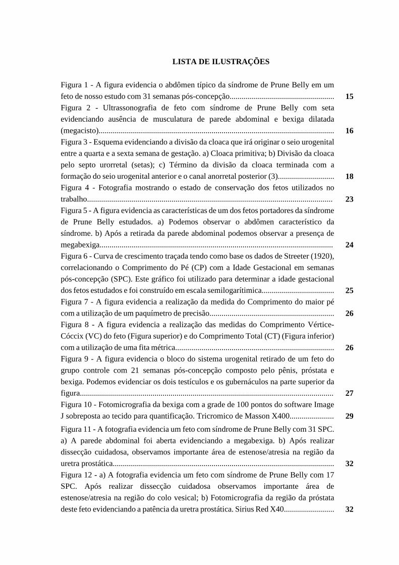
Figura 1 - A figura evidencia o abdômen típico da síndrome de Prune Belly em um feto de nosso estudo com 31 semanas pós-concepção.................................................... 15 Figura 2 - Ultrassonografia de feto com síndrome de Prune Belly com seta evidenciando ausência de musculatura de parede abdominal e bexiga dilatada (megacisto)..................................................................................................................... 16 Figura 3 - Esquema evidenciando a divisão da cloaca que irá originar o seio urogenital entre a quarta e a sexta semana de gestação. a) Cloaca primitiva; b) Divisão da cloaca pelo septo urorretal (setas); c) Término da divisão da cloaca terminada com a formação do seio urogenital anterior e o canal anorretal posterior (3)............................ 18 Figura 4 - Fotografia mostrando o estado de conservação dos fetos utilizados no trabalho.......................................................................................................................... 23 Figura 5 - A figura evidencia as características de um dos fetos portadores da síndrome de Prune Belly estudados. a) Podemos observar o abdômen característico da síndrome. b) Após a retirada da parede abdominal podemos observar a presença de megabexiga.................................................................................................................... 24 Figura 6 - Curva de crescimento traçada tendo como base os dados de Streeter (1920), correlacionando o Comprimento do Pé (CP) com a Idade Gestacional em semanas pós-concepção (SPC). Este gráfico foi utilizado para determinar a idade gestacional dos fetos estudados e foi construído em escala semilogarítimica.................................... 25 Figura 7 - A figura evidencia a realização da medida do Comprimento do maior pé com a utilização de um paquímetro de precisão.............................................................. 26 Figura 8 - A figura evidencia a realização das medidas do Comprimento Vértice-Cóccix (VC) do feto (Figura superior) e do Comprimento Total (CT) (Figura inferior) com a utilização de uma fita métrica............................................................................... 26 Figura 9 - A figura evidencia o bloco do sistema urogenital retirado de um feto do grupo controle com 21 semanas pós-concepção composto pelo pênis, próstata e bexiga. Podemos evidenciar os dois testículos e os gubernáculos na parte superior da figura.............................................................................................................................. 27 Figura 10 - Fotomicrografia da bexiga com a grade de 100 pontos do software Image J sobreposta ao tecido para quantificação. Tricromico de Masson X400...................... 29 Figura 11 - A fotografia evidencia um feto com síndrome de Prune Belly com 31 SPC. a) A parede abdominal foi aberta evidenciando a megabexiga. b) Após realizardissecção cuidadosa, observamos importante área de estenose/atresia na região dauretra prostática.............................................................................................................. 32 Figura 12 - a) A fotografia evidencia um feto com síndrome de Prune Belly com 17 SPC. Após realizar dissecção cuidadosa observamos importante área de estenose/atresia na região do colo vesical; b) Fotomicrografia da região da próstata deste feto evidenciando a patência da uretra prostática. Sirius Red X40......................... 32

Figura 13 - a) Fotomicrografias da bexiga de um feto do grupo controle sem evidenciar a presença das fibras do sistema elástico. Feto do sexo masculino com 21 SPC. Resorcina Fucsina de Weigert com prévia oxidação (40x). b) Fotomicrografia do mesmo feto do grupo controle evidenciando a presença de fibras elásticas na região da artéria umbilical (400x). Resorcina Fucsina de Weigert com prévia oxidação........... 33 Figura 14 - Análise do Colageno. a) Fotomicrografias da bexiga de um feto com síndrome de Prune Belly com 19 SPC evidenciando o predomínio da coloração verde. b) Fotomicrografia de um feto com 18 SPC do grupo controle mostrando opredomínio da coloração vermelha. Picro Sirius Red com polarização. 200x................ 34 Figura 15 - Fotomicrografias de bexigas de fetos mostrando a presença do tecido muscular (corado em vermelho) nos grupos. a) Bexiga de feto com síndrome de Prune Belly com 19 SPC. (b) Bexiga de feto normal com 18 SPC. Tricrômico de Masson. 400X............................................................................................................................... 35 Figura 16 - Análise da distribuição dos nervos. a) Fotomicrografia demostrando os nervos (setas) na bexiga de um feto do grupo controle com 35 SPC. Tubulina X200. b) Fotomicrografia demonstrando os nervos na bexiga de um feto portador desíndrome de Prune Belly com 31 SPC. Tubulina X200.................................................. 35 Figura 17 - Análise da distribuição dos nervos. a) Fotomicrografia demostrando os nervos (setas) na bexiga de um feto do grupo controle com 18SPC. Tubulina X200. b) Fotomicrografia demonstrando os nervos na bexiga de um feto portador desíndrome de Prune Belly com 19SPC. Tubulina X200.................................................. 36

LISTA DE TABELAS
Tabela 1 - A tabela evidencia a idade (em semanas pós-concepção) e os parâmetros fetais de nossa amostra (peso em gramas e comprimento vértice-coccix – CVC em cm)..................................................................................................................................... 31

LISTA DE ABREVIATURAS E SIGLAS
cm = centímetros mm = milímetros HPB = hiperplasia prostática benigna UERJ = Universidade do Estado do Rio de Janeiro CEP = Comitê de Ética em Pesquisa HUPE = Hospital Universitário Pedro Ernesto SPC = semanas pós-concepção µm = micrômetros VV = densidade volumétrica mg = miligramas HCl = ácido clorídrico g = gramas r = índice de correlação IC = intervalo de confiança

SUMÁRIO
1 INTRODUÇÃO ................................................................................................................... 14
1.1 Síndrome de Prune Belly ................................................................................................ 14
1.2 Embriologia da bexiga .................................................................................................... 17
2 OBJETIVO .......................................................................................................................... 21
3 MATERIAL E MÉTODOS ................................................................................................ 22
3.1 Estudo dos fetos .............................................................................................................. 22
3.2 Análise quantitativa ........................................................................................................ 28
3.3 Análise estatística ........................................................................................................... 30
4 RESULTADOS .................................................................................................................... 31
5 DISCUSSÃO ........................................................................................................................ 37
6 CONCLUSÕES .................................................................................................................... 41
7 REFERÊNCIAS BIBLIOGRÁFICAS .............................................................................. 42
ANEXOS ................................................................................................................................. 46
1 - Parecer do comitê de ética ............................................................................................... 47
2 - Artigo publicado na revista Neururology and Urodinamics ........................................... 48
3 - Artigo aceito para publicação na revista Biomedical Research International ................. 53

14
1 INTRODUÇÃO
As malformações congênitas acometem 1 em cada 40 gestações, com,
aproximadamente, 20 % dos fetos indo a óbito ainda no período uterino, como natimortos ou
como resultado de abortos terapêuticos (COPP; GREENE, 2012).
1.1 Síndrome de Prune Belly
A síndrome de Prune Belly (SPB) é uma anomalia congênita rara que afeta cerca de 1
em cada 30.000 a 50.000 nascimentos (HASSETT; SMITH; HOLLAND, 2012). Esta síndrome
é caracterizada por uma tríade clássica de sinais: (a) desenvolvimento deficiente dos músculos
abdominais que faz com que a pele do abdômen apresente um aspecto enrugado como uma
ameixa seca; (b) criptorquidia bilateral e (c) anormalidades do trato urinário, como hidronefrose
bilateral severa, megaureter e megabexiga (Figura 1) (STEPHENS; SMITH; HUTSON, 2002).
A obstrução uretral está presente em um terço dos pacientes com SPB, que pode ser a principal
causa das malformações nesta síndrome (STEPHENS; SMITH; HUTSON, 2002; ZUGOR;
SCHOTT; LABANARIS, 2012).
Frolich fez a primeira descrição de um neonato com ausência congênita da musculatura
da parede abdominal (GOULDING; GARRETT, 1978). Em 1895, Parker descreveu um
conjunto de anomalias do trato urinário (criptorquia, hidronefrose, hidroureter e megacisto
vesical) junto com a ausência da musculatura da parede abdominal (GOULDING; GARRETT,
1978). Porém, coube a Osler, em 1901, cunhar o termo síndrome de Prune Belly (GOULDING;
GARRETT, 1978). Esta síndrome é composta por três achados principais: flacidez da parede
abdominal, criptorquidismo intra-abdominal e anormalidades urológicas (ROUTH, 2010;
SEIDEL, 2015). Devido à ausência da musculatura da parede abdominal, esta assume uma
forma flácida, em aspecto de ameixa, achado característico desta síndrome (Figura 1).
Mais de 96 % dos casos são masculinos (GOULDING; GARRETT, 1978), com
mulheres sendo acometidas em menos de 5 % dos casos, as quais se apresentam com ausência
da musculatura abdominal, porém com poucas (ou nenhuma) alterações do trato urinário
(GOULDING; GARRETT, 1978) e sem anomalias gonadais (SEIDEL, 2015). PBS tem
incidência estimada de 3,6-3,8 a cada 100.000 nascimentos de bebês do sexo masculino nos
EUA (SEIDEL, 2015).

15
Figura 1 - A figura evidencia o abdômen típico da síndrome de Prune Belly em um feto de
nosso estudo com 31 semanas pós-concepção.
Fonte: <https://www.rrnursingschool.biz/newborns-
2/images/8245_39_225-prune-belly.jpg>
No estudo de Routh (2010), avaliou-se 133 neonatos masculinos com PBS, os quais 50
% eram brancos, 31 % negros e 10 % hispânicos, com 43 % prematuros, sendo responsáveis
por 56 % dos óbitos. A síndrome de Prune Belly pode ser classificada em três grupos (WEIN,
et al., 2007): 1) presença de displasia renal importante e hipoplasia pulmonar presente,
correspondente a 20 % dos casos, nos quais a maioria é natimorto ou morre logo após o
nascimento; 2) correspondente a 40 % dos casos, função renal adequada presente desde o
nascimento, apesar de várias anormalidades anatômicas e funcionais, porém pode tornar-se
comprometida pela obstrução ou infecção; 1/3 destes pacientes, se não tratados, vão a óbito nos
primeiros dois anos por insuficiência renal ou sepse; 3) pequenas anormalidades do trato
urinário, com função renal preservada e sobrevivência na maioria dos casos, correspondente a
40 % dos casos. PBS continua associada a alta mortalidade perinatal, provavelmente devido a
prematuridade e complicações pulmonares. De forma geral, o principal fator determinante para
a sobrevida é a gravidade das anomalias do trato urinário, em particular, o grau de displasia
renal. A orquidopexia foi o procedimento cirúrgico mais realizado em pacientes com PBS para
correção da criptorquia. Quando não tratados, apesar de níveis sérios de testosterona normais,
não apresentam espermatogênese após a puberdade. A PBS pode ser detectada ainda durante a
vida intrauterina (Figura 2).

16
A etiologia da SPB não é conhecida; no entanto, algumas hipóteses têm sido sugeridas.
Estudos revelam a possibilidade de herança genética e possível associação com a trissomia dos
cromossomas 18 e 21 (STEPHENS; SMITH; HUTSON, 2002; ZUGOR; SCHOTT;
LABANARIS, 2012). Outras hipóteses tais como uropatia obstrutiva distal e defeito
mesodérmico da parede abdominal, anomalias do alantoide e do trato urinário também vêm
sendo estudadas (HASSETT; SMITH; HOLLAND, 2012; STEPHENS; SMITH; HUTSON,
2002; ZUGOR; SCHOTT; LABANARIS, 2012; SHIMADA; HOSOKAWA; TOHDA;
MATSUMOTO; JOHNIN, 2000).
Figura 2 - Ultrassonografia de feto com síndrome de Prune Belly com seta evidenciando
ausência de musculatura de parede abdominal e bexiga dilatada (megacisto).
Fonte: Consenso de hidronefrose da SPU
A causa da criptorquidia na SPB é desconhecida. No entanto, especula-se que as
alterações anatômicas na parede abdominal impedem um aumento da pressão intra-abdominal,
que é um dos fatores necessários para a migração testicular. Também se especulou que a
megabexiga nesta síndrome torna o canal inguinal extraperitoneal, de modo que o gubernáculo

17
e seu processo vaginal não podem desenvolver-se normalmente (STEPHENS; SMITH;
HUTSON, 2002; ZUGOR; SCHOTT; LABANARIS, 2012).
O estudo de Costa (COSTA; COSTA; SAMPAIO; FAVORITO, 2015) mostrou que
fetos com PBS apresentam alterações no gubernáculo, o que poderia desempenhar um papel na
criptorquia desta síndrome. De fato, o processo de migração testicular depende de estímulos
hormonais e modificações anatômicas (BACKHOUSE, 1982; HEYNS; HUTSON, 1995). O
testículo migra do abdome para o escroto, atravessando a parede abdominal e o canal inguinal
entre a décima-quinta e vigésima-oitava semana pós-concepção (BACKHOUSE, 1982;
HEYNS; HUTSON, 1995; HEYNS, 1987; SAMPAIO; FAVORITO, 1998). O momento em
que começa essa descida é controverso: Backhouse (1982) relata que o processo se inicia
próximo à vigésima-quarta semana, enquanto Heyns (1987) e Sampaio & Favorito relatam
casos nos quais a descida começa já com 17 semanas (1998).
1.2 Embriologia da bexiga
A bexiga urinária é um órgão muscular oco que recolhe a urina produzida pelos rins e
funciona como um reservatório temporário (STEPHENS; SMITH; HUTSON, 2002). É
constituída por mucosa (revestida por epitélio de transição), camada muscular e adventícia
(STEPHENS; SMITH; HUTSON, 2002). A cloaca primitiva é dividida pelo septo urorretal,
processo que se estende da quarta até a sétima semana pós-concepção (SADLER, 1995;
MOORE, 1977; MAIZELS, 1992). A cloaca é dividida no canal anorretal, posterior e no seio
urogenital, localizado anteriormente (Figura 3). A membrana cloacal é dividida também em
duas porções, a membrana urogenital anteriormente e a membrana anal, posteriormente. O seio
urogenital, originado da cloaca primitiva, é dividido em três porções: vesical, pélvica e fálica.
A porção vesical é a mais superior e a mais larga do seio urogenital. Inicialmente ela é
contínua com o alantoide, cujo lúmen posteriormente se oblitera originando o úraco (SADLER,
1995; MOORE, 1977; MAIZELS, 1992). A segunda porção do seio urogenital, situada abaixo
da porção vesical, é a porção pélvica, de onde originará a próstata e a porção membranosa da
uretra (Figura 3). A porção distal do seio urogenital é a porção fálica, que é fechada
externamente pela membrana urogenital (Figura 3).
Em torno da quinta semana de desenvolvimento, a porção do ducto mesonéfrico situada
distalmente à origem dos brotos uretéricos se dilata e é absorvida pela região do seio urogenital

18
(STEPHENS; SMITH; HUTSON, 2002). Os dutos mesonéfricos se fundem na linha média
originando uma região triangular, o futuro trígono vesical (PARK, 2002).
A bexiga é, então, dividida sob o ponto de vista embriológico em duas porções: corpo
vesical e trígono. O corpo vesical é derivado do endoderma da região vesical do seio urogenital.
O epitélio desta região é derivado do endoderma do seio urogenital e a lâmina própria, as
camadas musculares e a adventícia derivam do mesênquima esplâncnico adjacente (SADLER,
1995; MOORE, 1977; MAIZELS, 1992).
O trígono se origina da incorporação dos dutos mesonéfricos na base da bexiga em
desenvolvimento (PARK, 2002). Inicialmente, esses dutos contribuem para a formação da
mucosa do trígono vesical, no entanto, o epitélio é substituído pelo epitélio endodérmico do
seio urogenital (SADLER, 1995; MOORE, 1977; MAIZELS, 1992).
Diversas anomalias congênitas podem acometer a bexiga, as principais são as anomalias
do úraco (originadas por falhas no fechamento do alantoide); as anomalias de septação; os
divertículos congênitos de bexiga e a extrofia vesical.
Figura 3 - Esquema evidenciando a divisão da cloaca que irá originar o seio urogenital entre a
quarta e a sexta semana de gestação
Legenda: A) Cloaca primitiva; B) Divisão da cloaca pelo septo urorretal (setas); C) Término
da divisão da cloaca terminada com a formação do seio urogenital anterior e o canal anorretal
posterior.
Fonte: STEPHENS; SMITH; HUTSON, 2002.

19
A bexiga fetal normal é visualizada, aproximadamente, na décima semana gestacional
devido a ser neste momento o início da produção de urina, no entanto, as opiniões sobre o início,
de fato, da produção de urina variam da 11ª até a 16ª semanas de gestação, baseado em
ultrassonografias (WILCOX; CHITTY, 2001; BRONSHTEIN; BAR-HAVA;
BLUMENFELD, 1993; PATTEN; MACK; WANG; CYR, 1990). Entre a 4ª e a 5ª semana do
desenvolvimento fetal, a bexiga, localizada próxima a região umbilical, migra para a região
pélvica e posiciona-se próxima ao púbis (STEPHENS; SMITH; HUTSON, 2002). Durante a
migração da bexiga, a luz do úraco estreita-se e progressivamente fecha-se, tornando-se um
cordão fibroso que conecta a cúpula da bexiga ao umbigo (STEPHENS; SMITH; HUTSON,
2002). Após o nascimento, essa estrutura vestigial recebe o nome de ligamento umbilical
mediano (ASHLEY; INMAN; SEBO; LEIBOVICH; BLUTE; KNOW; ZINCKE, 2006). O
momento do fechamento do úraco e as suas dimensões ainda são desconhecidos.
Newman (1989) descreveu que a bexiga fetal humana passa por uma série de
transformações em seu desenvolvimento da 13ª até a 21ª semanas gestacionais, finalmente
adquirindo um urotélio típico e uma camada muscular bem desenvolvida. Até a 11ª semana
gestacional a parede da bexiga é formada por mesenquima e este, gradualmente, amadurece em
tecido conjuntivo frouxo. O colágeno torna-se aparente por volta da décima terceira semana
gestacional.
O colágeno e a elastina são componentes importantes da parede da bexiga que
participam do seu funcionamento. O colágeno proporciona resistência de tensão, porém, um
acúmulo excessivo pode inibir a contratilidade da bexiga e a condução dos impulsos elétricos
através da parede. A elastina proporciona ao tecido elasticidade e pode ajudar na complacência
(KIM; KOGAN; MASSAD, 1991).
No tecido muscular, o seu papel é proporcionar sustentação, da mesma forma que os
tendões no tecido esquelético. Desta maneira, como o músculo liso não apresenta tendões,
necessita de uma maior quantidade de colágeno (KIM; KOGAN; MASSAD, 1991).
O músculo detrusor da bexiga é um dos que apresenta a camada mais espessa de
músculo liso. Ele é responsável pela principal função da bexiga, armazenamento e eliminação
da urina. Em algumas condições patológicas, como a HPB, válvulas posteriores de uretra,
espinha bífida e danos no cordão espinhal, a organização e a função do músculo detrusor são
profundamente alteradas. Essas alterações levam a complacência anormal de bexiga e
subsequente pressão intravesical elevada, a qual não sendo tratada ocasiona danos renais
(BASKIN; DISANDRO; LI; LI; HAYWARD; CUNHA, 2001).

20
Estudos qualitativos e quantitativos sobre a matrix extracelular, a musculutura e os
nervos da bexiga na síndrome de Prune Belly são extremamente raros. Diante de tal lacuna, a
hipótese deste trabalho é: a estrutura da bexiga em fetos humanos com síndrome de Prune Belly
apresenta diferença em relação aos fetos sem anomalias?

21
2 OBJETIVO
O objetivo desta tese foi realizar um estudo morfológico quantitativo e qualitativo de
fetos humanos portadores de sindrome de Prune Belly, avaliando por métodos quantitativos
possíveis alterações na matrix extracelular, nos nervos e na musculatura da bexiga.

22
3 MATERIAL E MÉTODOS
O Projeto foi realizado na Unidade de Pesquisa Urogenital e foi aprovado pela Comissão
de Ética em Pesquisa do Hospital Universitário Pedro Ernesto, da Universidade do Estado do
Rio de Janeiro (UERJ) com o protocolo número 923-662-CEP/HUPE (ANEXO 1).
3.1 Estudo dos fetos
Foram utilizados sete fetos humanos sem anomalias aparentes e três fetos portadores da
síndrome de Prune Belly, todos do sexo masculino. Nenhum dos fetos estudados apresentou
morte relacionada ao aparelho urogenital, todos apresentavam bom estado de preservação
(Figura 4). Os fetos portadores da síndrome de Prune Belly demonstravam as características
típicas desta anomalia, ou seja, criptorquia bilateral, megabexiga e agenesia da musculatura da
parede abdominal anterior (Figura 5). Os fetos utilizados neste estudo foram doados pelo
Instituto Fernandes Figueira.
Todos os fetos estudados encontravam-se em perfeito estado de conservação, com
classificação grau I, segundo Streeter (1920); ou seja, róseo, brilhante, tecidos firmes, sem
traumatismos ou hematomas. Após constatado o óbito, os fetos eram mantidos à uma
refrigeração menor que 04 graus centígrados, por um período de 24 a 72 horas. Ao chegarem
ao Laboratório de Pesquisa Urogenital da U.E.R.J., os fetos eram descongelados, limpos,
identificados e analisados quanto ao seu aspecto morfológico.
A idade gestacional dos fetos foi determinada em semanas pós-concepção (SPC), de
acordo com o critério do comprimento do maior pé (Figura 6). Eram feitas três medidas de
cada um dos pés, direito e esquerdo. Em seguida era feita a média entre as três medidas de cada
um dos pés. Aquele que apresentasse a maior média seria utilizado para a determinação da idade
gestacional. Atualmente, esse critério é o parâmetro mais aceito para o cálculo da idade
gestacional (HERN, 1984; MERCER; SKLAR; SHARIATMADAR; GILLIESON; ALTON,
1987; PLATT; MEDEARIS; DEVORE; HORENSTEIN; CARLSON; BRAR, 1988;
FAVORITO; COSTA; SAMPAIO, 2010; FAVORITO; SAMPAIO, 2014). Também foram
determinados o comprimento vértice-cóccix dos fetos e o seu peso corporal imediatamente
antes da dissecção. Todas as medidas foram feitas pelo mesmo examinador.
A primeira medida a ser tomada era o peso, utilizando-se uma balança de
precisão de 1.0 grama. Em seguida, eram tomadas medidas lineares: Comprimento Total (CT),

23
Comprimento Vértice-Cóccix (VC) e Comprimento do Maior Pé (Figura 7 e Figura 8). Para
as medidas lineares usou-se uma fita métrica e para o comprimento do maior pé (região mais
posterior do calcanhar até a extremidade do dedo mais proeminente, 1o ou 2o) foi utilizado um
paquímetro de 0.01 cm de precisão. Todas as medidas foram feitas pelo mesmo investigador.
Após os procedimentos descritos previamente, realizamos a dissecção dos órgãos do
sistema urinário e genital dos fetos com a retirada de um bloco contendo a bexiga, a próstata e
o pênis (Figura 9).
Figura 4 - Fotografia mostrando o estado de conservação dos fetos utilizados no trabalho.
Fonte: o autor.

24
Figura 5 - A figura evidencia as características de um dos fetos portadores da síndrome de
Prune Belly estudados.
(a) (b)
Legenda: a) Podemos observar o abdômen característico da síndrome; b) Após a retirada da
parede abdominal podemos observar a presença de megabexiga.
Fonte: o autor.

25
Figura 6 - Curva de crescimento traçada tendo como base os dados de Streeter (1920),
correlacionando o Comprimento do Pé (CP) com a Idade Gestacional em semanas pós-
concepção (SPC). Este gráfico foi utilizado para determinar a idade gestacional dos fetos
estudados e foi construído em escala semilogarítimica.
Equação alométrica relativa ao gráfico acima (Y = A + B * X)
Onde: X = Idade fetal em semanas pós-concepção e Y = Comprimento do maior pé em mm.
Parâmetro Valor Desvio padrão
A -20.67676 0.85318
B 2.76421 0.03304
SD 1.80236
P 4.3706E-38
R 0.99779
Fonte: SAMPAIO; LACERDA; PRATES, 1989; FAVORITO, 1997; FREITAS, 2000.

26
Figura 7 - A figura evidencia a realização da medida do Comprimento do maior pé com a
utilização de um paquímetro de precisão.
Fonte: o autor.
Figura 8 (a, b) - As figuras evidenciam a realização das medidas do comprimento vértice-
cóccix (VC) do feto (figura esquerda) e do comprimento total (CT) (figura diretia), com a
utilização de uma fita métrica.
(a) (b)
Fonte: o autor.

27
Figura 9 - A figura evidencia o bloco do sistema urogenital retirado de um feto do grupo
controle com 21 semanas pós-concepção composto pelo pênis, próstata e bexiga. Podemos
evidenciar os dois testículos e os gubernáculos na parte superior da figura.
Fonte: o autor.

28
3.2 Análise quantitativa
A bexiga e a próstata foram separadas das outras estruturas e fixadas em formalina
tamponada 10 %. Realizou-se processamento histológico de rotina para inclusão em parafina e,
em seguida, foram feitos cortes de 5 µm de espessura com intervalo de 200 µm entre cada um
deles. Foram estudados o tecido muscular, o tecido conjuntivo, as fibras do sistema elástico e
o colágeno através de métodos histoquímicos e imunohistoquímicos.
Os cortes eram corados com hematoxilina-eosina para verificar a integridade do
material. Foi utilizado o Tricrômico de Masson para caracterizar o tecido conjuntivo e o tecido
muscular; Resorcina Fucsina de Weigert com prévia oxidação para observação das fibras do
sistema elástico; e Vermelho de Picro-Sirius com polarização para observação dos diferentes
tipos de colágeno. O tecido muscular, o tecido conjuntivo e as fibras do sistema elástico foram
quantificadas por método estereológico (CHAGAS; BABINSKI; COSTA; SAMPAIO, 2002;
MANDARIM-DE-LACERDA, 2003; MANDARIM-DE-LACERDA; FERNANDES-
SANTOS; AGUILA, 2010).
Cinco cortes foram corados, e cinco campos foram selecionados em cada corte. Assim,
eram analisados 25 diferentes campos de cada um dos fetos estudados. Todos os campos
selecionados foram fotografados em aumento de 400x e as imagens capturadas com a utilização
do microscópio Olympus BX51 e da câmera Olympus DP70. As imagens, em seguida, eram
transferidas para o software Image Pro. As fibras foram quantificadas com a utilização do
software Image J (versão 1.34s; National Institute of Health, Bethesda, MD) para a
determinação da densidade volumétrica (Vv) de cada componente.
Era sobreposta uma grade de 100 pontos a área do tecido selecionada. Com relação aos
cortes corados pelo Tricrômico de Masson, os pontos que se encontravam sobre a coloração
azul eram colocados como tecido conjuntivo e os pontos que se encontravam sobre a coloração
vermelha eram colocados como tecido muscular (Figura 10). Era determinada a média dos
dados obtidos em cada feto para que fosse realizada a análise estatística.

29
Figura 10 - Fotomicrografia da bexiga com a grade de 100 pontos do software Image J
sobreposta ao tecido para quantificação. Tricrômico de Masson X400.
Fonte: o autor.

30
3.3 Análise estatística
Para comparar as medias foram utilizados os testes T não pareado e o Mann-Whitney
test. O teste de Wilcoxon foi usado para as variáveis contínuas. O valor de p ≤ 0.05 foi
considerado estatisticamente significativo.

31
4 RESULTADOS
Os fetos com síndrome de Prune Belly tinham idade entre 17 e 31 SPC, pesavam entre
240 e 2150 g e tinham o CVC entre 18 e 43 cm. Os fetos do grupo controle tinham idade entre
12 e 35 SPC, pesavam entre 210 e 2860 g e tinham o CVC entre 18 e 34 cm. Os dados da
morfometria fetal podem ser vistos na Tabela 1.
Tabela 1 - A tabela evidencia a idade (em semanas pós-concepção) e os parâmetros fetais de
nossa amostra (peso em gramas e comprimento vértice-cóccix – CVC em cm).
Feto Idade Anomalia Peso (g) CVC (cm)
1 17 Prune Belly 240 18
2 23 Prune Belly 1100 25
3 31 Prune Belly 2150 43
4 12 sem anomalia 210 18
5 16 sem anomalia 430 22 6 18 sem anomalia 250 18 7 19 sem anomalia 380 21 8 21 sem anomalia 401 21 9 21 sem anomalia 450 22 10 35 sem anomalia 2860 34
Os três fetos com SPB tinham o aspecto típico da parede abdominal anterior,
megabexigas e criptorquidia bilateral, além de hidronefrose bilateral. Em dois casos
observamos uma obstrução severa (atresia) na região da uretra prostática (Figura 11) e no outro
caso uma obstrução na região do colo vesical (Figura 12).
Não foi observada a presença de fibras do sistema elástico em nenhum dos fetos
analisados. Como mostram as imagens, as fibras do sistema elástico foram observadas apenas
nos vasos sanguíneos (Figura 13).

32
Figura 11 - A fotografia evidencia um feto com síndrome de Prune Belly com 31 SPC.
(a) (b)
Legenda: a) A parede abdominal foi aberta evidenciando a megabexiga. b) Após realizar
dissecção cuidadosa observamos importante área de estenose/atresia na região da uretra
prostática.
Fonte: o autor.
Figura 12 - a) A fotografia evidencia um feto com síndrome de Prune Belly com 17 SPC. Após
realizar dissecção cuidadosa observamos importante área de estenose/atresia na região do colo
vesical; b) Fotomicrografia da região da próstata deste feto evidenciando a patência da uretra
prostática. Sirius Red X40.
(a) (b)
Fonte: o autor.

33
Figura 13 - a) Fotomicrografias da bexiga de um feto do grupo controle sem evidenciar a
presença das fibras do sistema elástico. Feto do sexo masculino com 21 SPC. Resorcina Fucsina
de Weigert com prévia oxidação (40x). b) Fotomicrografia do mesmo feto do grupo controle
evidenciando a presença de fibras elásticas na região da artéria umbilical (400x). Resorcina
Fucsina de Weigert com prévia oxidação.
(a) (b)
Fonte: o autor.
Colágeno
As fotomicrografias dos cortes corados com Vermelho de Picro Sirius com polarização
apresentaram diferença de cores entre os grupos. Essa diferença de cores, como já foi descrito
anteriormente, pode sugerir alterações na organização das fibras de colágeno nas bexigas dos
fetos portadores de síndrome de Prune Belly. A análise qualitativa demonstrou a
predominância da coloração verde no grupo controle, sugerindo a presença de colágeno tipo 3
e a vermelha nos fetos com Prune Belly sugerindo colágeno tipo 1 neste grupo (Figura 14).
Tecido muscular
A análise quantitativa demonstrou que o tecido muscular era significativamente
menor (p = 0.04) nos fetos com síndrome de Prune Belly (9.67 % a 17.75 %, média = 13.2 %)
quando comparado com o grupo controle (13.33 % a 26.56 %, média = 17.43 %). A
disposição do tecido muscular nos dois grupos estudados pode ser observada na Figura 15.
34
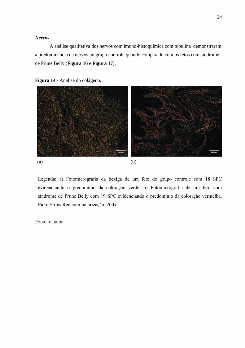
Nervos
A análise qualitativa dos nervos com imuno-histoquímica com tubulina demonstraram
a predominância de nervos no grupo controle quando comparado com os fetos com síndrome
de Prune Belly (Figura 16 e Figura 17).
Figura 14 - Análise do colágeno.
(a) (b)
Legenda: a) Fotomicrografia da bexiga de um feto do grupo controle com 18 SPC
evidenciando o predomínio da coloração verde. b) Fotomicrografia de um feto com
síndrome de Prune Belly com 19 SPC evidenciando o predomínio da coloração vermelha.
Picro Sirius Red com polarização. 200x.
Fonte: o autor.

35
Figura 15 - Fotomicrografias de bexigas de fetos mostrando a presença do tecido muscular
(corado em vermelho) nos grupos.
(a) (b)
Legenda: a) Bexiga de feto com síndrome de Prune Belly com 19 SPC. b) Bexiga de feto normal
com 18 SPC. Tricrômico de Masson. 400X.
Fonte: o autor.
Figura 16 - Análise da distribuição dos nervos.
(a) (b)
Legenda: a) Fotomicrografia demostrando os nervos (setas) na bexiga de um feto do grupo controle com 35 SPC. Tubulina X200. b) Fotomicrografia demonstrando os nervos na bexiga de um feto portador de síndrome de Prune Belly com 31 SPC. Tubulina X200. Fonte: o autor.

36
Figura 17 - Análise da distribuição dos nervos.
(a) (b)
Legenda: a) Fotomicrografia demostrando os nervos (setas) na bexiga de um feto do grupo
controle com 18 SPC. Tubulina X200. b) Fotomicrografia demonstrando os nervos na bexiga
de um feto portador de síndrome de Prune Belly com 19 SPC. Tubulina X200.
Fonte: o autor.
37
5 DISCUSSÃO
A etiologia da SPB não é conhecida, no entanto, algumas hipóteses têm sido sugeridas.
Estudos revelam a possibilidade de herança genética e possível associação com a trissomia dos
cromossomas 18 e 21 (HASSETT; SMITH; HOLLAND, 2012; STEPHENS; SMITH;
HUTSON, 2002). Outras hipóteses tais como uropatia obstrutiva distal e defeito mesodérmico
da parede abdominal, anomalias do alantoide e do tracto urinário também vêm sendo estudadas
(COPP; GREENE, 2012; HASSETT; SMITH; HOLLAND, 2012; STEPHENS; SMITH;
HUTSON, 2002; ZUGOR; SCHOTT; LABANARIS, 2012). A obstrução uretral está presente
em um terço dos pacientes com SPB, que pode ser a principal causa das malformações nesta
síndrome (HASSETT; SMITH; HOLLAND, 2012; STEPHENS; SMITH; HUTSON, 2002).
A causa da criptorquidia na SPB é desconhecida. No entanto, especula-se que as
alterações anatômicas na parede abdominal impedem um aumento da pressão intra-abdominal,
que é um dos fatores necessários para a migração testicular. Também se especulou que a
megabexiga nesta síndrome torna o canal inguinal extraperitoneal, de modo que o gubernáculo
e seu processo vaginal não podem desenvolver-se normalmente (HASSETT; SMITH;
HOLLAND, 2012; STEPHENS; SMITH; HUTSON, 2002; COSTA; COSTA; SAMPAIO;
FAVORITO, 2015).
Alguns estudos sugerem que a SPB pode surgir de obstrução anatômica de vários tipos
(uretra, próstata, colo vesical) ou obstrução funcional originando a megabexiga (VOLMAR;
FRITSCH; PERLMAN; HUTCHINS, 2001; DIAO; DIALLO; FALL; NGOM; FALL;
NDOYE; FALL; BA; NDOYE; DIAGNE, 2008; PELLEGRINO; VISCONTI; CATANIA;
D'ORIA; MANZONI; GRELLA; CARUSO; MASINI; NOIA, 2017; TAGHAVI; SHARPE;
STRINGER, 2016). Volmar (VOLMAR; FRITSCH; PERLMAN; HUTCHINS, 2001) analisou
11 casos de SPB e observou obstrução mecânica em oito dos casos. Na nossa amostra, todos os
três fetos de SPB apresentaram obstrução no nível da uretra prostática ou do colo vesical.
Nossos achados corroboram que a aparência fenotípica da SPB, juntamente com a megabexiga,
com espessamento acentuado e alterações histológicas pode ser explicada por obstrução do trato
urinário.
A maior parte da bexiga se origina da parte vesical do seio urogenital, enquanto que o
trígono vesical tem origem a partir da absorção da região caudal do duto mesonéfrico durante
o desenvolvimento da bexiga (STEPHENS; SMITH; HUTSON, 2002; LATERZA;
GENNARO; TUBARO, 2011). A bexiga fetal humana passa por uma série de transformações
em seu desenvolvimento da décima terceira até a vigésima primeira semanas gestacionais,

38
finalmente adquirindo um urotélio típico e uma camada muscular bem desenvolvida. Até a
décima primeira semana gestacional, a parede da bexiga é formada por mesênquima e este
gradualmente amadurece em tecido conjuntivo frouxo. O colágeno torna-se aparente por volta
da décima terceira semana gestacional (STEPHENS; SMITH; HUTSON, 2002; LATERZA;
GENNARO; TUBARO, 2011; TASIAN; CUNHA; BASKIN, 2010).
O colágeno e a elastina são componentes importantes da parede da bexiga que
participam do seu funcionamento. O colágeno proporciona resistência de tensão, porém, um
acúmulo excessivo pode inibir a contratilidade da bexiga e a condução dos impulsos elétricos
através da parede. A elastina proporciona ao tecido elasticidade e pode ajudar na complacência
(FREEDMAN; QURESHI; SHAPIRO, 1997). No tecido muscular o seu papel é proporcionar
sustentação, da mesma forma que os tendões no tecido esquelético. Desta maneira, como o
músculo liso não apresenta tendões, necessita de uma maior quantidade de colágeno
(FREEDMAN; QURESHI; SHAPIRO, 1997).
Freedman (1997) estudou a estrutura da bexiga de nove fetos com válvula de uretra
posterior e atresia uretral. Esse estudo demonstrou um aumento substancial na espessura da
parede da bexiga, mas não foram observadas alterações significativas na distribuição do tecido
conjuntivo e nos músculos lisos da bexiga fetal. Também sugeriu que os processos de obstrução
intra-útero não estão associados ao excesso de deposição de colágeno, mas sim com aumento
do desenvolvimento da musculatura vesical (FREEDMAN; QURESHI; SHAPIRO, 1997).
Pazos (2011) em um estudo feito em bexigas de fetos com anencefalia observou um
aumento significativo na quantidade de colágeno total nas bexigas de fetos anencéfalos com
predominância do colágeno tipo III. Segundo esse estudo, isto pode sugerir uma alteração
importante no desenvolvimento da camada muscular da bexiga. A lesão no sistema nervoso
com consequente alteração na regulação do nervo da bexiga poderia ser uma hipótese plausível
para explicar essas mudanças estruturais.
Os nervos da bexiga em fetos anencefálicos podem ser modificados devido a lesões
cerebrais, com consequentes danos no controle cerebral nos nervos da bexiga (PAZOS; LOBO;
COSTA; SAMPAIO; CARDOSO; FAVORITO; 2011). No entanto, nesse estudo, eles não
investigaram a distribuição dos nervos da bexiga em fetos anencefálicos, pelo que são
necessárias mais pesquisas para confirmar ou refutar essa hipótese.
As alterações do sistema elástico estão envolvidas na formação de tecidos fibróticos
(FREEDMAN; QURESHI; SHAPIRO, 1997), no entanto não foi observado em nenhum dos
casos estudados em nosso trabalho a presença de fibras do sistema elástico na bexiga. Isso pode
indicar que este componente da matriz extracelular forma-se apenas a partir do terceiro

39
trimestre gestacional na bexiga fetal. Estudos prévios evidenciaram a presença de fibras do
sistema elástico em outros órgãos do sistema genito-urinário em fetos humanos (BASTOS;
SILVA; COSTA; SAMPAIO, 2004). Neste estudo foi observado um sistema elástico fino e
escasso no tecido do corpo esponjoso de um feto com 15 semanas de gestação e em um feto
com 36 semanas de gestação observou a trabécula do corpo esponjoso delimitando extensos
espaços vasculares (BASTOS; SILVA; COSTA; SAMPAIO, 2004).
Segundo esse estudo, as fibras do sistema elástico são abundantes e organizadas em
fetos no terceiro trimestre gestacional (BASTOS; SILVA; COSTA; SAMPAIO, 2004). Nossa
amostra era composta de fetos do segundo trimestre gestacional, período no qual,
provavelmente, as fibras do sistema elástico ainda estão se formando na bexiga fetal.
Estudos anteriores mostraram que fetos com PBS e obstrução infravesical aumentaram
a espessura muscular e uma relação normal do tecido conjuntivo (VOLMAR; FRITSCH;
PERLMAN; HUTCHINS, 2001; WORKMAN; KOGAN, 1990). No presente trabalho,
encontramos uma diminuição na densidade volumetrica de células musculares lisas em bexigas
de SPB. Provavelmente isso aconteceu devido à obstrução observada na uretra prostática em
casos de SPB. A bexiga em SPB é submetida a uma pressão hidrostática muito maior, o que
pode causar um aumento na atividade fibroblástica (JUNKER; KRATZ; TOLLBÄCK;
KRATZ, 2008 ). Com base nestas observações, podemos especular que a redução muscular lisa
é um evento importante na obstrução da bexiga. Este evento provavelmente está correlacionado
com um processo de hidrodistensão.
Em nossa amostra foi observado um predomínio do colágeno tipo I na bexiga da SPB.
O colágeno é responsável por proporcionar tensão de resistência aos tecidos (USHIKI, 2002;
HIRSHBERG; LIB; KOZLOVSKY; KAPLAN, 2007). A quantidade de colágeno é aumentada
na obstrução crônica de bexiga nos adultos humanos. Porém, um estudo sobre a obstrução de
bexiga fetal humana mostrou que embora a quantidade total de colágeno tenha aumentado ele
é proporcional a quantidade de músculo, o qual aumenta também em resposta a obstrução (50).
Se todo o colágeno for removido do tecido muscular liso sua força de ativação será reduzida.
De maneira interessante, o acúmulo anormal de colágeno encontrado em bexigas obstruídas
pode diminuir a contratilidade muscular além de afetar a complacência da bexiga
(FREEDMAN; QURESHI; SHAPIRO, 1997).
Assim, com base nestes achados, os resultados da estrutura de colágeno sugerem que a
matriz de colágeno da bexiga na SPB é interrompida. Podemos especular que a ocorrência de
uma obstrução mecânica ou a pressão intra-abdominal alterada na SPB dificulta o

40
desenvolvimento da bexiga. O fornecimento nervoso da bexiga tem origem sacral, exceto o
trígono, que é inervado pelo plexo hipogástrico (VAN DUZEN; DUNCAN, 1953).
No presente estudo, observamos uma pequena quantidade de nervos na bexiga do grupo
SPB na análise qualitativa. Este é o primeiro estudo que avalia a distribuição dos nervos da
bexiga humana com SPB. A pequena quantidade de nervos presentes nas bexigas estudadas
poderia ser a consequência da obstrução posterior da uretra combinada com um processo de
hidrodistensão e degradação da matriz extracelular e os demais componentes da parede da
bexiga, mais pesquisas futuras com amostras maiores serão necessárias para esclarecer este
tópico.
Algumas limitações deste estudo devem ser mencionadas: a) distribuição desigual de
SPC entre SPB e fetos de controle; e b) pequeno tamanho da amostra, uma vez que os fetos
com SPB são raros. No entanto, apesar da limitação do tamanho da amostra, as observações
ainda fornecem novas informações importantes.

41
6 CONCLUSÕES
A bexiga em fetos com síndrome de Prune Belly apresenta redução de fibras musculares
lisas, colágeno tipo III e nervos. Essas alterações estruturais podem ser um dos fatores
envolvidos nas anomalias do trato urinário como a megabexiga em pacientes com síndrome de
Prune Belly.

42
7 REFERÊNCIAS BIBLIOGRÁFICAS
ASHLEY, R. A.; INMAN, B. A.; SEBO, T. J.; LEIBOVICH, B.; BLUTE, M. L.; KNOW, E. D.; ZINCKE, H. Urachal carcinoma: clinicopathologic features and long-term outcomes of an aggressive malignancy. Cancer, v. 107, n. 4, pp. 712-720, 2006.
BACKHOUSE, K. M. Embryology of testicular descent and maldescent. The Urologic clinics of North America, v. 9, n. 3, pp. 315-325, 1982.
BASKIN, L.; DiSandro, M.; Li, Y., Li, W.; Hayward, S.; Cunha, G. Mesenchymal-epithelial interactions in bladder smooth muscle development: effects of the local tissue environment. J Urol, v. 165, pp. 1283-1288, 2001.
BASTOS, A. L.; SILVA E. A.; COSTA, W. S.; SAMPAIO, F. J. The concentration of elastic fibers in the male urethra during human fetal development. BJU Int.; v. 94, n. 4, pp. 620-623, 2004.
BRONSHTEIN, M.; Bar-Hava, I.; Blumenfeld, Z. Differential diagnosis of the nonvisualized fetal urinary bladder by transvaginal sonography in the early second trimester. Obstet Gynecol, v. 82, pp. 490-493, 1993.
CHAGAS, M. A.; BABINSKI, M. A.; COSTA, W. S.; SAMPAIO, F. J. Stromal and acinar components of the transition zone in normal and hyperplastic human prostate. BJU Int. v. 89, n. 7, pp. 699-702, 2002.
COPP, A. J.; GREENE, N. D. E. Neural tube defects-disorders of neurulation and related embryonic processes. Wiley Interdisciplinary Reviews: Developmental Biology, v. 2, n. 2, pp. 213-27, 2012.
COSTA, S. F.; COSTA W. S.; SAMPAIO F. J.; FAVORITO, L. A. Structural study of gubernaculum testis in fetuses with prune belly syndrome. J Urol. v. 193, n. (5 suppl), pp.1830-1836, 2015.
DIAO, B.; DIALLO, Y.; FALL, P. A.; NGOM, G.; FALL, B.; NDOYE, A. K.; FALL, I.; BA, M.; NDOYE, M.; DIAGNE, B. A. Prune Belly syndrome: epidemiologic, clinic and therapeutic aspects. Prog Urol. v. 18, n. 7, pp. 470-474, 2008.
FAVORITO, L. A.; COSTA, W. S.; SAMPAIO, F. J. B. The position of the testis during the fetal period: an additional parameter to estimate fetal weight. International braz j urol, v. 36, n. 5, pp. 609–613, 2010.
FAVORITO, L. A.; SAMPAIO, F. J. B. Testicular migration chronology: do the right and the left testes migrate at the same time? Analysis of 164 human fetuses. BJU international, v. 113, n. 4, pp. 650–653, 2014.
FREEDMAN, A. L; QURESHI, F.; SHAPIRO, E, et al. Smooth muscle development in the obstructed fetal bladder. Urology, v. 49, p. 104, 1997.
GOULDING, F. J.; GARRETT, R. A. Twenty-five-year experience with prune belly syndrome. URL, v. 12, n. 3, pp. 329–332, 1978.
43
HASSETT, S.; SMITH, G. H.; HOLLAND, A. J. Prune Belly Syndrome. Pediatr Surg Int. v. 28, n. 3, pp. 219-28, 2012.
HERN, W. M. Correlation of fetal age and measurements between 10 and 26 weeks of gestation. Obst Gynecol. v. 63, n. 1, pp. 26-32, 1984.
HEYNS, C. F. The gubernaculums during testicular descent in the human fetus. J Anat. v. 153, pp. 93-112, 1987.
HEYNS, C. F.; HUTSON, J. M. Historical review of theories on testicular descent. Journal of Urology, v. 153, n. 3 Pt 1, pp. 754–767, 1995.
HIRSHBERG, A.; LIB, M.; KOZLOVSKY, A.; KAPLAN, I. The influence of inflammation on the polarization colors of collagen fibers in the wall of odontogenic keratocyst. Oral Oncol.v. 43, n. 3, pp. 278-82, 2007.
JUNKER, J. P.; KRATZ, C.; TOLLBÄCK, A.; KRATZ, G. Mechanical tension stimulates the transdifferentiation of fibroblasts into myofibroblasts in human burn scars. Burns. v. 34, n 7, pp. 942-46, 2008.
KIM, K. M.; KOGAN, B. A.; MASSAD, C. A.; HUANG, Y. C. Collagen and elastin in the normal fetal bladder. J Urol. v. 146, n. 2, pp. 524-27, 1991.
LATERZA, R. M.; GENNARO, M.; TUBARO, A. Female pelvic congenital malformations. Part I: embryology, anatomy and surgical treatment. Eur J Obst Gynecol and Repro Biol. v. 159, n. 1, p. 26, 2011.
MAIZELS, M. Normal development of the urinary tract. In: Campbell’s Urology, 6a. ed., p. 1301. New York: Saunders, 1992.
MANDARIM-DE-LACERDA, C. A.; FERNANDES-SANTOS, C.; AGUILA, M. B. Image analysis and quantitative morphology. Methods Mol Biol.; n. 611, pp. 211-25, 2010.
MANDARIM-DE-LACERDA, C. A. Stereological tools in biomedical research. An Acad Bras Cienc. v. 75, n. 4, pp. 469-486, 2003.
MERCER, B. M.; SKLAR, S.; SHARIATMADAR, A.; GILLIESON, M. S., D’ALTON, M. E. Fetal foot length as a predictor of gestational age. Am J Obst Gynecol. v. 156, n. 2, pp. 350-5, 1987.
MOORE, K. L. The Developing human. Clinically Oriented Embryology. Philadelphia: W.B Saunders, 1977.
NEWMAN, J.; ANTONAKOPOULOS, G. N. The fine structure of the human fetal urinary bladder. Development and maturation. A light, transmission and scanning electron microscopic study. J Anat, n. 166, pp. 135-50, 1989.
PARK, J. M. Normal and anomalus development of the urogenital system. In: Campbell’s Urology, 8a. ed., p. 1737. New York: Saunders, 2002.
44
PATTEN, R. M.; MACK, L. A.; WANG, K. Y.; CYR, D. R. The fetal genitourinary tract. Radiol Clin North Am, v. 28, n. 1, pp. 115-30, 1990.
PAZOS, H. M.; LOBO, M. L.; COSTA, W. S.; SAMPAIO, F. J.; CARDOSO, L. E.; FAVORITO, L. A. Do neural tube defects lead to structural alterations in the human bladder? Histol Histopathol, v. 26, n. 5, p. 581, 2011.
PELLEGRINO, M.; VISCONTI, D.; CATANIA, V. D.; D'ORIA, L.; MANZONI, C.; GRELLA, M. G.; CARUSO, A.; MASINI, L.; NOIA, G. Prenatal detection of megacystis: not always an adverse prognostic factor. Experience in 25 consecutive cases in a tertiary referral center, with complete neonatal outcome and follow-up. J Pediatr Urol, v. 13, n. 5, p. 486, 2017.
PLATT, L. D.; MEDEARIS, A. L.; DEVORE, G. R.; HORENSTEIN, J. M.; CARLSON, D. E.; BRAR, H. S. Fetal foot length: Relationship to menstrual age and fetal measurements in the second trimester. Obstet Gynecol. v. 71, n. 4, pp. 526-31, 1988.
ROUTH, J. C. et al. Congential AnomaliesContemporary Epidemiologyand Characterization of NewbornMales with Prune Belly Syndrome. URL, v. 76, n. 1, pp. 44–48, 2010.
SADLER, T. W. Langman’s Medical Embryology. 7th Ed., Baltimore, Maryland: Williams & Wilkins, 1995.
SAMPAIO, F. J.; MANDARIM-DE-LACERDA, C. A. Morphometry of the kidney. Applied study in urology and imaging. J Urol (Paris). v. 95, n. 2, pp. 77-80, 1989.
SAMPAIO, F. J.; FAVORITO, L. A. Analysis of testicular migration during the fetal period in humans. Journal of Urology, v. 159, n. 2, pp. 540–542, 1998.
SEIDEL, N. E. et al. Pediatric UrologyClinical Manifestations and Managementof Prune-belly Syndrome in a LargeContemporary Pediatric Population. URL, v. 85, n. 1, pp. 211–215, 2015.
SHIMADA, K.; HOSOKAWA, S.; TOHDA, A.; MATSUMOTO, F.; JOHNIN, K. Histology of the fetal prune belly syndrome with reference to the efficacy of prenatal decompression. Int J Urol. v. 7, n. 5, pp. 161-6, 2000.
STEPHENS, F. D.; SMITH, E. D.; HUTSON, J. M. Morphology and embryogenesis of the triad (prune belly) syndrome. In Congenital anomalies of the kidney, urinary and genital tracts Chapter 37, pp. 391-409. Londres: Martin Dunitz, 2002.
STREETER GL: Weight, sitting height, head size, foot lenght and menstrual age of the human embryo. Contr Embryol Carnegie Instn, v. 11, pp. 143-170, 1920.
TAGHAVI, K.; SHARPE, C.; STRINGER, M. D. Fetal megacystis: a systematic review. J Pediatr Urol.v. 13, n. 1, pp. 7-15, 2017.
TASIAN, G.; Cunha, G.; Baskin, L. Smooth muscle differentiation and patterning in the urinary bladder. Differentiation, v. 80, n. 2-3, p. 106, 2010.
USHIKI, T. Collagen fibers, reticular fibers and elastic fibers. A comprehensive understanding from a morphological viewpoint. Arch Histol Cytol. v. 65, n. 2, pp. 109-26, 2002.

45
VAN DUZEN, R. E., M.D.; DUNCAN, C. G., M.D. Anatomy and nerve supply of urinary bladder. JAMA. v. 153, n. 15, pp. 1345-47; 1953.
VOLMAR, K. E.; FRITSCH, M. K.; PERLMAN, E. J.; HUTCHINS, G. M. Patterns of congenital lower urinary tract obstructive uropathy: relation to abnormal prostate and bladder development and the prune belly syndrome. Pediatr Dev Pathol. v. 4, n. 5, pp. 467-72, Sep-Oct. 2001.
WEIN, A. J.; KAVOUSSI L. R.; NOVICK A. C.; PARTIN A. J.; PETERS C. A. Prune Belly Syndrome. In: Campbell-Walsh Urology. Tradução. [s.l.] Elsevier Health Sciences. Campbell Walsh Urology, 9a. ed., p. 3482-3496, Philadelphia: Saunders/Elsevier, 2007.
WILCOX, D. T.; CHITTY, L. S. Non-visualisations of the fetal bladder: aetiology and management. Prenat Diagn, v. 21, n. 11, pp. 977-83, 2001.
WORKMAN, S. J.; KOGAN BA. Fetal bladder histology in posterior urethral valves and the prune belly syndrome. J Urol. v. 144, n. (2 Pt 1), pp. 337-9, 1990.
ZUGOR, V.; SCHOTT, G. E.; LABANARIS, A. P. The Prune belly syndrome: Urological aspects and long-term outcomes of a rare disease. Pediatr Rep. v. 4, n. 2: p. e20, 2012.

46
ANEXOS
1 - Parecer do comitê de ética
2 - Artigo publicado na revista Neururology and Urodinamics
3 - Artigo aceito para publicação na revista Biomedical Research International

DADOS DO PARECER:
Número do Parecer: 923.662
Situação do Parecer: Aprovado
HOSPITAL UNIVERSITÁRIOPEDRO ERNESTO/
UNIVERSIDADE DO ESTADO
PARECER CONSUBSTANCIADO DO CEP
Pesquisador:
Título da Pesquisa:
Instituição Proponente:
Versão:CAAE:
Estudo dos órgãos urogenitais na síndrome de Prunne Belly e em fetos anencéfalos
Suelen Freitas Costa
Hospital Universitário Pedro Ernesto/UERJ
139418714.7.0000.5259
Área Temática:
DADOS DO PROJETO DE PESQUISA
Número do Parecer:Data da Relatoria:
923.66216/12/2014
DADOS DO PARECER
O projeto será realizado na unidade de pesquisa Urogenital, serão estudados 10 fetos masculinos semanomalias, 3 fetos masculinos com PruneBelly, e 3 fetos masculinos com anencefalia. Serão avaliados as próstatas, os gubernáculos, os testículos eas bexigas de cada feto. Os fetos utilizados em nosso estudo serão provenientes do Departamento deAnatomia da Universidade do Estado do Rio de Janeiro.
Apresentação do Projeto:
Objetivo Primário: Os estudos da estrutura dos órgãos urogenitais na síndrome de prunne-belly são raras naliteratura. O objetivo desse projeto é fazer um estudo morfológico destes órgãos em fetos portadores dasíndrome de prunne-belly e analisar as principais alterações estruturais desses órgãos nessa anomaliacongênita.
Objetivo da Pesquisa:
Prezado pesquisador: Caracteriza-se como risco direto para os sujeitos da pesquisa a possibilidade dedesconforto ou constrangimento no momento do preenchimento dos questionários. Os pesquisadoresdevem se comprometer a minimizar os riscos ou desconfortos indiretos que possam vir a ser causados.
Avaliação dos Riscos e Benefícios:
MINISTERIO DA EDUCACAOFUN CARLOS CHAGAS F. DE AMPARO A PESQUISA DO ESTADO DO RIO DEJANEIRO - FAPERJ
Patrocinador Principal:
20.551-030
(21)2868-8253 E-mail: [email protected]
Endereço:Bairro: CEP:
Telefone:
Avenida 28 de Setembro 77 - TérreoVila Isabel
UF: Município:RJ RIO DE JANEIROFax: (21)2264-0853
Página 01 de 03
ANEXO 1 - Parecer da Comissão de Ética 47

Received: 19 March 2017 | Accepted: 25 April 2017
DOI: 10.1002/nau.23327
ORIGINAL BASIC SCIENCE ARTICLE
Structural study of the bladder in fetuses with prune bellysyndrome
Helce R. Julio Junior | Suelen F. Costa | Waldemar S. Costa |
Francisco J. Barcellos Sampaio | Luciano A. Favorito
Urogenital Research Unit, State Universityof Rio de Janeiro, Rio de Janeiro, Brazil
CorrespondenceLuciano Alves Favorito, UrogenitalResearch Unit, State University of Rio deJaneiro, Rua Professor Gabizo, 104/201,Tijuca, RJ, CEP: 20271-320, Rio deJaneiro, Brazil.Email: [email protected]
Funding informationNational Council for Scientific andTechnological Development (CNPq–Brazil); Rio de Janeiro State ResearchFoundation (FAPERJ)
Aims: To study the bladder structure of fetuses with prune belly syndrome (PBS).
Methods: We studied three bladders obtained from three male fetuses with PBS and
seven bladders from seven male fetuses without anomalies. Each bladder was
dissected and embedded in paraffin, fromwhich5 μmthick sectionswere obtained and
stained with Masson’s trichrome (to quantify connective tissue and smooth muscle)
and picrosirius red with polarization (to observe collagen). Immunohistochemistry
with tubulin (Tubulin, beta III, Mouse Monoclonal Antibody) was applied to observe
the bladder nerves. The imageswere capturedwith anOlympus BX51microscope and
Olympus DP70 camera. The stereological analysis was done with the Image Pro and
Image J programs, using a grid to determine volumetric densities (Vv). Means were
statistically compared using the Mann-Whitney test (P< 0.05).
Results:Quantitative analysis documented that smoothmuscle fiberswere significantly
smaller (P= 0.04) in PBS fetuses (9.67% to 17.75%, mean = 13.2%) compared to
control group (13.33% to 26.56%, mean = 17.43%). The analysis of collagen fibers
showed predominance of green in the control group, suggesting collagen type III
presence, and predominance of red in the in PBS fetal bladders, suggesting collagen type
I presence in this group. The qualitative analysis of the nerves with immunohistochem-
istry with tubulin showed predominance of nerves in the control group.
Conclusion: The bladder in PBS had lower concentrations of smooth muscle fibers,
collagen type III, and nerves. These structural alterations can be one of the factors
involved in urinary tract abnormality such as distended bladder in patients with PBS.
KEYWORDS
bladder, histology, prune belly syndrome
1 | INTRODUCTION
Prune belly syndrome (PBS) is a congenital anomalycharacterized by deficiency or hypoplasia of the abdominalmuscles as well as malformations of the urinary tract, such as
large and hypotonic bladders, dilated and tortuous ureters, andbilateral cryptorchidism.1 Urethral obstruction occurs in one-third of patients with PBS and can be the primary cause of thebladder alterations in this syndrome.2,3
Studies of the bladder structure in PBS are scarce in theliterature and show that bladder histology is variable, withboth increased musculature and defective or dysplasticmuscles,4 along with normal ratio of connective tissue.5
Alan Wein led the peer-review process as the Associate Editor responsiblefor the paper.
Neurourology and Urodynamics. 2017;9999:1–5. wileyonlinelibrary.com/journal/nau © 2017 Wiley Periodicals, Inc. |
ANEXO 2 - Artigo publicado na revista Neururology and Urodinamics 48

Another study demonstrated that the walls of the bladder werethick or thin and the muscle, when present, was intermixedwith fibrocytes and large plaques of collagen.2
Recently, important alterations in the gubernaculum testisstructure were demonstrated in fetuses with PBS.6 Neverthe-less, structural, qualitative and quantitative analyses ofsmooth muscle cells, extra-cellular matrix, and nerves ofthe bladder during the human fetal period in PBS are scarce.
The hypothesis stated in our study is: Is the bladderstructure similar in PBS and normal fetuses? This paperdescribes the bladder structure in fetuses with prune bellysyndrome (PBS).
2 | MATERIALS AND METHODS
The experimental protocol described here was approved bythe ethics committee on human experimentation of ouruniversity. This study was carried out in accordance with theethical standards of the hospital’s institutional committee onhuman experimentation.
We studied three bladders obtained from three malefetuses with PBS and seven bladders obtained from sevenmale fetuses without anomalies. The fetuses were macro-scopically well preserved. The gestational age was deter-mined in weeks post conception (WPC), according to thefoot-length criterion, which is currently considered the mostacceptable parameter to calculate gestational age.7–9 Thefetuses were also evaluated regarding crown-rump length(CRL) and body weight immediately before dissection. Thesame observer made the measurements.
After the measurements, the fetuses were carefullydissected with the aid of a stereoscopic lens with 16/25×magnification. The abdomen and pelvis were opened toidentify and expose the urogenital organs.
The bladder of each fetus was separated from the otherstructures and fixed in 10% buffered formalin, and routinelyprocessed for paraffin embedding, after which 5 μm thicksections were obtained at 200 μm intervals. Sections werestained with hematoxylin-eosin to assess the integrity of thetissue. The following staining methods were used: Masson’strichrome, to quantify connective and smooth muscle tissue,Weigert’s resorcin fuchsin with previous oxidation to observeelastic system fibers, and picrosirius red with polarization forobservation of different collagen types.
The smooth muscle tissue was quantified by a stereologi-cal method.10–12We studied five microscopic fields chosen atrandom, totaling 25 test areas for each bladder for thequantitative analysis. We used the Image J software, version1.46r, loaded with its own plug-in (http://rsb.info.nih.gov/ij/).All sections were photographed with a digital camera (DP70,Olympus America, Inc., Melville, New York) under the sameconditions at a resolution of 2040 pixels, directly coupled to
the microscope (BX51, Olympus America, Inc.) and stored ina TIFF file.
To quantify the smooth muscle tissue we used the Image Jsoftware to determine the volumetric density (Vv) of thetubules (Fig. 1). Results for each field were obtained throughthe quantification assessment method, to count the musclecells of each section. The arithmetic mean of the quantifica-tion in five fields of each section was determined. Afterward,we obtained the mean quantification value for the fivesections studied from each bladder (total of 25 test areas).
Means were statistically compared using the unpairedT-test and the Mann-Whitney test for all categorical variablesand Wilcoxon rank sum tests were used for continuousvariables. All tests were two-sided and a P-value < 0.05was considered statistically significant.
3 | RESULTS
The prune belly fetuses ranged in age from 17 to 31 WPC,weighed between 240 and 2150 g, and had crown-rump lengthbetween 18 and 43 cm. The fetuses in the control groupranged in age from 12 to 35 WPC, weighed between 210 and2860 g and had crown-rump length between 18 and 34 cm(Table 1). The three fetuses with PBS had the typical aspect ofthe anterior abdominal wall, enlarged bladders, bilateralcryptorchidism, and bilateral hydronephrosis (Fig. 2). In twocases we observed severe obstruction in prostatic urethra andin one case obstruction at the level of the vesical neck.
We did not observe elastic fibers in the bladder in PBS andin the control group. Quantitative analysis documented thatsmooth muscle fibers were significantly smaller (P= 0.04)in PBS fetuses (9.67% to 17.75%, mean = 13.2%) when
FIGURE 1 Morphometric analysis of the bladder. To quantifythe smooth muscle number we used the Image J Test grid software todetermine the volumetric density (Vv). HE ×400
49

compared to the control group (13.33% to 26.56%,mean = 17.43%).
The analysis of collagen fibers showed predominance ofred in the control group, suggesting collagen type I presence,and predominance of green in the in PBS fetal bladder,suggesting collagen type III presence in this group.
The qualitative analysis of the nerves with immunohis-tochemistry with tubulin showed predominance of nerves inthe control group when compared to the PBS fetuses (Fig. 3).
4 | DISCUSSION
The etiology of PBS is controversial. Some studies suggestthat PBS may arise from either anatomic obstruction ofvarious types or functional obstruction from megacystis.13,14
Volmar13 analyzed 11 cases of PBS and observed mechanicalobstruction in eight cases. In our sample, all three PBS fetuseshad obstruction at the level of the prostatic urethra or vesicalneck. Our findings corroborate that the phenotypic appear-ance of PBS, along with the megacystis, with markedthickening, and histological alterations, can be explained by aurinary tract obstruction.
In our sample we did not observe elastic fibers in anyof the bladders analyzed. This may indicate that this
TABLE 1 The table shows the age and the fetal parameters of oursample: Seven male fetuses without anomalies and three male fetuseswith prune belly syndrome WPC, age in weeks post-conception;g, grams; CRL, crown-rump length; and cm, centimeters
Fetus Age (WPC) Anomaly Weight (g) CRL(cm)
1 17 Prune belly 240 18
2 23 Prune belly 1100 25
3 31 Prune belly 2150 43
4 12 none 210 18
5 16 none 430 22
6 18 none 250 18
7 19 none 380 21
8 21 none 401 21
9 21 none 450 22
10 35 none 2860 34
FIGURE 2 Fetus with 35 weeks post-conception with PBS. A)Aspect of the abdominal wall in PBS. B) The anterior abdominal wallis extirpated. We can observe the enlarged bladder (B) in PBS
FIGURE 3 Analysis of the nerves distribution. A)Photomicrograph showing the nerves of the bladder (arrowhead).bladder of control group fetus with 35 WPC. Tubulin ×200. B)Photomicrograph showing the nerves of the bladder (arrowhead).Bladder of a prune belly fetus with 31 WPC. Tubulin ×200
50

extracellular matrix component appears only in the thirdgestational trimester in the fetal testis. Previous studies haveshown the existence of elastic system fibers in other humanfetal genitourinary organs.15
Previous studies have shown that fetuses with PBS andinfra-vesical obstruction have increased muscle thickness anda normal ratio of connective tissue.13 In the present work, wefound a decrease in the Vv of smooth muscle cells in PBSbladders. Probably this happened due to the obstructionobserved in the prostatic urethra in cases of PBS. The bladderin PBS is submitted to a much higher hydrostatic pressure,which can cause an increase in fibroblast activity.16 Based onthese observations, we can speculate that the smooth musclereduction is an important event in bladder obstruction. Thisevent is probably correlated to a hydrodistension process.
In our sample we observed a predominance of collagentype I in the PBS bladders. Collagen provides tensilestrength, but over-accumulation can inhibit contractilityand conduction of electrical impulses through the visceralwall.17,18 In earlier phases of the remodeling and repair ofconnective tissues, the synthesis of type III collagen isenhanced.16 Thus, based on these findings, the results ofthe collagen structure suggest that the collagen matrix ofthe prune belly bladder is disrupted. We can speculate thatthe occurrence of a mechanical obstruction or the alteredintra-abdominal pressure in PBS hinders the bladderdevelopment.
The nerve supply of the bladder has sacral origin, exceptfor the trigone, which is innervated by the hypogastricplexus.19 In the present study, we observed a small quantity ofnerves in the bladder of the PBS group in the qualitativeanalysis. This is the first study assessing the distribution of thenerves of the human bladder with PBS. The small quantity ofnerves present in the bladders studied could be theconsequence of posterior urethra obstruction combinedwith a hydrodistension process and degradation of theextracellular matrix and the other components of the bladderwall, but future research with a larger samples will benecessary to clarify this topic.
Some limitations of this study should be mentioned: 1)unequal WPC distribution between PBS and control fetuses;and 2) small sample size, since prune belly fetuses are rare.However, despite the sample size limitation, the observationsstill provide important new information.
5 | CONCLUSIONS
The bladder in PBS has lower concentrations of smoothmuscle fibers, collagen type III, and nerves. Thesestructural alterations can be one of the factors involvedin urinary tract abnormality such as distended bladder inpatients with PBS.
ACKNOWLEDGMENTS
This study was supported by grants from the National Councilfor Scientific and Technological Development (CNPq −Brazil) and the Rio de Janeiro State Research Foundation(FAPERJ).
CONFLICTS OF INTEREST
None declared
REFERENCES:
1. Hassett S, Smith GH, Holland AJ. Prune belly syndrome. PediatrSurg Int. 2012 Mar;28:219–228.
2. Stephens FD, Smith ED, Hutson JM. Morphology and embryogen-esis of the triad (prune belly) syndrome. Congenital Anomalies ofthe Kidney, Urinary and Genital Tracts. London: Martin Dunitz;2002;Chapter;37:391–409.
3. Zugor V, Schott GE, Labanaris AP. The Prune belly syndrome:urological aspects and long-term outcomes of a rare disease.Pediatr Rep. 2012 Apr 2;4:e20.
4. Shimada K, Hosokawa S, Tohda A, Matsumoto F, Johnin K.Histology of the fetal prune belly syndrome with reference tothe efficacy of prenatal decompression. Int J Urol. 2000 May;7:161–166. PMID: 10830822.
5. Workman SJ, Kogan BA. Fetal bladder histology in posteriorurethral valves and the prune belly syndrome. J Urol. 1990Aug;144:337–339. PMID: 2374202.
6. Costa SF, Costa WS, Sampaio FJ, Favorito LA. Structural study ofgubernaculum testis in fetuses with prune belly syndrome. J Urol.2015 May;193:1830–1836.
7. Hern WM. Correlation of fetal age and measurements between 10and 26 weeks of gestation. Obst Gynecol. 1984 Jan;63:26–32.PMID: 6691014.
8. Mercer BM, Sklar S, Shariatmadar A, Gillieson MS, D’Alton ME.Fetal foot length as a predictor of gestational age. Am J ObstGynecol. 1987 Feb;156:350–355. PMID: 3548369.
9. Platt LD, Medearis AL, DeVore GR, Horenstein JM, Carlson DE,Brar HS. Fetal foot length: relationship to menstrual age and fetalmeasurements in the second trimester. Obstet Gynecol. 1988Apr;71:526–531. PMID: 3353042.
10. Chagas MA, Babinski MA, Costa WS, Sampaio FJ. Stromal andacinar components of the transition zone in normal and hyperplastichuman prostate. BJU Int. 2002 May;89:699–702.
11. Mandarim-de-lacerda CA. Stereological tools in biomedicalresearch. An Acad Bras Cienc. 2003 Dec;75:469–486. PMID:11966627.
12. Mandarim-de-Lacerda CA, Fernandes-Santos C, Aguila MB.Image analysis and quantitative morphology. Methods Mol Biol.2010;611:211–225.
13. Volmar KE, Fritsch MK, Perlman EJ, Hutchins GM. Patterns ofcongenital lower urinary tract obstructive uropathy: relation toabnormal prostate and bladder development and the prune bellysyndrome. Pediatr Dev Pathol. 2001 Sep-Oct;4:467–472.
14. Diao B, Diallo Y, Fall PA, et al. Prune Belly syndrome:epidemiologic, clinic and therapeutic aspects. Prog Urol. 2008Jul;18:470–474.
51

15. Bastos AL, Silva EA, Costa WS, Sampaio FJ. The concentration ofelastic fibers in the male urethra during human fetal development.BJU Int. 2004 Sep;94:620–623.
16. Junker JP, Kratz C, Tollbäck A, Kratz G. Mechanical tensionstimulates the transdifferentiation of fibroblasts into myofibroblastsin human burn scars. Burns. 2008 Nov;34:942–946.
17. Ushiki T. Collagen fibers, reticular fibers and elastic fibers. Acomprehensive understanding from a morphological viewpoint.Arch Histol Cytol. 2002 Jun;65:109–126.
18. Hirshberg A, Lib M, Kozlovsky A, Kaplan I. The influence ofinflammation on the polarization colors of collagen fibers in thewall of odontogenic keratocyst.Oral Oncol. 2007Mar;43:278–282.
19. Van Duzen RE, Duncan CG. Anatomy and nerve supply of urinarybladder. JAMA. 1953 Dec 12;153:1345–1347.
How to cite this article: Julio Junior HR, Costa SF,Costa WS, Barcellos Sampaio FJ, Favorito LA.Structural study of the bladder in fetuses with prunebelly syndrome. Neurourology and Urodynamics.2017;9999:1–5. https://doi.org/10.1002/nau.23327
52

Relationship between Undescended Testis Position andPrevalence of Testicular Appendices, Epididymal Anomalies, andPatency of Processus Vaginalis
Luciano A. Favorito, Helce Riberio Julio-Junior, and Francisco J. Sampaio
Urogenital Research Unit, State University of Rio de Janeiro, Rio de Janeiro, RJ, Brazil
Correspondence should be addressed to Luciano A. Favorito; [email protected]
Received 23 September 2017; Revised 13 November 2017; Accepted 3 December 2017; Published 28 December 2017
Academic Editor: Dragana Zivkovic
Copyright © 2017 Luciano A. Favorito et al. This is an open access article distributed under the Creative Commons AttributionLicense, which permits unrestricted use, distribution, and reproduction in any medium, provided the original work is properlycited.
Objectives. To assess the incidence of testicular appendices (Tas), epididymal anomalies (EAs), and processus vaginalis (PV) patencyin patients with undescended testis (UT) according to testicular position and to compare them with human fetuses.Methods. Westudied 85 patients (108 testes) with cryptorchidism and compared the features with those of 15 fetuses (30 testes) with scrotal testes.We analyzed the relationships among the testis and epididymis, patency of PV, and the presence of TAs. We used the Chi-squaretest for statistical analysis (𝑝 < 0.05). Results. In 108 UT, 72 (66.66%) had PV patent, 67 (62.03%) had TAs, and 39 (36.12%) had EAs.Of the 108 UT, 14 were abdominal (12.96%; 14 had PV patency, 9 TAs, and 7 EAs); 81 were inguinal (75%; 52 had PV patency, 45TAs, and 31 EAs), and 13 were suprascrotal (12.03%; 6 had PV patency, 13 TAs, and 1 EAs). The patency of PV was more frequentlyassociated with EAs (𝑝 = 0.00364). The EAs had a higher prevalence in UT compared with fetuses (𝑝 = 0.0005). Conclusions.Undescended testis has a higher risk of anatomical anomalies and the testes situated in abdomen and inguinal canal have a higherrisk of presenting patency of PV and EAs.
1. Introduction
During the human fetal period, the testes migrate fromthe abdomen to the scrotum, traversing the abdominal walland the inguinal canal between the 15th and the 28th weekpostconception [1, 2]. Cryptorchidism is one of the mostcommon congenital anomalies among males, with a ratebetween 2 and 5% of full-term births, a rate that can reach30% in premature babies [3, 4].
Cryptorchidism can be associated with various anatom-ical anomalies, but epididymal anomalies and patency ofthe vaginal process are among the most frequent [3, 5, 6].Epididymal anomalies are associated with cryptorchidismwith highly variable incidence reported in the literature:from 36 to 79% [7, 8]. The occurrence of inguinal herniasassociated with cryptorchidism is due to the persistence ofthe vaginal process [9, 10]. The vaginal process (PV) is aconduit that extends from the peritoneum to the scrotum andis covered by a coelomic epithelium. This conduit is usually
obliterated after the end of the testicular migration [9, 10]. Incases where the vaginal process does not close, the child maydevelop inguinal hernia or communicating hydrocele.
Testicular and epididymal appendices have been consid-ered congenital anomalies [9]; however some studies reportthat these structures are present in most normal individuals[11]. The functions of testicular appendages are controversial:they can control the amount of serous fluid in the vaginaltunica space [12].
Studies of epididymal anomalies and their relation to thepatency of the vaginal process as well as the analysis of theembryology and structure of the testicular and epididymalappendages are frequent [11, 13, 14]. The analysis of thecorrelation between the position of the cryptorchidic testisand the presence of abnormalities of the epididymis, patencyof the vaginal process, and testicular appendages are rare.To the best of our knowledge, no study has been publishedregarding these parameters using as a control group humanfetuses with testes that completed their migration.
ANEXO 3 - Artigo publicado na revista Biomedical Research InternationalHindawiBioMed Research InternationalVolume 2017, Article ID 5926370, 6 pageshttps://doi.org/10.1155/2017/5926370
Research Article
53

BioMed Research International
Table 1: The table shows the relation between the presence of testicular appendix, the patency of the vaginal process, and the presence ofepididymal anomaly in patients with cryptorchidism in relation to the position of the testis.
Testicular position Appendicespresence/absence
Processus vaginalisocclusion/patency
Epididymal anomaliespresence/absence
Abdominal (14—12.96%) 9 (64.29%)/5 (33.71%) 0 (0%)/14 (100%) 7 (50%)/7 (50%)Inguinal (81—75%) 45 (55.66%)/36 (44.34%) 29 (35.8%)/52 (64.2%) 31 (38.27%)/50 (61.72%)Suprascrotal (13—12.03%) 13 (100%)/0 (0%) 7 (53.84%)/6 (46.16%) 1 (7.69%)/12 (92.30%)Total (108—100%) 67 (62.03%)/41 (37.97%) 36 (33.34%)/72 (66.66%) 39 (36.12%)/69 (63.88%)
Theaimof this paper is to assess the incidence of testicularappendices (Tas), epididymal anomalies (EAs), and processusvaginalis (PV) patency in patients with undescended testis(UT) according to testicular position and compare theseaspects with human fetuses having testes situated in scrotum.
2. Material and Methods
This study was approved and was carried out in accordancewith the ethical standards of the hospital’s institutionalcommittee on human experimentation.
From January 2011 to January 2017, we studied 85 patientswith cryptorchidism (108 testicles). Fifteen male humanfetuses with the scrotal testes were also studied. Fetuses wereof gestational age between 30 and 35 weeks postconcep-tion (WPC) and their cause was prematurity or perinatalasphyxia.
All patients underwent orchidopexy through incision inthe inguinal region and the testicles were divided into threegroups according to their position: (A) abdominal: testicleslocated above the inner inguinal ring; (B) inguinal: testicleslocated between the inner and outer inguinal rings; and (C)suprascrotal: testicles located below the outer inguinal ring.During surgery, three parameters were analyzed: (A) persis-tence of PV; (B) relationship between testis and epididymis;and (C) presence of testicular appendages.
To analyze the relations between the testis and epididymisin surgical patients and fetuses, we used a previous classi-fication [15, 16]: Type I: epididymis attached to the testis atthe head and tail; Type II: epididymis totally attached to thetestis; Type III: epididymis attached to the testis only at thehead; Type IV: epididymis attached to the testis only at thetail; Type V: no visible connection between the testis andepididymis; and Type VI: epididymal atresia. Type I and IIrelationships are considered normal, while the other types areconsidered to be epididymal anomalies (EAs) [15]. To analyzethe structure of the PV, we considered two situations: (a)complete obliteration of the PV between the internal inguinalring and the upper pole of the testis and (b) complete patencyof the PV.
In relation to the testicular appendices, we analyzed thefollowing situations: (I) absence of testicular and epididymalappendages, (II) presence of a testicular appendix, (III)presence of appendix of the epididymis, and (IV) presenceof testicular appendix and epididymis (Figure 1).
The urogenital tract of the 15 fetuses studied was anatom-ically well preserved. To estimate the gestational age, themeasurement of the length of the largest foot [17, 18]was used.
We also measured the weight, total length and the vertex-coccyx length of the fetuses. All measurements were doneby the same investigator. After the measurements, the fetuseswere carefully dissected with the aid of a stereoscopic lenswith 16/25x magnification.
We used the Chi-square test for contingency analysisof the populations under study (𝑝 < 0.05), calculated byGraphPad.
3. Results
The fetuses presented age between 30 and 35 WPC (mean =31.66), weight between 1195 and 2860 g (mean = 1835.53), andVC length between 27 and 34 cm (mean = 30.67). PV wasfound in 7 cases (23.34%).We observed EAs (Type III) in only1 case (3.44%). In 18 cases (60%)we observed the presence of atesticular appendix, while in 7 cases (23.33%) no appendiceswere found. In 3 cases (10%) we observed the presence of atesticular appendix and epididymis and in 2 cases (6.6%) wefound a single epididymal appendix.
The 85 patients were aged between 1 and 10 years (mean= 5.16). Of the 108 testes, 14 were abdominal (12.96%); 81inguinal (75%); and 13 suprascrotal (12.03%).The correlationsbetween the position of the cryptorchid testicles, the presenceof ATs, the patency of the PV, and the EAs can be seen inTable 1.
PVwas found in 72 cases (66.66%) of cryptorchidism andin 7 cases (23.34%) in the fetuses (𝑝 < 0.0001). All abdominaltesticles had patent PV, and this patency is significant inrelation to fetuses (𝑝 < 0.001), the inguinal testicles (𝑝 =0.0072), and also the suprascrotal testes (𝑝 = 0.0014).We alsoobserved that the patency of PV in the testes located in thecanal was higher than the patency in the fetuses (𝑝 = 0.0001).We did not observe any difference in the patency of the PVbetween the suprascrotal testis and the fetus (𝑝 = 0.1345) andbetween the inguinal and suprascrotal testes (𝑝 = 0.2141).
EAs (Figure 2) were found in 39 cases of cryptorchidism(36.11%) and in only one case in the fetuses (𝑝 = 0.0005). Weobserved AEs in 7 abdominal (50%), 31 inguinal (38.27%),and 1 suprascrotal testes (7.69%). There was a differencebetween the presence of EAs in the abdominal (𝑝 = 0.0002)and inguinal (𝑝 = 0.0003) testicles compared to the fetusesand between the abdominal and suprascrotal testes (𝑝 =0.0161).There was no significant difference in the occurrenceof EAs between the suprascrotal testes and fetuses (𝑝 =0.5330); between the abdominal and inguinal testes (𝑝 =0.4082); and between the inguinal and suprascrotal testes(𝑝 = 0.0308).
54

BioMed Research International
(I) (II)
(III) (IV)
Figure 1: Schematic drawing showing the types of possible dispositions of the paratesticular appendages found during surgical intervention:(I) absence of testicular and epididymis appendages; (II) presence of testicular appendix; (III) presence of epididymal appendage; and (IV)presence of the testicular appendix and epididymis.
Of the 72 testicles with patent PV, 27 (37.5%) had EAs, andin the 36 testicles that had occluded PV, 12 (33.33%) had EAs(𝑝 = 0.67). In 40 cases of AEs (39 in cryptorchidism and 1in fetuses), the PV was patent in 29 (72.5%). On the otherhand, in the 96 cases of normal anatomy of the epididymis(69 cryptorchidism and 29 in the fetuses), the PV was patentin 51 (53.12%), which was significant (𝑝 = 0.0364).
There were no differences (𝑝 = 0.1367) in the occurrenceof TAs among the 108 cases of cryptorchidism (67 hadappendages) and 30 fetal testicles (23 had appendages).Whenanalyzing the position of the testes and the occurrenceof appendices we found a significant difference betweenthe inguinal testicles and fetuses (𝑝 = 0.0426), betweenthe abdominal and suprascrotal testes (𝑝 = 0.0170), and
between the inguinal and suprascrotal testes (𝑝 = 0.0022).No differences were found in the occurrence of appendicesbetween the abdominal testicles and fetuses (𝑝 = 0.3904);between suprascrotal and fetal (𝑝 = 0.0570); and betweenabdominal and inguinal (𝑝 = 0.5425).
Of the 67 testicles with appendages, 49 (73.13%) hadpatent PV and 19 (28.35%) had EAs, whereas in 41 testicleswithout appendages, 23 (56.09%) had patent PV and 17(41.46%) had EAs. This analysis showed that the presenceof appendices is associated more frequently with patency ofPV and with EAs (𝑝 = 0.0054). The correlation between PVpatency, the occurrence of EAs, and the presence of TAs canbe seen in Table 2.
55

BioMed Research International
Table 2: The table shows the correlation between the patency of the process and the presence of anomalies of epididymis and testicularappendices in the 108 testicles of patients with cryptorchidism studied.
Processus vaginalis Epididymal anomalies Testicular appendicesPatency: 72 (66.66%) 27 (69.23%) 49 (73.13%)Occlusion: 36 (33.33%) 12 (30.76%) 18 (26.86%)Total: 108 (100%) 39 (100%) 67 (100%)
Figure 2: A 4-year-old patient with suprascrotal testis presentedanatomical relationship between the testis and the epididymis TypeI, epididymis attached to the testis at the head and tail. Note thepresence of a testicular appendix (ap) and patency of the processusvaginalis (PV). m = mesorchium, T = testis, and E = epididymis.
4. Discussion
Knowledge of anomalies associated with cryptorchidism isrelevant in clinical practice, both to prevent accidents duringorchidopexy and to predict infertility in the future (and coun-sel patients/parents), such as in cases of epididymis atresiaand total disjunction between the testis and the epididymis[19, 20]. EAs are frequently found in cryptorchidism [5, 13,14]. The abdominal testicles present a higher index of theseanomalies [3, 19]. Previous studies on fetuses and childrenwithout cryptorchidism have demonstrated an incidence ofEAs below 4% [15, 16].
In the present study, we observed EAs in only 3.44%of the fetal testes and 36.12% in the cryptorchid testicles,a difference that was significant. When we analyzed thetesticular position, we observed that the abdominal testiclespresented EAs in half of the cases and those in the canal hadEAs inmore than 38%.Themost cranially positioned testicleshad a higher incidence of EAs than the suprascrotal ones andthe fetal testicles located in the scrotum, confirming findingsfrom previous studies [3, 5, 13].
Platt [18] questioned the high incidence of EAs associatedwith cryptorchidism [5, 7, 21]. The author considers thisa consequence of the lack of definition of the normalanatomical pattern of the epididymis in the various studies.We used the same standard proposed by Turek [15] to analyzethe relationship between the testis and the epididymis andfound an incidence of more than 30% of this type of anomaly(disjunction and/or atresia) in patients with cryptorchidism,confirming the high incidence of EAs in cryptorchidism.
The timing of vaginal closure is still unknown. Studiessuggest that at birth there would be patency of PV in up to
80% of boys, with progressively lower rates during the firstyear of life [22]. In a significant number of adult men, the PVis never obliterated. However, in the majority of these cases,there is no development of indirect inguinal hernia [5, 9]. In astudy of 137 patients with cryptorchidism, the authors foundno significant difference in the patency of PV in relation tothe age of the patients [23].
The patency of PV in patients with cryptorchidism rangesfrom 21.3 to 81.3% [24]. In our study, PV was found in morethan 66% of cases of cryptorchidism and 23% of fetuses,a significant difference. Regarding testicular position, weobserved that all abdominal testicles had patent PV and thatthe testicles located in the canal presented a PV patency indexof 64%, which was higher than the PV patency found in thesuprascrotal testes and fetal testes.
Patients with cryptorchidism with patent PV have ahigher EA index than in cases where the PV is closed [5, 7, 13].The index of EAs in patients with cryptorchidism and patentPV varies from 50 to 80% [5, 8]. Of the 72 testicles withpatent PV in our study, 27 (37.5%) had EAs and 12 (33.33%)had EAs in the 36 testicles that had occluded PV, a differencethat was not significant. This finding, discordant with severalstudies in the literature, can be attributed to the type ofclassification used to determine epididymal anomalies. In ourstudy we used the classification that is currently acceptedin the literature [2, 8, 15]. However, when analyzing andcomparing cases of EAs in fetuses and patients (72.5% of PVpatency) with cases of normal epididymis anatomy in the twogroups (53.12% of PV patency), the difference was significant,which confirms the association between EAs and PV patency.
TAs had a significantly lower incidence in patients withcryptorchidism, which could indicate a possible role of TAsin the process of testicular migration [12]. In our study,we observed that 62% of testicles with cryptorchidism hadtesticular appendages, a much larger number than the 24%reported by Jozsa [12], who in an elegant histologic studiedanalyzed 37 appendix testes that were collected intraopera-tively. The great majority of the population analyzed by Jozsawas Caucasian. In contrast, we analyzed more than 100 testesof patients with a great variety of ethnicities. The substantialdifference between our findings and those of Jozsa mightbe explained by geographical or racial causes. There maybe some differences in testicular appendix incidence in thedifferent ethnicities. More prospective studies are needed toconfirm this hypothesis.
We did not find a significant difference in the number ofappendages in the testicles with cryptorchidism in relation tothe control group, nor did we find a significant difference inthe incidence of appendices in relation to testicular positionin patients with cryptorchidism. Tostes [25] also did not
56

BioMed Research International
observe differences in the number of TAs in the testicles ofpatients with cryptorchidism in relation the control group.This study showed that the TAs in undescended testes hada larger quantity of elastic fibers and smaller quantity ofsmooth muscle cells and predominance of type III collagen.The collagenmatrix at the testicular appendix in patients withcryptorchidism is disrupted or degraded, rather than fibrotic,which is consistent with higher hydrostatic pressure. Thisfinding suggests that undescended testis involves histologicalteration in TAs [25].
In the testes where the TAs were present, 73% had patentPV and 28% EAs.When comparing the incidence of EAs andthe patency of PV in appendage patients with patients with-out appendices, we observed that the presence of appendiceswas associated more frequently with the patency of PV andEAs.Thenovelty of this report is the investigation of testicularappendages among the previously studied parameters, suchas the position of the cryptorchidic testis, the presence of theabnormalities of the epididymis, and patency of the vaginalprocess.
The main limitations of our study are as follows: (a) theideal control group for our study would consist of childrenwith the same age group as those with cryptorchidism andwithout genital anomalies, a very difficult group to obtain,and (b) use of human fetuses as a control group. Howcan one know if the testicles will not become ascending (apreviously documented scrotal testicle that later ascends intoan extrascrotal position)? We do not have this answer, butthe low PV patency index, lower than previously describedin the literature [22], in our opinion may be a factorthat demonstrates that the testicles present a lower risk ofascending.
5. Conclusions
The abdominal and inguinal testes were associated with theoccurrence of a greater number of anatomical abnormali-ties of the epididymis and in the patency of the PV. ThePV patency was not associated with a higher frequencyof epididymal anomalies. The presence of paratesticularappendages was associated more frequently with the occur-rence of epididymal anomalies and the patency of the vaginalprocess in patients with cryptorchidism.
Disclosure
An earlier version of this work has been presented at the 37thCongress of the Societe Internationale d’Urologie, Centro deCongressos de Lisboa, according to the following link: https://link.springer.com/article/10.1007%2Fs00345-017-2090-9.
Conflicts of Interest
The authors declare that they have no conflicts of interest.
Acknowledgments
This study was supported by grants from the National Coun-cil for Scientific and Technological Development (CNPq,
Brazil) and the Rio de Janeiro State Research Foundation (FAPERJ).
References
[1] C. F. Heyns and J. M. Hutson, “Historical review of theories ontesticular descent,” The Journal of Urology, vol. 153, no. 3, pp.754–767, 1995.
[2] F. J. B. Sampaio and L. A. Favorito, “Analysis of testicularmigration during the fetal period in humans,” The Journal ofUrology, vol. 159, no. 2, pp. 540–542, 1998.
[3] P. A. Lee and C. P. Houk, “Cryptorchidism,” Current Opinion inEndocrinology, Diabetes and Obesity, vol. 20, no. 3, pp. 210–216,2013.
[4] J. M. Hutson and J.Thorup, “Evaluation andmanagement of theinfant with cryptorchidism,” Current Opinion in Pediatrics, vol.27, no. 4, pp. 520–524, 2015.
[5] C. G. Scorer and G. H. Farrington, Congenital Deformities of theTestis and Epididymis, Butter Worths, London, UK, 1971.
[6] J. S. Barthold and J. F. Redman, “Association of epididymalanomalies with patent processus vaginalis in hernia, hydroceleand cryptorchidism,”The Journal of Urology, vol. 156, no. 6, pp.2054–2056, 1996.
[7] M. Mollaeian, V. Mehrabi, and B. Elahi, “Significance of epi-didymal and ductal anomalies associated with undescendedtestis: study in 652 cases,” Urology, vol. 43, no. 6, pp. 857–860,1994.
[8] L. A. Favorito, W. S. Costa, and F. J. B. Sampaio, “Analysisof anomalies of the epididymis and processus vaginalis inhuman fetuses and in patients with cryptorchidism treatedand untreated with human chorionic gonadotrophin,” BJUInternational, vol. 98, no. 4, pp. 854–857, 2006.
[9] T. E. B. Johansen, “Anatomy of the testis and epididymis incryptorchidism,” Andrologia, vol. 19, no. 5, pp. 565–569, 1987.
[10] O. B. Lao, R. J. Fitzgibbons, andR. A. Cusick, “Pediatric inguinalhernias, hydroceles, and undescended testicles,” Surgical Clinicsof North America, vol. 92, no. 3, pp. 487–504, 2012.
[11] D. Rolnick, S. Kawanoue, P. Szanto, and I.M. Bush, “Anatomicalincidence of testicular appendages,”The Journal of Urology, vol.100, no. 6, pp. 755-756, 1968.
[12] T. Jozsa, B. Dienes, A. Telek, Z. Hargitai, A. Por, and C. Kiss,“Differential expression of androgen and estrogen receptor ofappendix testis in patients with descended and undescendedtestes,” International Journal of Urology, vol. 15, no. 2, pp. 171–174, 2008.
[13] S.-O.Kim, S.W.Na,H. S. Yu, andD.Kwon, “Epididymal anoma-lies in boys with undescended testis or hydrocele: Significanceof testicular location,”BMCUrology, vol. 15, no. 1, article no. 108,2015.
[14] S. Sharma and A. Sen, “Complete testicular epididymal dissoci-ation in the abdominal cryptorchid testis,” Journal of PediatricUrology, vol. 9, no. 6, pp. 1023–1027, 2013.
[15] P. J. Turek, D. H. Ewalt, H. M. Snyder, and J. W. Duckett,“Normal epididymal anatomy in boys,” The Journal of Urology,vol. 151, no. 3, pp. 726-727, 1994.
[16] L. A. Favorito and F. J. B. Sampaio, “Anatomical relationshipsbetween testis and epididymis during the fetal period inhumans (10-36 weeks postconception),” European Urology, vol.33, no. 1, pp. 121–123, 1998.
[17] W. M. Hern, “Correlation of fetal age and measuramentsbetween 10 and 26 weeks of gestation,” Obst Gynecol, vol. 63,pp. 26–32, 1984.
57

BioMed Research International
[18] L. D. Platt, A. L. Medearis, G. R. DeVore, J. M. Horenstein, D.E. Carlson, and H. S. Brar, “Fetal foot length: relationship tomenstrual age and fetal measurements in the second trimester,”Obstetrics & Gynecology, vol. 71, no. 4, pp. 526–531, 1988.
[19] C. H. Han and S. H. Kang, “Epididymal anomalies associ-ated with patent processus vaginalis in hydrocele and cryp-torchidism,” Journal of KoreanMedical Science, vol. 17, no. 5, pp.660–662, 2002.
[20] E. Rachmani, Z. Zachariou, H. Snyder, and F. Hadziselimovic,“Complete testis-epididymis nonfusion anomaly: a typical asso-ciation with cryptorchid testis,”Urologia Internationalis, vol. 89,no. 3, pp. 355–357, 2012.
[21] M. Kubota, K. Nakaya, Y. Arai, T. Ohyama, N. Yokota, and Y.Nagai, “The area and attachment abnormalities of the gubernac-ulum in patients with undescended testes in comparison withthose with retractile testes,” Pediatric Surgery International, vol.30, no. 11, pp. 1149–1154, 2014.
[22] C. Burgmeier, J. Dreyhaupt, and F. Schier, “Comparison ofinguinal hernia and asymptomatic patent processus vaginalis interm and preterm infants,” Journal of Pediatric Surgery, vol. 49,no. 9, pp. 1416–1418, 2014.
[23] L. A. Favorito, W. S. Costa, and F. J. Sampaio, “Relationshipbetween the persistence of the processus vaginalis and age inpatients with cryptorchidism,” International Brazilian Journal ofUrology, vol. 31, no. 1, pp. 57–61, 2005.
[24] H. Aggarwal, B. A. Kogan, and P. J. Feustel, “One third ofpatients with a unilateral palpable undescended testis have acontralateral patent processus,” Journal of Pediatric Surgery, vol.47, no. 9, pp. 1711–1715, 2012.
[25] G. D. Tostes, S. F. Costa, J. P. D. Carvalho, W. S. Costa, F. J.B. Sampaio, and L. A. Favorito, “Structural analysis of testicu-lar appendices in patients with cryptorchidism,” InternationalBrazilian Journal of Urology, vol. 39, no. 2, pp. 240–247, 2013.
58