Embed Size (px)
Citation preview

I
UNIVERSIDADE FEDERAL DA BAHIA
INSTITUTO DE CIÊNCIAS DA SAÚDE PROGRAMA DE PÓS-GRADUAÇÃO EM IMUNOLOGIA
DISSERTAÇÃO DE MESTRADO
FATORES DE RISCO DE INFECÇÃO POR TOXOCARA CANIS E ASSOCIAÇÃO DESTA PARASITOSE COM ASMA E ATOPIA
LÍVIA RIBEIRO MENDONÇA
Salvador, Bahia
2010
PPGIm

II
LÍVIA RIBEIRO MENDONÇA
DISSERTAÇÃO DE MESTRADO
FATORES DE RISCO DE INFECÇÃO POR TOXOCARA CANIS E ASSOCIAÇÃO DESTA PARASITOSE COM ASMA E ATOPIA
Trabalho realizado no Laboratório de Alergia e
Acarologia, Departamento de Ciências da Biointeração,
Instituto de Ciências da Saúde, Universidade Federal
da Bahia, como requisito para obtenção do título de
Mestre em Imunologia.
Orientadora: Profª Dra. Neuza Maria Alcântara Neves
Co-orientadora: Profª Dra. Camila A. V. Figueiredo
Salvador, Bahia
2010

III
Folha de aprovaçao

IV
“Um pouco de ciência nos afasta de Deus,
muita, nos aproxima”.
Louis Pasteur
V
AGRADECIMENTOS
À Deus.
Aos meus pais, Francisco e Solange, por terem colocado a educação como prioridade em
minha vida mesmo nos momentos difíceis, pela confiança e, sobretudo pelo imenso
carinho.
Aos meus irmãos, André e Júlio, por serem os melhores irmãos que alguém pode desejar.
À Luiz, por ser acima de tudo meu companheiro, confidente, conselheiro e amigo.
À Profª Drª Neuza Maria Alcântara Neves, não só pela orientação acadêmica, estando
disponível em todos os momentos, com boa vontade e incentivo, mas também por ter me
dado a oportunidade de aprender com ela e formar uma segunda família, Família LAA.
À Profª Drª Camila Alexandrina Viana de Figueiredo pela atenção e disponibilidade nos
momentos difíceis.
Aos grandes amigos Alex, Sabynne, Alice, Marcos, Vitor, Rafael, Mariese, Joilson, Kelly,
João, Gustavo, Clélio, Jaqueline, Ana Tereza, Gabriela, Ryan, Leonardo, Rodrigo, enfim, a
família que eu fiz no LAA.
A todos os membros do Programa de Pós-graduação em Imunologia (PPGIm) do Instituto
de Ciências da Saúde, pelo apoio ao meu curso de mestrado e, principalmente, a Dilcéia
por nos receber sempre com um enorme e acolhedor sorriso.
A todas as pessoas, parentes, amigos e colegas, que direta ou indiretamente, participaram
desta longa jornada de maneira a torná-la mais leve e empolgante.
VI
SUMÁRIO
RESUMO ............................................................................................................................. VIII
ABSTRACT .......................................................................................................................... IIX
1. INTRODUÇÃO .................................................................................................................. 10
1.1.Caracterização da infecção por Toxocara canis e alguns aspectos
epidemiológicos........................................................................................................................10
1.2 Asma e outras doenças alérgicas.......................................................................................11
1.3 Toxocara canis e alergia....................................................................................................16
2.HIPÓTESES ........................................................................................................................ 19
3. OBJETIVO GERAL .......................................................................................................... 20
3.1. Objetivos específicos ......................................................................................................... 20
4.RESULTADOS .................................................................................................................... 21
4.1 Manuscrito 1: Risk factors for Toxocara canis infection in children from a Brazilian
urban setting………………..........................................................................……................22
4.2 Manuscrito 2: Toxocara canis infection as risk factor for atopy and atopic asthma in a
large set of children from a Latin American urban
center........................................................................................................................................46
5. DISCUSSÃO ....................................................................................................................... 71
6. CONCLUSÕES ................................................................................................................... 80
REFERÊNCIAS BIBLIOGRÁFICAS ................................................................................. 81
VII
LISTA DE ABREVIATURAS BAL – lavado bronco-alveolar CCR3 - “chemokine receptor” 3 Células Th - células T “helper” ELISA - Enzyme-linked Immunosorbent Assay FcεRI – receptor da porção Fc de imunoglobulina E tipo I FcεRII – receptor da porção Fc de imunoglobulina E tipo II IFN-γ– Interferon gama IgE - Imunoglobulina da classe E IgG- Imunoglobulina da classe G IL - Interleucina ISAAC - Estudo Internacional de Asma e Alergia na Infância LMV - larva migrans visceral OVA- ovoalbumina RANTES - Regulated on Activation, Normal T Expressed and Secreted T. cati - Toxocara cati TcESLA – T. canis excreted-secreted larval antigen (antígeno excretório/secretório de larvas de T. canis)
Th1 – célula T “helper” tipo 1 Th2 – célula T “helper” tipo 2 TLR – receptores do tipo Toll TPC - teste de puntura cutânea

VIII
RESUMO Introdução: O Toxocara canis é um parasito helminto cosmopolita de cães que pode infectar os seres humanos provocando a síndrome da Larva Migrans Visceral (LMV). Os sinais clínicos da LMV são muito inespecíficos e seu diagnóstico é realizado através da detecção da IgG anti-T. canis por ELISA, utilizando antígeno excretório/secretório das larvas de T. canis (TcESLA). Objetivos: Investigar possíveis associações entre a infecção por T. canis com eosinofilia, IgE total e IgE específica contra aeroalérgenos, teste de punctura cutâneo e asma. Objetivou-se ainda, investigar os fatores de risco associados á aquisição desta infecção. Métodos: Os pais ou responsáveis das 1.445 crianças do estudo responderam a um questionário ISAAC fase II adaptado para o português, sobre o histórico de sibilo nos últimos 12 meses das crianças. Em seguida estas foram submetidas ao teste de punctura cutâneo (TPC), coleta de sangue para contagem de células periféricas, cultivo para detecção de citocinas e determinação sorológica da IgE específica contra aeroalérgenos, pelo teste Unicap, IgE total determinada por ELISA e detecção de anticorpos IgG anti-T. canis por ELISA, utilizando TcESLA e soros pré-absorvido com antígenos de Ascaris lumbricoides. Na análise estatística estimou-se Odds Ratio (OR) e Intervalo de Confiança a 95% (IC 95%) na análise univariada e multivariada com regressão logística e análise politômica ajustada para sexo, idade, escolaridade materna, asma dos pais, mofo, esgotamento sanitário e infecções por A. lumbricoides e Trichuris
trichiura. Resultados: 53,7% das crianças eram do sexo masculino, 40,5% tinham idade entre seis e sete anos, 48,3% possuíam mães com segundo grau incompleto e em 70% das casas inspecionadas havia mofo nas paredes. Foi detectada infecção de 14,9% para A.
lumbricoides e 13,8% para T. trichiura. A prevalência da infecção pelo T. canis foi de 48,4%; 13,4% das crianças tinham pais alérgicos e 22,4% das crianças forma classificadas como asmáticas. Eosinofilia maior que 4% ocorreu em 74,2% e maior que 10% em 25,4% das crianças; IgE total acima do ponto de corte de 0,2 mg/ml ocorreu em 59,6%, e IgE específica para pelo menos um alérgeno de quatro investigados, nos pontos de cortes de ≥0,35 e ≥0,70 foi de 48,5% e 36,8%, respectivamente. Teste cutâneo positivo para pelo menos um dos sete alérgenos testados foi observado em 30,4% das crianças. Os fatores de risco para infecção por T. canis determinados neste estudo foram idade, baixa escolaridade materna, pavimentação da rua e contato com cão e/ou gato. A infecção por T. canis foi positivamente associada com eosinofilia tanto à 4% como à 10%, com IgE específica para aeroalérgenos ≥0,35 e ≥0,70, aumento de IL-10 e negativamente associada ao TPC. Não foi observada associação desta infecção e asma atópica e não-atópica. Conclusão: A soroprevalência da infecção pelo T. canis é alta em nossa população. A associação da infecção e o contato com gato é sugestivo que o TcESLA pode reagir cruzadamente com antígenos de T. cati. A relação entre baixa escolaridade materna com maior soroprevalência de T. canis suporta o caráter sócio-econômico desta patologia. Embora a infecção por T. canis seja um fator de risco para eosinofilia, IgE total e IgE específica para aeroalérgenos a infecção está associada negativamente a hipersensibilidade cutânea imediata e, possivelmente, pode impedir a degranulação de mastócitos seja por competição da IgE anti-T.canis com a IgE anti-alérgenos na ligação aos receptores de IgE destas células, ou seja pelo aumento da produção de IL-10 mostrado neste estudo. Isto pode explicar também ausência de associação com asma, ambas, atópica e não atópica. PALAVRAS-CHAVE: Toxocara canis, fatores de risco, eosinofilia, atopia, asma, IL-10.

IX
ABSTRACT Introduction: Toxocara canis is a cosmopolitan helminth parasite of dogs that can infect humans causing the syndrome of visceral larva migrans (VLM). Clinical signs of LMV are very nonspecific and its diagnosis is established by the detection of IgG anti-T. canis by ELISA using excretory/secretory larval T. canis antigen (TcESLA). Objectives: To investigate possible associations between T. canis infection with eosinophilia, total IgE and specific IgE against allergens, skin prick tests and asthma. The objective was also to investigate the risk factors associated with acquiring this infection. Methods: Parents or guardians of 1445 children in the study answered a ISAAC phase II questionnaire adapted for portuguese on the history of asthma in children in 12 months. Then they were subjected to skin prick test (SPT), blood sampling for peripheral cell count, culture for cytokine detection and determination of specific IgE in serum against aeroallergens at Unicap test, total IgE determined by ELISA and detection of IgG anti-T. canis by ELISA using TcESLA and serum pre-absorbed with antigens of Ascaris lumbricoides. Statistical analysis was estimated odds ratio (OR) and confidence interval 95% (95%) in univariate and multivariate polytomous logistic regression adjusted for sex, age, maternal education, parental asthma, mold, sewage health and infection by A. lumbricoides and Trichuris
trichura. Results: 53.7% of children were male, 40.5% were aged between six and seven years, 48.3% had mothers with incomplete secondary education and in 70% of inspected homes had mold on the walls. Infection was detected from 14.9% to A. lumbricoides and 13.8% for T. trichura. The prevalence of T. canis infection was 48.4%. 13.4% of the parents were allergic, and 22.4% of children were classified as asthmatic. Eosinophilia greater than 4% occurred in 74.2% and greater than 10% in 25.4% of children, total IgE above the cutoff of 0.2 mg/ml occurred in 59.6%, and specific IgE to at least one allergen four investigated in the cut-off points of ≥ 0.35 and ≥ 0.70 was 48.5% and 36.8% respectively. Positive skin test to at least one of the seven allergens tested was observed in 30.4% of children. Risk factors for T. canis infection determined in this study were age, low maternal education, paving the street and connect with dog and/or cat. Infection by T.
canis was positively associated with eosinophilia both the 4% to 10% as with specific IgE to aeroallergens ≥ 0.35 and ≥ 0.70, an increase of IL-10 and negatively associated with the SPT. There was no association of this infection and atopic and non-atopic asthma. Conclusion: The seroprevalence for T. canis is high in our population. The association of infection and contact with cats is suggestive that the TcESLA can cross-react with antigens of T. cati. The relationship between low maternal education with higher seroprevalence of T. canis supports the socio-economic development of this pathology. Although infection by T. canis is a risk factor for eosinophilia, total IgE and specific IgE to aeroallergens it is negatively associated with cutaneous hypersensitivity and may possibly prevent the degranulation of mast cells or by competition of anti-IgE T.canis with anti-IgE binding to allergens in IgE receptors of these cells, or by increased production of IL-10 shown in this study. This may also explain lack of association with asthma, both atopic and nonatopic Key-words: Toxocara canis, risk factors, eosinophilia, atopy, asthma, IL-10.

10
1. INTRODUÇÃO
1.1. Caracterização da infecção por Toxocara canis e alguns aspectos epidemiológicos
A toxocaríase é uma enfermidade parasitária que acomete canídeos e felídeos, domésticos e
selvagens, causada pelo nematódeo gastrintestinal Toxocara sp. A Síndrome da Larva
Migrans Visceral (LMV) é causada pela infecção acidental do homem, com ovos de T. canis
e mais raramente de T. cati liberados nas fezes dos hospedeiros definitivos (canídeos e
felídeos domésticos e selvagens). A ingestão acidental destes ovos pelo homem dá início ao
um ciclo incompleto, uma vez que no hospedeiro paratênico (homem) este parasito
permanece no estágio larval migrando através da circulação sanguínea podendo estabelecer-se
em qualquer órgão (JACOB e OSELKA, 1991).
A LMV segundo Lynch et al (1993), é tão ou mais prevalente do que a ascaridíase em
crianças de classe social baixa. Infecção humana por T. canis tem sido relatada em todo o
mundo (THEODORIDIS et al, 2001), porém a prevalência é maior em regiões tropicais e
entre populações de baixa renda (NOORDIN et al, 2005). Estudos sobre soropositividade para
anticorpos IgG anti-T.canis em Bali foi reportada em 63,2% (CHOMEL et al, 1993), na
Malásia em 20% (LOKMAN-HAKIN et al, 1993) e nos Estados Unidos variando de 4,6% a
7,3% (HOTEZ e WILKINS, 2009). Chieffi et al (2009) em um levantamento de trabalhos
realizados no Brasil, mostraram prevalências variando de 3,72% até 40% nos estados de São
Paulo, Pernambuco, Goiás, Acre, Minas Gerais, Espírito Santo e Mato Grosso do Sul.
Recentemente vários trabalhos demonstram o aumento da prevalência desta infecção,
evidenciada principalmente através da presença de anticorpos anti-T.canis nas populações
11
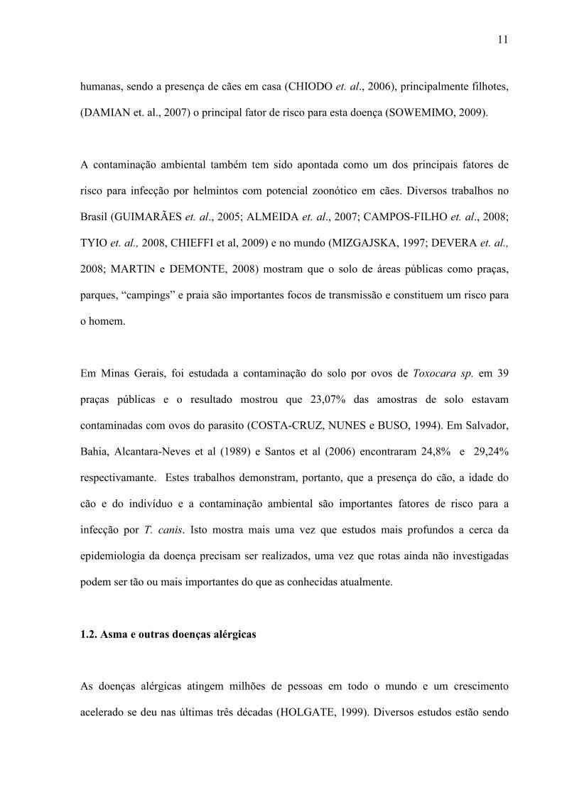
humanas, sendo a presença de cães em casa (CHIODO et. al., 2006), principalmente filhotes,
(DAMIAN et. al., 2007) o principal fator de risco para esta doença (SOWEMIMO, 2009).
A contaminação ambiental também tem sido apontada como um dos principais fatores de
risco para infecção por helmintos com potencial zoonótico em cães. Diversos trabalhos no
Brasil (GUIMARÃES et. al., 2005; ALMEIDA et. al., 2007; CAMPOS-FILHO et. al., 2008;
TYIO et. al., 2008, CHIEFFI et al, 2009) e no mundo (MIZGAJSKA, 1997; DEVERA et. al.,
2008; MARTIN e DEMONTE, 2008) mostram que o solo de áreas públicas como praças,
parques, “campings” e praia são importantes focos de transmissão e constituem um risco para
o homem.
Em Minas Gerais, foi estudada a contaminação do solo por ovos de Toxocara sp. em 39
praças públicas e o resultado mostrou que 23,07% das amostras de solo estavam
contaminadas com ovos do parasito (COSTA-CRUZ, NUNES e BUSO, 1994). Em Salvador,
Bahia, Alcantara-Neves et al (1989) e Santos et al (2006) encontraram 24,8% e 29,24%
respectivamante. Estes trabalhos demonstram, portanto, que a presença do cão, a idade do
cão e do indivíduo e a contaminação ambiental são importantes fatores de risco para a
infecção por T. canis. Isto mostra mais uma vez que estudos mais profundos a cerca da
epidemiologia da doença precisam ser realizados, uma vez que rotas ainda não investigadas
podem ser tão ou mais importantes do que as conhecidas atualmente.
1.2. Asma e outras doenças alérgicas
As doenças alérgicas atingem milhões de pessoas em todo o mundo e um crescimento
acelerado se deu nas últimas três décadas (HOLGATE, 1999). Diversos estudos estão sendo
12
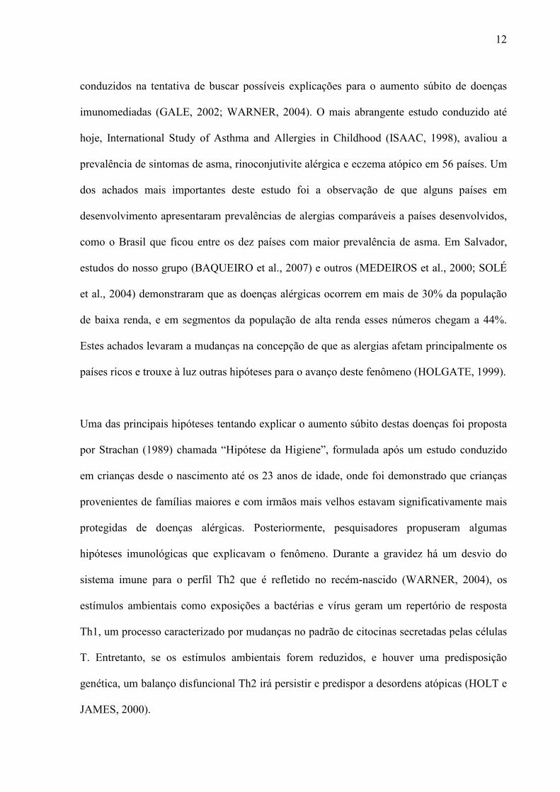
conduzidos na tentativa de buscar possíveis explicações para o aumento súbito de doenças
imunomediadas (GALE, 2002; WARNER, 2004). O mais abrangente estudo conduzido até
hoje, International Study of Asthma and Allergies in Childhood (ISAAC, 1998), avaliou a
prevalência de sintomas de asma, rinoconjutivite alérgica e eczema atópico em 56 países. Um
dos achados mais importantes deste estudo foi a observação de que alguns países em
desenvolvimento apresentaram prevalências de alergias comparáveis a países desenvolvidos,
como o Brasil que ficou entre os dez países com maior prevalência de asma. Em Salvador,
estudos do nosso grupo (BAQUEIRO et al., 2007) e outros (MEDEIROS et al., 2000; SOLÉ
et al., 2004) demonstraram que as doenças alérgicas ocorrem em mais de 30% da população
de baixa renda, e em segmentos da população de alta renda esses números chegam a 44%.
Estes achados levaram a mudanças na concepção de que as alergias afetam principalmente os
países ricos e trouxe à luz outras hipóteses para o avanço deste fenômeno (HOLGATE, 1999).
Uma das principais hipóteses tentando explicar o aumento súbito destas doenças foi proposta
por Strachan (1989) chamada “Hipótese da Higiene”, formulada após um estudo conduzido
em crianças desde o nascimento até os 23 anos de idade, onde foi demonstrado que crianças
provenientes de famílias maiores e com irmãos mais velhos estavam significativamente mais
protegidas de doenças alérgicas. Posteriormente, pesquisadores propuseram algumas
hipóteses imunológicas que explicavam o fenômeno. Durante a gravidez há um desvio do
sistema imune para o perfil Th2 que é refletido no recém-nascido (WARNER, 2004), os
estímulos ambientais como exposições a bactérias e vírus geram um repertório de resposta
Th1, um processo caracterizado por mudanças no padrão de citocinas secretadas pelas células
T. Entretanto, se os estímulos ambientais forem reduzidos, e houver uma predisposição
genética, um balanço disfuncional Th2 irá persistir e predispor a desordens atópicas (HOLT e
JAMES, 2000).

13
As mais variadas infecções bacterianas, fúngicas e virais vem sendo estudadas com alguns
resultados bastante controversos (MATRICARDI et al., 2000; JANSON, 2007 ; CHEN et al.,
2008). Entretanto, foram as infecções helmínticas que trouxeram os resultados mais
intrigantes. Sendo infecções caracterizadas por desencadearem uma resposta imune do tipo
Th2, semelhante à resposta alérgica, acreditava-se que ela seria uma potencializadora das
reações alérgicas. Entretanto, os achados obtidos através das pesquisas epidemiológicas e
experimentais mostraram que estas infecções protegiam o indivíduo do desenvolvimento de
alergias (COOPER et al, 2003; McCONCHIE et al, 2006).
Atualmente, a alergia é definida como uma doença dependente de uma resposta do sistema
imune a um antígeno exógeno, sob outros aspectos, inócuo. Segundo a classificação de Gell e
Coombs (1963) a resposta alérgica é ocasionada por uma reação de hipersensibilidade do tipo
I ou imediato, mediada por anticorpos da classe IgE que ligam-se aos receptores de alta
afinidade (FcεRI) de mastócitos e basófilos, num processo denominado de sensibilização.
Num segundo contato com o alérgeno, estes se ligam a duas IgEs específicas presentes na
membrana dos mastócitos levando a degranulação destas células. Os grânulos liberados são
ricos em leucotrienos, histamina e citocinas pro-inflamatórias, os quais acarretam espasmo da
musculatura lisa e iniciam a resposta inflamatória das vias aéreas, ocasionando coriza,
espirros e broncoespasmo. Esta resposta é também responsável pela reação de
hipersensibilidade imediata dos testes cutâneos aos aeroalérgenos (JANEWAY et al., 2007).
A IgE específica para estes antígenos liga-se ainda a receptores de baixa afinidade (FcεRII) de
eosinófilos, linfócitos, plaquetas e macrófagos, intensificando e modulando a resposta
inflamatória através da produção de IL-4, que estimula a produção de IL-5, IL-13 e demais

14
citocinas e moléculas inflamatórias envolvidas na resposta Th2 (BUSSE et al., 2001;
MURPHY e REINER, 2002; ABBAS e LICHTMAN, 2005). Na inflamação eosinofílica das
vias aéreas, a IL-5 está envolvida na diferenciação, ativação e sobrevivência dos eosinófilos,
aumentando sua responsividade para a eotaxina, através da regulação da expressão de
receptores CCR3 de eosinófilos para esta citocina (LEFORT et al., 1998).
A importância da IgE no curso das doenças alérgicas tem sido amplamente relatada, e estudos
de bloqueio da IgE circulante com anticorpos monoclonais diminuíram não só a IgE sérica
como reduziram a expressão dos receptores de alta afinidade para IgE (FcεRI), atenuando
tanto a fase inicial (mediada por mastócitos) como a fase tardia (inflamatória) da asma (KON
et al, 1998)..
A causa da alergia é multifatorial e depende da interação de fatores genéticos, tempo e
quantidade de exposição aos alérgenos, principalmente os derivados de ácaros da poeira
doméstica, de animais de estimação e fungos e de fatores ambientais ainda não bem
caracterizados. Os ácaros da poeira doméstica, Dermatophagoides farinae, D. pteronyssinus e
Blomia topicalis, são os principais agentes desencadeadores de fenômenos alérgicos descritos
em todo o mundo (PLATTS-MILLS et al., 2000).
Algumas propriedades do alérgeno são definitivas para sua maior capacidade de sensibilizar e
desencadear a resposta imune alérgica (ABBAS e LICHTMAN, 2005). A atividade
enzimática é uma das propriedades mais importantes no desencadeamento das reações
alérgicas provocadas pelos alérgenos, possivelmente por destruir as junções comunicantes das
células epiteliais aumentando a permeabilidade da mucosa bronquial a macromoléculas
(SCHULZ et al, 1999).
15
Dentre as alergias respiratórias, destaca-se a asma por ser uma das doenças crônicas mais
comuns na infância (WONG et al, 2001), atribuída a ativação e produção de citocinas pelos
linfócitos T CD4+ de forma imprópria, induzida principalmente por aeroalérgenos, resultando
em inflamação eosinofílica das vias aéreas, aumento da IgE sérica, degranulação dos
mastócitos submucosos do trato respiratório, constrição brônquica e secreção aumentada de
muco (ANDERSON, 2002).
Atualmente, a asma é classificada em duas formas muito específicas: asma atópica e asma
não-atópica. A asma atópica acomete principalmente indivíduos entre 4 e 40 anos, porém tem
sido também relatada em populações geriátricas (APTER et al., 1988). Na asma existe uma
relação temporal entre sintomas respiratórios e exposição aos alérgenos e presença de
anticorpos IgE contra alérgenos comuns. Por outro lado, a asma não atópica acomete
principalmente crianças com menos de 04 anos ou idosos com mais de 60 anos de idade.
Nesta enfermidade a inflamação das vias aéreas mediada por IgE específica para alérgenos
está ausente.
Embora recentes revisões tenham comentado o paralelo entre o aumento de doenças alérgicas
e auto-imunes no mundo ocidental, não é fácil explicar como as mesmas mudanças
ambientais podem promover o aumento de doenças com orientações imunes opostas e
mutuamente excludentes (BLACK, 2001; WILLS-KARP et al, 2001). Tem sido argumentado
que a secreção de citocinas anti-inflamatórias como a IL-10 produzidas pelas células T
regulatórias podem ser a chave para regular ambos os desvios imunes (YAZDANBAKHSH et
al, 2001). Portanto as infecções na infância ou mesmo durante a gestação exercem um
importante papel protetor contra o desenvolvimento de alergia como uma conseqüência do
16
estímulo crônico dos receptores semelhantes ao Toll (TLRs), sendo, portanto, a causa do
aumento dos fenômenos alérgicos a diminuição da atividade das células T regulatórias mais
do que a exacerbação da resposta Th2 (YAZDANBAKHSH et al, 2002; ROMAGNANI,
2004).
1.3. Toxocara canis e alergia
Diversos estudos epidemiológicos vêm demonstrando que a infecção por T. canis contribui
para o desenvolvimento de manifestações alérgicas no homem. Buijs et al (1997) mostrou
associação entre a soropositividade para T. canis e asma alérgica/bronquite recorrente e
aumento de IgE específica contra aeroalérgenos. Alteração na atividade respiratória e asma
tem sido observada em diversos estudos em associação com a infecção por T. canis
(FERREIRA et al, 2007; ESPINOZA et. al., 2008; MARTIN et. al., 2008; FERNANDO et al,
2007), assim como eosinofilia (TEIXEIRA et. al, 2006; ESPINOZA et. al., 2008; MARTIN
et. al., 2008). Chiodo et. al., (2006) observaram que a eosinofilia estava presente em 86,95%
dos indivíduos positivos para T. canis (p < 0,001, OR = 11,03) e Fernando et. al. (2007), no
Sri Lanka, observaram que a presença de eosinofilia foi significantemente maior nas crianças
soropositivas (77%) que nas soronegativas (40%) (p<0,001).
Paralelamente a estes achados, outros autores têm relatado ausência de associação entre
soropositividade para T. canis e atopia e asma. Sharghi et al (2001) não observaram
associação entre asma e infecção por T. canis em 324 crianças nos Estados Unidos, enquanto
Yong-Hun et al (2008) avaliaram a associação entre eosinofilia acima de 10% e
soropositividade para T. canis em 96 amostras de soro, mostrando ausência de correlação
entre a infecção e eosinofilia.
17
Na tentativa de entender os mecanismos envolvidos na imunopatologia desta infecção bem
como seus efeitos em patologias imunomediadas como a asma, modelos murinos têm sido
amplamente utilizados com este propósito (ESPINOZA et al, 2002a; PECINALI et al, 2005;
PINELLI et al 2007). Estes estudos vêm mostrando que infecção por T. canis resulta em
inflamação pulmonar persistente, eosinofilia, aumento da produção de IgE, hiperreatividade
das vias aéreas e produção de citocinas do tipo Th2 (PINELI et al, 2007).
Pineli et al (2007) estudaram os efeitos da infecção com T. canis nas manifestações alérgicas
combinando um modelo murino para toxocaríase e um modelo experimental para inflamação
das vias aéreas utilizando ovoalbumina (OVA). O efeito do tempo de infecção na inflamação
das vias aéreas também foi avaliado e classificado em inicial com 3 dias e tardio com 20 dias
de infecção. Foi demonstrado o aumento da expressão da citocina IL-4 e eosinófilos nos
pulmões dos camundongos, sendo estes mais significativos no início da infecção. Além disso,
foi observado aumento da IgE no plasma e no lavado broncoalveolar (BAL) durante a
infecção inicial e na tardia.
Além da típica indução de perfil Th2 ocasionada pelo T. canis, moléculas relacionadas à
ativação de padrão de resposta Th1, tais como o óxido nítrico, vêm sendo descrita na infecção
por T. canis e associada a efeitos deletérios no sistema vascular pulmonar e dano direto aos
hospedeiros murinos (ESPINOZA et al, 2002a). A via de sinalização citoplasmática envolvida
na produção de óxido nítrico após o estímulo com antígeno de T. canis foi estudada em
macrófagos alveolares de ratos, mostrando que a fosfolipase C e A2 induz a produção e
liberação do óxido nítrico por duas vias distintas: tanto pela indução do aumento do cálcio
intracelular; como pela liberação do ácido araquidônico (ESPINOZA et al, 2002b).
18
A lesão tecidual causada pela migração das larvas do T. canis pelos diversos órgãos induz um
aumento de citocinas pró-inflamatórias que pode comprometer as funções sistêmicas. Pecinali
et al (2005) avaliaram o nível das citocinas IL-6, IFN-γ, eotaxina e RANTES (Regulated on
Activation Normal T Cell Expressed and Secreted) no plasma e no lavado bronco-alveolar
(BAL) de camundongos infectados com T.canis. A IL-6 é um marcador bem caracterizado de
resposta inflamatória bem como o IFN-γ, enquanto o RANTES é importante na resposta
eosinofílica e a eotaxina é uma quimiotaxina de eosinófilos. Neste estudo todas as citocinas
estavam aumentadas durante a infecção pelo parasito mostrando uma atividade inflamatória
nas vias aéreas.
Diante do exposto, os achados da literatura ainda são muito controversos no que diz respeito a
capacidade do T. canis em induzir ou proteger de alergias o que sinaliza para a necessidade de
estudos mais aprofundados com este propósito. Essa disparidade entre os dados na literatura
pode ser uma conseqüência do baixo número de amostras em alguns trabalhos
epidemiológicos. Somam-se a isto as diferenças no diagnóstico sorológico da toxocaríase,
assim como a determinação da definição de atopia e asma utilizadas pelos diferentes autores.
A alta prevalência de asma e atopia em nossa população nos estimulam a buscar possíveis
fatores que podem estar determinando o aumento destas doenças.
19
2. HIPÓTESES
2.1
H0) Infecção causada por Toxocara canis não está associada com o nível socioeconômico
e condições de higiene da população.
H1) Infecção causada por Toxocara canis está associada com o nível socioeconômico e
condições de higiene da população.
2.2.
H0) A presença da infecção por Toxocara canis não modula o sistema imune, portanto
não influencia o desenvolvimento de atopia e asma
H1) A presença da infecção por Toxocara canis modula o sistema imune regulando
o desenvolvimento de atopia e asma.
20
3. OBJETIVO GERAL
Estudar os fatores de risco para infecção por Toxocara canis e investigar a associação desta
infecção com eosinofilia, IgE total, atopia e asma em crianças oriundas de população de baixa
renda de Salvador, Bahia.
3.1. Objetivos específicos
• Determinar os fatores de risco para a infecção por T.canis nas crianças do estudo.
• Investigar possível associação entre a soropositividade para T. canis com eosinofilia e
níveis de IgE total sérica.
• Investigar possível associação entre a soropositividade para T. canis com atopia e
asma nas crianças do estudo.
21
4. RESULTADOS
4.1 Manuscrito 1: Risk factors for Toxocara canis infection in children from a Brazilian
urban setting (formatado para submissão ao Transaction of the Society of Medicine and
Tropical Higiene)
4.2. Manuscrito 2: Toxocara canis infection as risk factor for atopy and atopic asthma in
a large set of children from a Latin American urban center (a ser submetido ao JACI –
Journal of Allergy and Clinical Immunology)
22
Risk factors for Toxocara canis infection in children from a Brazilian urban setting Lívia R. Mendonçaa, Vitor Camilo Cavalcante Dattolia, Camila A. Figueiredoa, Renata
Esquivelb, Rosemeire Fiacconec, Lain P-de-Carvalhod, Maurício L. Barretob, Neuza M.
Alcantara-Nevesa§
aDepartamento de Biointeração, Instituto de Ciências da Saúde, Universidade Federal da
Bahia, Av. Reitor Miguel Calmon, Sem no. Canela, Salvador, Bahia, CEP 40110-902 Brasil
bInstituto de Saúde Coletiva, Universidade Federal da Bahia, Brazil; Rua Basílio da Gama,
s/n – Canela, CEP: 40110-040 Salvador- BA
cInstituto de Matemática, Universidade Federal da Bahia, Brazil; End: Rua Barão de
Jeremoabo, s/nº - Campus Universitário de Ondina, CEP: 40170-115 Salvador- BA
dCentro de Pesquisas Gonçalo Moniz, Fundação Oswaldo Cruz, Rua Waldemar Falcão, 121,
Brotas, Salvador, Bahia, CEP 40296710, Brazil.
§Corresponding author
E-mail addresses:
LRM: [email protected]
VCCD: [email protected]
CAF: [email protected]
LCPC: [email protected]
MLB: [email protected]
NMAN: [email protected]
23
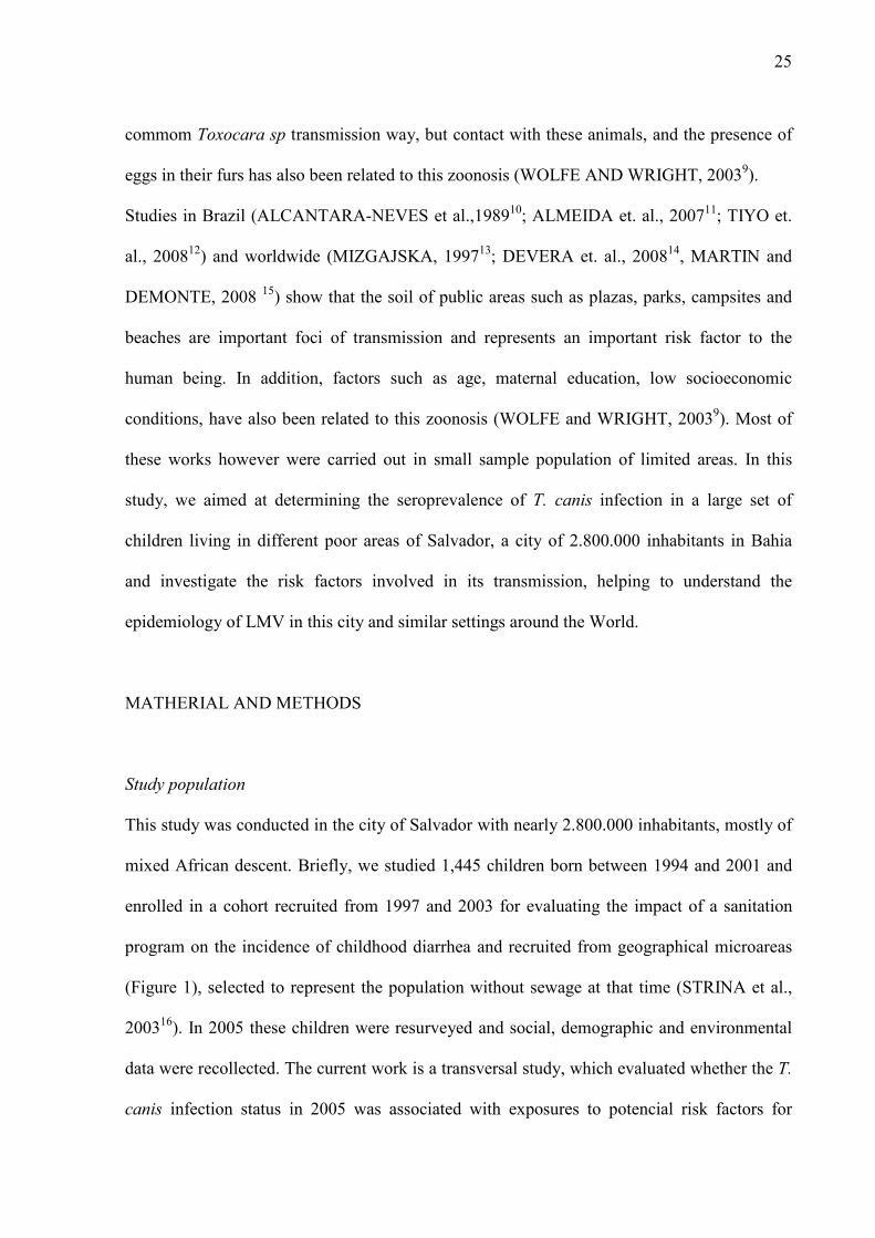
SUMMARY
Background: Visceral larva migrans syndrome is a zoonosis caused by migration of
Toxocara sp larvae in human organs. The improvement of its diagnosis showed that this
disease occurs worldwide. This study aimed to estimate the seroprevalence of T. canis
infection, and to identify potential risk factors for this infection in children living in poor
areas of Salvador, Brazil. Methodology: Parents of 1,445 children answered a validated
questionnaire containing possible risk factor for acquisition of this infection. Blood was
collected and the presence of IgG anti-T. canis antibodies was detected by indirect ELISA
using T. canis larval excretory-secretory antigens (TcESLA) in pre-absorbed sera with
Ascaris lumbricoides extract. Results: Seroprevalence of T. canis infection was 48.4%.
Among the risk factors studied, contact with dogs and cats, child´s age, low maternal
scholarity and household located in paved streets were shown to be risk factors for T. canis
infection. Conclusions: The seroprevalence of T. canis infection is high among children
living in a poor urban center of Brazil. The association of the infection with cat’s contact is
suggestive that T.canis ESLA reacts with anti-T. cati antibodies. The finding of association of
T. canis infection with living in paved streets may be secondary to the spreading of the eggs
and higher exposure of the population to cat and dog contaminated feces in this type of
ground . The relationship of low maternal education with higher infection by T. canis supports
previous studies showing that low socioeconomic status is a risk factor for the acquisition of
this infection.
Keywords: Toxocara canis, seroprevalence, children, risk factors, dog, cats, cross-reaction

24
INTRODUCTION
Visceral larva migrans (VLM) is a syndrome of human beings, transmitted by accidental
ingestion of embryonated eggs of Toxocara canis (dog round worms) or rarely of T. cati (cat
round worm). Their larvae do not migrate to intestine as occur in the definite hosts and remain
migrating through the organs and visceras leading to polymorphic clinical pictures which vary
from asymptomatic to severe systemic forms such as prolonged fever with hepato-
esplenomegaly, meningoencephalitis and asthma-like symptoms (DEPOMMIER, 20031;
HARALAMBIDOU et. al., 20052; SAPORITO et. al., 20083). Further than the clinical
pictures mentioned above, T. canis infection leads to a hypersensitivity reaction status, even
in asymptomatic subject, which may cause eosinophilia, increase in total IgE and high
susceptibility to asthma (BUIJS et. al., 19974; FERREIRA et. al., 20075). Although this
infection occur worldwide, its prevalence is higher in non-affluent population and countries
(COELHO et. al., 20046; ESPINOZA et. al., 20087), where its diagnosis is rarely done, being
considered a neglected disease. For example in Brazil, from our knowledge, only one
laboratory is able to diagnosis VLM. The disease diagnosis depended on the larvae
cultivation to produce the antigen used in ELISA, but actually commercial kits are available,
although expensive. This infection is also prevalent in many developed countries and its
global importance may be underestimated. In the United States, it is the most common
helminthic infection, affecting millions of individuals (HOTEZ E WILKINS, 20098).
Strain dogs and cats and domiciliated pets from low income population play an important role
in the transmition of Toxocara sp eggs providing environmental contamination, the
perpetuation of the cycle and spreading the diseases among the human population. The
contact with contaminated ground, hands or food with embryonated eggs is the most
25
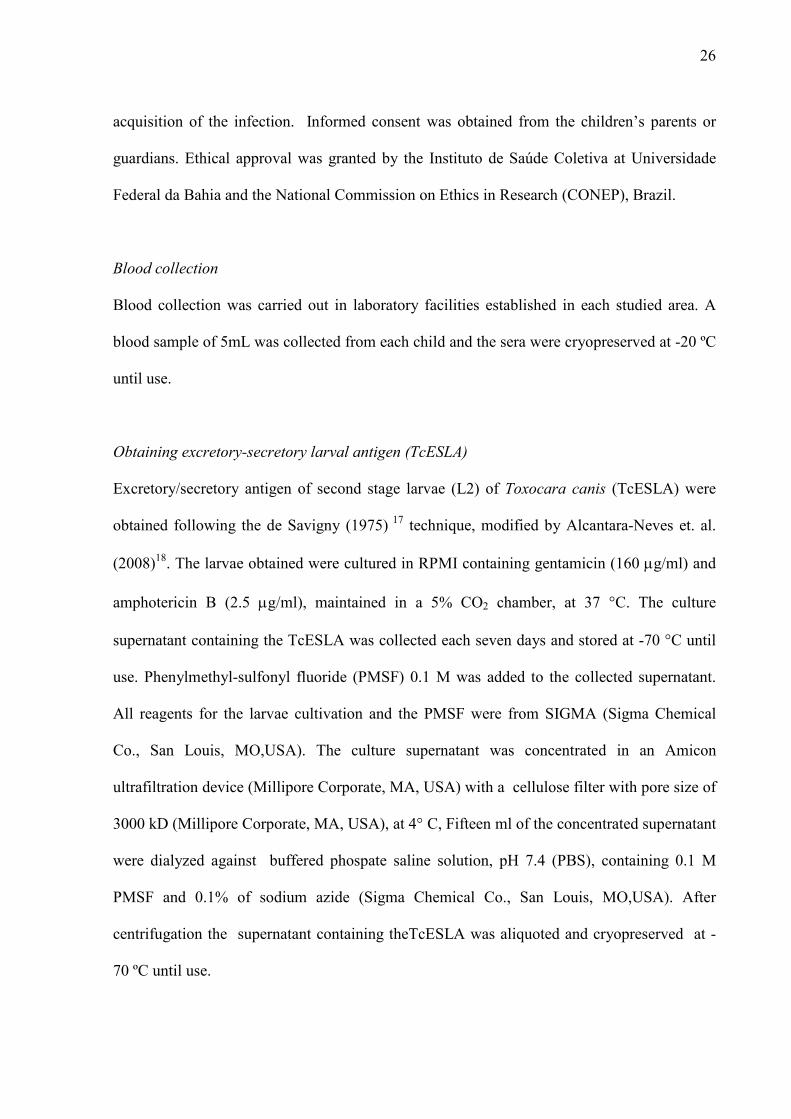
commom Toxocara sp transmission way, but contact with these animals, and the presence of
eggs in their furs has also been related to this zoonosis (WOLFE AND WRIGHT, 20039).
Studies in Brazil (ALCANTARA-NEVES et al.,198910; ALMEIDA et. al., 200711; TIYO et.
al., 200812) and worldwide (MIZGAJSKA, 199713; DEVERA et. al., 200814, MARTIN and
DEMONTE, 2008 15) show that the soil of public areas such as plazas, parks, campsites and
beaches are important foci of transmission and represents an important risk factor to the
human being. In addition, factors such as age, maternal education, low socioeconomic
conditions, have also been related to this zoonosis (WOLFE and WRIGHT, 20039). Most of
these works however were carried out in small sample population of limited areas. In this
study, we aimed at determining the seroprevalence of T. canis infection in a large set of
children living in different poor areas of Salvador, a city of 2.800.000 inhabitants in Bahia
and investigate the risk factors involved in its transmission, helping to understand the
epidemiology of LMV in this city and similar settings around the World.
MATHERIAL AND METHODS
Study population
This study was conducted in the city of Salvador with nearly 2.800.000 inhabitants, mostly of
mixed African descent. Briefly, we studied 1,445 children born between 1994 and 2001 and
enrolled in a cohort recruited from 1997 and 2003 for evaluating the impact of a sanitation
program on the incidence of childhood diarrhea and recruited from geographical microareas
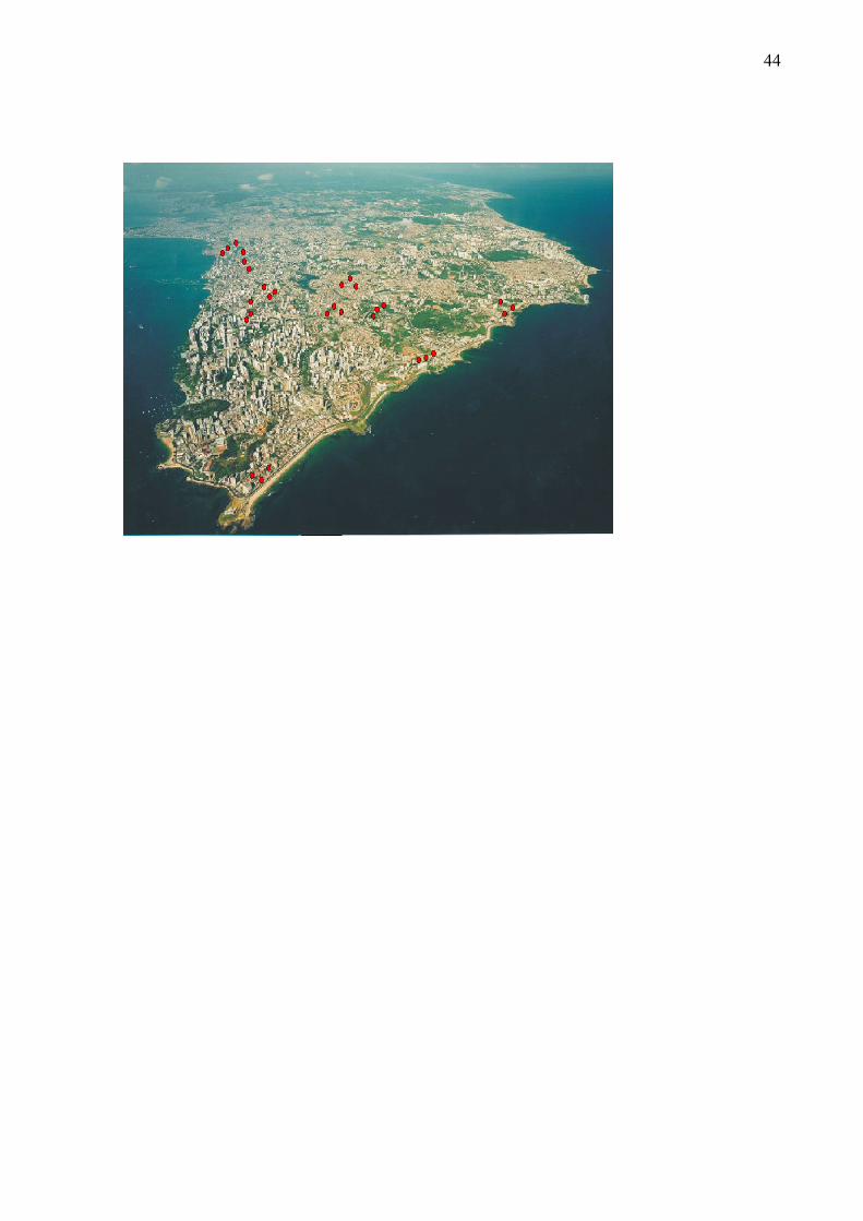
(Figure 1), selected to represent the population without sewage at that time (STRINA et al.,
200316). In 2005 these children were resurveyed and social, demographic and environmental
data were recollected. The current work is a transversal study, which evaluated whether the T.
canis infection status in 2005 was associated with exposures to potencial risk factors for
26
acquisition of the infection. Informed consent was obtained from the children’s parents or
guardians. Ethical approval was granted by the Instituto de Saúde Coletiva at Universidade
Federal da Bahia and the National Commission on Ethics in Research (CONEP), Brazil.
Blood collection
Blood collection was carried out in laboratory facilities established in each studied area. A
blood sample of 5mL was collected from each child and the sera were cryopreserved at -20 ºC
until use.
Obtaining excretory-secretory larval antigen (TcESLA)
Excretory/secretory antigen of second stage larvae (L2) of Toxocara canis (TcESLA) were
obtained following the de Savigny (1975) 17 technique, modified by Alcantara-Neves et. al.
(2008)18. The larvae obtained were cultured in RPMI containing gentamicin (160 µg/ml) and
amphotericin B (2.5 µg/ml), maintained in a 5% CO2 chamber, at 37 °C. The culture
supernatant containing the TcESLA was collected each seven days and stored at -70 °C until
use. Phenylmethyl-sulfonyl fluoride (PMSF) 0.1 M was added to the collected supernatant.
All reagents for the larvae cultivation and the PMSF were from SIGMA (Sigma Chemical
Co., San Louis, MO,USA). The culture supernatant was concentrated in an Amicon
ultrafiltration device (Millipore Corporate, MA, USA) with a cellulose filter with pore size of
3000 kD (Millipore Corporate, MA, USA), at 4° C, Fifteen ml of the concentrated supernatant
were dialyzed against buffered phospate saline solution, pH 7.4 (PBS), containing 0.1 M
PMSF and 0.1% of sodium azide (Sigma Chemical Co., San Louis, MO,USA). After
centrifugation the supernatant containing theTcESLA was aliquoted and cryopreserved at -
70 ºC until use.
27
Characterization of the TcESLA by sodium duodecyl sulphate polyacrylamide gel
electrophoresis (SDS-PAGE)
TcESLA SDS-PAGE was performed according to Laemmli (1970)19 using a Mini-PROTEAN
III Electrophoresis Cells (Bio-Rad Laboratories, Hercules, CA) and a 12% polyacrylamide
gel in the presence of 10% sodium dodecyl sulphate (Merck & Co., Inc., White house Station,
NJ, USA). Protein fractions were labeled with Coomassie Brilliant Blue R 250 (Sigma
Chemical Co., San Louis, MO, USA). The relative molecular weights were calculated using
prestained protein of standard molecular weight according to the relative electrophoretical
mobility (REM), using the following equation: REM = distance of the protein
migration/distance of bromophenol blue migration.
Sera absorption with Ascaris lumbricoides antigens
In order to eliminate cross-reaction between anti-A. lumbricoides and anti-T.canis IgG
antibodies the sera were pre-absorbed with an extract of adult A. lumbricoides in the presence
of polietilenoglicol (3%) (PEG 15.000 – Sigma Chemical Co., San Louis, MO,USA) and
0.1% sodium azide diluted in PBS. After incubation for 30 minutes in a homogenizer at room
temperature, the material was centrifuged at 5,724g for 10 minutes. The supernatant
containing the serum was re-absorbed as described above and frozen at -20 ºC until testing.
Immunoassay for detection of anti-Toxocara canis IgG antibodies
IgG antibodies were detected in sera by indirect ELISA assay using TcESLA as antigen.
Briefly, 96-well plates were sensitized with 3.2 µg/mL of TcESLA in carbonate/bicarbonate
buffer, overnight at 4°C. The plates were blocked with a solution of PBS containing 10% of
fetal calf serum (FCS - Cutilab, Campinas/SP, Brazil). The sera were applayed to the wells
diluted at 1:1,000 in a solution of PBS containing 0.05% tween 20 and 2.5% fetal calf serum
28
(PBS/T/FCS). The reaction was developed using an anti-human biotinylated IgG conjugate
(BD Pharmingen, San Diego, CA, USA) diluted at 1:2000 in PBS/T/FCS followed by
Streptavidin-peroxidase (Streptoavidin-HRP, BD Pharmingen, San Diego, CA, USA) diluted
at 1:1000 in PBS/T/FCS. Hydrogen peroxide and OPD (o-Phenylenediamine - Sigma
Chemical Co., San Louis, MO, USA) were used as substrate and chromogen. The plates were
incubated for one hour at room temperature after each step, except for the substrate which was
incubated for 1/2 hour and washings were done four times betwwen steps with PBS/T. The
reaction was stopped within 30 minutes with sulfuric acid 2N and the optical density was
determined using a 490nm filter. To determine the cutoff, 19 serum samples from individuals
who had no contact with dog and/or cat and higher socioeconomic status were using like
negative controls. The mean of optical density plus three standard deviation was considered
the cutoff. To determine the avidity of the antibodies binding to the antigen, the assay was
performed in duplicate, and for each serum two wells were washed after the serum incubation
for 5 minutes with PBS-T containing 6M urea (Ureia P.A. – VETEC, São Paulo, Brazil).
Following, the assay was continued as described above. Toxocara-specific IgG avidity was
calculated by using the for relative avidity index: Percentage of Reduction (PR) = 100 – O.D.
with urea x 100/ O.D. without urea. A serum was considered to have low avidity, when the
PR was above 50% (Dziemian et. al., 200820).
Statistical Analysis
Only children for whom complete data was available were included in the analysis. The
children´s gender and age in 2005 were treated as a priori confounders. The following
variables were studied as risk factors for acquisition of T. canis infection (outcome): whether
the child attended nursery, maternal schooling, presence of dogs or cats at home, if house
were served by a paved road. We first performed a univariate analysis between each potential
risk factor and outcome; built a multivariable model with standard logistic regression

29
including only significant variables from the univariate analysis; and then, we assessed each
non-significant variable a second time by including each one in the model (one at a time). If
the variable remained non-significant, it was completely removed from the analysis. This
process was repeated until no variables remained to be assessed. The association between
outcome and risk factors was estimated with odds ratio and 95% confidence interval.
RESULTS
Among the original 1,445 children enrolled in the 2005 survey, 1,308 had complete data sets
and were used in the analysis. No statistically significant differences were found in the
frequencies of the analyzed variables, between the excluded and the studied children.
Figure 2A shows the quality of the TcESLA, used as antigen to detect anti-T.canis IgG in an
indirect ELISA, determined by SDS-PAGE showing bands of 104, 95, 86, 71, 40, 21, 17, 16
and 13kDa. Figure 2B shows the result of the sera absortion with somatic antigen of A,
lumbricoides. Sera absorbed twice with PEG 15,000 had a decrease of 76.39% in optical
density. Figure 1C shows the determination of the assay cut-off which was performed with
sera from 19 children without history of contact with dogs, and five children with history of
contact with dogs and low social and economic condictions. The cut-off obtained, of 0.23 was
calculated by the mean of OD of the negative controls plus three standard deviations of this
mean. Figure 1D shows the results of the determination of anti-T. canis IgG antibodies in the
whole population by indirect ELISA.
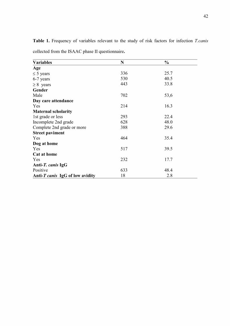
The majority of the children aged between six and seven years old (40.5%), approximately
half of them were male 702 (53.6%), 214 (16.3%) had attended nursery school for some time
and 64.6% of children lived in areas without paved street. 388 (29.6%) of their mothers had
30
completed a high school degree. Using the established cut-off, 48.4 % of the children were
seropositive for T. canis IgG. Only 2.8% of the 633 children seropositive showed IgG of low
avidity, indicating a recent infection (Table 1).
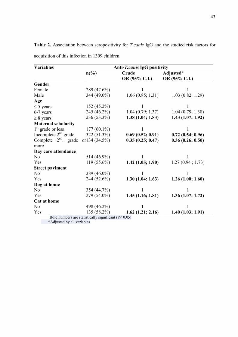
The following variables were significantly associated with an increased prevalence of T. canis
infection at both univariated and multivariated analyses: to be ≥ 8 years old (crude OR=1.38;
95%C.I.=1.04;1.83 and (adjusted OR=1.43; 95%C.I.=1.07;1.92); living in house placed in
paved streets (crude OR=1.30, 95%C.I.=1.04;1.63 and adjusted OR=1.26,
95%C.I.=1.00;1.60); presence of a dog at home (crude OR=1.45, 95%C.I.=1.16;1.81;
adjusted OR=1.36; 95%C.I.=1.07;1.72) and presence of a cat at home (crude OR=1.62,
95%C.I.=1.21;2.16; adjusted OR=1.40, 95%C.I.=1.03;1.91). Day care attendance was
associated only at univariated analysis (crude OR=1.42; 95%C.I.=1.05;1.90) and mother
with both, incomplete 2nd grade and complete 2nd grade or more were negatively associated
with T. canis infection when compared with mothers with 1st grade or less (crude OR=0.69;
95%C.I.=0.52;0.91; adjusted OR=0.72; 95%C.I.=0.54;0.96 and crude OR=0.35;
95%C.I.=0.25;0.4; adjusted OR=0.36, 95%C.I.=0.26;0.50) respectively, (Table 2).
DISCUSSION
The diagnosis of visceral larva migrans is performed almost exclusively by antibodies anti-
T.canis due to the difficulty of parasitological and clinically diagnosing the disease. Many
laboratories have developed in house assays for research purpose in order to determine the
prevalence of this zoonosis (AGUIAR-SANTOS, 2004)21. The TcESLA obtained in our
laboratory had bands with similar molecular weight as antigens previously described by other
authors (RUBINSKY-ELEFANT et. al., 200622; IDDAWELA et. al., 200723) and the
standardization provided more specificity for our assay when compared with the literature,
31
since the serum dilution was 1:1000 instaed of 1:200 (NUNES et al. 1997)24. Roldán and
Espinoza (2006)25, because we used a biotin instead of a peroxidase-congugate. Even using
the cut-off of the mean of negative controls plus 3 standard deviation we had a cut offt of 0.23
and some of the positive sera with optical density values above the up detection limit.
Furthermore we absordeb the sera with A. lumbricoides extract instead of A. suum as reported
by the above cited works. Absorption of sera with antigens from other parasites is a practice
that increases the specificity of the test, since many parasite species share similar antigens
giving rise to cross-reactivity between these antibodies (ISHIDA et al., 2003)26. For the
diagnosis of toxocariasis is usually the serum samples are absorbed with Ascaris suum
antigen (LYNCH et. al., 198827, NUNES et al. 199724; CAMPOS JUNIOR et. al. 200328;
ROLDÁN et al. 200625). Nunes et al. (1997) 24 determined at least one band of molecular
weight around 55-66 kDa responsible for cross-reactivity between T. canis and A. suum, and
that disappeared with the pre-absorption of sera with antigen A. suum. Roldán and Espinoza
(2009) 29 determined antigenic bands recognized by sera from patients with toxocariasis as
following, 24, 28, 30, 35, 56, 117, 136 and 152 kDa. However, only the bands of 24-35 kDa
were highly specific for Toxocara infection (98.3%), while the other was observed cross-
reactivity with sera from patients infected with other helminthic infections. In our population
the helminth infection are caused only by A. lumbricoides and Trichuris trichiura which occur
in 16.1% and 10.8% respectively in our children. The absorption with antigen Ascaris
lumbricoides decreased up to76.39% of optical density, indicating a higher removal of
specific antibodies to A. lumbricoides avoiding cross-reactions between this helminth and
anti-T. canis in antibodies. Additionally we performed absorption with T. trichiura somatic
antigen after absorption with A. lumbricoides antigen and observed not significant decrease in
optical density that justify its use, showing that pre-absortion with A. lumbricoides was
sufficient to avoiding cross-reaction with others helminthes (data not shown).We also have
32
also absorbed the sera with Ancylostoma braziliensis antigens and there was no decrease of
the anti-T. canis IgG, showing that this parasite do not share antigens reactive with IgG (data
not shown)..
In our work we found a prevalence of IgG anti-T.canis of 48.4%. Others studies conducted in
Latin America and Brasil reported small prevalences, except for Damian et. al. (2007)30 who
found a prevalence of 52% among adult population in Amazonas state in Brazil. Alonso et. al.
(2000)31 reported a positivity of 37.9% in children younger than 14 years in Argentina,
Espinoza et. al. (2008)7 determined a seroprevalence of 32.4% in Peru and in Brazil, Chieffi
(2009)32 in a review, cited prevalence of T. canis varying from 3.72% to 40%. In agreement
with our results, Radman et. al (2000) 33 in Argentina observed a prevalence of 39% infection.
Maternal education is an indicator of socioeconomic status of the family. The result of the
seroprevalence of IgG anti-T.canis found in this study was similar to those observed in other
low-income populations, where prevalence of infection in children of mothers with fewer
years of education was higher. Alderete et. al. (2003)34 diagnosed a prevalence of 38.8% in
children with a mean age of 9.4 years and determined that Toxocara infection was inversely
proportional to family income.
Contact with dogs has been shown in several studies as an important risk factor for
toxocariasis. A cross-sectional study estimated a frequency of 52% positivity for T.canis in 34
families in the Amazonas state. Individuals who had contact with dog at home, 60% were
positive (chi2 = 14.317, p = 0.026), and who had contact with puppies at home, 66.6% were
positive (chi2 = 22.149, p = 0.008), demonstrating the association between contact with the
dog and the presence of anti-T.canis IgG (Damian et. al., 2007)30. In Argentina, Chiodo et. al.
(2006) 35 evaluated 100 individuals for IgG anti-T. canis and 23% were positive, and all had

33
contact with dog at home. Our results confirm these findings of the presence of the dog at
home as a risk factor for T.canis infection in this study population.
Several epidemiological studies indicate contamination of soil as a determinant in infection by
T. canis. In the present study it was noted that paved street increased the chance of infection,
which makes us suppose that maybe dog and cat may defecate in those streets and the absence
of soil to absord the eggs may favour more contact of the children with contaminated feces.
Cross-reaction of IgG between the T. canis and T. cati ESLA occur (KENNEDY et al,
1987)36. Few studies were conducted to estimate the infection of cats and their potential role
as reservoir for human toxocariasis, Martinez-Barbosa et. al. (2003)37 determined a prevalence
of 42.5% of T. cati infection in cats which makes one think that this parasite may be common
and raises the importance of the development of a species-specific ELISA for detection anti-
Toxocara sp IgG, useful for studies on the epidemiology of LMV caused by both Toxocara
species.
Some studies refer that soil contamination is not the only effective route in human
toxocariasis and eggs of T. canis can be sprouted in fur and direct contact between humans
and dogs may be an alternative route of human infection (WOLFE and WRIGHT, 2003) 9.
Aydenizoz-Ozkayhan et. al. (2008) 38 collected 51 fur samples and observed that 21.56% of
the dogs had eggs in their fur. Roddie et.al. (2008) 39 examined 100 dogs for the presence of
eggs in fur and found Toxocara eggs in 67% of adult dogs and 95% of puppies. In the
Netherlands, Overgaauw et al (2009)40 found Toxocara eggs in 4.4% of dog fecal samples and
in 12.2% of their fur samples. Moreover, many of the owners allowed their dogs to climb and
sleep in their beds, and only 15% washed their hands after contact with your pet. Therefore,
this close physical contact between pets and their owners possibly increase the risk of
34
transmission of Toxocara sp and point to the need for greater attention to the potential risk to
which humans are exposed.
In conclusion this work shows that T. canis is a highly prevalent infection in the studied
population and it is closely related to social status and hygiene. The presence of the dog at
home proved to be an important risk factor for this disease and is necessary to adopt sanitary
measures more specific for resident dogs, since only control programs stray dogs is not the
only way to control the disease. The association with presence of cats in house is suggestive
that thre are antigenic similarities between T. canis and T. cati ESLA and that anti-T. cati
antibodies have influenced the outcome of the study. Paving the street which was associated
with increased risk of Toxocara infection may be influencing the increased exposure to dog
and cat feces probabily because the absence of soil to absorb the eggs make them to be more
exposed and accessible to be transmitted to the children.
Statements on the authors' contributions
LRM has done the laboratory assays and wrote the first draft of the manuscript. VCCD helped
in the laboratory assay and in analyze the data. CAF, has helped in obtaining the Toxocara
antigen and revised the manuscript. RE and RLF have analysed the data: ; LPC has helped in
the T. canis assay standardization and revised the manuscript. MLB has coordinated the
epidemiological work. NMAN planned the work, supervised the laboratory work, helped in
the manuscript elaboration and revised the text.
Acknowledgements
We thank the WELLCOME TRUST for funding this work and the Brazilian agencies
CAPES, CNPQ and FAPESB for scholarships that supported the author and some of the co-
authors.
35
Fundings
This study was conducted through the SCAALA (Social change, Asthma and Allergy in Latin
America) initiative, funded by the Wellcome Trust, Grant No. 072405/Z/03/Z and FABESP
grant (camila completar).
Ethical approval
Ethical approval was obtained from the Brazilian National Ethical Committee. Written,
informed consent detailing all procedures to be carried out on the children was signed by the
parents or legal guardian of each child.
REFERENCES
1. Despommier D. Toxocariasis: Clinical Aspects, Epidemiology, Medical Ecology, and
Molecular Aspects. Clinical Microbiology Reviews 2003, 16: 265–72.
2. Haralambidou S, Vlachaki E, Ioannidou E, Milioni V, Haralambidis S , Klonizakis I.
Pulmonary and myocardial manifestations due to Toxocara canis infection. European
Journal of Internal Medicine 2005, 16: 601–2.
3. Saporito L, Scarlata F, Colomba C, Infurnari L, Giordano S, Titone L. Human
toxocariasis: a report of nine cases. Acta Paediatr 2008, 97: 1301-2.
4. Buijs J, Borsboom G, Renting M, Hilgersom WJA, van Wieringen JC, Jansen G,
Neijens J. Relationship between allergic manifestations and Toxocara seropositivity: a
cross-sectional study among elementary school children. Eur Respir J 1997, 10: 1467–
75.
5. Ferreira MU, Rubinsky-Elefant G, Castro TG, Hoffmann EHE, Silva-Nunes M,
Cardoso MA, Munizd PT. Bottle Feeding and Exposure to Toxocara as Risk Factors

36
for Wheezing Illness among Under-five Amazonian Children: A Population-based
Cross-sectional Study. Journal of Tropical Pediatrics 2007, 53: 119-24.
6. Coelho LMPS, Silva MV, Dini CY, Giacon Neto AA, Novo NF, Silveira EPR. Human
Toxocariasis: a Seroepidemiological Survey in Schoolchildren of Sorocaba, Brazil.
Mem Inst Oswaldo Cruz Rio de Janeiro 2004, 99: 553-7.
7. Espinoza YA, Huapaya PH, Roldán WH, Jiménez S, Arce Z, Lopez E. Clinical and
serological evidence of Toxocara infection in school children from Morrope district,
Lambayeque, Peru. Rev Inst Med Trop Sao Paulo 2008, 50: 101-5.
8. Hotez PJ, Wilkins PP. Toxocariasis: America’s Most Common Neglected Infection of
Poverty and a Helminthiasis of Global Importance? PLOS Neglected Tropical
Diseases 2009, 3: 1-4.
9. Wolfe A, Wright IP. Human toxocariasis and direct contact with dogs. Vet Rec 2003,
152: 419–22.
10. Alcântara-Neves Nm, Bavia E, Silvão Rm, Carvalho E. Environmental contamination
by Toxacara sp eggs in public areas of Salvador,Bahia State, Brazil. Rev Soc
Brasileira de Med Trop 1989, 24: 187-190.
11. Almeida ABPF, Sousa VRF, Dalcin L, Justino CHS. Contaminação por fezes caninas
das praças públicas de Cuiabá, Mato Grosso. Braz J vet Res Anim Sci São Paulo 2007,
44: 132-6.
12. Tiyo R, Guedes TA, Falavigna DL, Falavigna-Guilherme AL. Seasonal contamination
of public squares and lawns by parasites with zoonotic potential in southern Brazil. J
Helminthol 2008, 82: 1-6.
13. Mizgajska H. The role of some environmental factors in the contamination of soil with
Toxocara spp. and other geohelminth eggs. Parasitology International 1997, 46: 67-
72.
37
14. Devera R, Blanco Y, Hernández H, Simoes D. Toxocara spp. and other helminths in
squares and parks of Ciudad Bolívar, Bolivar State (Venezuela). Enferm Infecc
Microbiol Clin 2008, 26: 23-6.
15. Martin UO, Demonte MA. Urban Contamination with Zoonotic Parasites in the
Central Region of Argentina. Medicina Buenos Aires 2008, 68: 363-6.
16. Strina A, Cairncross S, Barreto ML, Larrea C, Prado MS. Childhood diarrhea and
observed hygiene behavior in Salvador, Brazil. Am J Epidemiol 2003, 157: 1032–8.
17. De Savigny DH, Tizard IR. Serodiagnosis of Toxocara larva migrans visceral. Canad
J Publ Hlth 1975, 66: 52-6.
18. Alcântara-Neves NM, dos Santos AB, Mendonça LR, Figueiredo CAV, Pontes-de-
Carvalho L. An improved method to obtain antigen-excreting Toxocara canis larvae.
Experimental Parasitology 2008, 119: 349–51.
19. Laemmli UK. Cleavage of structural proteins during the assembly of the head of
bacteriophage T4. Nature 1970, 227: 680-685.
20. Dziemian E, Zarnowska H, Kolodziej-Sobociñska M, Machnicka B. Determination of
the relative avidity of the specific IgG antibodies in human toxocariasis. Parasite
Immunology 2008, 30: 187–90.
21. Aguiar-Santos AM, Andrade LD, Medeiros Z, Chieffi PP, Lescano SZ, Perez EP.
Human Toxocariasis: Frequency Of Anti-Toxocara Antibodies in Children and
Adolescents from an Outpatient Clinic for Lymphatic Filariasis in Recife, Northeast
Brazil. Rev. Inst. Med. trop. S. Paulo 2004, 46:81-84.
22. Rubinsky-Elefant G, Shimizu SH, Sanchez MCA, Jacob CMA, Ferreira AW. A
Serological Follow-up of Toxocariasis Patients After Chemotherapy Based on the
Detection of IgG, IgA, and IgE Antibodies by Enzyme-Linked Immunosorbent Assay.
Journal of Clinical Laboratory Analysis 2006, 20: 164–72.

38
23. Iddawela Rd, Rajapakse Rpvj, Perera Nand, Agatsuma T. Characterization of a
Toxocara canis species-specific excretory-secretory antigen (TcES-57) and
development of a double sandwich ELISA for diagnosis of visceral larva migrans.
Korean Journal of Parasitology 2007, 45: 19-26.
24. Nunes CM, Tundisi RN, Garcia JF, Heinemann MB, Ogassawara S, Richtzenhain LJ.
Cross-Reactions Between Toxocara Canis And Ascaris Suum In The Diagnosis Of
Visceral Larva Migrans By Western Blotting Technique. Rev. Inst. Med. trop. S.
Paulo 1997, 39.
25. Roldán W, Cornejo W, Espinoza Y. Evaluation of the dot enzyme-linked
immunosorbent assay in comparison with standard ELISA for the immunodiagnosis of
human toxocariasis. Mem Inst Oswaldo Cruz Rio de Janeiro 2006, 101: 71-4.
26. Ishida MMI, Rubinsky-Elefant G, Ferreira AW, Hoshino-Shimizu S, Vaz AJ.
Helminth antigens (Taenia solium, Taenia crassiceps, Toxocara canis, Schistosoma
mansoni and Echinococcus granulosus) and cross-reactivities in human infections and
immunized animals. Acta Tropica 2003, 89: 73–84.
27. Lynch NR, Wilkes LK, Hodgen AN, Turner KJ. Specificity of Toxocara ELISA in
tropical populations. Parasite Immunology 1988, 10: 323-37.
28. Campos Junior D, Elefant GR, Silva EOM, Gandolfi L, Jacob CMA, Tofeti A, Pratesi
R. Freqüência de Soropositividade para Antígenos de Toxocara canis em Crianças de
Classes Sociais Diferentes. Revista da Sociedade Brasileira de Medicina Tropical
2003, 36: 509-13.
29. Roldán WH, Espinoza YA. Evaluation of an enzyme-linked immunoelectrotransfer
blot test for the confirmatory serodiagnosis of human toxocariasis. Mem Inst Oswaldo
Cruz, Rio de Janeiro 2009, 104: 411-8.
39
30. Damian MM, Martins M, Sardinha JF, Souza LO, Chaves A, Tavares M. Freqüência
de anticorpo anti-Toxocara canis em comunidade do Rio Uatumã, no Estado do
Amazonas. Rev Soc Bras Med Trop 2007, 40: 661-4.
31. Alonso JM, Bojanich MVL, Chamorro M, Gorodner JO. Toxocara Seroprevalence in
Children from a Subtropical City in Argentina. Rev Inst Med trop S Paulo 2000, 42:
235-7.
32. Chieffi PP, Santos SV, Queiroz ML, Lescano SAZ. Human Toxocariasis: Contribution
by Brazilian Researchers. Rev. Inst. Med. trop. S. Paulo 2009, 51: 301-8.
33. Radman NE, Archelli SM, Fonrouge RD, Guardis MV, Linzitto OR. Human
Toxocarosis. Its Seroprevalence in the City of La Plata. Mem Inst Oswaldo Cruz Rio
de Janeiro 2000, 95: 281-5.
34. Alderete JMS, Jacob CMA, Pastorino AC, Elefant GR, Castro APM, Fomin ABF,
Chieffi PP. Prevalence of Toxocara infection in schoolchildren for the Butantã, region,
São Paulo, Brazil. Memórias do Instituto Oswaldo Cruz 2003, 98: 593-7.
35. Chiodo P, Basualdo J, Ciarmela L, Pezzani B, Apezteguía M, Minvielle M. Related
factors to human toxocariasis in a rural community of Argentina. Mem Inst Oswaldo
Cruz Rio de Janeiro 2006, 101: 397-0.
36. Kennedy MW, Maizels RM,Meghji M, Young L, Qureshi F, Smith HV. Species-
specific carbohydrate epitopes on the secreted and surface antigens of Toxocara cati
and Toxocara canis infective larvae. Parasite Immunol 1987, 9: 407-420.
37. Martínez-Barbabosa I, Tsuji OV, Cabello RR, Cárdenas EMG, Chasin AO. The
prevalence of Toxocara cati in domestic cats in Mexico City. Vet Parasitol 2003, 114:
43–9.
40
38. Aydenizöz-Ozkayhan M, Yagci BB, Erat S. Te investigation of Toxocara canis eggs in
coats of different dog breeds as a potential transmission route in human toxocariasis.
Vet Parasitol 2008, 152: 94-0.
39. Roddie G, Stafford P, Holland C, Wolfe A. Contamination of dog hair with eggs of
Toxocara canis. Vet Parasitol 2008, 152: 85–93.
40. Overgaauw PAM, Zutphen L, Hoek D, Yaya FO, Roelfsema J, Pinelli E, Knapen F,
Kortbeek LM. Zoonotic parasites in fecal samples and fur from dogs and cats in The
Netherlands. Vet Parasitol 2009, 163: 115–22.

41
Legend to Figures:
Figure 1. Aerophoto of the city of Salvador. The red spot are the microaraes chosen to
represent the city areas without sewage system.
Figure 2. Standardization procedures for immunoassay to detect anti-T. canis IgG and results
of the tested sera. A. 12% polyacrilamide gel eletroforesis of TcESLAof with blue Cooumasie
(a) molecular weight markers; (b, c and d , antigen dilutions of 10, 20, 40 and 80µg/ml
respectively); B. Results of the absortion of the children sera with somatic antigen of adult A.
lumbricoides.in the presence of poliethileneglicol as described im Material and Methods: C.
Determination of the ELISA for anti-T.canis IgG cut-off as described in Material and
Methods and E. Dispersion of the anti-T.canis IgG in serum samples of the study population.
42
Table 1. Frequency of variables relevant to the study of risk factors for infection T.canis
collected from the ISAAC phase II questionnaire.
Variables N % Age ≤ 5 years 6-7 years ≥ 8 years
336 530 443
25.7 40.5 33.8
Gender Male
702
53,6
Day care attendance Yes
214
16.3
Maternal scholarity 1st grade or less Incomplete 2nd grade Complete 2nd grade or more
293 628 388
22.4 48.0 29.6
Street paviment Yes
464
35.4
Dog at home Yes
517
39.5
Cat at home Yes
232
17.7
Anti-T. canis IgG Positive Anti-T canis IgG of low avidity
633 18
48.4 2.8
43
Table 2. Association between seropositivity for T.canis IgG and the studied risk factors for
acquisition of this infection in 1309 children.
Variables Anti-T.canis IgG positivity n(%) Crude
OR (95% C.I.) Adjusted* OR (95% C.I.)
Gender Female Male
289 (47.6%) 344 (49.0%)
1
1.06 (0.85; 1.31)
1
1.03 (0.82; 1.29) Age ≤ 5 years 6-7 years ≥ 8 years
152 (45.2%) 245 (46.2%) 236 (53.3%)
1
1.04 (0.79; 1.37) 1.38 (1.04; 1.83)
1
1.04 (0.79; 1.38) 1.43 (1.07; 1.92)
Maternal scholarity 1st grade or less Incomplete 2nd grade Complete 2nd. grade or more
177 (60.1%) 322 (51.3%) 134 (34.5%)
1
0.69 (0.52; 0.91) 0.35 (0.25; 0.47)
1
0.72 (0.54; 0.96) 0.36 (0.26; 0.50)
Day care attendance No Yes
514 (46.9%) 119 (55.6%)
1
1.42 (1.05; 1.90)
1
1.27 (0.94 ; 1.73) Street paviment No Yes
389 (46.0%) 244 (52.6%)
1
1.30 (1.04; 1.63)
1
1.26 (1.00; 1.60) Dog at home No Yes
354 (44.7%) 279 (54.0%)
1
1.45 (1.16; 1.81)
1
1.36 (1.07; 1.72) Cat at home No Yes
498 (46.2%) 135 (58.2%)
1
1.62 (1.21; 2.16)
1
1.40 (1.03; 1.91) Bold numbers are statistically significant (P< 0.05)
*Adjusted by all variables
44

45
C
D
B
A

46
Toxocara canis infection as risk factor for atopy and atopic asthma in a large set of
children from a Latin American urban center
Lívia Ribeiro Mendonça1, Vitor Camilo Cavalcante Dattoli1, Rafael Veiga1, Camila
Alexandrina Figueiredo1, Renata Esquivel2, Rosemeire Fiaccone3, Lain C. Pontes-de-
Carvalho, Phillip Cooper, Álvaro Cruz, Laura Rodrigues, Maurício Lima Barreto2, Neuza
Maria Alcântara-Neves1§
1 Instituto de Ciências da Saúde, Universidade Federal da Bahia, Salvador, Bahia, Brazil
2 Instituto de Saúde Coletiva, Universidade Federal da Bahia, Salvador, Bahia, Brazil
3 Instituto de Matemática, Universidade Federal da Bahia, Salvador, Bahia, Brazil
§Corresponding author:
Laboratório de Alergia e Acarologia
Instituto de Ciências da Saúde
Av. Reitor Miguel Calmon, s/n. sala 203.
Tel:
E-mail addresses:
LRM: [email protected]
VCCD: [email protected]
CAF: [email protected]
LPC: [email protected]
PJC: [email protected]
LR: [email protected];
MLB: [email protected]
NMAN: [email protected]
Author´s contribution
Conceived and designed the experiments: MLB, LR, PJC, NMAN; Performed the laboratory work :
LRM, VCCD and RVV; Contributed to development of the laboratory assays: LPC, CAV; Supervised
the laboratory work: NMAN; Analyzed the data: RE, RF, RVV; Supervised the field work: MLB;
LCR; Wrote the paper: LRM, CAVF, NMAN; Revised the paper: NMAN; LPC, PJC.
47
ABSTRACT
Background: Toxocara canis is a helminth of dogs which can infect human causing Visceral
Larva Migrans (VLM) as asymptomatic infection or with unspecific clinical signs associated
with eosinophilia and asthma-like symptoms. Objectives: To study the risk factors for
acquisition of T. canis infection, and to investigate possible associations between this
infection with specific IgE, skin prick test (SPT), asthma and IL-10 production in children.
Methods: Parents of 1,445 children answered questionnaires, and total and specific IgE for
aeroallergens and anti-T. canis IgG were measured. Statistical analyses estimated odds ratio
(OR) and 95% confidence intervals (CI) by univariate, multivariate and polytomous logystic
regression. Findings: The prevalence of T. canis infection was 47%, 22.4% of the children
had asthma, specific IgE to at least one of the tested aeroallergens was positive in 48.5%
(cut-off ≥0.35 kU/L) and 36.8% (cut-off ≥0.70 kU/L). Skin test reactivity for at least one of
the tested allergens was present in 30.4%. T. canis was positively associated with eosinophilia
at 4% and 10%, total IgE, specific IgE for aeroallergens but it was negatively associated with
SPT and not associated with asthma. Furthemore, it was also associated with more
spontaneous production of IL-10. Conclusion: T. canis infection had high prevalence in this
population and it seems to be a risk factor for eosinophilia, total IgE and specific IgE but it
may down modulate skin hipersensitivity and do not increase asthma in the studied children.
The mechanisms for these diverging actions maybe the presence of non-funtional polyclonal
IgE competing for the effector cells FCεRI with anti-allergen IgEs or/and the dowm
modulation of mast cell degranulation by IL-10 stimulated by the infection.
48
CLINICAL IMPLICATIONS
Association between T.canis infection and clinical characteristics of atopy, like skin prick test
positivity and increased total and specific IgE, showed in our results could improve
therapeutic approaches and knowledge in atopic diseases.
CAPSULE SUMMARY
A detailed investigation of the factors involved in increasing allergy and asthma showed that
T. canis was a risk factor to increased total IgE, specific IgE for aeroallergens, eosinophilia
and IL-10 production.
KEY WORDS
Toxocara canis, eosinophilia, atopy, asthma, cytokines

49
INTRODUCTION
The prevalence of allergic diseases has been growing at a rapid pace throughout the World,
especially in large urban centers with westernized lifestyle [1]Von Mutius et al, 1998). A
better understanding of the causes and risk factors related to this growing epidemic is an
important approach to prevention of these diseases [2](Franco & Pritchard, 2005).
Epidemiological studies conducted in various locations around the World, have pointed out to
the ability of certain helminth infections to reduce or exacerbate allergic diseases
[3,4,5](Yazdanbakhsh et. al., 2001; Fallon e Mangan, 2007; Cooper, 2009).
T. canis infection has been related to changes in the immune system associated to atopy and
respiratory allergies [6](Cooper, 2008). The migration of the larvae in the body gives rise to a
pulmonary infection, often confused with asthma [7](Despommier, 2003) which has been
shown to increases the predisposition to the development of allergic diseases especially in
children [8](Chan et. al., 2001). It has been demonstrated that infection with T. canis
increases the levels of specific IgE against aeroallergens [9](Buijs et. al.; 1997), serum total
IgE, eosinophil count [9](Buijs et. al., 1997) and predispose to the development of atopic
asthma in children [10](Kustimur et. al.; 2007). However, some authors have shown a
negative association between seropositivity for this infection and reactivity to skin prick test
(SPT).
Some authors suggest that Toxocara infection may contribute to the pathogenesis of allergic
diseases and atopy [8](Chan et. al., 2001), however the results obtained by different research
groups are contradictory [11,9,8,12,13] (Buijs et al., 1994, 1997; Chan et al., 2001, Lynch et
al., 1993; Sharghi et al., 2001). Therefore, the aims of this study were to determine the
association of seropositivity for T. canis with eosinophilia, total and aeroallergen specific
IgEs, SPT, asthma (wheezing plus symptoms) and IL-10 produced by unstimulated white
blood cells.
50
MATERIAL AND METHODS
Study population
This work was performed in Salvador, a 2,800, 000 population city of Northeastern
Brazil in a cohort of 1,445 children 4 to 12 years old, living in non-affluent scattered areas,
chosen to represent non-sanitated areas of the city, who had been previously investigated in
1997 to 2001, to assess the impact of a sanitation program on the occurrence of diarrhea
[14Barreto ML et al, 2007]. The children were resurveyed in 2005 for studing risk fact for
asthma and allergies [15 Barreto ML et al., 2006]. The legal guardian of each child filled out
an ISAAC Phase II - based questionnaire. Social, demographic and environmental data were
recollected using validated questionaire. Informed consent was obtained from the children
parents or guardians and ethical approval was granted by the Instituto de Saúde Coletiva at
Universidade Federal da Bahia and the National Commission on Ethics in Research
(CONEP), Brazil.
Parasitological analysis
Paired stool samples from each child were collected two days apart and analyzed for
parasites. Stools were analyzed using the gravitational sedimentation technique of Hoffman,
Pons & Janner [16] to detect eggs of Ascaris lumbricoides, Trichuris trichiura, hookworms
and Schistosoma mansoni. Because the last two worms were rare in the children, they were
dropped from the analyses. Quantification of helminth eggs was performed using the Kato-
Katz technique [17](Katz et al, 1972). All children with positive results were treated with
appropriate anti-parasitic drugs [15](Barreto et al, 2006).
51
Collection of blood, eosinophils counting and skin prick tests
The children were referred to an ambulatory facility set up in each area of study where they
were attended by a team of medical, laboratory technician and nursing students who
performed the blood collection and skin testing for airborne allergens. In addition, the results
of the stool examinations were given to the guardians and prescription and dispensation of
medicines for helminths and protozoa were accomplished. EDTA blood samples collected
from children were used for total and differencial blood cell count in automated counter
(Counter Electronics Hialeah FL, USA), plasma collection for anti- T. canis IgG detection,
and blood cell cultivation for IL-10 detection in the culture supernatants.
SPTs were applied in the right forearm of each child using extracts (ALK-Abello, São Paulo,
Brazil) of Dermatophagoides pteronyssinus, Blomia tropicalis, Blattella germanica,
Periplaneta americana, fungi, and dog and cat danders. Saline and histamine were used as
negative and positive controls, respectively, following the manufacturer´s instructions. The
reaction was read after 15 minutes of application of the allergens and was considered positive
when the average of the two major test wheal diameters was greater than three millimeters of
the two largest diameters of the wheal of the negative control.
Detection of serum anti-Toxocara canis IgG antibodies
IgG antibodies against T. canis were detected in sera by indirect ELISA, using as antigen,
excretory/secretory products of T. canis larvae (TcESLA) obtained according to de Savigny
and collaborators (1975)18 modified by [19 Alcantara-Neves and collaborators (2008)].
Briefly, 96-well plates were sensitized with 3.2 µg/mL of TcESLA in carbonate/bicarbonate
buffer, overnight at 4°C. The plates were blocked with phospate buffered saline, pH 7.4
(PBS), containing 10% fetal calf serum (Sigma, St Louis, MO, USA). A. lumbricoides pre-
absorbed sera diluted at 1:1000 in a solution of PBS, containing 0.05 tween 20 and 2.5% fetal
52
calf serum (PBS/T/FCS) were added to the plates. After incubation a biotinylated anti-human
IgG (BD, Pharmigen., San Jose, CA, EUA) was added, followed by streptavidin-peroxidase
(BD, Pharmigen., San Jose, CA, EUA), hydrogen peroxide and o-Phenylenediamine (Sigma,
St Louis, MO, USA) used for developing the reaction. Between all steps the plates were
washed for three times with PBS/T and once with PBS and all incubation were done for one
hour at room temperature, except for the streptavidin-peroxidade and the substrate which
were incubated for ½ hour. The reation was blocked with sulfuric acid 2N and read using a
490 nm filter spectrophotometer (Biotek EL-800, CA, USA).
Detection of total and specific IgE to aeroallergens and A. lumbricoides
The measurement of total IgE was performed as previously described [20] (Figueiredo et al,
2010). Briefly, high binding microassay plates (Costar, Cambridge, Me, USA) were coated
with 4 µg/mL of an anti- human IgE antibody (Pharmingen, San Diego, CA, USA) overnight
at 4oC. Plates were blocked with PBS, containing 15% FCS, 0.05% tween 20 overnight at
4°C. Samples were diluted 1:10 in PBS/FCS/T and incubated overnight at 4°C. Following
they were incubated in 3 steps with biotinylated anti-human IgE (Sigma Chemical Co., San
Louis, MO,USA), streptavidin/peroxidase (Pharmigen., San Jose, CA, EUA) and H2O2/OPD
substrate (Merck & Co., Inc., White house Station, NJ, USA). Washings, incubations and
reading were carried out as described in the assay above. A pool of parasite infected children'
sera was used as positive control. Umbilical cord serum from a newborn of a non-atopic and
non-parasitized mother was used as negative control. The assay cut-off for total IgE was
determined as the median plus the semi-interquartile deviation of negative controls (one
hundred sera from children with 3 negative stool samples collected serially, specific IgE
<0.35 and eosinophils less than 2% in peripheral blood).

53
Determination of specific IgE serum anti-Blomia tropicalis, Dermatophagoides
pteronyssinus, Periplaneta america, Blattella germanica and A. lumbricoides concentration
was carried out using the ImmunoCAP assay (Phadia Diagnostics AB, Uppsala Sweden). The
IgE cut-offs of ≥≥0.35kU/L or 0.70kU/L L were considered for the analyses.
Spontaneous production of IL-10
Heparinized whole blood was cultured at a 1:4 dilution in RPMI containing 10 mM glutamine
(Sigma, St. Louis, MO, USA) and 100 mg/ml gentamicin (Sigma, St. Louis, MO, USA).
Pookweed mitogen (Gibco, Auckland, NZ) was used as positive control at a concentration of
2.5 mg / ml and RPMI as negative control. Cultures were incubated in humid atmosphere of
5% CO2 for 24 hours to detect IL-10. After the incubation period, the supernatants were
collected and stored in a freezer-70ºC. Interleukin 10 (IL-10, BD PharMingen Duo Set
Pharmigen BD, San Diego, Ca, USA) was measured in the culture supernatant using a
commercial kit, employing the technique of capture ELISA following the manufacturer's
recommendations.
Definition of asthma and atopy
Children were classified as having asthma in 2005 if parents reported wheezing in the
previous 12 months and at least one of the following: (i) diagnosis of asthma ever; (ii)
wheezing with exercise in the last 12 months; (iii) 4 episodes wheezing in the last 12 months;
(iv) waking up at night because of wheezing in the last 12 months. All other children were
classified as non-asthmatic.
Serum sensitivity for each child was defined by the presence of at least one detectable
allergen specific IgE, and the highest level observed for any specific IgE defined a child’s

54
allergen specific IgE status. Atopy was defined by the presence of allergen specific IgE for at
least one allergen ≥ 0.35 kU/ L or ≥ 0.70 kU/L regardless the SPT results.
Statistical Analysis
For the association between T. canis (exposure) and the outcomes: eosinophilia, total IgE
specific IgEs and SPT, it was performed univariated and multivariated analyses, using logistic
regression. Moreover, for the association between the infection and different allergy and
asthma phenotypes polytomous analyses were performed. In all statistic models the following
variables were considered potential confounders: gender, age, maternal schooling, parental
asthma, mould, sewage system and intestinal helmith infection.
After the univariate analysis, we applied two filters. The first using a set of a priori
variables such as gender, age, mother´s educational level, parents’ asthma and the second set
which included the presence of A. lumbricoides and T. trichiura infections in order to assess
whether the associations obtained were being influenced by the presence of other helminth
infections. In addition, it was performed polytomous analysis, which allowed us to assess in
more detail how T. canis infection may influence the development of asthma and atopy.
RESULTS
From the 1445 children enrolled in the study, 1,148 with complete data setting were
analysed. No statistically significant differences were found in the prevalence of the
outcomes between the excluded and the studied children (data not shown). The
demographic, social and environmental variables as well as and the infection status of the
study population are presented in Table 1. 53.7% of the children were male, 40.5% aged
between six and seven years, mothers’ education below complete the second grade was found
in 70.2% and parental asthma occurred in 13,4% of the children. Inspection of the household
55
by the interviewers showed that 69.9% had mold on their walls and sewage system was found
in 70.1%. A. lumbricoides and T. trichiura eggs in stool were found in 14.9% and 13.8% of
the children respectively. 47% of the children had serum IgG anti-Toxocara canis and in 3.3
% of them were low avidity, which means that the infection was in acute phase.
Table 2 presents the prevalence of the studied outcomes. Eosinophilia above 4% and 10% was
found in 74.2% and 25.4% of the children respectively. 59.6% of the children had total IgE
above the assay cut-off. 48.5% and 36.8% had specific IgE for at least one of the aeroallergen
studied using cut off points of ≥0.35kU/L or ≥0.70kU/L, respectively. Positive SPT to at least
one of the tested allergens was present in 30.4% of the children and 22.4% of the children
were considered asthmatics.
Table 3 presents the association between T. canis infection and blood eosinophils and total
IgE in the 1148 studied children. Only the results of adjusted analysis was presented. We
observed a positive association between T. canis infection with total IgE (adjusted OR =
1.45; 95% CI = 1.12; 1.87), eosinophilia of 4% (adjusted OR = 2.89; 95% CI = 2.11;3.94)
and of 10% (adjusted OR = 2.25; 95 % CI = 1.66; 3.04). Table 4 shows the association at
both crude and adjusted analyses of T. canis infection with specific IgE (sIgE) and SPT with.
adjustment was done with all counfunding variables, including intestinal helminth infections.
At ≥ 0.35 kU/L sIgE cut-off, both crude and adjusted analyses were positively and
significantly associated (crude OR=1.44; 95% CI =1.14; 1.82 and adjusted OR=1.48; 95%CI=
1.15;1.91). At ≥ 0.70 kU/L cut-off although both analyses showed positive association, only
the adjusted analysis was statiscally significant (adjusted OR=1.36; 95%CI=1.04; 1.77).
Meanwhile, negative associations with SPT were found in both analyses but only in the crude
analysis there was statistical significant (crude OR = 0.73 95% CI = 0.56; 0.94). To
investigate if A. lumbricoides and T.trichiura infection could modify the effect of T. canis
infection, logistic analyses were performed in a sub-group of 914 children non-infected by

56
these helminths (Table 5). Only the adjusted analysis was shown. We observed that the
associations with IgE cut-offs were maintained positively and statistically significant and with
SPT it not only maintained negatively associated but became statistically significant (adjusted
OR = 0.73; 95% CI = 0.54; 0.98).
The association of T. canis nfection with asthma was investigated by polytomous analysis
using as the reference group non-atopic and non-asthmatic children (healthy), and to study
atopic asthma a atopic and non-asthmatic group was also used.. Using the healthy group the
infection was not associated with non-atopic asthma, but it was positively and statistically
associated with atopic asthma adjusted OR=1.62; 95%CI=1.06;2.48). When the atopic and
non-asthmatic group was used as reference this significance was lost.
Figure 1 showed that T. canis infection was positively and significantly associated to
spontaneous production of IL-10 in whole blood culture in crude analysis (OR = 1.97 95% CI
= 1.29; 3.00) and in adjusted analysis without intestinal helminth infection (adjusted OR =
1.84 95% CI = 1.20; 2.83). When the helminth were included as counfound variables it lost
the statistical significance (adjusted OR* = 1.41 95% CI = 0.91; 2.17).
DISCUSSION
The syndrome of visceral larva migrans (VLM) is a disease of difficult diagnosis, especially
because most of the infections are asymptomatic. This absence of symptoms sometimes leads
to the believe that the disease has a low incidence. However, high seroprevalence has been
reported in different locations in both developed and developing countries. [21]Won et. al.
(2008) found a prevalence of 13.9% in the U.S., while in Latin America, [22]Alonso et. al.
(2000) reported the positivity of 37.9% in children in Argentina, [23] Espinoza et. al. (2008)
determined the seroprevalence of 32.4% in Peru, [24]Coelho et. al. (2004) described the

57
prevalence of 38.3% in children in Brazil and in Amazonas, [25]Damian et. al. (2007) found a
seroprevalence of 52%, rates similar to those found in our study.
Toxocara infection has peculiar characteristics. Parasite can not complete their life cycle,
remain in the larval stage in the host for long time [26](Magnaval et al, 2001). Although
difficult to diagnose clinically, signs as eosinophilia, increased serum IgE, and in some cases,
pulmonary disorders like-asthma can be observed in the disease course [7](Despommier,
2003). Clearly, this is immunological profile resulting from the stimulation of Th2 cells which
has been also associated with atopic asthma and allergies in general. Moreover, it has been
observed in mice infected with T. canis, increased inflammatory activity, intense eosinophil
migration to the lungs and increased plasma levels of IL-6, IFN-γ, eotaxin and regulated upon
activation, normal T-cell expressed and secreted chemokine (RANTES), the former two
important markers of inflammatory response and the latter two of eosinophilic response
[27](Pecinali et al, 2005).
Our findings suggest that T. canis infection is associated with eosinophilia in both cut-offs of
≥4% and ≥10%, even when adjusted for other helminth infections demonstrating the ability of
T. canis to induce eosinophilia. A study conducted in Argentina showed that eosinophilia was
diagnosed in 86.95% of the T. canis infected individuals. [23]Espinoza et. al. (2008)
determined a frequency of seropositivity for T. canis, 32.4% and 77.96% of positive children
had respiratory symptoms and 38.98% had mild to moderate eosinophilia.
The cutoff for allergen specific IgE of the Pharmacia ImmunoCAP system is standardized in
≥ 0.35 kU/L, and refers to the detectable level of IgE that this positively associated with atopy
in developed countries. However, in tropical and developing countries, where there is high
prevalence of intestinal parasites, this cutoff may not be ideal, since most individuals have
circulating IgE [28,6](Baqueiro et al 2007; Cooper, 2008), many of them cross-reactive
among parasites and allergens[29](Acevedo et al, 2009). In the present study it was used,
58
therefore, two cutoff for specific IgE, of ≥0.35 and of ≥0.70 kU/L the second being more
representative of atopy for our study population because IgE may be increased due to high
parasites burdens in tropical countries. Our analysis showed that both specific IgE against
aeroallergens ≥ 0.35 as ≥0.70 proved to be positively associated with infection by T. canis.
Our results showed a negative association between T. canis infection and SPT. The absence of
skin reactivity despite high IgE values has been attributed to the following mechanisms: a.
high production of polyclonal IgE induced by the parasite that alters the late effector phase of
Th2 allergen-specific saturating mast cells and preventing cross-linking of anti-allergen IgEs
with allergens, also known as "IgE blocking hypothesis" [30,31](Holt et al, 1999;
Yazdanbakhah et al, 2002) and b. the so-called "modified Th2 response." Helminth infection
estimulates high IL-10 production that induces the class-switching to IgG4 which interacts
with the allergens by preventing their binding to anti-allergen IgE present on mast cells and
the release of this cell inflammatory mediators [32](Platts-Mills et al, 2001). And our data
had shown that T. canis infection was associated with the increase of basal IL-10.
Moreover, T. canis infection was not associated with asthma in the multivariate analysis (data
not showed). In the polytomous analysis T. canis infection was not associated with non-atopic
asthma; it was associated with the phenotype of atopic non-asthmatic and apparently with
atopic asthmatics (when it was compared with healthy group), but, the comparison with this
phenotype, using the atopic non-asthmatic as reference, lost the association with atopic
asthma. And we believe that the analysis using as reference the group of atopic non-asthmatic
children would be more representative to observe the association with atopic asthma, since in
many children both phenotypes coexist, atopic asthma as classified in this study could be the
coexistence of atopy and asthma and not exactly asthma caused by atopy. Therefore the
results of the polytomous analysis using the reference group non-atopic asthmatics showed us

59
that infection with T. canis is not associated with atopic nor non-atopic asthma and, therefore,
the positive association of T. canis infection is exclusively to atopy.
Although it was reported in the literature pulmonary inflammatory action caused by T. canis
infection in mice [33,34](ESPINOZA et al, 2002a, b) and asthma-like symptoms in humans,
these findings were not observed in our study. It is known that T. canis induces different
clinical patterns and that these depend on the period of infection [27](PECINALI et al, 2005).
So, we believe that the timing of infection may explain this fact, since the minority of
individuals in our population were positive for recent infection, demonstrated by anti-T.canis
IgG of low avidity (3.3%). Pinelli et al (2007)[35] demonstrated that the timing affected the
airways responsiviness (AHR) and the number of eosinophils in BAL of mice. While these
parameters were significantly higher in early infection, production of IgE was the opposite
observed, being higher only in chronic infections. Added to this the fact T. canis infection was
positively associated with the spontaneous production of IL-10, which leads us to believe that
chronic infection by T. canis has an immunomodulatory capacity that explains the lack of
association with asthma and the down modulation of SPT in this population. The findings of
association with asthma so reported in the literature [9,36 ( Buijs et al, 1997; Ferreira et. al.,
2007) may be due to population differences, the number of individuals of the study groups or
difference in the statistical analyses.
The association between respiratory allergies and atopy in helminth infections deserve further
investigation for a deep understanding of the epidemiology of asthma and atopy. A detailed
investigation of the factors involved in increasing allergy and asthma in the World is
important for more effective prevention and control. Studies of T. canis infection, until
recently considered a neglected desease, has been gaining some attention for its high
prevalence around the World and as an important risk factor for atopy and alteration of the

60
immune system. However other studies must be carried out to disantangle the role that this
infection plays in the human being immune system.
ACKNOWLEDGMENTS This study was conducted through the SCAALA (Social change, Asthma and Allergy in Latin
America) initiative, funded by the Wellcome Trust, Grant No. 072405/Z/03/Z. We thank
CAPES, CNPQ and FAPESB for scholarships that supported some of the co-authors.

61
REFERENCES
1. Von Mutius E, Weiland SK, Fritzsch C, Duhme H, Keil U (1998) Increasing prevalence of
hay fever and atopy among children in Leipzig, East Germany. Lancet 351: 862–866.
2. Franco HF, Pritchard DI (2005) Parasite role reversal: worms on trial. Trends Parasitol
21:157-160.
3. Yazdanbakhsh M, Van Den Biggelaar AHJ, Maizels RM (2001) Th2 responses without
atopy: immunoregulation in chronic helminth infections and reduced allergic disease. Trends
Immunol 22: 372–377.
4. Fallon PG, Mangan NE (2007) Suppression of TH2-type allergic reactions by helminth
infection. Nature Reviews Immunology, 7: 220-30.
5. Cooper PJ (2009) Interactions between helminth parasites and allergy. Curr Opin Allergy
Clin Immunol 9:29–37.
6. Cooper P J (2008) Toxocara canis infection: an important and neglected environmental risk
factor for asthma? Clinical and Experimental Allergy 38: 551–553.
7. Despommier D (2003) Toxocariasis: Clinical Aspects, Epidemiology, Medical Ecology,
and Molecular Aspects. Clinical Microbiology Reviews 16: 265–72.
8. Chan PWK, Anuar AK, Fong MY, Debruyne JA, Ibrahim J (2001) Toxocara
seroprevalence and childhood asthma among Malaysian Children. Pediatrics International 43:
350–353.
9. Buijs J, Borsboom G, Renting M, Hilgersom WJA, Van Wieringen JC, et al (1997)
Relationship between allergic manifestations and Toxocara seropositivity: a cross-sectional
study among elementary school children. Eur Respir J 10: 1467–75.
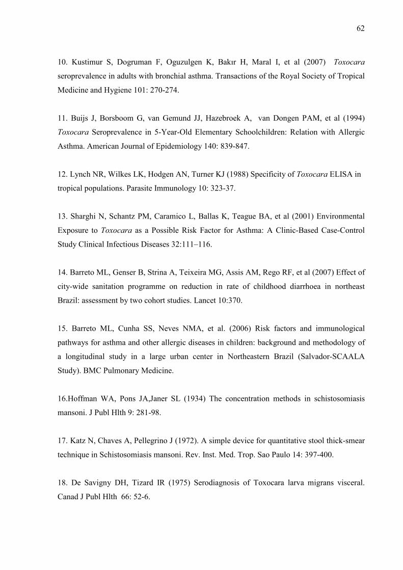
62
10. Kustimur S, Dogruman F, Oguzulgen K, Bakır H, Maral I, et al (2007) Toxocara
seroprevalence in adults with bronchial asthma. Transactions of the Royal Society of Tropical
Medicine and Hygiene 101: 270-274.
11. Buijs J, Borsboom G, van Gemund JJ, Hazebroek A, van Dongen PAM, et al (1994)
Toxocara Seroprevalence in 5-Year-Old Elementary Schoolchildren: Relation with Allergic
Asthma. American Journal of Epidemiology 140: 839-847.
12. Lynch NR, Wilkes LK, Hodgen AN, Turner KJ (1988) Specificity of Toxocara ELISA in
tropical populations. Parasite Immunology 10: 323-37.
13. Sharghi N, Schantz PM, Caramico L, Ballas K, Teague BA, et al (2001) Environmental
Exposure to Toxocara as a Possible Risk Factor for Asthma: A Clinic-Based Case-Control
Study Clinical Infectious Diseases 32:111–116.
14. Barreto ML, Genser B, Strina A, Teixeira MG, Assis AM, Rego RF, et al (2007) Effect of
city-wide sanitation programme on reduction in rate of childhood diarrhoea in northeast
Brazil: assessment by two cohort studies. Lancet 10:370.
15. Barreto ML, Cunha SS, Neves NMA, et al. (2006) Risk factors and immunological
pathways for asthma and other allergic diseases in children: background and methodology of
a longitudinal study in a large urban center in Northeastern Brazil (Salvador-SCAALA
Study). BMC Pulmonary Medicine.
16.Hoffman WA, Pons JA,Janer SL (1934) The concentration methods in schistosomiasis
mansoni. J Publ Hlth 9: 281-98.
17. Katz N, Chaves A, Pellegrino J (1972). A simple device for quantitative stool thick-smear
technique in Schistosomiasis mansoni. Rev. Inst. Med. Trop. Sao Paulo 14: 397-400.
18. De Savigny DH, Tizard IR (1975) Serodiagnosis of Toxocara larva migrans visceral.
Canad J Publ Hlth 66: 52-6.

63
19. Alcantara-Neves NM, Santos AB, Mendonça LR, Figueirêdo CAV, Carvalho LCP (2008).
An improved method to obtain antigen-excreting Toxocara canis. Experimental Parasitology
119: 349-351.
20. Figueiredo CA, Barreto ML, Rodrigues LC, Cooper PJ, Silva NB, Amorim LD,
Alcantara-Neves NM (2010). Chronic intestinal helminth infections are associated with
immune hyporesponsiveness and induction of a regulatory network. Infection and Immunity
78: 20404082.
21. Won KY, Kruszon-Moran D, Schantz PM, Jones JL (2008) National Seroprevalence and
Risk Factors for Zoonotic Toxocara spp. Infection .Am J Trop Med Hyg 79: 552–557
22. Alonso JM, Bojanich MVL, Chamorro M, Gorodner JO (2000) Toxocara Seroprevalence
in Children from a Subtropical City in Argentina. Rev Inst Med trop S Paulo 42: 235-237.
23. Espinoza YA, Huapaya PH, Roldán WH, Jiménez S, Arce Z, Lopez E (2008) Clinical and
serological evidence of Toxocara infection in school children from Morrope district,
Lambayeque, Peru. Rev Inst Med Trop Sao Paulo 50: 101-105.
24. Coelho LMPS, Silva MV, Dini CY, Giacon Neto AA, Novo NF, et al (2004) Human
Toxocariasis: a Seroepidemiological Survey in Schoolchildren of Sorocaba, Brazil. Mem Inst
Oswaldo Cruz Rio de Janeiro 99: 553-7.
25. Damian MM, Martins M, Sardinha JF, Souza LO, Chaves A, et al (2007) Freqüência de
anticorpo anti-Toxocara canis em comunidade do Rio Uatumã, no Estado do Amazonas. Rev
Soc Bras Med Trop 40: 661-664.
26. Magnaval JF, Glickman LT, Dorchies P, Morassin B (2001) Highlights of human
toxocariasis. The Korean Journal of Parasitology 39: 1-11.
27. Pecinali NR, Gomes RN, Amendoeira FC, Augusto CM, Pereira BC, et al (2005)
Influence of murine Toxocara canis infection on plasma and bronchoalveolar lavage fluid
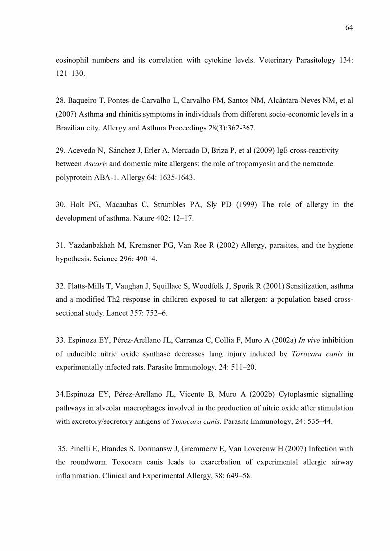
64
eosinophil numbers and its correlation with cytokine levels. Veterinary Parasitology 134:
121–130.
28. Baqueiro T, Pontes-de-Carvalho L, Carvalho FM, Santos NM, Alcântara-Neves NM, et al
(2007) Asthma and rhinitis symptoms in individuals from different socio-economic levels in a
Brazilian city. Allergy and Asthma Proceedings 28(3):362-367.
29. Acevedo N, Sánchez J, Erler A, Mercado D, Briza P, et al (2009) IgE cross-reactivity
between Ascaris and domestic mite allergens: the role of tropomyosin and the nematode
polyprotein ABA-1. Allergy 64: 1635-1643.
30. Holt PG, Macaubas C, Strumbles PA, Sly PD (1999) The role of allergy in the
development of asthma. Nature 402: 12–17.
31. Yazdanbakhah M, Kremsner PG, Van Ree R (2002) Allergy, parasites, and the hygiene
hypothesis. Science 296: 490–4.
32. Platts-Mills T, Vaughan J, Squillace S, Woodfolk J, Sporik R (2001) Sensitization, asthma
and a modified Th2 response in children exposed to cat allergen: a population based cross-
sectional study. Lancet 357: 752–6.
33. Espinoza EY, Pérez-Arellano JL, Carranza C, Collía F, Muro A (2002a) In vivo inhibition
of inducible nitric oxide synthase decreases lung injury induced by Toxocara canis in
experimentally infected rats. Parasite Immunology, 24: 511–20.
34.Espinoza EY, Pérez-Arellano JL, Vicente B, Muro A (2002b) Cytoplasmic signalling
pathways in alveolar macrophages involved in the production of nitric oxide after stimulation
with excretory/secretory antigens of Toxocara canis. Parasite Immunology, 24: 535–44.
35. Pinelli E, Brandes S, Dormansw J, Gremmerw E, Van Loverenw H (2007) Infection with
the roundworm Toxocara canis leads to exacerbation of experimental allergic airway
inflammation. Clinical and Experimental Allergy, 38: 649–58.

65
36. Ferreira MU, Rubinsky-Elefant G, Castro TG, Hoffmann EHE, Silva-Nunes M, Cardoso
MA, Munizd PT (2007) Bottle Feeding and Exposure to Toxocara as Risk Factors for
Wheezing Illness among Under-five Amazonian Children: A Population-based Cross-
sectional Study. Journal of Tropical Pediatrics, 53: 119-24.

66
Legend to Figure 1.
Association between T. canis infection and spontaneous production of IL-10 in whole blood
culture of 1,133 studied children. The results was showed in uni and multivarite logistic
regression (odds ration and confidence interval of 95%). Crude analysis (OR = 1.97 95% CI =
1.29; 3.00); Adjusted* - analysis adjusted by potencial confounders (gender, age, maternal
schooling, parental asthma, mould and sewage system) without helminth infection (OR =
1.84 95% CI = 1.20; 2.83) and Adjusted**-analysis adjusted by all potencial confounders,
including helminths (OR = 1.41 95% CI = 0.91; 2.17).
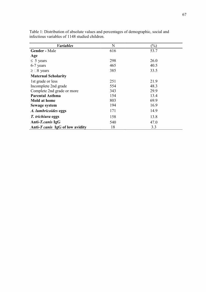
67
Table 1: Distribution of absolute values and percentages of demographic, social and infectious variables of 1148 studied children.
Variables N (%) Gender - Male 616 53.7 Age ≤ 5 years 298 26.0 6-7 years 465 40.5 ≥ 8 years 385 33.5 Maternal Scholarity 1st grade or less 251 21.9 Incomplete 2nd grade 554 48.3 Complete 2nd grade or more 343 29.9 Parental Asthma 154 13.4 Mold at home 803 69.9 Sewage system 194 16.9 A. lumbricoides eggs 171 14.9
T. trichiura eggs 158 13.8 Anti-T.canis IgG 540 47.0 Anti-T canis IgG of low avidity 18 3.3
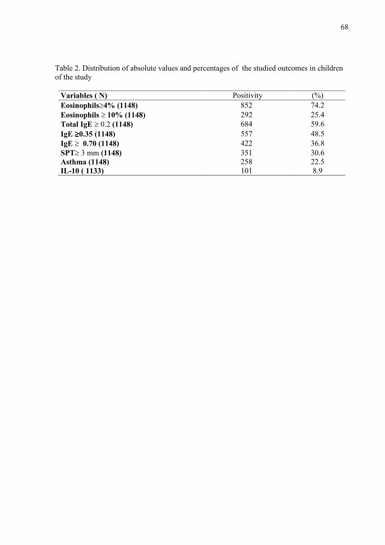
68
Table 2. Distribution of absolute values and percentages of the studied outcomes in children of the study
Variables ( N) Positivity (%) Eosinophils≥4% (1148) 852 74.2 Eosinophils ≥ 10% (1148) 292 25.4
Total IgE ≥ 0.2 (1148) 684 59.6 IgE ≥≥≥≥0.35 (1148) 557 48.5 IgE ≥ 0.70 (1148) 422 36.8 SPT≥ 3 mm (1148) 351 30.6 Asthma (1148) 258 22.5 IL-10 ( 1133) 101 8.9

69
Tabela 3: Association of T. canis infection and total IgE , eosinophilia at 4% and 10% in 1,148 studied children
T .canis
infection
Total IgE Eosinophilia (4%) Eosinophilia (10%)
n (%) *Adjusted OR (95%C.I.)
n (%) Adjusted OR (95%C.I.)
n (%) Adjusted OR (95%C.I.)
Negative 330(48.2) 1 392(46.0) 1 97 (33.2) 1
Positive 354(51.8) 1.45 460(54.0) 2.89 195(66.8) 2.25 (1.12; 1.87) (2.11; 3.94) (1.66; 3.04)
*Adjusted by gender, age, maternal scholarity, parental asthma, mold, sewage system, A.lumbricoides and T. trichiura infections Table 4: Association of T. canis infection with specific IgE for aeroallergens and skin prick test in 1148 studied children T.canis
infection (N=1148)
IgE (≥ 0.35 ) IgE (≥ 0.70 ) Skin prick test
n (%) Crude Adjusted
OR (95%C.I.)
n (%) Crude Adjusted OR
(95%C.I.)
n (%) Crude Adjusted OR
(95%C.I.) Negative 269
(48.3) 1 1 209
(49.5) 1 1 205
(58.4) 1 1
Positive 288 (51.7)
1.44 1.48 213 (50.5)
1.24 1.36 146 (41.6)
0.73 0.79 (1.14;1.82) (1.15 1.91) (0.98;1.58) (1.04; 1.77) (0.56;0.94) (0.60; 1.04)
*Adjusted by gender, age, maternal scholarity, parental asthma, mold, sewage system, A.lumbricoides and T. trichiura
infections. Tabela 5: Association of T. canis infection with specific IgE and skin prick test in 914 children non-infected by intestinal helminths
T. canis
infection
(N=914)
IgE ≥ 0.35 kU/L
IgE ≥ 0.70 kU/L
Skin prick test
n (%) Adjusted OR
(95%CI)
n (%) Adjusted OR
(95%CI)
n (%) Adjusted OR
(95% CI) Negative 191(35.4) 1 243(45.1) 1 187(34.7) 1
Positive
159(42.4)
1.35 (1.2; 1.79)
206(54.9)
1.52 (1.15; 1.99)
108(28.8)
0.73 (0.54; 0.98)
*Adjusted by gender, age, maternal sholarity, parental asthma, mold, sewage system.
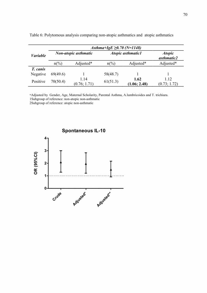
70
Table 6: Polytomous analysis comparing non-atopic asthmatics and atopic asthmatics
Variable
Asthma+IgE ≥0.70 (N=1148)
Non-atopic asthmatic
Atopic asthmatic1
Atopic
asthmatic2
n(%) Adjusted* n(%) Adjusted* Adjusted* T. canis
Negative 69(49.6) 1 58(48.7) 1 1
Positive 70(50.4) 1.14
(0.76; 1.71) 61(51.3)
1.62 (1.06; 2.48)
1.12 (0.73; 1.72)
*Adjusted by Gender, Age, Maternal Scholarity, Parental Asthma, A.lumbricoides and T. trichiura. 1Subgroup of reference: non-atopic non-asthmatic 2Subgroup of reference: atopic non-asthmatic
Spontaneous IL-10
Cru
de
Adju
sted
*
Adju
sted
**
0
1
2
3
4
OR
(95%
CI)

71
5. DISCUSSÃO
A síndrome da Larva Migrans Visceral (LMV) é uma patologia de difícil diagnóstico clínico,
principalmente por apresentar-se na maioria das vezes na forma assintomática ou com sinais
clínicos bem diversificados (HARALAMBIDOU et. al., 2005; GAVIGNET et. al., 2008;
SAPORITO et. al., 2008). Desta forma, o diagnóstico desta patologia é realizado quase que
exclusivamente através da detecção de anticorpos IgG anti-T.canis.
Muitos laboratórios padronizaram ensaios in house para determinar a prevalência desta
zoonose (AGUIAR-SANTOS et al, 2004). O antígeno ESLA do Toxocara canis obtidos em
nosso laboratório possui bandas com peso molecular semelhantes as já padronizadas por
outros autores (RUBINSKY-ELEFANT et. al., 2006; IDDAWELA et. al., 2007) o que
permite a comparação dos nossos resultados com as prevalências obtidas em outros locais do
mundo. A padronização do ensaio pelo nosso laboratório mostrou ser mais sensível quando
comparado com a literatura, uma vez que, a diluição do soro foi de 1:1000 e do anticorpo
conjugado 1:2000, com ponto de corte de 0,23. Outro ponto importante que pode ter
influenciado nessa maior sensibilidade de nosso ensaio foi o fato de termos absorvido (pré-
incubado) os soros com extrato de A. lumbricoides ao invés de A. suum como reportado na
literatura na maioria dos trabalhos (LYNCH et. al., 1988; CAMARGO et al. 1992; NUNES et
al. 1997; CAMPOS JUNIOR et. al., 2003; ROLDÁN et al. 2006). Acreditamos que a
absorção com antígeno de A. lumbricoides é mais eficaz em bloquear os anticorpos que
ligam-se cruzadamente por ser um parasito de humanos.
A absorção dos soros com antígeno de outros parasitos é uma prática que aumenta a
especificidade do ensaio, uma vez que muitas espécies de parasitos compartilham antígenos
72
semelhantes o que ocasiona a reação cruzada entre estes anticorpos Os nossos resultados
mostraram que a absorção com antígeno de A.lumbricoides diminuiu sensivelmente a
Densidade Óptica (D.O.) dos soros, o que tornou o ensaio mais específico. Devido ao fato de
encontrarmos freqüências moderadas de A. lumbricoides (14,9%) e T. trichuira (13,8%) nas
crianças do estudo e os soros terem sido absorvido com extrato somático de A. lumbricoides,
para evitar reação cruzada de IgG, acreditamos que nosso ensaio tem uma boa especificidade.
Além disso, realizamos absorção de alguns soros com antígeno somático de Trichuris
trichiura após a absorção com antígeno de A. lumbricóides e não observamos diminuição
significativa na densidade óptica que justificasse a realização deste procedimento,
principalmente devido à dificuldade de obtenção deste parasito. Os resultados mostraram quea
pré-absorção com A. lumbricóides foi suficiente para evitar a reação cruzada com outros
parasitos (dados não mostrados).
Nunes et al. (1997) determinaram pelo menos uma banda de peso molecular por volta de 55-
66 kDa responsável pela reatividade cruzada entre T. canis e A. suum, e que esta desaparecia
com a pré-absorção dos soros com antígeno de A. suum. Roldán e Espinoza (2009)
determinaram bandas antigênicas reconhecidas por soros de pacientes com toxocaríase de 24,
28, 30, 35, 56, 117, 136 e 152 kDa. Contudo, somente as bandas de 24-35 kDa foram
altamente específicas para infecção por Toxocara (98.3%), enquanto nas outras foram
observadas reações cruzadas com soro de pacientes infectados com outros parasitos.
Em nosso estudo verificamos uma prevalência de IgG anti-T.canis de 48,4%. Won et. al.
(2008) determinaram uma prevalência de 13,9% nos EUA, enquanto estudos conduzidos na
América Latina indicaram uma soroprevalência semelhante aos diagnosticados em nosso
trabalho. Alonso et. al. (2000) reportaram uma positividade de 37,9% em crianças menores de
73
14 anos na Argentina, Espinoza et. al. (2008) determinaram uma soropositividade de 32,4%
no Peru e no Brasil, Coelho et. al (2004) diagnosticaram infecção em 38,3% das crianças
estudadas. A soropositividade para T.canis, em geral, está associada à idade mais avançada
das crianças. Em concordância com nossos resultados, Lescano et. al. (1998) também
diagnosticou uma prevalência maior no grupo de crianças mais velhas. Entretanto, Radman et.
al (2000) observaram maior percentagem de infectados em indivíduos mais novos. Enquanto
que Alderete et.al. (2003) diagnosticaram uma soroprevalência de 38,8% em crianças com
idade média de 9,4 anos. Tem sido demonstrado, portanto, que crianças com idade mais
avançada são mais infectadas que crianças de menor idade, entretanto as referências ainda são
pouco conclusivas e estes achados podem ser conseqüência de um efeito acumulativo, uma
vez que o diagnóstico é sorológico e não se tem dados sobre a curva da queda de anticorpos
IgG anti-T. canis em humanos.
A escolaridade materna foi utilizada neste trabalho como um indicativo da condição sócio-
econômica da família. O resultado da soroprevalência de IgG anti-T.canis encontrado no
presente trabalho assemelham-se aos observados em outras populações de baixa renda, onde a
prevalência da infecção é maior dentre crianças de mães com menor nível de escolaridade.
Estes dados são semelhantes aos determinados por Alderete et.al. (2003) onde a infecção por
Toxocara foi inversamente proporcional a renda familiar.
O contato com cão tem sido apontado em diversos estudos como um importante fator de risco
para a toxocaríase. Um estudo de corte transversal estimou uma freqüência de 52% de
positividade para T.canis em 34 famílias no estado do Amazonas. Dos 55% dos indivíduos
que tinham contato com cão em casa, 60% foram positivos (chi2= 14,317; p = 0,026), e dos
que tinham contato com filhotes de cão em casa, 66,6% foram positivos (chi2= 22,149;

74
p=0,008), o que demonstra a associação entre o contato com o cão e a presença de anticorpos
anti-T.canis (DAMIAN et. al., 2007). Na Argentina, 100 indivíduos foram avaliados quanto à
frequência de anticorpos anti-T. canis e 23% foram positivos, sendo que todos tinham contato
com cão em casa (CHIODO et. al., 2006). Nossos resultados confirmam estes achados sendo
a presença do cão em casa um fator de risco para infecção por T.canis nesta população de
estudo (1.36 [1.07; 1.72])
Diversos trabalhos epidemiológicos apontam a contaminação do solo como determinante na
infecção por T. canis . Diversos trabalhos no Brasil (GUIMARÃES et. al., 2005; ALMEIDA
et. al., 2007; TYIO et. al., 2008) e no mundo (MIZGAJSKA, 1997; DEVERA et. al., 2008;
MARTIN e DEMONTE, 2008) mostram que o solo de áreas públicas como praças, parques,
campings e praia são importantes focos de transmissão. Em nosso trabalho não avaliamos a
contaminação ambiental por ovos de T.canis, mas foi observado que a pavimentação da rua
aumentou a chance de infecção o que nos faz supor que talvez os ovos de T. canis ficaram
mais expostos facilitando a contaminação. Este fato pode ser explicado pelo fato dos ovos
serem pequenos o suficiente para serem soterrados em áreas não pavimentadas dificultando a
contaminação. Além disso, os gatos, que também foram um fator de risco neste estudo,
possam estar contaminando o ambiente com ovos de T. cati, uma vez que com a
pavimentação estes não podem enterrar sua fezes deixando-as expostas. Poucos estudos são
realizados para estimar a infecção de gatos e seu potencial como causador da LMV. Martinez-
Barbabosa et. al. (2003) determinaram, no México, uma prevalência de infecção de T. cati em
gatos de 42,5%, o que nos faz pensar que este parasito possa ser freqüente em nosso meio e
levanta a importância de maiores estudos a cerca da epidemiologia da LMV.
75
Alguns estudos referenciam que a contaminação do solo não é a única rota efetiva na
toxocaríase humana e que os ovos de T. canis podem se embrionar no pêlo de cães, e o
contato direto entre estes e humanos pode ser uma rota alternativa para explicar a
epidemiologia da doença (WOLFE e WRIGHT, 2003). Aydenizoz-Ozkayhan et. al. (2008)
coletou amostras de pêlo de 51 cães e analisou quanto à presença de ovos de T.canis e 21,56%
dos cães tinham ovos nos seus pêlos. Roddie et.al. (2008) examinou 100 cães quanto à
presença de ovos no pêlo através de lavagem. Foi encontrado ovos de Toxocara no pêlo de
67% dos cães, sendo que 95% das amostras eram provenientes de filhotes.
Adicionalmente, acreditamos que os ovos de Toxocara sp. podem ser carregados também em
pêlo de gatos, o que pode explicar em parte a forte associação positiva entre a presença do
gato e a infecção por T. canis, uma vez que a prevalência de gatos foi menor que a de cães e
sua associação foi mais forte que a encontrada para presença de cães em casa. Portanto, este
contato físico tão próximo entre os cães e gatos e seus proprietários possivelmente aumentam
o risco de transmissão do Toxocara, e apontam para a necessidade de maior atenção ao risco
potencial a que os humanos estão expostos (OVERGAAUW et al, 2009).
A infecção por Toxocara tem características peculiares. O parasito não completa seu ciclo
evolutivo, permanecendo no estágio larval por tempo indeterminado no hospedeiro
(MAGNAVAL et al, 2001). Apesar da dificuldade em diagnosticar clinicamente, sinais como
marcada eosinofilia, aumento da IgE sérica e, em alguns casos, desordens pulmonares
similares a asma podem ser observados no curso da doença (DESPOMMIER, 2003).
Claramente este perfil imunológico é resultante do estímulo de células Th2, fenótipo este
associado também à asma atópica e às alergias em geral. Além disso, tem sido observado em

76
camundongos infectados com T.canis aumento de atividade inflamatória e intensa migração
eosinofílica para os pulmões (PECINALI et al, 2005).
Nossos dados sugerem que a infecção por Toxocara esta associada à maior predisposição a
eosinofilia em ambos os pontes de corte >4% e >10%, mesmo quando ajustada para outras
infecções helmínticas. Um estudo conduzido na Argentina determinou uma soroprevalência
de 23% sendo que foi diagnosticado eosinofilia em 86,95% dos indivíduos positivos (p <
0,001, OR = 11,03) e todos tinham contato com cão em casa (CHIODO et. al., 2006). Ainda
na Argentina, Martin et. al. (2008) determinou uma soropositividade em ELISA de 59% para
T. canis sendo que foi mais freqüente na população soropositiva a eosinofilia (p=0,017) e as
dificuldades respiratórias (p=0,05). Espinoza et. al. (2008) determinou uma freqüência de
soropositividade para T. canis de 32,4%, sendo que das crianças positivas 77,96% tinham
sintomas respiratórios, 61,02% manifestações oculares, 38,98% tinham sintomas hepáticos e
38,98% tinham eosinofilia leve a moderada.
Além da eosinofilia, outro sinal da infecção por Toxocara é o aumento de IgE sérica (BUIJS
et al, 1997). Dessa forma, neste trabalho avaliamos a associação entre IgG anti-T. canis e IgE
total e específica para aeroalérgenos. Com relação à IgE específica contra aeroalérgenos
utilizamos dois pontos de corte, o primeiro ≥0.35 kU/L e o segundo ≥0.70 kU/L, sendo este
mais representativo de atopia para nossa população de estudo.
O ponto de corte da IgE especifica contra aeroalérgenos diagnosticada pelo sistema de
immunoCAP da Pharmacia é utilizada mundialmente é ≥0.35 kU/L, ponto este que refere-se
ao nível detectável de IgE que está positivamente associada a atopia nos países desenvolvidos.
Contudo, nos países tropicais e em desenvolvimento, onde há alta prevalência de parasitoses

77
intestinais, este ponto de corte não é o ideal, uma vez que os indivíduos possuem mais IgE
circulante (BAQUEIRO, 2007, COOPER, 2008) e os médicos alergistas utilizam pontos de
corte mais altos para associarem com atopia e manifestações alérgicas. Nossas análises
mostraram que a infecção por T.canis associa-se com positividade a aeroalérgenos em ambos
pontos de corte utilizados o que nos leva a hipotetizar a presença de reações cruzadas de IgE
entre epítopos do T. canis e epítopos de alérgenos ambientais comuns como ácaros e baratas
em ambos pontos de corte. Estas reações já foram descritas para antígenos de A. lumbricoides
e ácaros (ACEVEDO et al, 2009) e entre Anisakis simplex e T. canis (PERTEGUER et al,
2003).
Por outro lado, nossos resultados mostraram que houve associação negativa entre teste
cutâneo (SPT) positivo e infecção por T. canis, mas que esta não foi mantida quando ajustada
para infecções para Ascaris e Trichuris. O ajuste realizado com as variáveis indicativas de
outras infecções helmínticas mostra que estas podem ser, possivelmente, potenciais
confundidoras. Entretanto a análise realizada nas crianças não infectadas com helmintos
intestinais mostrou que infecção por T. canis está realmente associada com a diminuição da
reatividade cutânea a aeroaléregnos. Este efeito sobre o SPT, ou seja, a ausência de
reatividade cutânea apesar dos altos valores de IgE tem sido atribuída dois mecanismsos: a. a
alta produção de IgE policlonal induzida pelo parasito que altera a fase efetora tardia da
resposta Th2 alérgeno-específica saturando os mastócitos e impedindo a ligação cruzada das
IgEs com os alérgenos, também conhecida como “hipótese do bloqueio da IgE” (HOLT et al,
1999; YAZDANBAKHAH et al, 2002) e b. a denominada “resposta Th2 modificada”
induzindo alta produção de IL-10 que suscita a mudança de isótipo para IgG4. Esta
imunoglobulina é reconhecida bloqueadora da reação alérgica, interagindo com os alérgenos e
78
prevenindo a ligação das IgEs presentes nos mastócitos aos mesmos, impedindo a liberação
dos mediadores inflamatórios destas células (PLATTS-MILLS et al, 2001).
Por outro lado, a infecção por T. canis não foi associada à asma na análise multivariada
(OR=1.17, 95%C.I.= 0.86;1.58) mas foi positivamente associada aos fenótipos de asmáticos
atópicos na análise politômica, usando como referência as crianças não atópicas e não
asmáticas (OR=1.62, 95%C.I.=1.06;2,48). Contudo, acreditamos que a análise utilizando
como referência as crianças atópicas não asmáticas seria mais representativa para
observarmos a asma atópica, uma vez que em muitas crianças ambos os fenótipos coexistem,
ou seja, a asma atópica como classificada neste estudo poderia ser a coexistência da atopia e
da asma e não exatamente asma atópica. Portanto os resultados da análise politômica
utilizando o grupo de referência atópicos não asmáticos nos mostrou a infecção por T. canis
associada à asma atópica e, portanto, essa associação positiva se deve somenta á associação
com atopia.
Apesar de ter sido relatado na literatura a ação inflamatória pulmonar causada pelo T. canis
em camundongos (ESPINOZA et al, 2002a,b), estes achados não foram observados em nosso
estudo. Sabe-se que o T. canis induz diferentes padrões clínicos e que estes dependem do
período de infecção (PECINALI et al, 2005). Acreditamos, portanto, que o “timing” da
infecção pode explicar este fato, uma vez que, a minoria de crianças positivos em nossa
população eram de infecções recentes, demonstrado pelo teste de avidez (3,3%). Pinelli et al
(2007) demonstraram que o “timing” afetou a reatividade das vias aéreas e o número de
eosinófilos no BAL de camundongos, sendo que a hiperreatividade só foi significativamente
alta no início da infecção, já em relação à produção de IgE o oposto foi observado, sendo
maior em infecções crônicas. Soma-se a isto o fato da infecção por T. canis estar

79
positivamente associada à produção espontânea de IL-10 em nossa população, o que nos leva
a crer que a infecção crônica por T. canis tem uma capacidade imunomoduladora o que
explica, em parte, a ausência de associação com asma e a negativação do SPT.
80
6. CONCLUSÔES
• A soroprevalência da infecção por Toxocara canis é alta na população de estudo,
devido às baixas condições sócio-econômicas e ambientais.
• Os fatores de risco para a infecção por Toxocara canis estão intimamente relacionadas
a presença da fonte transmissora (cão e gato) e principalmente ao baixo nível de
educação sanitária da população.
• O Toxocara canis é um importante indutor do aumento de eosinófilos e IgE total
sérica em indivíduos soropositivos para esta infecção.
• A infecção por Toxocara canis aumenta os níveis de IgE específica contra
aeroalérgenos, aumentando, consequentemente, a atopia na população.
• A associação negativa entre a infecção pot T. canis e teste cutâneo deve-se
possivelmente à indução policlonal de IgE dificultando a degranulação dos mastócitos
sensibilizados com IgE anti-aeroalérgenos.
• A infecção crônica por Toxocara canis tem uma capacidade imunomoduladora, por
induzir produção de IL-10, e pode ser um possível mecanismo da negativação do teste
cutâneo.

81
REFERÊNCIAS BIBLIOGRÁFICAS
ABBAS, A. K.; LICHTMAN, A. H.; POBER, J.S. Imunologia celular & molecular. 3 ed.
Rio de janeiro: Revinter, 2005. 486P. cap. 4.
ACEVEDO, N.; SÁNCHEZ, J.; ERLER, A.; MERCADO, D.; BRIZA, P.; KENNEDY, M.;
FERNANDEZ, A.; GUTIERREZ, M.; CHUA, K.Y.; CHEONG, N.; JIMÉNEZ, S.; PUERTA,
L.; CARABALLO, L. IgE cross-reactivity between Ascaris and domestic mite allergens: the
role of tropomyosin and the nematode polyprotein ABA-1. Allergy,64: 1635 – 1643, 2009.
AGUIAR-SANTOS, A.M.; ANDRADE, L.D.; MEDEIROS, Z.; CHIEFFI, P.P.; LESCANO,
S.Z.; PEREZ, E.P. Human toxocariasis: frequency of anti-Toxocara antibodies in children and
adolescents from an outpatient clinic for lymphatic filariasis in Recife, Northeast Brazil, Rev
Inst Med Trop São Paulo, 46:81-5, 2004.
ALCÂNTARA-NEVES, N.M.; BAVIA, E.; SILVÃO, R.M.; CARVALHO, E. Environmental
contamination by Toxacara sp eggs in public areas of Salvador,Bahia State, Brazil. Rev Soc
Brasileira de Med Trop, 24: 187-190, 1989.
ALCÂNTARA-NEVES, N.M.; DOS SANTOS, A.B.; MENDONÇA, L.R.; FIGUEIREDO,
C.A.V.; PONTES-DE-CARVALHO, L. An improved method to obtain antigen-excreting
Toxocara canis larvae. Experimental Parasitology, 119: 349–51, 2008.
ALDERETE, J.M.S.; JACOB, C.M.A.; PASTORINO, A.C.; ELEFANT, G.R.; CASTRO,
A.P.M.; FOMIN, A.B.F.; CHIEFFI, P.P. Prevalence of Toxocara infection in schoolchildren
82
for the Butantã, region, São Paulo, Brazil. Memórias do Instituto Oswaldo Cruz, 98: 593-7,
2003.
ALMEIDA, A.B.P.F.; SOUSA, V.R.F.; DALCIN, L.; JUSTINO, C.H.S. Contaminação por
fezes caninas das praças públicas de Cuiabá, Mato Grosso. Braz. J. vet. Res. Anim. Sci., São
Paulo, 44: 132-136, 2007.
ALONSO, J.M.; BOJANICH, M.V.L.; CHAMORRO, M.; GORODNER, J.O. Toxocara
Seroprevalence in Children from a Subtropical City in Argentina. Rev Inst Med trop S
Paulo,42: 235-7, 2000.
ANDERSON, G.P. The immunobiology of early asthma. Med J Aust, 177: 47-9, 2002.
APTER, A.; GRAMMER, L. C.; NAUGHTON, B.; PATTERSON, R. Asthma in the elderly:
a brief report. N. Engl. Allergy Proc., 9:153, 1988.
AYDENIZÖZ-OZKAYHAN, M.; YAGCI, B.B.; ERAT, S. Te investigation of Toxocara
canis eggs in coats of different dog breeds as a potential transmission route in human
toxocariasis. Vet Parasitol, 152: 94-0, 2008.
BAQUEIRO, T.; PONTES-DE-CARVALHO, L.; CARVALHO, F.M.; SANTOS,
N.M.; ALCÂNTARA-NEVES, N.M.; Medical Student´s Group. Asthma and rhinitis
symptoms in individuals from different socio-economic levels in a Brazilian city. Allergy
and Asthma Proceedings 28:362-367, 2007.
83
BARRETO, M.L.; GENSER, B.; STRINA, A.; TEIXEIRA, M.G.; ASSIS, A.M.; REGO,
R.F.; et al. Effect of city-wide sanitation programme on reduction in rate of childhood
diarrhoea in northeast Brazil: assessment by two cohort studies. Lancet, 10: 370, 2007.
BARRETO, M.L.; CUNHA, S.S.; NEVES, N.M.A.; et al. Risk factors and immunological
pathways for asthma and other allergic diseases in children: background and methodology of
a longitudinal study in a large urban center in Northeastern Brazil (Salvador-SCAALA
Study). BMC Pulmonary Medicine, 2006.
BLACK, P. Why is the prevalence of asthma and autoimmunity increasing? Trends
Immunol, 22: 354–355, 2001.
BUIJS, J.; BORSBOOM, G.; RENTING, M.; HILGERSOM, W.J.A.; VAN WIERINGEN,
J.C.; JANSEN, G.; NEIJENS, J. Relationship between allergic manifestations and Toxocara
seropositivity: a cross-sectional study among elementary school children. Eur Respir J, 10:
1467–75, 1997.
BUIJS, J.; BORSBOOM, G.; VAN GEMUND, J.J.; HAZEBROEK, A.; VAN DONGEN,
P.A.M, VAN KNAPEN, F.; NEIJENS, H.J. Toxocara Seroprevalence in 5-Year-Old
Elementary Schoolchildren: Relation with Allergic Asthma. American Journal of
Epidemiology, 140: 839-847, 1994.
BUSSE, W.W.; LEMANSKE JUNIOR, R.F. Asthma. N. Engl. J. Med., 344:350-362, 2001.
84
CAMARGO, E.D.; NAKAMURA, P.M.; VAZ, A.J.; SILVA, M.V.; CHIEFFI, P.P.; MELO,
E.O. Standadization of Dot-ELISA for the Serological Diagnosis of Toxocariasis and
Comparison of the Assay with ELISA. Rev. Med. Inst. trop. São Paulo, 34: 55-60, 1992.
CAMPOS-FILHO, P.C.; BARROS, L.M.; CAMPOS, J.O.; BRAGA, V.B.; CAZORLA, I.M.;
ALBUQUERQUE, G.R.; CARVALHO, S.M. Zoonotic parasites in dog feces at public
squares in the municipality of Itabuna, Bahia, Brazil. Rev Bras Parasitol Vet, 17:206-9,
2008.
CAMPOS JUNIOR, D.; ELEFANT, G.R.; SILVA, E.O.M.; GANDOLFI, L.; JACOB,
C.M.A.; TOFETI, A.; PRATESI, R. Freqüência de Soropositividade para Antígenos de
Toxocara canis em Crianças de Classes Sociais Diferentes. Revista da Sociedade Brasileira
de Medicina Tropical, 36: 509-13, 2003.
CHAN, P.W.K.; ANUAR, A.K.; FONG, M.Y.; DEBRUYNE, J.A.; IBRAHIM, J. Toxocara
seroprevalence and childhood asthma among Malaysian Children. Pediatrics International
43: 350–353, 2001.
CHEN, Y.; BLASER, M.J. Helicobacter pylori Colonization Is Inversely Associated with
Childhood Asthma. The Journal of Infectious Diseases, 198: 1-8, 2008.
CHIEFFI, P.P.; SANTOS, S.V.; QUEIROZ, M.L.; LESCANO, S.A.Z. Human Toxocariasis:
Contribution by Brazilian Researchers. Rev. Inst. Med. trop. S. Paulo, 51: 301-8, 2009.

85
CHIODO, P.; BASUALDO, J.; CIARMELA, L.; PEZZANI, B.; APEZTEGUÍA, M.;
MINVIELLE, M. Related factors to human toxocariasis in a rural community of Argentina.
Mem Inst Oswaldo Cruz, 101:397-400, 2006.
CHOMEL, B.B.; KASTEN, R.; ADAMS, C.; LAMBILLOTTE, D.; THEIS, J.;
GOLDSMITH, R.; KOSS, J.; CHIOINO, C.; WIDJANA, D.P.; SUTISNA, P. Serosurrvey of
some major zoonotic infections in children an teenagers in Bali, Indonesia, South East Asian
J. Trop. Med. Pub. Health, 24: 321-326, 1993.
COELHO L.M.P.S.; SILVA, M.V.; DINI, C.Y.; GIACON NETO, A.A.; NOVO, N.F.;
SILVEIRA, E.P.R. Human Toxocariasis: a Seroepidemiological Survey in Schoolchildren of
Sorocaba, Brazil. Mem Inst Oswaldo Cruz Rio de Janeiro, 99: 553-7, 2004.
COOPER, P.J.; CHICO, M.E.; RODRIGUES, L.C.; ORDONEZ, M.; STRACHAN, D.;
GRIFFIN, G.E.; NUTMAN, T.B. Reduced risk of atopy among school-age children infected
with geohelminth parasites in a rural area of the tropics. Environmental and Occupacional
Disordens, 995-1000, 2003.
COOPER, P.J. Interactions between helminth parasites and allergy. Curr Opin Allergy Clin
Immunol, 9: 29–37, 2009.
COOPER, P.J. Toxocara canis infection: an important and neglected environmental risk factor
for asthma? Clinical and Experimental Allergy, 38: 551–553, 2008.
86
COSTA-CRUZ, J. M.; NUNES, R. S.; BUSO, A. G. Presença de ovos de Toxocara spp. em
praças públicas da cidade de Uberlândia, Minas Gerais, Brasil. Revista do Instituto de
Medicina Tropical de São Paulo, 36: 39-42, 1994.
DAMIAN, M.M.; MARTINS, M.; SARDINHA, J.F.; SOUZA, L.O.; CHAVES, A.;
TAVARES, M. Freqüência de anticorpo anti-Toxocara canis em comunidade do Rio Uatumã,
no Estado do Amazonas. Rev Soc Bras Med Trop, 40:661-4, Nov-Dec, 2007.
DE SAVIGNY, D.H.; TIZARD, I.R. Serodiagnosis of Toxocara larva migrans visceral.
Canad J Publ Hlth, 66: 52-6, 1975.
DESPOMMIER, D. Toxocariasis: Clinical Aspects, Epidemiology, Medical Ecology, and
Molecular Aspects. Clinical Microbiology Reviews, 16: 265–72, 2003.
DEVERA, R.; BLANCO, Y.; HERNÁNDEZ, H.; SIMOES, D. Toxocara spp. and other
helminths in squares and parks of Ciudad Bolívar, Bolivar State (Venezuela). Enferm Infecc
Microbiol Clin, 26:23-6, 2008.
DZIEMIAN, E.; ZARNOWSKA, H.; KOLODZIEJ-SOBOCIÑSKA, M.; MACHNICKA, B.
Determination of the relative avidity of the specific IgG antibodies in human toxocariasis.
Parasite Immunology, 30: 187–90, 2008.
ESPINOZA, E.Y.; PÉREZ-ARELLANO, J.L.; CARRANZA, C.; COLLÍA, F.; MURO, A. In
vivo inhibition of inducible nitric oxide synthase decreases lung injury induced by Toxocara
canis in experimentally infected rats. Parasite Immunology, 24: 511–520, 2002a.
87
ESPINOZA, E.Y.; PÉREZ-ARELLANO, J.L.; VICENTE, B.; MURO, A. Cytoplasmic
signalling pathways in alveolar macrophages involved in the production of nitric oxide after
stimulation with excretory/secretory antigens of Toxocara canis. Parasite Immunology, 24:
535–544, 2002b.
ESPINOZA, Y.A.; HUAPAYA, P.H.; ROLDÁN, W.H.; JIMÉNEZ, S.; ARCE, Z.; LOPEZ,
E. Clinical and serological evidence of Toxocara infection in school children from Morrope
district, Lambayeque, Peru. Rev Inst Med Trop Sao Paulo, 50: 101-5, 2008.
FALLON, P.G.; MANGAN, N.E. Suppression of TH2-type allergic reactions by helminth
infection. Nature Reviews Immunology, 7: 220-30, 2007.
FERNANDO, S.D.; WICKRAMASINGHE, V.P.; KAPILANANDA, G.M.;
DEVASURENDRA, R.L.; AMARASOORIYA, J.D.; DAYARATNE, H.G. Epidemiological
aspects and risk factors of toxocariasis in a pediatric population in Sri Lanka. Southeast
Asian J Trop Med Public Health, 38:983-90, 2007.
FERREIRA, M.U.; RUBINSKY-ELEFANT, G.; CASTRO, T.G.; HOFFMANN, E.H.E.;
SILVA-NUNES, M.; CARDOSO, M.A.; MUNIZD, P.T. Bottle Feeding and Exposure to
Toxocara as Risk Factors for Wheezing Illness among Under-five Amazonian Children: A
Population-based Cross-sectional Study. Journal of Tropical Pediatrics, 53: 119-24, 2007.
FIGUEIREDO, C.A.; BARRETO, M.L.; RODRIGUES, L.C.; COOPER, P.J.; SILVA, N.B.;
AMORIM, L.D.; ALCANTARA-NEVES, N.M. Chronic intestinal helminth infections are

88
associated with immune hyporesponsiveness and induction of a regulatory network. Infection
and Immunity, 78: 2040-82, 2010.
FRANCO, H.F.; PRITCHARD, D.I. Parasite role reversal: worms on trial. Trends Parasitol,
21:157-160, 2005.
GALE, E.A.M. A missing link in the hygiene hypothesis? Diabetologia 45: 588–594, 2002.
GAVIGNET, B.; PIARROUX, R.; AUBIN, F.; MILLON, L.; HUMBERT, P. Cutaneous
manifestations of human toxocariasis. J Am Acad Dermatol, 59:1031-1042, 2008.
GELL, P.G.H.; COOMBS, R.R.A. eds. Clinical Aspects of Immunology. 1st ed. Oxford,
England: Blackwell; 1963
GUIMARÃES, A.M.; ALVES, E.G.L.; REZENDE, G.F.; RODRIGUES, M.C. Ovos de
Toxocara sp. e larvas de Ancylostoma sp. em praça pública de Lavras, MG . Rev Saúde
Pública, 39:293-5, 2005.
HARALAMBIDOU, S.; VLACHAKI, E.; IOANNIDOU, E.; MILIONI, V.;
HARALAMBIDIS, S.; KLONIZAKIS, I. Pulmonary and myocardial manifestations due to
Toxocara canis infection. European Journal of Internal Medicine, 16: 601 – 602, 2005.
HOFFMAN, W.A.; PONS, J.A.; JANER, S.L. The concentration methods in schistosomiasis
mansoni. J Publ Hlth, 9: 281-98, 1934.

89
HOLGATE, S.T. The epidemic of allergy and asthma. Nature, 402, 1999.
HOLT, P.G.; JAMES, C.A. The development of the immune system during pregnancy and
early life. Allergy, 55: 688–697, 2000.
HOLT, P.G.; MACAUBAS, C.; STRUMBLES, P.A.; SLY, P.D. The role of allergy in the
development of asthma. Nature, 402: 12–7, 1999.
HOTEZ, P.J.; WILKINS, P.P. Toxocariasis: America’s Most Common Neglected Infection of
Poverty and a Helminthiasis of Global Importance? PLOS Neglected Tropical Disease, 3: 1-
4, 2009.
IDDAWELA, R.D.; RAJAPAKSE, R.P.V.J.; PERERA, N.A.N.D.; AGATSUMA, T.
Characterization of a Toxocara canis species-specific excretory-secretory antigen (TcES-57)
and development of a double sandwich ELISA for diagnosis of visceral larva migrans.
Korean Journal of Parasitology, 45: 19-26, 2007.
ISAAC - The International Study of Asthma and Allergies in Childhood. Steering Committee.
Worldwide variation in prevalence of symptoms of asthma, allergic rhinoconjunctivitis, and
atopic eczema: ISAAC. The Lancet, 351: 1225-32, 1998.
ISHIDA, M.M.I.; RUBINSKY-ELEFANT, G.; FERREIRA, A.W.; HOSHINO-SHIMIZU, S.;
VAZ, A.J. Helminth antigens (Taenia solium, Taenia crassiceps, Toxocara canis,
Schistosoma mansoni and Echinococcus granulosus) and cross-reactivities in human
infections and immunized animals. Acta Tropica, 89: 73–84, 2003.
90
JACOB, C.M.A.; OSELKA, G.W. Toxocaríase na infância. Revisões e Ensaios,48-55, 1991.
JANEWAY, C. A.; TRAVERS, P.; WALPORT, M.; CAPRA, J. D. Imunobiologia. O
sistema imunológico na saúde e na doença. 6 ed. Porto Alegre: Artes Médicas Sul LTDA,
2007.
JANSON, C.; ASBJORNSDOTTIR, H.; BIRGISDOTTIR, A.; SIGURJONSDOTTIR, R.B.;
GUNNBJÖRNSDOTTIR, M.; GISLASON, D.; OLAFSSON, I.; COOK, E.; JOÖGI, R.;
GISLASON, T.; THJODLEIFSSON, B. The effect of infectious burden on the prevalence of
atopy and respiratory allergies in Iceland, Estonia, and Sweden. J. Allergy Clin. Immunol.
120(3):673-9, 2007.
KATZ, N.; CHAVES, A.; PELLEGRINO, J. A simple device for quantitative stool thick-
smear technique in Schistosomiasis mansoni. Rev. Inst. Med. Trop. Sao Paulo, 14: 397-400,
1972.
KENNEDY, M.W.; MAIZELS, R.M.; MEGHJI, M.; YOUNG, L.; QURESHI, F.; SMITH,
H.V. Species-specific carbohydrate epitopes on the secreted and surface antigens of Toxocara
cati and Toxocara canis infective larvae. Parasite Immunol, 9: 407-420, 1987.
KON, O.M .; SIHRA, B.S.; COMPTON, C.H.; LEONARD, T.B.; KAY, A.B.; BARNES,
N.C. Randomised, dose-ranging, placebo-controlled study of chimeric antibody to CD4
(keliximab) in chronic severe asthma. Lancet, 352: 1109-1113, 1998.

91
KUSTIMUR, S.; DOGRUMAN, F.; OGUZULGEN, K.; BAKIR, H.; MARAL, I.;
TURKTAS, H.; TUZUN, H. Toxocara seroprevalence in adults with bronchial asthma.
Transactions of the Royal Society of Tropical Medicine and Hygiene, 101: 270-274, 2007.
LAEMMLI, U.K. Cleavage of structural proteins during the assembly of the head of
bacteriophage T4. Nature, 227: 680-685, 1970.
LEFORT, J.; SINGER, M.; LEDUC, D.; RENESTO, P.; NAHORI, M. A.; HUERRE, M.;
CREMINON, C.; CHIGNARD, M.; VARGAFTIG, B. B. Systemic administration of
endotoxin induces bronchopulmonary hyperreactivity dissociated from TNF-alpha formation
and neutrophil sequestration into the murine lungs. J. Immunol., 161:474-480,1998.
LESCANO, S.A.Z.; CHIEFFI, P.P.; PERES, B.A.; DE MELLO, E.O.; VELARDE, C.N.;
SALINAS, A.AP.; ROJAS, C.E. Soil Contamination and Human Infection by Toxocara sp. in
the Urban Area of Lima, Peru. Mem Inst Oswaldo Cruz, Rio de Janeiro, 93: 733-734,
1998.
LOKMAN-HAKIM, S.; MAK, J. W.; LAM, P.L. Seropositivity for Toxocara canis
antibodies in Malaysia 1989-1991, Med. J. Malaysia, 48: 303-307, 1993.
LYNCH, N.R.; WILKES, L.K.; HODGEN, A.N.; TURNER, K.J. Specificity of Toxocara
ELISA in tropical populations. Parasite Immunology, 10: 323-37, 1988.

92
LYNCH, N.R.; HAGEL, I.; VARGAS, V.; ROTUNDO, A.; VARELA, M.C.; DI PRISCO,
M.C.; HODGEN, A.N. Comparable seropositivity for ascariasis and toxocariasis in tropical
slum children, Parasitol Res. 79(7):547-50, 1993.
MAGNAVAL, J.F.; GLICKMAN, L.T.; DORCHIES, P.; MORASSIN, B. Highlights of
human toxocariasis. The Korean Journal of Parasitology, 39: 1-11, 2001.
MARTIN, U.O.; DEMONTE, M.A.. Urban Contamination with Zoonotic Parasites in the
Central Region of Argentina. MEDICINA (Buenos Aires), 68: 363-366, 2008.
MARTIN, U.O.; MACHUCA, P.B.; DEMONTE, M.A.; CONTINI, L. Estudio En Niños Con
Diagnostico Presuntivo De Toxocariasis En Santa Fe, Argentina. MEDICINA (Buenos
Aires), 68: 353-357, 2008.
MARTÍNEZ-BARBABOSA, I.; TSUJI, O.V.; CABELLO, R.R.; CÁRDENAS, E.M.G.;
CHASIN, O.A.. The prevalence of Toxocara cati in domestic cats in Mexico City.
Veterinary Parasitology, 114: 43–49,2003.
MATRICARDI PM et al. Allergic asthma: epidemiological study versus airborne viruses in
relation to atopy and Exposure. BMJ 2000;320;412-7
McCONCHIE, B.W.; NORRIS, H.H.; BUNDOC, V.G.; TRIVEDI, S.; BOESEN, A.;
URBAN, J.F.; KEANE-MYERS, A.M. Ascaris suum-Derived Products Suppress Mucosal
Allergic Inflammation in an Interleukin-10-Independent Manner via Interference with
Dendritic Cell Function. Infection And Immunity,74: 6632–6641, 2006.
93
MEDEIROS JR., M.; FIGUEIREDO, J. P.; ALMEIDA, M. C.; MATOS, M. A.; PINHO, R.
S.; AMORIM, W. W. C. C.; CARVALHO, E. M.; CRUZ, A. A.; LOPES, A. A.; ATTA, A.
M.; ARAÚJO, M. Prevalência de alergia respiratória em indivíduos de área endêmica de
Schistosoma mansoni. Rev. Bras. Alergia Imunopatol., 23:036, 2000.
MIZGAJSKA, H. The role of some environmental factors in the contamination of soil with
Toxocara spp. and other geohelminth eggs. Parasitology International, 46: 67-72, 1997.
MURPHY, K. M.; REINER, S. L.The lineage decisions of helper T cells. Nat. Rev.
Immunol., 2: 933-944, 2002.
NOORDIN, R.; SMITH H.V.; MOHAMAD, S.; MAIZELS, R.M.; FONG, M.Y. Comparison
of IgG-ELISA and IgG4-ELISA for Toxocara serodiagnosis, Acta Tropical, 93:57-62, 2005.
NUNES CM, TUNDISI RN, GARCIA JF, HEINEMANN MB, OGASSAWARA S,
RICHTZENHAIN LJ. Cross-Reactions Between Toxocara Canis And Ascaris Suum In The
Diagnosis Of Visceral Larva Migrans By Western Blotting Technique. Rev. Inst. Med. trop.
S. Paulo 39, 1997.
OVERGAAUW, P.A.M.; ZUTPHEN, L.; HOEK, D.; YAYA, F.O.; ROELFSEMA, J.;
PINELLI, E.; KNAPEN, F.; KORTBEEK, L.M. Zoonotic parasites in fecal samples and fur
from dogs and cats in The Netherlands. Vet Parasitol, 163: 115–22, 2009.

94
PECINALI, N.R; GOMES, R.N.; AMENDOEIRA, F.C.; AUGUSTO C.M. PEREIRA
BASTOS C, MARTINS, M.J.Q.A.; PEGADO, C.S.; PEREIRA BASTOS, O.M.;. BOZZA,
P.T.; CASTRO-FARIA-NETO, H.C. Influence of murine Toxocara canis infection on plasma
and bronchoalveolar lavage fluid eosinophil numbers and its correlation with cytokine levels.
Veterinary Parasitology, 134: 121–130, 2005.
PERTEGUER, M.J.; CUÉLLAR, C.; GUILLÉN, J.L.; ÁGUILA, C.; FENOY, S.; CHIVATO,
T.; LAGUNA, R. Cross-reactivity between Anisakis simplex sensitization and visceral larva
migrans by Toxocara canis. Acta Tropica, 89: 85–89, 2003.
PINELLI, E.; BRANDES, S.; DORMANSW, J.; GREMMERW, E.; VAN LOVERENW, H.
Infection with the roundworm Toxocara canis leads to exacerbation of experimental allergic
airway inflammation. Clinical and Experimental Allergy, 38: 649–658, 2007.
PLATTS-MILLS, T. A.; BLUMENTHAL, K.; PERZANOWSKI, M.; WOODFOLK, J. A.
Determinants of clinical allergic disease. The relevance of indoor allergens to the increase in
asthma. Am. J. Respir. Crit. Care Med, 162:128-133, 2000.
PLATTS-MILLS, T.; VAUGHAN, J.; SQUILLACE, S.; WOODFOLK, J.; SPORIK, R.
Sensitization, asthma and a modified Th2 response in children exposed to cat allergen: a
population based cross-sectional study. Lancet 357: 752–6, 2001.
RADMAN, N.E.; ARCHELLI, S.M.; FONROUGE, R.D.; GUARDIS, M.V.; LINZITTO,
O.R. Human Toxocarosis. Its Seroprevalence in the City of La Plata. Mem Inst Oswaldo
Cruz Rio de Janeiro, 95: 281-5, 2000.

95
RODDIE, G.; STAFFORD, P.; HOLLAND, C.; WOLFE, A. Contamination of dog hair with
eggs of Toxocara canis. Vet Parasitol, 152: 85–93, 2008
ROLDÁN, W.; CORNEJO, W.; ESPINOZA, Y. Evaluation of the dot enzyme-linked
immunosorbent assay in comparison with standard ELISA for the immunodiagnosis of human
toxocariasis. Mem Inst Oswaldo Cruz Rio de Janeiro, 101: 71-4, 2006.
ROLDÁN, W.H.; ESPINOZA, Y.A. Evaluation of an enzyme-linked immunoelectrotransfer
blot test for the confirmatory serodiagnosis of human toxocariasis. Mem Inst Oswaldo Cruz,
Rio de Janeiro, 104: 411-8, 2009.
ROMAGNANI, S. The increased prevalence of allergy and the hygiene hypothesis: missing
immune deviation, reduced immune suppression, or both? Immunology, 112: 352–363,
2004.
RUBINSKY-ELEFANT, G.; SHIMIZU, S.H.; SANCHEZ, M.C.A.; JACOB, C.M.A.;
FERREIRA, A.W. A Serological Follow-up of Toxocariasis Patients After Chemotherapy
Based on the Detection of IgG, IgA, and IgE Antibodies by Enzyme-Linked Immunosorbent
Assay. Journal of Clinical Laboratory Analysis, 20: 164–72, 2006.
SAPORITO, L.; SCARLATA, F.; COLOMBA, C.; INFURNARI, L.; GIORDANO, S.;
TITONE, L. Human toxocariasis: a report of nine cases. Acta Paediatr, 97:1301-2, 2008.
96
SCHULZ, O.; SEWELL, H.F.; SHAKIB, F. The interaction between the dust mite antigen
Der P1 and cell signalling molecules in amplifying allergic disease. Clin. Exp. Allergy 29:
439-444, 1999.
SHARGHI, N.; SCHANTZ, P.M.; CARAMICO, L.; BALLAS, K.; TEAGUE, B.A.; HOTEZ,
P.J. Environmental Exposure to Toxocara as a Possible Risk Factor for Asthma: A Clinic-
Based Case-Control Study. Clinical Infectious Diseases, 32: 111–6, 2001.
SOLE, D.; CAMELO-NUNES, I. C.; VANA, A.T.; YAMADA, E.; WERNECK, F.; DE
FREITAS, L. S.; SOLOGUREN, M. J.; BRITO, M.; ROSARIO, F. N.A.; STEIN, R.T.;
NASPITZ, C. K. Prevalence of rhinitis and related-symptoms in schoolchildren from different
cities in Brazil. Allergol . Immunopathol., 32:7-12, 2004.
SOWEMIMO, A.O. The prevalence and intensity of gastrointestinal parasites of dogs in Ile-
Ife, Nigeria. J Helminthol, 83:27-31, 2009.
STRACHAN, D.P. Hay fever, hygiene and household size. BMJ, 299:1259-1260, 1989.
STRINA, A.; CAIRNCROSS, S.; BARRETO, M.L.; LARREA, C.; PRADO, M.S. Childhood
diarrhea and observed hygiene behavior in Salvador, Brazil. Am J Epidemiol, 157: 1032–8,
2003.
TEIXEIRA, C.R.; CHIEFFI, P.P.; LESCANO, S.A.; DE MELO SILVA E.O.; FUX, B.;
CURY, M.C. Frequency and risk factors for toxocariasis in children from a pediatric
outpatient center in southeastern Brazil. Rev Inst Med Trop Sao Paulo,48:251-5, 2006.
97
THEODORIDIS, I.; FRYDAS, S.; PAPAZAHARIADOU, M.; HATZISTILIANOU, M.;
ADAMAMA-MORAITOU, K.K.; DI GIOACCHINO, M.; FELACO, M. Toxocarosis as
zoonosis. A review of literature and the prevalence of Toxocara canis antibodies in 511 serum
samples, Int J Immunopathol Pharmacol.,14:17-23, 2001.
TIYO, R.; GUEDES, T.A.; FALAVIGNA, D.L.; FALAVIGNA-GUILHERME, A.L.
Seasonal contamination of public squares and lawns by parasites with zoonotic potential in
southern Brazil. J Helminthol, 82:1-6, 2008.
VON MUTIUS, E.; WEILAND, S.K.; FRITZSCH, C.; DUHME, H.; KEIL, U. Increasing
prevalence of hay fever and atopy among children in Leipzig, East Germany. Lancet, 351:
862–866, 1998.
WARNER, J.O. The early life origins of asthma and related allergic disorders. Arch. Dis.
Child., 89:97-102, 2004.
WILLS-KARP, M.; SANTELIZ, J.; KARP, C.L. The germless theory of allergic disease:
revisiting the hygiene hypothesis. Nat Rev Immunol, 1: 69–75, 2001.
WOLFE, A.; WRIGHT, I.P. Human toxocariasis and direct contact with dogs. Vet Rec, 152:
419–22, 2003.

98
WON, K.Y.; KRUSZON-MORAN, D.; SCHANTZ, P.M.; JONES, J.L. National
Seroprevalence and Risk Factors for Zoonotic Toxocara spp. Infection. Am. J. Trop. Med.
Hyg., 79: 552–557, 2008.
WONG, G. W. K.; HUI, D. S. C.; CHAN, H. H.; FOK, T. F.; LEUNG, R.; ZHONG, N. S.
Prevalence of respiratory and atopic disorders in Chinese schoolchildren. Clin. Exp. Allergy,
31:1225-12231, 2001.
YAZDANBAKHAH, M.; KREMSNER, P.G.; VAN REE, R. Allergy, parasites, and the
hygiene hypothesis. Science, 296: 490–4, 2002.
YAZDANBAKHSH, M.; VAN DEN BIGGELAAR, A.H.J.; MAIZELS, R.M. Th2 responses
without atopy: immunoregulation in chronic helminth infections and reduced allergic disease.
Trends Immunol, 22: 372–377, 2001.
YONG-HUN, K.; SUN H.; YOUNG-BAE, C. Seroprevalence of Toxocariasis among Healthy
People with Eosinophilia. Korean J Parasitol, 46: 29-32, 2008.