Embed Size (px)
Citation preview
UNIVERSIDADE FEDERAL DO CEARAacute
FACULDADE DE FARMAacuteCIA ODONTOLOGIA E ENFERMAGEM
POacuteS-GRADUACcedilAtildeO EM ODONTOLOGIA
GUILHERME DE ALENCAR TEMOacuteTEO
ANAacuteLISE DA CONTAMINACcedilAtildeO MICROBIANA DE DISPOSITIVOS
ACRIacuteLICOS CONFECCIONADOS EM LABORATOacuteRIOS DE PROacuteTESE
DENTAacuteRIA
FORTALEZA
2014
1
GUILHERME DE ALENCAR TEMOacuteTEO
ANAacuteLISE DA CONTAMINACcedilAtildeO MICROBIANA DE DISPOSITIVOS
ACRIacuteLICOS CONFECCIONADOS EM LABORATOacuteRIOS DE PROacuteTESE
DENTAacuteRIA
FORTALEZA
2014
Dissertaccedilatildeo apresentada ao Programa de Poacutes-Graduaccedilatildeo em Odontologia da Faculdade de Farmaacutecia Odontologia e Enfermagem da Universidade Federal do Cearaacute como um dos requisitos para a obtenccedilatildeo do tiacutetulo de Mestre em Odontologia Aacuterea de Concentraccedilatildeo Cliacutenica Odontoloacutegica Orientadora Profa Dra Karina de Matthes de Freitas Pontes
2
Dados Internacionais de Catalogaccedilatildeo na Publicaccedilatildeo Universidade Federal do Cearaacute Biblioteca de Ciecircncias da Sauacutede
T278a Temoacuteteo Guilherme de Alencar
Anaacutelise da contaminaccedilatildeo microbiana de dispositivos acriacutelicos confeccionados em laboratoacuterios de proacutetese dentaacuteria Guilherme de Alencar Temoacuteteo ndash 2014
51 f il color enc 30 cm Dissertaccedilatildeo (Mestrado) ndash Universidade Federal do Cearaacute Faculdade de
Farmaacutecia Odontologia e Enfermagem Programa de Poacutes-Graduaccedilatildeo em Odontologia Mestrado em Odontologia Fortaleza 2014
Aacuterea de Concentraccedilatildeo Cliacutenica Odontoloacutegica Orientaccedilatildeo Profa Dra Karina de Matthes de Freitas Pontes 1 Resinas Acriacutelicas 2 Proacutetese Dentaacuteria 3 Contaminaccedilatildeo 3 Desinfecccedilatildeo 4
Esterilizaccedilatildeo I Tiacutetulo
CDD 617695
3
GUILHERME DE ALENCAR TEMOacuteTEO
ANAacuteLISE DA CONTAMINACcedilAtildeO MICROBIANA DE DISPOSITIVOS
ACRIacuteLICOS CONFECCIONADOS EM LABORATOacuteRIOS DE PROacuteTESE
DENTAacuteRIA
Dissertaccedilatildeo apresentada ao Programa de Poacutes-Graduaccedilatildeo em Odontologia da Faculdade de Farmaacutecia Odontologia e Enfermagem da Universidade Federal do Cearaacute como um dos requisitos para a obtenccedilatildeo do tiacutetulo de Mestre em Odontologia
Aprovada em 27022014
BANCA EXAMINADORA
______________________________________________________________
Profa Dra Karina Matthes de Freitas Pontes (Orientadora)
Faculdade de Farmaacutecia Odontologia e Enfermagem ndash UFC
_______________________________________________________________
Prof Dr Marcus Aureacutelio Rabelo Lima Verde
Faculdade de Farmaacutecia Odontologia e Enfermagem ndash UFC
_______________________________________________________________
Prof Dr Eduardo Diogo Gurgel Filho
Curso de Odontologia - UNIFOR
4
AGRADECIMENTOS
A Deus por me iluminar com todo seu amor em todos os momentos
me fazendo ter dicernimento diante dos desafios da vida
Aos meus queridos pais Hermes e Celina por me proporcionarem uma
educaccedilatildeo de qualidade e me ensinarem valores morais como honestidade e
respeito Sou eternamente grato por todo amor e dedicaccedilatildeo
Agrave minha amada esposa Carol que sempre esteve ao meu lado em
todos os desafios me ajudando e incentivando
5
AGRADECIMENTOS ESPECIAIS
Aacute Profordf Drordf Karina Matthes de Freitas Pontes uma pessoa fantaacutestica
que une doccedilura e competecircncia numa soacute pessoa Chamou-me a atenccedilatildeo
quando foi preciso mas sempre me elogiou quando eu mereci Serei sempre
grato por todos os ensinamentos
Ao Carlos Eduardo Albuquerque pela amizade e pela forccedila para que
eu fizesse a seleccedilatildeo do mestrado Agrave Bruna Frota por toda disponibilidade e
boa vontade em ajudar nas pesquisas
Agrave Bruninha Albuquerque pela amizade e por ser meu braccedilo direito
durante toda a pesquisa Agrave Camila Caracas por natildeo medir esforccedilos para me
ajudar Agrave Naacutedia Dantas pelo apoio no inicio da pesquisa
Agrave minha turma de mestrado pela parceria durante essa caminhada
Em especial agrave Ximena Treacutevia por ter me ajudado durante vaacuterios momentos no
mestrado
Agrave Ramille Lima e David Queiroz por me orientarem muitas vezes
durante o periacuteodo de realizaccedilatildeo dos experimentos microbioloacutegicos
Ao Paulinho Goberlanio por todo o apoio e dedicaccedilatildeo na parte da
estatiacutestica
Agrave toda equipe de professores da Proacutetese Fixa aleacutem da Malu
Gonzaga e Janaina Cacircncio pelos bons momentos de convivecircncia e
aprendizado na clinica
Agrave coordenadora do Programa de Poacutes-graduaccedilatildeo em Odontologia da
UFC Profordf Drordf Lidiany Karla Azevedo Rodrigues pela atenccedilatildeo dada a todos
aacute nossa turma do mestrado Agrave Luacutecia e Janaine pela presteza em ajudar na
parte burocraacutetica do mestrado
6
Aos professores do Programa de Poacutes-graduaccedilatildeo em Odontologia
da UFC pelos ensinamentos
Aos professores titulares e suplentes da banca de defesa por sua
disponibilidade
7
ldquoA persistecircncia eacute o menor caminho do ecircxitordquo
(Charles Chaplin)
8
RESUMO
A possiacutevel presenccedila de microorganismos potencialmente patogecircnicos em
proacuteteses dentaacuterias receacutem-chegadas dos laboratoacuterios proteacuteticos deve ser
considerada Este estudo avaliou o niacutevel de contaminaccedilatildeo bacteriana e fuacutengica
de espeacutecimes de resina acriacutelica confeccionados em 14 laboratoacuterios de proacutetese
dentaacuteria inscritos no Conselho Regional de Odontologia do Cearaacute na cidade
de Fortaleza Cada laboratoacuterio foi solicitado a confeccionar 10 espeacutecimes de
resina acriacutelica a partir de modelos padronizados de silicona de adiccedilatildeo esteacutereis
desconhecendo os objetivos da pesquisa Os espeacutecimes recebidos dos
laboratoacuterios foram colocados em tubos individuais contendo BHI caldo e
incubados a 37degC por 48 horas e em seguida removidos lavados colocados
em soluccedilatildeo salina esteacuteril e agitados para desprendimento microbiano A
suspensatildeo obtida foi diluiacuteda em 1100 11000 e semeada em placas com Aacutegar
Sangue Sabouraud Dextrose Aacutegar e HICrome UTI Aacutegarreg para incubaccedilatildeo por
48 horas a 37degC Foi obtido o nuacutemero de unidades formadoras de colocircnias
(UFC) bacterianas e fuacutengicas viaacuteveis aleacutem da identificaccedilatildeo e quantificaccedilatildeo de
algumas espeacutecies de bacteacuterias comparando-se os laboratoacuterios por meio dos
testes de Kruskall-Wallis e Dunn (α=005) Houve contaminaccedilatildeo advinda de
todos os laboratoacuterios analizados com uma contagem de UFC meacutedia de 101438
de bacteacuterias e 71047 de fungos Pseudomonas spp foi o microorganismo a
mais prevalente identificado (plt005) Foi concluido que existe risco de
contaminaccedilatildeo por bacteacuterias potencialmente patogecircnicas e fungos em
dispositivos proteacuteticos receacutem chegados dos laboratoacuterios
Palavras-chave Resinas Acriacutelicas Proacutetese Dentaacuteria Contaminaccedilatildeo
Desinfecccedilatildeo Esterilizaccedilatildeo
9
ABSTRACT
The possible presence of potentially pathogenic microorganisms in
denture newly arrived from prosthetic laboratories should be considered This
study evaluated the level of bacterial and fungal contamination of specimens of
acrylic resin made in 14 dental laboratories registered with the Regional Council
of Dentistry of Cearaacute Fortaleza Each laboratory was asked to fabricate 10
specimens of acrylic resin from standard models of sterile silicone addition
unaware of the research objectives Specimens received from laboratories were
placed in individual tubes containing BHI broth incubated at 37degC for 48 hours
and then removed washed and placed in sterile saline and stirred for microbial
detachment The suspension obtained was diluted (1100 11000) and plated
on blood agar plates and Sabouraud Dextrose Agar and Agar HiCrome ICU by
incubation for 48 hours at 37degC The number of colony forming units (CFU)
bacterial and fungal viable was obtained besides the identification and
quantification of some species of bacteria comparing the laboratory by means
of the Kruskal-Wallis and Dunn (α = 005) tests There was contamination
originating from all laboratories analyzed with a mean CFU counts of 101438
bacteria and 71047 fungi Pseudomonas spp was the most prevalent
microorganism identified (p lt 005) It was concluded that there is a risk of
contamination with potentially pathogenic bacteria and fungi in prosthetic
devices newly arrived from dental laboratories
Keywords Acrylic Resin Dental Prosthesis Contamination Disinfection
Sterilization
10
SUMAacuteRIO
1 INTRODUCcedilAtildeO GERAL11
2 PROPOSICcedilAtildeO14
21 Objetivo Geral14
22 Objetivos Especiacuteficos14
3 CAPIacuteTULO15
Analysis of microbial contamination of device acrylic
manufactured in dental laboratories
4 CONCLUSOtildeES GERAIS41
REFEREcircNCIAS GERAIS42
APEcircNDICES47
ANEXO52
11
1 INTRODUCcedilAtildeO GERAL
Infecccedilatildeo cruzada entre consultoacuterio odontoloacutegico e laboratoacuterio de
proacutetese dentaacuteria pode ocorrer quando procedimentos de biosseguranccedila natildeo
satildeo executados adequadamente tanto por parte dos dentistas quanto dos
teacutecnicos de laboratoacuterio A desinfecccedilatildeo dos trabalhos proteacuteticos eacute uma etapa
importante para prevenccedilatildeo da contaminaccedilatildeo entre pacientes dentistas e
teacutecnicos de laboratoacuterio (Leung amp Schonfeld 1983 Kugel et al 2000 Boas amp
Quirino 2002)
Estudos tecircm sugerido que os laboratoacuterios de proacutetese dentaacuteria satildeo
fontes importantes de contaminaccedilatildeo cruzada Os teacutecnicos em laboratoacuterio
devem estar cientes dos riscos potenciais de contaminaccedilatildeo colocados pela
presenccedila de uma gama de patoacutegenos oportunistas em trabalhos proteacuteticos
(Verran et al 1996) Proacuteteses moldes modelos ou outros objetos que
mantiverem contato com a saliva ou sangue de pacientes podem servir como
via indireta de transmissatildeo de micro-organismos ao pessoal envolvido no
processamento laboratorial de proacuteteses dentaacuterias via contato direto ou pelos
aerossoacuteis produzidos durante os procedimentos de desgaste e polimento das
proacuteteses (Silva et al 2010 Abichandani amp Nadiger 2013)
A ADA (American Dental Association) preconiza que os materiais
impressotildees e proacuteteses intraorais devem ser limpas e desinfetadas antes de
serem manipuladas ajustadas ou enviadas para um laboratoacuterio de proacutetese
dentaacuteria (Bhat et al 2007) Em alguns paiacuteses recomendaccedilotildees relativas agrave
desinfecccedilatildeo de itens enviados para laboratoacuterios jaacute existem haacute vaacuterios anos no
entanto essas recomendaccedilotildees normalmente satildeo escassamente respeitadas e
muito negligenciadas (Wakefield1980 Verran et al 1996 Sofou et al 2002)
De acordo com o Centers for Disease Control (Atlanta Georgia EUA) sangue
e saliva devem ser minuciosamente limpos do material que foi usado na
cavidade oral tambeacutem devem ser limpos e desinfetados antes de serem
manipulados em laboratoacuterio de proacutetese dentaacuteria e antes que eles sejam
colocados na cavidade oral de um paciente (Powell 1990)
O procedimento padratildeo de enxaguamento de moldes com aacutegua
corrente imediatamente apoacutes sua remoccedilatildeo da cavidade oral elimina uma
12
contaminaccedilatildeo grosseira juntamente com a maioria de saliva e sangue No
entanto nem todos os micro-organismos satildeo removidos e eles podem ser uma
fonte de infecccedilatildeo (Merchant et al 1984) Estudo com moldes entregues a um
grande laboratoacuterio dental na Sueacutecia revelou que cerca de metade das cliacutenicas
relatou seguir algum tipo de rotina de desinfecccedilatildeo e no entanto 72 das
impressotildees apresentavam crescimento de bacteacuterias (Sofou et al 2002)
A formaccedilatildeo de biofilmes na cavidade oral pode acontecer natildeo soacute em
dentes mas tambeacutem em proacuteteses dentaacuterias com a adesatildeo de micro-
organismos patogecircnicos (Nikawa et al 1998) Diferentes espeacutecies de agentes
patogecircnicos orais e natildeo orais estatildeo associados com a placa da dentadura
incluindo Candida spp Staphylococcus spp Streptococcus spp Lactobacillus
spp Pseudomonas spp Enterobacter spp e Actinomyces spp (Glass et al
2001) A presenccedila desta microflora tem sido implicada em patologias locais e
sistecircmicas tais como caacuterie doenccedila periodontal inflamaccedilatildeo da mucosa
infecccedilotildees do trato urinaacuterio conjuntivite pneumonia meningite abcessos
endocardite e septicemia (Zarb amp Mackay 1980)
Agostinho et al (2004) encontraram um alto iacutendice de contaminaccedilatildeo
bacteriana e fuacutengica nas proacuteteses totais provenientes de seus usuaacuterios e
concluiacuteram que em virtude disto se procedimentos adequados de desinfecccedilatildeo
natildeo fossem implantados tambeacutem nos laboratoacuterios quando fossem recebidas
proacuteteses para ajustes consertos ou polimento os micro-organismos poderiam
contaminar os pacientes de outros consultoacuterios gerando uma infecccedilatildeo
cruzada
Em laboratoacuterios de proacutetese dentaacuteria tornos usados para polimento e
acabamento de proacuteteses tecircm sido descritos como uma das maiores fontes de
contaminaccedilatildeo Witt amp Hart (1990) publicaram que todas as amostras analisadas
de discos de feltro embebidos com pedra-pomes e aacutegua estavam
contaminadas com micro-organismos do tipo aeroacutebio bacilo Gram-positivo
incluindo B cereus B brevis B licheniformis e com os membros do grupo coli
Segundo Kahn et al (1982) viacuterus fungos e bacteacuterias patogecircnicas
podem ser facilmente transmitidos de paciente para paciente atraveacutes do
simples ato de polir uma dentadura Levando em consideraccedilatildeo que portadores
de proacuteteses dentaacuterias normalmente satildeo pessoas idosas que podem ter a
imunidade comprometida doenccedilas epidecircmicas relativamente comuns como a
13
gripe podem causar-lhes uma morbidade mais significativa Nestes pacientes
podem estar presentes tambeacutem problemas com higiene oral periodontite
doenccedilas sistecircmicas como pneumonia por aspiraccedilatildeo doenccedilas cardiovasculares
e diabetes Dentre estes a pneumonia por aspiraccedilatildeo eacute um das principais
causas de morte em idosos (Abaci et al 2010) Desta maneira o cuidado com
a infecccedilatildeo cruzada deve ser redobrado em pacientes imunocomprometidos ou
que tenham alguma outra condiccedilatildeo sistecircmica como diabetes ou cardiopatias
por exemplo O paciente diabeacutetico apresenta muitas alteraccedilotildees fisioloacutegicas que
diminuem a capacidade imunoloacutegica e a resposta inflamatoacuteria aumentando a
suceptibilidade agraves infecccedilotildees (Sousa et al 2003)
Os laboratoacuterios de proacutetese dentaacuteria de modo geral natildeo tecircm contato
direto com os pacientes e desta forma acreditam que natildeo estatildeo expostos a
material bioloacutegico (Silva et al 2010) Um estudo relatou que 395 de teacutecnicos
nunca usavam luvas ao trabalhar (Merchant et al 1984) Talvez a realidade
hoje natildeo seja diferente Satildeo escassos na literatura artigos atuais sobre a
problemaacutetica da biosseguranccedila em laboratoacuterios de proacutetese dentaacuteria e no
manejo de dispositivos proteacuteticos no consultoacuterio odontoloacutegico
14
2 PROPOSICcedilAtildeO
21 Objetivo Geral
O objetivo deste estudo observacional descritivo e transversal foi
avaliar o niacutevel de contaminaccedilatildeo bacteriana e fuacutengica em superfiacutecie de resina
acriacutelica de espeacutecimes confeccionados em diferentes laboratoacuterios de proacutetese
dentaacuteria da cidade de Fortaleza-CE
22 Objetivos Especiacuteficos
Os objetivos especiacuteficos desta dissertaccedilatildeo foram
fazer um levantamento da prevalecircncia meacutedia de contaminaccedilatildeo por bacteacuterias e
fungos a ser transmitida via laboratoacuterio de proacutetese dentaacuteria para a cliacutenica
odontoloacutegica
comparar os laboratoacuterios selecionados quanto ao niacutevel de contaminaccedilatildeo
bacteriana e fuacutengica dos espeacutecimes por eles produzidos
identificar alguns gecircneros eou espeacutecies bacterianas com potencial
patogecircnico presentes nos espeacutecimes de resina acriacutelica advindos dos
laboratoacuterios de proacutetese dentaacuteria apontando sua prevalecircncia
15
3 CAPIacuteTULO
Esta dissertaccedilatildeo baseia-se no Artigo 46 do Regimento Interno do
Programa de Poacutes-graduaccedilatildeo em Odontologia da Universidade Federal do
Cearaacute que regulamenta o formato alternativo para dissertaccedilotildees de mestrado e
teses de doutorado e permite a inserccedilatildeo de artigos cientiacuteficos de autoria e co-
autoria do candidato Assim sendo essa dissertaccedilatildeo eacute composta por um
capiacutetulo contendo um artigo submetido agrave publicaccedilatildeo ou em fase de redaccedilatildeo
conforme descrito na sequecircncia
Capiacutetulo 1 ndash artigo para publicaccedilatildeo
Analysis of microbial contamination of device acrylic manufactured in
dental laboratories
Pontes KMF Temoacuteteo GA Garcia BA Silva PGB Sousa CCV
Este artigo seraacute submetido agrave publicaccedilatildeo no perioacutedico ldquoThe International
Journal of Prosthodontics ldquo
16
Analysis of microbial contamination of device acrylic manufactured in
dental laboratories
Guilherme de Alencar Temoacuteteo a
Bruna Albuquerque Garcia a
Paulo Goberlacircnio de Barros Silva a
Camila Caracas Vieira de Sousa a
Karina Matthes de Freitas Pontes b
DDS graduate student Faculty of Pharmacy Dentistry and Nursing Federal University of Cearaacute ndash a PhD adjunct professor Faculty of Pharmacy Dentistry and Nursing Federal University of Cearaacute - b
Corresponding author Karina Matthes de Freitas Pontes Rua Monsenhor Furtado SN Rodolfo Teoacutefilo CEP 60430-350 Fortaleza CE Brazil E-mail kamatthesyahoocombr
17
Abstract
Purpose This study evaluated the level of both bacterial and fungal
contamination in acrylic resin specimens produced by different dental
laboratories Materials and Methods A total of fourteen laboratories
registered in the Regional Council of Dentistry of Cearaacute in Fortaleza were each
requested to make 10 acrylic resin specimens based on sterile addition silicon
models Neither the laboratories did not know the aims of the research nor were
their identifications informed to the experiment operator The specimens
brought from the laboratories were placed in individual tubes containing BHI
broth and then incubated at 37ordmC for 48 hours Afterwards they were removed
washed placed in sterile saline solution and then agitator to microbial release
The obtained microbial suspension was diluted 1100 11000 and plated in
dishes containing blood agar Sabouraud Dextrose agar and HICrome UTI
agarreg for incubation at 37degC for 48 hours The number of viable bacterial and
fungal colony forming units (CFU) was obtained besides the identification and
quantification of some bacterial species The analysis was carried out by means
of Kruskall-Wallis and Dunn tests (α=005) Results Contamination was found
in 14 laboratories There was an average of 101438 CFU of viable bacteria and
71047 viable fungi however two laboratories stood-out by presenting more
than 200000 CFU of bacteria and fungi (plt005) Pseudomonas spp
Enterococcus spp Staphylococcus aureus Klebsiella Staphylococcus
saprophyticus and Escherichia coli were identified being the first one the most
prevalent microorganism Conclusion There is a risk of contamination with
potentially pathogenic bacteria and fungi in prosthetic devices newly arrived
from laboratories
Descriptors acrylic resin dental prosthesis contamination disinfection
sterilization
18
Introduction
Cross-contamination among patients dentists and laboratory
technicians when biosafety care is neglected is real In the dental environment
there is a possibility of exposure to a wide variety of pathogenic microorganisms
in the blood and saliva such as hepatitis B virus (HBV) and hepatitis C (HCV)
HIV Pseudomonas Acinetobacter Diphteroids Lactobacillus Staphylococci
Streptococci Mycobacterium and other microorganisms that colonize the oral
cavity and respiratory tract These microorganisms can be transmitted through
direct or indirect contact1 Prosthesis impressions models or other objects that
had contact with the saliva or blood of patients can serve as an indirect route of
transmission of microorganisms to the staff involved in laboratory processing of
the prosthesis by contact or by aerosols produced during abrasion and polishing
of prothesis2
During dental treatment dentures are often transported from one place
to another and the lack of adequate disinfection is harmful to the dental office
staff patients and also to laboratory technicians3 If the rules of asepsis and
antisepsis are well established in dental clinics although not always strictly
followed the same might not be said for laboratories4
Studies have shown that microorganisms are transmitted from
impressions to the plaster models5 and from dentures to the pumice present in
lathers which remain viable impregnated in the felt cones or in the wet denim
wheels6 If the polishing material in laboratory is not sterilized or disposable it
can perpetuate this contamination back to the office through other prosthetic
materials that perhaps are polished Therefore in laboratories pumice used for
polishing dentures was identified as the major source of contamination and
potential source of infection transmission7
Concurrently with the increase in the proportion of the eldrely within
each population the number of people with impaired immunity due to senile
systemic health problems is increasing8 thus they are more susceptible to the
risks of contamination
Biosafety care in dental offices is well-established4 However
carelessness in the handling of impressions models and prosthetic devices that
are tested by the patient can still be seen as after testing they return to the lab
19
without going through processes of disinfection or sterilization In the
laboratories neglecting biosecurity is even greater since technicians generally
have no direct contact with the patient therefore they believe they are not
exposed to materials biologic materials2 As such a survey on the degree of
contamination of materials coming from prosthetic laboratories is of great
importance
The purpose of this study was to conduct a microbiological evaluation of
standard specimens produced by different dental laboratories located in a
Brazilian capital The amount of bacteria and viable yeast present in the surface
as well as the identification of some microorganisms were evaluated The null
hypothesis tested was that there would be no contamination of bacteria and
viable yeasts in the specimens
Materials and Methods
This is an observational descriptive cross-sectional double-blinded
trial
Eligibility criteria of dental laboratories
Based on a data collection carried out in the Regional Council of
Dentistry of Cearaacute it was found that there are 32 registered dental laboratories
in the city of Fortaleza A number of 14 laboratories that met the proposed
inclusion criteria were selected
Inclusion criteria for the laboratories were being registered in the
Regional Council of Dentistry of Cearaacute in Fortaleza and usually working with
acrylic resin
There was exclusion of those ones that have not worked with acrylic
resin laboratories whose address and phone number were incorrect since it
was not possible to make contact with them and also the laboratories that did
not accept the proposed work order due to the deadline stipulated by the
researchers
The order of approaching the laboratories was determined by draw
performed by one of the researchers called B The lot was unknown by the
20
main researcher named A who performed the microbiological procedures
blindly
The laboratories received no information on the research aims only a
work order containing a requirement on the trademark of acrylic resin finishing
system and silicone models to be reproduced in acrylic in order to standardize
specimens in the study
Sample size
Based on the results obtained from an initial sample in a pilot test the
calculation of the statistical power was carried out by means of the BioEstat 50
software (Institute of Sustainable Development of Mamirauaacute Manaus AM
Brazil) It was verified that the number of required specimens from each dental
laboratory for a minimum of 80 power with significance level of α = 005 would
be n equal to 10
Production of specimen models
The models delivered to the laboratories were made of addition silicon
(Adsil Vigodent Satildeo Paulo Brazil) measuring 10 cm in diameter and 2 mm
thick In this preparation stainless steel arrays were used (fig 1) where the
material was inserted according to the manufacturers instructions being
removed after setting Then the silicon models were wrapped in surgical paper
and autoclaved (Cristoacutefoli-Campo Mouratildeo Paranaacute Brazil) at 121degC for 30 min
The sterile silicon models in the amount of 10 and the work order
requesting the making of the acrylic resin specimens were delivered in each of
the 14 laboratories They were flasked using a number 6 flask (Jon Satildeo
Paulo Brazil) and then reprinted by pressing the indicated acrylic resin
Work order to dental laboratories
A number of ten specimens were ordered from each laboratory (n = 10)
The acrylic resin selected to make them was the autopolymerizing Claacutessico
(Artigos Odontoloacutegicos Claacutessico Ltda Satildeo Paulo Brazil) in medium pink color
21
The finishing and polishing were also performed by the laboratories
indicating the use of sandpaper numbers 220 400 and 600 This procedure was
followed by mechanical polishing using a polishing machine with felt cone and
denim wheel embedded in pumice and Spain white
The newly prepared specimens were packaged according to the
preference of each laboratory and then taken from the dental laboratory to the
laboratory of Microbiology of the Post Graduate Program in Dentistry of the
Federal University of Cearaacute The specimens were transported in coded
hermetically sealed sterile plastic box Microbiological procedures were
conducted after the fabrication and delivery of specimens The logistics were
taken by arrangements between researcher B and each laboratory in which the
dates for delivering the silicon models and for picking up ready acrylic
specimens were scheduled
Microbiological procedures
After bringing the specimens from the laboratories researcher B
delivered them to researcher A who was unaware of their origin The shipping
box received from each laboratory was only opened in the laminar flow for
removal of specimens
The specimens were placed individually in sterile Eppendorf tubes
containing Brain Heart Infusion broth (BHI Acumedia Michigan USA) After
that they were incubated for 48 hours at 37degC in bacteriological incubator
After this period the Eppendorf tubes were opened in laminar flow and
the specimens were placed individually into other tubes containing sterile saline
solution 09 after washing to remove BHI excess Each tube was placed in
vortex agitator (Vertex QL-901) to microbial release for one minute The
obtained microbial suspension went through a process of dilution 1100 11000
and the last two dilutions were 50 uL plated in Petri dishes containing blood
agar culture media (Eximlab LTDA Satildeo Paulo Brazil) Sabouraud Dextrose
agar (Eximlab LTDA Satildeo Paulo Brazil) and HiCrome UTI agarreg (Himedia
New York USA) The plates were incubated for 48 hours at 37degC for counting
the colony forming units (CFU) afterwards
22
The blood agar was used to allow the growth of colonies of viable
bacteria whereas Sabouraud agar permits the growth of viable yeasts The
HiCrome UTI agarreg culture medium is a chromogenic selective medium which
allows identification of specific colonies for differences in their colors
Escherichia coli Enterococcus spp Proteus Pseudomonas spp
Staphylococcus aureus Staphylococcus saprophyticus Klebsiella Citrobacter
Outcome analysis method
The data from the CFU counts in the petri dishes were exposed as
mean plusmn standard error of means and submitted to Shapiro-Wilk test for
normality to assess the pattern of sample distribution After analysis of
normality the elements were assessed using the Kruskal-Wallis test followed by
Dunns post-test (nonparametric data)
It was used confidence level of 95 (α = 005) in all analyzes and
GraphPad Prism 50 software (GraphPad Software Inc La Jolla Ca USA) for
all assessments
Results
Contamination was found in all laboratories The results of the CFU
counts regarding viable bacteria on plates with medium culture blood agar from
the 14 laboratories are shown in figure 2 The laboratory eight had the highest
count with an average of 430786 CFU whereas the laboratory 12 was the one
that had the lowest level with a mean of 1023 CFU
The results of the CFU counts regarding viable fungi on the plates with
the medium culture Sabourad Dextrose Agar coming from the 14 laboratories
are shown in figure 3 Laboratory eight was the one which got the highest level
with an average of 285667 CFU whereas the laboratory 10 had the lowest
score showing a mean of 6890 CFU
The bacteria found and identified by the culture medium HiCrome UTIreg
Agar were Enterococcus Pseudomonas spp Staphylococcus aureus
Klebsiella Escherichia coli and Staphylococcus saprophyticus are represented
according to a general differential count in figure 4 Pseudomonas spp is the
23
most common bacteria in all laboratories where it was identified found as
Escherichia coli was the one with appeared in a fewer quantity
Figures 5 to 10 show the comparison of each bacterium found in the
differential medium culture HICrome ITUreg agar regarding each studied
laboratory Pseudomonas spp (fig 5) was more present in the laboratory eight
(120500 CFU) and less present in laboratory nine (840 CFU) Enterococcus spp
(fig 6) was more present in the laboratory eight (43540 CFU) and absent in the
laboratory one Staphylococcus aureus (fig 7) was more present in the
laboratory seven (76740 CFU) and absent in laboratories 1 2 8 10 11 and 12
Staphylococcus saprophyticus (fig 8) was present mostly in the laboratory
seven (41350 CFU) and absent in laboratories 1 4 5 9 10 and 12 The genus
Klebsiella (fig 9) was more present in the laboratory 11 (16290 CFU) and
absent in laboratories two and three Escherichia coli (fig10) was more present
in the laboratory one (46790 CFU) and absent in laboratories 2 3 and 10
Discussion
As time passes concern about cross-contamination between dental
clinics and dental laboratories has been increasing9 During dental treatment
prosthesis are often transported from one place to another and the lack of
adequate disinfection is harmful to the dental office staff patients and also to
laboratory technicians3
Although there are implemented standards related to aseptic and
antiseptic materials for the practice in dental clinic Nevile amp Zarb (2007)
showed that over 60 of impressions that came from dental offices in the
laboratories were contaminated with Enterobacter cloacae Escherichia coli and
Klebsiella oxytoca10 Verran et al (1996) say that prosthesis that were checked
or adapted in the patientsrsquo mouths and then returned to adjustments in the
laboratory can also transfer microorganisms Thus if the laboratory technicians
are not careful enough to make a preventive disinfection of that material they
can run the risk of infecting themselves and their working materials11
According to Tatarciuc et al (2010) there is a lack of established well-
documented disinfecting protocols in dental laboratories4 Moreover although
the technician is aware of the possibility of contracting any disease it seems
24
that the lack of direct contact with the patient makes such professional not to
fear contamination and consequently not being protect adequately12 One study
reported that 395 of laboratory technicians never wear gloves when
working13
The literature says that the major source of contamination in dental
laboratories is mainly present in the polishing lathes4814 Junior (1974) adds
that both pumice and polishing lathes used can be contaminated by
microorganisms present on prosthesis coming from dental offices by
microorganisms present on hands nose and mouth of the technicians by
aerosols and particles in the air of the laboratory or by tap water15 According to
Orsi et al (2011) the teeth of immunocompromised patients may be regarded
as a greater source of contamination in dental laboratories when compared to
normosystemic patients as there is a higher possibility of presence of
pathogenic microorganisms8
In this study the acrylic resin was chosen because it is used in several
works of prosthesis that are in constant flux between dental offices and
laboratories for example total prosthesis removable partial dentures
myorelaxing plates surgical guides and provisional crowns
It was found that all laboratories participating in this study had some type of
contamination The presence of fungi and some bacteria type Enterococcus
spp Pseudomonas spp Staphylococcus aureus Escherichia coli Klebsiella
and Staphylococcus saprophyticus was observed
In relation to microorganisms present in blood agar culture laboratory
eight was the one which had the highest count of viable bacteria whereas
laboratory 12 had the lowest one In relation to the amount fungi present in
culture medium Sabourad agar the laboratory eight received the highest score
again whereas the laboratory 10 was the one showing the lowest count
statistically similar to laboratories two and nine Taking into consideration all the
laboratories the genus Pseudomonas spp was the most present in all
laboratories where it was identified as Escherichia coli was the species that
appeared in smaller quantities The study by Firoozeh et al (2013) evaluated
pumice samples from 24 laboratory and found both oral and non-oral
microorganisms in the following proportions Staphylococcus aureus (154)
Streptococcus viridance (108) Bacillus cereus (187) Pseudomonas
25
aeruginosa (128) Diphtheroids (73) Enterobacter cloace (43)
Escherichia coli (131) Klebsiella pneumonia (54) and Acinetobacter spp
(122) Among the isolated fungi it was included Candida albicans (367)
other yeasts (173) Fusarium spp (138) Aspergillus spp (224 ) and
Penicillium spp (98)15
Kahn et al (1982) in their study on contamination of removable
prosthesis when polished on lathes with denim wheel and pumice found the
following microorganisms Streptococcus Lactobacillus Neisseria and
Diphtheroids besides -hemolytic Streptococcus (group B) Staphylococcus
aureus Escherichia coli and Candida albicans16 In a study by Wakefield
(1990) it was shown that nine out of ten sterile prosthesis sent to the laboratory
for polishing were sent back contaminated with Gram negative bacilli such as
Pseudomonas Acinetobacter Escherichia coli and Moraxella17
Depending on the situation some microorganisms can cause mild to
severe inconvenience to patients A cold for an immunocompromised patient
for instance can bring great complications16 The coexistence of Pseudomonas
spp which was the most prevalent genus in this study and C albicans in
elderly people is a potential indicator of high risk for pneumonia and
endocarditis18 Moreover Candida may be present in denture stomatitis since it
can develop in both hard and soft tissues19 When Enterococcus faecalis enters
the bloodstream inadvertently it can cause endocarditis as well as urinary tract
and pelvic infections20 S aureus is related to a number of oral infections such
as osteomyelitis and stomatitis19 Gram-negative bacteria such as Escherichia
coli Enterobacter and Klebsiella when they enter the bloodstream of patients
particularly those who are weak can cause a strong infection21
Acinetobacter Pseudomonas and Moraxella microorganisms which are
not part of the normal oral flora can cause serious illness if they are passed to
patients by means of prosthesis polished with contaminated material or to
laboratory technician by exposure to contaminated aerosol22 Vojdani amp Zibaei
(2006) say that the prosthesis contaminated by potentially pathogenic
microorganisms such as gram negative bacilli can cause serious illness when
they enter the area of the oropharynx increasing the incidence of pneumonia7
In case of installation of immediate prosthesis after tooth extraction
one should pay special attention to this prosthesis disinfection due to the
26
greater possibility of contamination on the surgical wound23 Prosthesis are
considered as semi critical items and must be subjected to strict sterilization or
disinfection However because acrylic resins are heat-sensitive materials in
order to be undamaged the use of chemical disinfectants is necessary3
Merchant (1997) suggest the use of sodium hypochlorite and glutaraldehyde24
Despite the instructions there were small differences in the quality of
the polishing of acrylic resin specimens from one laboratory to another
According to the literature the surface roughness increases the adhesion of
microorganisms and biofilm formation25 A denture showing poor surface
smoothness or containing an old porous tissue conditioner can increase
infection substantially16 In a study carried out by Kuroki (2010) antibacterial
substances were placed in the composition of self-polymerizable acrylic
resinsTheir early results were encouraging decreasing the amount of
Streptococcus ssp26 According to Orsi et al (2010) several procedures are
recommended in order to reduce the risk of cross-infection such as sterilization
of polishing brushes and drills replacement of pumice or addition of
disinfectants for polishing materials However many of these procedures might
not be necessary if all devices were efficiently disinfected before being placed in
the oral cavity8
Staff should also be encouraged to use personal protective equipment
(PPE) more often not to contaminate the prosthetic work or not to be
contaminated In addition to that is worth remembering the importance of
investigating the presence of viruses When the dehydrated HIV virus is rapidly
inactivated however the hepatitis B virus (HBV) can survive in 42 humidity
for seven days11 In another study conducted in 1986 22 out of 155 (142)
examined laboratory technicians had a positive sorologic test for hepatitis27
The results of this study also show the need for the introduction in
laboratory of a good practical guide to biosafety and strict legislation regarding
this work for dental prosthesis in order to reduce the risk of cross-
contamination In future studies it is suggested to evaluate a greater number of
laboratories in other cities andor regions in Brazil also comparing with the
reality of other countries A microbiological evaluation of actual prosthetic
works which come to different dental clinics in different districts and cities
27
would also be important in order to strengthen or soften the findings of this
study
Conclusion
It was found bacterial and fungal contamination in all laboratories
especially in two of them The genus Pseudomonas was the most prevalent
whereas E coli was the least prevalent among the identified species
28
References
1 Al-Saadi AK Bacterial cross-contamination between clinic amp dental laboratory
during polishing procedure of complete denture Mustansiria Dental Journal
20118(3)288-292
2 Silva MCV Cartaxo JUQ Arioli Filho JN Batista AUL Evaluation of the
Biosecurity Measures Adopted in Dental Prosthesis Laboratories of the City of
Joatildeo Pessoa PB Brazil Pesq Bras Odontoped Clin Integr 201010(1)101-106
3 Chassot AL Poisl MI Samuel SM In vivo and in vitro evaluation of the
efficacy of a peracetic acid-based disinfectant for decontamination of acrylic
resins Brazilian Dental Journal 200617(2)117-121
4 Tatarciuc M Zamfirache IC Stefan M Vitalariu A Diaconu D Microbiologic
study regarding the risk of cross infection in the technical laboratory Sectiunea
Genetica si Biologie Moleculara 2010XI53-58
5 Leung RL Schonfeld SE Gypsum casts as a potential source of microbial
cross-contamination J Prosthet Dent 198349(2)210-211
6 Williams N The persistence of contaminated bacteria in dental laboratory
pumice J Dent Res 198564258
7 Vojdani M Zibaei M Frequency of bacteria and fungi isolated from pumice in
dental laboratories J Res Health Sci 2006633-38
8 Orsi IA Junior AG Villabona CA Fernandes FACN Ito IY Evaluation of
the efficacy of chemical disinfectants for disinfection of heat-polymerized acrylic
resin Gerodontology 201128253-257
9 Bellissimo-Rodrigues WT Bellissimo-Rodrigues F Machado AA Infection
control practices among a cohort of Brazilian dentists International Dental
Journal 20095953-81
10 Neville D Zarb M Bacterial atmospheric contamination during routine
dental activity Malta Medical Journal 200720(4)14-18
11 Verran J Kossar S McCord JF Microbiological study of selected risk areas
in dental technology laboratories J Dent 19962477-80
12 Glass RT Bullard JW Hadley CS Mix EW Conrad RS Partial spectrum of
microorganisms found in dentures and possible disease implications J Am
Osteopath Assoc 200110192-94
29
13 Merchant VA McNeight MK Ciborowski CJ Molinari JA Preliminary
investigation of a method for disinfection of dental impressions J Prosthet Dent
198452(6)877-879
14 Firoozeh F Zibaei M Zendedel A Rashidipour H Kamran A Microbial
contamination of pumice used in dental laboratories Healthcare in Low-
resource Settings 20131(5)18-21
15 Junior JWK Cross-contamination via the prosthodontic laboratory J
Prosthetic Dentistry 197432(4)412-419
16 Kahn RC Lancaster MV Junior WK The microbiologic cross-contamination
of dental protstheses The Journal of Prosthetic Dentistry 198247(5)556-559
17 Wakefield CW Laboratory contamination of dental prothesis J Prosthet
Dent 199044143-146
18 Abaci O Haliki-Uztan A Ozturk B Toksavul S Ulusoy M Boyacioglu H
Determining Candida spp incidence in denture wearers Mycopathologia
2010169365-372
19 Coco BJ Bagg J Cross LJ Jose A Cross J Ramage G Mixed Candida
albicans and Candida glabrata populations associated with the pathogenesis of
denture stomatitis OralMicrobiology and Immunology 200823377-383
20 Tankson JD Thaxton JP Vizzier-thaxton Y Pulmonary hypertension
syndrome in Broilers caused by Enteroccoccus faecalis Infect Immun
2001696318-6322
21 Schuoter GS Microbiology of the orofacial region in Topazian oral and
maxillofacial infection 4th ed Philadelphia PA WB Saunders 2002
22 Agostinho AM Miyoshi PR Gnoatto N Aparanhos HF Figueiredo LC
Salvador SL Cross-contamination in the dental laboratory thought the polishing
procedure of complete dentures Braz Dent J 200415(2)138-143
23 Autio KL Rosen S Reynolds NJ Bright JS Studies on cross-contamination
in the dental clinic J Am Dent Assoc 1980100-358
24 Merchant VA An update on infection control in the dental laboratory QDT
19972157-169
25 Yildirim MS Hasanreisoglu U Hasirci N Sultan N Adher-ence of Candida
albicans to glow-discharge modified acrylic denture base polymers Journal of
Oral Rehabilitation 200532518-525
30
26 Kuroki K Hayashi T Sato K Asai T Okano M Kominami Y Takahashi Y
Kawai T Effect of self-cured acrylic resin added with an inorganic antibacterial
agent on Streptococcus mutans Dental Materials Journal 201029(3)277-285
27 Bocircas MV Quirino MRS Controle da infecccedilatildeo cruzada Laboratoacuterio versus
consultoacuterio odontolgico Rev biociecircnc 20028(1)103-108
31
Figure1 Matrix with addition silicon sent to laboratories for making the acrylic
resin specimens
32
Figure 2 Counting colony forming units (CFU) of viable bacteria in different
laboratories
01
02
03
04 05 0
6 07 08 09 10 11 12
13 14
Gen
eral
0
50000
100000
150000
200000
250000
300000
350000
400000
450000
500000
550000
Laboratories
CF
U -
via
ble
bacte
ria
plt005 post-test of Kruskal-Wallis and Dunn (Mean plusmn EPM)
33
Figure 3 Counting colony forming units (CFU) of viable fungi in different
laboratories
01 02 03 04 05 06 07 08 0
9 10 11 12 13 14
Gen
eral
0
50000
100000
150000
200000
250000
300000
350000
400000
450000
500000
550000
Laboratories
CF
U -
via
ble
yeasts
p lt005 post-test of Kruskal-Wallis and Dunn (Mean plusmn EPM)
34
Figure 4 Counting colony forming units (CFU) of bacterial species identified in
specimens coming from laboratories
Pse
udomonas
Ente
roco
ccus
S a
ureus
S s
apro
phytic
us
Kle
bsiel
la
E c
oli0
10000
20000
30000
40000
50000
60000
CF
U
p lt005 post-test of Kruskal-Wallis and Dunn (Mean plusmn EPM)
35
Figure 5 Counting CFU of Pseudomonas spp found in different laboratories
01 02 03
04
05
06 07 08 09 10 11 12 13 14
Gen
eral
0
25000
50000
75000
100000
125000
150000
175000
200000
Laboratories
CF
U -
Pseu
do
mo
nas
p lt005 post-test of Kruskal-Wallis and Dunn (Mean plusmn EPM)
36
Figure 6 Counting CFU of Enterococcus spp found in different laboratories
01 02 03
04 05 06 07 0
8 09 10
11 12 1
3 14
Gen
eral
0
10000
20000
30000
40000
50000
60000
70000
80000
Laboratories
CF
U -
En
tero
co
ccu
s
p lt005 post-test of Kruskal-Wallis and Dunn (Mean plusmn EPM)
37
Figure 7 Counting CFU of Staphylococcus aureus found in different
laboratories
01 02 03 04 05 06 07 08 09 10 11 12 13 14
Gen
eral
0
12500
25000
37500
50000
62500
75000
87500
100000
112500
125000
Laboratories
CF
U -
S
au
reu
s
p lt005 post-test of Kruskal-Wallis and Dunn (Mean plusmn EPM)
38
Figure 8 Counting CFU Staphylococcus saprophyticus found in different
laboratories
01 02 03
04
05 06 07 0
8 0
9 10 11 12 13 14
Gen
eral
0
10000
20000
30000
40000
50000
60000
70000
Laboratories
CF
U -
S
sap
rop
hyti
cu
s
p lt005 post-test of Kruskal-Wallis and Dunn (Mean plusmn EPM)
39
Figure 9 Counting CFU of Klebsiella spp found in different laboratories
01 02
03 04 0
5 06 07 08 09 10 11 12 13 14
Gen
eral
0
4000
8000
12000
16000
20000
24000
28000
Laboratories
CF
U -
Kle
bsie
lla
p lt005 post-test of Kruskal-Wallis and Dunn (Mean plusmn EPM)
40
Figure 10 Counting CFU of Escherichia coli found in different laboratories
01 02 03 0
4 05 06 07 08
09 10 11 12 13 14
Gen
eral
0
10000
20000
30000
40000
50000
60000
70000
80000
90000
Laboratories
CF
U -
E
co
li
p lt005 post-test of Kruskal-Wallis and Dunn (Mean plusmn EPM)
41
4 CONCLUSOtildeES GERAIS
Ao final deste trabalho e diante de suas limitaccedilotildees foi possiacutevel concluir
que
nenhum laboratoacuterio de proacutetese dentaacuteria avaliado ficou isento da
presenccedila de contaminaccedilatildeo bacteriana em seus espeacutecimes
todos os laboratoacuterios avaliados tambeacutem apresentaram contaminaccedilatildeo
dos espeacutecimes por fungos
houve heterogeneidade entre os laboratoacuterios quanto ao grau de
contaminaccedilatildeo sendo que dois se destacaram por apresentarem
contaminaccedilotildees bacteriana e fuacutengica mais significativas
o gecircnero Pseudomonas foi o mais prevalente enquanto o E coli foi o
menos prevalente entre as espeacutecies identificadas
42
REFEREcircNCIAS GERAIS
ABACI O HALIKI-UZTAN A OZTURK B TOKSAVUL S ULUSOY M
BOYACIOGLU H Determining Candida spp incidence in denture wearers
Mycopathologia 2010 169 365ndash372
ABICHANDANI SJ NADIGER R Cross-contamination in dentistry A
comprehensive overview Chron Young Sci 2013451-8
AGOSTINHO AM MIYOSHI PR GNOATTO N APARANHOS HF
FIGUEIREDO LC SALVADOR SL Cross-contamination in the dental
laboratory thought the polishing procedure of complete dentures Braz Dent J
200415(2)138-143
AL-SAADI AK Bacterial cross-contamination between clinic amp dental laboratory
during polishing procedure of complete denture Mustansiria Dental Journal
20118288-92
AUTIO K L ROSEN S REYNOLDS N J AND BRIGHT J S Studies on
cross-contamination in the dental clinic J Am Dent Assoc 1980100~358
BELLISSIMO-RODRIGUES WT BELLISSIMO-RODRIGUES F MACHADO
AA Infection control practices among a cohort of Brazilian dentists
International Dental Journal 20095953mdash8
BURTON W E AND MILLER R I The Role of Aerobiology in Dentistry U
Prw Fir Int Symp Aerobiol Berkeley 1963
BHAT VS SHETTY MS SHENOY KK Infection control in the prosthodontic
laboratory The Journal of Indian Prosthdontic Society 2007 7(2)62-65
BOcircAS MV QUIRINO Controle de infecccedilatildeo cruzada laboratoacuterio de proacutetese
versus consultoacuterio odontoloacutegico Rev biociecircncTaubateacute 20028(1)103-108
43
CHASSOT AL POISL MI SAMUEL SM In vivo and in vitro evalu-ation of the
efficacy of a peracetic acid-based disinfectantfor decontamination of acrylic
resins Brazilian Dental Journal 200617117mdash21
COCO BJ BAGG J CROSS LJ JOSE A CROSS J RAMAGE G
MixedCandida albicans and Candida glabrata populations asso-ciated with the
pathogenesis of denture stomatitis OralMicrobiology and Immunology
200823377mdash83
DONALD L MITCHELL NADJMEH M HARIRIMANVILLE G DUNCANSON
JR NANCY L JACOBSEN RODERICK E MCCALLUM Quantitative study
of bacterial colonization of dental casts J Prosthet Dent 1997(5)78518-21
FIROOZEH F ZIBAEI M ZENDEDEL A RASHIDIPOUR H KAMRAN A
Microbial contamination of pumice used in dental laboratories Healthcare in
Low-resource Settings 2013 volume 1e5 18-21
GLASS RT BULLARD JW HADLEY CS MIX EW CONRAD RS Partial
spectrum of microorganisms found in dentures and possible disease
implications J Am Osteopath Assoc 200110192ndash94
JUNIOR JWK Cross-contamination via the prosthodontic laboratory J
Prosthetic Dentistry1974 Out 32(4) 412-19
KAHN RC LANCASTER MVJUNIOR WK The microbiologic cross-
contamination of dental protsthesesThe Journal of Prosthetic Dentistry1982
May 47(5)556-559
KUGEL G PERRY RD FERRAR M LALICATA P Disinfection and
communication practices a survey of US dental laboratories J Am Dent
Assoc 2000131(6)786-92
KUROKI K HAYASHI T SATO K ASAI T OKANO M KOMINAMI Y
TAKAHASHI Y KAWAI T Effect of self-cured acrylic resin added with an
44
inorganic antibacterial agent on Streptococcus mutans Dental Materials
Journal 2010 29(3) 277ndash285
LEUNG RL SCHONFELD SE Gypsum casts as a potential source of microbial
cross-contamination J Prosthet Dent 1983 Feb49(2)210-1
MERCHANT VA MCNEIGHT MK CIBOROWSKI CJ MOLINARI JA
Preliminary investigation of a method for disinfection of dental impressions J
Prosthet Dent 1984 Dec52(6)877-9
NEVILLE DEBATTISTA ZARB M 2007- Bacterial atmospheric copntamination
during routine dental activity Malta Medical Journal 20(4)14-18
NIKAWA H HAMADA T YAMAMOTO T Denture plaque-past and recent
concerns J Dent 199826299ndash304
ORSI IA JUNIOR AG VILLABONA CA FERNANDES FACN ITO IY
Evaluation of the efficacy of chemical disinfectants for disinfection of heat-
polymerised acrylic resin The Gerodontology Society and John Wiley amp
Sons AS Gerodontology 2011 28 253ndash257
POWELL GL RUNNELLS RD SAXON BA WHISENANT BK The presence
and identification of organisms transmitted to dental laboratories J Prosthet
Dent 1990 Aug64(2)235-7
SCHUOTER GS Microbiology of the orofacial region in Topazian oral and
maxillofacial infection 4th ed Philadelphia PA WB Saunders 2002
SILVA MCV CARTAXO JUQ ARIOLI FILHO JN BATISTA AUL Evaluation of
the Biosecurity Measures Adopted in Dental Prosthesis Laboratories of the City
of Joatildeo Pessoa PB Brazil Pesq Bras Odontoped Clin Integr
201010(1)101-106
45
SOFOU ALARSER T FIEHN NEOWELL B Contamination level of alginate
impressions arriving at a dental laboratory Clin Oral Investig 20026161-5
SOUSA RR CASTRO RD MONTEIRO CH SILVA SC NUNES AB O
Paciente Odontoloacutegico Portador de Diabetes Mellitus Uma Revisatildeo da
Literatura Pesq Bras Odontoped Clin Integr Joatildeo Pessoa 20033(2)71-77
TANKSON JD THAXTON JP VIZZIER-THAXTON Y Pulmonary hypertension
syndrome in Broilers caused by Enteroccoccus faecalis Infect Immun 2001
69 6318ndash 3322
TATARCIUC M ZAMFIRACHE IC STEFAM M VITALARIU A DIACONU D
Microbiologic study regarding the risk of cross infection in the technical
laboratory Analele Stiintifice ale Universitatii Alexandru Ioan Cuza Sectiunea
Genetica si Biologie Moleculara XI(4) pg53-59
VERRAN J KOSSAR S MCCORD JF Microbiological study of selected risk
areas in dental technology laboratories J Dent 19962477-80
VOJDANI M ZIBAEI M Frequency of bacteria and fungi isolated from pumice
in dental laboratories J Res Health Sci 2006633-8
WAKEFIELD CW Laboratory contamination of dental prostheses J Prosthet
Dent 1980 Aug44(2)143-6
WILLIAMS N The persistence of contaminatedbacteria in dental laboratory
pumice J Dent Res 198564258
WITT S HART P Cross-infection hazards associated with the use of pumice in
dental laboratories J Dent 199018281-3
YILDIRIM MS HASANREISOGLU U HASIRCI N SULTAN N Adher-ence of
Candida albicans to glow-discharge modified acrylicdenture base polymers
Journal of Oral Rehabilitation 200532518mdash25
46
ZARB GA MACKAY HF The partially edentulous patient I The biologic price
of prosthodontic intervention Aust Dent J 19802563ndash68
47
APEcircNDICE A ndash Tabelas de resultados
Tabela 1 Quantidade meacutedia de UFC de bacteacuterias no meio de cultura Agar
Sangue
Tabela 2 Quantidade meacutedia de UFC de fungos no meio de cultura Agar
Sabourad
Tabela 3 Quantidade meacutedia de UFC de Enterococcus no meio de cultura HICrome UTI
Aacutegarreg
Tabela 4 Quantidade meacutedia de UFC de Pseudomonas no meio de cultura HICrome
UTI Aacutegarreg
Contagem no HICrome UTI Aacutegarreg (Pseudomonas)
LAB 1 LAB 2 LAB 3 LAB 4 LAB 5 LAB 6 LAB 7 LAB 8 LAB 9 LAB 10 LAB 11 LAB 12 LAB 13 LAB 14 Geral
Meacutedia 77430 3030 50530 63880 54130 88970 110470 120500 840 9310 12510 15980 19800 5230 45186
Erro-padratildeo 51979 1130 14697 50232 17685 26183 28820 47336 6344 7760 3788 5625 6182 2594 7502
Tabela 5 Quantidade meacutedia de UFC de S Aureus no meio de cultura HICrome
UTI Aacutegarreg
Contagem no HICrome UTI Aacutegarreg (S aureus)
LAB 1 LAB 2 LAB 3 LAB 4 LAB 5 LAB 6 LAB 7 LAB 8 LAB 9 LAB 10 LAB 11 LAB 12 LAB 13 LAB 14 Geral
Meacutedia 0 0 330 3400 2810 9500 76740 0 1210 0 0 0 7920 2960 7491
Erro padratildeo 0 0 330 1554 1879 9064 29946 0 1103 0 0 0 2069 1334 2703
Contagem no Aacutegar Sangue
LAB 1 LAB 2 LAB 3 LAB 4 LAB 5 LAB 6 LAB 7 LAB 8 LAB 9 LAB 10 LAB 11 LAB 12 LAB 13 LAB 14 Geral
Meacutedia 117910 3440 52467 65320 37590 135811 216333 430786 64750 32400 134125 1023 124356 70289 101438
Erro-padratildeo 47291 2430 12461 27511 11310 42682 55534 64022 25560 8316 38579 2102 26651 9560 11979
Contagem no Saboraud
LAB 1 LAB 2 LAB 3 LAB 4 LAB 5 LAB 6 LAB 7 LAB 8 LAB 9 LAB 10 LAB 11 LAB 12 LAB 13 LAB 14 Geral
Meacutedia 102870 7713 66700 61160 32080 35740 219970 285667 8067 6890 116889 12000 91867 20160 71047
Erro-padratildeo 67865 2345 15845 46749 10592 7699 52540 58111 1137 1099 41035 5377 23545 4638 10617
Contagem no HICrome UTI Aacutegarreg (Enterococcus)
LAB 1 LAB 2 LAB 3 LAB 4 LAB 5 LAB 6 LAB 7 LAB 8 LAB 9 LAB 10 LAB 11 LAB 12 LAB 13 LAB 14 Geral
Meacutedia 0 7210 70 15280 19060 40240 20060 43540 8580 11770 35990 3780 2400 18230 16158
Erro-padratildeo 0 5417 423 8745 12281 14882 12917 24601 2481 1535 34974 1667 1678 10721 3669
48
Tabela 6 Quantidade meacutedia de UFC de S Saprophyticus no meio de cultura
HICrome UTI Aacutegarreg
Contagem no HICrome UTI Aacutegarreg (S saprophyticus)
LAB 1 LAB 2 LAB 3 LAB 4 LAB 5 LAB 6 LAB 7 LAB 8 LAB 9 LAB 10 LAB 11 LAB 12 LAB 13 LAB 14 Geral
Meacutedia 0 90 2130 0 0 7370 41350 22950 0 0 2370 0 38190 1400 8275
Erro-padratildeo 0 90 2130 0 0 3881 17169 13211 0 0 1993 0 10456 1035 2057
Tabela 7 Quantidade meacutedia de UFC de Klebsiella no meio de cultura HICrome
UTI Aacutegarreg
Contagem no HICrome UTI Aacutegarreg (Kleibsiella)
LAB 1 LAB 2 LAB 3 LAB 4 LAB 5 LAB 6 LAB 7 LAB 8 LAB 9 LAB 10 LAB 11 LAB 12 LAB 13 LAB 14 Geral
Meacutedia 3160 0 0 40 7020 6510 100 3000 15370 9090 16290 8760 7390 9990 6194
Erro-padratildeo 2118 0 0 3055 4826 2365 100 3000 5734 2076 8332 3217 7063 7748 1202
Tabela 8 Quantidade meacutedia de UFC de E Coli no meio de cultura HICrome
UTI Aacutegarreg
Contagem no HICrome UTI Aacutegarreg (E coli)
LAB 1 LAB 2 LAB 3 LAB 4 LAB 5 LAB 6 LAB 7 LAB 8 LAB 9 LAB 10 LAB 11 LAB 12 LAB 13 LAB 14 Geral
Meacutedia 46790 0 0 200 1840 730 400 500 8450 0 1940 1110 3300 1910 4798
Erro-padratildeo 38315 0 0 200 1443 730 400 500 7963 0 1284 1110 3300 1519 2860
Tabela 9 Prevalecircncia de quantidades de UFC de bacteacuterias especificas
Geral
Enterococcus Pseudomonas S aureus S saprophyticus Kleibsiela E coli
Meacutedia 16158 45186 7491 8275 6194 4798
Erro-padratildeo 3669 7502 2703 2057 1202 2860
49
APEcircNDICE B- Exemplo do crescimento bacteriano no meio de cultura Aacutegar
Sangue nas diluiccedilotildees 1100 (Lado esquerdo) e 11000 (Lado direito)
50
APEcircNDICE C - Exemplo do crescimento fuacutengico no meio de cultura Aacutegar
Sabourad nas diluiccedilotildees 1100 (Lado esquerdo) e 11000 (lado direito)
51
APEcircNDICE D - Exemplo do crescimento bacteriano especifico no meio de
cultura nas HICrome UTI Aacutegarreg nas diluiccedilotildees 1100 (Lado esquerdo) e 11000
(lado direito)
52
ANEXO
Identificaccedilatildeo dos microorganismos atraveacutes do meio de cultura HICrome UTI Aacutegarreg
Cor Tiacutepica da Colocircnia Microorganismo preacute-identificado
Vermelho Escherichia coli
Azul Turquesa Enterococcus spp
Azul Metaacutelico Klebsiella ssp Enterobacter spp
Citrobacter spp
Halo marrom Proteus spp
Creme transluacutecida Pseudomonas spp
Dourada opaca pequena Staphylococcus aureus
Rosa opaca pequena Staphylococcus saprophyticus
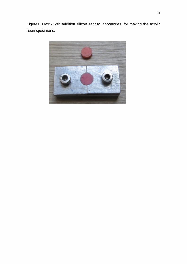
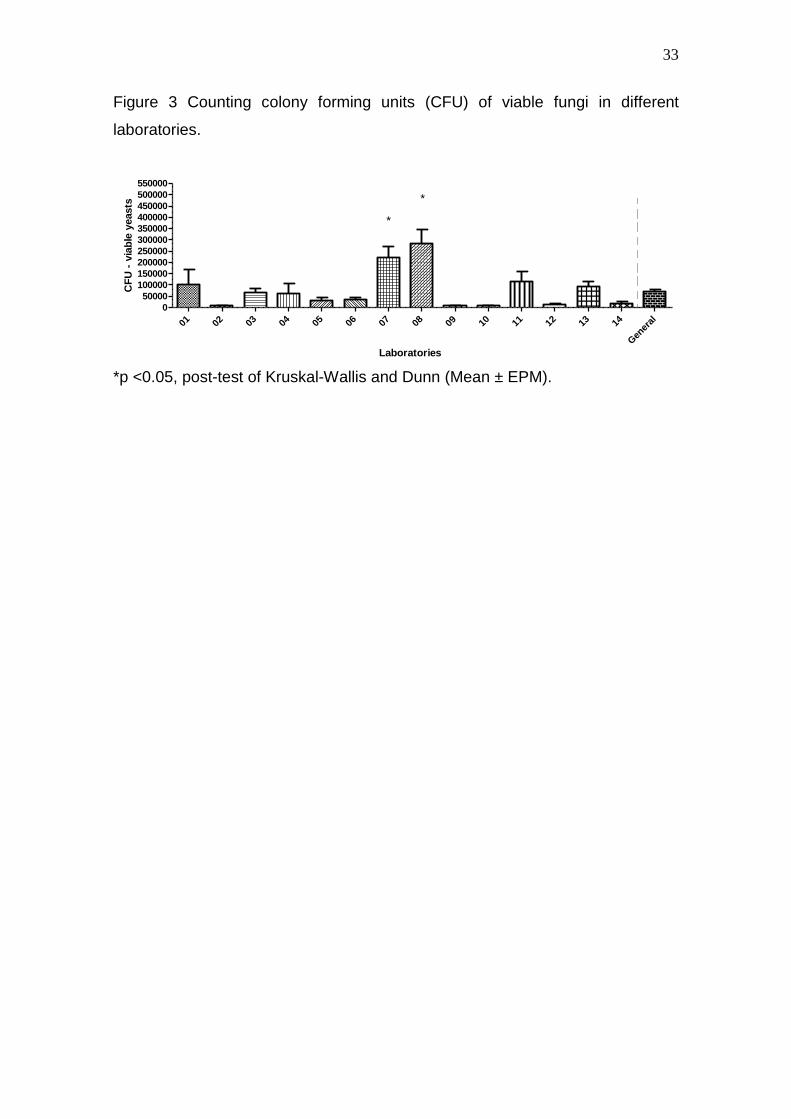
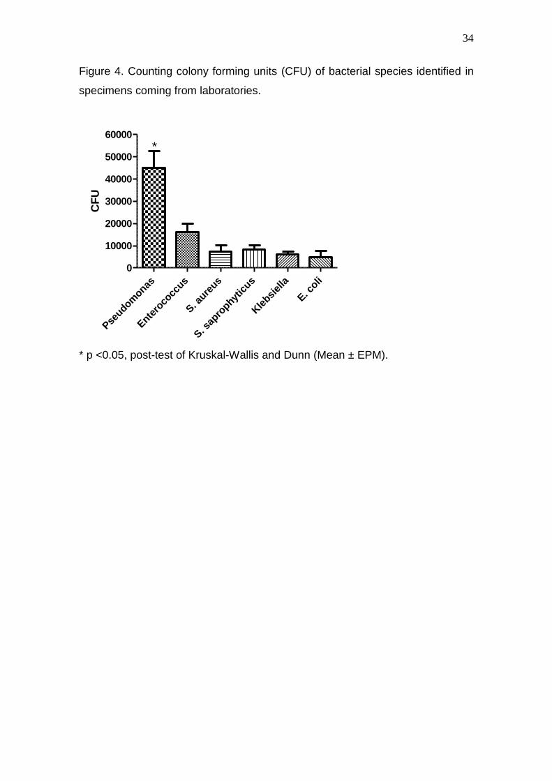
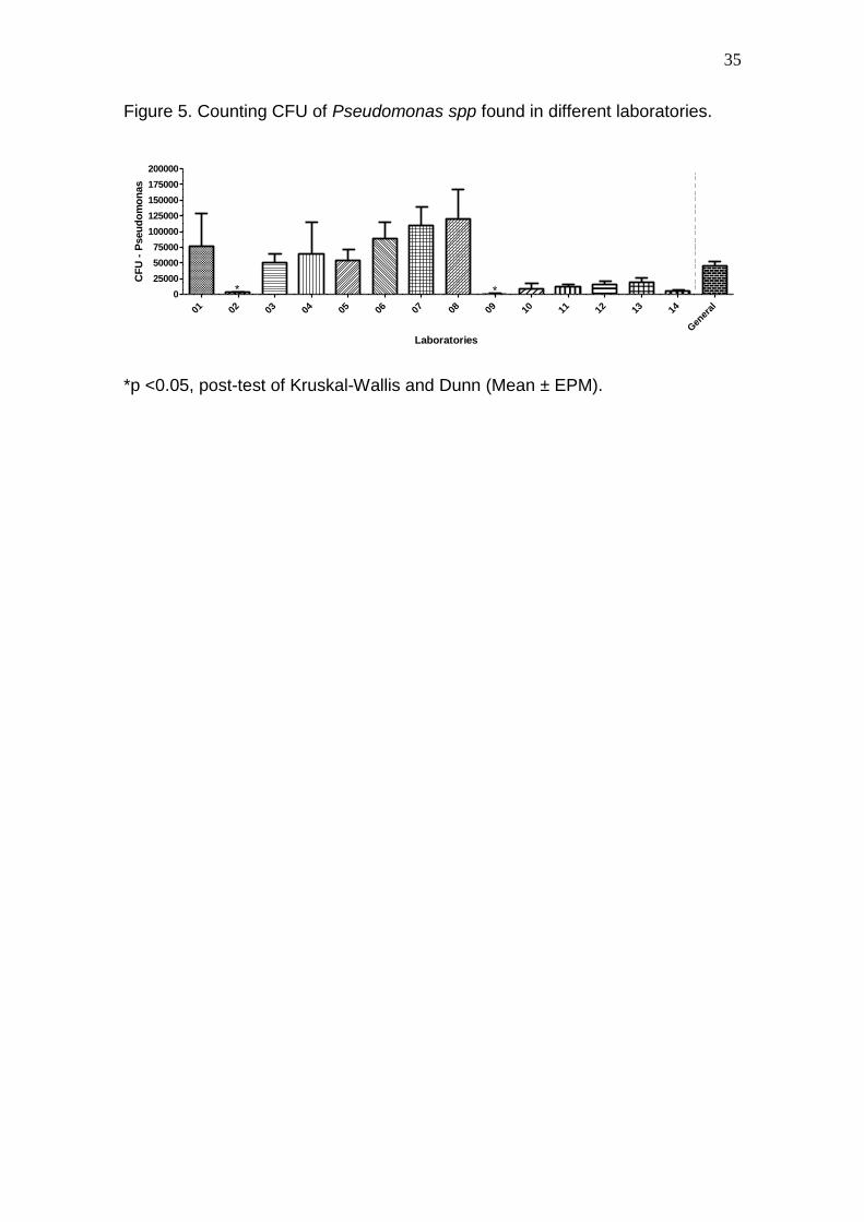
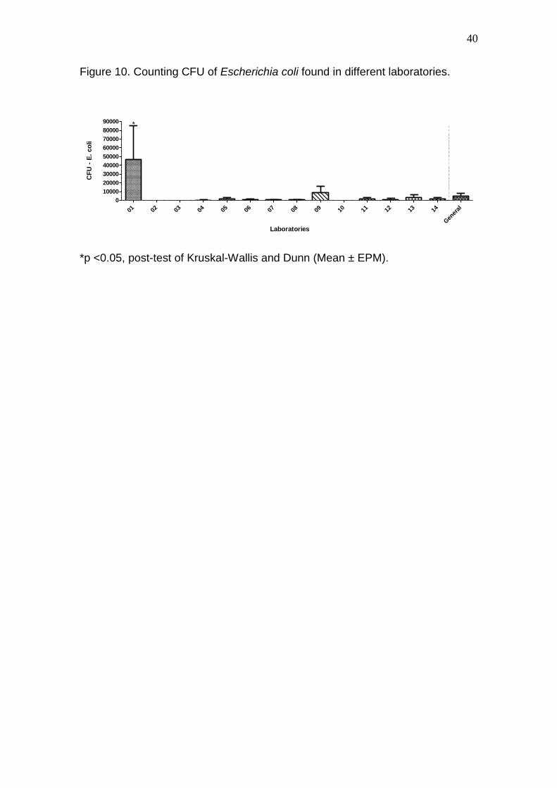
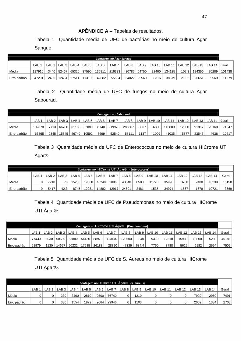
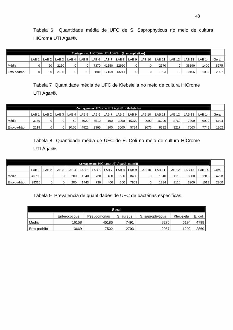
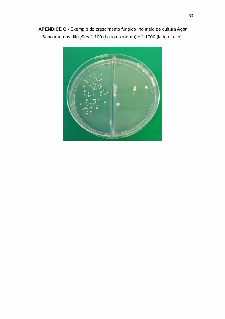