Embed Size (px)
Citation preview

UNIVERSIDADE DO EXTREMO SUL CATARINENSE – UNESC
PROGRAMA DE PÓS-GRADUAÇÃO EM CIÊNCIAS DA SAÚDE
GUSTAVO FEIER
ALTERAÇÕES COMPORTAMENTAIS E DO METABOLISMO
ENERGÉTICO NO TRANSTORNO DO HUMOR BIPOLAR:
EVIDÊNCIAS PRÉ-CLÍNICAS E CLÍNICAS
CRICIÚMA, JANEIRO DE 2012
1
GUSTAVO FEIER
ALTERAÇÕES COMPORTAMENTAIS E DO METABOLISMO
ENERGÉTICO NO TRANSTORNO DO HUMOR BIPOLAR:
EVIDÊNCIAS PRÉ-CLÍNICAS E CLÍNICAS
Tese de Doutorado apresentada ao Programa de Pós-Graduação em Ciências da Saúde para a obtenção do título de doutor em Ciências da Saúde.
Orientador: Prof. Dr. João Quevedo
CRICIÚMA, JANEIRO DE 2012

2
"Dedico este trabalho aos meus pais, Oswaldo e
Maristela, por acreditarem em mim."

3
AGRADECIMENTOS
Aos meus orientadores; Professor Dr. João Luciano de Quevedo, meu grande e eterno mestre, incentivador, amigo, irmão, com o qual tive o prazer de descobrir o mundo da ciência. Sua obra busca desvendar os grandes mistérios do cérebro humano. Sua inesgotável energia tornou-se fonte de inspiração para muitas pessoas e em especial para mim. A ele devo também o apoio na busca de recursos, a intermediação de contato com colegas, a amizade e amadurecimento como pessoa e cientista; Professor Dr. Emilio Streck, presença constante e disposição imperturbável nas inúmeras parcerias cientificas firmadas. Fonte de incansável dedicação, qualidade esta só encontrada naqueles que tem profundo conhecimento da área e a teimosia de não rejeitar desafios; Minha colega e amiga Samira Valvassori, cuja grandeza transcende quaisquer limites. Sua doação ao laboratório, à Educação, à Formação Acadêmica, somada a singular personalidade são objeto da minha maior e mais sincera admiração; Aos demais colegas de Laboratório por todo o apoio que me foi dado e pelas horas de convívio que passamos juntos, pelo carinho e amizade; Aos demais professores do Programa de Pós-Graduação em Ciências da Saúde da Universidade do Extremo Sul Catarinense, pela ajuda, dedicação e auxilio na minha formação pessoal e profissional; Ao meu colega de faculdade e doutorado Fabiano Rosa Agostinho pela amizade, apoio, carinho e por algumas vezes me aturar de mal humor; Aos meus pais pela oportunidade e confiança que em mim depositaram e pelo imenso apoio e carinho; Aos meus irmãos pela força e companheirismo; A minha esposa por tudo que tem sido para mim;
E, finalmente, a Deus por me permitir estar aqui, obrigado.

4
“Vence, quem se vence.”
Provérbio Latino

5
RESUMO
O modelo animal de mania induzido por dextroanfetamina (d-AMPH) é descrito na literatura como um bom modelo animal de transtorno bipolar (TB). Entretanto, tem sido relatado diferenças entre os tipos de anfetaminas em induzir alterações comportamentais e neuroquímicas. Além disso, vários estudos sugerem que o TB está associado à disfunções no metabolismo energético. No presente estudo foi avaliado a diferença entre d-AMPH e metanfetamina (m-AMPH) sobre o comportamento e disfunção no metabolismo energético no cérebro de ratos. Foi avaliado também os efeitos de lítio (Li) e valproato (VPA) sobre as alterações comportamentais e sobre metabolismo energético induzidos por m-AMPH. Finalmente, comparamos os níveis de creatina quinase (CK) no soro de pacientes bipolares nas fases depressiva, maníaca e eutímica. Para tanto, o trabalho foi dividido em três partes. Parte 1: Ratos Wistar receberam uma injeção intraperitoneal (i.p) de salina, d-AMPH (2mg/kg) ou m-AMPH (0,25, 0,5, 1 ou 2mg/kg) e foram submetidos ao teste do campo aberto para avaliação comportamental 2 h após. Foi avaliada também a atividade das enzimas do ciclo de Krebs (citrato sintase, succinato desidrogenase e malato desidrogenase), dos complexos da cadeia respiratória mitocondrial (I, II, II-III, IV) e da CK no cérebro dos ratos. Parte 2: Foi administrado m-AMPH ou salina i.p em ratos Wistar durante 14 dias e entre o 8º e o 14º dia os animais eram tratados com Li, VPA ou salina via i.p. Os animais foram submetidos aos mesmos testes comportamentais e bioquímicos descritos na Parte1. Parte 3: Foram avaliados os níveis de CK no soro de pacientes bipolares - eutímicos, depressivos e em mania – que foram comparados com voluntários saudáveis. Na primeira parte do trabalho foi demonstrado que d-AMPH e m-AMPH aumentaram a atividade locomotora nos ratos. As visitas ao centro do campo aberto aumentaram com a administração de ambas as drogas na dose de 2mg/kg. A administração de m-AMPH na dose de 2mg/kg aumentou o comportamento estereotipado dos animais (sniffing). A administração tanto de d-AMPH quanto de m-AMPH diminuiu a atividade das enzimas do ciclo de Krebs, dos complexos da cadeia respiratória mitocondrial e da CK; entretanto, estes efeitos variam de acordo com a região cerebral avaliada. Na segunda parte do trabalho foi demonstrado que Li e VPA revertem a hiperatividade e alterações no metabolismo energético induzida por m-AMPH. Por fim, na terceira parte do trabalho foi demonstrado que durante a mania, os níveis de CK estão aumentados no soro dos pacientes bipolares quando comparado com voluntários saudáveis. Estes dados demonstram que a m-AMPH, mas não d-AMPH, induz comportamento estereotípico em ratos; porém, as duas drogas parecem ter efeitos similares sobre o metabolismo energético; e que, assim como a d-AMPH, a m-AMPH é capaz de induzir hiperatividade e disfunção no metabolismo energético que são revertidos por Li e VPA. As fases maníaca, depressiva e eutímica do TB, além de apresentarem sintomatologia distinta, também podem ser diferenciadas pelo nível de CK presente no soro dos pacientes. Entretanto, mais estudos são necessários para entender as diferenças observadas na atividade da CK durante as fases do TB.
Palavras-chave: mania; creatina quinase; metanfetamina; cadeia respiratória mitocondrial; transtorno bipolar; ciclo de Krebs

6
ABSTRACT
The animal model of mania induced by dextroamphetamine (d-AMPH) has been considered a good model for the study of bipolar disorder (BD). However, some studies have shown differences between amphetamines to induce both behavioral and biochemical changes. Besides, several studies have suggested that dysfunctional energy metabolism have a central role in BD. In the present study was investigated the potency of the d-AMPH and methamphetamine (m-AMPH) on the behavior and energetic dysfunction in the brain of rats. Were investigated also the effects of the lithium (Li) and valproate (VPT) on behavioral and energy metabolism changes in brain of rats undergoing treatment with the m-AMPH. Finally, was to compare serum creatine kinase (CK) levels between bipolar disorder patients, in the various phases (depressive, manic, and euthymic). The work was divided into three parts. Part 1: Wistar rats were given single intraperitoneal (i.p) injections of saline, d-AMPH (2 mg/kg) or m-AMPH (0.25, 0.5, 1 or 2 mg/kg). Locomotor behavior was assessed using the open-field task and activities of Krebs cycle enzymes (citrate synthase and succinate dehydrogenase), mitochondrial respiratory chain complexes (I, II, III and IV) and CK were measured in brain of rats. Part 2: Wistar rats were first given m-AMPH or saline for 14 days, and then, between days 8 and 14, rats were treated with Li, VPA or saline i.p. The animals were submitted to the same behavioral and biochemical tests described in Part 1. Part 3: Was compared serum CK levels between BD patients, in the various phases (depressive, manic, and euthymic), and healthy volunteers. In the first part of the study was demonstrated that d-AMPH and m-AMPH (all doses administered) increased locomotor activity of animals. The numbers of visits to the centre were increased by d-AMPH and m-AMPH at 2 mg/kg. The m-AMPH administration at 2mg/kg increased the amount of stereotypic behavior. The amphetamines significantly decreased the activities of Krebs cycle enzymes, mitochondrial respiratory chain complexes and CK; nevertheless, this effect varied depending on the brain region evaluated. In the second part of this study we found that Li and VPA reversed m-AMPH-induced hyperactivity. Besides, Li and VPA reversed m-AMPH-induced energetic metabolism dysfunction; however, the effects of Li and VPA were dependent on the brain region analyzed. Finally, in the third part of this study was demonstrated that CK levels were higher in the manic patients than in the controls. Together these data show that: 1) at high doses, m-AMPH increased stereotyped (sniffing) behavior in rats, but d-AMPH did not. However, this study shows that d-AMPH and m-AMPH seem to have similar effects on the brains energetic metabolism; and the d-AMPH, m-AMPH is able to induced hyperactivity and energetic metabolism dysfunction, both seen in BD. In addition, Li and VPA reversed m-AMPH’s effects on locomotor activity and energetic metabolism. The clinical differences among the depressive, manic, and euthymic phases of BD are paralleled by contrasting levels of CK. However, further studies are needed in order to understand the state-dependent differences observed in serum CK activity.
Key-words: mania; creatine kinase; methamphetamine; mitochondrial respiratory chain;
bipolar disorder; Krebs cycle.

7
LISTA DE FIGURAS
Figura 1: Estrutura molecular da dextroanfetamina e da metanfetamina................................ 11
Figura 2: Ciclo de Krebs ......................................................................................................... 23
Figura 3: Formação de ATP pela Cadeia de Transporte de Elétrons ..................................... 24

8
LISTA DE ABREVIATURAS
ADP - Adenosina Difosfato
AMPHs - Anfetaminas
ATP - Adenosina-5'-Trifosfato
BDNF - Fator Neurotrófico Derivado do Cérebro
CK - Enzima Creatina Quinase
CS - Citrato Sintase
DA - Dopamina
d-AMPH - Dextroanfetamina
FADH2 - Dinucleotídeo Flavina Adenina Reduzida
FDA - Food and Drug Administration
GSK-3 - Glicogênio Sintase Quinase 3
GTP – Guanosina 5’Trifosfato
i.p - Injeção Intraperitoneal
Li - Lítio
m-AMPH - Metanfetamina
MD - Enzima Malato Desidrogenase
NADH - Dinucleotídeo Nicotinamida Adenina Reduzida
OMS - Organização Mundial de Saúde
PBI – Transtorno Bipolar do tipo I
PBII – Transtorno Bipolar do Tipo II
Pi - Fosfato Inorgânico
PKC - Proteína Quinase C
SD - Enzima Succinato Desidrogenase
SNC - Sistema Nervoso Central

9
TB - Transtorno Bipolar
UNODC – United Nations Office on Drugs and crime Reports
VPA – Valproato

10
SUMÁRIO
1. Introdução ........................................................................................................................... 11
1.1. Dependência Química a Anfetaminas ............................................................................ 11
1.2. Mecanismo de Ação das Anfetaminas ........................................................................... 14
1.3. Transtorno Bipolar .......................................................................................................... 15
1.4. Lítio e Valproato .............................................................................................................. 18
1.5. Modelo Animal de Mania Induzido por Anfetamina ................................................... 20
1.6. O Metabolismo Energético no Transtorno Bipolar ...................................................... 22
2. Justificativa ......................................................................................................................... 25
3. Objetivo ............................................................................................................................... 25
3.1. Objetivos Específicos ....................................................................................................... 25
4. Artigos Cientificos .............................................................................................................. 27
4.1. Artigo Cientifico 1 ........................................................................................................... 27
4.2. Artigo Cientifico 2 ........................................................................................................... 55
4.3. Artigo Cientifico 3 ........................................................................................................... 80
5. Discussão ............................................................................................................................. 86
Referências .............................................................................................................................. 95

11
1. Introdução
1.1. Dependência Química a Anfetaminas
Com o mundo à beira da guerra, e seus efeitos tóxicos ainda não bem descritos, os efeitos
clínicos das anfetaminas (AMPHs) foram pensados para serem ideais para os soldados em
combate: atenção aumentada, agressão, além de diminuição da fome e da necessidade de
dormir. Durante a Segunda Guerra Mundial, os Estados Unidos, Alemanha e Japão
empregaram o uso de AMPHs nas suas tropas (Suwaki et al., 1997; Anglin et al, 2000;
Rusyniak, 2011). Após a guerra, o Japão experimentou o abuso generalizado como excedentes
do exército inundando o mercado (Rusyniak, 2011). Embora o uso de AMPHs no Japão
declinasse na década de 1960, ele ressurgiu na década de 1970 e continua a ser um problema
de saúde publica até hoje (Suwaki et al., 1997; Hunt et al., 2006).
Os tipos mais comuns de AMPH são a dextroanfetamina (d-AMPH) e a metanfetamina
(m-AMPH). Esses dois tipos de AMPH têm estrutura química similar, sendo que a m-AMPH
é um análogo n-metilado da d-AMPH (Hoffman & Lefkowitz, 1996) (Figura 1).
Dextroanfetamina Metanfetamina
Figura 1. Estrutura molecular da dextroanfetamina e da metanfetamina.
Apesar da similaridade estrutural, alguns estudos mostram diferenças entre as duas
drogas, principalmente na indução de alterações comportamentais e neuroquímicas. Estudos

12
epidemiológicos mostram que a m-AMPH apresenta maiores taxas de abuso em relação à d-
AMPH (Samhsa, 2007).
Nas últimas seis décadas, o abuso de AMPHs passou a ocorrer em todo o mundo (Baberg
et al., 1996; Shaw, 1999). As AMPHs são as substâncias ilícitas mais usadas no mundo
ficando atrás apenas da cannabis sativa. Em 2008, o United Nations Office on Drugs and
Crime Reports (UNODC)- uma agencia dos Estados Unidos que controla drogas e previne
crimes - estimou em 25 milhões os abusadores de AMPHs no mundo, superando o número de
usuários tanto para a cocaína (14 milhões) quanto para a heroína (11 milhões). Uma das
razões que levou o consumo de AMPHs a ultrapassar o de cocaína em todo o mundo é que ela
tem uma meia-vida de 12 horas, permitindo que o viciado tenha um longo e sustentado
período de ação da droga (Rusyniak, 2011). As AMPHs podem ser sintetizadas a partir de
uma ampla variedade de matérias-primas e métodos em laboratórios clandestinos. Em
contraste, a cocaína deve ser extraída da planta, convertida em sua forma de uso, exportada,
para depois ser distribuída através de traficantes para os usuários (Streatfeild, 2001).
Baseado em informações do UNODC 2011, não há dados atualizados na prevalência de
substâncias do grupo das anfetaminas na América do Sul. Informações existentes mostram
que a prevalência anual do uso de substâncias do grupo das AMPHs, na América do Sul,
continua próxima da média mundial, com estimativas entre 0,5% e 0,7% da população entre
15-64 anos ou entre 1,34 e 1,89 milhões de pessoas nesse grupo de idade que fizeram uso
dessas substâncias no ano anterior. Porem em uma pesquisa nacional feita entre estudantes
universitários no Brasil em 2009 mostra que a prevalência anual do uso de anfetaminas entre
estudantes foi relatada com 10,5%. A prevalência anual foi maior entre estudantes mulheres
(14,1%) do que entre estudantes homens (5,5%), e também foi maior entre estudantes mais
velhos, isto é, aqueles de 35 anos ou mais (18,6%), seguidos por estudantes entre 25-34 anos
(13,7%). O uso de substâncias como AMPH é relatada como sendo mais comum entre

13
mulheres devido aos efeitos anoréxicos e a uma cultura predominante de uso de
medicamentos para propósitos de perda de peso (UNODC, 2011).
Os efeitos agudos das AMPHs são respostas do tipo luta ou fuga: aumento da freqüência
cardíaca, aumento da pressão arterial, vasoconstrição, broncodilatação e hiperglicemia
(Cruickshank et al., 2009; Rusyniak, 2011). As alterações decorrentes tanto dos efeitos
agudos como dos crônicos das AMPHs são resultados de sua farmacologia e toxicologia,
sendo que usuários crônicos desses psicoestimulantes desenvolvem sintomas que são muito
semelhantes aos sintomas de mania idiopática que ocorrem em condições de intoxicação,
abstinência ou ambos (Smith & Davis, 1977; Brauer & de Wit 1996).
A estimulação do sistema nervoso central (SNC) induzida por essas drogas podem
também resultar em euforia, energia acentuada, estado de alerta aumentado, intensa
curiosidade e auto-estima elevadas (Cruickshank et al., 2009; Rusyniak, 2011), sintomas estes
também encontrados na fase maníaca do transtorno bipolar (TB). Em usuários crônicos de
AMPHs estes estados psiquiátricos são por vezes prolongados com o surgimento de sintomas
residuais, que são facilmente exacerbados pelo uso de outros anfetaminoides, por sua
reutilização ou ainda por estresse psicológico (Sato et al., 1992; Konuma et al., 1994;
Buffenstein et al., 1999). Segundo o DSM-IV-TR, o início do TB induzido por AMPHs pode
ocorrer durante a intoxicação ou a abstinência. De modo geral, a intoxicação está associada a
características maníacas ou mistas; a abstinência, a característica de humor depressivo
(Kaplan & Sadock, 2007) produzindo sintomatologia semelhante à encontrada nas distintas
fases do TB.

14
1.2. Mecanismo de Ação das Anfetaminas
Uma vez no SNC a AMPH entra no neurônio através dos transportadores de dopamina
(DA). Dentro do neurônio a AMPH se liga a transportadores vesiculares de monoaminas,
facilitando a exocitose vesicular e conseqüentemente aumentando a liberação de DA na fenda
sináptica. Em concentrações mais elevadas, as AMPHs também podem atravessar as
membranas celulares independentemente da vinculação a um transportador (Rusyniak, 2011).
Além disso, o alto pKa da AMPH dificulta o gradiente de prótons - que normalmente mantém
as monoaminas dentro da vesícula - fazendo com que as monaminas sejam liberadas da
vesícula e se acumulem no citoplasma, onde serão transportadas para fora da célula através
dos transportadores reversos de dopamina (Brandle et al., 1992; Fleckenstein et al., 2007;
Schep et al., 2010; Rusyniak, 2011). Além de aumentar a liberação de DA, as AMPHs
também diminuem a sua recaptação e degradação (Suzuki et al., 1980), provocando um
aumento rápido e prolongado nas concentrações extracelulares desse neurotransmissor
(Rusyniak, 2011).
Estudos sugerem que os déficites cognitivos devido ao uso crônico das AMPHs podem
ser ainda devido a alterações no sistema glutamatérgico (Simões et al., 2007; Gross &
Marshall, 2009). Alguns trabalhos salientam a existência de projeções glutamatérgicas no
circuito de recompensa mesolímbico-dopaminérgico, o que possibilita uma base anatômica de
interação entre os dois sistemas (Gass & Olive, 2008). Foi relatado também que o aumento
dos níveis extracelulares de DA estriatal pode causar uma libertação secundária de glutamato
na via corticostriatal, através do circuito estriato-tálamo-corticostriatal (Mark et al., 2004;
2007). Juntos esses estudos sugerem que o aumento dos níveis de glutamato desempenha um
papel importante na neurotoxicidade induzida pelas AMPHs (Nash & Yamamoto, 1992; Mark
et al., 2004, 2007; Caligiuri & Buitenhuys, 2005).

15
Os efeitos clínicos subjacentes associados ao abuso das AMPHs envolvem a estimulação
excessiva do sistema nervoso simpático, sendo esta ativação a principal responsável pela
toxicidade dessa droga (Schep et al., 2010; Rusyniak, 2011). Foi demonstrado em estudos
clínicos e pré-clinicos que a administração crônica de AMPH provoca danos aos terminais
nervosos, levando ao esgotamento de DA, que por sua vez faz com que o usuário crônico
tenha a capacidade diminuída em sentir prazer (Anedonia), contribuindo, conseqüentemente,
para o abuso da substância. A administração repetida de AMPH em animais resulta em
sensibilização comportamental, manifestada por aumento da locomoção e comportamento
estereotipado (Rusyniak, 2011). Essa sensibilização parece envolver tanto o glutamato quanto
a DA, e, mais recentemente, a DA mediando diminuição de acetilcolina (Pierce & Kalivas,
1997; O’Sullivan et al., 2009; Aliane et al., 2010).Esses danos neurais induzidos pelas
AMPHs são eventos complexos, que envolvem o aumento das concentrações intracelulares e
extracelulares, principalmente, de DA e glutamato, os quais induzem uma cascata de eventos
que inclui o estresse oxidativo a neuroinflamação e neurotoxicidade excitatória (Yamamoto et
al., 2010).
1.3. Transtorno Bipolar
O Transtorno Bipolar (TB) é um dos mais graves transtornos psiquiátricos, caracterizado
pela presença de episódios recorrentes de mania e depressão (Belmaker, 2004). Apesar da
importância desse transtorno, a precisa origem do TB não foi estabelecida. Sabe-se que
múltiplos fatores podem estar envolvidos (genéticos, bioquímicos, psicodinâmicos e sócio-
ambientais).
A Organização Mundial de Saúde (OMS) estima que o TB seja a 5a maior causa de
incapacitação no mundo (Murray & Lopez, 1997). Mais de dois milhões de adultos

16
americanos, ou aproximadamente 1% da população, tem TB (Spearing, 2001; Hirschfeld et
al., 2003). Segundo o Estudo da Área de Captação Epidemiológica do Instituto Nacional de
Saúde Mental (ECA-NIMH), conduzido nos Estados Unidos, a taxa anual de incidência para
TB foi de 0,5%, muito próximo da prevalência anual de 0,6% (Lima et al., 2005). A alta
prevalência de TB é particularmente digna de nota por causa do significativo prejuízo
associado a esta condição, não somente aos pacientes, mas também aos parentes e cuidadores
(Ten Have et al., 2002; Calabreze et al., 2003). Pacientes bipolares passam metade de suas
vidas doentes, sendo que a maioria dos dias em depressão (Judd et al., 2003).
Talvez esta seja a doença mental com uma das maiores taxas de suicídio para casos não
tratados com índice 30 vezes maior do que o encontrado na população geral, o que representa
um enorme desafio para pacientes, familiares e médicos. A taxa de suicídio no TB foi
estimada em 19%, podendo igualar-se (e talvez superando) a de transtorno depressivo maior
(Goodwin & Jamison, 1990) e cerca de um terço dos indivíduos afetados pelo TB admite,
pelo menos, uma tentativa de suicídio (Müller-Oerlinghausen et al., 2002). Embora a fase de
mania, especificamente, identifique a doença, é na fase depressiva onde ocorre o maior índice
de suicídios (Baldessarini, 1999; Judd et al., 2002).
O termo bipolar expressa dois pólos de humor ou dois estados afetivos que se alternam
nesse transtorno: a depressão e seu oposto a hipomania ou mania dependendo da gravidade
(Lara, 2004). O TB é subdividido em vários tipos que compõem o espectro bipolar, porém a
classificação mais utilizada o divide em três principais tipos: TB do tipo I apresenta
transtornos de humor recorrentes, com um ou mais episódios maníacos ou mistos, ou ambos
maníacos e episódios mistos e pelo menos um episódio depressivo maior. O TB do tipo II é
caracterizado por um ou mais episódios de depressão maior e pelo menos um episódio
hipomanico. Finalmente, o TB do tipo III é diagnosticado como hipomania associada a
antidepressivos, caracterizada por pacientes que apresentam episódios de hipomania ou mania

17
quando em uso de antidepressivos ou psicoestimulantes. Usualmente ocorre em pacientes com
temperamento ciclotímico prévio (Müller-Oerlinghausen et al., 2002; Alcantara et al., 2003).
No episódio maníaco, o indivíduo apresenta o humor elevado, expansivo ou irritável,
autoestima exagerada ou grandiosidade, diminuição da necessidade do sono, pensamento e
fala acelerados, geralmente com fuga de ideias, distratibilidade, aumento da libido,
hiperatividade ou agitação psicomotora praticamente incontrolável e aumento da exposição a
riscos (ex: gastos excessivos, hipersexualidade) (CID-10, 1993). Nos episódios típicos de
cada um dos três graus de depressão – leve, moderado ou grave - o paciente apresenta o
humor deprimido, perda de interesse ou prazer nas atividades, aumento ou perda de peso,
insônia ou hipersonia, lentidão psicomotora importante, agitação, perda de apetite, perda da
libido, fadiga ou perda da energia, sentimento de inutilidade ou culpa, podendo ser
acompanhada por delírios, diminuição da capacidade de pensar ou concentração e
pensamentos de morte ou ideação suicida (CID-10, 1993).
O diagnóstico precoce e tratamento podem ajudar a reduzir a gravidade associada a esta
condição. Entretanto, pacientes com TB - quando buscam tratamento - geralmente se
apresentam no estágio depressivo e assim eles freqüentemente são diagnosticados apenas
como depressão maior (Manning, 1997; Hirschfeld et al., 2003). Um diagnóstico errado pode
levar a uma terapia inapropriada, ou seja, a monoterapia com antidepressivos, que pode levar
o paciente bipolar ao estado de mania aguda, estados mistos ou estados cíclicos rápidos (Peet,
1994; Ghaemi et al., 2004).
Mania ou hipomania são tratadas com fármacos antipsicóticos, anticonvulsivantes, ou
sais de Lítio (Li) e algumas vezes suplementados com um potente sedativo em curto prazo. Os
sais de Li e certos anticonvulsivantes, como Valproato (VPA), têm propriedades de
estabilizadores do humor, e são usados para prevenção em longo prazo de recorrências
(Baldessarini, 2001).

18
O tratamento de indivíduos com TB é uma tarefa altamente complexa, envolvendo
estratégias distintas nas diferentes fases da doença: mania, depressão e eutimia (Cheniaux,
2011). Atualmente, tem-se dado uma grande valorização dos sintomas depressivos no TB. A
grande questão, entretanto, após o diagnóstico correto, é saber quando, se e como se institui
uma terapêutica antidepressiva na depressão bipolar, pois o risco de virada hipomaníaca ou
maníaca é sempre uma incógnita com a introdução de um antidepressivo (Shansis & Cordioli,
2005). Aproximadamente 25 a 40% dos pacientes bipolares apresentam pelo menos uma
virada deste tipo ao longo da vida associada ao uso de antidepressivos e há uma tendência de
que esta ocorra no mesmo paciente independentemente da classe de antidepressivo que ele
esteja usando (Goldberg & Truman, 2003). Até o momento, receberam aprovação do Food
and Drug Administration (FDA) - a agência americana responsável pelo controle de
medicamentos e alimentos - a combinação olanzapina-fluoxetina e a quetiapina para o
tratamento da depressão bipolar (Cheniaux, 2011).
1.4. Lítio e Valproato
O Li e o VPA são fármacos clássicos usados no tratamento do TB e ambos possuem boa
ação antimaníaca e modesta ação antidepressiva. Os mecanismos de ação de Li e VPA ainda
não estão bem descritos, porém sabe-se que eles agem sobre diversos sistemas de
neurotransmissores e cascatas de sinalização intracelulares. Alguns sistemas em que Li e VPA
agem estão descritos abaixo.
Evidências indicam que o TB está associado a uma desregulação dopaminérgica. Em
apoio a esta hipótese, temos que os fármacos que inibem a transmissão dopaminérgica
exercem uma ação anti-maníaca no TB, enquanto drogas que estimulam a síntese de DA,
ativam os receptores de DA ou inibem a recaptação de DA, muitas vezes desencadeando

19
sintomas de mania (Yatham 2002; Schatzberg 2004; Silverstone & Silverstone, 2004). Porém,
enquanto neurolépticos convencionais e anti-psicóticos, que também são eficazes no
tratamento da mania aguda (McElroy e Keck 2000), agem diretamente inibindo os receptores
DA (Seeman & Lee, 1975; Creese et al 1976), os estabilizadores de humor Li e VPA agem
indiretamente nestes mesmos receptores por um mecanismo de ativação de segundos
mensageiros ativados por receptores do tipo D2 (Yatham et al. 2002).
Além disso, ambos os estabilizadores do humor, Li e VPA, atenuam a função
glutamatérgica atravez de múltiplos mecanismos. A administração crônica desses fármacos
promove o aumento da recaptação do glutamato (Dixon & Hokin, 1998), atenuando a função
dos seus receptores (Nonaka et al., 1998; Du et al., 2004) e, consequentemente, reduzindo a
cascata de sinalização intracelular que são ativadas pela ligação do glutamato ao seu receptor
(Manji & Lenox, 1999). Outro mecanismo de ação do VPA que é bem descrito na literatura é
em relação ao metabolismo de GABA, em diferentes fases da transmissão gabaérgica,
aumentando significativamente os níveis desse neurotransmissor, bloqueando a entrada de
sódio e facilitando a saída de potássio (Post et al., 1992).
Vários estudos têm demonstrado que os estabilizadores do humor, Li e VPA, também
exercem efeitos neurotróficos. Tanto Li quanto VPA ativam o promotor IV de fator
neurotrófico derivado do cérebro (BDNF), elevando os níveis dessa neurotrofina no
hipocampo (Einat et al, 2003; Frey et al, 2006; Yasuda et al, 2009). O BDNF é importante
para a neurogênese, diferenciação e sobrevivência neuronal. Essa neurotrofina é expressa em
grande quantidade no cérebro, principalmente, em áreas que estão ligadas a cognição e
comportamento emocional como o hipocampo e a amídala (Strakowski et al., 2005). Diversos
estudos mostram que o BDNF está envolvido na fisiopatologia do TB, encontrando-se
diminuído tanto nas fases maníacas quanto nas depressivas (Cunha et al., 2006; Grande et al.,
2010).

20
Foi demonstrado que, em concentrações terapêuticas Li e VPA são inibidores da enzima
glicogênio sintase quinase 3 (GSK-3) (Klein & Melton, 1996). A GSK-3 é uma proteína
constantemente ativada, regula múltiplos substratos de serina/ treonina e é regulada por
diversas vias de sinalização (ex. Wnt, PI3K, PKA, PKC entre muitas outras) (Doble &
Woodgett, 2003). Em mamíferos existem duas isoformas de GSK3: GSK-3α e GSK-3β. Essa
enzima regula apoptose e plasticidade celular (Crowder & Freemam, 2000; Franco et al.,
2004; Zhao et al., 2007), sendo que a atividade aumentada de GSK-3 estimula processos
apoptóticos enquanto que sua inibição atenua ou prevene apoptose (Gould et al., 2006).
Outro alvo de Li e VPA é a proteína quinase C (PKC). A inibição da PKC foi sugerida
como um dos mecanismos dos estabilizadores do humor de grande importância (Manji &
Lenox, 2000; Manji & Chen, 2002). A PKC é encontrada principalmente no cérebro, sendo
essencial para os processos de neurotransmissão pré e pós-sináptica, regulando a
excitabilidade neuronal, a liberação dos neurotransmissores e a plasticidade celular (Zarate &
Manji, 2009). Estudos pré-clinicos mostram que a administração do Li leva a redução de PKC
no córtex frontal e hipocampo de ratos (Lenox et al., 1992; Manji et al., 1993; 1999). Chen e
colegas (1994) mostram que VPA produz efeitos similares aos do Li, sugerindo que a inibição
da PKC tem também um importante papel nos efeitos terapêuticos desse estabilizador do
humor.
1.5. Modelo Animal de Mania Induzido por Anfetamina
Apesar do TB ser um transtorno psiquiátrico comum que leva a sérios problemas de
saúde, pouco se sabe sobre sua fisiopatologia. Como discutido anteriormente, o TB é um
transtorno multifatorial que possui diversos sintomas, incluindo episódios de mania
recorrentes, depressão e estados mistos - o que dificulta o desenvolvimento de um modelo

21
animal adequado (Machado-Vieira et al., 2004). Embora haja uma grande dificuldade em
mimetizar o TB em animais, vários modelos animais de mania ou depressão têm sido
desenvolvidos com o intuito de mimetizar alguns aspectos comportamentais dessas condições
psiquiatricas (Machado-Vieira et al., 2004; Frey et al., 2006a; Herman et al., 2007; Jornada et
al., 2010).
Os modelos animais são considerados importantes ferramentas para o estudo do TB,
através dos quais, tem-se proposto novos sistemas de neurotransmissão e vias de sinalização
intracelular envolvidas na modulação desta psicopatologia (Machado-Vieira et al., 2004). Para
ser válido um modelo animal em transtornos psiquiátricos devem ser consideradas três
características principais: 1) se o modelo mimetiza os sintomas da doença determinada
(validade de face), 2) a habilidade do modelo em reproduzir alguns aspectos fisiopatológicos
da doença (validade de constructo) e, finalmente, 3) se os agentes terapêuticos usados no
tratamento revertem os sintomas induzidos no modelo animal (validade preditiva) (Ellenbroek
& Cools, 1990).
A hiperatividade induzida por psicoestimulantes em ratos é o modelo animal de mania
mais aceito na literatura; porque contém os três principais critérios para o desenvolvimento de
um modelo animal adequado. A administração de AMPHs em roedores induz
hiperlocomoção, comportamento de risco, insônia e aumento da atividade sexual (validade de
face); além disso, aumenta os níveis de DA no cérebro – também visto em pacientes bipolares
- (validade de constructo) e responde a fármacos antimaníacos como antipsicóticos, Li e VPA
(validade preditiva) (Fiorino & Phillips, 1999; Frey et al., 2006 b,c,d).
Como descrito anteriormente, os tipos mais comum de AMPHs são a d-AMPH e a m-
AMPH, que são análogas e possuem estruturas químicas semelhantes (Hoffman & Lefkowitz,
1996). Recentemente, nosso grupo de pesquisa mostrou que m-AMPH induz dano oxidativo
maior que d-AMPH (da-Rosa et al., 2011). Além disso, Li e VPA revertem e previnem

22
hiperatividade induzida por m-AMPH somente quando administrada em uma dose muito
baixa, de 0,25 mg/kg, (da-Rosa, 2011), enquanto que esses estabilizadores do humor revertem
e previnem a hiperatividade induzida por d-AMPH na dose de 2 mg/kg (Frey et al.,
2006a,b,c,d).
1.6. O Metabolismo Energético no Transtorno Bipolar
Os seres vivos precisam de energia para realizar várias funções, como, por exemplo, o
transporte ativo de íons e moléculas, síntese de macromoléculas e outras biomoléculas. A
maior parte da energia necessária para realizar essas funções é obtida com a oxidação de
substâncias pela respiração celular. O ATP é o principal combustível da célula na maioria dos
processos que precisam de energia. A energia é liberada pela hidrólise de ATP e serve para
impulsionar uma série de reações (Nelson & Cox, 2000).
Vários estudos têm sugerido que alterações no metabolismo energético celular têm um
papel chave no TB. Anormalidades no metabolismo energético cerebral de pacientes bipolares
foram encontradas a partir de testes de neuroimagem funcional e espectroscopia de
ressonância magnética (Deicken et al., 1995; Dager et al., 2004; Frey et al., 2007; Regenold et
al., 2009).
Após a glicólise, o piruvato é descarboxilado a acetil CoA pela piruvato desidrogenase. A
oxidação completa de acetil CoA no ciclo de Krebs resulta na produção de NADH, FADH2 e
GTP. O ciclo de Krebs é um sistema bioquímico composto por várias enzimas e etapas. Na
primeira etapa, a enzima citrato sintase (CS) catalisa a condensação de oxalaceto com
grupamento acetil-coA, visando a formação de ácido cítrico (Shepherd and Garland, 1969).
Na ultima etapa do ciclo de Krebs, a enzima malato desidrogenase (MD) catalisa a
desidrogenação de malato à oxaloacetato (Kelly et al., 1989). A succinato desidrogenase (SD)

23
faz parte tanto do ciclo de Krebs quanto da cadeia de transporte de elétrons (complexo II) por
isso essa enzima é um dos marcadores mais importantes do ciclo de Krebs (Tyler, 1992).
A disfunção no ciclo de Krebs pode alterar a taxa de metabolismo no cérebro e a
produção de radicais livres. O ciclo de Krebs ocorre na matriz mitocondrial e consiste de uma
sequência de reações onde, em cada volta do ciclo, são formadas três moléculas de NADH,
uma de FADH2, duas de CO2 e uma de GTP. O NADH e FADH2 produzidos no ciclo de
Krebs são carreadores de elétrons e são oxidados na cadeia respiratória para a produção de
ATP pela fosforilação oxidativa (Marks et al., 1996; Nelson & Cox, 2000) (Figura 1).
Figura 2. Ciclo de Krebs (Adaptado de NELSON e COX, 2000).
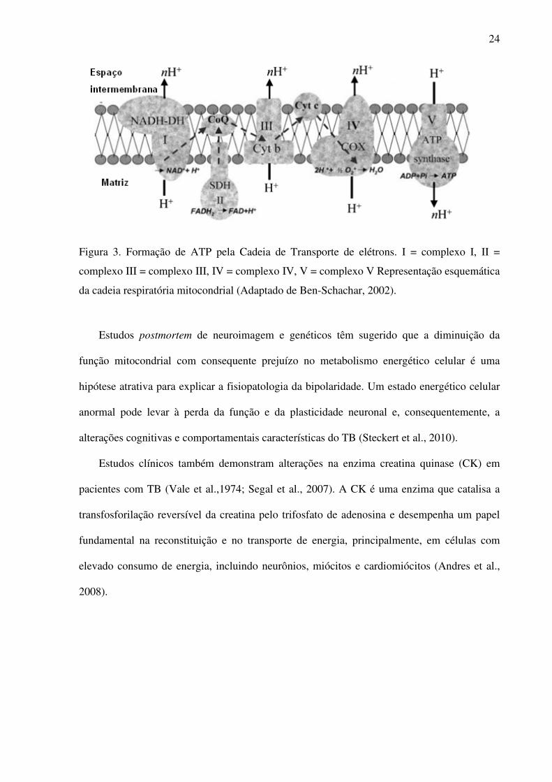
Após o ciclo de Krebs, ocorre outro processo chamado fosforilação oxidativa, no qual os
elétrons passam por uma série de complexos enzimáticos (complexo I, II, III, IV) -
localizados na membrana interna da mitocôndria – juntamente com essa transferência ocorre o
bombeamento de prótons para o espaço intermembrana. O gradiente eletroquímico resultante
desse processo permite que o complexo adenosina 5’-trifosfato ATP sintase, ou complexo V,
sintetize ATP a partir de ADP mais fosfato inorgânico (Pi) (Horn & Barrientos, 2008)
(Figura 3).
24
Figura 3. Formação de ATP pela Cadeia de Transporte de elétrons. I = complexo I, II =
complexo III = complexo III, IV = complexo IV, V = complexo V Representação esquemática
da cadeia respiratória mitocondrial (Adaptado de Ben-Schachar, 2002).
Estudos postmortem de neuroimagem e genéticos têm sugerido que a diminuição da
função mitocondrial com consequente prejuízo no metabolismo energético celular é uma
hipótese atrativa para explicar a fisiopatologia da bipolaridade. Um estado energético celular
anormal pode levar à perda da função e da plasticidade neuronal e, consequentemente, a
alterações cognitivas e comportamentais características do TB (Steckert et al., 2010).
Estudos clínicos também demonstram alterações na enzima creatina quinase (CK) em
pacientes com TB (Vale et al.,1974; Segal et al., 2007). A CK é uma enzima que catalisa a
transfosforilação reversível da creatina pelo trifosfato de adenosina e desempenha um papel
fundamental na reconstituição e no transporte de energia, principalmente, em células com
elevado consumo de energia, incluindo neurônios, miócitos e cardiomiócitos (Andres et al.,
2008).

25
2. Justificativa
O conhecimento bioquímico e enzimático peculiar ao TB é uma ferramenta
importante para o desenvolvimento de novos fármacos e para auxiliar na compreensão da
fisiopatologia desenvolvida neste transtorno do humor, melhorando assim, a terapêutica
utilizada bem como o prognóstico dos pacientes bipolares. Para tanto, faz-se necessário o
desenvolvimento de um modelo animal que mimetize o TB, bem como o conhecimento do
metabolismo energético envolvido nesta patologia tanto em um modelo animal de TB quanto
em pacientes bipolares.
3. Objetivo
Avaliar alterações comportamentais e parâmetros do metabolismo energético no sangue
periférico de pacientes bipolares e em cérebro de ratos em um modelo animal de mania
induzido por metanfetamina.
3.1. Objetivos Específicos
1) Avaliar os efeitos da administração aguda de dextro-anfetamina e meta-
anfetamina sobre a atividade locomotora, exploratória, comportamento de risco e
estereotipia em ratos Wistar.
2) Avaliar os efeitos da administração aguda de dextro-anfetamina e meta-
anfetamina sobre a atividade das enzimas do ciclo de Krebs (Citrato Sintase, Succinato
Desidrogenase e Malato Desidrogenase) em cérebro de ratos Wistar.

26
3) Avaliar os efeitos da administração aguda de dextro-anfetamina e meta-
anfetamina sobre a atividade dos complexos da cadeia respiratória mitocondrial
(Complexo I, II, II-III e IV) em cérebro de ratos Wistar.
4) Avaliar os efeitos da administração aguda de dextro-anfetamina e meta-
anfetamina sobre a atividade da enzima Creatina Quinase em cérebro de ratos Wistar.
5) Avaliar a atividade locomotora e exploratória em um modelo animal de mania
induzido por meta-anfetamina.
6) Avaliar os efeitos da administração crônica de meta-anfetamina na atividade
das enzimas do ciclo de Krebs (Citrato Sintase, Succinato Desidrogenase e Malato
Desidrogenase) em um modelo animal de mania induzido por meta-anfetamina.
7) Avaliar os efeitos da administração crônica de meta-anfetamina na atividade
dos complexos da cadeia respiratória mitocondrial (Complexo I, II, II-III e IV) em um
modelo animal de mania induzido por meta-anfetamina.
8) Avaliar os efeitos da administração crônica de meta-anfetamina na atividade da
enzima Creatina Quinase em cérebro de ratos Wistar em um modelo animal de mania
induzido por meta-anfetamina.
9) Avaliar a atividade da enzima Creatina Quinase em sangue periférico de
pacientes bipolares, comparando as fases de mania, depressão e eutimia.

27
4. Artigo Cientifico
4.1. Artigo Cientifico 1
BEHAVIORAL CHANGES AND BRAIN ENERGY METABOLISM DYSFUNCTION
IN RAT TREATED WITH METHAMPHETAMINE OR DEXTROAMPHETAMINE.
Gustavo Feier, Samira S. Valvassori, Jéssica Lopes-Borges, Roger B. Varella, Felipe Ornell,
Giselli Scaini, Meline O. Morais, Monica L. Andersen, Emilio L. Streck, João Quevedo
Artigo cientifico submetido para publicação no periódico International Journal of
Developmental Neuroscience, Dezembro de 2011.

28
Behavioral changes and brain energy metabolism dysfunction in rats treated with
methamphetamine or dextroamphetamine.
Gustavo Feiera, Samira S. Valvassoria, Jéssica Lopes-Borgesa, Roger B. Varellaa, Felipe
Ornella, Giselli Scainib, Meline O. Moraisb, Monica L. Andersenc, Emilio L. Streckb, João
Quevedoa*
aLaboratory of Neurosciences and National Institute for Translational Medicine (INCT-TM),
Postgraduate Program in Health Sciences, Health Sciences Unit, University of Southern Santa
Catarina, 88806-000 Criciúma, SC, Brazil. bLaboratory of Experimental Pathophysiology and National Institute for Translational
Medicine (INCT-TM), Postgraduate Program in Health Sciences, Health Sciences Unit,
University of Southern Santa Catarina, 88806-000 Criciúma, SC, Brazil. cDepartment of Psychobiology, Universidade Federal de São Paulo, 04024-002 São Paulo,
SP, Brazil.
*Prof. João Quevedo, MD, PhD - Laboratório de Neurociências, PPGCS, UNASAU,
Universidade do Extremo Sul Catarinense, 88806-000 Criciúma, SC, Brazil. Fax: #55 48
3443 4817. E-mail: [email protected]

29
Abstract
Previous studies have demonstrated that amphetamines (AMPHs), such as
dextroamphetamine (d-AMPH) and methamphetamine (m-AMPH) differentially induce
oxidative damage in the rat brain. However, the mechanism to explain this difference is
unknown. It is well described in literature that AMPHs inhibit the mitochondrial respiratory
chain and cause changes to behaviour when administered to experimental animals. Besides
this, mitochondria play a key role in energy metabolism and are also the main producers of
reactive oxygen species as well as being the source of pro- and anti-apoptotic key factors. The
goal of the present study was thus to investigate the potency of the two drugs on the
behaviour and energetic dysfunction in the brain of rats. Male adult Wistar rats were given
single intraperitoneal injections of saline (0.9% NaCl), d-AMPH (2 mg/kg) or m-AMPH
(0.25, 0.5, 1 or 2 mg/kg). The animals were submitted to the open field task for behavioral
assessment. The energy metabolism parameters were measured in the prefrontal cortex,
amygdala, hippocampus and striatum. D-AMPH and m-AMPH (all doses administered)
increased the crossing and rearing behaviours of animals, but no significant difference
between d-AMPH and m-AMPH was observed. The numbers of visits to the centre were
increased by d-AMPH and m-AMPH only at 2 mg/kg. Likewise, at a high dose (2mg/kg), the
injection of m-AMPH increased the amount of sniffing, but this was not seen with d-AMPH.
The AMPHs significantly decreased the activities of Krebs cycle enzymes (citrate synthase
and succinate dehydrogenase), mitochondrial respiratory chain complexes (I, II, III, IV) and
creatine kinase; nevertheless, this effect varied depending on the brain region evaluated.
However, only in the prefrontal did m-AMPH (at 0.5 and 1 mg/kg) induce complex IV
inhibition greater than d-AMPH. In summary, this study demonstrated that at high doses, m-
AMPH, increased stereotyped (sniffing) behaviour in rats, but d-AMPH did not. However,
this study shows that d-AMPH and m-AMPH seem to have similar effects on the brains
energetic metabolism.
Key Words: methamphetamine; dextroamphetamine; Krebs cycle enzymes; mitochondrial
chain; creatine kinase; stereotypy
30
Introduction
Amphetamines (AMPHs) are central nervous system stimulants and act on multiple
sites of pre-synaptic dopaminergic neurotransmission, however, the three primary targets are
considered to be competitive blockade of the dopamine transport (DAT), depletion of
vesicular dopamine (DA) stores, and reversal of DAT function driving a non-exocytotic form
of DA release called efflux (Kuczenski and Segal 1994; Fleckenstein et al. 2007).
The abuse of AMPHs has become a major public health problem worldwide and the
use of these stimulants has significant psychiatric and medical consequences, including
psychosis, dependence, overdose and death (Chen et al., 2010) plus there are concerns about
potential neurotoxicity (Volkow et al., 2001). Clinical studies have shown that the chronic use
of AMPHs leads to functional alterations and neurodegerative changes in various brain
regions (Wilson et al., 1996; Ernst et al., 2000).
AMPHs are synthetic chemicals with a structure similar to amphetamine, comprising a
broad range of psychoactive derivatives such as methamphetamine (m-AMPH) and
dextroamphetamine (d-AMPH). Previous studies demonstrated that amphetamines (AMPHs),
such as dextroamphetamine (d-AMPH) and methamphetamine (m-AMPH) differentially
induce oxidative damage in the rat brain. However, the mechanism to explain this difference
is unknown (da-Rosa et al., 2011).
Mitochondria are membrane-enclosed organelles which generate most of the cell's
supply of adenosine triphosphate (ATP) by a process called oxidative phosphorylation
(Calabrese et al., 2001). Electrons are passed along a series of respiratory enzyme complexes
located in the inner mitochondrial membrane, and the energy released by this electron transfer
is used to pump protons across the membrane. The resultant electrochemical gradient enables
another complex, adenosine 5′-triphosphate (ATP) synthase, to synthesize the energy carrier
ATP (Horn and Barrientos, 2008).

31
Citrate synthase (CS) is localized in the mitochondrial matrix and catalyzes the
condensation of oxaloacetate and the acetyl group of acetyl coenzyme-A, the first step of
Krebs cycle (Shepherd and Garland, 1969). On the other hand, malate dehydrogenase (MD)
catalyzes the dehydrogenation of l-malate to oxaloacetate in the final step of TCA cycle
(Kelly et al., 1989). Succinate dehydrogenase (SD) is still one of the most reliable markers of
the mitochondrial capability to supply an adequate amount of ATP, as it is part of both the
Krebs cycle and the respiratory chain (complex II) (Tyler, 1992).
The increase in extracellular DA concentration induced by AMPHs (Sulzer and
Rayport 1990) causes overproduction of the toxic metabolite of DA oxidation (Wrona et al.
1997), leading to oxidative damage to proteins, lipids and DNA in the neurons (Gluck et al.
2001, Eyerman and Yamamoto 2007; Yamamoto & Zhu 1998). Valvassori and colleagues
(2010) showed that chronic amphetamine administration resulted in a marked inhibition of
complexes I, II, III and IV of the mitochondrial respiratory chain in the rat brain. It is well
described in literature that mitochondria are the major source of reactive oxygen species
(ROS), which are produced in the complexes of the electron transport chain (Mattiasson et al.,
2003). Moreover, a shift in the antioxidant/pro-oxidant balance toward oxidative stress may
inhibit electron transport chain complexes, leading to decreases in ATP production and
cellular dysfunction (Calabrese et al., 2001).
Besides that, creatine kinase (CK) is an enzyme that catalyses the reversible
transphosphorylatin of creatine by ATP and plays a key role in cellular energy buffering and
energy transport, particularly in cells with high and fluctuating energy requirements, including
neurons. The CK system plays a significant role in the brain, and a functional impairment of
this system leads to a deterioration in energy metabolism (Andres et al., 2008).
In the present study m-AMPH and d-AMPH were compared to determine the potency
of the two drugs on behavior, activities of Krebs cycle enzymes (citrate synthase and

32
succinate dehydrogenase), mitochondrial respiratory chain complexes (I, II, III, IV) and
creatine kinase energetic metabolism in the brain of rats.
Experimental methods
Animals
The subjects were adult male Wistar rats (weighting 250–350 g) obtained from our breeding
colony. The animals were housed five to a cage, with food and water available ad libitum and
were maintained on a 12-h light/dark cycle (lights on at 7:00 a.m.) at a temperature of 22 ±
1°C. All experimental procedures were performed in accordance with, and with the approval
of the local Ethics Committee in the use of animals at the Universidade do Extremo Sul
Catarinense. All experiments were performed at the same time during the day to avoid
circadian variations.
Drugs and pharmacological procedures
The drugs, d-AMPH and m-AMPH (Sigma, St Louis, Missouri, USA), were dissolved in
saline (0.9% NaCl). The solutions were prepared immediately before use and were protected
from the light during the experimental session. The total number of rats used in this
experiment were 72 (n = 12 animals per group). Animals received a single injection of m-
AMPH (0.25, 0.5, 1 or 2 mg/kg body weight) or d-AMPH (2 mg/kg body weight) in a volume
of 1 mL/kg, administered intraperitoneally (i.p.). The control group received an injection of
saline (0.9% NaCl) in a volume of 1 mL/kg. Locomotor activity was measured 2 h after the
injection and the rats were killed by decapitation right after the open-field task.

33
Behavior patterns of rat in the open field test
The task was performed in a 40×60 cm open field surrounded by 50 cm high walls. The floor
of apparatus was constructed from varnished wood and divided into 12 equal rectangles by
black lines. The animals were gently placed on the left rear rectangle and left to explore the
arena for 5 min. The following behavioural parameters were assessed in the open field test:
Crossings: Total number of square crossings during the entire test period (Ericson et al.,
1991; Prut and Belzung, 2003; Wultz et al., 1990).
Rearings: Total number of erect postures during the entire test period (Ericson et al., 1991;
Prut and Belzung, 2003).
Visits to center: Total number of visits to the centre of open-field. A center square of
30x30cm was defined as the “center” area of the field.
Grooming: Total time (in seconds) of grooming behaviour during the entire test period.
Include rat paw licking, nose/face grooming, head washing, body grooming/scratching, leg
licking and tail/genitals grooming (Kalueff and Tobimaa, 2004, 2005; Kalueff et al., 2007).
Sniffing: Total time (in seconds) of sniffing behaviour during the entire test period. Rat sniffs
the environment in moving (walking + rearing) (Casarrubea et al., 2008, 2009a, 2009b,
Meyerson and Höglund, 1981).
Tissue and homogenate preparation
The prefrontal cortex, amygdala, hippocampus and striatum were removed and homogenized
(1:10, w/v) in SETH buffer, pH 7.4 (250 mM sucrose, 2 mM EDTA, 10 mM Trizma base, 50
IU/ml heparin). The homogenates were centrifuged at 800 × g for 10 min at 4°C and the
supernatants kept at −70oC until being used for enzymes activity determination. The maximal
period between homogenate preparation and enzyme analysis was always less than 5 days.

34
The protein content was determined by the method described by Lowry and colleagues
(Lowry et al., 1951) using bovine serum albumin as standard.
Activities of enzymes of Krebs cycle
Citrate synthase activity: Citrate synthase activity was assayed according to the method
described by Shepherd and Garland (1969). The reaction mixture contained 100 mM Tris, pH
8.0, 100 mM acetyl CoA, 100 mM 5,5′-di-thiobis-(2- nitrobenzoic acid), 0.1% triton X-100,
and 2–4 μg supernatant protein and was initiated with 100 μM oxaloacetate and monitored at
412 nm for 3 min at 25 °C.
Malate dehydrogenase activity: Malate dehydrogenase was measured as described by Kitto
(1969). Aliquots (20 mg protein) were transferred into a medium containing 10 mM rotenone,
0.2% Triton X-100, 0.15 mM NADH, and 100 mM potassium phosphate buffer, pH 7.4, at
37°C. The reaction was started by the addition of 0.33 mM oxaloacetate. Absorbance was
monitored as described above.
Succinate dehydrogenase activity: Succinate dehydrogenase activity was determined
according to the method of Fischer and colleagues (1985), and measured by following the
decrease in absorbance due to the reduction of 2,6-di-chloro-indophenol (2,6-DCIP) at 600nm
with 700nm as a reference wavelength (ε = 19.1 mM−1 cm−1) in the presence of phenazine
methasulphate (PMS). The reaction mixture consisting of 40mM potassium phosphate, pH
7.4, 16mM succinate and 8μM 2,6-DCIP was pre-incubated with 40–80μg homogenate
protein at 30oC for 20 min. Subsequently, 4mM sodium azide, 7μM rotenone and 40μM 2,6-
DCIP were added and the reaction was initiated by the addition of 1mM PMS and was
monitored for 5 min.
35
Activities of mitochondrial respiratory chain enzymes
Complex I activity: NADH dehydrogenase (complex I) was evaluated according to Cassina
and Radi (1996) by the determination of the rate of NADH-dependent ferricyanide reduction
at λ = 420 nm.
Complex II activity: The activities of succinate-2,6- dichloroindophenol (DCIP)-
oxidoreductase (complex II) was determined by the method described by Fischer and
colleagues (1985). Complex II activity was measured by following the decrease in absorbance
due to the reduction of 2,6-DCIP at λ = 600 nm.
Complex II-III activity: The activity of succinate:cytochrome c oxidoreductase (complex
III) was determined by the method described by Fischer and colleagues (1985). Complex II-
III activity was measured by cytochrome c reduction using succinate as substrate at λ = 550
nm.
Complex IV activity: The activity of cytochrome c oxidase (complex IV) was assayed
according to the method described by Rustin and colleagues (1994), measured by following
the decrease in absorbance due to the oxidation of previously reduced cytochrome c (prepared
by reduction of cytochrome with NaBH4 and HCl) at λ = 550 nm with 580 nm as the
reference wavelength (ɛ= 19.1 mM−1 cm−1). The activities of the mitochondrial respiratory
chain complexes were calculated as nmol. min-1. mg protein-1.
Activity of creatine kinase enzyme
Creatine kinase activity was measured in brain homogenates pretreated with 0.625 mM lauryl
maltoside. The reaction mixture consisted of 60mM Tris–HCl, pH 7.5, containing 7 mM
phosphocreatine, 9 mM MgSO4 and approximately 0.4–1.2 μg protein in a final volume of
100 μL. After 15 min of pre-incubation at 37 °C, the reaction was started by the addition of
3.2 mmol of ADP plus 0.8 mmol of reduced glutathione. The reaction was stopped after 10

36
min by the addition of 1 μmol of p-hydroxymercuribenzoic acid. The creatine formed was
estimated according to the colorimetric method of Hughes (1962). The color was developed
by the addition of 100 μL 2%α-naphtol and 100 μL 0.05% diacetyl in a final volume of 1 mL
and read spectrophotometrically after 20 min at 540 nm. Results were expressed as
units/min×mg protein.
Statistical analysis
Data were analyzed by one-way analysis of variance followed by the Tukey test when F was
significant and are expressed as mean ± standard deviation. All analyses were performed
using the Statistical Package for the Social Science (SPSS; version 16.0) software.
Results
The effects of d-AMPH and m-AMPH administration on open-field behaviors:
The results for locomotor activity are shown in Fig. 1A. We replicated here previous data
from our group (da-Rosa et al., 2011), the administration of d-AMPH (2 mg/kg) and m-
AMPH at 0.25, 0.5, 1 and 2 mg/kg increased rat spontaneous locomotion (crossing) and
exploration (rearing), when compared with the control group. No significant difference was
observed between any dose of m-AMPH and d-AMPH, indicating that all tested doses of m-
AMPH are equivalent to 2 mg/kg of d-AMPH in activating locomotor and exploratory
activity. The administration of d-AMPH and m-AMPH at 2 mg/kg increased visits to center
of open-field, suggesting that the two drugs at a dose of 2mg/kg increased risk behavior in
animals. In addition, m-AMPH at 2mg/kg displayed an increase in sniffing (Fig. 1B),
indicating stereotyped behavior.

37
The effects of d-AMPH and m-AMPH administration on activities of enzymes of Krebs cycle.
Citrate synthase (CS), malate dehydrogenase (MD) and succinate dehydrogenase (SD)
activities were measured in prefrontal cortex, hippocampus and striatum of rats.
m-AMPH administration at 1 mg/kg inhibited CS activity in the prefrontal and hippocampus
of rats. Administration of d-AMPH (2 mg/kg) and m-AMPH at 0.5, 1 and 2 mg/kg also
inhibited CS in the amygdala and striatum of rats (Fig. 2A). We also observed decreased
activity of SD (Fig. 2B) in the striatum after d-AMPH (2mg/kg) and m-AMPH at 0.5 and 1
mg/kg. No change was observed in MD activity after d-AMPH or m-AMPH injection in any
brain structure evaluated (Fig. 2C).
The effects of d-AMPH and m-AMPH administration on activities of mitochondrial
respiratory chain enzymes. Complex I, II, III and IV activities were measured in the
prefrontal cortex, hippocampus and striatum of rats.
Administration of d-AMPH (2mg/kg) and m-AMPH at all doses (0.25, 0.5, 1 and 2 mg/kg)
administered significantly inhibited complex I activity in the rat’s amygdala (Fig. 3A). m-
AMPH at 0.5 and 1 mg/kg induced a decrease of complex II activity in the amygdala of rats
(Fig. 3B). The activity of complex II–III was decreased in prefrontal, amygdala and
hippocampus after d-AMPH or m-AMPH at 0.5, 1 and 2 mg/kg administration (Fig. 3C). In
the striatum the complex II activity was decreased after d-AMPH and m-AMPH at all doses
(0.25, 0.5, 1 and 2 mg/kg) was administered (Fig. 3C). Complex IV activity was also
decreased in prefrontal after d-AMPH and m-AMPH at 0.5 and 1 mg/kg; in the amygdala
following administration of d-AMPH and m-AMPH at 1mg/kg; in the hippocampus after d-
AMPH and m-AMPH at all doses administered and finally in the striatum after d-AMPH and
m-AMPH at 0.5, 1 and 2 mg/kg.

38
The effects of d-AMPH and m-AMPH administration on creatine kinase activity (CK) in the
prefrontal cortex, hippocampus and striatum of rats.
d-AMPH and m-AMPH at 0.5, 1 and 2 mg/kg significantly inhibited CK activity in the
prefrontal. In the amygdala only the highest dose of m-AMPH (2 mg/kg) decreased activity of
CK. Finally, in the striatum d-AMPH decreased and m-AMPH increase de activity of this
enzyme.
Discussion
The present study was designed to compare the doses of 0.25, 0.5, 1 or 2 mg/kg of m-
AMPH with 2 mg/kg of d-AMPH - known to cause hyperactivity and inhibition of
mitochondrial respiratory chain complexes in the brain of rats (Valvassori et al., 2010) - on
behavior and energetic metabolism parameters in the prefrontal cortex, amygdala,
hippocampus and striatum of rats.
We replicated here previous data from our group (da-Rosa et al., 2011); the
administration of d-AMPH (2 mg/kg) and m-AMPH at 0.25, 0.5, 1 and 2 mg/kg were
equipotent at increased rat spontaneous locomotion (crossing) and exploration (rearing). An
additional element observed in this study was that m-AMPH and d-AMPH both at 2 mg/kg,
increased the number of visits to the center of the open-field, showing the equivalence of the
two drugs to induce risk-taking behavior in rats. According to Einat (2006), risk-taking
behaviour may represent the mirror image of anxiety in many tests. Administration of
AMPHs in rodents increases risk-taking behavior by reducing anxiety–like measures such as
increasing the time spent in the centre of an open-field (Einat et al., 2003).
In our study, we found that the administration of m-AMPH (but not d-AMPH) in high
doses (2 mg/kg), induces sniffing behaviour. AMPHs and nicotine increased stereotypy until
80 min after the administration of these psychotropic drugs in rats (Izawa et al., 2006).

39
Commonly abused drugs, such as cocaine and methamphetamine stimulate dopamine
transmission in the nucleus accumbens, inducing behavioural activation in rodents and other
laboratory animals (Di Chiara, 2002).
In the present work, we observed that the activities of enzymes of the Krebs cycle -
CS, SD - mitochondrial respiratory chain – complex I, II, II-II, IV - and CK were inhibited in
the brain of rats submitted to the administration of d-AMPH and m-AMPH; however, these
changes varied according to the brain structure and biochemical analysis. m-AMPH and d-
AMPH decreased complex II-III, complex IV and CK activities in the prefrontal;
nevertheless, m-AMPH also inhibits the CS activity in this brain structure. In the amygdala,
both m-AMPH and d-AMPH reduced CS, complex I, II-III and IV and CK activities; in
addition, m-AMPH administration also decreased complex II. In the hippocampus, m-AMPH
and d-AMPH administration decreased complex II-III and complex IV activities; although m-
AMPH injection also decreased CS activity. Finally, in the striatum, both amphetamines
inhibited the activities of CS, SD, complex II-III, complex IV and CK. As can be seen, the
effects of m-AMPH and d-AMPH on cerebral metabolism is heterogeneous between the
structures.
It is well known that the amygdala modulates the limbic system, controlling an
iterative circuit, prefrontal–striatal–thalamic, which controls complex socioemotional
behaviours (Strakowski et al., 2000; Strakowski et al., 2005). Amphetamines act on the
mesocorticolimbic dopamine system which projects from the ventral tegmental area to the
nucleus accumbens, olfactory tubercle, frontal cortex and amygdala (Kalivas and Stewart
1991; Volkow et al. 2003). Above the nucleus accumbens (or ventral striatum) is the dorsal
striatum that has been implicated in the locomotor response to amphetamines (Hamamura et
al. 1991; Nestler 2001). The frontal cortex is an important cerebral area involved in working
memory, in decision making, inhibitory control and in selecting and retaining information to

40
produce executive control (Royall 2002; Huang et al., 2004; Rinaldi et al., 2007).
Glutamatergic projections from the prefrontal cortex to the nucleus accumbens also play an
important role in the action of amphetamines (Kalivas et al. 2005). In addition, dopaminergic
innervation is critical for long term changes in synaptic efficacy in hippocampus and the
interactions between dopamine and glutamate receptors are essential for prefrontal and
hippocampal cognitive functions (Gurden et al., 2000; Li et al., 2003; Huang et al., 2004;
Granado et al., 2008; Yang et al., 2000; Chen et al., 2004; Tseng and O’Donnell, 2004;
Nowak and Corces, 2000).
According to the data presented in this article, previous studies from our and other
laboratories have demonstrated that administration of d-AMPH and m-AMPH in rats also
inhibited enzymes of energetic metabolism (Valenzuela and Villanueva, 1987; Valvassori et
al., 2010; Corrêa et al., 2007; Streck et al., 2008; Moretti et al., 2011; Bachmann et al., 2009).
However, there are no studies comparing the two drugs on energy metabolism.
The amphetamines have been associated to long-term deficits in dopaminergic and
serotonergic systems in the brain, resulting from dopamine- and glutamate-generated reactive
oxygen species (LaVoie et al., 1999; Page et al., 2001). Several studies suggest that dopamine
autoxidation can form reactive quinones that attack and potentially inhibit the function of
intracellular proteins. In addition to dopamine autoxidation, metabolism of dopamine by
monoamine oxidase can increase H2O2 production and iron-dependent ROS production (Spina
and Cohen, 1989). Monoamine oxidase is located in the outer membrane of mitochondria and
could be a significant source of ROS production mainly in dopaminergic neurons (Adam-
Vizi, 2005). Mitochondria are the main source of reactive oxygen species (ROS), which are
produced in the complexes of the electron transport chain (Mattiasson et al., 2003). Moreover,
a shift in the antioxidant/pro-oxidant balance toward oxidative stress may inhibit electron

41
transport chain complexes, leading to decreases in ATP production and cellular dysfunction
(Calabrese et al., 2001).
Oxidative stress induced by the amphetamines (da-Rosa et al., 2011) with consequent
impairment in mitochondrial complex activity can be a possible explanation for the results of
this study. We note here that in general, even lower doses of m-AMPH (most of the doses
administered) were equivalent to 2mg/kg of d-AMPH to inhibit enzymes of energy
metabolism. In contrast, a recent study from our laboratory showed that d-AMPH and m-
AMPH increased oxidative lipid and protein damage, but m-AMPH even at lower doses, was
more potent than d-AMPH (da-Rosa et al., 2011). This discrepancy can be explained at least
in part by the fact that mitochondria play a key role in ROS formation but also in the
antioxidant response (Starkov, 2008).
Superoxide produced in the mitochondria is rapidly dismutated by enzyme manganese
superoxide dismutase in the mitochondrial matrix (Forman and Azzi, 1997) or by copper/zinc
superoxide dismutase in the intermembrane space and the cytosol reducing the superoxide
radical into hydrogen peroxide (H2O2) (Fridovich, 1995; Loschen et al., 1973). H2O2 is also
a ROS, which is moderately membrane-permeable and able to leave the mitochondrial matrix
and, when produced in excess, is released from the cell. In the presence of Fe3+, hydroxyl
radical (•OH), the more reactive radical, is also formed from H2O2 and superoxide. On the
whole, H2O2 is eliminated by glutathione or it is converted to H2O in the enzyme reactions
by catalase or glutathione peroxidase/glutathione reductase present in both the mitochondrial
matrix and the cytosol (Halliwell and Gutteridge, 1999).
In summary, this study demonstrated that at high doses, m-AMPH, (but not d-AMPH)
increased stereotyped behaviour (sniffing) in rats. However, this study shows that d-AMPH
and m-AMPH seem to have similar effects on brain energetic metabolism.

42
References
Adam-Vizi, V., 2005. Production of reactive oxygen species in brain mitochondria:
contribution by electron transport chain and non-electron transport chain sources. Antioxid
Redox Signal. 7,1140-9.
Andres, R.H., Ducray, A.D., Schlattner, U., Wallimann, T., Widmer, H.R., 2008. Functions
and effects of creatine in the central nervous system. Brain Res Bull. 76, 329–43.
Bachmann, R.F., Wang, Y., Yuan, P., Zhou, R., Li, X., Alesci, S., Du, J., Manji, H.K., 2009.
Common effects of lithium and valproate on mitochondrial functions: protection against
methamphetamine-induced mitochondrial damage. Int J Neuropsychopharmacol. 12, 805-22.
Calabrese, V., Scapagnini, G., Giuffrida Stella, A.M., Bates, T.E., Clark, J.B., 2001.
Mitochondrial involvement in brain function and dysfunction: relevance to aging,
neurodegenerative disorders and longevity. Neurochemical Research. 6, 739-64.
Casarrubea, M., Sorbera, F., Crescimanno, G., 2008. Multivariate analysis of the
modifications induced by an environmental acoustic cue on rat exploratory behavior. Physiol
Behav. 93, 687–96.
Casarrubea, M., Sorbera, F., Crescimanno, G., 2009a Structure of rat behavior in holeboard: I.
Multivariate analysis of response to anxiety. Physiol. Behav. 96, 174–9.
Casarrubea, M., Sorbera, F., Crescimanno, G., 2009b. Structure of rat behavior in holeboard:
II. Multivariate analysis of modifications induced by diazepam. Physiol. Behav. 96, 683–92.
Cassina, A., Radi, R., 1996. Differential inhibitory Aation of nitric oxide and peroxynitrite on
mitochondrial electron transport. Arch. Biochem. Biophys. 328, 309-16.
Chen, G., Greengard, P. and Yan, Z., 2004. Potentiation of NMDA receptor currents by
dopamine D1 receptors in prefrontal cortex. Proc. Natl. Acad. Sci. USA. 101, 2596–2600.
Chen, H., Wu, J., Zhang, J., Hashimoto, K., 2010. Recent topics on pharmacotherapy for
amphetamine-type stimulants abuse and dependence. Curr Drug Abuse Rev. 3, 222-38.

43
Corrêa, C., Amboni, G., Assis, L.C., Martins, M.R., Kapczinski, F., Streck, E.L., Quevedo, J.,
2007. Effects of lithium and valproate on hippocampus citrate synthase activity in an animal
model of mania. Prog. Neuropsychopharmacol. Biol. Psychiatry. 31, 887-891.
da-Rosa, D.D., Valvassori, S.S., Steckert, A.V., Arent, C.O., Ferreira, C.L., Lopes-Borges, J.,
Varela, R.B., Mariot, E., Dal-Pizzol, F., Andersen, M.L., Quevedo, J., 2011. Differences
between dextroamphetamine and methamphetamine: behavioral changes and oxidative
damage in brain of Wistar rats. J. Neural. Transm. [Epub ahead of print].
Di Chiar, G., 2002. Nucleus accumbens shell and core dopamine: differential role in behavior
and addiction. Behav. Brain Res. 137, 75–114.
Einat, H., Yuan, P., Dogra, S., Manji, H.K., 2003. Does the PKC signaling pathway play a
role in the pathophysiology and treatment of bipolar disorder? Biological Psychiatr. 53,
(Suppl. 8): S-399.
Einat, H., 2006. Modelling facets of mania--new directions related to the notion of
endophenotypes. J. Psychopharmacol. 20, 714-22.
Ernst, T., Chang, L., Leonido-Yee, M., Speck, O., 2000. Evidence for long-term neurotoxicity
associated with methamphetamine abuse: A 1H MRS study. Neurology. 54, 1344–49.
Ericson, E., Samuelsson, J., Ahlenius, S., 1991. Photocell measurements of rat motor activity.
A contribution to sensitivity and variation in behavioral observations. J. Pharmacol. Methods.
25, 111-22.
Eyerman, D.J., Yamamoto, B.K., 2007. A rapid oxidation and persistent decrease in the
vesicular monoamine transporter 2 after methamphetamine. J. Neurochem. 103, 1219–27.
Fischer, J.C., Ruitenbeek, W., Berden, J.A., Trijbels, J.M., Veerkamp, J.H., Stadhouders,
A.M., Sengers, R.C., Janssen, A.J., 1985. Differential investigation of the capacity of
succinate oxidation in human skeletal muscle. Clin. Chim. Acta. 153, 23-6.

44
Fleckenstein, A.E., Volz, T.J., Riddle, E.L., Gibb, J.W., Hanson, G.R., 2007. New insights
into the mechanism of action of amphetamines. Annu. Rev. Pharmacol. Toxicol. 47, 681–98.
Forman, H.J., Azzi, A., 1997. On the virtual existence of superoxide anions in mitochondria:
thoughts regarding its role in pathophysiology. FASEB J. 11, 374–75.
Fridovich, I., 1995. Superoxide radical and superoxide dismutases. Annu Rev Biochem 64,
97–112.
Granado, N., Ortiz, O., Suárez, L.M., Martín, E.D., Ceña, V., Solís, J.M. and Moratalla, R.,
2008 D1 but not D5 dopamine receptors are critical for LTP, spatial learning, and LTP-
induced arc and zif268 expression in the hippocampus. Cereb. Cortex. 18, 1–12.
Gluck, M. R., Moy, L.Y., Jayatilleke, E., Hogan, K.A., Manzino, L., Sonsalla, P.K., 2001.
Parallel increases in lipid and protein oxidative markers in several mouse brain regions after
methamphetamine treatment. J. Neurochem. 79, 152–60.
Gurden, H., Takita, M. and Jay, T.M., 2000. Essential role of D1 but not D2 receptors in the
NMDA receptor-dependent long-term potentiation at hippocampal–prefrontal cortex synapses
in vivo. J. Neurosci. 20, RC106.
Halliwell, B., Gutteridge, J.M., 1999. Free Radical in Biology and Medicine. Oxford, UK:
Oxford University Press.
Hamamura, T., Akiyama, K., Akimoto, K., Kashihara, K., Okumura, K., Ujike, H., Otsuki, S.,
1991. Coadministration of either a selective D1 or D2 dopamine antagonist withy
methamphetamine prevents methamphetamine-induced behavioural sensitization and
neurochemical change, studied by in vivo intracerebral dialysis. Brain Res. 546, 40–46.
Horn, D., Barrientos, A., 2008. Mitochondrial copper metabolism and delivery to cytochrome
c oxidase. IUBMB Life. 60, 421–429.

45
Huang, Y.Y., Simpson, E., Kellendonk C. and Kandel, E.R., 2004. Genetic evidence for the
bidirectional modulation of synaptic plasticity in the prefrontal cortex by D1 receptors. Proc.
Natl. Acad. Sci. USA. 101, 3236–3241.
Hughes, B.P., 1962. A method for estimation of serum creatine kinase and its use in
comparing creatine kinase and aldolase activity in normal and pathologic sera. Clin. Chim.
Acta. 7, 597-604.
Izawa, J., Yamanashi, K., Asakura, T., Misu, Y., Goshima, Y., 2006. Differential effects of
methamphetamine and cocaine on behavior and extracellular levels of dopamine and 3,4-
dihydroxyphenylalanine in the nucleus accumbens of conscious rats. Eur. J. Pharmacol. 549,
84-90.
Kalivas, P.W., Stewart, J., 1991. Dopamine transmission in the initiation and expression of
drug- and stress-induced sensitization of motor activity. Brain Res. Brain. Res. Rev. 16, 223–
244.
Kalivas, P.W., Volkow, N., Seamans, J., 2005. Unmanageable motivation in addiction: a
pathology in prefrontal-accumbens glutamate transmission. Neuron. 45, 647–650.
Kalueff, A.V., Aldridge, J.W., LaPorte, J.L., Murphy, D.L., Tuohimaa, P., 2007. Analyzing
grooming microstructure in neurobehavioral experiments. Nat. Protoc. 2, 2538–44.
Kalueff, A.V., Tuohimaa, P., 2004. Grooming analysis algorithm for neurobehavioural stress
research. Brain Res. Brain Res Protoc. 13, 151–58.
Kalueff, A.V., Tuohimaa, P., 2005. The grooming analysis algorithm discriminates between
different levels of anxiety in rats: potential utility for neurobehavioural stress research. J.
Neurosci. Methods. 143, 169–77.
Kelly, D., Gordon, J., Alpers, R., Strauss, A.W., 1989 The tissue-specific expression and
developmental regulation of two nuclear genes encoding rat mitochondrial proteins. Medium

46
chain acyl-CoA dehydrogenase and mitochondrial malate dehydrogenase. J. Biol. Chem. 264,
18921–18925.
Kitto, G.B., 1969. Intra- and extramitochondrial malate dehydrogenases from chicken and
tuna heart. Methods Enzymol. 23, 106-16.
Kuczenski, R., Segal, D.S., 1994. Neurochemistry of Amphetamine. In: Cho AK, Segal DS,
editors. Amphetamine and Its Analogs. San Diego: Academic Press, 81–113.
LaVoie, M.J., Hastings, T.G., 1999. Dopamine quinone formation and protein modification
associated with the striatal neurotoxicity of methamphetamine: evidence against a role for
extracellular dopamine. J. Neurosci. 19, 1484–1491.
Li, S., Cullen, W.K., Anwyl R. and Rowan, M.J., 2003. Dopamine-dependent facilitation of
LTP induction in hippocampal CA1 by exposure to spatial novelty. Nat. Neurosci. 6, 526–31.
Loschen, G., Azzi, A., Flohe, L., 1973. Mitochondrial H2O2 formation: relationship with
energy conservation. FEBS Lett. 33, 84–7.
Lowry, O.H., Rosebrough, N.J., Farr, A.L., Randall, R.J., 1951. Protein measurement with the
Folin phenol reagent. J. Biol. Chem. 193, 265-75.
Mattiasson, G., Shamloo, M., Gido, G., Mathi, K., Tomasevic, G., Yi, S., Warden, C.H.,
Castilho, R.F., Melcher, T., Gonzalez-Zulueta, M., Nikolich, K., Wieloch, T., 2003
Uncoupling protein-2 prevents neuronal death and diminishes brain dysfunction after stroke
and brain trauma. Nature Medicine. 9, 1062-8.
Meyerson, B.J., Höglund, A.U., 1981. Exploratory and socio-sexual behaviour in the male
laboratory rat: a methodological approach for the investigation of drug action. Acta
Pharmacol Toxicol Copenh. 48, 168–80.
Moretti, M., Valvassori, S.S., Steckert, A.V., Rochi, N., Benedet, J., Scaini, G., Kapczinski,
F., Streck, E.L., Zugno, A.I., Quevedo, J., 2011. Tamoxifen effects on respiratory chain
47
complexes and creatine kinase activities in an animal model of mania. Pharmacol. Biochem.
Behav. 98, 304-10.
Nestler, E.J., 2001. Molecular basis of long-term plasticity underlying addiction. Nat Rev
Neurosci. 2, 119–128.
Nowak, S.J. and Corces V.G., 2000. Phosphorylation of histone H3 correlates with
transcriptionally active loci. Genes Dev. 14, 3003–3013.
Page, G., Peeters, M., Najimi, M. et al., 2001. Modulation of the neuronal dopamine
transporter activity by the metabotropic glutamate receptor mGluR5 in rat striatal
synaptosomes through phosphorylation mediated processes. J. Neurochem. 76, 1282–1290
Prut, L., Belzung, C., 2003. The open field as a paradigm to measure the effects of drugs on
anxiety-like behaviors: a review. Eur. J. Pharmacol. 463, 3–33.
Rinaldi, A., Mandillo, S., Oliverio A. and Mele, A., 2007. D1 and D2 receptor antagonist
injections in the prefrontal cortex selectively impair spatial learning in mice.
Neuropsychopharmacology. 32, 309–319.
Royall, D.R., 2002. Executive control function: a review of its promise and challenges for
clinical research. A report from the Committee on Research of the American Neuropsychiatric
Association. J. Neuropsychiatry Clin. Neurosci. 14, 377–405.
Rustin, P., Chretien, D., Bourgeron, T., Gérard, B., Rötig, A., Saudubray, J.M., Munnich, A.,
1994. Biochemical and molecular investigations in respiratory chain deficiencies. Clin. Chim.
Acta. 228, 35-51.
Shepherd, D., Garland, P.B., 1996. The kinetic properties of citrate synthase from rat liver
mitochondria. Biochem. J. 114, 597–610.
Spina, M.B., Cohen, G., 1989. Dopamine turnover and glutathione oxidation: implications for
Parkinson disease. Proc. Natl. Acad. Sci. USA. 86, 1398–1400.

48
Starkov, A.A., 2008. The role of mitochondria in reactive oxygen species metabolism and
signaling. Ann N Y Acad Sci. 1147, 37-52.
Strakowski, S.M., Delbello, M.P., Adler, C.M., 2005. The functional neuroanatomy of bipolar
disorder: a review of neuroimaging findings. Mol Psychiatry. 10, 105-16.
Strakowski, S.M., Delbello, M.P., Adler, C., Cecil, D.M., Sax, K.W., 2000. Neuroimaging in
bipolar disorder. Bipolar Disord. 2, 148-64. Review.
Sulzer, D., Rayport, S., 1990. Amphetamine and other psychostimulants reduce pH gradients
in midbrain dopaminergic neurons and chromaffin granules: a mechanism of action. Neuron.
5, 797-808.
Streck, E.L., Amboni, G., Scaini, G., Di-Pietro, P.B., Rezin, G.T., Valvassori, S.S., Luz, G.,
Kapczinski, F., Quevedo, J., 2008. Brain creatine kinase activity in an animal model of mania.
Life Sci. 82, 424-9.
Tseng, K.Y. and O’Donnell P., 2004. Dopamine–glutamate interactions controlling prefrontal
cortical pyramidal cell excitability involve multiple signaling mechanisms. J. Neurosci. 24,
5131–5139.
Tyler, D., 1992. The Mitochondrion in Health and Diseases, VCH Publishers, New York .
Valenzuela, A., Pla, A., Villanueva, E., 1987. Effects of chronic administration of
dextroamphetamine on enzymes of energy metabolism in regions of the rat brain.
Neuropharmacology. 26, 627-31.
Valvassori, S.S., Rezin, G.T., Ferreira, C.L., Moretti, M., Gonçalves, C.L., Cardoso, M.R.,
Streck, E.L., Kapczinski, F., Quevedo, J., 2010. Effects of mood stabilizers on mitochondrial
respiratory chain activity in brain of rats treated with d-amphetamine. J Psychiatr. Res. 44,
903-9.
Volkow, N.D., Chang, L., Wang, G., Fowler, J.S., Leonido-Yee, M., Franceschi, D., Sedler,
M.J., Gatley, S.J., Hitzemann, R., Ding, Y.S., Logan, J., Wong, C., Miller, E.N., 2001.

49
Association of dopamine transporter reduction with psychomotor impairment in
methamphetamine abusers. Am. J. Psychiatry. 158, 377–82.
Volkow, N.D., Fowler, J.S., Wang, G-J., 2003. The addicted human brain: insights from
imaging studies. J. Clin. Invest. 111, 1444–1451.
Wilson, J.M., Kalasinsky, K.S., Levey, A.I., Bergeron, C., Reiber, G., Anthony, R.M.,
Schmunk, G.A., Shannak, K., Haycock, J.W., Kish, S.J., 1996. Striatal dopamine nerve
terminal markers in human, chronic methamphetamine users. Nat. Med. 2, 699-703.
Wrona, M.Z., Yang, Z., Zhang, F., Dryhurst, G., 1997. Potential new insights into the
molecular mechanisms of methamphetamine-induced neurodegeneration. NIDA Res.
Monogr. 173, 146–74.
Yamamoto, B.K., Zhu, W., 1998. The effects of methamphetamine on the production of free
radicals and oxidative stress. J. Pharmacol. Exp. Ther. 287, 107–14.
Yang, S.N., 2000. Sustained enhancement of AMPA receptor- and NMDA receptor-mediated
currents induced by dopamine D1/D5 receptor activation in the hippocampus: an essential
role of postsynaptic Ca2+. Hippocampus. 10, 57–63.

50
Legends to Figures
Figure 1: Open field test. A) Free movement in the open field: crossings (dF=5; F=54.842; p<0.0001), rearings (dF=5; F=30.766; p<0.0001) and visits to center (dF=5; 51.101; p<0.0001). B) Stereotypy: grooming (dF=5; F=5.869; p<0.0001) and sniffing (dF=5; F=34.515; p<0.0001). *Different from Saline group. Figure 2: Activities of enzymes of Krebs cycle. A) Citrate synthase activity: In the prefrontal (dF=5; F=10.261; p<0.0001), amygdala (dF=5; F=14.819; p<0.0001), hippocampus (dF=5; F=2.809; p=0.034) and striatum (dF=5; F=6.189; p=0.001). B) Succinate dehydrogenase activity: In the prefrontal, amygdala, hippocampus and striatum (dF=5; F=8.714; p<0.0001). C) Malate dehydrogenase activity: In the prefrontal, amygdala, hippocampus and striatum. *Different from Saline group. Figure 3: Activities of mitochondrial respiratory chain enzymes. A) Complex I activity: In the prefrontal, amygdala (dF=5; F=10.533; p<0.0001), hippocampus and striatum. B) Complex II activity: In the prefrontal, amygdala (dF=5; F=3.819; p=0.018), hippocampus and striatum. C) Complex II-III activity: In the prefrontal (dF=5; F=18.526; p<0.0001), amygdala (dF=5; F=9.714; p<0.0001), hippocampus (dF=5; F=6.967; p<0.0001) and striatum (dF=5; F= 34.235; p<0.0001). D) Complex IV activity: In the prefrontal (dF=5; F=23.848; p<0.0001), amygdala (dF=5; F=6.283; p=0.002), hippocampus (dF=5; F=7.522; p=0.001) and striatum (dF=5; F=8.655; p<0.0001). *Different from Saline group. #Different from d-AMPH 2mg/kg group. Figure 4: Activity of creatine kinase: In the prefrontal (dF=5; F=12.053; p<0.0001), amygdala (dF=5; F=9.633; p<0.0001), hippocampus and striatum (dF=5; F=13.712; p<0.0001). *Different from Saline group. #Different from d-AMPH 2mg/kg group.

51

52
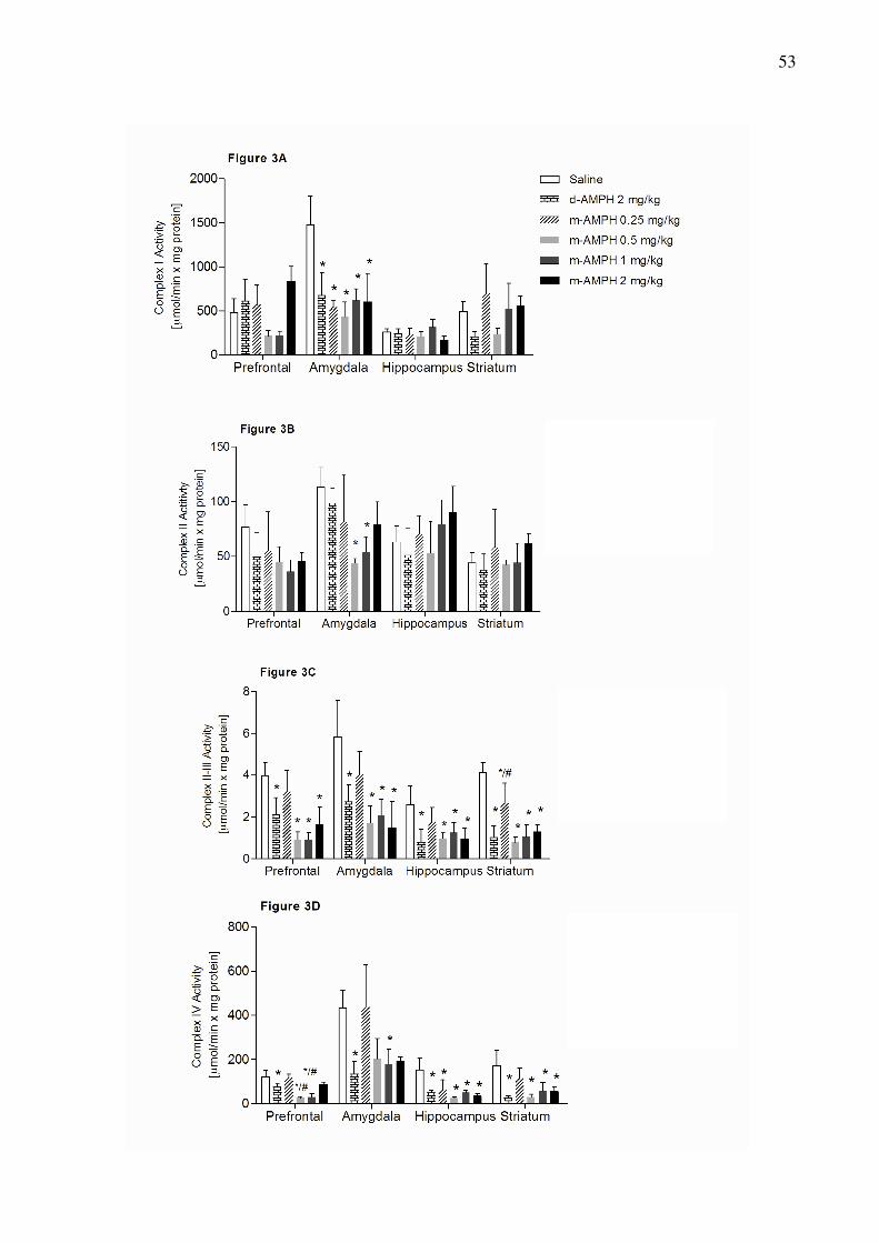
53

54

55
4.2 Artigo Cientifico 2
LITHIUM AND VALPROATE MODULATE ENERGY METABOLISM IN AN
ANIMAL MODEL OF MANIA INDUCED BY METHAMPHETAMINE.
Gustavo Feier, Samira S. Valvassori, Felipe Ornel, Wilson R. Resende, Meline O. Morais,
Lis M. Mello-Santos, Monica L. Andersen, Emilio L. Streck, João Quevedo
Artigo cientifico submetido para publicação no periódico Brain Research Bulletin,
Dezembro de 2011.
56
Brain Research Bulletin
Lithium and Valproate Modulate Energy Metabolism in an Animal Model of Mania Induced
by Methamphetamine
Gustavo Feiera, Samira S. Valvassoria, Felipe Ornela, Wilson R. Resendea, Meline O. Moraisb,
Lis M. Mello-Santosb, Monica L. Andersenc, Emilio L. Streckb, João Quevedoa*
aLaboratory of Neurosciences and National Institute for Translational Medicine (INCT-TM),
Postgraduate Program in Health Sciences, Health Sciences Unit, University of Southern Santa
Catarina, 88806-000 Criciúma, SC, Brazil. bLaboratory of Experimental Pathophysiology and National Institute for Translational
Medicine (INCT-TM), Postgraduate Program in Health Sciences, Health Sciences Unit,
University of Southern Santa Catarina, 88806-000 Criciúma, SC, Brazil. cDepartment of Psychobiology, Universidade Federal de São Paulo, 04024-002 São Paulo,
SP, Brazil.
*Prof. João Quevedo, MD, PhD - Laboratório de Neurociências, PPGCS, UNASAU,
Universidade do Extremo Sul Catarinense, 88806-000 Criciúma, SC, Brazil. Fax: #55 48
3443 4817. E-mail: [email protected]
Abstract
Animal models have been important to study intracellular systems involved in bipolar
disorder (BD). The animal model of mania induced by amphetamine has been considered a
good model for the study of BD. However, some studies have shown differences between
amphetamines to induce both behavioral and biochemical changes. In recent years our
research laboratory has validated animal model of mania induced by d-AMPH and has found
behavioral and biochemical changes induced by d-AMPH as well as in BD. The aim of this

57
study is to assess behavioral and energy metabolism changes in an animal model of mania
induced by metamphetamine (m-AMPH). Wistar rats were first given m-AMPH or saline for
14 days, and then, between days 8 and 14, rats were treated with lithium (Li), valproate
(VPA) or saline (Sal). Locomotor behavior was assessed using the open-field task and
activities of Krebs cycle enzymes (citrate synthase and succinate dehydrogenase),
mitochondrial respiratory chain complexes (I, II, III and IV) and creatine kinase were
measured in brain structures (prefrontal, amygdala, hippocampus and striatum). Li and VPA
reversed m-AMPH-induced hyperactivity. The administration of m-AMPH inhibited activities
of Krebs cycle enzymes and complexes of mitochondrial respiratory chain in all analyzed
structures. Li and VPA reversed m-AMPH-induced energetic metabolism dysfunction;
however, the effects of Li and VPA were dependent on the brain region analyzed. These
findings suggested that m-AMPH also is able to induced hyperactivity and energetic
metabolism dysfunction, both seen in BD. Besides, the results suggest that the animal model
of mania induced by m-AMPH fulfills adequate face, construct and predictive validity as an
animal model of mania.
Key words: m-AMPH; animal model of mania; Krebs cycle enzymes; complexes of
mitochondrial respiratory chain
Introduction
Although bipolar disorder (BD) is a common psychiatric disorder that leads to serious
health problems, little is known about its pathophysiology. BD is a multifactorial illness and
has diversity symptoms including recurrences of mania, depression, and mixed states; this
hampers the development of a suitable animal model (Machado-Vieira et al., 2004). Despite
the difficulties inherent in modeling BD in animals, several behavioral animal models of
mania or depression have been developed to try to mimic some aspect of behavioral changes

58
found in this psychiatric condition (Frey et al., 2006a; Jornada et al., 2010; Herman et al.,
2007; Machado-Vieira et al., 2004).
The psychostimulant-induced hyperactivity is the best established animal model of
mania; because it contains the three major criteria for the development of an adequate animal
model: face validity, construct validity and predictive validity. The amphetamines AMPHs
administration in rodents causes hyperlocomotion, insomnia and increased sexual drive (face
validity) (Fiorino and Phillips, 1999); besides, reproduces pathophysiological characteristics
of the bipolar condition (construct validity) (Frey et al., 2006d) and responses to antimanic
agents, such as antipsychotics and mood stabilizers (predictive validity) (Frey et al., 2006b;
Frey et al., 2006c).
The most common types of AMPHs are dextroamphetamine (d-AMPH) and
methamphetamine (m-AMPH). m-AMPH and d-AMPH have similar chemical structures,
since, m-AMPH is the N-methylated analog of d-AMPH (Hoffman and Lefkowitz, 1996).
Despite their structural similarities, some studies have reported differences between two
drugs, particularly in the induction of behavioral and neurochemical changes.
Epidemiological evidence indicates that m-AMPH abuse rates are greater than those of d-
AMPH (SAMHSA, 2007). Recently, our research group showed that m-AMPH induces
oxidative damage greater than d-AMPH (da-Rosa et al., 2011). Besides, Li and VPA reversed
and prevented m-AMPH-induced hyperactivity when m-AMPH was administered at 0.25
mg/kg (da-Rosa et al., in press), while the mood stabilizers reversed and prevented
hyperactivity induced by d-AMPH at 2mg/kg (Frey et al., 2006a,b,c,d).
Several studies have suggested that dysfunctional cellular energy metabolism have a
central role in BD. Abnormalities in energy metabolism were found in functional assays and
in magnetic resonance spectroscopy studies (Regenold et al., 2009; Dager et al., 2004;
Deicken et al., 1995; Frey et al., 2007). In an animal model of mania, Li and VPA were able

59
to reverse and prevent d-AMPH-induced mitochondrial dysfunction, suggesting that one of
the mechanisms of action of mood stabilizers may be decreasing the amount of dopamine
available, and stabilizing mitochondrial function in the pathophysiology of BD (Valvassori et
al., 2010).
Dysfunctions in the Krebs cycle can be capable of alter the rate of brain metabolism
and the production of free radicals. After glycolisis, pyruvate is decarboxylated to acetyl CoA
by the pyruvate dehydrogenase. The conversion of acetyl CoA to CO2 in the Krebs cycle
results in production of NADH for the electron transport chain and subsequent production of
ATP. Krebs cycle is a chemical system made up of several enzymes and steps. In the first
step, citrate synthase (CS) catalyzes the condensation of oxaloacetate and the acetyl group of
acetyl coenzyme-A (Shepherd and Garland, 1969). In the final step of Krebs cycle, malate
dehydrogenase (MD) catalyzes the dehydrogenation of l-malate to oxaloacetate (Kelly et al.,
1989). Succinate dehydrogenase (SD) is part of both the Krebs cycle and the respiratory chain
(complex II); therefore, this enzyme is one of the most important markers of the
mitochondrial ability to supply an adequate amount of ATP (Tyler, 1992).
After the Krebs cycle, there is another process called oxidative phosphorylation, which
electrons are passed along a series of respiratory enzyme complexes (complex I, II, III and
IV) located in the inner mitochondrial membrane, and the energy released by this electron
transfer is used to pump protons across the membrane. The resultant electrochemical gradient
enables another complex, adenosine 5′-triphosphate (ATP) synthase, to synthesize ATP from
ADP plus Pi (Horn and Barrientos, 2008).
The main symptoms of BD are neurovegetative abnormalities, impulsivity, and
psychosis, suggesting that anterior limbic brain networks controlling these behaviors are
dysfunctional. It is well known that the amygdala modulates the limbic system, controlling an
60
iterative circuit, prefrontal–striatal–thalamic, which control complex socioemotional
behaviors (Strakowski et al., 2000; Strakowski et al., 2005).
Thus, we examined the activities of mitochondrial enzymes of the Krebs cycle (CS,
MD and SD) and respiratory enzyme complexes (complex I, II, III and IV) in amygdala,
prefrontal, striatum and hippocampus of rats submitted to an animal model of mania induced
by m-AMPH.
Experimental methods
Animals
The subjects were adult male Wistar rats (weighting 250–350 g) obtained from our breeding
colony. The animals were housed five to a cage, with food and water available ad libitum and
were maintained on a 12-h light/dark cycle (lights on at 7:00 a.m.) at a temperature of 22 ±
1°C. All experimental procedures were performed in accordance with, and with the approval
of the local Ethics Committee in the use of animals at the Universidade do Extremo Sul
Catarinense. All experiments were performed at the same time during the day to avoid
circadian variations.
Drugs and pharmacological procedures
The animals received one daily intraperitoneal injection (i.p.) of m-AMPH 0.25mg/kg or
saline (Sal) for 14days (45 animals per group). On the 8th day of treatment, the animals in the
saline and d-AMPH group were divided in 3 groups (15 animals per group): 1) treatment with
Li (47.5 mg/kg i.p.); 2) treatment with VPA (200 mg/kg i.p.) and 3) treatment with Sal for 7
days twice a day for all drugs. On the 15th day of treatment, the animals received a single
injection of m-AMPH or Sal and locomotor activity was assessed 2 h after the last injection.
The rats were killed by decapitation immediately after the open-field task and amygdala,

61
prefrontal, striatum and hippocampus were dissected, rapidly frozen and stored -70º C until
assayed.
Locomotor Activity
Locomotor activity was assessed using the open-field task as previously described (Barros et
al., 2002; Frey et al., 2006a; b; c). This task was performed in a 40x60 cm open field
surrounded by 50 cm high walls, made of brown plywood, with the floor divided into 12
equal rectangles by black lines. The animals were gently placed on the left rear rectangle, and
left free to explore the arena for 5 min. Crossings of the black lines (locomotor activity/
horizontal activity) and rearings (exploratory activity/ vertical activity) were counted.
Tissue and homogenate preparation
The prefrontal cortex, amygdala, hippocampus and striatum were removed and homogenized
(1:10, w/v) in SETH buffer, pH 7.4 (250 mM sucrose, 2 mM EDTA, 10 mM Trizma base, 50
IU/ml heparin). The homogenates were centrifuged at 800 × g for 10 min at 4°C and the
supernatants kept at −70oC until being used for enzymes activity determination. The maximal
period between homogenate preparation and enzyme analysis was always less than 5 days.
The protein content was determined by the method described by Lowry and colleagues
(Lowry et al., 1951) using bovine serum albumin as standard.
Activities of enzymes of Krebs cycle
Citrate synthase activity: Citrate synthase activity was assayed according to the method
described by Shepherd and Garland (1969). The reaction mixture contained 100 mM Tris, pH
8.0, 100 mM acetyl CoA, 100 mM 5,5′-di-thiobis-(2- nitrobenzoic acid), 0.1% triton X-100,

62
and 2–4 μg supernatant protein and was initiated with 100 μM oxaloacetate and monitored at
412 nm for 3 min at 25 °C.
Malate dehydrogenase activity: Malate dehydrogenase was measured as described by Kitto
(1969). Aliquots (20 mg protein) were transferred into a medium containing 10 mM rotenone,
0.2% Triton X-100, 0.15 mM NADH, and 100 mM potassium phosphate buffer, pH 7.4, at
37°C. The reaction was started by the addition of 0.33 mM oxaloacetate. Absorbance was
monitored as described above.
Succinate dehydrogenase activity: Succinate dehydrogenase activity was determined
according to the method of Fischer and colleagues (1985), and measured by following the
decrease in absorbance due to the reduction of 2,6-di-chloro-indophenol (2,6-DCIP) at 600nm
with 700nm as a reference wavelength (ε = 19.1 mM−1 cm−1) in the presence of phenazine
methasulphate (PMS). The reaction mixture consisting of 40mM potassium phosphate, pH
7.4, 16mM succinate and 8μM 2,6-DCIP was pre-incubated with 40–80μg homogenate
protein at 30oC for 20 min. Subsequently, 4mM sodium azide, 7μM rotenone and 40μM 2,6-
DCIP were added and the reaction was initiated by the addition of 1mM PMS and was
monitored for 5 min.
Activities of mitochondrial respiratory chain enzymes
Complex I activity: NADH dehydrogenase (complex I) was evaluated according to Cassina
and Radi (1996) by the determination of the rate of NADH-dependent ferricyanide reduction
at λ = 420 nm.
Complex II activity: The activities of succinate-2,6- dichloroindophenol (DCIP)-
oxidoreductase (complex II) was determined by the method described by Fischer and
colleagues (1985). Complex II activity was measured by following the decrease in absorbance
due to the reduction of 2,6-DCIP at λ = 600 nm.

63
Complex II-III activity: The activity of succinate:cytochrome c oxidoreductase (complex
III) was determined by the method described by Fischer and colleagues (1985). Complex II-
III activity was measured by cytochrome c reduction using succinate as substrate at λ = 550
nm.
Complex IV activity: The activity of cytochrome c oxidase (complex IV) was assayed
according to the method described by Rustin and colleagues (1994), measured by following
the decrease in absorbance due to the oxidation of previously reduced cytochrome c (prepared
by reduction of cytochrome with NaBH4 and HCl) at λ = 550 nm with 580 nm as the
reference wavelength (ɛ= 19.1 mM−1 cm−1). The activities of the mitochondrial respiratory
chain complexes were calculated as nmol. min-1. mg protein-1.
Activity of creatine kinase enzyme
Creatine kinase activity was measured in brain homogenates pretreated with 0.625 mM lauryl
maltoside. The reaction mixture consisted of 60mM Tris–HCl, pH 7.5, containing 7 mM
phosphocreatine, 9 mM MgSO4 and approximately 0.4–1.2 μg protein in a final volume of
100 μL. After 15 min of pre-incubation at 37 °C, the reaction was started by the addition of
3.2 mmol of ADP plus 0.8 mmol of reduced glutathione. The reaction was stopped after 10
min by the addition of 1 μmol of p-hydroxymercuribenzoic acid. The creatine formed was
estimated according to the colorimetric method of Hughes (1962). The color was developed
by the addition of 100 μL 2%α-naphtol and 100 μL 0.05% diacetyl in a final volume of 1 mL
and read spectrophotometrically after 20 min at 540 nm. Results were expressed as
units/min×mg protein.

64
Statistical analysis
Data were analyzed by one-way analysis of variance followed by the Tukey test when F was
significant and are expressed as mean ± standard deviation. All analyses were performed
using the Statistical Package for the Social Science (SPSS; version 16.0) software.
Results
Li and VPA reversed hyperlocomotion induced by m-AMPH in rats
Results for locomotor activity are shown in Figure 1. There was a significant effect of m-
AMPH and mood stabilizers in the number of both crossings [df=5, F=27.57, p<0.001] and
rearings [df=5, F=7.43, p<0.001]. Further analysis with Tukey post-hoc tests showed that
administration of m-AMPH increased locomotion and rearing behavior in rats treated with Sal
(both Ps < 0.001 compared to the group treated with SAL followed by SAL). Treatment with
Li and VPA reversed m-AMPH-related hyperlocomotion. Animals given Li or VPA followed
by m-AMPH did not differ from the control group given Sal followed by Sal.
Li and VPA treatment attenuate m-AMPH-induced decreases in activities of enzymes of Krebs
cycle in rat brain
Results for citrate sinthase activity are shown in Figure 2A. The m-AMPH administration
caused a marked decreased of the citrate synthase activity of rat prefrontal, amygdala,
hippocampus and striatum. Seven days of treatment with Li and VPA reversed m-AMPH’s
effects on citrate synthase activity in the amygdala, hippocampus and striatum. In the
prefrontal, Li reversed and VPA partially reversed m-AMPH’s effects on citrate synthase
activity. Chronic use of Li and VPT in saline-treated animals had no influence in citrate

65
synthase activity in the amygdala hippocampus and striatum. In the prefrontal, VPA alone,
but not Li, decreased the citrate synthase activity.
Results for succinato dehydrogenase activity are shown in Figure 2B. The m-AMPH
administration decreased succinato dehydrogenase activity in all brain structures evaluated.
Treatment with Li reversed the reduction of succinato dehydrogenase activity induced by m-
AMPH in the amygdala and hippocampus, but not in the striatum. In the prefrontal, the Li
administration in the m-AMPH pretreated rats increased succinate dehydrogenase activity
when compared to control group. Besides, in the hippocampus, succinate dehydrogenase
activity was significantly decreased in the Sal+Li group. Seven days of treatment with VPA
reversed m-AMPH’s effects on succinate dehydrogenase in the striatum, but not in the
prefrontal and amygdala. In the hippocampus, succinate dehydrogenase activity was
significantly increased in the VPA+m-AMPH group. We found also that treatment with VPA
alone, succinate dehydrogenase activity was significantly enhanced in the prefrontal when
compared to control group.
Results for malate dehydrogenase activity are shown in Figure 2C. After m-AMPH
administration, malate dehydrogenase activity also decreased in the amygdala, hippocampus
and striatum. The Li and VPA treatment reversed m-AMPH’s effects on the malate
dehydrogenase activity in the hippocampus and striatum. In the amygdala, VPA, but not Li,
partially reversed m-AMPH’s effects on malate dehydrogenase activity.
Li and VPA treatment attenuate m-AMPH-induced decreases in activities of respiratory chain
complexes in rat brain
m-AMPH administration inhibited the respiratory chain complexes (I, II, II-III and IV) in all
brain structure evaluated.

66
Results for complex I activity are shown in Figure 3A. The Li and VPA administration
reversed m-AMPH’s effects on complex I activity in the prefrontal and amygdala of rats. The
VPA treatment, but not Li, reversed the m-AMPH-induced the decreased in complex I activity
in the hippocampus and striatum.
Results for complex II activity are shown in Figure 3B. The Li and VPA treatment reversed
m-AMPH’s effects on complex II activity in the hippocampus and striatum. However, in the
prefrontal and amygdala VPA, but not Li, significantly increased the complex II activity when
compared to control group.
Results for complex II-III activity are shown in Figure 3C. The Li and VPA treatment
attenuate the m-AMPH-induced the decreased in complex II-III activity in the hippocampus
and striatum. Seven days of treatment with VPA attenuate m-AMPH’s effects on complex II-
III activity in the prefrontal and amygdala; unlike, Li administration potentiates the m-
AMPH’s effects in these cerebral structures.
Results for complex IV activity are shown in Figure 3D. Treatment with Li and VPA reversed
m-AMPH-related complex IV dysfunction in the amygdala and striatum. In the prefrontal,
VPA, but not Li, reversed m-AMPH’s effects on complex IV activity. In the amygdala, Li and
VPA partially reversed the m-AMPH’s effects on complex IV activity.
Effects of Li and VPA on creatine kinase activity in the rats submitted to animal model of
mania induced by m-AMPH
The m-AMPH administration decreased CK activity in the prefrontal of rats and the mood
stabilizers, Li and VPA, were not able to reverse this damage. In the amygdala and
hippocampus, CK activity was decreased in the Sal+VPA and m-AMPH+Li groups. In the
striatum CK activity was decreased in the Sal+VPA group.

67
Discussion
Initially, we reproduced data from previous studies from our laboratory, which Li and
VPA reversed the hyperactivity induced by m-AMPH at 0.25mg/kg (da-Rosa et al., in press).
It is know that mood stabilizers reversed and prevented hyperactivity induced by d-AMPH at
2mg/kg (Frey et al., 2006a; b; c; d).
Accumulating evidence suggests that energetic metabolism dysfunction contribute to
the pathogenesis of BD. Impairment of complex I was found in the prefrontal cortex of
bipolar patients (Andreazza et al., 2010). In a large-scale DNA microarray analysis in
postmortem brains was showed a global down-regulation of mitochondrial genes, such as
those encoding respiratory chain components, in BD (Iwamoto et al., 2005). In an elegant
study, Cataldo and colleagues (2010) have demonstrated that mitochondria from patients with
BD have size and distributional abnormalities compared with control group. In the brain,
individual mitochondria profiles had significantly smaller areas, on average, in BD samples.
In peripheral cells, mitochondria in BD samples were concentrated proportionately more
within the perinuclear region than in distal processes. These changes in mitochondrial
morphology and distribution appear to be linked to diverse cellular events, such as
differentiation, aging, and apoptosis. In addition, abnormalities in energy metabolism of BD
patients were also found in functional assays and in magnetic resonance spectroscopy studies
(Regenold et al., 2009; Dager et al., 2004; Deicken et al., 1995; Frey et al., 2007).
In previous studies, we have demonstrated also that d-AMPH at 2mg/kg inhibited
citrate synthase (Correa et al., 2007) and mitochondrial respiratory chain complexes I, II, II-
III and IV (Valvassori et al., 2010) activities in brain of rats, which was reversed by Li and
VPA. As described above, changes in energy metabolism are strongly linked to BD; therefore,
it is interesting the development of animal models with this feature. In order to study the
action mechanisms of classical drugs on this system and still test new drugs.

68
Here, we demonstrated that chronic administration of m-AMPH at 0.25mg/kg was
able to inhibit the cycle Krebs enzymes activities: citrate synthase, succinate dehydrogenase
and malate dehydrogenase. Moreover, we demonstrated also that m-AMPH at 0.25mg/kg
inhibit mitochondrial respiratory chain complexes (I, II, II-III and IV) in all brain structure
evaluated, as well as d-AMPH at 2mg/kg as demonstrated in previous studies (Valvassori et
al., 2010). From the results obtained in this study, we suggested that the decreased Krebs
cycle enzymes activity induced by m-AMPH inhibit energy production by mitochondrial
respiratory chain via Krebs cycle. Recently, was showed that m-AMPH administration
decreased Krebs cycle intermediates in the urine and increased glucose in the plasma of rats
(Shima et al., 2011). Kim and colleagues (2009) have demonstrated dose-dependent frontal
hypometabolism on fluorodeoxy-D-glucose-positron emission tomography (FDG-PET) in
methamphetamine abusers. Impaired energy metabolism results in depletion of ATP in cells,
and ATP depletion as a consequence of mitochondrial toxicity may be related to a number of
m-AMPH-induced toxicities.
Another significant finding in this study is that mood stabilizers, Li and VPA,
attenuated the AMPH’s effects on Krebs cycle enzymes activity and, consequently, reduced
the impairment on respiratory chain complexes activity. Our results are in agreement with a
previous study, which showed that that long-term treatment with Li or VPA protected against
m-AMPH-induced toxicity at the mitochondrial level (Bachmann et al., 2009). Additionally,
the mood stabilizers prevented the m-AMPH-induced reduction of mitochondrial cytochrome
c, the mitochondrial anti-apoptotic Bcl-2/Bax ratio, and mitochondrial cytochrome oxidase
activity (Bachmann et al., 2009).
Kazuno (2008) and colleagues have showed that VPA may stabilize intracellular
calcium in cells with high mitochondrial calcium levels. The excessive release of glutamate
by m-AMPH may induce neuronal damage through receptor-mediated intracellular Ca2+

69
overload and increased reactive oxygen species (ROS) production (Arundine and Tymianski,
2004; Choi, 1995; Lu et al., 2008). The Li and VPA treatment exert neuroprotective effects
against cytotoxicity by inhibiting the glutamate-induced increase of intracellular free Ca2+
concentration (Shao et al., 2005). Besides, it is well known that m-AMPH-induced increased
DA oxidized metabolites inhibit the mitochondrial respiratory system, both in vivo and in
vitro (Przedborsk et al., 1993). The Li and VPA treatment inhibit the dopaminergic system by
a mechanism of second messenger activation, which are activated by D2 receptors (Yatham et
al., 2002). Together with our results, these studies suggest that mood stabilizers, Li e VPA,
attenuate the glutamate and DA systems, protecting the mitochondria against m-AMPH-
induced damage.
Finally, we can observe that m-AMPH inhibit the CK activity only in the prefrontal;
however, Li and VPA were not able to reverse this enzyme alteration. In a previous study of
our laboratory we have showed that d-AMPH at 2mg/kg inhibited CK activity in
hippocampus, striatum and cortex, but not in prefrontal cortex, and administration of Li or
VPA did not reverse the enzyme inhibition (Streck et al., 2008). This discrepancy can be
explain by the difference, in the structure and in the dose administered, between the two
drugs. Here we demonstrated also that chronic VPA administration, in the saline pretreated
rats, decreased CK activity in all brain structures evaluated. Additionally, despite m-AMPH
alone did not alter CK activity in the amygdala and hippocampus, the Li treatment (Li+m-
AMPH) decreased CK activity in this brain structures. Couthon and colleagues (1997) have
demonstrated that incubation of dimeric MM-creatine kinase (MM-CK) with LiCl results in
dissociation of the subunits and complete enzyme inactivation.
These findings suggested that m-AMPH also is able to induced hyperactivity and
energetic metabolism dysfunction, both seen in BD. Besides, the results suggest that the

70
animal model of mania induced by m-AMPH fulfills adequate face, construct and predictive
validity as an animal model of mania.

71
References
Andreazza, A.C., Shao, L., Wang, J.F., Young, L.T., 2010 Mitochondrial complex I activity
and oxidative damage to mitochondrial proteins in the prefrontal cortex of patients with
bipolar disorder. Arch Gen Psychiatry. 67, 360-8.
Arundine, M. and Tymianski, M., 2004. Molecular mechanisms of glutamate-dependent
neurodegeneration in ischemia and traumatic brain injury. Cell. Mol. Life Sci. 61,657–668.
Bachmann, R.F., Wang, Y., Yuan, P., Zhou, R., Li, X., Alesci, S., Du, J., Manji, H.K., 2009.
Common effects of lithium and valproate on mitochondrial functions: protection against
methamphetamine-induced mitochondrial damage. Int J Neuropsychopharmacol. 12, 805-22.
Barros, D.M., Izquierdo, L.A., Medina, J.H., Izquierdo, I., 2002. Bupropion and sertraline
enhance retrieval of recent and remote long-term memory in rats. Behav Pharmacol. 13, 215-
20.
Calabrese, V., Scapagnini, G., Giuffrida Stella, A.M., Bates, T.E., Clark, J.B., 2001.
Mitochondrial involvement in brain function and dysfunction: relevance to aging,
neurodegenerative disorders and longevity. Neurochemical Research. 6, 739-64.
Cataldo, A.M., McPhie, D.L., Lange, N.T., Punzell, S., Elmiligy, S., Ye, N.Z., Froimowitz,
M.P., Hassinger, L.C., Menesale, E.B., Sargent, L.W., Logan, D.J., Carpenter, A.E., Cohen,
B.M., 2010. Abnormalities in mitochondrial structure in cells from patients with bipolar
disorder. Am J Pathol. 177, 575-85.
Choi, D.W., 1995. Calcium: still center-stage in hypoxic-ischemic neuronal death. Trends
Neurosci., 18, 58–60.
Corrêa, C., Amboni, G., Assis, L.C., Martins, M.R., Kapczinski, F., Streck, E.L., Quevedo, J.,
2007. Effects of lithium and valproate on hippocampus citrate synthase activity in an animal
model of mania. Prog Neuropsychopharmacol Biol Psychiatry. 9, 887-91.
Couthon, F., Clottes, E., Vial, C., 1997. High salt concentrations induce dissociation of
dimeric rabbit muscle creatine kinase. Physico-chemical characterization of the monomeric
species. Biochim Biophys Acta. 1339, 277-88.
Dager S.R., Friedman S.D., Parow A., Demopulos C., Stoll A.L., Lyoo I.K., Dunner D.L.,
Renshaw P.F., 2004. Brain metabolic alterations in medication-free patients with bipolar
disorder. Arch. Gen. Psychiatry. 61, 450-58.
Deicken R.F., Fein G., Weiner M.W., 1995. Abnormal frontal lobe phosphorous metabolism
in bipolar disorder. Am. J. Psychiatry. 152 , 915-18.

72
Fiorino, D.F., Phillips, A.G., 1999. Facilitation of sexual behavior and enhanced dopamine
efflux in the nucleus accumbens of male rats after d-amphetamine-induced behavioral
sensitization. J Neurosci. 1, 456–63.
Fischer, J.C., Ruitenbeek, W., Berden, J.A., Trijbels, J.M., Veerkamp, J.H., Stadhouders,
A.M., Sengers, R.C., Janssen, A.J., 1985. Differential investigation of the capacity of
succinate oxidation in human skeletal muscle. Clin. Chim. Acta. 153, 23-6.
Frey B.N., Stanley J.A., Nery F.G., Monkul E.S., Nicoletti M.A., Chen H.H., Hatch J.P.,
Caetano S.C., Ortiz O., Kapczinski F., Soares J.C., 2007. Abnormal cellular energy and
phospholipid metabolism in the left dorsolateral prefrontal cortex of medication-free
individuals with bipolar disorder: an in vivo 1H MRS study. Bipolar Disord. 9, 119-27.
Frey B.N., Valvassori S.S., Réus G.Z., Martins M.R., Petronilho F.C. Bardini K., Dal-Pizzol,
F., Kapczinski, F., Quevedo, J., 2006c. Effects of lithium and valproate on amphetamine-
induced oxidative stress generation in an animal model of mania. J Psychiatry Neurosci. 31,
326–32.
Frey B.N., Valvassori S.S., Réus G.Z., Martins M.R., Petronilho F.C. Bardini K., Dal-Pizzol,
F., Kapczinski, F., Quevedo, J., 2006d. Changes in antioxidant defense enzymes after d-
amphetamine exposure: implications as an animal model of mania. Neurochem Res. 31, 699–
703.
Frey, B.N., Andreazza, A.C., Ceresér, K.M., Martins, M.R., Valvassori, S.S., Réus, G.Z.,
Quevedo, J., Kapczinski, F., 2006a. Effects of mood stabilizers on hippocampus BDNF levels
in an animal model of mania. Life Sci. 13, 281-6.
Frey, B.N., Martins, M.R., Petronilho, F.C., Dal-Pizzol, F., Quevedo, J., Kapczinski, F.,
2006b. Increased oxidative stress after repeated amphetamine exposure: possible relevance as
a model of mania. Bipolar Disord. 8, 275–80.
Herman, L., Hougland, T., El-Mallakh, R.S., 2007. Mimicking human bipolar ion
dysregulation models mania in rats. Neurosci Biobehav Rev. 31, 874-81.
Hoffman, B.B., Lefkowitz RJ (1996). Catecholamines, sympathomimetic drugs, and
adrenergic receptor antagonists. In:Hardman JG, Limbird LE, Molinoff PB, et al, editors.
Goodman and Gilman’s The Pharmacological Basis of Therapeutics. 9th ed. New York:
McGraw-Hill; pp. 199-227.
Horn, D., Barrientos, A., 2008. Mitochondrial copper metabolism and delivery to cytochrome
c oxidase. IUBMB Life. 60, 421–429.

73
Iwamoto, K., Bundo, M., Kato, T., 2005. Altered expression of mitochondria-related genes in
postmortem brains of patients with bipolar disorder or schizophrenia, as revealed by large-
scale DNA microarray analysis. Hum Mol Genet. 15, 241-53.
Jornada, L.K., Moretti, M., Valvassori, S.S., Ferreira, C.L., Padilha, P.T., Arent, C.O., Fries,
G.R., Kapczinski, F., Quevedo, J., 2010. Effects of mood stabilizers on hippocampus and
amygdala BDNF levels in an animal model of mania induced by ouabain. J Psychiatr Res. 44,
506-10.
Kelly, D., Gordon, J., Alpers, R., Strauss, A.W., 1989 The tissue-specific expression and
developmental regulation of two nuclear genes encoding rat mitochondrial proteins. Medium
chain acyl-CoA dehydrogenase and mitochondrial malate dehydrogenase. J. Biol. Chem. 264,
18921–25.
Kim, Y.T., Lee, S.W., Kwon, D.H., Seo, J.H., Ahn B.C., Lee, J., 2009. Dose-dependent
frontal hypometabolism on FDG-PET in methamphetamine abusers. J. Psychiatr. Res. 43,
1166–70.
Kitto, G.B., 1969. Intra- and extramitochondrial malate dehydrogenases from chicken and
tuna heart. Methods Enzymol. 23, 106-16.
Lowry, O.H., Rosebrough, N.J., Farr, A.L., Randall, R.J., 1951. Protein measurement with the
Folin phenol reagent. J. Biol. Chem. 193, 265-75.
Lu, M., Hu, L.F., Hu G., Bian, J.S., 2008. Hydrogen sulfide protects astrocytes against
H(2)O(2)-induced neural injury via enhancing glutamate uptake. Free Radic. Biol.
Med., 45, 1705–1713.
Machado-Vieira, R., Kapczinski, F., Soares, J.C., 2004. Perspectives for the development of
animal models of bipolar disorder. Prog Neuropsychopharmacol Biol Psychiatry. 28, 209-24.
Przedborski, S., Jackson-Lewis, V., Muthane, U., Jiang, H., Ferreria, M., Naini, A.B, et
al., 1993. Chronic levodopa administration alters cerebral mitochondrial respiratory
chainactivity. Ann. Neurol. 34, 715–23.
Regenold, W.T., Phatak, P., Marano, C.M., Sassan, A., Conley, R.R., Kling, M.A., 2009.
Elevated cerebrospinal fluid lactate concentrations in patients withbipolar disorder and
schizophrenia: implications for the mitochondrial dysfunction hypothesis. Biol. Psychiatry.
65, 489-94.
SAMHSA (Substance Abuse and Mental Health Services Administration), Office of Applied
Studies. Treatment Episode Data Set (TEDS) Highlights - - 2007 National Admissions to
Substance Abuse Treatment Services. 2009; OAS Series #S-45, HHS Publication No. (SMA)
09-4360, Rockville, MD.

74
Shao, L., Young, L.T., Wang, J.F., 2005. Chronic treatment with mood stabilizers lithium and
valproate prevents excitotoxicity by inhibiting oxidative stress in rat cerebral cortical cells.
Biological Psychiatry 58, 879–884.
Shepherd, D., Garland, P.B., 1996. The kinetic properties of citrate synthase from rat liver
mitochondria. Biochem. J. 114, 597–610.
Shima, N., Miyawaki, I., Bando, K., Horie, H., Zaitsu, K., Katagi, M., Bamba, T.,
Tsuchihashi, H., Fukusaki, E., 2011. Influences of methamphetamine-induced acute
intoxication on urinary and plasma metabolic profiles in the rat. Toxicology. 287, 29-37.
Strakowski, S.M., DelBello, M.P., Adler, C., Cecil, K.M., Sax, K.W., 2000. Neuroimaging in
bipolar disorder. Bipolar Disord. 2, 148-64.
Strakowski, S.M., Delbello, M.P., Adler, C.M., 2005. The functional neuroanatomy of bipolar
disorder: a review of neuroimaging findings. Mol Psychiatry. 10, 105-16.
Streck, E.L., Amboni, G., Scaini, G., Di-Pietro, P.B., Rezin, G.T., Valvassori, S.S., Luz, G.,
Kapczinski, F., Quevedo, J., 2008. Brain creatine kinase activity in an animal model of mania.
Life Sci. 13, 424-9.
Tyler, D., 1992. The Mitochondrion in Health and Diseases, VCH Publishers, New York .
Valvassori, S.S., Rezin, G.T., Ferreira, C.L., Moretti, M., Gonçalves, C.L., Cardoso, M.R.,
Streck, E.L., Kapczinski, F., Quevedo, J., 2010. Effects of mood stabilizers on mitochondrial
respiratory chain activity in brain of rats treated with d-amphetamine. J Psychiatr Res. 44,
903-9.
Van der Geer, J., Hanraads, J.A.J., Lupton, R.A., 2010. The art of writing a scientific article.
J. Sci. Commun. 163, 51–59.

75
Legends to Figures Figure 1: Open field test. Free movement in the open field: crossings (dF=5; F=27.568; p<0.0001), rearings (dF=5; F=7.430; p<0.0001). *Different from Sal+Sal group. #Different from m-AMPH+Sal group. Figure 2: Activities of enzymes of Krebs cycle. A) Citrate synthase activity: In the prefrontal (dF=5; F=27.845; p<0.0001), amygdala (dF=5; F=16.176; p<0.0001), hippocampus (dF=5; F=8.799; p<0.0001) and striatum (dF=5; F=6.796; p<0.0001). B) Succinate dehydrogenase activity: In the prefrontal (dF=5; F=20.922; p<0.0001), amygdale (dF=5; F=22.592; p<0.0001), hippocampus (dF=5; F=25.361; p<0.0001) and striatum (dF=5; F=14.491; p<0.0001). C) Malate dehydrogenase activity: In the prefrontal (dF=5; F=4.332; p<0.0001), amygdala (dF=5; F=38.111; p<0.0001), hippocampus (dF=5; F=8.945; p<0.0001) and striatum (dF=5; F=15.033; p<0.0001). *Different from Sal+Sal group. #Different from m-AMPH+Sal group. Figure 3: Activities of mitochondrial respiratory chain enzymes. A) Complex I activity: In the prefrontal (dF=5; F=17.512; p<0.0001), amygdala (dF=5; F=16.018; p<0.0001), hippocampus (dF=5; F=44.667; p<0.0001) and striatum(dF=5; F=32.298; p<0.0001). B) Complex II activity: In the prefrontal (dF=5; F=23.680; p<0.0001), amygdala (dF=5; F=19.278; p<0.0001), hippocampus (dF=5; F=6.466; p<0.001) and striatum (dF=5; F=10.529; p<0.0001). C) Complex II-III activity: In the prefrontal (dF=5; F=58.859; p<0.0001), amygdala (dF=5; F=111.150; p<0.0001), hippocampus (dF=5; F=26.548; p<0.0001) and striatum (dF=5; F= 33.750; p<0.0001). D) Complex IV activity: In the prefrontal (dF=5; F=46.454; p<0.0001), amygdala (dF=5; F=18.474; p=0.002), hippocampus (dF=5; F=11.713; p=0.001) and striatum (dF=5; F=18.617; p<0.0001). *Different from Sal+Sal group. #Different from m-AMPH+Sal group. Figure 4: Activity of creatine kinase: In the prefrontal (dF=5; F=25.298; p<0.0001), amygdala (dF=5; F=9.96; p<0.0001), hippocampus (dF=5; F=3.08; p<0.03) and striatum (dF=5; F=6.103; p<0.002). *Different from Sal+Sal group. #Different from m-AMPH+Sal group.

76

77

78

79
80
4.3 Artigo Cientifico 3
CREATINE KINASE LEVELS IN PATIENTS WITH BIPOLAR DISORDER: DEPRESSIVE, MANIC, AND EUTHYMIC PHASES
COMPARAÇÃO DAS FASES DE DEPRESSÃO, MANIA E EUTIMIA SOBRE OS NÍVEIS DE CREATINA QUINASE EM PACIENTES BIPOLARES
Gustavo Feier, Samira S. Valvassori, Gislaine T. Rezin, Márcio Búrigo, Emilio L. Streck, Flávio Kapczinski, João Quevedo
Artigo cientifico publicado na Revista Brasileira de Psiquiatria em Junho de 2011.
81

82
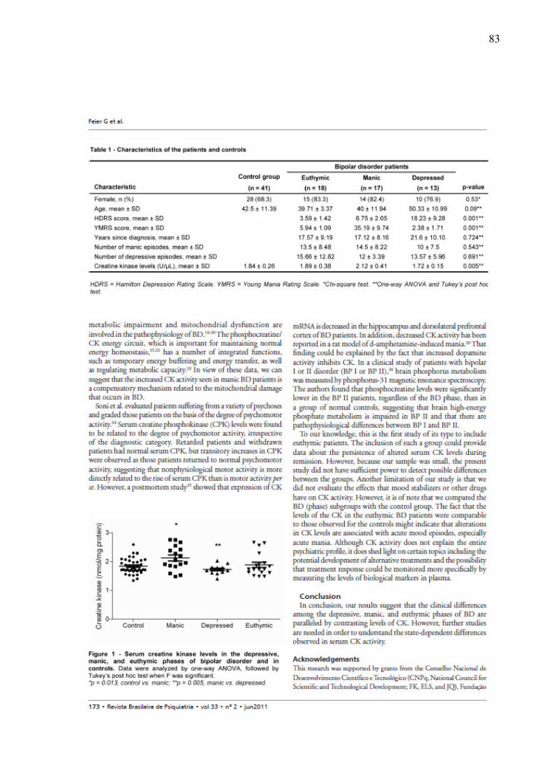
83

84

85

86
5. Discussão
O modelo animal de mania induzido por psicoestimulantes é descrito na literatura
como um bom modelo animal de TB (Machado-Vieira et al., 2004). Nos ultimos seis anos
nosso grupo de pesquisa tem validado o modelo animal de mania induzido por d-AMPH, com
o qual temos sugerido que a dopamina tem um papel fundamental tanto nos sintomas
maníacos quanto nos danos neuronais observados em pacientes bipolares (Frey et al., 2006a,
b, c, d). Entretanto, relata-se diferenças entre os tipos de anfetaminas em induzir alterações
comportamentais e neuroquímicas (Samhsa, 2007; da-Rosa et al., 2011). Tendo em vista o
fato, torna-se importante avaliar se existem diferenças entre tipos de AMPHs em mimetizar
comportamentos do tipo maníacos em animais experimentais. Além disso, cada vez mais
estudos sugerem que o TB está associado à disfunções no metabolismo energético (Deicken et
al., 1995; Dager et al., 2004; Frey et al., 2007; Regenold et al., 2009).
No presente estudo foi avaliada a diferença entre d-AMPH e m-AMPH sobre o
comportamento e disfunções no metabolismo energético no cérebro de ratos. Foram avaliados
também os efeitos de Li e VPA sobre as alterações comportamentais e do metabolismo
energético induzidos por m-AMPH. Em adição, foram comparados os níveis de CK no soro
de pacientes bipolares nas fases depressiva, maníaca e eutímica.
Na primeira fase do estudo foram comparadas as doses de 0,25, 0,5, 1 ou 2mg/kg de
m-AMPH com 2mg/kg de d-AMPH – dose já estabelecida pelo grupo de pesquisa, a qual
induz hiperatividade e inibição dos complexos da cadeia respiratória mitocondrial no cérebro
de ratos (Frey et al., 2006 a, b, c, d; Valvassori et al., 2010) – sobre tarefas comportamentais
e sobre parâmetros do metabolismo energético em córtex pré-frontal, amígdala, hipocampo e
estriado de ratos.

87
No presente estudo reproduzimos dados prévios do nosso grupo (da-Rosa et al, 2011),
onde a administração de d-AMPH (2 mg/kg) e m-AMPH nas doses de 0,25, 0,5, 1 e 2mg/kg
foram equivalentes em aumentar a atividade locomotora e exploratória nos ratos.
Adicionalmente, foi observado no presente estudo que m-AMPH e d-AMPH, ambos na dose
de 2mg/kg, aumentaram o número de visitas ao centro do campo aberto, mostrando a
equivalência das duas anfetaminas em induzir comportamento de risco. De acordo com Einat
(2006), comportamento de risco pode representar a ansiedade em muitos testes. Naturalmente
quando colocado em um campo iluminado aberto, pela primeira vez, ratos e camundongos
tendem a permanecer na periferia do aparelho ou contra as paredes, pois espaços abertos
constituem situações de risco predador. A administração de AMPHs em roedores aumenta
comportamentos de risco, pois reduzem a ansiedade e, consequentemente, os animais
aumentam o tempo gasto no centro do campo aberto (Einat et al., 2003).
No presente estudo encontramos também que a administração de m-AMPH (mas não
d-AMPH) em altas doses (2mg/kg) induz comportamento estereotípico (sniffing e grooming).
Em um estudo prévio foi demonstrado que AMPHs e nicotina aumentam estereotipia em ratos
- em até 80 min após a administração dessas drogas (Izawa et al., 2006). É bem descrito na
literatura que drogas de abuso, como a cocaína e a m-AMPH, que estimulam a transmissão de
dopaminérgica no núcleo accumbens, induzem ativação comportamental em roedores e outros
animais de laboratório (Di Chiara, 2002).
Foi observado, ainda, que a atividade das enzimas do ciclo de Krebs, citrato sintase e
succinato desidrogenase, da cadeia respiratória mitocondrial (complexos I, II, II-III e IV) e
CK foram inibidas no cérebro de ratos submetidos à administração de d-AMPH e m-AMPH;
no entanto, essas alterações no metabolismo energético variaram de acordo com a estrutura
cerebral analisada. m-AMPH e d-AMPH diminuiram a atividade dos complexos II-III e IV e a
atividaden da CK no pré-frontal dos ratos; além disso, m-AMPH inibiu a atividade da citrato
88
sintase nesta estrutura cerebral. Na amígdala, tanto d-AMPH quanto m-AMPH diminuiu a
atividade da citrato sintase, dos complexos I, II-III e IV e da CK; adicionalmente, m-AMPH
também diminuiu a atividade do complexo II nessa estrutura cerebral. No hipocampo, a
administração de ambas as AMPHs diminuiu a atividade dos complexos I e II-III; porém, uma
única injeção de m-AMPH também inibiu a atividade da citrato sintase. Finalmente, no
estriado, ambas as AMPHs inibiram as atividades da citrato sintase, da succinato
desidrogenase, dos complexos I, II, III e IV e da CK. Como podemos observar os efeitos de d-
AMPH e m-AMPH são heterogênios entre as estruturas cerebrais.
Quanto às estruturas cerebrais estudadas no presente trabalho podemos citar que a
amídala modula o sistema límbico, controlando um sistema interativo, pré-frontal-estriado-
tálamo, que controla comportamentos sócio-emocionais complexos (Strakowski et al., 2000;
Strakowski et al., 2005). As AMPHs agem sobre o sistema dopaminérgico
mesocorticolimbico, o qual projeta prolongamentos neuronais da área tegmental ventral para
o núcleo accumbens, tubérculo olfatório, córtex pré-frontal e amígdala (Kalivas & Stewart,
1991; Volkow et al., 2003). O estriado também está envolvido nos efeitos dos
psicoestimulantes, principalmente, na resposta locomotora e exploratória (Hamamura et al.,
1991;. Nestler, 2001). O córtex pré-frontal é uma importante área cerebral envolvida na
memória de trabalho, na tomada de decisões, controle inibitório, na seleção e retenção de
informações e controle executivo (Royall, 2002; Huang et al., 2004; Rinaldi et al., 2007).
Projeções glutamatérgica do córtex pré-frontal para o estriado também desempenham um
papel importante na ação das AMPHs (Kalivas et al., 2005). Além disso, o sistema
dopaminérgico é fundamental na eficácia sináptica e, consequentemente, para mudanças de
longo prazo no hipocampo. Vale citar também que a interação entre DA e receptores
glutamatérgicos são essenciais para funções cognitivas no pré-frontal e hipocampo (Gurden et

89
al., 2000; Nowak & Corces, 2000; Yang et al., 2000; Li et al., 2003; Chen et al., 2004; Huang
et al., 2004; Tseng & O'Donnell, 2004; Granado et al, 2008).
Estudos anteriores do nosso e de outros laboratórios têm demonstrado que a
administração de d-AMPH e m-AMPH em ratos inibem as enzimas do metabolismo
energético (Valenzuela & Villanueva, 1987; Corrêa et al., 2007; Streck et al, 2008; Bachmann
et al., 2009; Valvassori et al., 2010; Moretti et al., 2011). No entanto, não existem estudos
comparando as duas drogas sobre o metabolismo energético.
Na segunda parte do trabalho, reproduzimos dados de estudos anteriores de nosso
laboratório, no qual Li e VPA reverteram a hiperatividade induzida por m-AMPH na dose de
0.25mg/kg (da-Rosa et al., 2011b). É bem descrito na literatura que os estabilizadores do
humor revertem e previnem a hiperatividade induzida por d-AMPH na dose de 2mg/kg (Frey
et al, 2006; a, b, c, d).
Evidências sugerem que a disfunção no metabolismo energético está fortemente ligada
a fisiopatologia do TB. Em um estudo postmortem foi encontrado diminuição da expressão de
subunidades do complexo I no córtex pré-frontal de pacientes bipolares (Andreazza et al.,
2010). Em uma análise, de DNA microarray em larga escala feito em cérebros de pacientes
bipolares postmortem, foi demonstrado diminuição da expressão de genes mitocondriais, tais
como os que codificam componentes da cadeia respiratória (Iwamoto et al., 2005). Em um
elegante estudo, Cataldo e colegas (2010) demonstraram que as mitocôndrias de pacientes
com TB possuem tamanho menor e anormalidades na sua distribuição celular, quando
comparado com grupo controle. Essas alterações na morfologia e distribuição mitocondrial
parecem estar ligadas a diversos eventos celulares, como diferenciação, envelhecimento e
apoptose. Além disso, anormalidades no metabolismo energético de pacientes com TB
também foram encontrados em ensaios funcionais e em estudos de espectroscopia de
ressonância magnética (Deicken et al., 1995; Dager et al., 2004; Frey et al., 2007; Regenold et

90
al., 2009).
Em estudos anteriores, foi demonstrado que a administração de d-AMPH na dose
2mg/kg inibe a atividade da enzima citrato sintase (Correa et al., 2007) e dos complexos da
cadeia respiratória mitocondrial I, II, II-III e IV (Valvassori et al., 2010) no cérebro de ratos,
sendo que essas alterações induzidas por d-AMPH foram revertidas por Li e VPA. Como
descrito anteriormente, as alterações no metabolismo energético estão fortemente ligados à
TB, por isso, é interessante o desenvolvimento de modelos animais que mimetizem essa
característica para que posteriormente possamos estudar os mecanismos de ação dos fármacos
clássicos (Li e VPA) ou ainda testar novos fármacos sobre esse sistema.
Aqui, nós demonstramos que a administração crônica de m-AMPH na dose de
0,25mg/kg foi capaz de inibir a atividade das enzimas do ciclo de Krebs: citrato sintase,
succinato desidrogenase e malato desidrogenase. Além disso, demonstramos também que a
m-AMPH na dose de 0,25mg/kg inibiu os complexos da cadeia respiratória mitocondrial (I,
II, II-III e IV), em todas as estruturas cérebrais avaliadas, assim como a d-AMPH na dose de
2mg/kg como demonstrado em estudo anterior (Valvassori et al., 2010). A partir dos
resultados obtidos neste estudo, sugerimos que a diminuição da atividade das enzimas do
ciclo de Krebs induzida por m-AMPH pode inibir os complexos da cadeia respiratória
mitocondrial via ciclo de Krebs. Kim e colegas (2009) demonstraram hipometabolismo
frontal em usuários de m-AMPH em um estudo utilizando tomografia por emissão de
pósitrons. O prejuízo no metabolismo energético resulta na diminuição de ATP para a célula e
a diminuição de ATP como conseqüência da toxicidade mitocondrial pode estar relacionada a
uma série de toxicidade induzida pela m-AMPH.
Outro achado significativo do presente estudo é que os estabilizadores do humor, Li e
VPA, atenuam os efeitos da AMPH sobre atividade das enzimas do ciclo de Krebs e,
conseqüentemente, reduzem a atividade dos complexos da cadeia respiratória mitocondrial.

91
Nossos resultados estão de acordo com um estudo anterior, que mostrou que o tratamento
crônico com Li ou VPA protege a mitocondria contra a toxicidade induzida por m-AMPH
(Bachmann et al., 2009).
Kazuno e colegas (2008) mostraram que VPA pode estabilizar o cálcio intracelular em
células com altos níveis de cálcio mitocondrial. A liberação excessiva de glutamato por m-
AMPH pode induzir dano neuronal através do aumento intracelular Ca2+ mediado por
receptores e do aumento da produção de EROs (Choi, 1995; Arundine & Tymianski, 2004; Lu
et al., 2008). O tratamento com Li e VPA exerce efeitos neuroprotetores contra a
citotoxicidade por inibir o sistema glutamatérgico e, consequentemente, diminuir a
concentração de Ca2+ livre (Shao et al., 2005). Além disso, é bem descrito na literatura que a
m-AMPH aumenta os níveis de DA oxidada, inibindo o sistema respiratório mitocondrial,
tanto in vivo quanto in vitro (Przedborsk et al., 1993). O tratamento com Li e VPA inibe
indiretamente o sistema dopaminérgico por um mecanismo de ativação de segundos
mensageiros, que são ativadas por receptores D2 (Yatham et al., 2002). Juntamente com
nossos resultados, estes estudos sugerem que os estabilizadores do humor, Li e VPA,
diminuem a sinalização glutamatérgico e a DA, protegendo as mitocôndrias contra os danos
induzidos por m-AMPH.
Finalmente, podemos observar que m-AMPH inibiu a atividade de CK somente no
pré-frontal e que Li e VPA não foram capazes de reverter essa alteração. Em um estudo
anterior de nosso laboratório, nós demostramos que a d-AMPH na dose de 2mg/kg inibiu a
atividade de CK no estriado, hipocampo e no córtex, mas não no córtex pré-frontal, e que a
administração de Li ou VPA não reverteu a inibição desta enzima induzida por d-AMPH
(Streck et al., 2008). Esta discrepância pode ser explicada pela diferença, na estrutura
molecular e na dose administrada das duas anfetaminas. No presente estudo, foi demonstrado
também que a administração crônica de VPA, nos ratos pré-tratados com salina, diminuiu a

92
atividade de CK em todas as estruturas do cérebro avaliadas. Além disso, apesar de m-AMPH
por si só não alterar a atividade de CK na amígdala e no hipocampo, o tratamento Li (Li + m-
AMPH) diminui a atividade de CK nestas estruturas cerebrais. Couthon e colegas (1997)
demonstraram que a incubação de quinase dimérica MM-creatina (MM-CK) com LiCl causou
dissociação das subunidades e inativação total das enzimas.
Na terceira parte do nosso trabalho, foi encontrado que os níveis séricos de CK são
maiores em pacientes bipolares na fase de mania, quando comparados com voluntários
saudáveis refletindo possivelmente o aumento da atividade motora. Nós podemos observar
também que nível de CK foi maior nos pacientes em mania do que nos pacientes em
depressão. Entretanto, não houve diferença significativa nos níveis de CK quando os
pacientes em depressão foram comparados ao grupo controle. Outro dado interessante
encontrado no presente estudo é que não houve diferença significativa nos níveis de CK entre
os pacientes bipolares eutímicos e o grupo controle.
Na prática clínica, os níveis de CK sérica são mensurados como marcador de infarto
do miocárdio (ataque cardíaco), rabdomiólise (ruptura muscular grave), distrofia muscular e
lesão renal aguda. Em vista disso, deve-se ter em mente que elevados níveis séricos de CK
visto nos pacientes bipolares, durante a fase de mania, pode simplesmente representar
aumento da atividade motora (Manor et al., 1998).
De acordo com nossos resultados, Segal e colegas (2007) mostraram que os níveis de
CK nos pacientes bipolares são maiores na mania do que na depressão. Além disso, foi
demonstrado que os níveis de CK no líquido cefalorraquidiano e no soro estavam aumentados
em pacientes bipolares após um episódio de mania aguda (Vale et al., 1974; Taylor &
Abichandani, 1980; Manor et al., 1998). No entanto, há fortes evidências de que alterações
metabólicas e disfunções mitocondriais estão fortemente envolvidas na fisiopatologia da TB
(Hough & Chuang, 2000; Streck et al., 2008). O circuito energético fosfocreatina/CK é

93
importante para manter a homeostase energética (Khuchua et al., 1998; Schlattner &
Wallimann, 2000). Essa enzima possui várias funções integradas, como depósito de energia
temporária, transferência de energia, bem como a regulação da capacidade metabólica (Saks
et al., 1985).
Soni e colegas (1976) avaliaram pacientes com os mais variados tipos de psicoses e os
classificou com base no grau de atividade psicomotora. Os níveis de CK foram relacionados
com o grau de atividade psicomotora, independentemente da categoria de diagnóstico.
Pacientes com retardo mental e em abstinência apresentaram níveis séricos de CK normais.
Entretanto, foi observado aumento transitório nos níveis séricos de CK em pacientes que
retornavam ao estado psicomotor normal, sugerindo que a atividade motora não fisiológica é
mais diretamente relacionada ao aumento dos níveis séricos de CK do que a atividade motora
per se. Em um estudo postmortem foi demonstrado diminuição na expressão de RNAm de CK
no hipocampo e no córtex pré-frontal dorsolateral de pacientes com TB (Hermesch et al.,
2002). Em um estudo clínico, foi avaliado o metabolismo do fósforo cerebral de pacientes
bipolares do tipo I e do tipo II (PBI e PBII), através de espectroscopia de resonancia
magnética (Kato et al., 1994). Os autores encontraram que os níveis de fosfocreatina cerebral
dos PBII eram significativamente menores, quando comparado com voluntários saudáveis,
sugerindo que existem diferenças fisiopatológicas entre PBI e PBII.
Este é o primeiro estudo, que avalia os níveis de CK no soro de pacientes bipolares,
que incluiu pacientes eutímicos. A inclusão desse grupo nos estudos é de extrema
importância, pois, pode fornecer dados sobre a persistência das alterações bioquímicas
durante a remissão. Entretanto, pelo fato de a nossa amostra ser pequena, o presente estudo
não teve poder suficiente para detectar possíveis diferenças entre os grupos. Outra limitação
do nosso estudo é que nós não avaliamos os efeitos que os estabilizadores do humor ou outros
fármacos poderiam ter sobre a atividade da CK. Contudo, o fato de os níveis de CK nos

94
pacientes eutímicos serem equivalentes aos observados no grupo controle, pode indicar que as
alterações na CK estão associadas com episódios de humor agudo, especialmente mania
aguda. Embora a atividade da CK não explique todo o perfil psiquiátrico, nos faz pensar em
importantes tópicos, incluindo o desenvolvimento de tratamentos alternativos e a
possibilidade de que a resposta ao tratamento possa ser monitorada mais especificamente
através dos níveis de marcadores biológicos no plasma dos pacientes.
Em resumo os resultados do presente estudo demonstraram que: 1) altas doses de
m-AMPH, mas não de d-AMPH, aumentam o comportamento estereotipado em ratos. 2) d-
AMPH e m-AMPH tem efeitos similares sobre o metabolismo energético. 3) O tratamento
crônico com m-AMPH na dose de 0,25mg/kg é capaz de induzir hiperatividade e disfunção do
metabolismo energético, ambos vistos no TB. 4) O modelo animal de mania induzido por m-
AMPH cumpre adequadamente os critérios de validade (face, constructo e preditiva) para o
desenvolvimento de um modelo animal de mania. 5) As fases maníaca, depressiva e eutímica
do TB, além de apresentarem sintomatologia distinta, também podem ser diferenciadas pelo
nível de CK presente no soro dos pacientes. Entretanto, mais estudos são necessários para
entender as diferenças observadas na atividade da CK durante as fases do TB. Juntos o corpo
de resultados mostram que a disfunção do metabolismo energético está fortemente ligada ao
TB sendo uma ferramenta importante para auxiliar na compreensão da fisiopatologia
desenvolvida neste transtorno do humor, melhorando assim, a terapêutica utilizada bem como
o prognóstico destes pacientes bipolares.

95
REFERÊNCIAS
ADAM-VIZI V. Production of reactive oxygen species in brain mitochondria: contribution by
electron transport chain and non-electron transport chain sources. Antioxid Redox
Signal. 7: 1140-9. 2005.
ALCANTARA I; SCHMITT R; SCHWARZTHAUPT AW; CHACHAMOVICH E;
SULZBACH MFV; PADILHA RTL; CANDIAGO RH; LUCAS RM. Avanços no
diagnóstico do transtorno do humor bipolar. Rev Psiquiatr Rio Gd Sul. 25: 22-32.
2003.
ALIANE V; PEREZ S; BOHREN Y; DENIAU JM; KEMEL ML. Key role of striatal
cholinergic interneurons in processes leading to arrest of motor stereotypies. Brain.
134: 110–8. 2010.
ANDREAZZA AC; SHAO L; WANG JF; YOUNG LT. Mitochondrial complex I activity and
oxidative damage to mitochondrial proteins in the prefrontal cortex of patients with
bipolar disorder. Arch Gen Psychiatry. 67: 360-8. 2010.
ANDRES RH; DUCRAY AD; SCHLATTNER U; WALLIMANN T; WIDMER HR.
Functions and effects of creatine in the central nervous system. Brain Res Bull.
76:329–43. 2008.
ANGLIN MD; BURKE C; PERROCHET B; STAMPER E; DAWUD-NOURSI S. History of
the methamphetamine problem. J Psychoactive Drugs. 32:137–41. 2000.
ARUNDINE M; TYMIANSKI M. Molecular mechanisms of glutamate-dependent
neurodegeneration in ischemia and traumatic brain injury. Cell. Mol. Life Sci. 61: 657–
668. 2004.
BABERG HT; NELESEN RA; DIMSDALE JE. Amphetamine use: return of an old scourge
in a consultation psychiatry setting. Am J Psychiatry. 153: 789–793. 1996.

96
BACHMANN RF; WANG Y; YUAN P; ZHOU R; LI X; ALESCI S; DU J; MANJI HK.
Common effects of lithium and valproate on mitochondrial functions: protection against
methamphetamine-induced mitochondrial damage. Int J Neuropsychopharmacol. 12:
805-22. 2009.
BALDESSARINI RJ; TONDO L; HENNEN J. Effects of lithium treatment and its
discontinuation on suicidal behavior in bipolar manic-depressive disorder. J Clin
Psychiatry. 60: 77-84. 1999.
BALDESSARINI RJ; GOODMAN & GILMAN. The pharmacological basis of therapeutics;
Drugs and the treatment of Psychiatric Disorders. In: Depression and anxiety
Disorders. 10.ed. United States. Mc Graw Hill.2001. 2148p.
BELMAKER RH. Bipolar disorder. N Engl J Med. 351: 476-486. 2004.
BRANDLE E; FRITZSCH G; GREVEN J. Affinity of different local anesthetic drugs and
catecholamines for the contraluminal transport system for organic cations in proximal
tubules of rat kidneys. J Pharmacol Exp Ther. 260: 734–41. 1992.
BRAUER LH; DE WIT H. Subjective responses to d-amphetamine alone and after pimozide
pretreatment in normal, healthy volunteers. Biol Psychiat 39: 26–32. 1996.
BUFFENSTEIN A; HEASTER J; KO P. Chronic psychotic illness from methamphetamine
(letter). Am J Psychiatry. 156: 662. 1999.
CALABRESE V; SCAPAGNINI G; GIUFFRIDA STELLA AM; BATES TE; CLARK JB.
Mitochondrial involvement in brain function and dysfunction: relevance to aging,
neurodegenerative disorders and longevity. Neurochem Research. 6: 739-64. 2001.
CALABREZE JR; HIRSCHFIEL RM; REED M. Impact of bipolar disorder on
U.S.community sample. J Clin Psychiatry 64: 425-432. 2003.

97
CALIGIURI MP; BUITENHUYS C. Do preclinical findings of Methamphetamine-induced
motor abnormalities translate to an observable clinical phenotype?
Neuropsychopharmacol. 30: 2125-2134. 2005.
CATALDO AM; MCPHIE DL; LANGE NT; PUNZELL S; ELMILIGY S; YE NZ;
FROIMOWITZ MP; HASSINGER LC; MENESALE EB; SARGENT LW; LOGAN
DJ; CARPENTER AE; COHEN BM. Abnormalities in mitochondrial structure in cells
from patients with bipolar disorder. Am J Pathol. 177: 575-85. 2010.
CHEN G; MANJI HK; HAWVER DB;WRIGHT CB; POTTER WZ. Chronic sodium
valproate selectively decreases protein kinase C alpha and epsilon in vitro. J
Neurochem. 63: 2361-4. 1994.
CHEN G; GREENGARD P; YAN Z. Potentiation of NMDA receptor currents by dopamine
D1 receptors in prefrontal cortex. Proc. Natl. Acad. Sci. USA. 101: 2596–2600. 2004.
CHENIAUX E. O tratamento farmacológico do transtorno bipolar: uma revisão sistemática e
crítica dos aspectos metodológicos dos estudos clínicos modernos. Rev Bras Psiquiatr.
33: 72-80. 2011
CHOI DW. Calcium: still center-stage in hypoxic-ischemic neuronal death. Trends
Neurosci. 18: 58–60. 1995.
CID-10 - Organização Mundial Da Saúde. Classificação de transtornos mentais e de
comportamento da CID-10: descrições clínicas e diretrizes diagnósticas. Artmed,
Porto Alegre, pp. 108-129. 1993.
CORRÊA C; AMBONI G; ASSIS LC; MARTINS MR; KAPCZINSKI F; STRECK EL;
QUEVEDO J. Effects of lithium and valproate on hippocampus citrate synthase activity
in an animal model of mania. Prog Neuropsychopharmacol Biol Psychiatry. 9: 887-
91. 2007.

98
CREESE I; BURT DR; SNYDER SH. Dopamine receptor binding predicts clinical and
pharmacological potencies of antischizophrenic drugs. Science 192: 481–483. 1976.
CROWDER RJ; FREEMAN RS. Glycogen synthase kinase-3 beta activity is critical for
neuronal death caused by inhibiting phosphatidylinositol 3-kinase or Akt but not for
death caused by nerve growth factor withdrawal. J Biol Chem 275: 34266-34271. 2000.
CRUICKSHANK CC; DYER KR. A review of the clinical pharmacology of
methamphetamine. Addiction. 104: 1085–99. 2009.
CUNHA AB; FREY BN; ANDREAZZA AC; GOI JD; ROSA AR; GONCALVES CA;
SANTIN A; KAPCZINSKI F. Serum brain-derived neurotrophic factor is decreased in
bipolar disorder during depressive and manic episodes. Neurosci Lett. 398: 215-9.
2006.
DAGER SR; FRIEDMAN SD; PAROW A; DEMOPULOS C; STOLL AL; LYOO IK;
DUNNER DL; RENSHAW PF. Brain metabolic alterations in medication-free patients
with bipolar disorder. Arch. Gen. Psychiatry. 61: 450-58. 2004.
DA-ROSA DD. Diferenças entre dextroanfetamina e metanfetamina: alterações
comportamentais e neuroquímicas. Dissertação (Mestrado) - Universidade do Extremo
Sul Catarinense, Programa de Pós-graduação em Ciências da Saúde, Criciúma, 2011.
DA-ROSA DD; VALVASSORI SS; STECKERT AV; ARENT CO; FERREIRA CL;
LOPES-BORGES J; VARELA RB; MARIOT E; DAL-PIZZOL F; ANDERSEN ML;
QUEVEDO J. Differences between dextroamphetamine and methamphetamine:
behavioral changes and oxidative damage in brain of Wistar rats. J. Neural. Transm.
[Epub ahead of print]. 2011b.
DEICKEN RF; FEIN G; WEINER MW. Abnormal frontal lobe phosphorous metabolism in
bipolar disorder. Am. J. Psychiatry. 152: 915-18. 1995.
99
DI CHIAR G. Nucleus accumbens shell and core dopamine: differential role in behavior and
addiction. Behav. Brain Res. 137: 75–114. 2002.
DIXON JF; HOKIN LE. Lithium acutely inhibits and chronically up-regulates and stabilizes
glutamate uptake by presynaptic nerve endings in mouse cerebral cortex. Proceedings
of the National Academy of Sciences of the U.S.A. 95: 8363–8368. 1998.
DOBLE BW; WOODGETT JR. GSK-3: tricks of the trade for a multi-tasking kinase. J Cell
Sci. 116: 1175-86. 2003.
DU J; GRAY NA; FALKE CA; CHEN W; YUAN P; SZABO ST; EINAT H; MANJI HK.
Modulation of synaptic plasticity by antimanic agents: the role of AMPA glutamate
receptor subunit 1 synaptic expression. J Neurosc 24: 6578–6589. 2004.
EINAT H. Modelling facets of mania--new directions related to the notion of
endophenotypes. J. Psychopharmacol. 20: 714-22. 2006.
EINAT H; YUAN P; DOGRA S; MANJI HK. Does the PKC signaling pathway play a role in
the pathophysiology and treatment of bipolar disorder? Biological Psychiatr. 53: 399.
2003.
ELLENBROEK BA; COOLS AR. Animal models with construct validity for schizophrenia.
B Pharmacol 1: 469-490. 1990.
FIORINO DF; PHILLIPS AG. Facilitation of sexual behavior and enhanced dopamine efflux
in the nucleus accumbens of male rats after D-amphetamine-induced behavioral
sensitization. J Neurosc 1: 456-63. 1999.
FLECKENSTEIN AE; VOLZ TJ; RIDDLE EL; GIBB JW; HANSON GR. New insights into
the mechanism of action of amphetamines. Annu. Rev. Pharmacol. Toxicol. 47: 681–
98. 2007.
FORMAN HJ; AZZI A. On the virtual existence of superoxide anions in mitochondria:
thoughts regarding its role in pathophysiology. FASEB J. 11: 374–75. 1997.
100
FRANCO B; BOGDANIK L; BOBINNEC Y; DEBEC A; BOCKAERT J; PARMENTIER
ML; GRAU Y. Shaggy, the homolog of glycogen synthase kinase 3, controls
neuromuscular junction growth in Drosophila. J Neurosc 24: 6573-6577. 2004.
FREY BN; STANLEY JA; NERY FG; MONKUL ES; NICOLETTI MA; CHEN HH;
HATCH JP; CAETANO SC; ORTIZ O; KAPCZINSKI F; SOARES JC. Abnormal
cellular energy and phospholipid metabolism in the left dorsolateral prefrontal cortex of
medication-free individuals with bipolar disorder: an in vivo 1H MRS study. Bipolar
Disord. 9: 119-27. 2007.
FREY BN; VALVASSORI SS; RÉUS GZ; MARTINS MR; PETRONILHO FC; BARDINI
K; DAL-PIZZOL F; KAPCZINSKI F; QUEVEDO J. Changes in antioxidant defense
enzymes after d-amphetamine exposure: implications as an animal model of mania.
Neurochem Res. 31: 699–703. 2006d.
FREY BN; ANDREAZZA AC; CERESÉR KMM; MARTINS MR; VALVOSSORI SS;
RÉUS GZ; QUEVEDO J; KAPCZINSKI F. Effects of mood stabilizers on
hippocampus BDNF levels in an animal model of mania. Life Sciences 79: 281-286.
2006a.
FREY BN; ANDREAZZA AC; ROSA AR; MARTINS MR; VALVASSORI SS; RÉUS GZ;
HATCH JP; QUEVEDO J; KAPCZINSKI F. Lithium increases nerve growth factor
levels in the rat hippocampus in an animal model of mania. Behav Pharmacol 17: 311-
318. 2006c.
FREY BN; VALVASSORI SS; RÉUS GZ; MARTINS MR; PETRONILHO FC; BARDINI
KBA; DAL-PIZZOL F; KAPCZINSKI F; QUEVEDO J. Effects of lithium and
valproate on amphetamine induced oxidative stress generation in an animal model of
mania. Journal Psychiatry Neuroscience 31: 326-332. 2006b.

101
FRIDOVICH I. Superoxide radical and superoxide dismutases. Annu Rev Biochem. 64: 97–
112. 1995.
GASS JT; OLIVE MF. Glutamatergic substrates of drug addiction and alcoholism. Biochem
Pharmacol 75: 218-65. 2008.
GHAEMI SN; ROSENQUIST KJ; KO JY; BALDASSANO CF; KONTOS NJ;
BALDESSARINI RJ. Antidepressant treatment in bipolar versus unipolar depression.
Am J Psychiatry. 161: 163-5. 2004.
GOLDBERG J; TRUMAN C. Antidepressant-induced mania: an overview of current
controversies. Bipolar Disord. 5: 407-20. 2003.
GOODWIN FK; JAMISON KR. Manic-Depressive Illness: Bipolar and Recurrent
Unipolar Disorders, Oxford University Press, 2nd New York, 2003.
GOULD TD; PICCHINI AM; EINAT H; MANJI HK. Targeting glycogen synthase kinase-3
in the CNS: implications for the development of new treatments for mood disorders.
Curr Drug Targets 7: 1399–1409. 2006.
GRANADO N; ORTIZ O; SUÁREZ LM; MARTÍN ED; CEÑA V; SOLÍS JM;
MORATALLA R. D1 but not D5 dopamine receptors are critical for LTP, spatial
learning, and LTP-induced arc and zif268 expression in the hippocampus. Cereb.
Cortex. 18: 1–12. 2008.
GRANDE I; FRIES GR; KUNZ M; KAPCZINSKI F. The role of BDNF as a mediator of
neuroplasticity in bipolar disorder. Psychiatry Investig. 7: 243-50. 2010.
GROSS N; MARSHALL J. Striatal dopamine and glutamate receptors modulate
methamphetamine-induced cortical Fos expression. Neuroscience 21: 1114-1125. 2009.

102
GURDEN H; TAKITA M; JAY TM. Essential role of D1 but not D2 receptors in the NMDA
receptor-dependent long-term potentiation at hippocampal–prefrontal cortex synapses in
vivo. J. Neurosci. 20: 106. 2000.
HALLIWELL B; GUTTERIDGE JM. Free Radical in Biology and Medicine. Oxford, UK:
Oxford University Press. 1999.
HAMAMURA T; AKIYAMA K; AKIMOTO K; KASHIHARA K; OKUMURA K; UJIKE
H; OTSUKI S. Coadministration of either a selective D1 or D2 dopamine antagonist
withy methamphetamine prevents methamphetamine-induced behavioural sensitization
and neurochemical change, studied by in vivo intracerebral dialysis. Brain Res. 546:
40–46. 1991.
HERMAN L; HOUGLAND T; EL-MALLAKH RS. Mimicking human bipolar ion
dysregulation models mania in rats. Neurosci Biobehav Rev. 31: 874-81. 2007.
HERMESH H; STEIN D; MANOR I; SHECHTMANN T; BLUMENSOHN R; MEGED S;
SHILOH R; BENJAMINI Y; WEIZMANN A. Creatine kinase levels in untreated
hospitalized adolescents during acute psychosis. J Am Acad Child Adolesc
Psychiatry. 41: 1045–53. 2002.
HIRSCHFELD RM; LEWIS L; VORNIK LA. Perceptions and impact of bipolar disorder:
how far have we really come? results of National Depressive and Manic-Depressive
Association 2000 survey of individuals with bipolar disorder. J Clin Psychiatry 64:
161-174. 2003.
HOFFMAN BB; LEFKOWITZ RJ. Catecholamines, sympathomimetic drugs, and adrenergic
receptor antagonists. In: The Pharmacological Basis of Therapeutics (Goodman and
Gilman’s ed.) McGraw-Hill, New York, pp. 199-227. 1996.
HORN D; BARRIENTOS A. Mitochondrial copper metabolism and delivery to cytochrome c
oxidase. IUBMB Life. 60: 421–429. 2008.

103
HOUGH CJ; CHUANG DM. The mitochondrial hypothesis of bipolar disorder. Bipolar
Disord. 2: 145-7. 2000.
HUANG YY; SIMPSON E; KELLENDONK C; KANDEL ER. Genetic evidence for the
bidirectional modulation of synaptic plasticity in the prefrontal cortex by D1 receptors.
Proc. Natl. Acad. Sci. USA. 101: 3236–3241. 2004.
HUNT D; KUCK S; TRUITT L. Methamphetamine use: lessons learned: report to the
National Institute of Justice. Cambridge (MA): Abt Associates Inc; 2006. Available at:
http://www.ncjrs.gov/pdffiles1/nij/grants/209730.pdf. Accesso em 10 de Maio de 2011.
IWAMOTO K; BUNDO M; KATO T. Altered expression of mitochondria-related genes in
postmortem brains of patients with bipolar disorder or schizophrenia, as revealed by
large-scale DNA microarray analysis. Hum Mol Genet. 15: 241-53. 2005.
IZAWA J; YAMANASHI K; ASAKURA T; MISU Y; GOSHIMA Y. Differential effects of
methamphetamine and cocaine on behavior and extracellular levels of dopamine and
3,4-dihydroxyphenylalanine in the nucleus accumbens of conscious rats. Eur. J.
Pharmacol. 549: 84-90. 2006.
JORNADA LK; MORETTI M; VALVASSORI SS; FERREIRA CL; PADILHA PT; ARENT
CO; FRIES GR; KAPCZINSKI F; QUEVEDO J. Effects of mood stabilizers on
hippocampus and amygdala BDNF levels in an animal model of mania induced by
ouabain. J Psychiatr Res. 44: 506–10. 2010.
JUDD LL; AKISKAL HS; SCHETTLER PJ; CORYELL W; ENDICOTT J; MASER JD;
SOLOMON DA; LEON AC; KELLER MB. A prospective investigation of the natural
history of the long-term weekly symptomatic status of bipolar II disorder. Archives of
General Psychiatry 60: 261-269. 2003.
JUDD LL; AKISKAL HS; SCHETTLER PL. The long-term natural history of the weekly
symptomatic status of bipolar I disorder .Arch Gen Psychiatry 59: 530-537. 2002.

104
KALIVAS PW; STEWART J. Dopamine transmission in the initiation and expression of
drug- and stress-induced sensitization of motor activity. Brain Res. Brain. Res. Rev.
16: 223–244. 1991.
KALIVAS PW; VOLKOW N; SEAMANS J. Unmanageable motivation in addiction: a
pathology in prefrontal-accumbens glutamate transmission. Neuron. 45: 647–650. 2005.
KAPLAN HI; SADOCK BJ; SADOCK VA. Compêndio de psiquiatria: ciência do
comportamento e psiquiatria clínica. 9. ed. Porto Alegre: Artmed, 2007. 1584 p.
KATO T; TAKAHASHI S; SHIOIRI T; MURASHITA J; HAMAKAWA H; INUBUSHI T.
Reduction of brain phosphocreatine in bipolar II disorder detected by phosphorus-31
magnetic resonance spectroscopy. J Affect Disord. 31: 125-33. 1994.
KAZUNO AA; MUNAKATA K; KATO N; KATO T. Mitochondrial DNA-dependent effects
of valproate on mitochondrial calcium levels in transmitochondrial cybrids. Int J
Neuropsychopharmacol. 11: 71-8. 2008
KELLY D; GORDON J; ALPERS R; STRAUSS AW. The tissue-specific expression and
developmental regulation of two nuclear genes encoding rat mitochondrial proteins.
Medium chain acyl-CoA dehydrogenase and mitochondrial malate dehydrogenase. J.
Biol. Chem. 264: 18921–18925. 1989.
KHUCHUA ZA; QIN W; BOERO J; CHENG J; PAYNE RM; SAKS V; STRAUSS AW.
Octamer formation and coupling of cardiac sarcomeric mitochondrial creatine kinase are
mediated by charged N-terminal residues. J Biol Chem. 273: 22990-6. 1998.
KIM YT; LEE SW; KWON DH; SEO JH; AHN BC; LEE J. Dose-dependent frontal
hypometabolism on FDG-PET in methamphetamine abusers. J. Psychiatr. Res. 43:
1166–70. 2009.
KLEIN PS; MELTON DA. A molecular mechanism for the effect of lithium on development.
Proc Natl Acad Sci USA 93: 8455-9. 1996.

105
KONUMA K; HIRAI S; KASAHARA M. Use and abuse of amphetamines in Japan, in
Amphetamine and Its Analogs. Edited by Cho AK, Segal DS. San Diego, Academic
Press, 1994, pp 459– 478
LARA D. Temperamento Forte e Bipolaridade. 4.ed. Porto Alegre. Sandra Simon. 2004.
120p.
LAVOIE MJ; HASTINGS TG. Dopamine quinone formation and protein modification
associated with the striatal neurotoxicity of methamphetamine: evidence against a role
for extracellular dopamine. J. Neurosci. 19: 1484–1491. 1999.
LENOX RH; WATSON DG; PATEL J; ELLIS J. Chronic lithium administration alters a
prominent PCK substrate in rat hippocampus. Brain Res. 570: 333-40. 1992.
LI S; CULLEN WK; ANWYL R; ROWAN MJ. Dopamine-dependent facilitation of LTP
induction in hippocampal CA1 by exposure to spatial novelty. Nat. Neurosci. 6: 526–
31. 2003.
LIMA MS DE; TASSI J; NOVO IP; MARI JJ. Epidemiologia do transtorno bipolar. Rev.
Psiq. Clín. 32: 15-20. 2005.
LOSCHEN G; AZZI A; FLOHE L. Mitochondrial H2O2 formation: relationship with energy
conservation. FEBS Lett. 33: 84–7. 1973.
LU M; HU LF; HU G; BIAN JS. Hydrogen sulfide protects astrocytes against H(2)O(2)-
induced neural injury via enhancing glutamate uptake. Free Radic. Biol.
Med. 45: 1705–1713. 2008.
MACHADO-VIEIRA R; KAPCZINSKI F; SOARES JC. Perspectives for the development of
animal models of bipolar disorder. Progress in Neuro-Psychopharmacology &
Biological Psychiatry 28: 209-224. 2004.

106
MANJI HK; BEBCHUK JM; MOORE GL; GLITZ D; HASANAT K; CHEN G. Modulation
of CNS signal transduction pathways and gene expression by mood stabilizing agents:
therapeutics implications. J Clin Psychiatry. 60: 27-39. 1999.
MANJI HK; LENOX RH. Ziskind-Somerfeld Research Award. Protein kinase C signaling in
the brain: molecular transduction of mood stabilization in the treatment of manic-
depressive illness. Biol Psych. 46: 1328–1351. 1999.
MANJI HK; CHEN G. PKC, MAP kinases and bcl-2 family of proteins as long-term targets
for mood stabilizers. Mol Psych 7: 46-56. 2002.
MANJI HK; ETCHEBERRIGARAY R; CHEN G; OLDS JL. Lithium dramatically decreases
membrane-associated protein kinase C in the hippocampus: selectivity for the alpha
isozyme. Journal of Neurochemistry 61: 2303-2310. 1993.
MANJI HK.; LENOX RH. Signaling: Cellular Insights into the Pathophysiology of Bipolar
Disorder. Biol Psychiatry 48: 518-530. 2000.
MANNING JS; HAYKAL RF; CONNOR PD. On the nature of depressive and anxious states
in a family practice setting: the high prevalence of bipolar II and related disorders in a
cohort followed longitudinally. Compreh Psych 38: 102-108. 1997.
MANOR I; HERMESH H; VALEVSKI A; BENJAMIN Y; MUNITZ H; WEIZMAN A.
Recurrence pattern of serum creatine phosphokinase levels in repeated acute psychosis.
Biol Psychiatry. 43: 288-92. 1998.
MARK KA; QUINTON MS; RUSSEK SJ; YAMAMOTO BK. Dynamic Changes in
vesicular glutamate transporter 1 function and expression related to methamphetamine-
induced glutamate release. J Neurosci, 27: 6823-6831. 2007.
MARK KA; SOGHOMONIAN JJ; YAMAMOTO BK. High dose methamphetamine acutely
activates the striatonigral pathway to increase striatal glutamate and mediate long-term
dopamine toxicity. J Neurosci, 24: 11449-11456. 2004.

107
MARKS DB; MARKS AD; SMITH CM. Basic Medial Biochemistry. In: A Clinical
Approach, Maryland: Williams & Wilkins, 1996.
MATTIASSON G; SHAMLOO M; GIDO G; MATHI K; TOMASEVIC G; YI S; WARDEN
CH; CASTILHO RF; MELCHER T; GONZALEZ-ZULUETA M; NIKOLICH K;
WIELOCH T. Uncoupling protein-2 prevents neuronal death and diminishes brain
dysfunction after stroke and brain trauma. Nature Medicine. 9: 1062-8. 2003.
MCELROY SL; KECK PE JR. Pharmacologic agents for the treatment of acute bipolar
mania. Biol. Psychiatry 48: 539–557. 2000.
MORETTI M; VALVASSORI SS; STECKERT AV; ROCHI N; BENEDET J; SCAINI G;
KAPCZINSKI F; STRECK EL; ZUGNO AI; QUEVEDO J. Tamoxifen effects on
respiratory chain complexes and creatine kinase activities in an animal model of mania.
Pharmacol. Biochem. Behav. 98: 304-10. 2011.
MÜLLER-OERLINGHAUSEN B; BERGHÖFER A; BAUER M. Bipolar disorder. Lancet
359: 241–247. 2002.
MURRAY CJ; LOPEZ AD. Global mortality, disability, and the contribution of risk factors:
Global Burden of Disease Study. Lancet 349: 1436-1442. 1997.
NASH JF; YAMAMOTO BK. Methamphetamine neurotoxicity and striatal glutamate
release: comparison to 3, 4-methylenedioxymethamphetamine. Brain Res, 581: 237-
243. 1992.
NELSON DL; COX MM. Lehninger Principles of Biochemistry. 3rd ed. Worth Publishers,
New York, 1152p. 2000.
NESTLER EJ. Molecular basis of long-term plasticity underlying addiction. Nat Rev
Neurosci. 2: 119–128. 2001.

108
NONAKA S; HOUGH CJ; CHUANG DM. Chronic lithium treatment robustly protects
neurons in the central nervous system against excitotoxicity by inhibiting N-methyl-D-
aspartate receptor-mediated calcium influx. Proceed Nat Acad Sci U.S.A. 95: 2642–
2647. 1998.
NOWAK SJ; CORCES VG. Phosphorylation of histone H3 correlates with transcriptionally
active loci. Genes Dev. 14: 3003–3013. 2000.
O’SULLIVAN SS; EVANS AH; LEES AJ. Dopamine dysregulation syndrome: an overview
of its epidemiology, mechanisms and management. CNS Drugs. 23: 157–70. 2009.
PAGE G; PEETERS M; NAJIMI M; MALOTEAUX JM; HERMANS E. Modulation of the
neuronal dopamine transporter activity by the metabotropic glutamate receptor mGluR5
in rat striatal synaptosomes through phosphorylation mediated processes. J.
Neurochem. 76: 1282–1290. 2001.
PEET M. Induced of mania with selective serotonin re-uptake inhibitors and tricyclic
antidepressants. Br J Psychiatry 164: 549-550. 1994.
PIERCE RC; KALIVAS PW. A circuitry model of the expression of behavioral sensitization
to amphetamine-like psychostimulants. Brain Res Brain Res Rev. 25: 192–216. 1997.
POST RM; WEISS SRB; CHUANG DM. Mechanisms of action of anticonvulsants in
affective disorders: comparison with lithium. J Clin Psychopharmacol. 12: 235-355.
1992.
PRZEDBORSKI S; JACKSON-LEWIS V; MUTHANE U; JIANG H; FERRERIA M; NAINI
AB; FAHN S. Chronic levodopa administration alters cerebral mitochondrial
respiratory chainactivity. Ann. Neurol. 34: 715–23. 1993.
REGENOLD WT; PHATAK P; MARANO CM; SASSAN A; CONLEY RR; KLING MA.
Elevated cerebrospinal fluid lactate concentrations in patients withbipolar disorder and

109
schizophrenia: implications for the mitochondrial dysfunction hypothesis. Biol.
Psychiatry. 65: 489-94. 2009.
RINALDI A; MANDILLO S; OLIVERIO A; MELE A. D1 and D2 receptor antagonist
injections in the prefrontal cortex selectively impair spatial learning in mice.
Neuropsychopharmacology. 32: 309–319. 2007.
ROYALL DR. Executive control function: a review of its promise and challenges for clinical
research. A report from the Committee on Research of the American Neuropsychiatric
Association. J. Neuropsych Clin. Neurosci. 14: 377–405. 2002.
RUSYNIAK DE. Neurologic Manifestations of Chronic Methamphetamine Abuse. Neurol
Clin 29: 641–655. 2011.
SAKS VA; KUZNETSOV AV; KUPRIYANOV VV; MICELI MV; JACOBUS WE.
Creatine kinase of rat heart mitochondria. The demonstration of functional coupling to
oxidative phosphorylation in an inner membrane matrix preparation. J Biol Chem. 260:
7757-64. 1985.
SAMHSA (Substance Abuse and Mental Health Services Administration), Office of Applied
Studies. Treatment Episode Data Set (TEDS) Highlights - - 2007 National Admissions
to Substance Abuse Treatment Services. 2009; OAS Series #S-45, HHS Publication
No. (SMA) 09-4360, Rockville, MD.
SATO M; NUMACH Y; HAMAMURA T. Relapse of paranoid psychotic state in
methamphetamine model of schizophrenia. Schizophr Bull. 18: 115–122. 1992.
SCHATZBERG AF. Employing pharmacologic treatment of bipolar disorder to greatest
effect. J. Clin. Psychiatry 65: 15–20. 2004.
SCHEP LJ; SLAUGHTER RJ; BEASLEY DM. The clinical toxicology of metamfetamine.
Clin Toxicol 48: 675–94. 2010.

110
SCHLATTNER U; WALLIMANN T. Octamers of mitochondrial creatine kinase isoenzymes
differ in stability and membrane binding. J Biol Chem. 275: 17314-20. 2000.
SEEMAN P; LEE T. Antipsychotic drugs. direct correlation between clinical potency and
presynaptic action on dopamine neurons. Science 188: 1217–1219. 1975.
SEGAL M; AVITAL A; DROBOT M; LUKANIN A; DEREVENSKI A; SANDBANK S;
WEIZMAN A. CK levels in unmedicated bipolar patients. Eur
Neuropsychopharmacol. 17: 763-7. 2007.
SHANSIS FM; CORDIOLI AV. Farmacoterapia dos Transtornos do Espectro do Humor
Bipolar: diretrizes e algoritmo. In: Psicofármacos: Consulta Rápida; Porto Alegre,
Artmed, 2005, p.329
SHAO L; YOUNG LT; WANG JF. Chronic treatment with mood stabilizers lithium and
valproate prevents excitotoxicity by inhibiting oxidative stress in rat cerebral cortical
cells. Biol Psych 58: 879–884. 2005.
SHAW KP. Human methamphetamine-related fatalities in Taiwan during 1991–1996. J
Forensic Sci 44: 27–31. 1999.
SHEPHERD D; GARLAND PB. The kinetic properties of citrate synthase from rat liver
mitochondria. Biochem J. 114: 597–610. 1969.
SILVERSTONE PH; SILVERSTONE T. A review of acute treatments for bipolar depression.
Int. Clin. Psychopharmacol. 19: 113–124. 2004.
SIMÕES P; SILVA A; PEREIRA F; MARQUES E; GRADE S; MILHAZES N; BORGES F;
RIBEIRO C; MACEDO T. Methamphetamine induces alteration on hippocampal
NMDA and AMPA receptor subunit levels and impairs spatial working memory.
Neuroscience 150: 433–441. 2007.
SMITH RC; DAVIS JM. Comparative effects of d-amphetamine, l-amphetamine, and
methylphenidate on mood in man. Psychopharmacology 53: 1–12. 1977.

111
SONI SD. Serum creatine phosphokinase in acute psychosis. Br J Psychiatry. 128: 181-3.
1976.
SPEARING M. Bipolar Disorder, NIH Publication, Washington, DC, 2001.
SPINA MB; COHEN G. Dopamine turnover and glutathione oxidation: implications for
Parkinson disease. Proc. Natl. Acad. Sci. USA. 86: 1398–1400. 1989.
STARKOV AA. The role of mitochondria in reactive oxygen species metabolism and
signaling. Ann N Y Acad Sci. 1147: 37-52. 2008.
STECKERT AV; VALVASSORI SS; MORETTI M; DAL-PIZZOL F; QUEVEDO J. Role of
oxidative stress in the pathophysiology of bipolar disorder. Neurochemical Research
35: 1295-1301. 2010.
STRAKOWSKI SM; DELBELLO MP; ADLER C; CECIL DM; SAX KW. Neuroimaging in
bipolar disorder. Bipolar Disord. 2: 148-64. 2000.
STRAKOWSKI SM; DELBELLO MP; ADLER CM. The functional neuroanatomy of
bipolar disorder: a review of neuroimaging findings. Mol Psychiatry. 10: 105-16. 2005.
STREATFEILD D. Cocaine: an unauthorized biography. New York: Picador; 2001.
STRECK EL; AMBONI G; SCAINI G; DI-PIETRO PB; REZIN GT; VALVASSORI SS;
LUZ G; KAPCZINSKI F; QUEVEDO J. Brain creatine kinase activity in an animal
model of mania. Life Sci. 13: 424-9. 2008.
SUWAKI H; FUKUI S; KONUMA K. Methamphetamine abuse in Japan: its 45 year history
and the current situation. In: Klee H, editor. Amphetamine misuse: international
perspectives on current trends. Reading (UK): Harwood Academic Publishers; 1997.
SUZUKI O; HATTORI H; ASANO M; OYA M; KATSUMATA Y. Inhibition of monoamine
oxidase by d-methamphetamine. Biochem Pharmacol. 29: 2071–3. 1980.
TAYLOR JR; ABICHANDANI L. Creatine phosphokinase elevations and psychiatric
symptomatology. Biol Psychiatry. 15: 865-70. 1980.

112
TEN HAVE M; VOLLEBER GHW; BIJL R. Bipolar disorder in the general population in the
Netherlands(prevalence, consequence and care utilization): results from the Netherlands
Mental Health Surved and Incidence Study (NEMESIS).J Affect Disorder 68: 203-
213. 2002.
TSENG KY; O’DONNELL P. Dopamine–glutamate interactions controlling prefrontal
cortical pyramidal cell excitability involve multiple signaling mechanisms. J. Neurosci.
24: 5131–5139. 2004.
TYLER D. The Mitochondrion. In: Health and Diseases, VCH Publishers, New York . 1992.
UNODC- UNITED NATIONS OFFICE FOR DRUG CONTROL AND CRIME
PREVENTION. Word Drug Report 2011 - Viena: UNDCP , 2011 .- 1vol; 267pp;
Disponível em: http://www.unodc.org/documents/data-
andanalysis/WDR2011/World_Drug_Report_2011_ebook.pdf. Acesso em 15 de
dezembro de 2011.
VALE S; ESPEJEL MA; CALCANEO F; OCAMPO J; DIAZ-DE-LEON J. Creatine
phosphokinase. Increased activity of the spina fluid in psychotic patients. Arch Neurol.
30: 103-4. 1974.
VALENZUELA A; PLA A; VILLANUEVA E. Effects of chronic administration of
dextroamphetamine on enzymes of energy metabolism in regions of the rat brain.
Neuropharmacology. 26: 627-31. 1987.
VALVASSORI SS; REZIN GT; FERREIRA CL; MORETTI M; GONÇALVES CL;
CARDOSO MR; STRECK EL; KAPCZINSKI F; QUEVEDO J. Effects of mood
stabilizers on mitochondrial respiratory chain activity in brain of rats treated with d-
amphetamine. J Psychiatr. Res. 44: 903-9. 2010.
VOLKOW ND; FOWLER JS; WANG GJ. The addicted human brain: insights from imaging
studies. J. Clin. Invest. 111: 1444–1451. 2003.

113
YAMAMOTO BK; MOSZCZYNSKA A; GUDELSKY GA. Amphetamine toxicities. Ann N
Y Acad Sci. 1187: 101–21. 2010.
YANG SN. Sustained enhancement of AMPA receptor- and NMDA receptor-mediated
currents induced by dopamine D1/D5 receptor activation in the hippocampus: an
essential role of postsynaptic Ca2+. Hippocampus. 10: 57–63. 2000.
YASUDA S; LIANG MH; MARINOVA Z; YAHYAVI A; CHUANG DM. The mood
stabilizers lithium and valproate selectively activate the promoter IV of brain-derived
neurotrophic factor in neurons. Mol Psychiatry. 14: 51–59. 2009.
YATHAM LN. The role of novel antipsychotics in bipolar disorders. J. Clin. Psychiatry 63:
10–14. 2002.
YATHAM LN; LIDDLE PF; LAM RW; SHIAH IS; LANE C; STOESSL AJ; SOSSI V;
RUTH TJ. PET study of the effects of valproate on dopamine D(2) receptors in
neuroleptic- and mood-stabilizer-naive patients with nonpsychotic mania. Am J
Psychiatry. 159:1718-23. 2002.
ZARATE CA; MANJI HK. Protein kinase C inhibitors: rationale for use and potential in the
treatment of bipolar disorder. CNS Drugs 23: 569-582. 2009.
ZHAO Y; ALTMAN BJ; COLOFF JL; HERMAN CE; JACOBS SR; WIEMAN HL;
WOFFORD JA; DIMASCIO LN; ILKAYEVA O; KELEKAR A; REYA T;
RATHMELL JC. Glycogen synthase kinase 3alpha and 3beta mediate a glucose-
sensitive antiapoptotic signaling pathway to stabilize Mcl-1. Mol Cel Biol 27: 4328-
4339. 2007.


![[Apostila] Transistor Bipolar - SENAI](https://img.document.onl/doc/110x75/577ce5221a28abf1038fe37f/apostila-transistor-bipolar-senai.jpg)