Embed Size (px)
Citation preview

UNIVERSIDADE FEDERAL DA GRANDE DOURADOS
ATIVIDADE ANTI-INFLAMATÓRIA, ANTI-HIPERALGÉSICA E ANTI-DEPRESSIVA DO EXTRATO ETANÓLICO E DO COMPOSTO
ISOLADO FRUTICULINA A DE Salvia lachnostachys Benth. EM ROEDORES
ANA CLAUDIA PICCINELLI
DOURADOS MS 2014

ANA CLAUDIA PICCINELLI
ATIVIDADE ANTI-INFLAMATÓRIA, ANTI-HIPERALGÉSICA E ANTI-DEPRESSIVA DO EXTRATO ETANÓLICO E DO COMPOSTO
ISOLADO FRUTICULINA A DE Salvia lachnostachys Benth. EM ROEDORES
Dissertação apresentada à Universidade
Federal da Grande Dourados – Faculdade de
Ciências da Saúde, para obtenção do Título de
Mestre em Ciências da Saúde.
Orientador: Dra. Cândida Aparecida Leite Kassuya
DOURADOS MS
2014

Agradecimentos
Agradeço à Deus em primeiro lugar, por ter me dado a vida, a força necessária
frente às adversidades e as oportunidades de grandes realizações pessoais e profissionais,
pois as bênçãos e aprendizados me permitiram ser quem sou.
Aos meus pais, Mauro e Dânia e meus irmãos Caio e Lívia, os quais amo
imensamente e agradeço pelo amor, apoio e vida em família. Chegar aonde cheguei sem
vocês seria muito mais difícil.
Ao meu namorado, companheiro e amigo Egidio Tsuji, pelo incentivo, ajuda e
carinho. Por sempre acreditar que eu estava no caminho certo. Te amo.
Á minha orientadora Dra. Cândida Kassuya, por confiar no meu trabalho e minha
capacidade, por entender as minhas limitações e ajudar no meu desenvolvimento
profissional e pessoal, sempre atenciosa. Agradeço ainda pela oportunidade de
trabalharmos juntas por mais alguns anos.
Ás minhas amigas Natielli Oshiro, Daniela Oshiro, Pamela Vilela, Ranielle Vilela,
Camila Colla, Débora Elisi e Kamila Postaue, pelos momentos de descontração, apoio,
carinho e amizade. Cada uma é especial e única para mim.
Às colegas de mestrado e hoje amigas, Diana, Alexsandra, Juliane, Giselle, Joyce,
Priscila, Isabela, Thaísa e Viviane, pelo aprendizado e amizade.
Às professoras da minha banca, Dra. Priscila Morato, Dra. Fabíola Soares e
Mônica Kadri, pelas contribuições e disponibilidade em ajudar-nos neste trabalho.
Aos técnicos da Faculdade de Ciências da Saúde e professores do Mestrado em
Ciências da Saúde, pela dedicação, ajuda e suporte, em especial aos que trabalharam
diretamente conosco e possibilitaram a realização do nosso trabalho.
ii

Dedicatória
Dedico este trabalho aos meus pais, Mauro Antônio Piccinelli e Dânia Elisabeth
Klein Piccinelli, que me ensinaram que caráter, humildade, honestidade e persistência
devem ser praticados em todos os lugares e frente às diversas situações. Por terem
acreditado na minha capacidade e me apoiado sempre. Á vocês o meu eterno amor e
agradecimento.
iii

“Talvez não tenha conseguido fazer o melhor, mas lutei para que o melhor fosse feito. Não
sou o que deveria ser, mas Graças a Deus, não sou o que era antes”.
Marthin Luther King
iv

Sumário
Resumo................................................................................................................................ vi
Abstract...............................................................................................................................vii
Lista de Siglas e Abreviaturas .........................................................................................viii
1 INTRODUÇÃO ............................................................................................................ 01
2 REVISÃO DA LITERATURA ..................................................................................... 03
2.1 Desenvolvimento de fármacos a partir de plantas medicinais.................................... 03
2.2 Gênero Salvia e sua atividade biológica na dor e inflamação ....................................04
2.2.1 S. lachnostachys e sua composição química ..............................................................05
2.3 Diterpenos com atividade biológica na dor e inflamação ...........................................07
2.3.1 Fruticulina A ..............................................................................................................07
2.4 Processo inflamatório ...................................................................................................08
2.5 Dor ................................................................................................................................11
2.5.1 Mecanismo periférico de transmissão sensorial ........................................................12
2.5.2 Transmissão sensorial no corno dorsal .....................................................................13
2.5.3 Via nociceptiva descendente ......................................................................................14
2.5.4 A sensibilização central ..............................................................................................15
2.5.5 Tipos de dor ................................................................................................................16
2.5.6 Dor neuropática .........................................................................................................17
2.5.7 Mecanismo de geração da dor neuropática ...............................................................17
2.5.8 O papel das citocinas na dor neuropática .................................................................18
2.5.9 Tratamento da dor neuropática .................................................................................19
2.6 Depressão ......................................................................................................................21
2.7 Relação entre dor e depressão ......................................................................................21
3 OBJETIVOS ............................................................................................................... 23
4 REFERÊNCIAS BIBLIOGRÁFICAS ....................................................................... 24
ANEXO I ........................................................................................................................ 33
ANEXO II ...................................................................................................................... 59
ANEXO III ..................................................................................................................... 80
v

Resumo
Salvia lachnostachys Benth, Lamiaceae, é uma espécie endêmica do sul do Brasil. O
extrato das suas folhas é constituído principalmente por ácido dodecanóico e fruticulina A,
dentre outros componentes. A escassez de informações sobre as possíveis atividades
biológicas desta espécie e também a evidência de um raro composto, a fruticulina A,
ressalta a importância de estudos farmacológicos relacionados ao uso desta planta. O
presente trabalho avaliou os efeitos anti-inflamatório, anti-hiperalgésico e anti-depressivo
do extrato etanólico de Salvia lachnostachys (SLCH) e seu composto isolado fruticulina A.
Modelos experimentais de inflamação aguda (edema de pata e pleurisia induzidos por
injeção de carragenina) e hiperalgesia aguda (Von Frey eletrônico) foram desenvolvidos
em camundongos. Em modelos de dor neuropática, a partir de lesão do nervo ciático,
seguido por um modelo de depressão (teste do nado forçado) foram realizados em ratos. A
administração oral de SLCH nas doses de 30, 100 e 300 mg/kg diminuíram
significativamente a formação do edema de pata, enquanto na pleurisia as doses de 100 e
300 mg/kg diminuíram significativamente o número total de leucócitos no lavado pleural e
no extravasamento de proteínas. A administração oral de fruticulina A nas doses de 0,3 e 3
mg/kg diminuíram significativamente o total de leucócitos no lavado pleural,
extravasamento protéico e edema de pata. Ambos o SLCH (100 mg/kg) e seu composto
isolado fruticulina A (3 mg/kg) exibiram atividade anti-hiperalgésica em modelo de
hiperalgesia mecânica induzida pela carragenina em camundongos. Em modelo de lesão de
nervo ciático, o SLCH administrado na forma oral por 15 dias inibiu a sensibilidade
mecânica e ao frio, assim como, diminuiu a imobilidade dos animais no teste do nado
forçado. Desta forma, o presente estudo demonstrou que o SLCH é um agente anti-
inflamatório, anti-hiperalgésico e anti-depressivo natural e que a fruticulina A é um dos
compostos envolvidos nas propriedades do SLCH.
Palavras-chave: Salvia lachnostacys, inflamação, hiperalgesia, dor neuropática, fruticulina
A.
vi

Abstract
Salvia lachnostachys Benth., Lamiaceae, is an endemic specie from southern Brazil. The
extract from leaves is mainly constituted by dodecanoic acid, fruticuline A and others
components. The lack of information on the possible biological activities of this specie and
also the evidence of a rare compound, fruticuline A, emphasizes the importance of
pharmacological studies related to the use of this plant. The present work evaluated the
anti-inflammatory, antihyperalgesic and antidepressive effects of the ethanolic extract of
Salvia lachnostachys (SLCH) and its isolated compound fruticuline A. Experimental
models of acute inflammation (paw oedema and pleurisy induced by carrageenan injection)
and acute hyperalgesy (Electronic Von Frey) were developed in mice. In neuropathic pain
model, with sciatic nerve injury, followed by a depression model (forced swim test) were
done with rats. The oral administration of SLCH at doses of 30, 100 and 300 mg/kg
significantly decreased paw oedema formation, while in pleurisy the dose of 100 and 300
mg/kg significantly decrease the total leucocytes number in pleural lavage and also the
protein leakage. The oral administration of fruticuline A at doses of 0.3 and 3 mg/kg
significantly decreased the total leucocytes number in pleural lavage, protein extravasation
and paw edema. Both SLCH (100 mg/kg) and isolated compound fruticuline A (3 mg/kg)
exhibited antihyperalgesic activity in carrageenan induced mechanical hyperalgesy in
mice. In sciatic nerve injury model, SLCH administrated for 15 days by oral route inhibited
mechanical and cold sensitivity, as well as reduced immobility in forced swim test.
Therefore, the present study demonstrated that SLCH is a natural anti-inflammatory,
antihyperalgesic and antidepressive agent and that fruticuline A is one of the compounds
involved in SLCH properties.
Keywords: Salvia lachnostachys; inflammation; hyperalgesy; neuropathic pain; fruticuline
A.
vii

Lista de Siglas e Abreviaturas
AINE- anti-inflamatório não-esteroidal
Cg- carragenina
COX- cicloxigenase
FST- teste do nado forçado
i.p- intraperitoneal
IFN- interferon
IL- interleucina
ISRS- inibidor seletivo da recaptação de serotonina
MAPK- proteína quinase
NK1- receptor neuroquinina 1
PG- prostaglandina
PLP- potenciação à longo prazo
SLCH- extrato etanólico de Salvia lacnostachys
SNC- sistema nervoso central
SNI- lesão do nervo ciático
TGF- fator de crescimento
TLR- receptor Toll-like
TNF- fator de necrose tumoral
viii

1. INTRODUÇÃO
O uso de plantas medicinais tem sido base para a medicina tradicional mundial por
milhares de anos, e ainda hoje é considerado uma fonte importante de substâncias químicas
que geram o desenvolvimento de novas drogas (Bellik et al., 2012). O Brasil, por sua vez,
possui a maior diversidade natural do mundo e faz uso das plantas medicinais mais
comumente a partir das observações populares. Esse conhecimento da medicina popular é
responsável por disseminar as propriedades terapêuticas das plantas, e é a partir dessas
indicações que muitos estudos químicos e farmacológicos são iniciados (Matias et al.,
2013).
A resposta protetora do organismo humano frente a lesões e infecções é
caracterizada pela inflamação e dor. Os medicamentos anti-inflamatórios disponíveis no
mercado são ineficazes em curar doenças dolorosas crônicas e possuem diversos efeitos
adversos quando utilizados em longo prazo. Evidências atuais demonstram que muitos
produtos naturais são capazes de atuar como anti-inflamatórios ou analgésicos, a partir de
seu mecanismo de ação, inibindo a liberação de mediadores inflamatórios, como o ácido
araquidônico ou as citocinas, produzindo ou ativando segundos mensageiros, expressando
fatores de transcrição ou ainda induzindo moléculas anti-inflamatórias (Barbosa et al.,
2013; Bellik et al., 2013; Maroon et al., 2010).
A dor neuropática, da mesma forma, possui poucos medicamentos efetivos no
alívio da mesma ou das consequências dessa condição, como por exemplo a depressão. A
dor neuropática é classificada como um tipo de dor crônica causada por uma lesão ou uma
doença no sistema somatossensorial, possui mecanismos fisiológicos não completamente
esclarecidos. Além disso, pacientes com dor neuropática apresentam diversos sintomas
neurológicos como, por exemplo, hiperalgesia, queimação, disestesia ou ainda anestesia,
com grande incidência de doenças psicológicas, como a depressão. Esta união de fatores
dificulta a escolha do tratamento, que muitas vezes se mostra ineficaz (Barraza-Sandoval et
al., 2012; Snedecor et al., 2013). Portanto, alternativas de tratamento com o uso de
produtos naturais para a dor neuropática e suas doenças associadas, tem sido foco de
alguns estudos atualmente (Garg and Adams, 2012; Quintans et al., 2013).
O gênero Salvia (Lamiaceae) é distribuído mundialmente e encontrado no Brasil

especialmente na região sul do país, e suas varias espécies têm impulsionado diversos
estudos farmacológicos e de isolamento de seus compostos (Mossi et al., 2011). Diferentes
estudos farmacológicos descrevem as atividades anti-inflamatória e anti-nociceptiva de
algumas de suas espécies, como por exemplo, a S.officinalis (Rodrigues et al., 2012),
S.plebéia (Jung et al., 2009), S.miltiorrhiza (Jiang et al., 2013), S.leriifolia (Hosseinzadeh
et al., 2003) entre outras.
S. lachnostachys Benth não possui relatos de uso medicinal popular ou estudos
farmacológicos. Porém, sabe-se que esta espécie possui uma composição de ácidos
terpênicos e fenólicos nas suas folhas e caule. Estudo fitoquímico recente observou a
presença dos ácidos ursólico e oleanólico e o diterpeno fruticulina A, um composto raro na
natureza e já isolado anteriormente em outras espécies do mesmo gênero (Erbano et al.,
2012; Kassuya et al., 2009). Estudos in vitro com a fruticulina A demosntraram atividade
anti-bacteriana (Bisio et al., 2008; Schito et al., 2011) e quimiopreventiva contra o câncer
(Giacomelli et al., 2013). Não existem estudos in vivo com a fruticulina A.
A escassez de informações sobre as possíveis atividades biológicas desta espécie,
assim como a evidência da presença de fruticulina A, ressalta a importância de estudos
farmacológicos e toxicológicos relacionados com o uso desta planta. Portanto, o presente
trabalho avaliou a atividade anti-inflamatória, anti-hiperalgésica e anti-depressiva do
extrato de S.lachnostachyse seu composto fruticulina A em modelos animais.
.

3
2. REVISÃO DA LITERATURA
2.1 Desenvolvimento de fármacos a partir de plantas medicinais
Os primeiros relatos do uso de plantas medicinais aparecem em 2600a.C. na
Mesopotâmia com cerca de 1000 substâncias documentadas (Cragg and Newman, 2013),
dentre elas podemos citar os óleos da espécie Cedrus (Cedar) e Cupressussem pervirens
(Cypress), Glycyrrhiza glabra (o alcaçuz), espécies de Commiphora (mirra) e a Papaver
somniferum (suco de papoula), os quais são utilizados ainda hoje para o tratamento de
doenças como tosse, resfriado, infecções e inflamação (Gurib-Fakim, 2006).
Desde essas épocas mais remotas, estudos estão sendo realizados e novas moléculas
foram descobertas e apesar dessa intensa investigação, na flora terrestre estima-se que
apenas 6% das aproximadamente 300.000 espécies de plantas tenham sido investigadas do
ponto de vista farmacológico e 15% do fitoquímico (Cragg and Newman, 2013). Desta
forma, os produtos naturais são considerados fonte de diversidade química para as
descobertas farmacêuticas, sendo os metabólitos secundários resultados de diversos
mecanismos de defesa (Mishra e Tiwari, 2011).
Diferentes áreas terapêuticas, como de doenças infecciosas e oncologia, têm se
beneficiado com os fármacos naturais, capazes de interagir com alvos específicos no
interior das células (Mishra e Tiwari, 2011). O conhecimento bioquímico e celular é capaz
de fornecer ferramentas para a mensuração da potência e seletividade das drogas (Rishton,
2008). Para que essas drogas sejam estudadas, é necessário que seja realizado o
isolamento, a separação e a determinação química dos produtos naturais, para que então os
ensaios in vitro e in vivo sejam realizados (Phillipson, 2007).
O Brasil possui diversidade vegetal e animal abundantes, com mais de 55.000
espécies catalogadas, sendo que muitas delas não possuem estudos de seus efeitos
biológicos. O Brasil, assim como outros países em desenvolvimento, faz uso das plantas
medicinais em larga escala para o tratamento de enfermidades. As observações populares
no uso das plantas medicinais possuem uma grande relevância, pois elas são responsáveis
por disseminar as propriedades terapêuticas das plantas que não possuem estudos e não
tem seus compostos químicos conhecidos. São a partir dessas indicações que muitos
estudos químicos e farmacológicos são iniciados, com objetivo de comprovar a atividade

4
de determinada planta, quais compostos químicos estão envolvidos e garantir a segurança
do seu uso (Matias et al., 2013).
2.2 Gênero Salvia e sua atividade biológica na dor e inflamação
O gênero Salvia, pertencente à família Lamiaceae, compreende em
aproximadamente 1000 espécies distribuídas principalmente na América Central e do Sul
(500 espécies) e na Ásia (300 espécies) (Walker e Sytsma, 2007). Seu cultivo é voltado
especialmente para fins ornamentais, culinários e medicinais, sendo desta forma de grande
importância econômica e etnofarmacológica. No Brasil, especialmente na região sul do
país, a Salvia é considerada uma espécie bem adaptada, apesar de não pertencer
originalmente ao país. Assim, sua utilização medicinal tem impulsionado diversos estudos
farmacológicos e de isolamento de seus compostos (Mossi et al., 2011).
A Tabela 1 descreve diferentes estudos realizados com espécies do gênero Salvia
que apresentam atividade anti-inflamatória e anti-nociceptiva. Dentre essas espécies, dois
compostos isolados de S. miltiorrhizae e S. divinorum, o salvinorina A e o ácido
salvianólico B, demonstraram efeito anti-hiperalgésico em modelo animal de dor
neuropática e efeito anti-inflamatório, respectivamente (Capasso et al., 2008; Isacchi et al.,
2011). Pode-se, desta forma, observar que há grande variedade de espécies neste gênero
com atividades anti-inflamatória e anti-nociceptiva, apesar das diferenças dos compostos
ativos, o que ressalta a importância dos estudos de caracterização fitoquímica e
farmacológica deste gênero.

5
Tabela 1. Estudos farmacológicos de espécies do gênero Salvia com atividade anti-
inflamatória e anti-nociceptiva.
Espécie de Sálvia Tipo de extrato Resultados obtidos Autor e Ano
S. officinalis Hidroalcoólico das folhas
Efeito anti-inflamatório e anti-nociceptivo
Rodrigues et al., 2012
S. plebeia Etanólico das partes inteiras
Efeito anti-inflamatório, anti-angiogênico, anti-nociceptivo e antioxidante
Jung et al., 2009
S. miltiorrhiza
Fração PF2401-SF Efeito anti-inflamatório nos macrófagos e na artrite aguda
Jiang et al., 2013
Extrato em pó liofilizado
Efeito anti-inflamatório na colite Wen et al., 2013
Extrato lipossolúvel Efeito anti-inflamatório pela inibição da expressão genética e proteica dos mediadores inflamatórios
Li et al., 2012
S. leriifolia Aquoso da semente Efeito anti-inflamatório agudo e crônico e anti-nociceptivo
Hosseinzadeh et al., 2003
S. Hypoleuca Aquoso-metanólico das partes aereas
Efeito anti-nociceptivo Karami et al., 2013
S. divinorum Etanólico das folhas Efeito anti-inflamatório Capasso et al., 2008
S. splendens Aquoso-metanólico das folhas
Efeitos anti-inflamatório, hipoglicemiante e antioxidante
Moharram et al., 2012
S. bicolor Extrato de éter de petróleo e extrato metanólico
Efeitos anti-inflamatório, analgésico e antioxidante
Ibrahim, 2012
2.2.1 S. lachnostachys e sua composição química
S. lachnostachys Benth (Figura 1) é composta de ácidos terpênicos e fenólicos,
presentes nas suas folhas e caule. A partir do seu estudo fitoquímico foi possível observar a
presença dos ácidos ursólico e oleanólico, muito comuns em diversas famílias botânicas
incluindo a Lamiaceae, e o diterpeno fruticulina A, um composto muito raro na natureza e
já isolado anteriormente em outras espécies do mesmo gênero, a S. fruticulosa Benth e a S.
corrugata Vahl (Erbano et al., 2012). Estudo anterior do óleo essencial das folhas de S.

lachnostchys, evidenciou a presença de compostos alifáticos saturados em grande parte,
principalmente o ácido dodecanóico (61.6% do total) e terpenos (aproxima
sendo ao todo isolados 27 componentes
Atualmente, não existem
farmacológicos com a S. lachnostachys
possíveis atividades biológicas desta espécie, assim como
estudada e a evidência de um raro composto, ressalta a importância de estudos
farmacológicos e toxicológicos relacionados com
baseou-se em relatos de outras espécies da mesma família
aos seus efeitos anti-nociceptivos e anti
efeitos na S. lacnhostachys
farmacológicos, a fruticulina A.
Fig. 1- Salvia lachnostachys
bairro Cidade Industrial de Curitiba. Fonte: acervo
, evidenciou a presença de compostos alifáticos saturados em grande parte,
principalmente o ácido dodecanóico (61.6% do total) e terpenos (aproximadamente 3%),
27 componentes (Kassuya et al., 2009).
ão existem relatos do uso medicinal popular ou estudos
S. lachnostachys. Desta forma, a escassez de informações sobre as
possíveis atividades biológicas desta espécie, assim como a composição química já
estudada e a evidência de um raro composto, ressalta a importância de estudos
farmacológicos e toxicológicos relacionados com o uso desta planta. O presente trabalho
se em relatos de outras espécies da mesma família, anteriormente estudadas quanto
nociceptivos e anti-inflamatórios, a fim de investigar esses possíveis
S. lacnhostachys e seu composto isolado, também escasso em estudos
culina A.
via lachnostachys Benth. no campo. Foto tirada em Curitiba/ PR, no
bairro Cidade Industrial de Curitiba. Fonte: acervo pessoal da Dr. Élide P. Santos.
6
, evidenciou a presença de compostos alifáticos saturados em grande parte,
damente 3%),
relatos do uso medicinal popular ou estudos
Desta forma, a escassez de informações sobre as
composição química já
estudada e a evidência de um raro composto, ressalta a importância de estudos
o uso desta planta. O presente trabalho
estudadas quanto
, a fim de investigar esses possíveis
composto isolado, também escasso em estudos
em Curitiba/ PR, no
pessoal da Dr. Élide P. Santos.

7
2.3 Diterpenos com atividade biológica na dor e inflamação
Diterpenos ou diterpenóides são metabólitos secundários da classe dos terpenos,
que contêm quatro unidades de isopreno e possuem estrutura de base C20H32. Essas
substâncias naturais, comumente encontradas em plantas, possuem diversos efeitos
terapêuticos, como propriedades antibacteriana, antifúngica, citotóxica e em sua maioria
efeitos anti-inflamatórios (Salminen et al., 2008).
Estudos com o diterpeno Kahweol e o Cafestol, extraídos do café, demonstraram
atividades anti-inflamatória e anti-nociceptiva, respectivamente (Cárdenas et al., 2011;
Guzzo et al., 2012). Da mesma forma, o Neorogioltriol, extraído de uma alga vermelha
chamada Laurencia glandulifera apresentou atividade anti-inflamatória em estudos in vivo
e in vitro (Chatter et al., 2011). Em sementes de Vitex negundo foram isolados sete
diferentes diterpenos, dentre estes cinco deles demonstraram resultados significativos na
diminuição da expressão de proteínas pró-inflamatórias, demonstrando assim seu potencial
como agente anti-inflamatório (Zheng et al., 2010).
Em espécies de Salvia, os diterpenos salvinorina A e o ácido salvianólico B,
demonstraram seus efeitos anti-hiperalgésico em modelo animal de dor neuropática e
efeito anti-inflamatório , como anteriormente citado (Capasso et al., 2008; Isacchi et al.,
2011; Lamb et al., 2012).
2.3.1 Fruticulina A
A fruticulina A (Figura 2) é um diterpenóide raramente encontrado na natureza.
Três diferentes espécies de sálvia, sendo elas S. fruticulosa (Rodríguez-Hahn et al., 1989),
S. lachnostrachys (Erbano et al., 2012) e a S.corrugata (Bisio et al., 2008), possuem a
fruticulina A na sua composição e poucos estudos foram realizados, a fim de relatar as
propriedades biológicas deste composto.
A Fruticulina A demonstrou atividade bacteriostática em bactérias gram-positivas
(Bisio et al., 2008), assim como efetividade na diminuição do biofilme desse tipo de
bactéria (Schito et al., 2011) e, em estudo mais recente o composto apresentou atividade
quimiopreventiva no câncer (Giacomelli et al., 2013). Não existem estudos científicos in
vivo com o composto.

8
Figura 2. Estrutura química da Fruticulina A.
2.4 Processo inflamatório
O processo inflamatório é considerado uma resposta patofisiológica inata a uma
infecção ou dano tecidual (Ortega-Gómez et al., 2013). Apesar de existirem relatos sobre
inflamação em textos médicos antigos, os sintomas clínicos, denominados sinais cardinais,
foram descritos apenas no primeiro século d.C. por Cornelius Celsus em Roma, os quais
são conhecidos até hoje: calor, tumor, rubor e dor. As referências fisiológicas dos sinais
cardinais caracterizadas por migração de leucócitos e mudanças vasculares na inflamação
aguda, somente foram descritas posteriormente por Augustus Waller em 1846 e Julius
Cohnheim em 1867(Medzhitov, 2010; Ryan e Majno, 1977).
O tipo de via inflamatória é dependente do indutor do processo inflamatório. No
caso de patógenos bacterianos, a ativação ocorre a partir dos receptores do sistema imune
inato, como os receptores Toll-like (TLRs) (Medzhitov, 2010). O início da cascata
inflamatória está associado com a liberação de mediadores pró-inflamatório e células
teciduais, como os macrófagos e mastócitos (Walzog e Gaehtgens, 2000). A resposta
imune inata ou adaptativa só ocorre quando os leucócitos atravessam os vasos sanguíneos,
através do processo de diapedese, ou seja, a partir da interação leucócito-endotélio (Muller,
2013).
Mediadores como a histamina, bradicinina, serotonina, produtos da cascata do
ácido araquidônico, adenosina, neuropeptídios como a susbstância P e citocinas como a
interleucina-1 (IL-1), fator de necrose tumoral (TNF) e as quimiocinas são responsáveis
por induzir a expressão de moléculas de adesão e a secreção de mediadores solúveis, os
quais permitem a interação dos leucócitos com as células endoteliais (Walzog e Gaehtgens,
2000). Além disso, agem no tecido-alvo, promovendo a vasodilatação e extravasamento de

9
neutrófilos e de plasma para o tecido afetado. Os neutrófilos, macrófagos e mastócitos
recrutados, auxiliados pelos componentes plasmáticos como os anticorpos, são
responsáveis por procurar e destruir os patógenos invasores. Além dos mediadores
inflamatórios é importante ressaltar que o organismo frente a uma inflamação também
induz a liberação/produção de substância anti-inflamatórias e/ou resolutórias do processo
inflamatório como resolvinas, protectinas e lipoxinas (Medzhitov, 2010).
Quando existe a liberação dos mediadores inflamatórios há também alteração no
mecanismo periférico do estímulo nociceptivo, resultando em resposta exagerada dos
estímulos nociceptivos (hiperalgesia), dor espontânea (alodinia) e diminuição na percepção
do estímulo doloroso. Com isso, outras substâncias são liberadas, como a substância P, que
ocasiona vasodilatação, degranulação de mastócitos, liberação de enzimas lisossômicas,
prostaglandinas e IL-1 e IL-6 (Carvalho e Lemônica, 1998).
As citocinas são proteínas de sinalização celular que são liberados pelos neurônios,
células do sistema imunológico ou ainda pelas células gliais que induzem uma resposta
inflamatória, contribuindo para o dano axonal e também modulando a sensibilidade
espontânea, regulação homeostática e atividade dos nociceptores. As citocinas podem ser
classificadas em cinco categorias: as interleucinas (IL), os interferons (IFN), os fatores de
necrose tumoral (TNF), os fatores de crescimento (TGF) e as quimiocinas (Gwak et al.,
2012; Wang et al., 2012)..
As principais citocinas pró-inflamatórias como a IL-1-α, IL-1-β, IL-6 e o TNF são
responsáveis pela resposta aguda e agem como pirógenos endógenos, regulando a síntese
de mediadores secundários e atraindo células inflamatórias. Por outro lado, a IL-4, IL-10,
IL-16 e TGF-β são responsáveis por inibir a produção das citocinas pró-inflamatórias, a
fim de controlar a magnitude do processo inflamatório (Ren et al., 2013).
As quimiocinas, por sua vez, são induzidas nos mastócitos e macrófagos e agem
como reguladores endógenos estimulando a migração dos neutrófilos e monócitos e
garantindo a homeostasia celular. Este grupo de pequenas proteínas (C, CC, CXC e CX3C)
pode ser classificado por seu padrão de expressão, que pode ser induzível, homeostático ou
constitutivo (Koenen e Weber, 2011).
A persistência e intensidade da inflamação alteram as respostas sistêmicas, como
por exemplo, a leucopoiese da medula óssea ou ainda a resposta de fase aguda do fígado.
Essas consequências negativas do processo inflamatório precisam ser controladas ou então

10
o tecido afetado pode sofrer danos irreversíveis através do estresse oxidativo, liberação
enzimática ou ainda choque generalizado (Rauch et al., 2013). Quando persistente o
processo inflamatório torna-se crônico, com mecanismos patológicos mais complexos e
consequências prejudiciais. Dentre as doenças caracterizadas por inflamação crônica
podemos citar a artrite reumatóide, o lúpus eritematoso, a aterosclerose e doenças
inflamatórias intestinais (Carvalho e Lemônica, 1998).
Os anti-inflamatórios não esteroidais (AINES) são amplamente prescritos para o
alívio da dor e inflamação. Seu mecanismo de ação anti-inflamatório está envolvido com
sua habilidade de inibir a liberação da cicloxigenase-1 constitutiva (COX-1) e da
cicloxigenase-2 (COX-2), e consequentemente, a síntese e liberação das prostaglandinas
(PGs), responsáveis pela formação do edema, vasodilatação e sensibilização nociceptiva.
Porém, sabe-se ainda que devido à inibição das PGs estomacais, esses medicamentos estão
relacionados com efeitos adversos gastrointestinais e renais (Barbosa et al., 2013; Tarp et
al., 2012).
Os AINES seletivos para a cicloxigenase-2 (COX-2), como o rofecoxibe e o
celecoxibe foram desenvolvidos a fim de evitar os efeitos adversos gastrointestinais e
clinicamente possuem eficácia semelhante aos AINES não-seletivos, como por exemplo, o
ibuprofeno e o naproxeno, porém, efeitos adversos cardiovasculares (infarto do miocárdio,
trombose, hipertensão arterial e hipertensão sistólica isolada), além de edema,
hepatotoxicidade e distúrbio visual terem sido observados após sua liberação comercial
(Sostres et al., 2010; Tarp et al., 2012). Neste caso, os efeitos adversos estão relacionados
com a inibição da síntese e liberação das prostaciclinas, responsáveis pela ativação e
agregação plaquetária e vasodilatação. Além disso, o balanço alterado entre as
prostaglandinas e os tromboxanos contribui para o aumento da inibição da agregação
plaquetária e vasocontrição (Mattia e Coluzzi, 2005).
Os glicocorticóides, classificados como anti-inflamatórios esteroidais (AIES),
possuem eficácia em diversas doenças inflamatórias e imunológicas, como artrite
reumatóide, asma e dermatites. Seu mecanismo de ação relaciona-se a sua capacidade de
interferir em múltiplas vias de transdução de sinal, além disso, são capazes de ativar genes
anti-inflamatórios (IL-10 e a lipocortin-1) e reprimir genes inflamatórios (citocinas e COX-
2). Muitos efeitos adversos estão relacionados ao seu uso prolongado e ação metabólica,
dentre eles podemos citar: osteoporose, fraqueza do músculo esquelético, dislipidemia,

11
hipertensão, resistência periférica à insulina, irritação gástrica, úlcera péptica, entre outros
(Barbosa et al., 2013; Torres et al., 2012).
2.5 Dor
Atualmente existem diversas definições para dor; a Associação Internacional para o
Estudo da Dor (IASP- International Association for the Study of Pain) define dor como
sendo “uma experiência sensorial e emocional desagradável associada com o dano tecidual
real ou potencial ou descrita em termos de tais lesões” (Loeser e Melzack, 1999; Lumley et
al., 2011), ou seja, a dor é uma experiência multidimensional que envolve processos
sensoriais, emocionais, sociais e culturais que respondem a um estímulo provocando
reação motora, reflexo, vocalização, reajuste postural e mudança comportamental a fim de
reduzir a sensação dolorosa (Chudler e Dong, 1995; Rijavec e Grubic, 2012).
A dor é considerada como uma experiência individual e subjetiva, dificultando
assim sua mensuração (Sousa, 2002). Desta forma uma experiência física de dor depende
de substratos neurais distintos e pode ser dividida em dois componentes: o sensorial e o
afetivo. O sensorial é aquele que determina os aspectos discriminativos da dor como a
duração do estímulo, sua intensidade ou localização; já o afetivo determina a aflição e o
sofrimento, que são os aspectos desagradáveis da dor (Eisenberger, 2012).
Os nociceptores, presentes na pele, articulações, músculos e órgãos, são
responsáveis por conduzir a informação sensorial ao sistema nervoso central que existe um
dano tecidual periférico e que este deve ser reparado (Geffeney e Goodman, 2012). Essa
percepção da dor (ou nocicepção) tem grande importância e é fundamental para a
sobrevivência (Khuong e Neely, 2013).
Por outro lado o componente afetivo se faz extremamente complexo. Os processos
emocionais influenciam e por vezes agravam a saúde física em geral e a dor é capaz de
provocar grande desconforto e reações negativas (Lumley et al., 2011; Perl, 2011). Os
mecanismos de ação centrais no desenvolvimento da dor bem como as alterações
emocionais negativas se devem a uma perturbação límbica e um desequilíbrio biológico
patológico (Chapman e Gavrin, 1993).
Desta forma a dor pode ser gerada mesmo que não exista a nocicepção, mas quando
o sistema nervoso seja ele central ou periférico é danificado de alguma forma. A partir daí
a dor passa de uma resposta natural do organismo e se torna uma doença crônica. As
consequências negativas desse processo geram no cérebro reações como depressão, medo

12
ansiedade entre outras alterações comportamentais além da presença da dor (Julius e
Basbaum, 2001; Loeser, 2000).
De forma geral, a dor pode ser classificada por seu mecanismo, local ou natureza. A
dor aguda ou nociceptiva possui curta duração, servindo como um sinal de alerta a danos
agudos nos tecidos e a ativação dos nociceptores. Já a dor crônica tem longa duração e não
possui função de defesa, se tornado uma doença caracterizada por hipersensibilidade
nociceptiva ou dor espontânea (Abbadie, 2005; Loeser e Melzack, 1999; Lumley et al.,
2011).
2.5.1 Mecanismo periférico da transmissão sensorial
O estímulo nociceptivo, o principal desencadeador da dor, é responsável por ativar
os nociceptores (órgãos sensoriais periféricos) que são as terminações das fibras nervosas
presentes nos tecidos de todo o corpo, e que são ativadas quando existe um estímulo de alta
intensidade, causando dano ou potencial dano tecidual (Coutaux et al., 2005). Os nervos
periféricos incluem as fibras aferentes mielinizadas, que podem ser de pequeno ou médio
diâmetro (fibras Aδ) ou de grande diâmetro (fibras Aα e Aβ) e as fibras aferentes não
mielinizadas de pequeno diâmetro (fibras C) (Tabela 1). O diâmetro de cada fibra
determina sua capacidade de condução e seu potencial de ação nos nervos periféricos.
Como a maioria dos nociceptores são fibras Aδ e C, suas diferentes velocidades de
condução representam a primeira (rápida) e a segunda resposta (lenta) de dor,
respectivamente (Basbaum et al., 2009; Julius e Basbaum, 2001).
Tabela 2. Fibras aferentes primárias e suas características. Adaptado de Julius e Basmaum, 2001.
Fibras Aα e Aβ Fibras Aδ Fibras C
Mielinizadas Levemente mielinizada Não mielinizada
Diâmetro grande Diâmetro médio Diâmetro pequeno
Toque leve e Propiocepçao
Nocicepcao (térmica mecânica e química)
Temperatura inócua, coceira e nocicepção (térmica e química)

13
As fibras Aδ são divididas em duas classes principais: o tipo I, que são aquelas que
respondem aos estímulos mecânicos e térmicos elevados (< 50º C) e o tipo II que tem
menor resposta aos estímulos térmicos e maior resposta aos estímulos mecânicos. As fibras
C, por sua vez, são mais sensíveis aos estímulos químicos (capsaicina e histamina) e a uma
variedade de pruritogenios produtores de coceira, mas também apresentam fibras
chamadas de nociceptores silenciosos que são responsivos ao calor e mecanicamente
insensíveis (Tabela 2) (Basbaum et al., 2009).
Os corpos celulares dos nociceptores localizam-se nos gânglios da raiz ganglionar
dorsal e na cadeia ganglionar trigeminal, composta por nervos da face. Ambos possuem
uma ramificação axonal central e periférica responsável pela inervação do órgão-alvo e da
medula espinhal, respectivamente (Basbaum et al., 2009). Esses neurônios sensoriais
primários (nociceptores) são ativados através de estímulos térmicos, mecânicos ou
elétricos, como anteriormente citado, e esses sinais geram correntes despolarizantes por
receptores especializados no terminal dos nociceptores (Costigan e Woolf, 2000). Os
nociceptores podem ser modulados a partir de suas propriedades receptoras, isto é, são
responsáveis pelos sinais de dor aguda e também contribuem para a evolução da dor
crônica (alodinia) (Geffeney e Goodman, 2012; Julius e Basbaum, 2001).
Quando ocorre o estímulo nocivo, este é traduzido em sinais elétricos nas
terminações nervosas e a propagação deste sinal elétrico entre a periferia e a medula
espinhal ou tronco cerebral segue um caminho axonal direito. Os nociceptores têm como
seu principal neurotransmissor o glutamato e outros componentes (substancia P e
calcitonina, por exemplo), que são importantes nas sinalizações sinápticas centrais e
eferentes na pele. Mudanças bioquímicas, como a ativação das MAPK, podem ocorrer
devido ao potencial de ação dos nociceptores e alteram a expressão genética e o fenótipo
funcional doloroso (Dubin e Patapoutian, 2010).
2.5.2 Transmissão sensorial no corno dorsal
O axônio central, localizado na raiz ganglionar dorsal na medula espinhal inerva
vários segmentos da coluna vertebral e termina predominantemente nas lâminas I, II e V
do corno dorsal sobre os neurônios do relé e neurônios locais, que são responsáveis pela
modificação do sinal recebido. Os neurônios do tálamo, mesencéfalo e relé conduzem até o
córtex singulado e somatosensorial os aspectos sensório-discriminativos e afetivo-
cognitivos da dor (Dubin e Patapoutian, 2010).

14
No corno dorsal, os neurônios excitatórios e inibitórios modulam a transmissão de
sinais nociceptivos, que por fim transmitem a percepção de dor e as necessidades
comportamentais e homeostáticas necessárias (Dubin e Patapoutian, 2010). A saída do
corno dorsal para os centros superiores do cérebro ocorre através de neurônios com
projeção espinhal através das vias nociceptivas ascendentes (D´Mello e Dickenson, 2008).
A medula espinhal faz ligação com os nociceptores através de vários tipos de
células neuronais e dependendo da especificidade do sinal sináptico existem diferentes
tipos de informação sensorial. A estimulação repetitiva dos neurônios induz uma resposta
aumentada, que também é influenciada por interneuronios GABAérgicos inibitórios e
glutamatérgicos excitatórios que podem aumentar ou diminuir a resposta das células
nociceptivas, influenciando na transmissão da resposta pelo corno dorsal. Estudos têm
demonstrado que outros tipos de células neuronais, como os astrócitos e a microglia, são
capazes de influenciar a transmissão dolorosa através do corno dorsal, principalmente em
condições patológicas (D´Mello e Dickenson, 2008).
2.5.3 Via nociceptiva descendente
Grande parte dos neurônios que se projetam para o cérebro se encontram na lâmina
I e a maioria deles expressam o receptor da substância P, chamado de neuroquinina 1
(NK1), uma substância responsiva à estimulação nociva e que é liberada pelos
nociceptores. Essas células NK1-positivas projetam os sinais de dor para o tálamo, a
matéria cinzenta periaquedutal e parabraquial. Além disso, sua projeção também abrange
algumas áreas do tronco cerebral como a medula ventromedial, uma área que transmite
sinais descendentes de volta para o corno dorsal (D´Mello e Dickenson, 2008) .
Áreas límbicas do cérebro influenciam a via descendente da dor e incorporam a ela
o componente emocional e afetivo. Outro caminho que fornece a experiência sensorial da
dor é encontrado nas lâminas mais profundas (III e IV) que projetam sinais
predominantemente para o tálamo, e é a partir deste que a transmissão nociceptiva chega às
regiões corticais (D´Mello e Dickenson, 2008; Heinricher et al., 2009). No córtex existem
várias regiões corticais, chamadas de “matrizes da dor”, que podem ou não serem ativadas
durante a experiência dolorosa, tornando esta particular (D´Mello e Dickenson, 2008).
Durante muitas décadas, as áreas de controle das vias inibitórias descendentes
responsáveis pela anti-nocicepção (analgesia endógena) eram o foco dos estudos. Nos dias
de hoje sabe-se que o controle descendente pode ser facilitatório ou inibitório, podendo ser

15
alterado em diferentes estados patológicos, emocionais e comportamentais. Por exemplo,
em casos de estresse intenso ocorre a diminuição da resposta aos estímulos nocivos
(hipoalgesia) enquanto que em casos de inflamação ou lesão nervosa ocorre um aumento
dessa resposta (hiperalgesia). Desta forma, quando existe a facilitação descendente ocorre
também a sensibilização central, causando o desenvolvimento da hiperalgesia e
favorecendo a transição da dor aguda para a dor crônica (Heinricher et al., 2009).
2.5.4 A sensibilização central
Quando não existe lesão tecidual a sensibilidade elevada retorna aos níveis normais
e para que uma nova sinalização nociceptiva aconteça é necessário que haja novamente
estímulos de alta intensidade. Esse sistema somatossensorial adaptativo é responsável por
sensibilizar os nociceptores, e esta sensibilização desencadeada pelo sistema nervoso
central (SNC) é causada pela plasticidade sináptica que foi o primeiro exemplo de
sensibilização central do mecanismo de dor estudado. A partir daí descobriu-se outras
formas de sensibilização nociceptiva que causam dor em condições patológicas ou
normais, dentre elas podemos citar o mecanismo químico, funcional ou ainda de
plasticidade estrutural (Latremoliere e Woolf, 2009).
O mecanismo de sensibilização central é uma possível explicação para as mudanças
no limiar da sensibilidade dolorosa alterada e compreende em alterações no SNC. Essa
sensibilização central revela a dor como uma sensação sensorial ilusória, sem que haja
qualquer estimulo periférico e nesse caso o que ocorre é o desenvolvimento ou aumento da
atividade espontânea e diminuição da ativação dos estímulos periféricos. O estímulo
nocivo deve ser intenso, repetido e duradouro para que haja a sensibilização central e o
mecanismo envolvido é dependente dos receptores NMDA, do glutamato e dos canais
iônicos (Basbaum et al., 2009; Latremoliere e Woolf, 2009).
A sensibilização central dos neurônios do corno dorsal ocorre quando existem
alterações neuroplásticas dos nociceptores-C, levando a uma atividade espontânea
aumentada. Esse processo envolve a despolarização induzida pelo influxo de cálcio nos
canais de cálcio voltagem-dependente, liberando assim, neuropeptídios e glutamato dos
nociceptores, desencadeando processos secundários, como a produção de prostaglandinas,
síntese de óxido nítrico e ativação glial. O objetivo desse processo é induzir uma reposta
adaptativa de proteção, porém, a entrada aferente prolongada faz com que a sensibilização
central dure mais que o necessário e desenvolva a dor neuropática (Taylor, 2009).

16
Desta forma, sabe-se que a sinalização nociceptiva é fundamental para a
sobrevivência; porém quando existem certas circunstâncias fisiopatológicas, pode ocorrer
uma dor não benéfica ao organismo devido a uma estimulação sensorial deturpada,
provocando respostas exageradas e distorcidas, causando dor através de estímulos não
dolorosos ou ainda um estímulo doloroso maior do que o normal (hiperalgesia ou
hipernocicepção). Em casos mais graves, os nociceptores periféricos deturpados começam
a estimular os potenciais de ação sem que haja qualquer estímulo externo e essa condição,
que dura meses ou anos e aparece, por exemplo, em casos de dor neuropática e artrite,
causando uma condição debilitante e sofrida para o paciente (Linley et al., 2010).
2.5.5 Tipos de dor
A dor pode ser classificada de duas formas: dor aguda e dor crônica, como
anteriormente citado. Na dor aguda ou nociceptiva existe normalmente a ativação dos
nociceptores no local do dano tecidual, portanto é necessário que haja o estimulo local para
que esse tipo de dor se manifeste, normalmente depois de procedimentos cirúrgicos ou
algumas doenças, sendo sempre acompanhada do processo de cicatrização e inflamação.
Assim sua principal característica é sua curta duração e seu mecanismo envolve a ativação
dos nociceptores com a finalidade de sinalizar um perigo eminente ao organismo (Abbadie,
2005; Loeser e Melzack, 1999; Lumley et al., 2011).
A dor crônica, por outro lado, é comumente ligada a doenças ou danos teciduais,
mas diferente da dor aguda ela é prolongada por outros fatores. A dor nesses casos excede
a capacidade do corpo de cicatrização e afeta o sistema nervoso central que sofre
deturpação e se torna incapaz de retornar ao seu estado normal. As dores crônicas, como a
fibromialgia, dor neuropática ou artrite e outras doenças inflamatórias se caracterizam por
dor intensa e normalmente não possuem tratamentos eficazes, pois fatores afetivos,
ambientais e individuais contribuem para a persistência e intensidade da dor (Afilalo e
Morlion, 2013; Loeser e Melzack, 1999).
Para ser considerada crônica, a dor precisa se estender por no mínimo três meses e
normalmente apresenta maiores complicações. O ponto inicial ocorre quando o alarme de
dor aguda perde sua eficiência e ocorrem mudanças neurobiológicas, psicológicas e
sociais, impossibilitando o estado de homeostasia. Consequentemente, o tratamento
tradicional de dor deve ser concomitante ao acompanhamento psicológico, devido às

17
alterações de humor, abuso de substâncias e dificuldade sociais que os pacientes com dor
crônica apresentam (Lumley et al., 2011).
Acredita-se hoje que a plasticidade que o sistema nervoso central sofre pela
estimulação dos nociceptores envolve algumas regiões do cérebro como o córtex e o
hipocampo, responsáveis pela aprendizagem e memória, além do corno dorsal e o córtex
cingulado (Price e Ghosh, 2013). Assim, as diferentes terapias, com drogas de diferentes
mecanismos não são capazes de abordar tanto o mecanismo nociceptivo quanto o
mecanismo central, dificultando o tratamento da dor crônica (Afilalo e Morlion, 2013).
2.5.6 Dor neuropática
A dor neuropática é classificada atualmente pela Associação para o Estudo da Dor
como um tipo de dor crônica causada por uma lesão ou uma doença no sistema
somatossensorial. A falta de uma fonte para a nocicepção é uma das características dessa
manifestação e para sua avaliação clínica são necessários testes de diagnósticos adequados
e habilidades clínicas especiais, normalmente realizadas quando existe a suspeita do
comprometimento nervoso. A dor neuropática apresenta diversos sintomas neurológicos
como, por exemplo, hiperalgesia, queimação, disestesia ou ainda anestesia, podendo se
manifestar de forma contínua ou como episódios isolados (Barraza-Sandoval et al., 2012;
Vargas-Espinosa et al., 2012).
Podemos classificar a dor neuropática com base na localização anatômica do
envolvimento neurológico, isto é, como periférica ou central. Na clínica ela pode se
apresentar em diversas doenças, como: em lesões da medula espinhal, esclerose múltipla,
nevralgias, inflamação, invasão tumoral, estresse, doença auto-imune entre outras. Porém,
independentemente se periférica ou central esse tipo de dor é causada por uma
hipersensibilidade neuronal em diferentes áreas deturpadas no SNC, podendo apresentar
muitas características em comum (Del Rey et al., 2012; Vargas-Espinosa et al., 2012; Xu et
al., 2012). Estima-se que no mundo existam milhões de pacientes que tem sua saúde geral
e qualidade de vida (trabalho, humor, sono, vida social) comprometida pela dor
neuropática (Xu et al., 2012).
2.5.7 Mecanismo da geração da dor neuropática
Os fenômenos de sensibilização periférica e central, que causam alodinia e
hiperalgesia em casos de dor neuropática, são dependentes de uma variedade de processos
18
neuromodulatórios. Entre eles podemos citar a ativação dos receptores NMDA do corno
dorsal, vigorosa ativação da resposta inflamatória na medula espinhal, microglia e
astrócitos, bem como a produção de TNF, interleucinas e CCL2 (Swartjes et al., 2013).
Estudos mostram que na periferia, as lesões neuropáticas iniciam uma
sensibilização, induzindo uma atividade anormal em longo prazo nas vias aferentes
primárias, o que está associado a alterações na excitabilidade dos nociceptores (Taylor,
2009; Xu et al., 2012). Na medula espinhal, ocorre uma potenciação de longo prazo (PLP)
na transmissão excitatória sensorial. A regulação positiva dos receptores AMPA pós-
sinápticos recrutam sinapses silenciosas que contribuem para a PLP. No córtex, por sua
vez, a dor neuropática está ligada a indução da PLP de fase tardia em sinapses corticais,
onde a modulação facilitatória descendente reforça a transmissão sensorial no corno dorsal
da medula (Xu et al., 2012).
Modelos animais tem sido de grande importância para que as alterações
moleculares e celulares que ocorrem na dor neuropática sejam identificadas. Em uma
compressão do nervo ciático, por exemplo, sabe-se que existe uma PLP das sinapses das
fibras C aumentando a atividade da coluna vertebral para que esta configure a dor crônica,
conduzindo potenciais de ação nos neurônios do corno dorsal (Taylor, 2009; Xu et al.,
2012). Apesar das particularidades de cada modelo experimental, todos eles apresentam
alterações inflamatórias pós-lesão, além de recrutamento de macrófagos e neutrófilos, o
que indica que o microambiente inflamatório e a liberação de mediadores estão
diretamente ligados ao desenvolvimento da dor neuropática (Leung e Cahill, 2010)
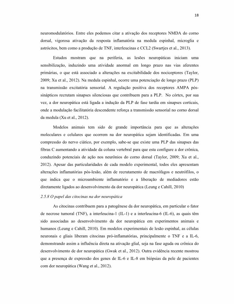
2.5.8 O papel das citocinas na dor neuropática
As citocinas contribuem para a patogênese da dor neuropática, em particular o fator
de necrose tumoral (TNF), a interleucina-1 (IL-1) e a interleucina-6 (IL-6), as quais têm
sido associadas ao desenvolvimento da dor neuropática em experimentos animais e
humanos (Leung e Cahill, 2010). Em modelos experimentais de lesão espinhal, as células
neuronais e gliais liberam citocinas pró-inflamatórias, principalmente o TNF e a IL-6,
demonstrando assim a influência direta na ativação glial, seja na fase aguda ou crônica do
desenvolvimento de dor neuropática (Gwak et al., 2012). Outra evidência recente mostrou
que a presença de expressão dos genes de IL-6 e IL-8 em biópsias da pele de pacientes
com dor neuropática (Wang et al., 2012).

19
No contexto da dor neuropática o TNF está envolvido no aumento da produção de
agentes próalgésicos. Evidências mostram que a injeção de TNF no nervo ciático está
associada com o edema do nervo e com o desenvolvimento da hiperalgesia e alodinia, além
de ativação de macrófagos e lesão nas células de Schwann (Leung e Cahill, 2010; Xu e
Yaksh, 2011). Além disso, o TNF diminuiu o limiar mecânico dos nociceptores-C,
causando dano permanente a alguns deles. No corno dorsal os níveis de TNF aumentaram
após dano no nervo periférico, assim como os níveis de dois receptores de TNF (TNFR1 e
o TNFR2). Quando os neurônios do corno dorsal são lesionados eles respondem a
quantidades muito baixas de TNF e apresentam descargas extremamente avançadas, o que
provavelmente explicaria a alodinia e o comportamento doloroso nos casos de dor
neuropática (Xu e Yaksh, 2011).
A família da IL-1 por sua vez, inclui a IL-1α e a IL-1β, sendo a última encontrada
em maior quantidade na microglia e nos macrófagos. Evidências mostram que existe uma
relação em potencial entre a IL-1β, a apoptose neuronal e a dor neuropática, apesar de não
existirem mecanismos bem estabelecidos para esta relação (Mika, 2008). Sabe-se apenas
que essa interleucina está envolvida na regulação da plasticidade sináptica e nos
processamentos de memória. Experimentos com animais e humanos têm demonstrado que
mudanças na expressão de IL-1β interferem nas funções cognitivas e sintomas depressivos
quando associados com a dor crônica (Del Rey et al., 2012).
Finalmente, a IL-6, também dentre as citocinas mais importantes na dor
neuropática, é um importante mediador inflamatório e modulador da resposta imune.
Recentemente, estudos têm demonstrado a presença da IL-6 em diversas neuropatologias
como a demência, o Alzheimer, a AIDS, a esclerose múltipla, traumas do SNC entre
outras. Os efeitos inibitórios da IL-6 na hiperexcitabilidade neuronal, após uma injúria,
demonstram que esta citocina está envolvida e possivelmente modula a dor neuropática,
porém com mecanismos ainda não determinados (Mika, 2008).
2.5.9 Tratamentos na dor neuropática
Atualmente, a forma mais importante de tratamento em casos de dor neuropática é a
abordagem farmacológica, porém, com resultados insatisfatórios quando se trata do alívio
da dor. Esse fato acontece, pois as recomendações farmacológicas se baseiam apenas na
intensidade da dor do paciente e os sintomas apresentados, e não é levada em conta a

20
heterogeneidade dos mecanismos responsáveis pela dor neuropática e os aspectos
emocionais e psicológicos coexistentes (Baron et al., 2010; Finnerup et al., 2010).
Os pacientes devem ser orientados quanto aos efeitos colaterais, à eficácia e a
tolerabilidade do tratamento farmacológico para que expectativas sejam evitadas, já que
resultados significativos são de no mínimo 30% na redução da dor e outros pontos como a
qualidade de vida e saúde, aspectos sociais e emocionais e distúrbios de sono também
devem ser considerados quando a eficácia do medicamento é analisada. Na clínica, a
abordagem terapêutica se faz, normalmente, com uma abordagem interdisciplinar,
incluindo tratamentos farmacológicos e não-farmacológicos como terapia, por exemplo,
principalmente em casos em que a dor coexiste com outros distúrbios como a ansiedade e a
depressão (Baron et al., 2010). Dentre as drogas utilizadas no tratamento da dor
neuropática temos os opióides, a lidocaína tópica, os antidepressivos tricíclicos, os
inibidores seletivos da recaptação de serotonina (ISRS) e os anticonvulsivantes (Baron et
al., 2010; Schestatsky, 2009; Snedecor et al., 2013; Swartjes et al., 2013).
Estudo recente mostrou através de uma revisão sistemática que não existem
estudos clínicos e dados suficientes para que haja uma comparação entre as terapias
farmacológicas utilizadas para a dor neuropática e que a droga pregabalina
(anticonvulsivante) foi a mais estudada apresentando uma eficácia favorável no tratamento
da dor neuropática (Snedecor et al., 2013). Outro estudo analisou a eficácia dos opióides,
mostrando que apesar de não existirem evidências, alguns opióides podem apresentar
maior efetividade que outros em pacientes com dor neuropática. Sabe-se, porém, que essa
diferença de eficácia deve-se a muitas variantes como a individualidade do paciente, o
receptor opióide a qual o medicamento tem afinidade, diferentes subtipos de proteína-G
envolvidos na doença e os diferentes estados de dor neuropática (Smith, 2012).
Os antidepressivos tricíclicos, por sua vez, apresentam um efeito analgésico
independente dos efeitos antidepressivos, podendo assim ser eficazes não somente na dor,
mas como também na depressão associada a esta. Os ISRS, também se mostraram eficazes
no controle da dor em casos de polineuropatias, porém ambas as classes não apresentam
estudos de eficácia em outras síndromes de dor neuropática (Baron et al., 2010). Desta
forma, pode-se observar como são limitadas as opções farmacológicas para o tratamento da
dor neuropática e que maiores estudos são necessários para avaliar a eficácia dos
medicamentos atualmente utilizados.

21
2.6 Depressão
A depressão, um distúrbio mental, possui uma prevalência mundial de 10-20%,
sendo que 15% dos pacientes cometem suicídio. Tais valores demonstram o quanto comum
são os casos de depressão e como é alta a taxa de mortalidade dos pacientes acometidos.
Estima-se que no ano de 2020 esse distúrbio será o número um no mundo (Rijavec e
Grubic, 2012). De acordo com a Associação Americana de Psiquiatria (American
Psychiatric Association), os sintomas característicos da depressão são: anorexia, perda de
peso, letargia, fadiga, distúrbios do sono, hiperalgesia, falta de concentração e redução da
atividade locomotora (Maes et al., 2012).
O transtorno depressivo maior é caracterizado por uma interação entre fatores
genéticos, biológicos e ambientais com uma patogênese complexa e que envolve diversos
sistemas biológicos. Desta forma, muitos estudos têm sido realizados a fim de entender
melhor os mecanismos envolvidos nesta doença, e sabe-se que uma evidência importante é
que as alterações fisiológicas que ocorrem não são apenas no SNC, mas também na
periferia (Hepgul et al., 2013).
Ainda de acordo com Hepgul e colaboradores (2013), pacientes com depressão
apresentam anormalidades hormonais e imunológicas, como níveis aumentados de
citocinas pró-inflamatórias, mudanças de neuroplasticidade, alterações no eixo hipotálamo-
hipófise-adrenal, além de mudanças nos mecanismos de estresse oxidativo, demonstrando
assim a natureza neuroprogressiva desta doença.
Muitas outras teorias existem para explicar a patogênese da depressão, dentre elas
temos a hipótese das monoaminas, baseada na deficiência das monoaminas, principalmente
a serotonina e norepinefrina; teorias comportamentais e cognitivas; a hipótese neurogênica
e a hipótese neurodegenerativa. Porém atualmente, os mecanismos da neuroinflamação e
da neurodegeneração têm ganhado maior relevância, principalmente, pois ainda não se
sabe qual seria o possível papel dos glicocorticóides em ambos os mecanismos (Zunszain
et al., 2011).
2.7 A relação entre dor e depressão
Em torno de 50% dos pacientes com depressão apresentam sintomas físicos
dolorosos inexplicáveis como fadiga, dores musculares, nas juntas, nas costas e dores de
cabeça, demonstrando dessa forma como a dor e as depressões estão interligadas. A

22
coexistência de ambas as doenças envolve principalmente vias neurobiológicas como
alguns neurotransmissores (glutamato, substância P, serotonina, norepinefrina, dopamina,
fator neurotrópico e ácido gama-aminobutírico), os quais são ativados tanto na dor quanto
na depressão sem descartar a importância de outros fatores, como acima citado (Rijavec e
Grubic, 2012).
Por outro lado, a depressão afeta de 30 a 100% dos pacientes com dor crônica, causando
deficiências emocionais e cognitivas (Wang et al., 2011). Provas científicas relacionam a
depressão com a dor crônica, como em casos de dor neuropática e fibromialgia, através do
mecanismo de sensibilização central, pois ambas as doenças possuem bases
neurobiológicas de alterações neuroplásticas e de expressão gênica semelhantes. Dessa
forma, a etiologia de dor crônica, depressão e transtornos de ansiedade têm sido postulados
como “neurosensibilização”.
Wang e colaboradores (2011) demonstraram que a cirurgia de lesão do nervo
ciático em roedores, comumente utilizada como modelo de dor neuropática, induziu
rapidamente um comportamento depressivo nos animais operados e que a depressão
crônica apresentada é simultânea com a hipersensibilidade sensorial. Porém os
mecanismos responsáveis por tal relação ainda não estão bem esclarecidos, e uma das
causas poderia envolver eventos inflamatórios ou o stress pós-operatório. Sabe-se ainda,
que a dor é capaz de alterar a conectividade sináptica no córtex pré-frontal e no
hipocampo, alterando dessa forma a sinalização dopaminérgica, conhecida por induzir
sintomas na depressão, não podendo ser descartada como uma possível explicação.

23
3 OBJETIVOS
3.1 Geral
Avaliar a atividade anti-inflamatória, anti-hiperalgésica e anti-depressiva do extrato de
Salvia lachnostachys em roedores.
3.2 Específicos
Verificar se o extrato de S. lachnostachys e da fruticulina A possuem ação
anti-inflamatória no modelo de indução de pleurisia e edema de pata com carragenina em
camundongos;
Verificar se o extrato de S. lachnostachys e da fruticulina A possuem ação
anti-hiperalgésica no modelo de hiperalgesia mecânica induzia pela carragenina, de forma
local ou sistêmica, avaliada com von Frey eletrônico em camundongos;
Analisar se o extrato de S. lachnostachys possui ação anti-hiperalgésica em
modelo de lesão de nervo ciático avaliada com o Von Frey eletrônico em ratos;
Analisar se o extrato de S. lachnostachys diminui a sensibilidade ao frio em
modelo de lesão de nervo ciático no teste de acetona em ratos;
Avaliar se o extrato de S. lachnostachys possui ação anti-depressiva no teste
de nado forçado em modelo de lesão de nervo ciático em ratos.

24
4. REFERÊNCIAS BIBLIOGRÁFICAS
Abbadie C. Chemokines, chemokine receptors and pain. Trends of Immunology. 2005; 10:
529-534.
Afilalo M, Morlion B. Efficacy of tapentadol ER for managing moderate to severe chronic
pain. Pain physician. 2013; 16: 27-40.
Barbosa FL, Moris LS, Riva D, Stefanello MEA, Zampronio AR. Antinociceptive and
anti-inflammatory activities of the ethanolic extract, fractions and 8-methoxylapachenol
from Sinningia allagophylla tubers. Basic & Clinical Pharmacology & Toxicology. 2013;
113: 1-7.
Baron R, Binder A, Wasner G. Neuropathic pain: diagnosis, pathophysiological
mechanisms, and treatment. The Lancet Neurology. 2010; 9: 807-819.
Barraza-Sandoval G, Casanova-Mollá J, Valls-Solé J. Neurophysiological assessment of
painful neuropathies. Expert Review of Neurotherapeutics. 2012; 12: 1297-1310.
Basbaum AI, Bautista DM, Scherrer G, Julius D. Cellular and molecular mechanisms of
pain. Cell. 2009; 139: 267-284.
Bellik Y, Boukraâ L, Alzahrani HA, Bakhotmah BB, Abdellah F, Hammoudi SM, Iguer-
Ouada M. Molecular Mechanism Underlying Anti-Inflammatory and Anti-Allergic
Activities of Phytochemicals: An Update. Molecules. 2012; 18: 322-353.
Bisio A, Romussi G, Russo E, Cafaggi S, Schito AM, Repetto B, De Tommasi N.
Antimicrobial activity of the ornamental species Salvia corrugata, a potential new crop for
extractive purposes. Journal of Agricultural and Food Chemistry. 2008; 56: 10468-10472.
Capasso R, Borrelli F, Zjawiony J, Kutrzeba L, Aviello G, Sarnelli G, Capasso F, Izzo A.
The hallucinogenic herb Salvia divinorum and its active ingredient salvinorin A reduce
inflammation-induced hypermotility in mice. Neurogastroenterology & Motility. 2008; 20:
142-148.
Cárdenas C, Quesada AR, Medina MA. Anti-angiogenic and anti-inflammatory properties
of kahweol, a coffee diterpene. Plos One. 2011; 6: 23407.

25
Carvalho WA, Lemônica L. Mecanismos celulares e moleculares da dor inflamatória.
Modulação periférica e avanços terapêuticos. Revista Brasileira de Anestesiologia. 1998;
48: 137-158.
Chapman C R, Gavrin J. Suffering and its relationship to pain. Journal of Palliative Care.
1993; 9: 5-13.
Chatter R, Othman RB, Rabhi S, Kladi M, Tarhouni S, Vagias C, Roussis V, Guizani-
Tabbane L, Kharrat R. In vivo and In vitro anti-inflammatory activity of neorogioltriol, a
new diterpene extracted from red algae Laurencia glandulifera. Marine Drugs. 2011; 9:
1293-1306.
Chudler EH, Dong WK. The role of the basal ganglia in nociception and pain. Pain. 1995;
60: 3-38.
Costigan M, Woolf C J. Pain: molecular mechanisms. Journal of Pain. 2000; 3: 35-44.
Coutaux A. Adam F, Willer JC, Le Bars D. Hyperalgesia and allodynia: peripheral
mechanisms. Joint Bone Spine. 2005; 72: 359-371.
Cragg GM, Newman D J. Natural products: A continuing source of novel drug leads.
Biochimica et Biophysica Acta. 2013; 1830: 3670-3695.
D'Mello R, Dickenson A. Spinal cord mechanisms of pain. British Journal of Anaesthesia.
2008; 101: 8-16.
Del Rey A, Apkarian AV, Martina M, Basedovsky HO.. Chronic neuropathic pain–like
behavior and brain-borne IL-1β. Annals of the New York Academy of Sciences. 2012;
1262: 101-107.
Dubin AE, Patapoutian A. Nociceptors: the sensors of the pain pathway. The Journal of
Clinical Investigation. 2010; 11: 3760-3772.
Eisenberger NI. The neural bases of social pain: evidence for shared representations with
physical pain. Psychosomatic Medicine. 2012; 74: 126-35.
Erbano M, Ehrenfried CA, Stefanello MEA, Dos Santos EP. Morphoanatomical and
phytochemical studies of Salvia lachnostachys (Lamiaceae). Microscopy Research and
Technique. 2012; 75: 1737-1744.

26
Finnerup NB, Sindrup SH, Jensen TS. The evidence for pharmacological treatment of
neuropathic pain. Pain. 2010; 150: 573-581.
Garg G, Adams JD. Treatment of neuropathic pain with plant medicines. Chinese Journal
of Integrative Medicine. 2012; 18: 565-570.
Geffeney S L, Goodman, MB. How we feel: ion channel partnerships that detect
mechanical inputs and give rise to touch and pain perception. Neuron. 2012; 74: 609-619.
Giacomelli E, Bertrand S, Nievergelt A, Zwick V, Simoes-Pires C, Marcourt L, Rivara-
Minten E, Cuendet M, Bisio A, Wolfender JL. Cancer chemopreventive diterpenes from
Salvia corrugata. Phytochemistry. 2013; 96: 257-264.
Gurib-Fakim A. Medicinal plants: traditions of yesterday and drugs of tomorrow.
Molecular Aspects of Medicine. 2006; 27: 1-93.
Guzzo LS, Perez AC, Romero TR, Azevedo AO, Duarte ID. Cafestol, a coffee-specific
diterpene, induces peripheral antinociception mediated by endogenous opioid peptides.
Clinical and Experimental Pharmacology and Physiology. 2012; 39: 412-416.
Gwak Y S, Kang J, Unabia GC, Hulsebosch CE. Spatial and temporal activation of spinal
glial cells: role of gliopathy in central neuropathic pain following spinal cord injury in rats.
Experimental Neurology. 2012; 234: 362-372.
Heinricher MM, Tavares I, Leith JL, Lumb BM. Descending control of nociception:
specificity, recruitment and plasticity. Brain Research Reviews. 2009; 60: 214-225.
Hepgul N, Cattaneo A, Zunszain PA, Pariante CM. Depression pathogenesis and
treatment: what can we learn from blood mRNA expression? BMC Medicine. 2013; 11:
28, 2013.
Hosseinzadeh H, Haddadkhodaparast MH, Arash AR. Antinociceptive, antiinflammatory
and acute toxicity effects of Salvia leriifolia Benth. seed extract in mice and rats.
Phytotherapy Research. 2003; 17: 422-425.
Ibrahim TA. Chemical composition and biological activity of extracts from Salvia bicolor
Desf. growing in Egypt. Molecules. 2012; 17: 11315-11334.

27
Isacchi B, Fabbri V, Galeotti N, Bergonzi MC, Karioti A, Ghelardini C, Vannucchi MG,
Bilia AR. Salvianolic acid B and its liposomal formulations: anti-hyperalgesic activity in
the treatment of neuropathic pain. European Journal of Pharmaceutical Sciences. 2011; 44:
552-558.
Jiang WY, Jeon BH, Kim YC, Lee SH, Sohn DH, Seo GS. PF2401-SF, standardized
fraction of Salvia miltiorrhiza shows anti-inflammatory activity in macrophages and acute
arthritis in vivo. International Immunopharmacology. 2013; 16: 160-164.
Julius D, Basbaum AI. Molecular mechanisms of nociception. Nature. 2001; 413: 203-210.
Jung HJ, Song YS, Lim CJ, Park EH. Anti-inflammatory, anti-angiogenic and anti-
nociceptive activities of an ethanol extract of Salvia plebeia R. Brown. Journal of
Ethnopharmacology. 2009; 126: 355-360.
Karami M, Shamerani MM, Alemy S, Gohari A, Vostacolaee SE. Comparison
antinociceptive activity of the aqueous methanolic extracts of Salvia hypoleuca. European
Review for Medical and Pharmacological Sciences. 2013a; 20: 2755-2759.
Kassuya CA, Wisniewski Jr A, Simionatto EL, Santos EP, Stefanello ME. Composição dos
óleos essenciais de Salvia lachnostachys e S. melissiflora (Lamiaceae). Latin American
Journal of Pharmacy. 2009; 28: 919-921.
Khuong TM, Neely GG. Conserved systems and functional genomic assessment of
nociception. FEBS Journal. 2013; 280: 5298-5306.
Koenen RR, Weber C. Chemokines: established and novel targets in atherosclerosis.
EMBO Molecular Medicine. 2011; 3: 713-725.
Lamb K, Tidgewell K, Simpson DS, Bohn LM, Prisinzano TE. Antinociceptive effects of
herkinorin, a MOP receptor agonist derived from salvinorin A in the formalin test in rats:
New concepts in mu opioid receptor pharmacology. Drug and Alcohol Dependence. 2012;
121: 181-188.
Latremoliere A, Woolf CJ. Central sensitization: a generator of pain hypersensitivity by
central neural plasticity. The Journal of Pain. 2009; 10: 895-926.

28
Leung L, Cahill CM. TNF-α and neuropathic pain-a review. Journal of
Neuroinflammation. 2010; 16: 7-27.
Li M, Zhang L, Cai RL, Gao Y, Qi Y.. Lipid-soluble Extracts from Salvia miltiorrhiza
Inhibit Production of LPS-induced Inflammatory Mediators via NF-κB Modulation in
RAW 264.7 Cells and Perform Antiinflammatory Effects In Vivo. Phytotherapy Research.
2012; 26: 1195-1204.
Linley JE, Rose K, Ooi L, Gamper N. Understanding inflammatory pain: ion channels
contributing to acute and chronic nociception. Pflügers Archiv-European Journal of
Physiology. 2010; 459: 657-669.
Loeser JD. Pain and suffering. The Clinical Journal of Pain. 2000; 16:2-6.
Loeser JD, Melzack R. Pain: an overview. Lancet. 1999; 353: 1607-1609.
Lumlwy MA, Cohen JL, Borszcz GS, Cano A, Raddcliffe AM, Porter LS, Schubiner H,
Keefe FJ. Pain and emotion: a biopsychosocial review of recent research. Journal of
Clinical Psychology. 2011; 67: 942-968.
Maes M, Berk M, Goehler L, Song C, Anderson G, Gatecki P, Leonard B. Depression and
sickness behavior are Janus-faced responses to shared inflammatory pathways. BMC
Medicine. 2012; 10: 66.
Maroon JC, Bost JW, Maroon A. Natural anti-inflammatory agents for pain relief. Surgical
Neurology International. 2010; 1: 80.
Matias EF, Alves EF, Santos BS, de Souza CES, Ferreira JVA, de Lavor AKS, Figueiredo
FG, de Lima LF, dos Santos FAV, Peixoto FSN, Colares AV, Boligon AA, Saraiva RA,
Athayde ML, da Rocha JB, Menezes IRA, Coutinho HDM, da Costa JG. Biological
Activities and Chemical Characterization of Cordia verbenacea DC. as Tool to Validate
the Ethnobiological Usage. Evidence-Based Complementary and Alternative Medicine.
2013; 2013: 164215.
Mattia C, Coluzzi F. COX-2 inhibitors: pharmacological data and adverse effects. Minerva
Anestesiologica. 2005; 71: 461.

29
Medzhitov R. Inflammation 2010: new adventures of an old flame. Cell. 2010; 140: 771-
776.
Mika J. Modulation of microglia can attenuate neuropathic pain symptoms and enhance
morphine effectiveness. Pharmacological Reports. 2008; 60: 297-307.
Mishra BB, Tiwari VK. Natural products: an evolving role in future drug discovery.
European Journal of Medicinal Chemistry. 2011. 46: 4769-4807.
Moharram FA, Marzouk MS, El-Shenawy SM, Gaara AH, El Kady WM. Polyphenolic
profile and biological activity of Salvia splendens leaves. Journal of Pharmacy and
Pharmacology. 2012; 64: 1678-1687.
Mossi A, Cansian R, Paroul N, Toniazzo G, Oliveira J, Pierozan M, Pauletti G, Rota L,
Santos A, Serafini L. Morphological characterisation and agronomical parameters of
different species of Salvia sp. (Lamiaceae). Brazilian Journal of Biology. 2011; 71: 121-
129.
Muller W. Getting Leukocytes to the Site of Inflammation. Veterinary Pathology Online.
2013; 50: 7-22.
Ortega-Gómez A, Perretti M, Soehnlein O. Resolution of inflammation: an integrated
view. EMBO Molecular Medicine. 2013; 5: 661-674.
Perl ER. Pain mechanisms: a commentary on concepts and issues. Progress Neurobiology.
2011; 94: 20-38.
Phillipson JD. Phytochemistry and pharmacognosy. Phytochemistry. 2007; 68: 2960-2972.
Price TJ, Ghosh S. ZIPping to pain relief: The role (or not) of PKMzeta in chronic pain.
Molecular Pain. 2013 9: 6.
Quintans JS, Antoniolli AR, Almeida JR, Santana-Filho VJ, Quintans-Júnior LJ. Natural
Products Evaluated in Neuropathic Pain Models–A Systematic Review. Basic & Clinical
Pharmacology & Toxicology. 2013.
Rauch I, Müller M, Decker T. The regulation of inflammation by interferons and their
STATs. JAK-STAT. 2013; 2:1.

30
Ren L, Hu H, Sun X, Li F, Zhou JJ, Wang YM. The roles of inflammatory cytokines in the
pathogenesis of ossification of ligamentum flavum. American Journal of Translational
Research. 2013; 5: 582.
Rijavec N, Grubic VN. Depression and pain: often together but still a clinical challenge: a
review. Psychiatria Danubina. 2012; 4: 346-352.
Rishton GM. Natural products as a robust source of new drugs and drug leads: past
successes and present day issues. The American Journal of Cardiology. 2008; 101:43-49.
Rodrigues MRA, Kanazawa LKS, Neves TLM, Silva CF, Horst H, Pizzolatti MG, Santos
ARS, Baggio CH, Werner MF. Antinociceptive and anti-inflammatory potencial of extract
and isolated compounds from leaves of Salvia officinalis in mice. Journal of
Ethnopharmacology. 2012; 139: 519-526..
Rodríguez-Hahn L, Baldomero E, Sánchez C, Estebanes L, Cárdenas J, Soriano-Gárcia,
Rúben Toscano TP.. Abietane type diterpenoids from Salvia fruticulosa. A revision of the
structure of fruticulin B. Phytochemistry. 1989; 28: 567-570.
Ryan G, Majno G. Acute inflammation. A review. The American Journal of Pathology.
1977; 86: 183.
Salminen A, Lehtonen M, Suuronen T, Kaarniranta K, Huuskonen J. Terpenoids: natural
inhibitors of Nf-κB signaling with anti-inflammatory and anticancer potential. Celular and
Molecular Life Sciences.2008; 65: 2979-2999.
Schestatsky P. Definição, diagnóstico e tratamento da dor neuropática. Revista HCPA.
2009; 28: 3.
Schito A, Piatti G, Stauder M, Bisio A, Giacomelli E, Romussi G, Pruzzo C. Effects of
demethylfruticuline A and fruticuline A from Salvia corrugate Vahl. on biofilm production
in vitro by multiresistant strains of Staphylococcus aureus, Staphylococcus epidermidis
and Enterococcus faecalis. International Journal of Antimicrobial Agents. 2011; 37: 129-
134.
Smith HS. Opioids and neuropathic pain. Pain Physician. 2012; 15: 93-110.

31
Snedecor SJ, Sudharshan L, Cappelleri JC, Sadosky A, Desai P, Julundhwala YJ,
Botteman M. Systematic review and comparison of pharmacologic therapies for
neuropathic pain associated with spinal cord injury. Journal of Pain Research. 2013; 6:
539-547.
Sostres C, Gargallo CJ, Arroyo MT, Lanas A. et al. Adverse effects of non-steroidal anti-
inflammatory drugs (NSAIDs, aspirin and coxibs) on upper gastrointestinal tract. Best
Practice & Research Clinical Gastroenterology. 2010; 24: 121-132.
Sousa FAEF. Dor: o quinto sinal vital. Revista Latino-Americana de Enfermagem. 2002;
10: 446-447.
Swartjes M, Niesters M, Heij L, Dunne A, Aarts L, Hand CC, Kim H, Brines M Cerami A,
Dahan A.. Ketamine Does Not Produce Relief of Neuropathic Pain in Mice Lacking the β-
Common Receptor (CD131). PLOS ON. 2013; 8: e71326.
Tarp S, Bartels EM, Bliddal H, Furst DE, Boers M, Danneskiold-Samsoe B, Rasmussen M,
Christensen R. et al. Effect of nonsteroidal antiinflammatory drugs on the C-reactive
protein level in rheumatoid arthritis: A meta-analysis of randomized controlled trials.
Arthritis & Rheumatism. 2012; 64: 3511-3521.
Taylor BK. Spinal inhibitory neurotransmission in neuropathic pain. Current Pain and
Headache Reports. 2009; 13: 208-214.
Torres RC, Insuela DBR, Carvalho VDF. Mecanismos celulares e moleculares da ação
antiinflamatória dos glicocorticoides. Corpus et Scientia. 2012; 8: 36-51.
Vargas-Espinosa ML, Sanmaetí-García G, Vázquez-Delgado E, Gay-Escoda C.
Antiepileptic drugs for the treatment of neuropathic pain: A systematic review. Medicina
Oral, Patologia Oral y Cirugia Bucal. 2012; 17: e786.
Walker JB, Sytsma KJ. Staminal evolution in the genus Salvia (Lamiaceae): molecular
phylogenetic evidence for multiple origins of the staminal lever. Annals of Botany. 2007;
100: 375-391.

32
Walzog B, Gaehtgens P. Adhesion molecules: the path to a new understanding of acute
inflammation. Physiology. 2000; 15: 107-113.
Wang J, Goffer Y, Xu D, Tukey DS, Shamir DB, Eberle SE, Zou AH, Blanck TJ, Ziff EB..
A Single Sub-anesthetic Dose of Ketamine Relieves Depression-like Behaviors Induced by
Neuropathic Pain in Rats. Anesthesiology. 2011: 115: 812-821.
Wang XM, Lehky TJ, Brell JM, Dorsey SG. Discovering cytokines as targets for
chemotherapy-induced painful peripheral neuropathy. Cytokine. 2012; 59: 3-9.
Wen XD, Wang CZ, Yu C, Zhang Z, Calway T, Wang Y, Li P, Yuan CS. Salvia
miltiorrhiza (Dan Shen) Significantly Ameliorates Colon Inflammation in Dextran Sulfate
Sodium Induced Colitis. The American Journal of Chinese Medicine. 2013; 41: 1097-1108.
Xu B, Descalzi G, Ye HR, Zhuo M , Wang YW. Translational investigation and treatment
of neuropathic pain. Molecular Pain. 2012; 8: 15.
Xu Q, Yaksh TL. A brief comparison of the pathophysiology of inflammatory versus
neuropathic pain. Current Opinion in Anaesthesiology. 2011; 24: 400.
Zheng CJ, Huang BK, Wang Y, Ye Q, Han T, Zhang QY, Zhang H, Qin LP. Anti-
inflammatory diterpenes from the seeds of Vitex negundo. Bioorganic & Medicinal
Chemistry. 2010; 18: 175-181.
Zunszain PA, Anacker C, Cattaneo A, Carvalho LA, Pariante CM. Glucocorticoids,
cytokines and brain abnormalities in depression. Progress in Neuro-Psychopharmacology
and Biological Psychiatry. 2011; 35: 722-729.

33
Artigo 1 submetido a revista Phytomedicine (fator de impacto- 2.972) :
ANTI-INFLAMMATORY AND ANTI-HYPERALGESIC ACTIVITY OF EXTRACT
AND FRUTICULINE A FROM Salvia lachnostachys IN MICE

34
ANTI-INFLAMMATORY AND ANTIHYPERALGESIC ACTIVITY OF
EXTRACT AND FRUTICULIN A FROM Salvia lachnostachys IN MICE
A. C. Piccinellia, D. F. S. Aquinoa, R. L. B. Strapassonb, E. P. Santosc, M. E. A.
Stefanellob R.J. Oliveirad, C. A. L. Kassuyaa
aFederal University of Grande Dourados, College of Health Science, Dourados, MS,
Brazil
bFederal University of Paraná, Chemistry Department, Curitiba, PR, Brazil.
cFederal University of Paraná, Botany Department, Curitiba, PR, Brazil.
dFederal University of Mato Grosso do Sul, “Dr. Hélio Mandetta” School of Medicine,
Campo Grande, MS, Brazil.
*Corresponding author: Faculdade de Ciências da Saúde, Universidade Federal da
Grande Dourados, Dourados, 79825-070, MS, Brazil, Phone: +55 67 3410-2326 Fax:
+55 67 3410-2326. E-mail address:[email protected]

35
ABSTRACT
Salvia lachnostachys Benth., Lamiaceae, is an endemic specie from southern Brazil.
The ethanolic extract from leaves is mainly constituted by fruticulin A and a mixture of
oleanolic and ursolic acids. Our study aimed to evaluate the anti-inflammatory and
analgesic effects of ethanolic extract of Salvia lachnostachys (SLCH) and its isolated
compound fruticulin A in experimental models of inflammation (paw oedema and
pleurisy induced by carrageenan injection) and hyperalgesia (Electronic Von Frey),
performed in mice. The oral administration of SLCH at doses of 30, 100 and 300 mg/kg
significantly decreased paw oedema formation, while in pleurisy the dose of 100 and
300 mg/kg significantly decreased the total leucocytes number in pleural lavage and
also the protein leakage. The compound fruticulin A was isolated from SLCH and
identified by NMR analysis. The oral administration of fruticulin A at doses of 0.3 and
3 mg/kg significantly decreased the total leucocytes number in pleural lavage, protein
extravasation and paw edema. Both SLCH (100 mg/kg) and isolated compound
fruticulin A (3 mg/kg) exhibited antihyperalgesic activity in carrageenan induced
mechanical hyperalgesia in mice. Local administration of fruticulin A (3 mg/kg) also
significantly prevented mechanical hyperalgesia, demonstrating a local antihyperalgesic
effect. In the study of the mechanism of action, local administration of fruticulin A (3
mg/kg), significantly inhibited TNF-, but not L-DOPA induced mechanical
hyperalgesia. In conclusion, the present study showed that SLCH is an anti-
inflammatory and analgesic natural agent. The results lead us to conclude that fruticulin
A is a compound involved in the anti-inflammatory and antihyperalgesic properties of
SLCH with a TNF related mechanism.
Keywords: Lamiaceae; inflammation; hyperalgesia; Salvia lachnostachys; fruticulin A;
TNF.

36
INTRODUCTION
Since ancient times, plants have been used across the world as a safe natural
source of therapeutic agents because of their pharmacological principles. Even with a
relative safe in use, the population feels more confident in spreading their usage to
others. According to the World Health Organization (WHO), nowadays 70-80% of the
world population has the traditional medicine as a primary healthcare (D´Cruz et al.,
2010).
The available therapy for inflammatory and pain states is often inadequate. The
reasons for that involve severe side effects, resulted from long use and lacks efficacy.
Thus, studies with new analgesic and anti-inflammatory drugs that provide efficacy and
safety are needed, and natural products are an option for the development of new drugs
as an extensive therapeutic source (Recio et al., 2012; da Silva et al., 2012).
The genus Salvia, Lamiaceae family, consists of about 900 species distributed in
tropical and subtropical regions. They are usually used as condiments and/or used for
several therapeutic properties in folk medicine (Kassuya et al., 2009). In Brazil, there
are 61 native species however, there are a few pharmacological studies about them
(Erbano et al., 2012).
Salvia lachnostachys Benth. is endemic to southern Brazil and according to a
chemical study, the essential oil of its leaves and flowers has mostly long-chain
aliphatic acids and terpenoids as minor components (Kassuya et al., 2009). The
phytochemical study demonstrated the presence of the diterpene fruticulin A and ursolic
and oleanolic acids (Erbano et al., 2012). Fruticulin A has been recently studied and
presented antibacterial (Bisio et al., 2008; Schito et al., 2011) and cancer
chemopreventive activities (Giacomelli et al., 2013).

37
Therefore the lack of pharmacological studies with this specie of Salvia and the
presence of terpenoids in its composition, led us to evaluate the anti-inflammatory and
antihyperalgesic effect of the ethanolic extract of Salvia lachnostachys and its isolated
compound fruticulin A in experimental models of inflammation and hyperalgesia in
mice.
MATERIALS AND METHODS
Plant Material
The plant material was collected from natural populations in Curitiba, Parana
state, Brazil (25º30'44.6''S, 49º18'7.13''W), by E.P. Santos (number 1251), and a
voucher was included in the herbaria UPCB (Herbarium of the Federal University of
Paraná).
Preparation of the ethanolic extract and isolation of fruticulin A
Dried and powdered leaves of S. lachnostachys were extracted with hexane
followed by ethanol, yielding the ethanolic extract (SLCH) after solvent removal. The
phytochemical study of SLCH lead to isolation of fruticulin A (Figure 1). Detailed
experimental procedures were previously described (Erbano et al., 2012).
Animals
To the experimental procedures were used adult male and female Swiss mice
with approximately weight of 20 g and 50 days age at the beginning of the experiments.
These animals were provided from the Federal University of Grande Dourados (UFGD)
biotherium. The animals were kept in collective cages (5 animals per cage) under
controlled temperature (23 ± 1 º C) and light cycle (12 h light / dark), treated with water
and commercial diet ad libitum. All experimental procedures were approved for
research by the ethics committee on laboratory animals of UFGD (Nbr. 013/2013).

38
Reagents
λ-Carrageenan, TNF, L-DOPA, Bradford reagent, and dexamethasone were
purchased from Sigma-Aldrich ® Co. LLC (St. Louis, MO, USA).
Model of Carrageenan induced Paw Oedema
Each experimental methodology was conducted in two stages. In the first stage
we tested the crude ethanolic extract at 3 different doses and in the second stage we
tested the isolated compound, fruticulin A also in 3 different doses. Doses were
determined in a pilot study and according to literature data (Karami et al., 2013b).
Male animals were divided into experimental groups (n = 5 animals / group).
Group 1 (control) was treated orally with vehicle (10 mL / kg of saline 0.9% saline).
Different groups of mice received by oral route doses of the crude ethanolic extract of S.
lachnostachys (30, 100 and 300mg/kg) or isolated compound fruticulin A (0.3, 1 and 3
mg) dissolved in 0.9% saline solution. The positive control group received
dexamethasone at the dose of 1 mg/kg by subcutaneous route. 1 hour after the
treatments, animals received, in the right hind paw, 50 L of 0.9% saline solution
containing 300 g of carrageenan. The same volume of saline solution was administered
in the left hind paws which were used as control. Edema was measured with a paw
plethysmometer (PANLAB- Havard) at the times 0.5, 1, 2 and 4 hours after carrageenan
application (Formagio et al., 2013; Kassuya et al., 2005; Winter et al., 1962).
Model of Carrageenan induced pleurisy
Female animals were divided into experimental groups (n=5 animals/ group).
SLCH was tested orally at 3 different doses (30, 100, and 300 mg/kg) and the isolated
compound fruticulin A was tested orally at three different doses (0.3, 1 and 3 mg/kg)
one hour before carrageenan injection. Positive control group received 1 mg/kg of

39
dexamethasone, subcutaneously, 1 hour before carrageenan application. Pleurisy was
induced applying 0.25 ml suspension containing 300 μg of carrageenan (dilution in
phosphate buffered saline PBS, pH = 7.4) in the mice pleural cavity according to the
technique previously described (Vinegar et al., 1973). Four hours after induction of
pleurisy, animals were euthanized and the pleural inflammatory exudate was collected
through pleural lavage with 1 ml of sterile saline. The exudate volume was measured,
and an aliquot of 50 μL was diluted with Turk's solution (1:20). Total leukocytes were
counted in a Neubauer chamber, considering the four external quadrants, in a light
microscope.
In each ELISA microwell plate was added 10 µL of pleural lavage and 300 µL
of Bradford. Protein measurement was performed colorimetrically (TP-photometer
Reader / Thermo Plate ®) at the laboratory of Microbiology, Faculty of Health
Sciences, FCS - UFGD.
Model of Carrageenan-induced hyperalgesia
One day earlier the basal measurement was performed in all animals. Mice were
housed in containment boxes (WxDxH 230 x 200 x 180 mm - Insight ®) in a steel mesh
with 1 cm diameter spacing for a period of 1 hour and the digital analgesymeter (Insight
® - EFF 301 - Digital analgesymeter - von Frey) was used in order to determine the
similar mean baseline of mechanical stimulus in the right hind paw (Chaplan et al.,
1994; Mori et al., 2011).
At the following day, each group of male mice (n = 6 / group) received orally
(gavage) 0.9% saline solution or SLCH 300 mg/kg or fruticulin A 3 mg/kg. One hour
after oral treatment, animals received 300 µg of carrageenan injection, subcutaneously
(λ-carrageenan - Sigma-Aldrich ®), in the right hind paw. Then, each animal was
housed in the same containment boxes under the same steel mesh. Mechanical

40
hyperalgesia was measured at the times 3 and 4 hours after carrageenan injection using
the digital analgesymeter previously described.
The same procedures were performed with an injection of fruticulin A
(1µg/paw, 20 µL) and a control group (saline 0.9% v/v) to evaluate local
antihyperalgesic activity of the compound (Barbosa et al., 2013).
TNF- and L-DOPA-induced hyperalgesia
To evaluate the effect of the compound on hyperalgesia induced by different
stimuli, basal mechanical withdrawal threshold of mice was assessed with digital
analgesymeter as described before, and then the animals were treated with fruticulin A
(1µg/paw, 20 µL) or the same volume of vehicle (saline 0.9% v/v), and after 15
minutes, they received TNF (1 pg/paw in a volume of 20 µL) or L-DOPA (10 µg/paw)
in a volume of 20 µL (Cunha et al., 2005; Cunha et al., 2008) . Mechanical hyperalgesia
was also measured after 3 h (Barbosa et al., 2013).
Statistical Analysis
Data are presented as mean ± S.E.M. Differences between groups was evaluated
by analyses of variance (one-way ANOVA) followed by the Newman-Keuls test (the
symbols were included in the figures) or Bonferroni test (symbols were not included in
the figures). The number of animals per group is indicated in the legends. Statistical
differences were considered to be significant at P<0.05. *Asterisks or # denote
significant difference compared with groups.
RESULTS
Effects of SLCH and fruticulin A against carrageenan induced oedema
The results of first test using SLCH (30, 100, and 300mg/kg) were showed in

41
Figure 2. The Cg injection in the right hind paw of the animals induced edema, peaking
at 2 h. Oral treatment with SLCH was able to significantly inhibit the edema formation
in a dose-dependent manner. The maximum inhibitions were 84 ± 3% at the dose of 300
mg/kg and 77 ± 3% at the dose of 100 mg/kg after 2 hours of Cg injection, with
P<0.01.
The statistical analyses (symbols not included in the figures) with ANOVA with
post-test Bonferroni (comparing all pairs of columns) showed that all doses tested of
SLCH (except the group treated with 30 mg/kg – Figure 2C), fruticulin A and and
dexamethasone (Figure 2 and 3) had statistical differences from control group. All doses
tested of SLCH did not differ between groups and dexamethasone group in Figure 2A,
2B, and 2D therefore in Figure 2C the dose of 300 mg/kg group were significantly
different from treated group with dose of 30 mg/kg and group.
We can also observe that administration of fruticulin A significantly decreased
paw edema in mice at doses of 1 and 3 mg/kg (Figure 3C), with no significant
difference between both doses. The maximum inhibitions were 54 ± 11% at the dose of
1 mg/kg and 55 ± 11% at the dose of 3 mg/kg after 2 hours of Cg injection, with
P<0.05. The statistical analyses (symbols not included in the figures) with ANOVA
with post-test Bonferroni (comparing all pairs of columns) showed that all doses tested
of fruticulin A did not differ between groups and dexamethasone group in Figure 2A,
2B, and 2D, therefore in Figure 2C the dose of 30 mg/kg group were significantly
different from 100 and 300 mg/kg and dexamethasone group.
The anti-inflammatory activity of SLCH and fruticulin A on carrageenan induced
leukocyte migration and protein extravasation in pleural cavity
In the pleurisy test, the number of total leukocyte counted in a Neubauer

42
chamber significantly (P<0.01) decreased in groups SLCH 30, 100 and 300 mg/kg
(Figure 4A), when compared to the control group, with no significant difference
between both doses. The maximum inhibitions were respectively 46 ± 6% and 24 ± 6%.
There was also a significant decrease in protein dosage of pleural lavage for the
group SLCH 100 and 300 mg/kg (P<0.01), when compared to the control group (Figure
4B). The anti-inflammatory steroidal group (DEX) had a decrease in leukocyte
migration and also in protein extravasation (Figure 4).
In the evaluation of fruticulin A in the model of pleurisy, the two tested doses
(0.3, 1 and 3 mg/kg) showed significant effects with P<0.01, decreasing the total
leukocyte count in a dose-dependent manner compared to the control group (Figure
5A). The maximum inhibitions were respectively 31 ± 7% and 54 ± 3%.
The amount of protein measured in pleural lavage was also significantly lower
for fruticulin A groups when compared to the control group, with P<0.05 at dose of 0.3
and 1 mg/kg and P<0.01 at dose of 3 mg/kg (Figure 5B).
Analyzing the results by ANOVA with post-test Bonferroni, all naïve groups
differ significantly from control groups in Figure 4 and 5. Moreover, all doses tested of
SLCH (except the group treated with 30 mg/kg – Figure 4B), fruticulin A, and
dexamethasone (Figure 2 and 3) had statistical differences from control group. In figure
4A, the group treated with SLCH at dose of 100 mg/kg differed from others group. And
in figure 5A, the group treated with dexamethasone had statistical differences from
others group. All doses tested of SLCH did not differ between groups and
dexamethasone group in Figure 5.
The anti-hyperalgesic activity of oral administration of SLCH and local and oral
administration of fruticuline A on carrageenan induced mechanical sensitivity in

43
mice
Carrageenan injection decreased significantly the basal values, measured by
electronic von Frey mechanical stimulation. It is possible to see in Figure 6 that the
substances orally tested (SLCH 300 mg/kg and fruticulin A 3 mg/kg) relieved
hyperalgesia, so that the animals withstood the strength applied to the von Frey
filaments. Maximum inhibitions were 61 ± 15% for SLCH 300 mg/kg after 3 hours of
Cg injection and 72 ± 13% and 53 ± 11% for SLCH 300 mg/kg and fruticulin A 3
mg/kg, respectively, after 4 hours of Cg injection.
Local administration of compound fruticulin A (1µg/paw, 20 µL) significantly
prevented the decrease of the threshold of sensitivity, with maximum inhibition of 72 ±
15% after 3 hours and 72 ± 15% after 4 hours of Cg injection, with P< 0.05 (Figure 7).
The anti-allodynic activity of local administration of fruticulin A on TNF- or L-
DOPA- induced hypernociception
TNF injection decreased significantly the basal values of measure by electronic
von Frey mechanical stimulation. Local administration of fruticulin A inhibited the
hyperalgesic effects of TNF, but not L-DOPA, significantly preventing the decrease of
the threshold of sensitivity, being of 72 ±7 at a dose of 1µg/paw, after 3 hours of TNF
injection, with P<0.05 (Figure 8).
DISCUSSION
In the present study we evaluated the anti-inflammatory and antihyperalgesic
actions of extract from leaves of S. lachnostachys in oedema, pleurisy and
hypernociception induced by mechanical stimuli in carrageenan models in mice. It was
also observed that the dose based in yield of the isolated compound fruticulin A, but not
the mixture of ursolic and oleanolic acids (results not shown), was able to reduce the
oedema, the leukocytes migration, protein leakage and hypernociception induced by

44
carrageenan. Fruticulin A also demonstrated an antihyperalgesic effect when
administered locally, inhibiting both carrageenan and TNF hyperalgesia. However, there
is little pharmacological information in the literature to support the possible activity of
extracts or compounds derived from S. lachnostachys. To our knowledge, the present
study represents the first research that demonstrates the pharmacological properties of
extract of S. lachnostachys leaves in adult male and female mice.
Salvia genus, belonging to the Lamiaceae family, comprises approximately 100
species distributed mainly in Central and South America (500 species) and Asia (300
species) (Walker and Systsma, 2007). It is commonly used for ornamental, culinary and
medicinal purposes, and has great economic and ethnopharmacological importance
(Mossi et al., 2011). Several studies demonstrated different species of Salvia with anti-
inflammatory and anti-nociceptive activity, among them we have S. officinalis
(Rodrigues et al., 2012), S. plebeian (Jung et al., 2009), S. miltiorriza (Wen et al.,
2013), S. leriifolia (Hosseinzadeh et al., 2003), S. hypoleuca (Karami et al., 2013a), S.
divinorum (Capasso et al., 2008), S. splendens (Moharram et al., 2012), S. bicolor
(Ibrahim, 2012). Currently, there are no reports of folk use or pharmacological studies
with S. lachnostachys and, our study showed for the first time the ability of the extract
from leaves to inhibit some parameters of inflammation such as mechanical pain,
leukocyte migration, oedema, and plasma leakage in experimental inflammatory
models.
Previous study from our group showed that the essential oil from the leaves of S
.lachnostachys contains mainly aliphatic fatty acids, such as, dodecanoic acid (61.6%
of total), and a minor fraction of sesquiterpenes (about 12%) (Kassuya et al., 2009). In
contrast, ethanolic extract is rich in diterpenes and triterpenes. Diterpenes, commonly
found in plants belonging to Lamiaceae family, have various therapeutic effects, such

45
as antibacterial, antifungal, cytotoxic and, mostly anti-inflammatory activity (Salminen
et al., 2008). Studies with the diterpene kahweol and cafestol, isolated from coffee
extract, showed antinociceptive and antiinflammatory activities, respectively (Cardenas
et al., 2011; Guzzo et al., 2012). Likewise, Neorogioltriol , extracted from a red alga
called Laurencia glandulifera demonstrated anti-inflammatory activity in vivo and in
vitro (Chatter et al., 2011). In the seeds of Vitex negundo seven different diterpenes
have been isolated, among these, five of them showed significant results in decreased
expression of proinflammatory proteins, demonstrating its potential as an anti-
inflammatory agent (Zheng et al., 2010).
In species of Salvia, S. miltiorrhizae and S. divinorum, diterpenes salvinorin A
and B salvianolic acid demonstrated its anti-hyperalgesic effects in animal models of
neuropathic pain and anti-inflammatory effect, respectively (Capasso et al., 2008;
Isacchi et al., 2011; Lamb et al., 2012). The diterpene fruticulin A, assayed in this
work, is a rare compound in nature and, has been previously isolated only from S.
fruticulosa Benth (Rodriguez-Hahn et al, 1989) and S. corrugata Vahl (Bisio et al.,
2008). In addition to previously reported antibacterial (Bisio et al., 2008; Schito et al.,
2011) and cancer chemopreventive (Giacomelli et al, 2013) activities of fruticulin A, the
present work describes the effects of this compound in leukocyte migration, protein
extravasation and oedema. The results suggest that the fruticulin A is responsible, at
least in part for anti-inflammatory action of SLCH.
The carrageenan inflammatory agent is capable to induced inflammatory pain by
a mechanism of nociceptor sensibilization (Barbosa et al., 2013). Oedema formation
induced by carrageenan is characterized as a biphasic event: the first phase (0-2 hours)
is mediated by histamine and serotonin followed by bradykinin, prostaglandins and
lysosome while the late phase (2-6 hours) is related with the production of TNF,

46
interleukins (IL-1β and IL-6) and nitric oxide (Li et al., 2013). Similarly, carrageenan
induced hypernociception is also dependent of mediators as bradykinin, TNF,
prostaglandin, keratinocyte-derived chemokine (KC) and sympathetic amines (Cunha et
al., 2005; Cunha et al., 2008).
TNF is produced by mononuclear phagocytes and is responsible for immune
responses and induction of other inflammatory cytokines (Ma et al., 2013). Therefore,
TNF may be involved in both oedema formation and hyperalgesia, which may explain
the effects of fruticulin A on the animal models performed. The other pathway of
carrageenan-induced hyperalgesia involves the sympathetic amines, the L-DOPA
injection activates this pathway but fruticulin A did not interfere in this activation
showing that this pathway is not important for fruticulin A anti-hyperalgesic effects.
These results suggested that the main mechanism responsible by SLCH and fruticulin A
antihyperalgesic actions could involves TNF pathway.
CONCLUSION
The present study showed that the extract from leaves of S. lachnostachys
(SLCH) has anti-inflammatory and antihyperalgesic effects in oedema, pleurisy and
hyperalgesia induced by carrageenan models in mice. Fruticulin A, a diterpene found in
S. lachnostachys, seems to be responsible for SLCH effects, when tested in a dose based
in extract yield orally and locally, and was able to reduce the oedema, the leukocytes
migration, protein leakage and hyperalgesia induced by carrageenan. TNF seems to be
involved on its mechanism of action however other possible mechanisms should be
investigated. To our knowledge, the present study represents the first research that
demonstrates the pharmacological properties of extract of S. lachnostachys leaves and
fruticulin A in mice.

47
Conflict of interest
No conflict to disclose.
Acknowledgement
The authors are thankfull to CAPES and CNPq for the financial support.
REFERENCES
Barbosa FL, Moris LS, Riva D, Stefanello MEA, Zampronio AR. Antinociceptive and
anti-inflammatory activities of the ethanolic extract, fractions and 8-methoxylapachenol
from Sinningia allagophylla tubers. Basic & Clinical Pharmacology & Toxicology.
2013; 113: 1-7.
Bisio A, Romussi G, Russo E, Cafaggi S, Schito AM, Repetto B, De Tommasi N.
Antimicrobial activity of the ornamental species Salvia corrugate, a potencial new crop
for extractive purposes. Journal of Agricultural and Food Chemistry. 2008; 56: 10468-
10472.
Capasso R, Borrelli F, Zjawiony J, Kutrzeba L, Aviello G, Sarnelli G, Capasso F, Izzo
A. The hallucinogenic herb Salvia divinorum and its active ingredient salvinorin A
reduce inflammation-induced hypermotility in mice. Neurogastroenterology & Motility.
2008; 20: 142-148.
Cárdenas C, Quesada AR, Medina MA. Anti-angiogenic and anti-inflammatory
properties of kahweol, a coffee diterpene. Plos One. 2011; 6: 23407.
Chaplan S, Bach F, Pogrel J, Chung J, Yaksh T. Quantitative assessment of tactile
allodynia in the rat paw. Journal of Nuroscience Methods. 1994; 53: 55-63.
Chatter R, Othman RB, Rabhi S, Kladi M, Tarhouni S, Vagias C, Roussis V, Guizani-
Tabbane L, Kharrat R. In vivo and In vitro anti-inflammatory activity of neorogioltriol,
a new diterpene extracted from red algae Laurencia glandulifera. Marine Drugs. 2011;
9: 1293-1306.

48
Cunha T, Verri W, Silva J, Poole S, Cunha F, Ferreira S. A cascade of cytokines
mediates mechanical inflammatory hypernociception in mice. Proceedings of the
National Academy of Sciences. 2005; 102: 1755-1760.
Cunha TM, Verri WA, Schivo IR, Napimoga MH, Parada CA, Poole S, Teixeira MM,
Ferreira SH, Cunha FQ. Crucial role of neutrophils in the development of mechanical
inflammatory hypernociception. Journal of Leukocyte Biology. 2008; 83: 824-832.
D´Cruz SC, Vaithinathan S, Jubendradass R, Mathur PP. Effects of plants and plant
products on the testis. Asian Journal of Andrology. 2010; 12: 468- 479.
da Silva KA, Manjavachi MN, Paszcuk AF, Pivatto M, Viegas Jr C, Bolzani VS,
Calixto JB. Plant derived alkaloid (-)- cassine induces anyi-inflammatory and anti-
hyperalgesic effects in both acute and chronic inflammatory and neuropathic pain
models. Neuropharmacology. 2012; 62: 967-977.
Erbano M, Ehrenfried CA, Stefanello MEA, Dos Santos EP. Morphoanatomical and
phytochemical studies of Salvia lachnostachys (Lamiaceae). Microscopy Research and
Technique. 2012; 75: 1737-1744.
Formagio AS, Kassuya CA, Neto FF, Volobuff CR, Iriguchi EK, Vieira MC, Foglio
MA. The flavonoid content and antiproliferative, hipoglycaemic, anti-inflammatory and
free radical scavenging activities of Annona dioica St. Hill. BMC Complementary and
Alternative Medicine. 2013; 13:14.
Giacomelli E, Bertrand S, Nievergelt A, Zwick V, Simoes-Pires C, Marcourt L, Rivara-
Minten E, Cuendet M, Bisio A, Wolfender JL. Cancer chemopreventive diterpenes from
Salvia corrugata. Phytochemistry. 2013; 96: 257-264.
Guzzo LS, Perez AC, Romero TR, Azevedo AO, Duarte ID. Cafestol, a coffee-specific
diterpene, induces peripheral antinociception mediated by endogenous opioid peptides.
Clinical and Experimental Pharmacology and Physiology. 2012; 39: 412-416.
Hosseinzadeh H, Haddadkhodaparast MH, Arash AR. Antinociceptive,
antiinflammatory and acute toxicity effects of Salvia leriifolia Benth. seed extract in
mice and rats. Phytotherapy Research. 2003; 17: 422-425.

49
Ibrahim TA. Chemical composition and biological activity of extracts from Salvia
bicolor Desf. growing in Egypt. Molecules. 2012; 17: 11315-11334.
Isacchi B, Fabbri V, Galeotti N, Bergonzi MC, Karioti A, Ghelardini C, Vannucchi
MG, Bilia AR. Salvianolic acid B and its liposomal formulations: anti-hyperalgesic
activity in the treatment of neuropathic pain. European Journal of Pharmaceutical
Sciences. 2011; 44: 552-558.
Jung HJ, Song YS, Lim CJ, Park EH. Anti-inflammatory, anti-angiogenic and anti-
nociceptive activities of an ethanol extract of Salvia plebeia R. Brown. Journal of
Ethnopharmacology. 2009; 126: 355-360.
Karami M, Shamerani MM, Alemy S, Gohari A, Vostacolaee SE. Comparison
antinociceptive activity of the aqueous methanolic extracts of Salvia hypoleuca.
European Review for Medical and Pharmacological Sciences. 2013a; 20: 2755-2759.
Karami M, Shamerani MM, Hossini E, Gohari A, Ebrahimzadeh MA, Nosrati A.
Antinociceptive activity and effect of methanol extracts of three Salvia spp. on
withdrawal syndrome in mice. Advanced Pharmaceutical Bulletin. 2013b; 3: 457-459.
Kassuya CA, Leite DF, de Melo LV, Rehder VL, Calixto JB. Anti-inflammatory
properties of extracts, fractions and lignans isolated from Phyllanthus amarus. Planta
medica. 2005; 71: 721-726.
Kassuya CA, Wisniewski Jr A, Simionatto EL, Santos EP, Stefanello ME. Composição
dos óleos essenciais de Salvia lachnostachys e S. melissiflora (Lamiaceae). Latin
American Journal of Pharmacy. 2009; 28: 919-921.
Lamb K, Tidgewell K, Simpson DS, Bohn LM, Prisinzano TE. Antinociceptive effects
of herkinorin, a MOP receptor agonist derived from salvinorin A in the formalin test in
rats: New concepts in mu opioid receptor pharmacology. Drug and Alcohol
Dependence. 2012; 121: 181-188.
Li CW, Wu XL, Zhao XN, Su ZQ, Chen HM, Wang XF, Zhang XJ, Zeng HF, Chen JN,
Li YC. Anti-inflammatory property of the ethanol extract of the root and rhizome of
Pogostemon cablin (Blanco) Benth. Scientific World Journal. 2013; 2013: 434151.

50
Ma Y, Li Y, Li X, Wu Y. Anti-inflammatory effects of 4-Methylcyclopentadecanone on
edema models in mice. International Journal of Molecular Science. 2013; 14: 23980-
23992.
Moharram FA, Marzouk MS, El-Shenawy SM, Gaara AH, El Kady WM. Polyphenolic
profile and biological activity of Salvia splendens leaves. Journal of Pharmacy and
Pharmacology. 2012; 64: 1678-1687.
Mori LS, Boller S, Kassuya CAL, Stefanello MEA, Zampronio AR. Analgesic effects
of the ethanolic extract from Magnolia ovate (Magnoliaceae) trunk bark and of N-
acetylxylopine, a semi-synthetic analogue of xylopine. Phytomedicine. 2011; 18: 143-
147.
Mossi A, Cansian R, Paroul N, Toniazzo G, Oliveira J, Pierozan M, Pauletti G, Rota L,
Santos A, Serafini L. Morphological characterisation and agronomical parameters of
different species of Salvia sp. (Lamiaceae). Brazilian Journal of Biology. 2011; 71: 121-
129.
Recio MC, Andujar I, Rios JL. Anti-inflammatory agents from plants: progress and
potential. Current Medicinal Chemistry. 2012; 19: 10468- 10472.
Rodrigues MRA, Kanazawa LKS, Neves TLM, Silva CF, Horst H, Pizzolatti MG,
Santos ARS, Baggio CH, Werner MF. Antinociceptive and anti-inflammatory potencial
of extract and isolated compounds from leaves of Salvia officinalis in mice. Journal of
Ethnopharmacology. 2012; 139: 519-526.
Salminen A, Lehtonen M, Suuronen T, Kaarniranta K, Huuskonen J. Terpenoids:
natural inhibitors of Nf-κB signaling with anti-inflammatory and anticancer potential.
Celular and Molecular Life Sciences.2008; 65: 2979-2999.
Schito A, Piatti G, Stauder M, Bisio A, Giacomelli E, Romussi G, Pruzzo C. Effects of
demethylfruticuline A and fruticuline A from Salvia corrugate Vahl. on biofilm
production in vitro by multiresistant strains of Staphylococcus aureus, Staphylococcus
epidermidis and Enterococcus faecalis. International Journal of Antimicrobial Agents.
2011; 37: 129-134.

51
Vinegar R, Truax JF, Selph JL. Some quantitative temporal characteristics of
carragenan-induced pleurisy in the rat. Proceedings of the Society for Experimental
Biology and Medicine. 1973; 143: 711-714.
Walker JB, Sytsma KJ. Staminal evolution in the genus Salvia (Lamiaceae): molecular
phylogenetic evidence for multiple origins of the stamina lever. Annals of Botany.
2007; 100: 375-391.
Wen XD, Wang CZ, Yu, C, Zhang Z, Calway T, Wang Y, Li P, Yuan CS. Salvia
miltiorrhiza (Dan Shen) significantly ameliorates colon inflammation in dextran sulfate
sodium induced colitis. The American Journal of Chinese Medicine. 2013; 41: 1097-
1108.
Winter CA, Risley EA, Nuss GW. Carrageenin-induced edema in hind paw of the rat as
an assay for anti-inflammatory drugs. Proceedings of the Society for Experimental
Biology and Medicine. 1962; 111: 544-547.
Zheng CJ, Huang BK, Wang Y, Ye Q, Han T, Zhang QY, Zhang H, Qin LP. Anti-
inflammatory diterpenes from seeds of Vitex negundo. Bioorganic and Medicinal
Chemistry. 2010; 18: 175-181.
Legends to figures
Figure 1 - Structure of fruticulin A
Figure 2 – Effect of oral administration of SLCH on carrageenan-induced paw edema in
mice. Animals received the SLCH (30, 100 or 300 mg/kg, p.o.) or vehicle, and after 1h
an intraplantar injection of carrageenan (300μg/ paw). Graphics A, B, C and D represent
the evaluation of paw oedema after 0,5, 1, 2 and 4hs respectively, after carrageenan
injection. Each bar represents the mean ± SEM of 5 animals. * P <0.05, **P < 0.01
when compared the control-treated group. Differences between groups were analyzed
by analysis of variance (one-way ANOVA) followed by the Newman-Keuls test.

52
Figure 3 – Effect of oral administration of Fruticulin A on carrageenan -induced paw
edema in mice. Animals received fruticulin A (0,3, 1 and 3 mg/kg, p.o.) or vehicle, and
after 1h an intraplantar injection of carrageenan (300 μg/ paw). Graphics A, B, C and D
represent the evaluation of paw oedema after 0.5, 1, 2 and 4hs respectively, after
carrageenan injection. Each bar represents the mean ± SEM of 5 animals. * P < 0.05, **
P < 0.01 compared with the control-treated group. Differences between groups were
analyzed by analysis of variance (one-way ANOVA) followed by the Newman-Keuls
test.
Figure 4 - Effect of oral administration of SLCH at inhibition of the leukocyte migration
(A) and protein extravasation (B) on pleurisy test in mice. Mice were treated one hour
before an intrapleural injection of carrageenan, with SLCH (30, 100 and 300 mg/kg
p.o.), dexamethasone (DEX- 1 mg/kg, s.c.) or saline solution (V). Naive group (N), also
treated with saline p.o., received an intrapleural injection of sterile saline. Pleural cavity
was washed with PBS/EDTA 10mM. Cells were counted and plasma leakage was
analyzed. In A number of cells that migrated to pleural cavity 4 hours after carrageenan
injection. In B, plasma leakage was measured by Bradford´s reaction. The bars express
the mean±SEM of 5 animals. The symbol * compared with vehicle (V) vs SLCH or
DEX treated groups and # compared the vehicle treated group with Naive. #P<0.001,
*P<0.05, **P<0.01. Differences between groups were analyzed by analysis of variance
(one-way ANOVA) followed by the Newman-Keuls test.
Figure 5 - Effect of oral administration of fruticulin A at inhibition of the leukocyte
migration (A) and protein extravasation (B) on pleurisy test in mice. Mice were treated
one hour before an intrapleural injection of carrageenan, with Fruticulin A (0,3, 1 and 3
mg/kg p.o.), dexamethasone (DEX- 1 mg/kg, s.c.) or saline solution (V). Naive group
(N), also treated with saline p.o., received an intrapleural injection of sterile saline.

53
Pleural cavity was washed with PBS/EDTA 10mM. Cells were counted and plasma
leakage was analyzed. In A number of cells that migrated to pleural cavity 4 hours after
carrageenan injection. In B, plasma leakage was measured by Bradford´s reaction. The
symbol * compared with vehicle (V) vs fruticulin A or DEX treated groups and #
compared the vehicle treated group with Naive. #P<0.001, *P<0.05,
**P<0.01.Differences between groups were analyzed by analysis of variance (one-way
ANOVA) followed by the Newman-Keuls test.
Figure 6 - Effect of oral administration of SLCH or fruticulin A at paw withdraw
threshold, on Von Frey test in mice. Animals received SLCH (300 mg/kg) or fruticulin
A (3 mg/kg) or vehicle and after one hour 300 µg of carrageenan injection in the right
hind paw. Mechanical hyperalgesia was measured at the times 3 and 4 hours after
carrageenan injection using the digital analgesymeter. The bars express the mean±SEM
of 6 animals. The symbol * compared with vehicle (V) vs SLCH or fruticulin A treated
groups and # compared the control group before (basal) and after carrageenan´
treatment. #P<0.001, *P<0.05, **P<0.01 ***P<0.001. Differences between groups were
analyzed by analysis of variance (one-way ANOVA) followed by the Newman-Keuls
test.
Figure 7 - Effect of local administration of fruticulin A at paw withdraw threshold, on
Von Frey test in mice. Animals received fruticulin A (1µg/paw) or vehicle and after one
hour 300 µg of carrageenan injection in the right hind paw. Mechanical hyperalgesia
was measured at the times 3 and 4 hours after carrageenan injection using the digital
analgesymeter. The bars express the mean±SEM of 5 animals. The bars express the
mean±SEM of 6 animals. The symbol * compared with vehicle (V) vs fruticulin A
treated groups and # compared the control group before (basal) and after carrageenan´

54
treatment. #P<0.001, *P<0.05 Differences between groups were analyzed by analysis of
variance (one-way ANOVA) followed by the Newman-Keuls test.
Figure 8- Effect of local administration of fruticulin A at paw withdraw threshold, on
mechanical allodynia induced by TNF or L-DOPA in mice. Animals received fruticulin
A (1 µ/paw) or vehicle and after 15 minutes 1pg/paw of TNF injection in the right hind
paw. Mechanical hyperalgesia was measured at the times 3 hours after TNF injection
using the digital analgesymeter. The bars express the mean ± SEM of 5 animals. The
bars express the mean±SEM of 6 animals. The symbol * compared with vehicle (V) vs
fruticulin A treated groups and # compared the control group before (basal) and after
TNF´ or L-DOPA´ treatment. #P<0.001, * P < 0.05, # difference in comparison with
basal threshold levels or the vehicle-treated group. Differences between groups were
analyzed by analysis of variance (one-way ANOVA) followed by the Newman-Keuls
test.
Figure 1 - Piccinelli et al., 2014
O
O
OH
CH3O

55
0
20
40
60
80
****
Control 30 100 300 1 mg/kg
SLCH
0.5 h after carrageenan
A
**
DEXA
*
Pa
w e
de
ma
(
L)
0
20
40
60
80
****
B
***
Control 30 100 300 1 mg/kg
SLCH
1 h after carrageenan
DEXA
*
Pa
w e
de
ma
(
L)
0
20
40
60
80
***
C
******
Control 30 100 300 1 mg/kg
SLCH
2 h after carrageenan
DEXA
***
Pa
w e
de
ma
(
L)
0
20
40
60
80
******
D
***
Control 30 100 300 1 mg/kg
SLCH
4 h after carrageenan
DEXA
***
Pa
w e
de
ma
(
L)
Figure 2- Piccinelli et. al., 2014

56
0
20
40
60
80
****
Control 0.3 1 3 1 mg/kg
Fruticulin A
0.5 h after carrageenan
A
*
DEXA
Pa
w e
de
ma
(
L)
0
20
40
60
80
****
B
***
Control 0.3 1 3 1 mg/kg
Fruticulin A
1 h after carrageenan
DEXA
*
Pa
w e
de
ma
(
L)
0
20
40
60
80
****
C
Control 0.3 1 3 1 mg/kg
Fruticulin A
2 h after carrageenan
DEXA
**
Pa
w e
de
ma
(
L)
0
20
40
60
80
****
D
**
Control 0.3 1 3 1 mg/kg
Fruticulin A
4 h after carrageenan
DEXA
**
Pa
w e
de
ma
(
L)
Figure 3- Piccinelli et. al., 2013

57
0
1
2
3
A
N V 30 100 300 DEX SLCH (mg/kg, v.o.) (1mg/kg,s.c)
Carrageenan (1%)
**
******L
eu
co
cyte
s
X 1
06
cell
s/c
avit
y
#
0.0
0.5
1.0
1.5
2.0
B
N V 30 100 300 DEX SLCH (mg/kg, v.o.) (1mg/kg,s.c)
Carrageenan (1%)
*
****
Pro
tein
s (m
g/m
L)
#
Figure 4- Piccinelli et. al., 2013

58
0
1
2
3
A
N V 0.3 1 3 DEX
Fruticulin A (mg/kg, p.o.) (1mg/kg,s.c)
Carrageenan (1%)
**** **
**Leu
co
cyte
s
X 1
06
cell
s/c
avit
y
#
0.0
0.5
1.0
1.5
2.0
**
B
N V 0.3 1 3 DEX Fruticulin A (mg/kg, p.o.) (1mg/kg,s.c)
Carrageenan (1%)
**
* *
Pro
tein
s (m
g/m
L)
#
Figure 5- Piccinelli et al., 2014

59
Contr
ol
SLCH
Frutic
ulin A
Contr
ol
SLCH
Frutic
ulin A
Contr
ol
SLCH
Frutic
ulin A
0
600
1200
Basal 3h after Cg 4h after Cg
****
**
Time after carrageenan injection (300 g/paw)
Mech
an
ical th
resh
old
50 %
vo
n F
rey (
mg
)
#
#
Figure 6- Piccinelli et. al., 2014
Contr
ol
Frutic
ulin A
i.pl.
Contr
ol
Frutic
ulin A
i.pl.
Contr
ol
Frutic
ulin A
i.pl.
0
600
1200
Basal 3h after Cg 4h after Cg
Time after carrageenan injection (300 g/paw)
**
# #
Mech
an
ical th
resh
old
50 %
vo
n F
rey (
mg
)
Figure 7- Piccinelli et. al., 2014

60
Bas
al
Bas
al
Contr
ol (TN
F)
Frutic
ulin A
i.pl
Contr
ol (L-D
OPA
)
Frutic
ulin A
i.pl.
0
500
1000
1500
#
*
#
Me
ch
an
ica
l th
res
ho
ld 5
0 %
vo
n F
rey
(m
g)
Figure 8- Piccinelli et. al., 2014

61
ANEXO II
Artigo 2 submetido a revista Nutritional Neuroscience (fator de impacto- 1.647) :
ANTI-HYPERALGESIC AND ANTIDEPRESSIVE ACTIONS OF EXTRACT OBTAINED FROM
Salvia lachnostachys Benth LEAVES IN NEUROPATHIC PAIN

62
ANTI-HYPERALGIC AND ANTIDEPRESSIVE ACTIONS OF THE EXTRACT
OBTAINED FROM Salvia lachnostachys Benth LEAVES IN NEUROPATHIC
PAIN
Ana Claudia Piccinellia, Maria Élida Stefanellob, Elide Pereirac, Edward
Benjamim Ziffd, Cândida Aparecida Leite Kassuyaa.
aFederal University of Grande Dourados, College of Health Science, Dourados,
MS, Brazil
bFederal University of Grande Dourados, College of chemistry, Curitiba, PR,
Brazil
cFederal University of Grande Dourados, College of Botany, Curitiba, PR, Brazil
dDepartment of Biochemistry and Molecular Pharmacology, New York
University
School of Medicine, New York NY 10016, USA
* Corresponding author. Address for correspondence: Faculdade de Ciências
da Saúde, Universidade Federal da Grande Dourados, Rodovia Dourados -
Itaum, km 12, Dourados, Mato Grosso do Sul, CEP 79804-970, Brazil. Phone: +
55 67 34102327. Fax: + 55 67 34102320. E-mail: [email protected]

63
ABSTRACT
Objectives: The present work has investigated the antihyperalgesic and
antidepressant activities of the ethanolic extract of S. lacnostachys leaves in
SNI experimental model in rats.
Methods: In the present work, ethanolic extract from S. lachnostachys leaves
were assayed for their effects on spared nerve injury (SNI) induced mechanical
hyperalgesia, cold hypersensivity and depressive-like behavior (immobility in
forced swim test) in rats.
Results: Oral administration for up to 15 days of ethanolic extract of S.
lachnostachys (SLCH), and also subcutaneous 10 mg/kg dose of ketamine
(positive control) significantly inhibited SNI-induced mechanical hyperalgesia
and increased immobility in the forced swim test. Finally, on the 15th day of oral
treatment SLCH prevented the SNI-induced increase in sensitivity to a cold
stimulus.
Conclusion: Together, the results of the present work show that S.
lachnostachys exhibit antihyperalgesic effects against mechanical and cold
stimuli and anti-depressive activity in SNI rats.
Keywords: Salvia lachnostachys; rat; depression; neuropathic pain, fruticuline
A.

64
INTRODUCTION
Neuropathic pain is a type of chronic pain developed from a disease or a
direct damage on the somatosensory nervous system, which contrasts with the
normal plasticity in nociceptive pain (Xu et al., 2012). This persisting kind of
pain characterized by hyperalgesia, hyperpatia and allodynia, affects millions of
people worldwide and has been considered the most difficult type of chronic
pain to treat, disturbing patients’ quality of life and society (Baron et al., 2010;
Leung and Cahill, 2010). Spared nerve injury (SNI) experimental model induces
chronic mechanical and thermal hypersensitivity, with an immediate beginning
and prolonged action and it is largely reproduced as a good neuropathic pain
model in animals (Decosterd and Woolf, 2000).
Studies suggest that neuropathic pain is commonly associated with
cognitive and emotional comorbidities, such as depression, and the mechanism
seems to be involved with central sensitization and structural changes in the
brain (Raison, 2009). Due to this complex interaction, there are no effective
therapeutic strategies to control both pain and behavioral and cognitive
changes, which are based on anticonvulsants and opioids, normally ineffective
(Finnerup et al., 2010; Snedecor et al., 2013).
Salvia genus, belonging to the Lamiaceae family, comprises
approximately 1000 species distributed worldwide (Walker and Sytsma, 2007).
The antidepressive and anxiolytic effects have been observed in some reports
with species of salvia, as Salvia divinorum and Salvia officinalis (Braida et al.,
2009; Kennedy et al., 2005). Many others studies demonstrated anti-
inflammatory and anti-nociceptive activities in different species from Salvia

65
genus (Jiang et al., 2013; Jung et al., 2009; Karami et al., 2013; Rodrigues et
al., 2012).
Salvia lachnostachys Benth is composed of terpene and phenolic acids
present in its leaves and stems. From its phytochemical study it was observed
the presence of oleanolic and ursolic acids and a diterpene called Fruticulina A,
a very rare compound in nature which has been previously isolated from other
species of the same genus (Erbano et al., 2012). Previous study from our group
observed anti-inflammatory and analgesic activities of the ethanolic extract of S.
lachnostachys in experimental models with mice (unpublished data).
For these reasons, we have investigated the antihyperalgesic and
antidepressant activities of the ethanolic extract of S. lachnostachys leaves
using SNI as an experimental model for hypernociception and depression in
rats.
MATERIALS AND METHODS
Plant Material
The plant material was collected in the natural habitat in the steep
vegetation in Curitiba, Parana´ state, Brazil (25830044.6@S, 4981807.13@W,
932 m above sea level), by E.P. Santos (number 1251). Part of the material was
separated for phytochemical studies, and a voucher was included in the
herbaria UPCB (Herbarium of the Federal University of Paraná). The remaining
material, including mature leaves and young stem fragments, was fixed in a
solution of formalin:acetic acid:alcohol (FAA; 1:1:18, 70% ethanol,v/v) and
stored in 70% ethanol.

66
Extractions and isolation.
Dried and powdered leaves of S. lachnostachys were extracted with
hexane followed by ethanol, yielding the ethanolic extract (SLCH) after solvent
removal. The phytochemical study of SLCH lead to isolation of fruticuline A.
Detailed experimental procedures were previously described (Erbano et al.,
2012).
Animals
Experiments were conducted using male Wistar rats (250—350 g),
maintained on a 12-h light/12-h dark cycle in a room kept at a constant
temperature of 22 1 C, controlled humidity (60—80%) and with food and
water provided ad libitum. The animals were acclimatized to the
experimentation room for at least 2 h before testing and were used only once
throughout the experiments. All experimental procedures were approved for
research by the ethics committee on laboratory animals of UFGD (Nbr.
17/2013).
Spared nerve injury model and treatments
Animals were anesthetized using ketamine hydrochloride (60 mg/kg, i.p.)
and xylazine (10 mg/kg i.p.). An incision was made through the biceps femoralis
muscle and the right sciatic nerve was exposed at the level of its trifurcation into
the sural, tibial and common peroneal nerves. Each of the tibial and common
peroneal nerves was tightly ligated by two knots 4 mm apart using 6.0 silk and
then completely severed in between, leaving the sural nerve intact (Bourquin et
al., 2006; Decosterd and Woolf, 2000). After transection, the muscle fascia and
skin were sutured. The SNI model was implemented in 40 animals. An

67
additional 16 animals were used as sham controls, in which the sciatic nerve
was exposed but not manipulated.
SNI-treated groups.
Extract, ketamine or control vehicle was administered to SNI animals and
divided in groups: i) the control group, which received orally (once a day) the
vehicle used to dissolved the extract (in physiological saline); ii) the SNI group
that received orally (once a day) ethanolic extract of S. lachnostachys leaves
(SLCH) at a 100 mg/kg dose, iii) the SNI animals that received only ketamine
(10 mg/kg, intraperitoneal route) as positive control. The dose of SLCH was
determined based on a previous study from our group (unpublished results) and
according to literature data (Karami et al., 2013).
Pain behavioral testing
All operated animals (SNI, SNI plus treated groups and sham rats) were
studied behaviorally at 10 and 15 days post-surgery. Mechanical and cold
sensitivities (3, 5, 7, 10, and 15 days after SNI procedures) as well as forced
swim behavior (10 and 15 days after SNI procedures) were examined in all
animals.
Mechanical sensitivity
Mechanical sensitivity of the hind paw was measured by determining
withdrawal thresholds. Nociceptive thresholds (g) were estimated using an
electronic version of the von Frey test. A constantly increasing pressure was
applied to the right hind paw until the rats vocalized or withdrew their hind paws.
This indicated the level at which the rats felt pain (Ito et al., 2001). The animals
were placed individually in a small (35×20×15 cm) plastic cage with an open

68
wire mesh bottom. Before testing, the rats were left in the test cages for 15–20
min until their grooming and exploratory behaviors ceased and all four paws
were placed on the ground. All tests were performed on the left hind paws. The
analgesimeter was applied perpendicularly to the planter surface of the paw
with an upward force just sufficient to bend the apparatus. When testing SNI
animals, special care was taken to stimulate the lateral plantar surface, which is
the area of the skin innervated by the sural nerve (Decosterd and Woolf, 2000).
Paw withdrawals due to locomotion or weight shifting were not counted and
such trials were repeated.
Cold sensitivity
Cold hyperalgesia was measured by the acetone drop test as described
by Decosterd and Woolf (2000). A blunt needle connected to a syringe was
used to drop 30 μl of acetone on the paw and the duration (in seconds) of the
paw withdrawal was recorded. Minimal and maximal cut-offs were assigned at
0.5 and 20 sec, respectively. Paw withdrawals due to locomotion or weight
shifting were not counted and such trials were repeated.
Forced swim test (FST)
One day before SNI surgery, each rat was exposed to forced swim stress
in a 15 minute swim session. The test was conducted using the method of
Porsolt et al. (Porsolt et al., 1977) with modifications. Rats were individually
forced to swim in an open cylindrical container (diameter 60 cm, height 100 cm),
containing 30 cm of water at 25±1 °C; the total duration of immobility during a 5
min test was observed.
Statistical analysis

69
Data are presented as mean ± S.E.M. Differences between groups was
evaluated by analyses of variance (one-way ANOVA) followed by the Newman-
Keuls test. The number of animals per group is indicated in the legends.
Statistical differences were considered to be significant at p<0.05. *Asterisks
denote significant difference compared to vehicle-treated group.
RESULTS
Extract from S. lachnostachys leaves prevented SNI induced mechanical
sensitivity in rats
The development of mechanical hyperalgesia was evaluated at 3, 5, 7,
10 and 15 days after surgery. Figure 1 shows that mechanical hyperalgesia can
be detected from the 3 day post surgery in the SNI group and that the SNI
group differs significantly from the sham-operated and basal groups. At 3 and 5
days (Figure 1A and 1B) SLCH did not show significant increased sensitivity to
a mechanical stimulus. On the other hand, it is possible to observe significant
results from 7 day (Figure 1C) of treatment.
At 10 and 15 days, the sensitivity to a mechanical stimulus increased
approximately 5 fold in the SNI group relative to the controls (Figure 1D and
1E). Furthermore, treatment with the SLCH at 100 mg/kg, given once a day for
15 days after surgery, prevented approximately 100% (p<0.001) the SNI
induced increase in sensitivity to a mechanical stimulus when measured post
surgery. The treatment with ketamine was able of blocked the mechanical
hyperalgesia induced by SNI in all days tested.
Extract from S. lachnostachys leaves prevented SNI induced immobility

70
Increases in immobility in the forced swim test were induced by SNI in
rats both at 10 and 15 days post surgery. Figure 2 shows an increase of
approximately 2 fold in immobility in forced swimming at the 15th day post
surgery when compared to sham animals. The essential oil from SLCH (100
mg/kg) was given once a day for 15 days after surgery. On the 15th day of oral
treatment, the SNI increased in immobility was almost completely blocked
(p<0.05). The treatment with ketamine was able of reduced significantly
immobility induced by SNI (Figure 2).
Extract from S. lachnostachys leaves prevented SNI induced cold sensitivity in
rats
The development of cold sensitivity was evaluated at 10 and 15 days
after surgery. Figure 5 shows that cold sensitivity could be detected, that
sensitivity to a cold stimulus was increased approximately 7 fold and that the
SNI-operated group differs significantly from the sham-operated and basal
groups (p<0.05) (Figure 3). On the 15th day of daily oral treatment with extract
from S. lachnostachys leaves the SNI-induced increase in sensitivity to a cold
stimulus was significantly reduced (Figure 3B). The treatment with ketamine
was able of inhibit significantly the cold sensitivity induced by SNI.
DISCUSSION
In the present investigation, administration of SLCH (100 mg/kg, daily
for 15 days) attenuated SNI painful responses (i.e., mechanical hyperalgesia,
cold sensibility) and depressive behavior (immobility) as well. Based on data
and with support from the literature, it may be proposed that SLCH produced an

71
ameliorative effect in SNI-induced painful peripheral neuropathy by virtue of its
multiple effects: anti-inflammatory, antinociceptive and antidepressive actions
manifested in terms of alleviation of SNI-induced behavioral alterations
(mechanical hyperalgesia, cold hypersensibility and forced swimming).
Chronic neuropathic pain is a health problem and the peripheral nerve
injury is one of etiological factor, which can result in many changes, including
associated pain, cognitive and depressive-live bahavior (Alvarado et al., 2013).
The molecular mechanisms to explain the relation between pain and depression
is not well understood. Pain has been shown to alter synaptic connectivity at the
prefrontal cortex and hippocampus, changing dopamine signaling, which may
trigger negative symptoms of depression. Another possible hypothesis is that
neuropathy, caused by SNI, develops chronic stress or neuroinflammation and
that may generate depression (Wang et al., 2011).
As the therapeutic strategies for control of pain is limited for unspecific
drugs, new strategies are required to improve neuropathic pain treatment. For
drug discovery some researches are focused in experimental models testing
natural products from medicinal plants. For example, Salvianolic acid B, from
Salvia miltiorrhiza, and new pharmaceutical formulation obtained was effective
against chronic constriction injury of the sciatic nerve (CCI) in rats (Isacchi et
al., 2011). Additionally, several studies demonstrated different species of Salvia
with anti-nociceptive or antidepressive activity. Among them we have S.
verticilata (Naderi et al., 2011), S. sclarea (Seol et al., 2010) and S. divinorum
(Hanes, 2001) studied for their antidepressive activity and S. officinalis
(Rodrigues et al., 2012), S. plebeia (Jung et al., 2009), S. hypoleuca (Karami et

72
al., 2013), S. bicolor (Ibrahim, 2012) and S. leriifolia (Hosseinzadeh et al., 2003)
for their analgesic effects.
Currently, there are no reports of pharmacological studies with S.
lachnostachys and our study showed for the first time the ability of the extract
from leaves to reduce neuropathic pain and depression in SNI model. Previous
results from our group showed that the main chemical composition of Salvia
lachnostachys Benth extract (SLCH) is oleanolic and ursolic acids, very
common compounds in several plants of Lamiaceae family, and a diterpene
called fruticuline A (Erbano et al., 2012). SLCH and fruticuline A also exhibited
anti-inflammatory and antihypernociceptive effects in carrageenan induced
oedema, pleurisy and mechanical pain (unpublished results).
In animal models, pain is assessed by sensory hypersensitivity while
depression can be measured by forced swim test or sucrose preference (Wang
et al., 2011). The forced swim test is the most commonly used behavioral test
for assessment of depression in animal models and screening new
antidepressant agents and exploring the underlying mechanism of depression
because of its quick application (Porsolt et al., 1977; Zhang et al., 2013). Some
studies have shown that ketamine produces fast antidepressant effects in
rodents because it can rapidly increase synaptic proteins and the number and
function of synaptic connections in the prefrontal cortex and reduce synaptic
deficits (Gould et al., 2004; Yang et al., 2013; Zhang et al., 2013).
Porsolt et al. (1977) tested antidepressants standards and noted that
these reduced immobility in the FST, which would be a supposed
antidepressant effect. In the present study, the administration of ketamine at a
dose of 10 mg/kg significantly decreased the immobility time of rats testing FST.

73
This result is consistent with previous studies done by Gould et al. (2004) and
Yang et al. (2013), and confirmed that antidepressants activity inhibits these
models such as SLCH.
Our results also demonstrated that SLCH (100mg/kg) administered for 15
days induced antihyperalgesic and antidepressive actions in SNI induced
mechanical hypernociception, cold sensitivity and depressive-like behavior in
rats. The presence of diterpenes in extract composition, especially fruticuline A,
may explain these effects supported by our previous study that shows its anti-
inflammatory and antihyperalgesic activities. Chronic pain and depression
resulting from neuropathy remain without effective treatment. Thus, the present
study may have clinical relevance for the development of new anti-hyperalgesic
and/or antidepressive drugs.
ACKNOWLEDGMENTS
The authors would like to thank FUNDECT - Fundação de Apoio ao
Desenvolvimento do Ensino, Ciência e Tecnologia do Estado de Mato Grosso
do Sul, FNDCT - Fundo Nacional de Desenvolvimento Científico e tecnológico,
MCT - Ministério da Ciência e Tecnologia, CNPq - National Council for
Technological and Scientific Development and UFGD - Universidade Federal da
Grande Dourados. This work was supported by a grant from CAPES.
REFERENCES
Alvarado, S., Tajerian, M., Millecamps, M., Suderman, M., Stone, L.S., Szyf, M., 2013. Peripheral nerve injury is accompanied by chronic transcriptome-wide changes in the mouse prefrontal cortex. Mol Pain 9, 21.

74
Baron, R., Binder, A., Wasner, G., 2010. Neuropathic pain: diagnosis, pathophysiological mechanisms, and treatment. The Lancet Neurology 9, 807-819.
Bourquin, A.F., Suveges, M., Pertin, M., Gilliard, N., Sardy, S., Davison, A.C., Spahn, D.R., Decosterd, I., 2006. Assessment and analysis of mechanical allodynia-like behavior induced by spared nerve injury (SNI) in the mouse. Pain 122, 14 e11-14.
Braida, D., Capurro, V., Zani, A., Rubino, T., Viganò, D., Parolaro, D., Sala, M., 2009. Potential anxiolytic-and antidepressant-like effects of salvinorin A, the main active ingredient of Salvia divinorum, in rodents. British Journal of Pharmacology 157, 844-853.
Decosterd, I., Woolf, C.J., 2000. Spared nerve injury: an animal model of persistent peripheral neuropathic pain. Pain 87, 149-158.
Erbano, M., Ehrenfried, C.A., Stefanello, M.É.A., Dos Santos, É.P., 2012. Morphoanatomical and phytochemical studies of Salvia lachnostachys (Lamiaceae). Microscopy Research Techniques 75, 1737-1744.
Finnerup, N.B., Sindrup, S.H., Jensen, T.S., 2010. The evidence for pharmacological treatment of neuropathic pain. Pain 150, 573-581.
Gould, T.D., Einat, H., Bhat, R., Manji, H.K., 2004. AR-A 014418, a selective GSK-3 inhibitor, produces antidepressant-like effects in the forced swim test. The International Journal of Neuropsychopharmacology 7, 387-390.
Hanes, K.R., 2001. Antidepressant effects of the herb Salvia divinorum: a case report. Journal of Clinical Psychopharmacology 21, 634-635.
Hosseinzadeh, H., Haddadkhodaparast, M.H., Arash, A.R., 2003. Antinociceptive, antiinflammatory and acute toxicity effects of Salvia leriifolia Benth. seed extract in mice and rats. Phytotherapy Research 17, 422-425.
Ibrahim, T.A., 2012. Chemical Composition and Biological Activity of Extracts from Salvia bicolor Desf. Growing in Egypt. Molecules 17, 11315-11334.
Isacchi, B., Fabbri, V., Galeotti, N., Bergonzi, M.C., Karioti, A., Ghelardini, C., Vannucchi, M.G., Bilia, A.R., 2011. Salvianolic acid B and its liposomal formulations: Anti-hyperalgesic activity in the treatment of neuropathic pain. European Journal of Pharmaceutical Sciences 44, 552-558.
Ito, S., Okuda-Ashitaka, E., Minami, T., 2001. Central and peripheral roles of prostaglandins in pain and their interactions with novel neuropeptides nociceptin and nocistatin. Neuroscience Research 41, 299-332.
Jiang, W.-Y., Jeon, B.-H., Kim, Y.-C., Lee, S.H., Sohn, D.H., Seo, G.S., 2013. PF2401-SF, standardized fraction of Salvia miltiorrhiza shows anti-inflammatory activity in macrophages and acute arthritis in vivo. International Immunopharmacology.
Jung, H.-J., Song, Y.S., Lim, C.-J., Park, E.-H., 2009. Anti-inflammatory, anti-angiogenic and anti-nociceptive activities of an ethanol extract of Salvia plebeia R. Brown. Journal of Ethnopharmacology 126, 355-360.
Karami, M., Shamerani, M., Alemy, S., Gohari, A., Vostacolaee, S.E., 2013. Comparison antinociceptive activity of the aqueous methanolic extracts of Salvia Hypoleuca. European Journal of Pharmaceutical Sciences.
Kennedy, D.O., Pace, S., Haskell, C., Okello, E.J., Milne, A., Scholey, A.B., 2005. Effects of cholinesterase inhibiting sage (Salvia officinalis) on mood, anxiety and

75
performance on a psychological stressor battery. Neuropsychopharmacology 31, 845-852.
Leung, L., Cahill, C.M., 2010. Review TNF-α and neuropathic pain-a review. Journal of Neuroinflammation.
Naderi, N., Akhavan, N., Aziz Ahari, F., Zamani, N., Kamalinejad, M., Shokrzadeh, M., Ahangar, N., Motamedi, F., 2011. Effects of Hydroalcoholic Extract from Salvia verticillata on Pharmacological Models of Seizure, Anxiety and Depression in Mice. Iranian Journal of Pharmaceutical Research 10, 535-545.
Porsolt, R., Bertin, A., Jalfre, M., 1977. Behavioral despair in mice: a primary screening test for antidepressants. Archives Internationales de Pharmacodynamie et de Thérapie 229, 327.
Raison, V.M.C.L., 2009. Neurobiology of depression, fibromyalgia and neuropathic pain. Frontiers in Bioscience 14, 5291-5338.
Rodrigues, M.R.A., Kanazawa, L.K.S., Neves, T.L.M.d., Silva, C.F.d., Horst, H., Pizzolatti, M.G., Santos, A.R.S., Baggio, C.H., Werner, M.F.d.P., 2012. Antinociceptive and anti-inflammatory potential of extract and isolated compounds from the leaves of Salvia officinalis in mice. Journal of Ethnopharmacology 139, 519-526.
Seol, G.H., Shim, H.S., Kim, P.-J., Moon, H.K., Lee, K.H., Shim, I., Suh, S.H., Min, S.S., 2010. Antidepressant-like effect of Salvia sclarea is explained by modulation of dopamine activities in rats. Journal of Ethnopharmacology 130, 187-190.
Snedecor, S.J., Sudharshan, L., Cappelleri, J.C., Sadosky, A., Desai, P., Jalundhwala, Y.J., Botteman, M., 2013. Systematic review and comparison of pharmacologic therapies for neuropathic pain associated with spinal cord injury. Journal of pain research 6, 539.
Walker, J.B., Sytsma, K.J., 2007. Staminal evolution in the genus Salvia (Lamiaceae): molecular phylogenetic evidence for multiple origins of the staminal lever. Annuals of Botany 100, 375-391.
Wang, J., Goffer, Y., Xu, D., Tukey, D.S., Shamir, D., Eberle, S.E., Zou, A.H., Blanck, T.J., Ziff, E.B., 2011. A Single Sub-anesthetic Dose of Ketamine Relieves Depression-like Behaviors Induced by Neuropathic Pain in Rats. Anesthesiology 115, 812.
Xu, B., Descalzi, G., Ye, H.-R., Zhuo, M., Wang, Y.-W., 2012. Translational investigation and treatment of neuropathic pain. Molecular Pain 8, 15.
Yang, L.-M., Yu, L., Jin, H.-J., Zhao, H., 2013. Substance P receptor antagonist in lateral habenula improves rat depression-like behaviors. Brain Research Bulletin.
Zhang, L., Ridgway, L.D., Wetzel, M.D., Ngo, J., Yin, W., Kumar, D., Goodman, J.C., Groves, M.D., Marchetti, D., 2013. The Identification and Characterization of Breast Cancer CTCs Competent for Brain Metastasis. Science Translational Medicine 5, 180ra148.

76
Legends to figures
Figure 1 – Effect of oral administration of the ethanolic extract of S.
lachnostachys leaves on SNI-induced increase in mechanical sensitivity in rats.
Animals received SLCH (100 mg/kg, p.o.), ketamine (10 mg/kg, i.p.) or vehicle
once a day for 15 days after SNI and sham procedures. In (A, B, C, D and E)
bars show the basal value (before surgery), ketamine and mechanical
hyperalgesia values at 3, 5, 7, 10 and 15 days after SNI, respectively. Bars
express the mean ± SEM of 6 animals compared with vehicle (SNI) vs treated,
sham group. *P<0.001 or #P<0.001. Differences between groups were analyzed
by analysis of variance (one-way ANOVA) followed by the Newman-Keuls test.
Figure 2 – Effect of oral administration of the ethanolic extract of S.
lachnostachys leaves on SNI-induced increase in immobility in the forced swim
test in rats. Animals received SLCH (100 mg/kg, p.o.), ketamine (10 mg/kg, i.p.)
or vehicle once a day for 15 days after SNI and sham procedures. Bars show
the time of immobility on the 10th and 15th day after SNI. Bars express the mean
± SEM of 8 animals compared with vehicle (SNI) vs treated, sham group.
*P<0.05 or #P<0.001. Differences between groups were analyzed by analysis of
variance (one-way ANOVA) followed by the Newman-Keuls test.
Figure 3 – Effect of oral administration of ethanolic extract of S. lachnostachys
leaves on SNI-induced increase in cold sensitivity in rats. Animals received

77
SLCH (100 mg/kg, p.o.), ketamine (10 mg/kg, i.p.) or vehicle once a day for 15
days after SNI and sham procedures. In (A), bars show the basal value (before
surgery), the time of response to cold stimulus on the 10th day after SNI
analyzed in Sham, SNI (control) and SNI rats treated with SLCH or ketamine. In
(B), bars show the basal value (before surgery) of cold sensitivity on 15th day
after SNI analyzed in Sham, SNI (control) and SNI rats treated with SLCH or
ketamine. The bars express the mean ± SEM of 8 animals compared with
vehicle (SNI) vs treated, sham group. *P<0.05, or #P<0.05. Differences between
groups were analyzed by analysis of variance (one-way ANOVA) followed by
the Newman-Keuls test.

78
Figure 1 – Piccinelli et al., 2014
0
5
10
15
20
25
BASAL
#
Sham SNI SNI+ SNI+ SLCH Ketamin
A3 days after SNI
Paw
wit
hd
raw
th
resh
old
(g
)
*
0
5
10
15
20
25
BASAL
#
Sham SNI SNI+ SNI+ SLCH Ketamin
B5 days after SNI
Paw
wit
hd
raw
th
resh
old
(g
)
*
0
5
10
15
20
25
BASAL
#
Sham SNI SNI+ SNI+ SLCH Ketamin
D10 days after SNI
Paw
wit
hd
raw
th
resh
old
(g
)
**
0
5
10
15
20
25
BASAL
#
Sham SNI SNI+ SNI+ SLCH Ketamin
C7 days after SNI
Paw
wit
hd
raw
th
resh
old
(g
)
*
*
0
5
10
15
20
25
BASAL
#
Sham SNI SNI+ SNI+ SLCH Ketamin
E15 days after SNI
Paw
wit
hd
raw
th
resh
old
(g
)
*
*

79
Figure 2 – Piccinelli et al., 2013
0
20
40
60
80
100
120
10 days after SNI
Sham SNI SNI+ SNI+ SLCH Ketamin
15 days after SNI
# #
** *
*
Sham SNI SNI+ SNI+ SLCH Ketamin
Imm
ob
ilit
y (s
)

80
Figure 3 – Piccinelli et al., 2013
0
5
1010 days after SNI
A
Sham SNI SNI+ SNI+ SLCH Ketamin
#
**
Co
ld h
yp
ers
en
sit
ivit
y (
seco
nd
s)
0
5
10
1515 days after SNI
B
#
*
Sham SNI SNI+ SNI+ SLCH Ketamin
*
Co
ld h
yp
ers
en
sit
ivit
y (
seco
nd
s)

81
ANEXO III