Embed Size (px)
Citation preview
Maria Cleonice Aguiar JustinoInstituto de Ciências da Saúde, Universidade Federal do Pará, Belém, Pará, Brasil
Eliana Canen Pinto SoaresFundação Santa Casa de Misericórdia do Pará, Belém, Pará, Brasil
Dubin-Johnson syndrome: an important cause of obstructive jaundice in children
Síndrome Dubin-Johnson: importante causa de icterícia colestática na infância
El síndrome de Dubin-Johnson: importante causa de ictericia colestática en la infancia
Cláudio Sérgio Carvalho de AmorimInstituto de Ciências da Saúde, Universidade Federal do Pará, Belém, Pará, Brasil
INTRODUCTION
In 1954, the first descriptions of benign and familial jaundice with concurrent plasmatic hyperbilirubinemia at the expense of the direct fraction were described, characterized by the deposition of characteristic melanic
1pigment in hepatic cells . Dubin-Johnson syndrome is a rare, genetically-determined pathology of autosomal recessive inheritance, thus favored by co-sanguinity. Jaundice evolves in spurts, which are frequently precipitated by fatigue, string emotion, physical exercise or interspersed infections, and is often followed by discrete
2,3,4hepatomegaly and choluria .
In this syndrome, jaundice begins in infancy, at around two years of age, although it can also occur infrequently
4during the neonatal period . The onset of symptoms can be severe and accompanied by fever, similar to a viral
5,3hepatitis . There are no haematological signs attributed to hemolyses; hyperbilirubinemia occurs intermittently, with a predominance of the direct fraction, and the
5,3various liver function tests show normal values . In puberty, the symptoms include jaundice, in 100% of the cases, abdominal pain, especially in the right
hypochondrium, hepatomegaly, weakness, anorexia, 1choluria and fecal hipocholia . Some patients have
changes in the urinary excretion of coproporphyrins, such as the reduction of coproporphyrin III and a relative increase in the isomer I. These changes may be found to
6different degrees in family members.
The syndrome occurs due to the defective expression of the MRP2 gene, an ATP-dependent canalicular membrane
7,8,9transporter . The diagnosis is established by performing an oral cholecystography, a bromsulphalein test and a liver
5,3,4biopsy, associated with the clinical frame .
A liver biopsy is the gold standard test for diagnosing the syndrome. It shows the presence of granular brown
10,11pigment in centrilobular hepatocyte lysosomes .
In the literature, most reported cases involve young 1,5,4,11adults , and, in reports involving children, there is
usually a history of cholestatic jaundice of spontaneous remission in the neonatal period without an established
2,12,3,13,9,14diagnosis .
CASE PRESENTATION
ANAMNESIS
Patient A.M.A. was female, two years and eight months of age, parda (brown-skinned), native and resident of the municipality of Augusto Corrêa in northeastern Pará State. The patient was admitted to the pediatric ward, sick for eight months, with symptoms of abdominal distension and intermittent episodes of fever, associated with cutaneous-mucosal pallor and fluctuating jaundice. The child was born
Correspondence / Correspondência / Correspondencia:Serviço de Pediatria, Instituto de Ciências da Saúde, Universidade Federal do ParáPraça Camilo Salgado, 01. Bairro: UmarizalCEP: 66050-060 Belém-Pará-BrasilE-mail: [email protected] by / Traduzido por / Traducido por:
American Journal Experts
http://revista.iec.pa.gov.br
CASE REPORT RELATO DE CASO | | RELATO DE CASO
Rev Pan-Amaz Saude 2010; 1(3):133-136 133
doi: 10.5123/S2176-62232010000300018
ABSTRACT
The Dubin-Johnson syndrome is clinically characterized by recurrent episodes of benign and familial obstructive jaundice. It is identified by the presence of melanic pigment in the hepatocytes. The authors report a case of Dubin-Johnson syndrome in a child with jaundice and hepatosplenomegaly, whose diagnosis was confirmed by the presence of dark brown pigment on microscopy of liver biopsy. They suggest the suspicion of this syndrome in cases of fluctuating obstructive jaundice in children.
Keywords: Cholestasis; Jaundice; Jaundice, Chronic Idiopathic.
to consanguineous parents and had a history of three episodes of malaria over the past eight months. She was treated in her hometown. There were no previous reports of jaundice.
PHYSICAL EXAMINATION
Jaundice, mucocutaneous pallor and malnutrition were present. The abdomen was distended, normotensive, painful to superficial and deep palpation in the right upper quadrant, with the liver palpable 7 cm from the right costal margin and the spleen palpable 11 cm from the left costal margin.
COMPLEMENTARY TESTS
The total bilirubin was 3.2 mg/dL, including 2.5 mg/dL direct bilirubin. Hemoglobin and protein electrophoresis values were within the normal ranges. The serology was negative for viral hepatitis, toxoplasmosis, syphilis, rubella, herpes, cytomegalovirus, HIV and visceral leishmaniasis. A plasmodium search was negative, and the myelogram presented no alterations.
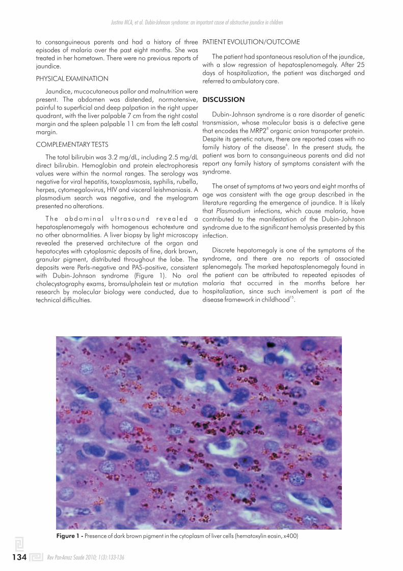
T h e a b d o m i n a l u l t r a s o u n d r e v e a l e d a hepatosplenomegaly with homogenous echotexture and no other abnormalities. A liver biopsy by light microscopy revealed the preserved architecture of the organ and hepatocytes with cytoplasmic deposits of fine, dark brown, granular pigment, distributed throughout the lobe. The deposits were Perls-negative and PAS-positive, consistent with Dubin-Johnson syndrome (Figure 1). No oral cholecystography exams, bromsulphalein test or mutation research by molecular biology were conducted, due to technical difficulties.
PATIENT EVOLUTION/OUTCOME
The patient had spontaneous resolution of the jaundice, with a slow regression of hepatosplenomegaly. After 25 days of hospitalization, the patient was discharged and referred to ambulatory care.
DISCUSSION
Dubin-Johnson syndrome is a rare disorder of genetic transmission, whose molecular basis is a defective gene
8that encodes the MRP2 organic anion transporter protein. Despite its genetic nature, there are reported cases with no
6family history of the disease . In the present study, the patient was born to consanguineous parents and did not report any family history of symptoms consistent with the syndrome.
The onset of symptoms at two years and eight months of age was consistent with the age group described in the literature regarding the emergence of jaundice. It is likely that Plasmodium infections, which cause malaria, have contributed to the manifestation of the Dubin-Johnson syndrome due to the significant hemolysis presented by this infection.
Discrete hepatomegaly is one of the symptoms of the syndrome, and there are no reports of associated splenomegaly. The marked hepatosplenomegaly found in the patient can be attributed to repeated episodes of malaria that occurred in the months before her hospitalization, since such involvement is part of the
15disease framework in childhood .
Figure 1 - Presence of dark brown pigment in the cytoplasm of liver cells (hematoxylin eosin, x400)
Justino MCA, et al. Dubin-Johnson syndrome: an important cause of obstructive jaundice in children
Rev Pan-Amaz Saude 2010; 1(3):133-136134
FINAL CONSIDERATIONS
Although Dubin-Johnson syndrome is a rare disease, it is extremely important to include it in the diagnostic investigation of cases of floating jaundice in childhood. The prognosis is favorable, its course is benign, and no
treatment is required. Therefore, establishing the correct diagnosis is important to prevent future unnecessary procedures. The carriers of the syndrome are later during adulthood guided not to use oral contraceptives, which can compete with the hepatocyte secretion of organic anions.
Síndrome Dubin-Johnson: importante causa de icterícia colestática na infância
RESUMO
A síndrome Dubin-Johnson é caracterizada clinicamente por episódios de icterícia colestática recorrente, de caráter benigno e familiar, sendo definida pela presença de pigmento melânico nos hepatócitos. Os autores relatam um caso de síndrome Dubin-Johnson em uma criança portadora de icterícia e hepatoesplenomegalia, cujo diagnóstico foi confirmado pela presença do pigmento castanho-escuro à microscopia hepática realizada a partir de biópsia, e alertam para a necessidade de suspeição dessa síndrome em casos de icterícia colestática flutuante na infância.
Palavras-chave: Colestase; Icterícia; Icterícia Idiopática Crônica.
El síndrome de Dubin-Johnson: importante causa de ictericia colestática en la infancia
RESUMEN
El síndrome Dubin-Johnson se caracteriza clínicamente por episodios de ictericia colestática recurrente, de carácter benigno y familiar, siendo definido por la presencia de pigmento melánico en los hepatocitos. Los autores relatan un caso de síndrome Dubin-Johnson en una niña portadora de ictericia y hepatoesplenomegalia, diagnóstico que fue confirmado por la presencia del pigmento castaño oscuro en la microscopia hepática realizada a partir de biopsia, y alertan para la necesidad de sospecha de ese síndrome en casos de ictericia colestática fluctuante en la infancia.
Palabras clave: Cholestasis; Ictericia; Ictericia Idiopática Crónica.
Justino MCA, et al. Dubin-Johnson syndrome: an important cause of obstructive jaundice in children
REFERENCES
1 Dubin IN, Johnson FB. Chronic idiopathic jaundice with unidentifield pigment in liver cells: a new clinicopathologic entity with a report of 12 cases. Medicine (Baltimore). 1954 Sep;33(3):155-97.
2 Haimi-Cohen Y, Amir J, Merlob P. Neonatal and infantile Dubin-Johnson syndrome. Pediatr Radiol. 1998 Nov;28(11):900.
3 Kondo T, Yagy R, Kuchiba K. Dubin-Johnson syndrome in neonate. N Engl J Med. 1975; 292:1028-9.
4 Rastogi A, Krishnani N, Pandey R. Dubin-Johnson syndrome, a clinicopathologic study of twenty cases. Indian J Pathol Microbiol. 2006;49(4):500-4.
5 Dubin IN. Chronic idiopathic jaundice with: a review of fifty cases. Am J Med. 1958 Feb;24(2):268-92.
6 Lanosa RA, Mazzini O, Pietriangelo C, Celia EJ, Monserrat JM. Contribucion al diagnostico del síndrome de Dubin-Johnson. Acta Gastroenterol Latinoam.1980;10:1-12.
7 Cebecauerova D, Jirasek T, Budisova L, Mandys V, Volf U, Novotna Z, et al. Dual hereditary jaundice: simultaneous occurrence of mutations causing Gi lbe r t ' s and Dub in- Johnson syndrome. Gastroenterology. 2005 Jul;129(1):135-320.
8 Paulusma CC, Kool M, Bosma PJ, Scheffer GL, Borg F, Sheper RJ, et al. A mutation in the human canalicularmultispecific organic anion transporter gene causes the Dubin-Johnson syndrome. Hepatology. 1997;25(6):1539-42.
9 Stapelbroek JM, Van Erpecum KJ, Klomp LWJ, Houwen RHJ. Liver disease associated with canalicular transport defects: current and future therapies. Journal of Hepatology. 2010 Feb;52(2):258-71.
10 Baba N, Ruppert RD. The Dubin-Johnson syndrome: electron microscopic observation of hepatic pigment-a case study. Am J Clin Pathol. 1972 Mar;57(3):306-10.
11 Sobaniec-Lotwska ME, Lebensztejn DM. Ultrastructure of Kupffer cells and hepatocytes in the Dubin-Johnson syndrome: a case report. World J Gastroenterol. 2006 Feb;12(6):987-9.
Rev Pan-Amaz Saude 2010; 1(3):133-136 135
Justino MCA, et al. Dubin-Johnson syndrome: an important cause of obstructive jaundice in children
12 Kimura A, Ushijima K, Kage M, Mahara R, Tohma M, Inokuchi T, et al. Neonatal Dubin-Johnson syndrome with severe cholestasis: effective phenobarbital therapy. Acta Paediatr Scand. 1991 Mar;80(3):381-5.
13 Shieh CC, Chang MH, Chen CL. Dubin-Jhonson syndrome presenting with neonatal cholestasis. Arch Dis Child. 1990;65:898-9.
14 Tsai WH, Teng RJ, Chu JS, Chang MH, Ho MM. Neonatal Dubin-Johnson syndrome. J Pediatr Gastroenterol Nutr. 1994;18(2):253-4.
15 Amaral CN, Albuquerque YD, Pinto AYN, Souza JM. A importância do perfil clínico-laboratorial no diagnóstico diferencial entre malária e hepatite aguda viral. J Pediatr (Rio J). 2003;79(5):429-34.
Received / Recebido em / Recibido en: 29/7/2010Accepted / Aceito em / Aceito en: 28/9/2010
Rev Pan-Amaz Saude 2010; 1(3):133-136136