Embed Size (px)
Citation preview
UNIVERSIDADE FEDERAL DE UBERLÂNDIAFACULDADE DE MEDICINA
PROGRAMA DE PÓS-GRADUAÇÃO EM CIÊNCIAS DA SAÚDE
EFEITO AGUDO DA BANDAGEM ELÁSTICA NO PADRÃO DE ATIVAÇÃO DOSMÚSCULOS ESTABILIZADORES DO TORNOZELO, NA CINEMÁTICA E NO
DESEMPENHO, DURANTE ATIVIDADES FUNCIONAIS EM JOVENS ADULTOSATIVOS.
GABRIEL PAGLIONI GARCIA
UBERLÂNDIA2017

GABRIEL PAGLIONI GARCIA
EFEITO AGUDO DA BANDAGEM ELÁSTICA NO PADRÃO DE ATIVAÇÃO DOSMÚSCULOS ESTABILIZADORES DO TORNOZELO, NA CINEMÁTICA E NODESEMPENHO, DURANTE ATIVIDADES FUNCIONAIS EM JOVENS ADULTOSATIVOS.
Dissertação apresentada ao Programa dePós-Graduação em Ciências da Saúde daFaculdade de Medicina da UniversidadeFederal de Uberlândia, como requisitoparcial para a obtenção do título de Mestreem Ciências da Saúde.
Área de concentração: Ciências da Saúde.
Orientador: Camilla Zamfolini Hallal
Co-orientador: Nise Ribeiro Marques
UBERLÂNDIA2017
Dados Internacionais de Catalogação na Publicação (CIP)Sistema de Bibliotecas da UFU, MG, Brasil.
G216e2017
Garcia, Gabriel Paglioni, 1991Efeito agudo da bandagem elástica no padrão de ativação dos
músculos estabilizadores do tornozelo, na cinemática e no desempenho,durante atividades funcionais em jovens adultos ativos / Gabriel PaglioniGarcia. - 2017.
66 f. : il.
Orientadora: Camilla Zamfolini Hallal.Coorientadora: Nise Ribeiro Marques.Dissertação (mestrado) - Universidade Federal de Uberlândia,
Programa de Pós-Graduação em Ciências da Saúde.Inclui bibliografia.
1. Ciências Médicas - Teses. 2. Tornozelos - Teses. 3. Eletromiografia- Teses. 4. Cinemática - Teses. I. Hallal, Camilla Zamfolini. II. Marques,Nise Ribeiro. III. Universidade Federal de Uberlândia. Programa de Pós-Graduação em Ciências da Saúde. IV. Título.
CDU: 61

FOLHA DE APROVAÇÃO
Gabriel Paglioni Garcia
EFEITO AGUDO DA BANDAGEM ELÁSTICA NO PADRÃO DE ATIVAÇÃO DOSMÚSCULOS ESTABILIZADORES DO TORNOZELO, NA CINEMÁTICA E NODESEMPENHO, DURANTE ATIVIDADES FUNCIONAIS EM JOVENS ADULTOSATIVOS.
Presidente da banca: Profa. Dra. Camilla Zamfolini Hallal
Dissertação apresentada ao Programa de Pós-
Graduação em Ciências da Saúde da Faculdade de
Medicina da Universidade Federal de Uberlândia,
como requisito parcial para a obtenção do título de
Mestre em Ciências da Saúde.
Área de concentração: Ciências da Saúde.
BANCA EXAMINADORA
Titular: Prof. Dr. Valdeci C. Dionisio
Instituição: Universidade Federal de Uberlândia - UFU
Titular: Prof. Dr. Luciano Fernandes Crozara
Instituição: Faculdade de Medicina de Marília FAMEMA

"O conhecimento não é seu, o conhecimento não é
meu, ele não é de ninguém, ele circula e só morre
quando o prendemos dentro de nós."

RESUMO
Introdução: O tornozelo está entre as articulações que mais estão sujeitas a lesões
durante a pratica de atividades esportivas. Contudo, ainda há uma lacuna sobre a
importância da bandagem elástica na estabilidade do tornozelo. Objetivo: Analisar o
efeito imediato da bandagem elástica no padrão de recrutamento dos músculos do
tornozelo, na cinemática e no desempenho, durante atividades funcionais em jovens
adultos ativos e saudáveis. Material e métodos: Participaram do estudo, 21
voluntários, com idade de 27,09 ± 5,83 anos, de ambos os sexos, praticantes de
Crossfit. A coleta ocorreu em dois dias com intervalo de 48 horas. No primeiro dia
foram realizados: a anamnese, testes ortopédicos específicos, familiarização com os
testes Star Excursion Balance Test (SEBT) e Multiple hop Test (MHT) e três
contrações isométricas voluntárias máximas (CIVM) de tibial anterior e gastrocnêmio
medial. Após a familiarização ocorreu a coleta de dados eletromiográficos,
cinemáticos e de desempenho nos testes SEBT e MHT. No segundo dia, foi realizado
familiarização dos gestos: salto vertical e mudança de direção, onde posteriormente
foram coletados os sinais eletromiográficos (SEMG) e dados cinemáticos dos gestos
familiarizados. Foi avaliado o desempenho apenas em MHT e SEBT.Todos os testes,
em ambos os dias, foram realizados nas condições com e sem bandagem elástica,
definidos de maneira aleatória por meio de sorteio. O processamento do sinal
eletromiográfico e cinemático, ocorreu por rotinas especificas do Matlab. A
normalização dos SEMGs ocorreu por meio das CIVMs. Para cinemática, o
acelerômetro foi posicionado entre terceiro metatarso e cuneiforme lateral,
considerando os dados dos eixos: transversal (X) e horizontal (Z). Para as
comparações foi utilizado o teste: ANOVA one-way medidas repetidas. O nível de
significância foi ajustado em p < 0,05. Resultados: Bandagem elástica possibilitou
aumento de 120,8% do pico de ativação de gastrocnêmio medial durante mudança de
direção (p =0,001) e também um aumento de 19,5% da média de ativação de
gastrocnêmio medial (p = 0,006) no teste de saltos com um único membro inferior.
Para desempenho, nas direções ântero medial e póstero medial do SEBT, ocorreu um
aumento na distância alcançada de 3,8% e 5,6% (p= 0,001 e p=0,005)
respectivamente, associado ao uso da bandagem elástica. Nos testes, de cruzar seis
metros saltando e de saltos com um único membro inferior, houve redução no tempo
de execução, de 5,7% e 7,1% (p=0,018 e p=0,009) respectivamente, associado ao

uso da bandagem elástica. Conclusão: De acordo com nossos achados a bandagem
elástica possui limitações como recurso terapêutico preventivo e de reabilitação,
entretanto pode ser benéfica para desempenho em atividades funcionais.
Palavras chaves: Tornozelo. Eletromiografia. Cinemática. Prevenção. Lesão.
ABSTRACT
Introduction: The ankle is among the joints that most subject to injury during a practice
of sports activities. However, there is still a gap on the importance of elastic taping in
ankle stability. Objective: Analyze the immediate effect of the elastic taping in the
recruitment pattern of the ankle muscles, in the kinematics and performance during
functional activities in young adults healthy and active. Material and methods:Participated in this study, 21 volunteers, aged 27,09 ± 5,83 years, of both sexes,
practitioners of Crossfit. The collection has occurred in two days with an interval of 48
hours. On the first day were carried out: anamnesis, orthopedic tests, the
familiarisation with the test Star Excursion Balance Test (SEBT) and the Multiple hop
Test (MHT) and three maximum voluntary isometric contraction (MVIC) of the tibialis
anterior and gastrocnemius medialis. After the familiarization has occurred in the data
collection electromyographic, kinematic, and performance in the tests, SEBT and MHT.
On the second day, was carried out familiarisation of the gestures: the vertical jump
and side cutting, where they were subsequently collected signals electromyography
(SEMG) and kinematic data. It was evaluated the performance only in MHT and
SEBT.All tests on both days were conducted under the conditions with and without
elastic taping randomly defined by lottery. The processing of electromyographic signal
and kinematic, occurred by routines specific to Matlab. The normalization of the
SEMGs occurred by means of the MVIC. For kinematics, the accelerometer was
positioned between the third metatarsal and lateral cuneiforme, considering the data
of the axes: transverse (X) and horizontal (Z). For the comparisons were used in the
test: ANOVA one-way repeated measures. The level of significance was set at p <0,05.
Results: Elastic taping increase 120,8% of the peak activation of the gastrocnemius
medialis during site cutting (p =0.001) and also an increase of 19.5% from the mean
activation of the gastrocnemius medialis (p = 0.006) in the test of single limb hopping
course. For performance, in the directions anteromedial e posteromedial in SEBT,
there was an increase in the distance reached 3.8% and 5.6% (p= 0.001 and p=0.005),
respectively, associated with the use of the elastic taping. In tests, cross six meter hop
for time and single limb hopping course, there was a reduction in run-time, 5.7% and
7.1% (p=0.018 and p=0.009), respectively, associated with the use of the elastic
taping. Conclusion: According our findings, the elastic taping has limitations as a

therapeutic resource for preventive and rehabilitation, however it can be beneficial to
performance in functional activities.
Key words: Ankle. Electromyography. Kinematics. Prevention. Injury.

LISTA DE ILUSTRAÇÕES
Figure 1. Flowchart of the study procedures..............................................................
Figure 2. Positioning the elastic taping in steps 1, 2, 3 and 4. ..................................
Figure 3. SEBT ..............................................................
Figure 4. Platform used to perform the test of one legged hop for distance..............36
Figure 5. Accelerometer pattern to determine the phases.........................................37
31
33
34
35
36
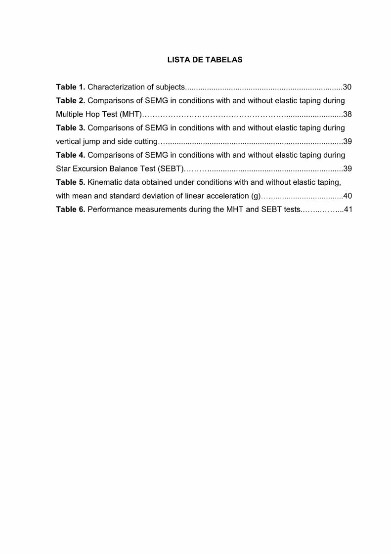
LISTA DE TABELAS
Table 1. Characterization of subjects........................................................................30
Table 2. Comparisons of SEMG in conditions with and without elastic taping during
...........................38
Table 3. Comparisons of SEMG in conditions with and without elastic taping during
vertical jump and side cutting .................................................................................39
Table 4. Comparisons of SEMG in conditions with and without elastic taping during
Star Excursion Balance Test (SEBT) ..............................................................39
Table 5. Kinematic data obtained under conditions with and without elastic taping,
with mean and standard deviation of .................................40
Table 6. Performance measurements during the MHT ... ....41

LISTA DE ABREVIATURAS E SÍMBOLOS
MHT Multiple Hop Test.
SEBT Star Excursion Balance Test.
Hz Hertz.
LTFA Ligamento talofibular anterior.
LTFP Ligamento talofibular posterior.
LCF Ligamento calcâneo fibular.
MVIC Maximum voluntary isometric contractions
GM Gastrocnêmio Medial / Gastrocnemius Medialis
TA Tibial Anterior / Tibialis Anterior
J1 Test of One legged hop for distance
J2 Test of triple legged hop for distance
J3 Test of Six meter hop for time
J4 Test of Cross six meter hop for time
J5 Test of Single limb hopping course
VJ Vertical Jump
SC Side Cutting
AcX Accelerometer axis X
AcZ Accelerometer axis Z
Am Anteromedial
M Medial
Pm Posteromedial
MEAN Mean
PEAK Maximum activation
Cocont Cocontraction
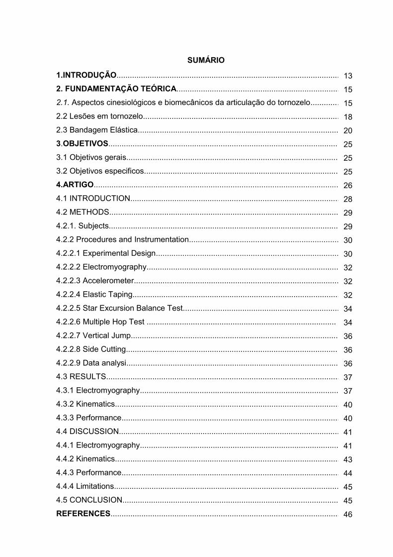
SUMÁRIO
1.INTRODUÇÃO........................................................................................................
2. FUNDAMENTAÇÃO TEÓRICA.............................................................................
2.1. Aspectos cinesiológicos e biomecânicos da articulação do tornozelo................
2.2 Lesões em tornozelo............................................................................................
2.3 Bandagem Elástica...............................................................................................
3.OBJETIVOS........................................................................................................
3.1 Objetivos gerais................................................................................................
3.2 Objetivos especificos.........................................................................................
4.ARTIGO...................................................................................................................
4.1 INTRODUCTION.....................................................................................................
4.2 METHODS..............................................................................................................
4.2.1. Subjects.............................................................................................................
4.2.2 Procedures and Instrumentation......................................................................
4.2.2.1 Experimental Design....................................................................................
4.2.2.2 Electromyography...............................................................................................
4.2.2.3 Accelerometer..................................................................................................
4.2.2.4 Elastic Taping..............................................................................................
4.2.2.5 Star Excursion Balance Test............................................................................
4.2.2.6 Multiple Hop Test ......................................................................................
4.2.2.7 Vertical Jump......................................................................................................
4.2.2.8 Side Cutting................................................................................................
4.2.2.9 Data analysi................................................................................................
4.3 RESULTS.................................................................................................................
4.3.1 Electromyography..................................................................................................
4.3.2 Kinematics..........................................................................................................
4.3.3 Performance......................................................................................................
4.4 DISCUSSION..........................................................................................................
4.4.1 Electromyography..................................................................................................
4.4.2 Kinematics............................................................................................................
4.4.3 Performance........................................................................................................
4.4.4 Limitations........................................................................................................
4.5 CONCLUSION..........................................................................................................
REFERENCES........................................................................................................
13
15
15
18
20
25
25
25
26
28
29
29
30
30
32
32
32
34
34
36
36
36
37
37
40
40
41
41
43
44
45
45
46

REFERÊNCIAS DA DISSERTAÇÃO...................................................................
APÊNDICE 1................................................................................................................
APÊNDICE 2................................................................................................................
51
60
61

13
1. INTRODUÇÃO
A articulação do tornozelo é comumente lesada durante à pratica de atividades
esportivas. De acordo com o estudo de FONG (2007), as lesões de maior ocorrência
no tornozelo, em um total de 43 modalidades esportivas, foram: entorse (33 esportes,
76,7%) e fratura (7 esportes, 16,3%). A entorse de tornozelo, por exemplo, pode levar
o indivíduo a limitações como: instabilidade funcional, redução da força muscular,
perda proprioceptiva, limitação de mobilidade e pode favorecer a degeneração
articular devido a recorrência de entorses (HERTEL,2002; WILLAMS et al., 2012; FU;
HUI-CHAN, 2005; AIRAKSINEN, 1999; DRAWER; FULLER, 2002).
Sabe que uma maior magnitude de impacto, durante gesto de aterrissagem de um
salto, maior a chance de ocorrência de lesões em tornozelo (WRIGHT et al., 2000).
Além disso, a velocidade de deslocamento articular pode vir a ser outro fator de risco
para ocorrência de lesões em tornozelo, visto que uma maior velocidade de
deslocamento articular leva a uma maior possibilidade de lesão (LOHRER; ALT;
GOLLHOFER,1999; HA et al., 2015).
Para manutenção da estabilidade articular, é necessário o trabalho em conjunto do
sistema ósseo, ligamentar e muscular (HERTEL, 2002). Onde a ação muscular,
através de contrações concêntricas e excêntricas, tem por objetivo gerar estabilidade
articular dinâmica (HERTEL, 2002). O tibial anterior, por meio de sua contração
excêntrica, controla movimentos excessivos de flexão plantar, consequentemente,
protege a articulação de movimentos excessivos que podem vir a favorecer lesões
(SINKJÆR et al., 1988; TERADA; PIETROSIMONE; GRIBBLE, 2012).
Já gastrocnêmio medial, também auxilia na estabilização de movimentos ativos do
tornozelo, essa estabilização se dá através da absorção de impacto e aumento da
rigidez muscular. (IIDA et al., 2011; LEE; PIAZZA, 2008). Estudos anteriores relatam
maior atividade de gastrocnêmio medial e tibial anterior durante atividades funcionais,
como aterrisagem e atividades que envolviam equilíbrio dinâmico, onde este aumento
de ativação, tanto de gastrocnêmio quanto de tibial anterior, possivelmente, seja uma
estratégia adaptativa para manutenção da estabilidade articular (MCKAY et al., 2001;
WOODS et al., 2003; LEE; PIAZZA, 2008; IIDA et al., 2011; KOSHINO et al., 2015;
POZZI; MOFFAT; GUTIERREZ, 2015; GUTTIERREZ et al., 2012).
A concontração é outro mecanismo muscular que poderia vir a contribuir com
a estabilização articular, ela é definida como uma ativação neuromuscular simultânea

14
de dois ou mais músculos antagonistas, que tem por objetivo gerar um ajuste dinâmico
da rigidez das partes móveis e por consequência manter a estabilidade articular
durante movimentos dinâmicos (DA FONSECA et al., 2001; NUNES, 2004 BARATTA
et al., 1988; DI NARDO et al.,2015; AQUINO, 2004; CANDOTTI et al.,2012).
Assim, a bandagem elástica pode vir atuar em mecanismos da estabilidade articular
do tornozelo, possivelmente, alterando parâmetros eletromiográficos e cinemáticos
(HSU et al., 2009; KUNI et al., 2015). A popularidade da bandagem elástica cresceu
após os jogos olímpicos de Pequim 2008, onde seu uso visava a prevenção e
reabilitação de lesões em atletas (WILLIAMS et al., 2012; MARTÍNEZ-GRAMAGE et
al., 2014). Alguns efeitos são atribuídos ao uso da bandagem elástica como: aumento
da excitabilidade muscular, correção do posicionamento articular, melhora da
circulação sanguínea e linfática e redução da dor através de supressão neurológica
(KASE, HASHIMOTO, TOMOKI; 1996). Esses efeitos podem ter ocorrido devido a um
aumento da estimulação somatosensorial do sistema proprioceptivo, que permite
respostas de diferentes aplicações, como inibição e ativação (FU et al., 2008).
Diversos estudos têm relatado que a bandagem elástica pode aumentar o
recrutamento neuromuscular (SLUPIK et al., 2006; HSU et al., 2009; HUANG et al.,
2011; KONISHI, 2013; GÓMEZ SORIANO et al., 2014), assim como, alterações
cinemáticas, estas que podem vir a reduzir mecanismos lesivos, como por exemplo,
evitar movimentos excessivos de flexão plantar (HO et al.,2015; Kuni et al., 2015). A
melhora no desempenho de atividades funcionais associado ao uso da bandagem
elástica, também tem sido relatado na literatura (BICICI; KARATAS; BALTACI, 2012).
Todavia, alguns estudos, relatam que a bandagem elástica não alterou parâmetros
eletromiográficos, cinemáticos ou no desempenho (BRIEM et al., 2011; DE ALMEIDA
LINS et al., 2013; MAGALHÃES et al., 2016; HETTLE et al., 2013).
Portanto, existe uma grande discrepância de resultados nos estudos que
investigam o efeito da bandagem elástica. Esta variabilidade de resultados pode estar
associada, as diferentes metodologias de aplicação da bandagem elástica. Assim, é
importante o entendimento, de como um recurso terapêutico, no caso bandagem
elástica, atua sobre parâmetros eletromiográficos e cinemáticos, pois assim seria
possível entender melhor os mecanismos lesivos do tornozelo e por consequência
elaborar reabilitações e prevenções mais precisas, visto que ainda existe uma lacuna
na literatura, em relação ao efeito real da bandagem elástica na prevenção e
reabilitação de lesões.
15
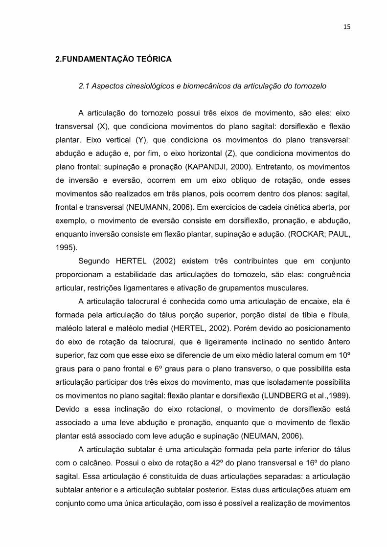
2.FUNDAMENTAÇÃO TEÓRICA
2.1 Aspectos cinesiológicos e biomecânicos da articulação do tornozelo
A articulação do tornozelo possui três eixos de movimento, são eles: eixo
transversal (X), que condiciona movimentos do plano sagital: dorsiflexão e flexão
plantar. Eixo vertical (Y), que condiciona os movimentos do plano transversal:
abdução e adução e, por fim, o eixo horizontal (Z), que condiciona movimentos do
plano frontal: supinação e pronação (KAPANDJI, 2000). Entretanto, os movimentos
de inversão e eversão, ocorrem em um eixo obliquo de rotação, onde esses
movimentos são realizados em três planos, pois ocorrem dentro dos planos: sagital,
frontal e transversal (NEUMANN, 2006). Em exercícios de cadeia cinética aberta, por
exemplo, o movimento de eversão consiste em dorsiflexão, pronação, e abdução,
enquanto inversão consiste em flexão plantar, supinação e adução. (ROCKAR; PAUL,
1995).
Segundo HERTEL (2002) existem três contribuintes que em conjunto
proporcionam a estabilidade das articulações do tornozelo, são elas: congruência
articular, restrições ligamentares e ativação de grupamentos musculares.
A articulação talocrural é conhecida como uma articulação de encaixe, ela é
formada pela articulação do tálus porção superior, porção distal de tíbia e fíbula,
maléolo lateral e maléolo medial (HERTEL, 2002). Porém devido ao posicionamento
do eixo de rotação da talocrural, que é ligeiramente inclinado no sentido ântero
superior, faz com que esse eixo se diferencie de um eixo médio lateral comum em 10º
graus para o pano frontal e 6º graus para o plano transverso, o que possibilita esta
articulação participar dos três eixos do movimento, mas que isoladamente possibilita
os movimentos no plano sagital: flexão plantar e dorsiflexão (LUNDBERG et al.,1989).
Devido a essa inclinação do eixo rotacional, o movimento de dorsiflexão está
associado a uma leve abdução e pronação, enquanto que o movimento de flexão
plantar está associado com leve adução e supinação (NEUMAN, 2006).
A articulação subtalar é uma articulação formada pela parte inferior do tálus
com o calcâneo. Possui o eixo de rotação a 42º do plano transversal e 16º do plano
sagital. Essa articulação é constituída de duas articulações separadas: a articulação
subtalar anterior e a articulação subtalar posterior. Estas duas articulações atuam em
conjunto como uma única articulação, com isso é possível a realização de movimentos

16
como a pronação e supinação, bem como, auxilio nos movimentos de adução e
abdução (VILADOT et al., 1984; ROCKAR; PAUL, 1995; HARMON, 2004).
A sindesmose fíbular distal é a articulação distal entre tíbia e fíbula. Essa
articulação tem como principal estabilizador a membrana interóssea, que permite uma
ligação muito forte entre as extremidades distais de tíbia e fíbula. Essa articulação
também é estabilizada por ligamentos, como: tibiofibular anterior e tibiofibular posterior
(HERTEL, 2002). O papel funcional dessa articulação é limitar o movimento de
translação e rotação da articulação talocrural, durante os movimentos de dorsiflexão
e flexão plantar e, com isso, promover maior congruência da articulação talocrural e,
consequentemente, maior estabilidade articular (PENA; COETZEE, 2006; HERTEL,
2002).
A anatomia óssea da articulação do tornozelo é responsável pela estabilidade
articular na posição neutra quando sujeito a cargas compressivas mais elevadas
(STORMONT et al., 1985; KERKHOFFS et al., 2007). O tálus, possui importante
contribuição na estabilidade do tornozelo, pois, anatomicamente, o tálus é maior
anteriormente do que posteriormente. Durante a flexão plantar, o tálus roda
posteriormente e se desloca anteriormente, assim a pinça bimaleolar, aproximação do
maléolo medial e lateral, promove uma redução no espaço interósseo para a
manutenção da estabilidade articular (KAPANDJI, 2000). Na dorsiflexão, existe um
contato da região superior do tálus com a margem anterior da tíbia, limitando o
movimento de dorsiflexão, além disso durante dorsiflexão ocorre um aumento do
espaço interósseo provido pela abertura da pinça bimaleolar, afastamento do maléolo
medial e lateral (KAPANDJI, 2000; NEUMANN, 2006). A fíbula se estende,
ligeiramente mais, até maléolo lateral do que a tíbia em relação ao maléolo medial,
devido a essa configuração óssea, há uma maior amplitude de movimento em
inversão do que em eversão (KAPANDJI, 2000)
Em condições de ausência de carga, a estabilização passa a ser promovida
pelas estruturas ligamentares (STORMONT et al., 1985; KERKHOFFS et al., 2007).
Entre os ligamentos que estabilizam o complexo articular do tornozelo, temos: o
ligamento talofibular anterior (LTFA), o ligamento talofibular posterior (LTFP), e o
ligamento calcâneo fibular (LCF) na face lateral do tornozelo. Na face medial, o
ligamento Deltoideo é o principal estabilizador medial (BOZKURT; DORAL, 2006).
Quando ocorrem perturbações cinemáticas externas, a magnitude da carga determina
quais ligamentos estarão sujeitos à lesão. Em termos de resistência à tensão, o LTFA
17
é o ligamento mais frágil e, geralmente, lesionado ao primeiro momento, o LCF é mais
resistente do que LTFA e o LTFP é o mais difícil de se lesionar, sendo que a lesão
deste, geralmente, está associado a fraturas de tornozelo e luxações (BROSTRÖM,
1966; SAFRAN et al., 1999; HA; FONG; CHAN, 2016). Em contrapartida os ligamentos
mediais são mais resistentes do que os ligamentos laterais (SAVAGE-ELLIOTT et al.,
2013), onde os mesmos são responsáveis por alinhar o tálus e o maléolo medial,
resistindo à adução do tálus em relação a tíbia e estabilizam essas estruturas quando
ocorre um estresse em valgo do tornozelo. (BEALS; CRIM; NICKISCH, 2012).
Além dos elementos passivos, como ósseos e ligamentos, para que ocorra a
estabilização articular é preciso que alguns músculos sejam recrutados em conjunto.
Esses músculos devem gerar estabilidade articular dinâmica por meio de contrações
concêntricas e excêntricas (HERTEL, 2002).
,
18
2.2 Lesões em tornozelo
O tornozelo está entre as articulações que mais estão sujeitas a lesões durante
a prática de atividades esportivas. Estudo anterior, constatou que em 24 modalidades
esportivas, em um total de 70 modalidades, apresentavam como região anatômica de
maior ocorrência de lesão, a articulação do tornozelo (FONG et al., 2007). Outro dado
interessante à cerca de lesões em tornozelo se trata do estudo de NABHAN (2016),
que relata, que no ano de 2014, durante os jogos olímpicos da juventude, a equipe
dos Estados Unidos, teve como regiões anatômicas mais acometidas por lesões,

19
joelho e tornozelo, sendo que 12% das lesões ocorreram no tornozelo. Já em relação
à tipos de lesões, foi feito um levantamento em 43 esportes, com históricos de lesões
em tornozelo e que a entorse do tornozelo foi a lesão mais comum (33 esportes,
76,7%), seguida de fratura (7 esportes, 16,3%) dentro destas 43 modalidades (FONG
et al., 2007).
A entorse de tornozelo é definida como uma lesão que afeta um ou mais
ligamentos que estão no tornozelo. As lesões de tornozelo podem ser definidas como
entorses agudos e entorses crônicos, sendo que 85% de todas as lesões no tornozelo
estão relacionadas a mecanismos lesivos laterais, ou seja, entorses que lesam os
ligamentos laterais (BAHR; ENGEBRETSEN, 2009). A classificação de entorse de
tornozelo é baseada no exame clínico da área afetada e é dividida em três tipos: grau
1, ocorre estiramento ligamentar; grau 2, ocorre lesão ligamentar parcial e grau 3,
ocorre lesão ligamentar total (BERNETT; SCHIRMANN, 1989). O mecanismo lesivo
mais comum da entorse do tornozelo ocorre por meio do movimento de inversão, esse
mecanismo é responsável pela entorse lateral de tornozelo, que ocorre quando há
supinação excessiva do retro pé, combinado com rotação externa da tíbia no início do
contato do pé com o solo durante as mudanças rápidas de direção (WRIGHT et al.,
2000).
Quando não há lesão, as informações proprioceptivas, tais como cinestesia e
senso de posição articular são obtidos a partir de mecanorreceptores, logo após a
detecção de deslocamentos comuns ou perturbações (HUGHES; ROCHESTER,
2008). Após uma lesão por entorse, por exemplo, há redução da informação
proprioceptiva, pois os receptores da pele são atingidos, como por exemplo
mecanorreceptores, diminuindo a capacidade de identificar a posição do corpo, dos
segmentos corporais e a percepção do movimento das articulações, o que resulta em
um mal posicionamento do pé durante gestos funcionais, aumentando-se assim, a
possibilidade de lesões por entorse (AMATUZZI et al., 2004; DELAHUNT;
MONAGHAN; CAULFIELD 2007). Outro fator que pode predispor a lesão por entorse
é a velocidade de deslocamento articular, uma maior velocidade de deslocamento
articular contribui como fator de risco adicional para a ocorrência de lesões em
tornozelo (LOHRER; ALT; GOLLHOFER, 1999; HA; FONG; CHAN, 2016).

20
2.3 Bandagem Elástica
A bandagem elástica surgiu em 1973, sendo utilizada a primeira vez em grande
escala nos Jogos Olímpicos de Seul de 1988 (MATHEUS et al., 2016). Nas olimpíadas
de Pequim 2008, a popularidade da bandagem elástica cresceu substancialmente e
tinha como finalidade a prevenção e reabilitação de lesões em atletas (WILLIAMS et
al., 2012; MARTÍNEZ-GRAMAGE et al., 2014). A bandagem elástica é uma fita
elástica, sem látex, permeável e resistente a água sendo possível utiliza-la por vários
dias, feita de algodão que pode ser aplicada sobre músculos e articulações, se difere
de outras bandagens pelo fato de se distender de 130% a 140% do seu comprimento
original. (BICICI; KARATAS; BALTACI, 2012; MARTÍNEZ-GRAMAGE et al., 2014).
Entretanto em estudo recente, de MATHEUS (2016), foi possível observar que a
distensão da fita foi maior do que os autores da literatura relatavam, atingindo maiores
valores de distensão, além disso o mesmo relata que diferentes marcas de bandagem
elástica, possuem características distintas, como por exemplo: tensão e rigidez.
Alguns efeitos terapêuticos têm sido atribuídos ao uso da bandagem elástica
como: excitabilidade muscular, correção do posicionamento articular, melhora da
circulação sanguínea e linfática e redução da dor através de supressão neurológica
(KASE, HASHIMOTO, TOMOKI; 1996). Esses efeitos podem estar relacionados com
uma estimulação somatosensorial, que permite diferentes respostas a aplicação da
bandagem, como uma maior ativação ou até inibição (FU et al.,2008).
Essa estimulação somatosensorial tem início no sistema tegumentar. Esse
sistema fornece informações do ambiente para SNC. Existem diversas terminações
nervosas (receptores), na pele, que são capazes de captar estímulos como:
dolorosos, térmicos e mecânicos. Estes receptores podem ser fásicos, aqueles que
respondem enquanto o estímulo é mantido ou tônicos, que se adaptam lentamente a
um estimulo constante (JUNIOR, 2015). A bandagem elástica visa estimular a
sensibilidade tátil e assim estimular estes receptores específicos.
O tato pode ser dividido em fino e grosso. O tanto fino possui os seguintes
receptores:
Discos de Merkel: Receptores de adaptação lenta e sensíveis a pressão
vertical local e não respondem ao alongamento lateral da pele. Assim, os movimentos
de pressão e tração sobre a epiderme desencadeiam os estímulos que são enviados
para o sistema nervoso centra (JUNIOR, 2015).

21
Corpúsculos de Meissner: Receptores de adaptação rápida e sensíveis a
pressão e a vibração local constante (JUNIOR, 2015).
Corpúsculos de Ruffini: Receptores de adaptação lenta e respondem ao
alongamento da pele sobre uma grande área. Adaptam-se lentamente à um
alongamento constante e emitem descargas em respostas aos ângulos articulares
estáticos para o sistema nervoso central (JUNIOR, 2015).
Corpúsculos de Pacini: Receptores de adaptação rápida, detectam estímulos
de pressão que se alteram rapidamente. Esta alteração de pressão é enviada aos
centros nervosos correspondentes, para que se elabora uma resposta ao estímulo
(JUNIOR, 2015).
O tato grosso, possui terminações nervosas por toda a pele, esses receptores
não são proprioceptivos, mas contribuem com informações cinestésicas (JUNIOR,
2015).
Assim, quando a pele é estimulada, por exemplo, pela bandagem elástica, os
estímulos gerados pela bandagem são enviados ao córtex sensorial primário, que
discrimina a intensidade e qualidade do estimulo. Posteriormente a isso, a informação
é enviada para o córtex de associação, que interpreta o estimulo e seleciona metas
do que fazer com o estímulo. Após isto, a informação chega até a área de
planejamento motor, que analisa como será a composição e sequenciamento da
resposta ao estimulo inicial da pele, provido pela bandagem elástica. Por fim o
estimulo inicial se transforma em um estimulo motor, que por consequência irá gerar
uma resposta ao estímulo inicial. Resumidamente, a bandagem elástica visa estimular
a sensação tátil, para que esta informação possa ser interpretada e utilizada na
motricidade e consciência corporal (JUNIOR, 2015).
Pesquisas anteriores, relatam um aumento de ativação eletromiográfica associada ao
uso da bandagem elástica. HSU (2009), avaliou o efeito da bandagem elástica, tanto
na eletromiografia quanto cinemática, do trapézio inferior, em indivíduos com
síndrome do impacto e observou que o uso da bandagem elástica proporcionou um
aumento de ativação neuromuscular assim como alteração na cinemática escapular.
HUANG (2011), em sua pesquisa com jovens adultos sedentários, verificou o efeito
da bandagem elástica durante o gesto de salto vertical e constatou uma tendência de
aumento da ativação de gastrocnêmio medial associado ao uso da bandagem
elástica. GÓMEZ-SORIANO (2014), verificou o efeito da bandagem elástica em
gastrocnêmio medial, sobre parâmetros eletromiográficos e de torque, em indivíduos

22
saudáveis. Observou-se um aumento da ativação eletromiográfica à curto prazo, no
caso, após 10 minutos de aplicação, entretanto após 24 horas, o efeito se perdeu.
Outros estudos relatam aumento da ativação eletromiográfica associado ao uso da
bandagem elástica (SLUPIK et al., 2006; KONISHI, 2013). Mas, em contrapartida,
alguns estudos, também relatam que o uso da bandagem elástica não altera
parâmetros eletromiográficos. MAGALHÃES (2016) avaliou o efeito da bandagem
elástica sobre eletromiografia e desenvolvimento de força do tríceps sural de jovens
ativos. Não houve alteração da eletromiografia e sim, apenas uma melhora na taxa de
desenvolvimento de força, no início da contração. BRIEM (2011), avaliou o efeito tanto
da bandagem elástica quanto rígida, durante teste em plataforma de inversão. Uma
maior ativação eletromiográfica de fibulares foi observada durante a condição
bandagem rígida, entretanto a condição, bandagem elástica não alterou o
recrutamento neuromuscular de fibulares. Outros estudos, relatam que o uso da
bandagem elástica não possibilitou a alteração do recrutamento neuromuscular (DE
ALMEIDA LINS et al., 2013; TRÉGOUËT; MERLAND; HORODYSK, 2012; SIMON;
GARCIA; DOCHERTY, 2014).
23
Para desempenho, BICICI (2012), observou os efeitos de bandagem rígida,
elástica, placebo e sem bandagem durante a realização de atividades funcionais que
envolviam saltos com único membro inferior e salto vertical. Embora sem diferença
significativa, a condição bandagem elástica apresentou resultados benéficos, como,
redução do tempo de execução dos testes que envolviam, saltos com único membro
e também uma maior altura durante a realização do salto vertical. Já em relação a
déficits funcionais, o uso da bandagem elástica de maneira longitudinal tem
apresentado resultados benéficos. KIM (2015), em um estudo de caso, aplicou a
bandagem elástica em indivíduo com instabilidade funcional de tornozelo durante dois
meses e observou que o uso longitudinal possibilitou redução na instabilidade
funcional durante atividades funcionais como subir, descer escada e saltar, além disso
apresentou melhora na pontuação do questionário Cumberland Ankle Instability Tool
(CAIT), que classifica instabilidade funcional de tornozelo. LEE (2016) realizou um
estudo de caso, de um indivíduo com instabilidade funcional, onde o tempo de
aplicação da bandagem elástica foi de quatro semanas e como resultado, obteve uma
redução na instabilidade funcional, melhora no quadro álgico, uma melhor pontuação
foi observada no questionário CAIT e maiores distancias foram alcançadas durante o
teste Y-balance. Todavia, HETTLE (2013), discorda dos autores citados
anteriormente, pois o mesmo, não encontrou em seus resultados, uma melhora de
desempenho associada ao uso da bandagem elástica, durante o teste funcional Star
Excursion Balance Test que foi aplicado em indivíduos com instabilidade funcional de
tornozelo.
24
Esta melhora em desempenho e déficits funcionais, pode ser explicada devido
ao aumento da percepção subjetiva de segurança que a bandagem pode gerar
através da estimulação de mecanorreceptores (GÓMEZ- SORIANO et al., 2014).
SAWKINS (2007) relata que em seu estudo, que indivíduos que estavam sujeitos
aplicação de algum tipo de bandagem, seja ela elástica ou placebo, relataram maior
sensação de segurança. SIMON (2014), verificou o efeito da bandagem elástica sobre
a propriocepção, em indivíduos com instabilidade funcional de tornozelo e saudáveis,
durante movimento de eversão e o mesmo relata que a melhora encontrada na
propriocepção, em ambos os grupos, estava associada a maior sensibilidade de
segurança e confiança proporcionados pela bandagem elástica. Nota-se, portanto, a
discrepância de resultados em estudos com bandagem elástica, possivelmente
associados a divergências metodológicas do recurso terapêutico como por exemplo:
posicionamento, amostra, teste proposto e tempo de aplicação.

25
3. OBJETIVOS
3.1 Objetivos Gerais
Avaliar o efeito de aplicação imediata da bandagem elástica, sobre padrões
eletromiográficos dos músculos estabilizadores do tornozelo, na cinemática do
tornozelo e no desempenho, durante atividade funcionais, em jovens adultos
saudáveis e ativos.
3.2 Objetivos específicos
- Comparar o efeito de aplicação imediata da bandagem elástica com a
condição sem bandagem elástica no recrutamento neuromuscular de gastrocnêmio
medial e tibial anterior durante atividades funcionais.
- Comparar o efeito de aplicação imediata da bandagem elástica com a
condição sem bandagem elástica na aceleração de deslocamento articular do
tornozelo, nos eixos: transversal e horizontal, durante atividades funcionais.
- Comparar o efeito de aplicação imediata da bandagem elástica com a
condição sem bandagem elástica no desempenho, durante os testes Multiple Hop
Test e Star Excursion Balance Test.

26
4. ARTIGO
IMMEDIATE EFFECT OF ELASTIC TAPING IN THE PATTERN OF ACTIVATION OFSTABILIZING MUSCLES AND KINEMATICS AND PERFORMANCE OF THEANKLE DURING FUNCTIONAL ACTIVITIES IN YOUNG ACTIVE ADULTS.
Gabriel Paglioni Garcia1, Ricardo José Tecchio Serrão1, Nise Ribeiro Marques2,
Camilla Zamfolini Hallal1
1 Programa de Pós-Graduação em Ciências da Saúde, Faculdade de Medicina,
Universidade Federal de Uberlândia, Brasil,
2 Centro de Ciências da Saúde, Universidade do Sagrado Coração, USC, Bauru, BRA
Correspondence:
Rua Irmã Arminda, 10-50, Vila Cardia, Bauru, SP. Centro de Ciências da Saúde,
Universidade do Sagrado Coração.
E-mail: [email protected]
Key words: Ankle. Electromyography. Kinematics. Prevention. Injury.

27
ABSTRACT
Objectives: Analyze the effect of elastic taping on electromyography of ankle muscles,
in kinematics and performance, during functional tests, in young healthy and active
adults. Methods: Twenty one healthy volunteers, aged 27.09 ± 5.83 years, of both
sexes, participated in the study. Electromyographic and kinematic data were collected
during tests: Multiple Hop Test (MHT), Star Excursion Balance Test (SEBT), vertical
jump and side cutting. Performance was measured only in MHT and SEBT. For the
electromyography, were considered, the tibialis anterior and gastrocnemius medialis
and in kinematics, plantar flexion and supination were considered for data collection.
All volunteers performed the tests in the condition with and without elastic taping. For
the comparisons was used the test, ANOVA one-way repeated measures. Results:Elastic taping allowed a 120.8% increase in the gastrocnemius medialis activation
peak during side cutting (p = 0.001) and also a 19.5% increase in the gastrocnemius
medialis activation mean (p = 0.006) in the test of single limb hopping course. For
performance, elastic taping provided an increase in the distances reached in the
anteromedial and posteromedial directions of the SEBT, of 3.8% and 5.6% (p = 0.001
and 0.005) respectively, as well as a reduction in the execution time of the
cross six meter hop for time and single limb hopping course of 5.7% and 7.1% (p =
0.018 and p = 0.009) respectively. Conclusion: Elastic taping has limitations as a
preventive and rehabilitative therapeutic resource, however it may be beneficial for
performance in functional activities.

28
4.1 INTRODUCTION
The ankle is between the joints that are most prone to injury while practicing
sports activities. A previous study verified the incidence of ankle injuries in 70 sports
modalities. It was found that approximately 34% of these modalities presented the
ankle as the anatomical region with the highest incidence of injuries (Fong et al., 2007).
At the 2014 Youth Olympic Games, for example, 12% of injuries occurred in the United
States delegation, occurred in ankle. (Nabhan et al., 2016). A survey was made of the
most common types of ankle injuries, in a total of 43 sports, where the ankle sprain
was the most common injury (33 sports, 76.7%), followed by fracture (7 sports, 16.3%)
(Fong et al., 2007).
Some factors such as landing phase of a jump and intensity of the impact in the
landing may cause ankle injuries (Wright et al., 2000). This may also be related to a
joint displacement velocity, a reduction in joint displacement velocity may be
considered an additional risk factor for the occurrence of ankle injuries. (Lohrer et al.,
1999; Ha et al., 2015). Another factor that can cause ankle injuries is the alteration of
neuromuscular activity, since the muscular action through concentric and eccentric
contractions assists in the dynamic stabilization of the movement. (Stormont et al.,
1985; Hertel, 2002).
Thus, tibialis anterior and gastrocnemius medialis action may assist in the
stabilization of the ankle joint, considering tibialis anterior, eccentrically controls the
movement of plantar flexion, reducing excessive amplitudes of this movement (Sinkjær
et al., 1988). Gastrocnemius medialis, through the absorption of impact and increase
of muscle stiffness, contributions to dynamic stability maintenance during dynamic
movements (Lee and Piazza, 2008; Iida et al., 2011). In addition, tibialis anterior and
gastrocnemius medialis can perform the cocontraction, which is defined as a
simultaneous neuromuscular activation of two or more antagonistic muscles, with the
purpose of generating an adjustment of the stiffness of the involved muscles aiming at
maintaining stability during unstable activities (Fonseca et al. Al., 2001; Nunes, 2004;
Aquino, 2004; Candotti et al., 2012).
Ankle stabilization can be supplement with the help of an external resource. This
resource may be an elastic taping (Kinesio Taping®), which was create by Kenzo
Kase, in the 70's. This resource has some effects like increase neuromuscular
recruitment and a correction of joint position (Kase et al., 1996;Gómez-Soriano et al.,

29
2014).These effects may help to maintain joint stability of the ankle. Previous studies
have reported that an increase in muscle recruitment during functional activity was
possible through the elastic taping (Hsu et al., 2009; Huang et al., 2011), as well
reduction of ankle injury mechanisms, such a maximal plantar flexion during dynamic
movement (Ho et al., 2015; Kuni et al., 2015). In terms of performance, BICICI (2012)
reports that the use of the elastic taping compared to other types of bandage presented
differences. Although this difference was not significant, individuals who used elastic
taping had a higher mean height during vertical jump and performed activities with a
single lower limb, with a lower mean time to perform the task, when compared to other
conditions. However, some studies also report that the use of elastic taping does not
chances neuromuscular recruitment, kinematics or performance (Briem et al., 2011;
Magalhães 2016; Hettle et al., 2013).
A divergence of results is observed with respect to studies with elastic taping.
This may be associated to methodological variability of the application of such
therapeutic resource, such as positioning, distension and time of application. Thus, it
is important to understand the effect of elastic taping on electromyography and
kinematic parameters during functional activities, since with elastic taping, it would be
possible to generate effects on these parameters, enabling a more precise and
coherent prevention and rehabilitation, as well as a lower recovery time and inactivity.
Therefore, the objective of this study was to evaluate the immediate effect of elastic
taping on the neuromuscular recruitment pattern, kinematics and performance during
functional activities in healthy and active young adults. We hypothesized that elastic
taping may change electromyography, kinematic, and performance parameters during
functional activities.
4.2 METHODS
4.2.1. Subjects
This research is a cross-sectional case-control. All volunteers signed the Informed
Consent Term. The study was approved by the Research Ethics Committee of the
Universidade Estadual Paulista, Campus of Marília, under the number: 073586/2015.
Participated in this research, 21 Crossfit practitioners (7 women and 14 men), aged
between 18 and 35 years, who frequently trained three times a week for one hour per

30
day, without lower limb injury and without mechanical joint instability. The
characterization of volunteers is present in Table 1. The sample size was calculated
using G*Power 3.1 software (Universitat, Kiel, Germany) with data obtained through a
pilot study. The analyzed variable was side cutting was calculated out by means of
pilot studies. Thus, considering a significance level of p <0.05, effect size of 0.45 and
with 90% test power for a sample size of 40 volunteers
Table 1.Characterization of subjects
Mean ± Standard Deviation
Age (Years)Body Mass Index (BMI)Weekly Training (Days)Crossfit Practice time (Months)
27,1±5,824,5±1,74,9±0,9
23,9±10,7
4.2.2 Procedures and Instrumentation
4.2.2.1 Experimental Design
Data collection occurred in two days, with a 48 hours interval between sessions.
On the first day, were evaluated the volunteers, by specific evaluation form, which
recorded anthropometric data, history of sprains, training frequency and evaluation of
mechanical stability through with specific orthopedic tests: drawer test and talar tit test
(Cipriano and Cipriano, 1999; Clanton and Schon, 2007). The volunteers were
instructed to use the daily training shoes to perform the proposed activities (Kuhman
et al., 2016) and the dominant lower limb was defined for the positioning of the elastic
taping and the performance of the tests (Hoffman et al., 1998).
After the initial evaluation, the volunteers performed three maximal voluntary
isometric contractions (MVICs) of the muscles: tibialis anterior and Gastrocnemius
medialis muscles, lasting five seconds for each contraction and 30 seconds of rest
between each contraction. The MVIC placements followed the following instructions:
Tibialis Anterior - The volunteers should perform the dorsiflexion and supination
movement against manual resistance (Kendall and Mccreay, 1995). Gastrocnemius
medialis - Volunteer with extension knee, perform the plantar flexion movement against
resistance (Kendall and Mccreay, 1995).
31
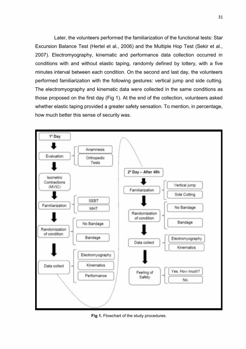
Later, the volunteers performed the familiarization of the functional tests: Star
Excursion Balance Test (Hertel et al., 2006) and the Multiple Hop Test (Sekir et al.,
2007). Electromyography, kinematic and performance data collection occurred in
conditions with and without elastic taping, randomly defined by lottery, with a five
minutes interval between each condition. On the second and last day, the volunteers
performed familiarization with the following gestures: vertical jump and side cutting.
The electromyography and kinematic data were collected in the same conditions as
those proposed on the first day (Fig 1). At the end of the collection, volunteers asked
whether elastic taping provided a greater safety sensation. To mention, in percentage,
how much better this sense of security was.
Fig 1. Flowchart of the study procedures.
32
4.2.2.2 Electromyography
For the collection of the electromyographic signal (SEMG), a biological signal
acquisition module (Noraxon, Arizona, USA) was used, adjusted with sampling rate of
1000 Hz, total gain of 2000 times (20 in the preamplifier and 100 in the amplifier) and
90 dcB common mode rejection.
Circular disposable electrodes of Ag / Agl, positioned in a bipolar configuration,
with a capture area of 1 cm2 and distance between 2 cm electrodes were positioned
on the tibialis anterior and gastrocnemius medialis muscles according to SENIAM
standards (Hermens et al., 2000). The positioning of the electrode consisted of tibialis
anterior - proximal third between the distance of the head of the fibula and medial
malleolus. Gastrocnemius medialis - most prominent portion of muscle. Before the
electrodes placed, we performed trichotomy and skin cleaning with alcohol, to reduce
possible interference in the SEMG acquisition (Hermens et al., 2000). The reference
electrode positioned on the patella.
4.2.2.3 Accelerometer
The kinematic data were captured by a 3D accelerometer (Noraxon, Arizona,
USA) that was previously collected, positioned between the third metatarsus and the
lateral cuneiform, in order to capture the acceleration of the segment in the transverse
axis (X), which conditions movements in the sagittal plane (plantar flexion) and
horizontal axis (Z), which conditions movements of the frontal plane (supination)
(Kapandji, 2000).
4.2.2.4 Elastic Taping
Were used elastic tapings (Kinesiosport, Marília, Brazil), 500 cm long by 5 cm
wide. Prior to the positioning of the elastic taping, were performed trichotomy and
hygiene of the area. The steps for the positioning of the elastic taping were adapted
for the study of KIM (2015). One physiotherapist with specific training in the method,
applied the elastic taping. The description of the steps followed for the positioning of
the elastic taping were:

33
Step 1 All the bandages were divided into three parts: two extremities and a
central part, where extremities were not tensioned. In the first step, the central part of
the bandage was positioned on the talocrural joint, with 50% of the maximum tension
(Therapist applied the highest possible tension on the bandage and identified the
percentage required for application). The extremities positioned on medial malleolus
and lateral malleolus (Fig. 2).
Step 2: Central part of the tape was placed on retro foot (plantar fascia -
calcaneal region), following caudal - cranial direction, with 50% of maximum tension.
The extremities of the bandage positioned over the medial and lateral malleolus (Fig.
2).
Step 3: One of the extremity positioned above medial malleolus, the central part
passed behind the calcaneus tendon and was directed to the lateral region of the retro
foot with 40% of maximum tension. The other extremity of the tape positioned medially
to the plantar fascia, more precisely on the navicular bone region (Fig. 2).
Step 4: One of the extremity positioned above the lateral malleolus, the central
part passed behind the tendon of the calcaneus and directed to the medial region of
the retro foot with 40% of maximum tension and the other end of the tape positioned
laterally to fascia plantar, more precisely on the cuboid bone (Fig. 2).
Fig. 2. Positioning the elastic taping in steps 1, 2, 3 and 4. The arrows indicate the positioning
and direction of the tapings.

34
4.2.2.5 Star Excursion Balance Test
The test should be performed in unilateral support, with the contralateral lower
limb to be touched as far as possible in three different directions, being them:
anteromedial, medial and posteromedial (Fig. 3).
Initially the volunteers were familiar as recommended by HERTEL (2006). The
test was performed three times, with an interval of 5 minutes between each evaluation
and 10 seconds of rest between each trial (Gribble and Hertel, 2003). For validation of
movements, the volunteer should keep his or her hands on the waist, always keep the
support foot on the ground, not transfer the body weight to the foot that will reach
maximum distance and maintain balance during reaching and during the end of the
movement (Steib et al., 2013, Delahunt et al., 2010). The maximum distance reached
was marked and the test applicator measured the distance. For the analysis, the mean
obtained in each direction were considered (Hertel et al., 2006).
Fig. 3. SEBT - Posteromedial direction. A. Anterior view B. Medial view C. Rear view.
4.2.2.6 Multiple Hop Test
Jump 1 - One legged hop for distance: Volunteers performed a single jump
forward, as far as possible, with only one lower limb and without losing balance. The
distance reached was marked and measured (Sekir et al., 2007).

35
Jump 2 - Triple legged hop for distance: Volunteers performed three
consecutive jumps with a single lower limb to reach as far as possible. The distance
was marked and measured. (Sekir et al., 2007).
Jump 3 - Six meter hop for time: this test, the volunteers were instructed to jump
as fast as possible on a six meters straight course with a single lower limb. The time
taken for a test was measured (Sekir et al., 2007).
Jump 4 - Cross six meter hop for time: The volunteers were executed with a
single lower limb, crossing a straight line, width of 10 cm and with total distance of six
meters, as fast as possible. The time taken for a test was measured (Sekir et al., 2007).
Jump 5: Single limb hopping course: consists of jumping during course formed
by eight squares, four of them leveled, one with a slope of 15 °, the other with a slope
of 15 °, and the others with a slope lateral of 15º. The volunteers were instructed to
perform the jumps on a single lower limb in each square as quickly as possible. The
test was quantified by the time used to complete the route, and for its evaluation, each
square was delimited by a line that, when touched, was considered as a fault, in which
case an extra second was added in the total time of the course (Fig.4; Sekir et al.,
2007).
For all tests two valid attempts were made. Attempts were invalidated if
assistance from the other foot occurred during landing or one step ahead of the same
foot that landed. The mean of the valid attempts used for data analysis.
Fig. 4. Platform used to perform the test of one legged hop for distance.

36
4.2.2.7 Vertical Jump
The volunteers jumped with countermovement, as high as possible with the aid
of the arms during the impulse phase, in a total of five attempts (Cardoso et al., 2005).
If the volunteer lost his balance, the attempt was invalidated.
4.2.2.8 Side Cutting
The volunteers were instructed to increase the walking speed in a straight line
(demarcated on the ground), during a course of two meters, at the end of this course,
consecutively, should perform with dominant lower limb one step laterally the straight
line and immediately after this step, perform the side cutting an angle of 45 degrees,
for a total of five repetitions.
4.2.2.9 Data analysis
Only the data corresponding to the landing phase of the jumps were considered.
It was possible to find a pattern between accelerometer and footswitch during pilot
studies, which allowed the delimitation of the phases of impulse, flight and landing
(Fig.5).
Fig 5. Accelerometer pattern to determine the phases. Between arrows 1 and 2 - impulse, between
arrows 2 and 3 - flight and between arrows 3 and 4 - landing.
TA: Tibialis Anterior SEMG, GM: Gastrocnemius medialis SEMG, Axis X: Transversal Axis and Axis Z:
Horizontal Axis.

37
For the analysis of the percentage of cocontraction the tibialis anterior medial
and gastrocnemius, was used Equation 1 of WINTER (2005)
Equation 1:
At where:
% COCON = percentage of cocontraction between the two antagonistic
muscles.
Area A = area below the enveloped EMG signal of the A muscle curve.
Area B = area below the enveloped EMG signal of the curve of muscle B.
areacomum A & B = common area of activity between two antagonistic muscles.
The SEMGs of the tibialis anterior and gastrocnemius medialis muscles and the
kinematic data were processed into specific routines using the MATLAB software
(Mathworks, Natick, USA). The SEMG was filtered by a 20-500 Hz bandpass filter,
rectified by the full wave method, smoothed by a 4th order Butterworth lowpass filter
with a cutoff frequency of 6 Hz and normalized by the MIVMs. The accelerometer signal
was filtered by a Butterworth low pass filter of 4th order with a cutoff frequency of 6 Hz.
For performance, the data were tabulated and analyzed statistically. For the statistical
analysis, was used the statistical package PASW 21.0 (SPSS inc, Armonk, USA). For
the comparisons of conditions, the test was used: ANOVA repeated measures. A
significance level of p <0.05 was adopted.
4.3 RESULTS
4.3.1. Electromyography
The ANOVA one-way repeated measures showed significant difference,
between the conditions, in the single limb hopping course and side cutting.
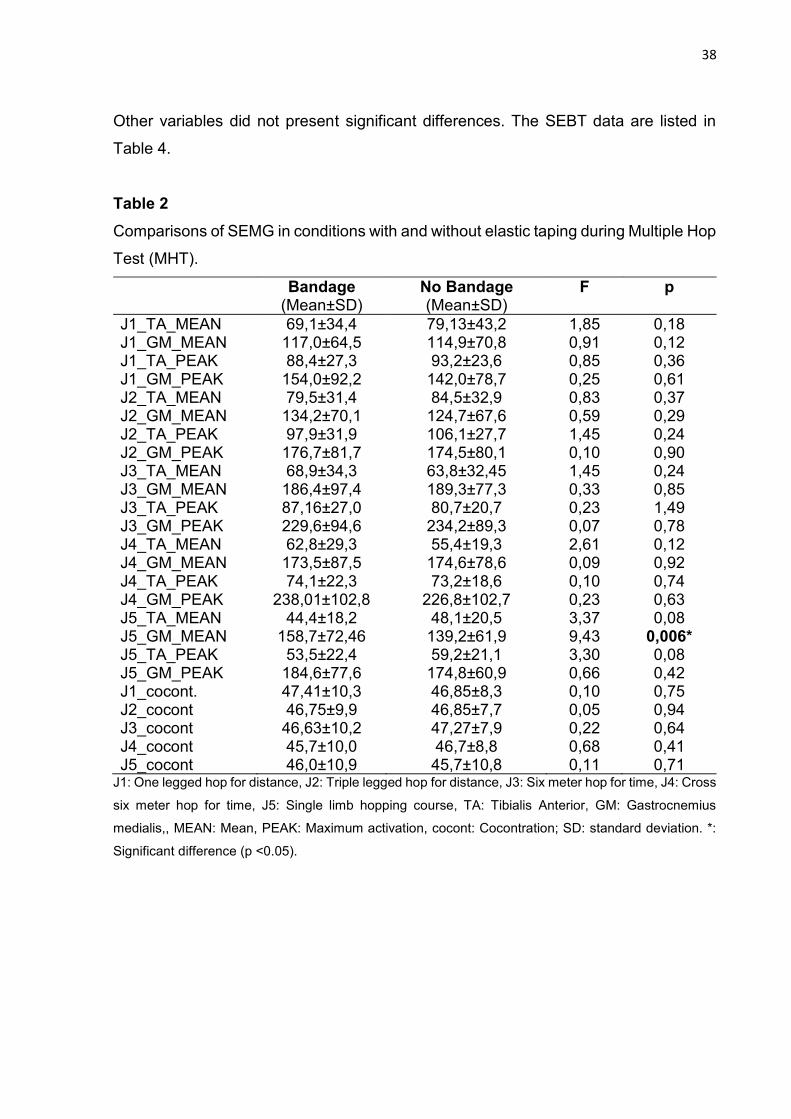
During the test, single limb hopping course, there was an increase of 19.5% of
the SEMG mean of gastrocnemius medialis associated with the use of elastic taping
(p = 0.006; Table 2).
For side cutting, there was a 120.8% increase of the SEMG peak of
gastrocnemius medialis associated with the use of elastic taping (p = 0.001; Table 3).
%COCON = 2x areacomum A&B x 100%
area A + area B
38
Other variables did not present significant differences. The SEBT data are listed in
Table 4.
Table 2Comparisons of SEMG in conditions with and without elastic taping during Multiple Hop
Test (MHT).
Bandage(Mean±SD)
No Bandage(Mean±SD)
F p
J1_TA_MEAN 69,1±34,4 79,13±43,2 1,85 0,18J1_GM_MEAN 117,0±64,5 114,9±70,8 0,91 0,12J1_TA_PEAK 88,4±27,3 93,2±23,6 0,85 0,36J1_GM_PEAK 154,0±92,2 142,0±78,7 0,25 0,61J2_TA_MEAN 79,5±31,4 84,5±32,9 0,83 0,37J2_GM_MEAN 134,2±70,1 124,7±67,6 0,59 0,29J2_TA_PEAK 97,9±31,9 106,1±27,7 1,45 0,24J2_GM_PEAK 176,7±81,7 174,5±80,1 0,10 0,90J3_TA_MEAN 68,9±34,3 63,8±32,45 1,45 0,24J3_GM_MEAN 186,4±97,4 189,3±77,3 0,33 0,85J3_TA_PEAK 87,16±27,0 80,7±20,7 0,23 1,49J3_GM_PEAK 229,6±94,6 234,2±89,3 0,07 0,78J4_TA_MEAN 62,8±29,3 55,4±19,3 2,61 0,12J4_GM_MEAN 173,5±87,5 174,6±78,6 0,09 0,92J4_TA_PEAK 74,1±22,3 73,2±18,6 0,10 0,74J4_GM_PEAK 238,01±102,8 226,8±102,7 0,23 0,63J5_TA_MEAN 44,4±18,2 48,1±20,5 3,37 0,08J5_GM_MEAN 158,7±72,46 139,2±61,9 9,43 0,006*J5_TA_PEAK 53,5±22,4 59,2±21,1 3,30 0,08J5_GM_PEAK 184,6±77,6 174,8±60,9 0,66 0,42J1_cocont. 47,41±10,3 46,85±8,3 0,10 0,75J2_cocont 46,75±9,9 46,85±7,7 0,05 0,94J3_cocont 46,63±10,2 47,27±7,9 0,22 0,64J4_cocont 45,7±10,0 46,7±8,8 0,68 0,41J5_cocont 46,0±10,9 45,7±10,8 0,11 0,71
J1: One legged hop for distance, J2: Triple legged hop for distance, J3: Six meter hop for time, J4: Cross
six meter hop for time, J5: Single limb hopping course, TA: Tibialis Anterior, GM: Gastrocnemius
medialis,, MEAN: Mean, PEAK: Maximum activation, cocont: Cocontration; SD: standard deviation. *:
Significant difference (p <0.05).
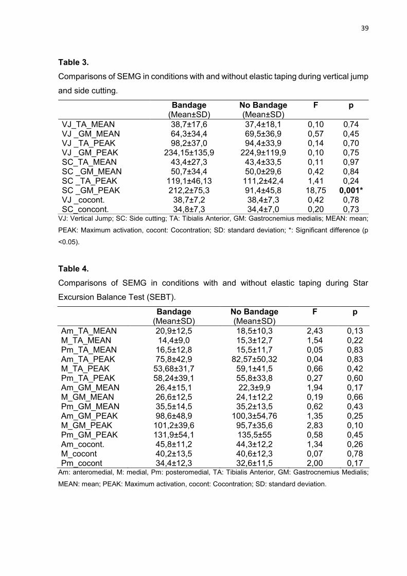
39
Table 3.Comparisons of SEMG in conditions with and without elastic taping during vertical jump
and side cutting.
Bandage(Mean±SD)
No Bandage(Mean±SD)
F p
VJ_TA_MEAN 38,7±17,6 37,4±18,1 0,10 0,74VJ _GM_MEAN 64,3±34,4 69,5±36,9 0,57 0,45VJ _TA_PEAK 98,2±37,0 94,4±33,9 0,14 0,70VJ _GM_PEAK 234,15±135,9 224,9±119,9 0,10 0,75SC_TA_MEAN 43,4±27,3 43,4±33,5 0,11 0,97SC _GM_MEAN 50,7±34,4 50,0±29,6 0,42 0,84SC _TA_PEAK 119,1±46,13 111,2±42,4 1,41 0,24SC _GM_PEAK 212,2±75,3 91,4±45,8 18,75 0,001*VJ _cocont. 38,7±7,2 38,4±7,3 0,42 0,78SC_concont. 34,8±7,3 34,4±7,0 0,20 0,73
VJ: Vertical Jump; SC: Side cutting; TA: Tibialis Anterior, GM: Gastrocnemius medialis; MEAN: mean;
PEAK: Maximum activation, cocont: Cocontration; SD: standard deviation; *: Significant difference (p
<0.05).
Table 4.Comparisons of SEMG in conditions with and without elastic taping during Star
Excursion Balance Test (SEBT).
Bandage(Mean±SD)
No Bandage(Mean±SD)
F p
Am_TA_MEAN 20,9±12,5 18,5±10,3 2,43 0,13M_TA_MEAN 14,4±9,0 15,3±12,7 1,54 0,22Pm_TA_MEAN 16,5±12,8 15,5±11,7 0,05 0,83Am_TA_PEAK 75,8±42,9 82,57±50,32 0,04 0,83M_TA_PEAK 53,68±31,7 59,1±41,5 0,66 0,42Pm_TA_PEAK 58,24±39,1 55,8±33,8 0,27 0,60Am_GM_MEAN 26,4±15,1 22,3±9,9 1,94 0,17M_GM_MEAN 26,6±12,5 24,1±12,2 0,19 0,66Pm_GM_MEAN 35,5±14,5 35,2±13,5 0,62 0,43Am_GM_PEAK 98,6±48,9 100,3±54,76 1,35 0,25M_GM_PEAK 101,2±39,6 95,7±35,6 2,83 0,10Pm_GM_PEAK 131,9±54,1 135,5±55 0,58 0,45Am_cocont. 45,8±11,2 44,3±12,2 1,34 0,26M_cocont 40,2±13,5 40,6±12,3 0,07 0,78Pm_cocont 34,4±12,3 32,6±11,5 2,00 0,17
Am: anteromedial, M: medial, Pm: posteromedial, TA: Tibialis Anterior, GM: Gastrocnemius Medialis;
MEAN: mean; PEAK: Maximum activation, cocont: Cocontration; SD: standard deviation.

40
4.3.2 Kinematics
ANOVA one way repeated measures no found significant difference for any
of the kinematic comparisons performed (Table 5).
Table 5.Kinematic data obtained under conditions with and without elastic taping, with mean
and standard deviation of linear acceleration (g).
Bandage(Mean±SD)
No Bandage(Mean±SD)
F P
MHT _J1_AcX 7,27±2,12 7,70±2,68 1,00 0,32MHT _J1_AcZ 17,78±2,32 17,85±3,41 0,10 0,89MHT _J2_AcX 5,75±2,49 6,76±2,73 1,31 0,26MHT _J2_AcZ 14,66±2,94 15,43±3,56 0,70 0,40MHT _J3_AcX 8,36±2,85 9,53±2,65 2,64 0,12MHT _J3_AcZ 14,42±3,55 14,66±3,38 0,05 0,81MHT _J4_AcX 9,31±2,54 9,77±2,12 0,42 0,52MHT _J4_AcZ 13,93±2,83 14,16±2,78 0,17 0,68MHT _J5_AcX 8,86±1,70 0,18 0,67MHT _J5_AcZ 19,86±2,12 0,03 0,86VJ_AcX 3,47±2,48 0,38 0,54VJ_AcZ 6,13±4,53 0,01 0,91SC_AcX 36,73±0,29 0,64 0,43SC_AcZ 36,79±0,69 0,12 0,72
MHT: Multiple Hop Test; VJ: Vertical Jump; SC: Side cutting; J1: One legged hop for distance, J2: Triple
legged hop for distance, J3: Six meter hop for time, J4: Cross six meter hop for time, J5: Single limb
hopping course; AcX: Delta between the maximum and minimum values obtained in the axis X of the
accelerometer - plantar flexion, AcZ: Delta between the maximum and minimum values obtained in the
axis Z of the accelerometer - supination.
4.3.3 Performance
For SEBT, there was an increase in the mean maximum range of 3.8% and
5.6% for the anteriormedial (p = 0.001) and posteromedial (0.005) directions,
respectively, during the condition with elastic taping. In the MHT, there was a reduction
in the execution time, in 5.7% and 7.1% of the jumps 4 (p = 0.018) and 5 (p = 0.009)
respectively, during condition with elastic taping. The data are present in Table 6. The
volunteers reported a 40% improvement over their sense of security.

41
Table 6.Performance measurements during the MHT and SEBT tests.
Bandage(Mean±SD)
No Bandage(Mean±SD)
F P
SEBT_Am (cm) 85,01± 6,15 81,84± 6,18 14,81 0,001*SEBT_M (cm) 81,84± 7,39 83,86± 6,10 3,91 0,061SEBT _Pm (cm) 90,56± 9,15 85,71± 7,49 9,67 0,005*MHT _S1 (cm) 167,55± 29,94 164,88± 33,83 0,43 0,515MHT _S2 (cm) 472,97± 94,29 467,94± 92,49 0,30 0,587MHT _S3 (s) 2,34± 0,38 2,36± 0,34 0,15 0,695MHT _S4 (s) 2,79± 0,54 2,96± 0,61 6,60 0,018*MHT _S5 (s) 3,64± 0,46 3,92± 0,46 8,20 0,009*
MHT: Multiple Hop Test, SEBT: Star Cursion Balance Test, Am: anteromedial, M: medial, Pm:
pósteromedial; J1: One legged hop for distance, J2: Triple legged hop for distance, J3: Six meter hop
for time, J4: Cross six meter hop for time, J5: Single limb hopping course, *: Significant difference (p
<0.05).
4.4 DISCUSSION
4.4.1 Electromyography
The main results of this study were increases SEMGs of gastrocnemius
medialisduring the side cutting and single limb hopping course of the MHT. This finding
may have occurred because elastic taping may provide cutaneous stimulation of
mechanoreceptors that stimulate central nervous system responses and increase
muscle excitability (Gómez-Soriano et al., 2014). These responses consist of the
activation of areas of the primary sensory cortex, which discriminates the intensity and
quality of the sensory stimulus. After this, the information arrives the association cortex,
it performs the selection of goals and interpretations according to the stimulus, with
this information is sent to the area of motor planning, which analyzes how the
composition and sequencing of the response will be, Finally, the stimulus that entered
the primary sensory cortex becomes a motor stimulus and consequently the response
to the initial stimulus occurs. Thus, the elastic taping aims to stimulate the tactile
sensation, so that this tactile information can be interpreted and used in the motricity
and corporal conscience (Junior, 2015).
During landing activity, bi articular muscles may be selectively activated during
periods when the moment potencies resulting from adjacent joints are opposite in
direction. This creates a flow of power between adjacent joints. When this power flow
42
occurs from distal to proximal, this enables a distribution of force through the muscle
tendon, which has a physiologically larger cross sectional area. Thus, during landing
of a jump, the power of the resulting joint moment in the ankle and knee, provides
potential for a transfer of energy through the biarticular tendon (Zatsiorsky, 2004).
Thus, a greater activation of gastrocnemius medialis would allow a greater energy
absorption, reducing the magnitude of impact during landing gestures and maintaining
joint stability.
The increase in muscle recruitment found in the present study corroborates the
findings of HUANG (2011), who investigated the effect of elastic taping on inactive
young adults during the vertical jump gesture. There was a tendency for an increase
in the neuromuscular recruitment of gastrocnemius medialis associated with the use
of elastic Taping. GÓMEZ-SORIANO (2014) also reports in his study with healthy
subjects that elastic taping provided an increase in electromyographic activation of
short term, gastrocnemius medialis during passive dorsiflexion motion testing.
However, this increase in muscle recruitment occurred only after 10 minutes of
application and after 24 hours this increase was no longer present. However BRIEM
(2011) disagrees with the findings presented here, because in his study with unstable
individuals and control, he observed that elastic taping did not alter fibular
electromyography during simulation of platform sprain
This discrepancy in results may be associated with methodological differences,
such as: Elastic taping position, where there is no consensus on which position is most
adequate to generate alterations in muscle recruitment, characteristics such as:
bandage direction, percentage of distension of the bandage and the type of material
that the tape is made may contribute to this variability of results in elastic taping
searches. Another factor that may contribute to this divergence of results is the
proposed activities: There is also no consensus in which type of activity elastic taping
demonstrates its therapeutic effects or attenuates these effects, such as: simulation of
sprains and jumps (impulse and landing), have their own characteristics of movement
mechanics and elastic taping may reveal its therapeutic effects in specific gestures and
not necessarily in all gestures.
The increase in electromyographic activation of gastrocnemius medialis
occurred only in side cutting and single limb hopping course. It was expected that more
variables would be altered due to the use of elastic taping and this fact may have
occurred due to some factors, such as the residual effect, where a 5 minutes rest was

43
standardized between the conditions with and without elastic taping, so volunteers that
perform the condition without elastic taping could finally present residual effects of the
therapeutic effects of the elastic taping, since the elastic taping can generate changes
in the electromyography after 10 minutes of application (Gómez-Soriano et al.,
2014).Thus, the time of 5 minutes between the conditions may not be enough to
eliminate the effects that the elastic taping can generate on the central nervous system
(Gómez-Soriano et al., 2014). Another factor that may have masked alterations in the
other variables may be the percentage of distension of the tape, because according to
MATHEUS (2016), hypothetically, the greater the distension applied, the greater
physiological changes may occur. Therefore, the distension applied in the elastic
taping in the present study may not have been enough to generate electromyographic
changes in other variables.
4.4.2 Kinematics
Our findings did not demonstrate alteration of the kinematics of the segment,
through the use of the elastic taping. Previous studies report divergent results from the
presented here. HO (2015), in his research with individuals with functional ankle
instability (FAI), observed that during the gestures of side cutting and vertical jump, the
elastic taping reduced the peak of the plantar flexion movement, as well as KUNI
(2015),were your study with healthy individuals and FAI observed a reduction of the
peak of plantar flexion in both groups during a gesture that involved
landing.TRÉGOUËT (2012), in his study with healthy individuals, observed a reduction
in peak and the speed of movement of inversion during test in an inversion platform,
associated with the use of the elastic taping.
Again, there is a divergence of results between the previous studies in the
literature and in the present study. This difference in results may have occurred due to
the study population, where the studies of HO (2015) and KUNI (2015) worked with
individuals with FAI and this may have evidenced the therapeutic effect of elastic
taping, since a previous study was possible to reduce proprioceptive deficits in this
population through the use of elastic taping (Simon et al., 2014). The time of application
of the elastic taping may have been another parameter that attenuated the therapeutic
effect of the elastic taping since the time of application may have been insufficient to
generate a change in kinematics since the elastic taping may have provided only
44
sensory learning, without functional and structural alterations of the central nervous
system and therefore the response to the stimulus is not continuous and therefore less
effective (Shumway-cook and Woollacott 2003). In addition, the short application time
may not have sufficiently stimulated the Ruffini corpuscles, which are nerve endings
that adapt slowly to a stimulus and are related to the static articular angles (Junior,
2015). Thus, long term application may provide us with relevant information about the
effect of elastic taping, since some studies report reduction of functional deficits in
individuals with dysfunctions such as FAI (Kim et al., 2015; Lee and Lee, 2016).
Finally, when we related the kinematic data with the electromyographic data, we
observed that the elastic taping may have operated in the central nervous system, but
not enough to alter the ankle kinematics. Showing that elastic taping has limitations as
a preventive and rehabilitative therapeutic resource.
4.4.3 Performance
The use of the elastic taping allowed an improvement in the performance of the
functional tests: SEBT (anteromedial and posteromedial directions) and MHT (Cross
test six meters jumping and Jumps test with a single lower limb). BICICI (2012), in his
study with individuals with recurrent sprains, verified the effect of applying various
banding conditions during functional activities, such as: jumps test with a single lower
limb and vertical jump. Although no significant difference, the elastic tapinge condition
allowed a higher height achieved during vertical jump and shorter time for performing
jumps test with a single lower limb, these results resemble those found in the present
study.
This improvement in performance may be associated with increased sensory
feedback through the stimulation of cutaneous mechanoreceptors, thereby increasing
the subjective perception of safety and individuals feel more confident performing the
tasks proposed (De-La-Torre-Domingo et al., 2015; SIMON et al., 2014). SAWKINS
(2007) reports that individuals subjects applying some type of bandage, relayed
greater sensation if safety. This suggests that the improvement in performance may
be related to the subjective perception of safety that the bandage can provide, however
this hypothesis should be tested in future studies.

45
4.4.4 Limitations
One of the limitations of the present study was the absence of a placebo
bandage to see if increased muscle recruitment and improved performance were
associated with elastic taping or a placebo effect. Another limitation is the sample
number, where possibly with the increase of the sample number, it would be possible
to find other significant differences in other variables of the study.
4.5 CONCLUSION
The immediate effect of elastic taping may increase muscle recruitment,
however, without reflecting on the kinematics of movement, suggesting that elastic
taping of immediate application has limitations as a preventive and rehabilitative
therapeutic resource. However, elastic taping may contribute beneficially to
performance in functional activities.
Conflict of interests
There is no conflict of interest.
Acknowledgment
We would like to thank the funding agencies: Coordenação de Aperfeiçoamento
de Pessoal de Nível Superior (CAPES), Fundação de Amparo à Pesquisa de Minas
Gerais (FAPEMIG; Processo APQ 00327-14) and Conselho Nacional de
Desenvolvimento Científico e Tecnológico (CNPq; Processo n. 459592/2014). In
addition to the partnership and extreme collaboration of CROSSFIT Marília during data
collection.
46
REFERENCES
Aquino CF, Viana SO, Fonseca ST, Bricio RS, Vaz DV. Mecanismos neuromusculares
de controle da estabilidade articular. Rev. bras. ciênc. Mov 2004;12(2):35-42.
Bicici S, Karatas N, Baltaci G. Effect of athletic taping and kinesiotaping on
measurements of functional performance in basketball players with chronic inversion
ankle sprains. International journal of sports physical therapy 2012;7(2):154-166.
Briem K, Eythörsdöttir H, Magnúsdóttir RG, Pálmarsson R, Rúnarsdöttir T, Sveinsson
T.Effects of kinesio tape compared with nonelastic sports tape and the untaped ankle
during a sudden inversion perturbation in male athletes. journal of orthopaedic & sports
physical therapy 2011;41(5):328-335.
Candotti CT, Carvalho KVD, La Torre M, Noll M, Varela M. Ativação e co-contração
dos músculos gastrocnêmio e tibial anterior na marcha de mulheres utilizando
diferentes alturas de saltos. Rev Bras Cienc Esporte 2012;34(1):27-39.
Cardoso JR, Guerino CDSM, Santos MB, Mustafá TADA, Lopes AR, Paula MCD.
Influência da utilização da órtese de tornozelo durante atividades do voleibol:
avaliação eletromiográfica. Revista Brasileira de Medicina e Esporte 2005;11(5):276-
280.
Cipriano JJ, Cipriano JJ. Testes ortopédicos do ombro. Cipriano JJ. Manual fotográfico
de testes ortopédicos e neurológicos 1999;1:71-112.
Clanton TO, Schon LC. Athletic injuries to the soft tissues of the foot and
ankle. Surgery of the Foot and Ankle 1999;2:1125-1126.
Da Fonseca, ST. Análise de um método eletromiográfico para quantificação de co-
contração muscular. Rev. Bras. Ciên. e Mov. Brasília, 2001; 9(3):23-30.
Delahunt E, McGrath A, Doran N, Coughlan GF.Effect of taping on actual and
perceived dynamic postural stability in persons with chronic ankle instability. Archives
of physical medicine and rehabilitation 2010;91(9):1383-1389.
47
De-La-Torre-Domingo C, Alguacil-Diego IM, Molina-Rueda F, López-Román A,
Fernández-Carnero J. Effect of Kinesiology Tape on Measurements of Balance in
Subjects With Chronic Ankle Instability: A Randomized Controlled Trial. Archives of
physical medicine and rehabilitation,2015;96(12):2169-2175.
Fong DTP, Hong Y, Chan LK, Yung PSH, Chan KM. A systematic review on ankle
injury and ankle sprain in sports. Sports Medicine 2007;37(1)73-94
Gómez-Soriano J, Abián-Vicén J, Aparicio-García C, Ruiz-Lázaro P, Simón-Martínez
C, Bravo-Esteban E,Fernández-Rodríguez JM.The effects of Kinesio taping on muscle
tone in healthy subjects: a double-blind, placebo-controlled crossover trial. Manual
therapy 2014;19(2):131-136.
Gribble PA, Hertel J. Considerations for normalizing measures of the Star Excursion
Balance Test. Measurement in physical education and exercise Science 2003;7(2):89-
100.
Ha SCW, Fong DTP, Chan KM. Review of ankle inversion sprain simulators in the
biomechanics laboratory. Asia-Pacific Journal of Sports Medicine, Arthroscopy,
Rehabilitation and Technology 2015;2(4):114-121.
Hermens HJ, Freriks B, Disselhorst-Klug C, Rau G. Development of recommendations
for SEMG sensors and sensor placement procedures. Journal of electromyography
and Kinesiology 2000;10(5):361-374.
Hertel J, Braham RA, Hale SA, Olmsted-Kramer LC.Simplifying the star excursion
balance test: analyses of subjects with and without chronic ankle instability. Journal of
Orthopaedic & Sports Physical Therapy 2006;36(3):131-137.
Hertel J. Functional anatomy, pathomechanics, and pathophysiology of lateral ankle
instability. Journal of athletic training 2002;37(4):364
Hettle D, Linton L, Bake JS, Donoghue O. The effect of kinesiotaping on functional
performance in chronic ankle instability-preliminary study. Clinical Research on Foot &
Ankle 2013:1-5.

48
Ho YH, Lin CF, Chang CH, Wu HW. Effect of ankle kinesio taping on vertical jump with
run-up and countermovement jump in athletes with ankle functional instability. Journal
of physical therapy Science 2015;27(7):2087.
Hoffman M, Schrader J, Applegate T, Koceja D. Unilateral postural control of the
functionally dominant and nondominant extremities of healthy subjects. J. Athl. Train
1998;33(4)319-322.
Hsu YH, Chen WY, Lin HC, Wang WT, Shih Y. F.The effects of taping on scapular
kinematics and muscle performance in baseball players with shoulder impingement
syndrome. Journal of electromyography and kinesiology 2009;19(6)1092-1099.
Huang CY, Hsieh TH, Lu SC, Su FC. Effect of the Kinesio tape to muscle activity and
vertical jump performance in healthy inactive people. Biomedical engineering online
2011;10(1):1
Iida Y, Kanehisa H, Inaba Y,Nakazawa K. Activity modulations of trunk and lower limb
muscles during impact-absorbing landing. Journal of Electromyography and
Kinesiology 2011;21(4):602-609.
Junior NM. Taping Terapêutica: Conceito de Estimulação Tegumentar. São Paulo:
Roca, 2013
Kapandji AI. Fisiologia articular: esquemas comentados de mecânica humana, volume
2: membros inferiores. 5rd ed. São Paulo: Panamericana. 2000.
Kase K, Hashimoto T, TomokI O. Development of kinesio taping perfect manual.
Kinesio Taping Association, v. 6, n. 10, p. 117-8, 1996
Kendall FP, Mccreay EK, Provance PG. Músculos: provas e funções, 4ª edição. São
Paulo: Manole, 1995.
Kim BJ, Lee JH, Kim CT, Lee SM. Effects of ankle balance taping with kinesiology tape
for a patient with chronic ankle instability. Journal of physical therapy Science
2015;27(7):2405.

49
Kuhman DJ, Paquette MR, Peel SA, Melcher DA. Comparison of ankle kinematics and
ground reaction forces between prospectively injured and uninjured collegiate cross
country runners. Human movement Science 2016; 47:9-15.
Kuni B, Mussler J, Kalkum E, Schmitt H, Wolf SI. Effect of kinesiotaping, non-elastic
taping and bracing on segmental foot kinematics during drop landing in healthy
subjects and subjects with chronic ankle instability. Physiotherapy 2015;102(3): 287
293
Lee SM, Lee JH. Effects of ankle eversion taping using kinesiology tape in a patient
with ankle inversion sprain. Journal of Physical Therapy Science 2016;28(2):708.
Lee SS, Piazza, SJ. Inversion eversion moment arms of gastrocnemius and tibialis
anterior measured in vivo. Journal of biomechanics 2008;41(16):3366-3370.
Lohrer H, Alt W, Gollhofer A. Neuromuscular properties and functional aspects of taped
ankles. The American Journal of Sports Medicine 1999;27(1): 69-75.
Magalhães I, Bottaro M, Mezzarane RA, Neto FR, Rodrigues BA, Ferreira-Júnior JB,
Carregaro, RL. Kinesiotaping enhances the rate of force development but not the
neuromuscular efficiency of physically active young men. Journal of Electromyography
and Kinesiology 2016;28:123-129.
Matheus JPC, Zille RR, Gomide LB, Lemos TV, Carregaro RL, Shimano AC.
Comparison of the mechanical properties of therapeutic elastic tapes used in sports
and clinical practice 2016. Physical Therapy in Sport.
Nabhan D, Walden T, Street J, Linden H, Moreau B. Sports injury and illness
epidemiology during the 2014 Youth Olympic Games: United States Olympic team
surveillance. British journal of sports medicine 2016;50(11):688-693.
Nunes LCBG. (2004). EFEITOS DA ELETROESTIMULAÇÃO NEUROMUSCULAR
NO MÚSCULO TIBIAL ANTERIOR DE PACIENTES HEMIPARÉTICOS
ESPÁSTICOS (Doctoral dissertation, Universidade Estadual de Campinas).
Sawkins K. The placebo effect of ankle taping on ankle instability, 2007.
50
Sekir U, Yildiz Y, Hazneci B, Ors F, Aydin T. Effect of isokinetic training on strength,
functionality and proprioception in athletes with functional ankle instability. Knee
surgery, sports traumatology, arthroscopy 2007;15(5):654-664.
Shumway-Cook A, Woollacott MH. Controle postural normal. Controle Motor-teoria e
aplicações práticas. 2ª ed. Barueri: Manole, 2003
Simon J, Garcia W, Docherty CL. The effect of kinesio tape on force sense in people
with functional ankle instability. Clinical Journal of Sport Medicine 2014;24(4):289-294.
Sinkjaer T, Toft E, Andreassen S, Hornemann BC. Muscle stiffness in human ankle
dorsiflexors: intrinsic and reflex components. Journal of Neurophysiology
1988;60(3)1110-1121.
Steib S, Hentschke C, Welsch G, Pfeifer K, Zech A. Effects of fatiguing treadmill
running on sensorimotor control in athletes with and without functional ankle instability.
Clinical Biomechanics 2013;28(7):790-795.
Stormont DM, Morrey BF, An KN, Cass JR.Stability of the loaded ankle Relation
between articular restraint and primary and secondary static restraints. The American
journal of sports medicine 1985;13(5):295-300
Trégouët P, Merland F, Horodyski MB. A comparison of the effects of ankle taping
styles on biomechanics during ankle inversion. Annals of physical and rehabilitation
medicine 2013;56(2):113-122.
Winter DA. Biomechanics and motor control of human movement. New York: Wiley,
2005:250-280.
Wright IC, Neptune R, Van Den Bogert AJ, Nigg BM.The influence of foot positioning
on ankle sprains. J Biomech 2000;33(5):513-519
Zatsiorsky VM. Biomecânica do esporte. Performance do desempenho e prevenção
de lesão. Rio de Janeiro: Guanabara,2004.
51
REFERÊNCIAS DISSERTAÇÃO
AIRAKSINEN, O. Changes in posttraumatic ankle joint mobility, pain, and edema
following intermittent pneumatic compression therapy. Arch Phys Med Rehabil, v. 70,
n. 4, p. 341-4, 1989.
AMATUZZI, M. M. et al. Reabilitação em medicina do esporte. Editora Roca, 2004.
AQUINO, C. F. et al. Mecanismos neuromusculares de controle da estabilidade
articular. Revista Brasileira de Ciência e Movimento, Brasília, v.12, n. 2, p. 35-42,
jun. 2004.
BAHR, R.; ENGEBRETSEN, L. Handbook of sports medicine and science, sports
injury prevention. John Wiley & Sons, 2011.
BARATTA, R. et al. Muscular coactivation The role of the antagonist musculature in
maintaining knee stability. The American journal of sports medicine, v. 16, n. 2, p.
113-122, 1988.
BEALS, T. C; CRIM, J.; NICKISCH, F. Deltoid ligament injuries in athletes: techniques
of repair and reconstruction. Operative Techniques in Sports Medicine, v. 18, n. 1,
p. 11-17, 2010.
BERNETT, P.; SCHIRMANN, A. Acute sporting injuries of the ankle joint (author's
transl). Unfallheilkunde, v. 82, n. 4, p. 155, 1979.
BICICI, S.; KARATAS, N.; BALTACI, G. Effect of athletic taping and kinesiotaping on
measurements of functional performance in basketball players with chronic inversion
ankle sprains. International Journal of Sports Pysical Therapy, v.7, n.2, p.154-166,
2012
BOZKURT, M.; DORAL, M.N. Anatomic factors and biomechanics in ankle
instability. Foot and ankle clinics, v. 11, n. 3, p. 451-463, 2006.
BRIEM, K. et al. Effects of kinesio tape compared with nonelastic sports tape and the
untaped ankle during a sudden inversion perturbation in male athletes. Journal oforthopaedic & sports physical therapy, v. 41, n. 5, p. 328-335, 2011.

52
BROSTRÖM, L. Sprained ankles. V. Treatment and prognosis in recent ligament
ruptures. Acta chirurgica Scandinavica, v. 132, n. 5, p. 537-550, 1966.
CANDOTTI, C. T. et al. Ativação e co-contração dos músculos gastrocnêmio e tibial
anterior na marcha de mulheres utilizando diferentes alturas de saltos. Rev. Bras.Ciênc. Esporte, v. 34, n. 1, p. 27-39, 2012.
CARDOSO, J. R. et al. Influência da utilização da órtese de tornozelo durante
atividades do voleibol: avaliação eletromiográfica. Rev Bras Med Esporte, v. 11, n. 5,
p. 276-80, 2005.
CIPRIANO, J. J.; CIPRIANO, J. J. Testes ortopédicos do ombro. Cipriano JJ. Manualfotográfico de testes ortopédicos e neurológicos, v. 1, p. 71-112, 1999.
CLANTON, Thomas O.; SCHON, L. C. Athletic injuries to the soft tissues of the foot
and ankle. Surgery of the Foot and Ankle, v. 2, p. 1125-1126, 1999
CRAIG, C. E.; GOBLE, D. J.; DOUMAS, M. Proprioceptive acuity predicts muscle co-
contraction of the tibialis anterior and gastrocnemius medialis
postural control.Neuroscience, v. 322, p. 251-261, 2016.
DA FONSECA, S. T. Análise de um método eletromiográfico para quantificação de co-
contração muscular. Rev. Bras. Ciên. e Mov. Brasília , v. 9, n. 3, p. 23-30, 2001.
DE ALMEIDA LINS, C. A. et al. Kinesio Taping® does not alter neuromuscular
performance of femoral quadriceps or lower limb function in healthy subjects:
Randomized, blind, controlled, clinical trial. Manual Therapy, v. 18, n. 1, p. 41-45,
2013.
DELAHUNT, E. et al. Effect of taping on actual and perceived dynamic postural stability
in persons with chronic ankle instability. Archives of physical medicine andrehabilitation, v. 91, n. 9, p. 1383-1389, 2010.
DELAHUNT, E.; MONAGHAN, K.; CAULFIELD, B. Ankle function during hopping in
subjects with functional instability of the ankle joint.Scandinavian journal of medicine& science in sports, v. 17, n. 6, p. 641-648, 2007.

53
DE-LA-TORRE-DOMINGO, C. et al. Effect of Kinesiology Tape on Measurements of
Balance in Subjects With Chronic Ankle Instability: A Randomized Controlled
Trial. Archives of physical medicine and rehabilitation, v. 96, n. 12, p. 2169-2175,
2015.
DI NARDO, F. et al. Assessment of the ankle muscle co-contraction during normal gait:
A surface electromyography study. Journal of Electromyography and Kinesiology,
v. 25, n. 2, p. 347-354, 2015.
DRAWER, S.; FULLER, C. W. Perceptions of retired professional soccer players about
the provision of support services before and after retirement.British journal of sportsmedicine, v. 36, n. 1, p. 33-38, 2002.
FONG, D. T. et al. A systematic review on ankle injury and ankle sprain in sports.
Sports Medicine, v. 37, n. 1, p. 73-94, 2007.
FU, A.S.N.; HUI-CHAN, C.W.Y. Ankle joint proprioception and postural control in
basketball players with bilateral ankle sprains. The American journal of sportsmedicine, v. 33, n. 8, p. 1174-1182, 2005.
FU, T. et al. Effect of Kinesio taping on muscle strength in athletes a pilot
study. Journal of science and medicine in sport, v. 11, n. 2, p. 198-201, 2008.
GÓMEZ-SORIANO, J. et al. The effects of Kinesio taping on muscle tone in healthy
subjects: a double-blind, placebo-controlled crossover trial. Manual therapy, v. 19, n.
2, p. 131-136, 2014.
GRIBBLE, P. A.; HERTEL, J. Considerations for normalizing measures of the Star
Excursion Balance Test. Measurement in physical education and exercisescience, v. 7, n. 2, p. 89-100, 2003.
GUTIERREZ, G. M. et al. Examining neuromuscular control during landings on a
supinating platform in persons with and without ankle instability. The Americanjournal of sports medicine, v. 40, n. 1, p. 193-201, 2012.
54
HA, S. C.; FONG, D. T.; CHAN, K.M. Review of ankle inversion sprain simulators in
the biomechanics laboratory. Asia-Pacific Journal of Sports Medicine,Arthroscopy, Rehabilitation and Technology, v. 2, n. 4, p. 114-121, 2015.
HARMON, K. G. The ankle examination. Primary Care: Clinics in Office Practice, v.
31, n. 4, p. 1025-1037, 2004.
HERMENS, H. J. et al. Development of recommendations for SEMG sensors and
sensor placement procedures. Journal of electromyography and Kinesiology, v.
10, n. 5, p. 361-374, 2000.
HERTEL, J. et al. Simplifying the star excursion balance test: analyses of subjects with
and without chronic ankle instability. Journal of Orthopaedic & Sports PhysicalTherapy, v. 36, n. 3, p. 131-137, 2006.
HERTEL, J. Functional anatomy, pathomechanics, and pathophysiology of lateral
ankle instability. Journal of athletic training, v. 37, n. 4, p. 364, 2002.
HETTLE, D. et al. The effect of kinesiotaping on functional performance in chronic
ankle instability-preliminary study. Clinical Research on Foot & Ankle, p. 1-5, 2013.
HO, Y. et al. Effect of ankle kinesio taping on vertical jump with run-up and
countermovement jump in athletes with ankle functional instability.Journal of physicaltherapy science, v. 27, n. 7, p. 2087, 2015.
HOFFMAN, M. et al. Unilateral postural control of the functionally dominant and
nondominant extremities of healthy subjects. Journal of athletic training, v. 33, n. 4,
p. 319, 1998.
HSU, Y. et al. The effects of taping on scapular kinematics and muscle performance
in baseball players with shoulder impingement syndrome. Journal ofelectromyography and kinesiology, v. 19, n. 6, p. 1092-1099, 2009.
HUANG, C. et al. Effect of the Kinesio tape to muscle activity and vertical jump
performance in healthy inactive people. Biomedical engineering online, v. 10, n. 1,
p. 70, 2011.
55
HUGHES, T.; ROCHESTER, P. The effects of proprioceptive exercise and taping on
proprioception in subjects with functional ankle instability: A review of the literature.
Physical Therapy in Sport, v. 9, p. 136-147, 2008.
IIDA, Y. et al. Activity modulations of trunk and lower limb muscles during impact-
absorbing landing. Journal of Electromyography and Kinesiology, v. 21, n. 4, p.
602-609, 2011.
JUNIOR, N. M. Bandagem Terapêutica: Conceito de Estimulação Tegumentar. São
Paulo: Roca, p. 106-13, 2015.
KAPANDJI, A. I. Fisiologia articular: esquemas comentados de mecânica humana.
São Paulo: Panamericana. 2000.
KASE, K., HASHIMOTO T., TOMOKI O. Development of kinesio taping perfect
manual. Kinesio Taping Association, v. 6, n. 10, p. 117-8, 1996.
KENDALL, F. P.; MCCREAY, E.K.; Provance P.G. Músculos: provas e funções, 4ª
edição. São Paulo: Manole, 1995.
KERKHOFFS, G.M.M.J. et al. Surgical versus conservative treatment for acute injuries
of the lateral ligament complex of the ankle in adults. The Cochrane Library, 2007.
KIM, B. et al. Effects of ankle balance taping with kinesiology tape for a patient with
chronic ankle instability. Journal of physical therapy science, v. 27, n. 7, p. 2405,
2015.
KOLDENHOVEN, R. M. et al. Surface electromyography and plantar pressure during
walking in young adults with chronic ankle instability. Knee Surgery, SportsTraumatology, Arthroscopy, v. 24, n. 4, p. 1060-1070, 2016.
KONISHI, Y. Tactile stimulation with Kinesiology tape alleviates muscle weakness
attributable to attenuation of Ia afferents. Journal of Science and Medicine in Sport,v. 16, n. 1, p. 45-48, 2013.
KOSHINO, Yuta et al. Kinematics and muscle activities of the lower limb during a side-
cutting task in subjects with chronic ankle instability. Knee Surgery, SportsTraumatology, Arthroscopy, p. 1-10, 2015.

56
KUHMAN, D.J. et al. Comparison of ankle kinematics and ground reaction forces
between prospectively injured and uninjured collegiate cross country runners. Humanmovement science, v. 47, p. 9-15, 2016.
KUNI, B. et al. Effect of kinesiotaping, non-elastic taping and bracing on segmental
foot kinematics during drop landing in healthy subjects and subjects with chronic ankle
instability. Physiotherapy, 2015.
LEE, S.; LEE, J. Effects of ankle eversion taping using kinesiology tape in a patient
with ankle inversion sprain. Journal of Physical Therapy Science, v. 28, n. 2, p. 708,
2016.
LEE, S.S.M.; PIAZZA, S.J. Inversion eversion moment arms of gastrocnemius and
tibialis anterior measured in vivo. Journal of biomechanics, v. 41, n. 16, p. 3366-
3370, 2008.
LOHRER, H.; ALT, W.; GOLLHOFER, A. Neuromuscular properties and functional
aspects of taped ankles. The American Journal of Sports Medicine, v. 27, n. 1, p.
69-75, 1999.
LUNDBERG, A. et al. Kinematics of the ankle/foot complex: plantarflexion and
dorsiflexion. Foot & Ankle International, v. 9, n. 4, p. 194-200, 1989.
MADHAVAN, S.; SHIELDS, R. K. Influence of age on dynamic position sense:
evidence using a sequential movement task. Experimental brain research, v. 164, n.
1, p. 18-28, 2005.
MAGALHÃES, I. et al. Kinesiotaping enhances the rate of force development but not
the neuromuscular efficiency of physically active young men. Journal ofElectromyography and Kinesiology, v. 28, p. 123-129, 2016.
MARTÍNEZ-GRAMAGE, J. et al. Effect of Kinesio Taping on gastrocnemius activity
and ankle range of movement during gait in healthy adults: a randomized controlled
trial. Physical Therapy in Sport, 2014.
MATHEUS, J. P. C. et al. Comparison of the mechanical properties of therapeutic
elastic tapes used in sports and clinical practice. Physical Therapy in Sport, 2016.

57
MCKAY, G. D. et al. Ankle injuries in basketball: injury rate and risk factors. BritishJournal of Sports Medicine, v. 35, n. 2, p. 103-108, 2001.
NABHAN, D. et al. Sports injury and illness epidemiology during the 2014 Youth
Olympic Games: United States Olympic Team Surveillance. British Journal of SportsMedicine, p. bjsports-2015-095835, 2016.
NEUMANN, D. A. Cinesiologia do Aparelho Musculoesquelético Fundamentos para
a Reabilitação Física. 1 ed.Rio de Janeiro:Guanabara Koogan, 2006
NUNES, L. C. B. G. Efeitos da eletroestimulação neuromuscular no músculo tibial
anterior de pacientes hemiparéticos espásticos. 2004. 105 f. Dissertação (Mestrado
em Engenharia Rev. Bras. Ciênc. Esporte, Florianópolis, v. 34, n. 1, p. 27-39, jan./mar.
2012 39 Elétrica) - Faculdade de Engenharia Elétrica e de Computação, Universidade
Estadual de Campinas, Campinas, 2004.
PENA, F. A.; COETZEE, J. C. Ankle syndesmosis injuries. Foot and ankle clinics, v.
11, n. 1, p. 35-50, 2006.
POZZI, F.; MOFFAT, M.; GUTIERREZ, G. Neuromuscular control during performance
of a dynamic balance task in subjects with and without ankle instability. Internationaljournal of sports physical therapy, v. 10, n. 4, p. 520, 2015.
ROCKAR, J.R.; PAUL, A. The subtalar joint: anatomy and joint motion. Journal ofOrthopaedic & Sports Physical Therapy, v. 21, n. 6, p. 361-372, 1995.
SAFRAN, M. R. et al. Lateral ankle sprains: a comprehensive review: part 1: etiology,
pathoanatomy, histopathogenesis, and diagnosis. Medicine and science in sportsand exercise, v. 31, n. 7 Suppl, p. S429-37, 1999.
SAVAGE-ELLIOTT, I. et al. The deltoid ligament: an in-depth review of anatomy,
function, and treatment strategies. Knee Surgery, Sports Traumatology,Arthroscopy, v. 21, n. 6, p. 1316-1327, 2013.
SAWKINS, K. The placebo effect of ankle taping on ankle instability. 2007.
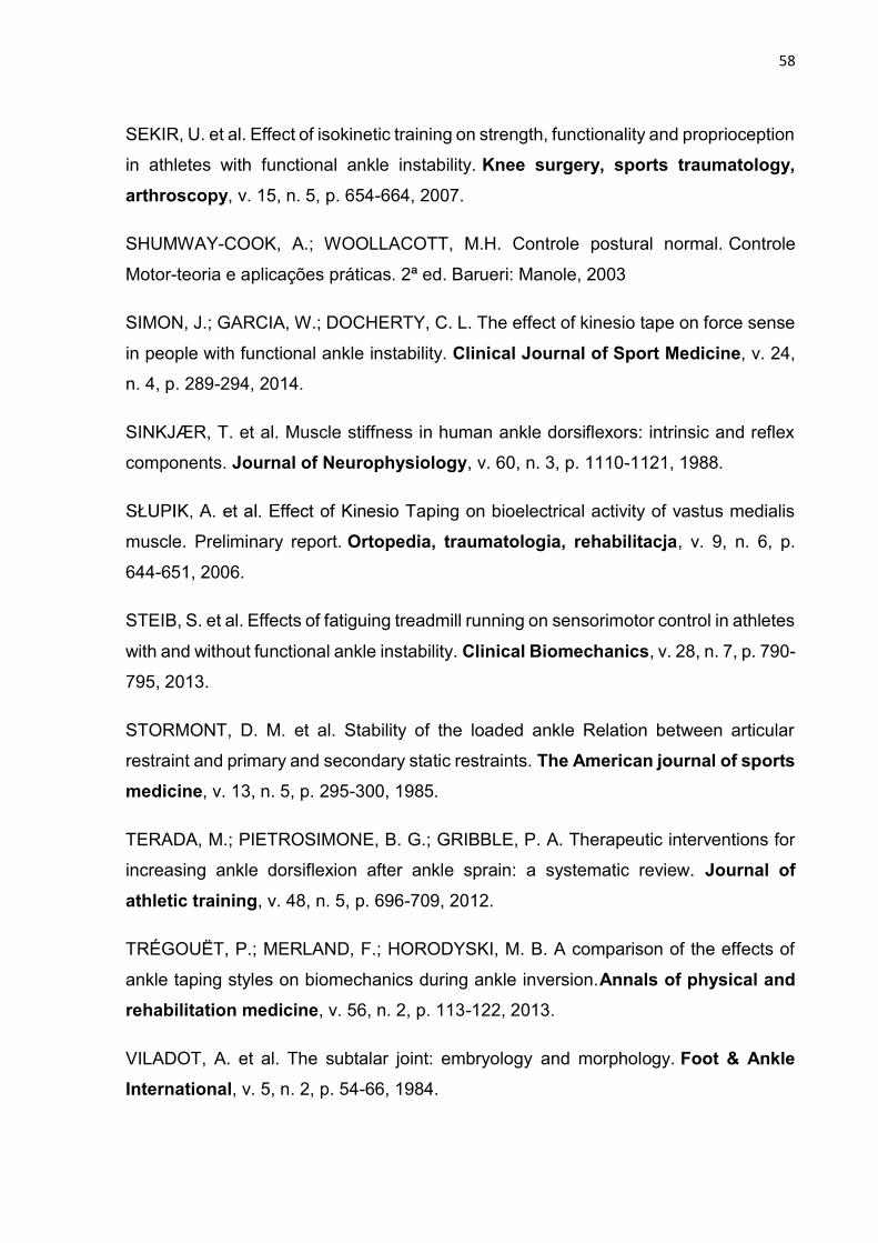
58
SEKIR, U. et al. Effect of isokinetic training on strength, functionality and proprioception
in athletes with functional ankle instability. Knee surgery, sports traumatology,arthroscopy, v. 15, n. 5, p. 654-664, 2007.
SHUMWAY-COOK, A.; WOOLLACOTT, M.H. Controle postural normal. Controle
Motor-teoria e aplicações práticas. 2ª ed. Barueri: Manole, 2003
SIMON, J.; GARCIA, W.; DOCHERTY, C. L. The effect of kinesio tape on force sense
in people with functional ankle instability. Clinical Journal of Sport Medicine, v. 24,
n. 4, p. 289-294, 2014.
SINKJÆR, T. et al. Muscle stiffness in human ankle dorsiflexors: intrinsic and reflex
components. Journal of Neurophysiology, v. 60, n. 3, p. 1110-1121, 1988.
Taping on bioelectrical activity of vastus medialis
muscle. Preliminary report. Ortopedia, traumatologia, rehabilitacja, v. 9, n. 6, p.
644-651, 2006.
STEIB, S. et al. Effects of fatiguing treadmill running on sensorimotor control in athletes
with and without functional ankle instability. Clinical Biomechanics, v. 28, n. 7, p. 790-
795, 2013.
STORMONT, D. M. et al. Stability of the loaded ankle Relation between articular
restraint and primary and secondary static restraints. The American journal of sportsmedicine, v. 13, n. 5, p. 295-300, 1985.
TERADA, M.; PIETROSIMONE, B. G.; GRIBBLE, P. A. Therapeutic interventions for
increasing ankle dorsiflexion after ankle sprain: a systematic review. Journal ofathletic training, v. 48, n. 5, p. 696-709, 2012.
TRÉGOUËT, P.; MERLAND, F.; HORODYSKI, M. B. A comparison of the effects of
ankle taping styles on biomechanics during ankle inversion.Annals of physical andrehabilitation medicine, v. 56, n. 2, p. 113-122, 2013.
VILADOT, A. et al. The subtalar joint: embryology and morphology. Foot & AnkleInternational, v. 5, n. 2, p. 54-66, 1984.

59
WILLIAMS, S. et al. Kinesio taping in treatment and prevention of sports
injuries. Sports medicine, v. 42, n. 2, p. 153-164, 2012.
WINTER, D.A. Biomechanics and motor control of human movement. New York:
Wiley, 2005.
WONG, O.M.H.; CHEUNG, R.T.H.; LI, RAYMOND, C.T. Isokinetic knee function in
healthy subjects with and without Kinesio taping. Physical Therapy in Sport, v. 13, n.
4, p. 255-258, 2012.
WOODS, C. et al. The Football Association Medical Research Programme: an audit of
injuries in professional football: an analysis of ankle sprains.British Journal of SportsMedicine, v. 37, n. 3, p. 233-238, 2003.
WRIGHT, I. C. et al. The influence of foot positioning on ankle sprains. Journal ofbiomechanics, v. 33, n. 5, p. 513-519, 2000
ZATSIORSKY, V.M. Biomecânica do esporte. Performance do desempenho e
prevenção de lesão. Rio de Janeiro: Guanabara,2004.

60
APÊNDICE 1 ANAMNESE

61
APÊNDICE 2 - Termo de Consentimento Livre e Esclarecido
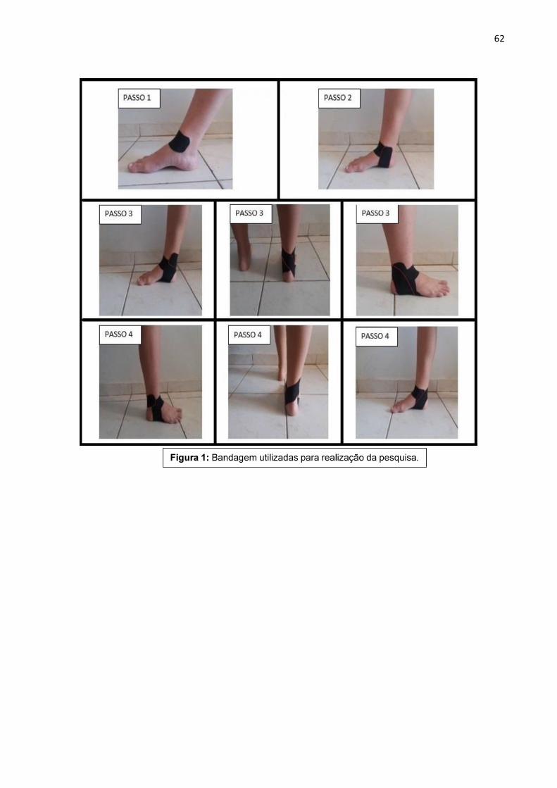
62
63
64

65

66
3.