Embed Size (px)
Citation preview
ISSN: 2320-5407 Int. J. Adv. Res. 6(7), 634-648
634
Journal Homepage: - www.journalijar.com
Article DOI: 10.21474/IJAR01/7408
DOI URL: http://dx.doi.org/10.21474/IJAR01/7408
RESEARCH ARTICLE
TREATMENT TECHNIQUES FOR OROANTRAL COMMUNICATIONS AND FISTULAS.
Leonardo Gomes de Lima1, Ana Larisse Carneiro Pereira
1, Jefferson David Melo de Matos
2, Guilherme da
Rocha Scalzer Lopes3, John Eversong Lucena de Vasconcelos
4 and Ivo Cavalcante Pita Neto
5.
1. Graduated in Dentistry, Centro Universitário UNILEÃO, Juazeiro do Norte – CE, Brazil.
2. Post Graduate Student - Masters Degree Program, Department of Prosthodontics, Universidade Estadual
Paulista Júlio de Mesquita Filho UNESP, São José dos Campos - SP, Brazil.
3. Post Graduate Student - Ph.D Program, Department of Prosthodontics, Universidade Estadual Paulista Júlio de
Mesquita Filho UNESP, São José dos Campos - SP, Brazil.
4. Professor of Oral Implantology, Department of Dentistry, Centro Caririense de Pós-Graduação CECAP,
Juazeiro do Norte – CE.
5. Professor of Oral and Maxillofacial Surgery, Dentistry Department, Centro Universitário UNILEÃO, Juazeiro
do Norte – CE, Brazil
……………………………………………………………………………………………………....
Manuscript Info Abstract
……………………. ……………………………………………………………… Manuscript History
Received: 14 May 2018
Final Accepted: 16 June 2018
Published: July 2018
Keywords:- Oroantral Fistula; Oral
Fistula; Maxillary Sinus; Surgery, Oral.
Aim: To describe the main characteristics of the
treatment techniques for oroantral communications and
fistulas. Methodology: This work was accomplished
from scientific articles found in the bibliographic
catalogued databases PubMed, Scielo, Google Scholar
and ResearchGate searcher, under the English
descriptors Oroantral Fistula, Oral Fistula, Maxillary
Sinus and Surgery Oral, published until the end of the
first trimester of the year 2018. Results: The oroantral
communications allow the passage of food, liquids, and
microorganisms, causing maxillary sinusitis; the
diagnosis is made from anamnesis, clinical examination
and complementary imaging tests. The treatment need an
approach that presents the capacity to promote closure of
the communication or fistula, leading to the recovery of
health and depending on the characteristics of each case,
it may vary from the need to use a simpler and easier to
handle flap such as vestibular flap, palatal flap rotation
or a combination of these, until the need to perform a
more complex as the rotation of the adipose Bichat's
bulla. Conclusion:The professional who performs dental
extractions must be able to treat these complications,
otherwise he should consider referral to a specialist
when, clinical and complementary examinations, the
communication risk is perceived.
Copy Right, IJAR, 2018,. All rights reserved.
……………………………………………………………………………………………………....
Corresponding Author:-Leonardo Gomes de Lima.
Address:-Graduated in Dentistry, Centro Universitário UNILEÃO, Juazeiro do Norte – CE, Brazil.

ISSN: 2320-5407 Int. J. Adv. Res. 6(7), 634-648
635
Introduction:- Oroantral fistula is a pathological condition in which the oral cavity and sinus have permanent communication
through fibrous connective tissues lined by epithelium, and the closure of this communication is important because
fluids, water, food and bacteria migrate from the buccal cavity to the maxillary sinus, often causing chronic
sinusitis1. The choice of a type of surgical intervention will depend on the type, the location and, mainly, the size of
the communication2.
Oral communication comprises a frequent complication in dental extractions due to the proximity of the maxillary
sinus and the oral cavity3. Operative accidents are the main causes, occurring after extractions of teeth or root
remains, curettage of the alveoli after extraction, surgical removal of included teeth, fractures of buccal bone boards
or during enucleation of cysts or tumors in close relation to the maxillary sinus4.
It is considered an etiological factor that promotes destruction of bone continuity between the oral cavity and the
maxillary sinus5. There are other etiological factors of lesser incidence, such as traumatic injuries to sharp objects,
stab wounds or firearm projectiles, extensive pathological lesions involving the maxillary sinus or oral cavity,
infections of the maxillary sinus or buccal cavity causing bone destruction and the formation of kidnapping bones,
as well as less common necrotic causes such as radiation or mercury, phosphorus, and bismuth poisoning6.
For this type of communication, the immediate and the late treatment are described in the literature; the immediate
one is performed preferably at the moment of communication, while the late one, in situations in which the patient
presents an already established oroantral fistula7. The importance of knowing the main techniques for the treatment
of oroantral fistulas is demonstrated by the high incidence of these complications after extraction of posterior
maxillary teeth, and dental surgeons must be able to recognize situations that favor the complication, which points to
the need to know the local anatomy and have an integral vision when performing procedures in areas around the
maxillary sinuses, which is important for the prevention of communication and consequent formation of a possible
fistula; have a clear sense of when they will be able to handle simpler techniques or need to refer to some advanced
techniques, knowing how to indicate the type of treatment appropriate to the characteristics of the injury8.
In view of this the present study aims to describe, from the scientific point of view, the main characteristics of the
treatment techniques for oroantral communications and fistulas described in the literature.
Methodology:- The review of the literature was based on scientific articles found in the electronic bibliographic cataloging
databases of PubMed, Scielo and Google Scholar search engine, with the descriptors in Portuguese: Fístula
Bucoantral, Fístula Bucal, Seio Maxilar and Cirurgia Bucal; in English: Oroantral Fistula, Oral Fistula, Maxillary
Sinus e Surgery Oral, published between the 1950s, due to the importance of some works of this era, and the end of
the first quarter of 2018.
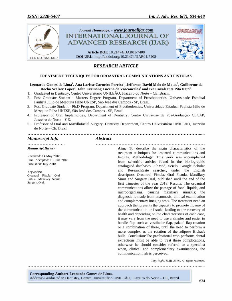
The bibliographical research resulted in 1522 articles, which were initially submitted to the reading of their
respective titles, resulting in the exclusion of 1418 of these, whose title did not indicate relevance for the
construction of the work, followed by the summaries of other works. , those containing significant contents for the
elaboration of the work, which should contain information on the main treatment techniques for oroantral and fistula
communications, as well as reports of cases treated surgically, and 55 articles were excluded at this stage, since they
did not contain relevant information of the The remaining 57 articles were submitted to the complete analysis, of
which 6 were excluded because they were repeated, leaving 51 articles for the elaboration of the work. (Fig.1).
ISSN: 2320-5407 Int. J. Adv. Res. 6(7), 634-648
636
Figure 1:-Flowchart of the criterion of inclusion of articles.
Results:- The maxillary sinuses, the largest of the paranasal sinuses, are pneumatic spaces contained bilaterally within the
maxillary bones, covered by mucus-secreting epithelium. Embryologically they are the first paranasal sinuses to
develop and begin around the seventieth day of gestation, only being complete in puberty, and its final size is very
variable9.
These cavities expand with growth and in adults and the elderly they are marginal to the dental apices; in edentulous
areas the maxillary sinuses are separated from the oral cavity only by internal mucosa, a thin bone board and oral
mucosa, which facilitates the establishment of communications in the face of traumatic or surgical conditions10
. To
establish a correct diagnosis of vestibular spinal communication, adequate anamnesis and physical examination
should be performed, assisted by imaging tests; the importance of performing the valsalva maneuver, which is not a
consensus among the authors due to the risk of increasing or even creating communication, being a technique in
which the nostrils should be compressed with the fingers, blowing in order to promote pressure, in order verify if
there is or is no passage of air or pus from the sinus cavity to the alveolus11-13
.
Patients affected by communication or fistula of the buccal sinus frequently present with fluid and food to the nose,
anonymous dizziness, reported discomfort, painful face sensation or frontal headache due to the presence of chronic
or acute maxillary sinusitis, rhinorrhea14
. An adequate imaging study using a computerized tomography of the
sinuses with three-dimensional reconstruction, combined with panoramic radiography, constitutes an important
complementary diagnostic or planning exam, both for the prevention of communication and fistula formation, and
for the treatment schedule surgical. The most frequent radiographic findings are the discontinuity of the sinus floor,
and consequent direct communication between the oral cavity and sinus, ipsilateral sinus veilation, focal areas of
alveolar atrophy and associated periodontal disease. Computed tomography is the test indicated for this evaluation,
because it presents a wealth of information and does not show magnification or overlap15
.
Discussion:- If the communication presents up to 2mm, there is no need to be intervened, because it closes spontaneously, that
between 2mm and 6mm a compressive suture should be performed, to promote the maintenance of the blood clot in
the site, to prescribe nasal decongestant and of antibiotic, to orient the patient to sneeze with open mouth, not to
blow the nose and to avoid suctioning of liquids with straws, avoiding to promote negative pressure and consequent
rupture of the clot, if the opening is of 7mm or more, perform surgical intervention of the flap16
. In cases of
ISSN: 2320-5407 Int. J. Adv. Res. 6(7), 634-648
637
communication between 3 and 5mm, they present good results being treated with suture and cases above 5mm
treated with Bichat fat17
.
The primary closure of oroantral fistulas within 48 hours has a success rate of 90-95%, and decreases to 67% when
closure is secondary. Of the 101 cases of oroantral communication treated immediately, 60% of the cases were
treated with sutures, 28% with cheek fat, 9% with vestibular flap, 2% with palatal flap and 1% with dental
transplantation, achieving success in each technique used, except for one case in which recurrence occurred and
required secondary18
. Before surgical intervention in the fistulas, if there is a sign or symptom of sinus infection
installed should be treated, the presence of infection or a maxillary sinusitis is a condition that prevents successful
treatment, especially when there is suppuration. And the main techniques described in the literature will be
discussed below19
.
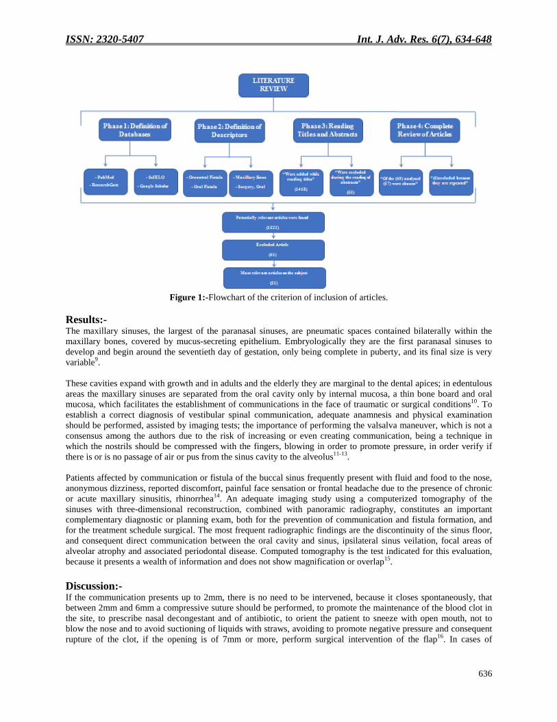
Buccal Flap:-
This flap is widely used because of the excellent characteristics it presents, such as the ease of manipulation, due to
its ipsilateral location to the lesion, an extension suitable for most cases and good blood perfusion20
. When
vestibular flaps are used (Fig. 2), there is a risk of fibrous bridges forming in the bundle, promoting what is
commonly referred to as groove erasure (Fig.3), which may compromise the adaptation and stability of dental
prostheses made to replace lost dental elements21
.
Figure.2:-Buccal tissue flap. Source: DYM and WOLF., 2012, p. 241.
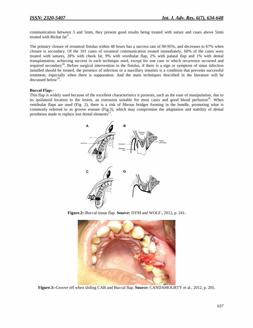
Figure.3:-Groove off when sliding CAB and Buccal flap. Source: CANDAMOURTY et al., 2012, p. 205.
ISSN: 2320-5407 Int. J. Adv. Res. 6(7), 634-648
638
The use of this type of flap is inadvisable because it leads to a considerable loss of vestibule fund requiring a second
surgical intervention aiming to return the depth or to minimize the effect of erasure, since they need wide pedicles
since they are irrigated by small blood vessels and that frequently the patient is subject to the presence of bruising
due to location and infections, the latter are inherent to any surgical procedure, and that there is a possibility of
injury to the facial nerve and the parotid duct22
.
Slippage of the flap is indicated in cases that do not promote changes in the facial contour and the bottom of the
vestibule lost is restored eight weeks after surgery. In addition, its use for small and medium fistulas, and in cases
who additional surgery should be considered to restore depth of the vestibular groove23
.
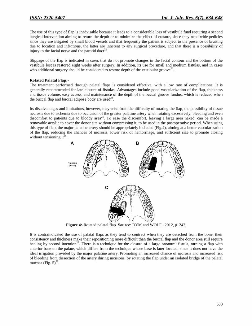
Rotated Palatal Flap;-
The treatment performed through palatal flaps is considered effective, with a low rate of complications. It is
generally recommended for late closure of fistulas. Advantages include good vascularization of the flap, thickness
and tissue volume, easy access, and maintenance of the depth of the buccal groove fundus, which is reduced when
the buccal flap and buccal adipose body are used24
.
Its disadvantages and limitations, however, may arise from the difficulty of rotating the flap, the possibility of tissue
necrosis due to ischemia due to occlusion of the greater palatine artery when rotating excessively, bleeding and even
discomfort to patients due to bloody area25
. To ease the discomfort, leaving a large area naked, can be made a
removable acrylic to cover the donor site without compressing it, to be used in the postoperative period. When using
this type of flap, the major palatine artery should be appropriately included (Fig.4), aiming at a better vascularization
of the flap, reducing the chances of necrosis, lower risk of hemorrhage, and sufficient size to promote closing
without tensioning it26
.
Figure 4:-Rotated palatal flap. Source: DYM and WOLF., 2012, p. 242.
It is contraindicated the use of palatal flaps as they tend to contract when they are detached from the bone, their
consistency and thickness make their repositioning more difficult than the buccal flap and the donor area still require
healing by second intention27
. There is a technique for the closure of a large oroantral fistula, turning a flap with
anterior base on the palate, which differs from the technique whose base is later located, since it does not have the
ideal irrigation provided by the major palatine artery. Promoting an increased chance of necrosis and increased risk
of bleeding from dissection of the artery during incisions, by rotating the flap under an isolated bridge of the palatal
mucosa (Fig. 5)28
.

ISSN: 2320-5407 Int. J. Adv. Res. 6(7), 634-648
639
Figure 5. A:-Incision of the flap; B. Palatal flap under mucosal bridge. Source: Yalçin et al., 2011, p. 336.
Oral adipose body:-
This adipose tissue was first described by Bichat in 1802 and initially used as a pedicled graft in association with the
skin graft for the treatment of oroantral and buconasal communications29
. The buccal adipose body (BAD) is a mass
of adipose tissue encapsulated by a thin layer of connective tissue, located outside the buccinator muscle and the
front of the anterior portion of the masseter, consisting of a central body and four extensions: buccal, pterygoid,
superficial and deep temporal (Fig. 6). It has a mechanical function, acting as a cushion to facilitate the movement
and separation of muscles, besides contributing to the external morphology of the face30
.
Figure 6:-Oral adipose body. Source: KIM et al., 2017, p. 40.
The BAD (Fig.7) has been used to treat fistulas and oroantral (Fig.8) and buconasal communications, reconstruction
after tumor resection, rehabilitation of patients with fissures, and graft recovery for implants because it is a surgical
procedure relatively fast and with a high success rate31
.
A3 B3

ISSN: 2320-5407 Int. J. Adv. Res. 6(7), 634-648
640
Figure 7:-Oral adipose body. Source: KIM et al., 2017, p. 42.
Figure 8.A:-Displacement of BAD; B. BAD of the Positioning; C. Metaplasia of the graft and healing. Source:
MEYER et al., 2012, p. 47.
The advantages of its use are the high success rate, with minimal incidence of failures and complications when
appropriately performed, generation of fewer disturbances and vestibular scars compared to vestibular flap, its
epithelialization occurs in about 2 to 3 weeks , it is a simple and quick procedure, without tooth or bone removal,
minimal discomfort to the patient, it is present in the same region of the surgical intervention, it can be performed
undr local anesthesia, with low morbidity and possibility of being associated with other patchwork32
.
The greatest failure rate of the BAD use occurs due to a necrosis of the adipose tissue supposedly caused by the
excessive manipulation or tension of the same. Therefore, it is suggested that a tension-free suture be performed in
order to avoid the failure of the procedure. And the main disadvantages are the slight vestibular erasure and
alteration in the facial contour of the operated side33
.
Bone Graft:-
This type of graft can be used for closure of nasosinusal, oroantral fistulas, promoting a good bone base for a future
breast lift, and the closure of fistulas that extend along the surface of exposed roots. Since autogenous grafts are
preferable to homogenous and/or xenogenes34
. The use of monocortical bone grafts in a block as a great option for
treatment of oroantral fistulas, when removed from the areas of ment (Fig.9) and mandibular retromolar region,
present good results; these are accompanied by patches of soft tissue for covering, and the palatal and vestibular
rotational flaps are used35
.
A3 B3 C3

ISSN: 2320-5407 Int. J. Adv. Res. 6(7), 634-648
641
Figure 9:-Obtaining and adapting bone graft. Source: HAAS et al., 2003, p. 264.
Matos et al., 2017 points out that the basic principle for the application of this type of graft consists of congruent
fitting of the bone block by pressure in the defect to guarantee the primary stability (Fig.10), and for this to be
possible the preparation of the receptor site is made with a circular trephine drill with the lowest possible external
diameter, and the block graft is obtained by the use of a circular trephine drill whose internal diameter is equal to the
external diameter of the one used for the preparation of the receptor site (Fig.11); if the block is loose, miniplates,
bone screws or some other osteosynthesis technique can be used36
.
Figure 10:-Adaptation of the graft to the bone defect. Source: WATZAK et al., 2005, p. 1291.

ISSN: 2320-5407 Int. J. Adv. Res. 6(7), 634-648
642
Figure 11 A:-Schedule of the diameter of the drills used in the preparation of the receptor site; B. To obtain the
graft. Source: WATZAK et al., 2005, p. 1290.
The bone graft block of the iliac crest can be obtained, however its use should be reserved for cases of major bone
defect, due to the morbidity associated with its obtaining. The advantage of using bone graft block, especially in a
secondary approach, is by providing a solid base for the soft tissue flap and for a future sinus lift, as well as
preserving the adjacent teeth of the fistula. The main disadvantages are the increased working time during the
surgical procedure and consequently increased costs, in addition to being more difficult compared to soft tissue
flaps, and the need for intervention of a medical assistant when obtaining a crest graft iliac37
.
Although closure can be successfully performed using autogenous grafts for the treatment of oroantral
communications, the treatment performed with the rotated or buccal palatal flap without bone graft follow-up is
shown to be quite effective and its use made easier by clinicians, constituting one of the reasons for being widely
used38
.
Dental Transplant:-
It is a surgical modality in which a tooth is avulsed from its place of origin and repositioned in another natural or
surgically created alveolus. The success of the technique is influenced by the integrity of the periodontium and
follicular tissue, surgical asepsis, minimal trauma during trans-surgical management and among other factors, the
time that the element remains outside the alveolus (Fig.12)39
.
A3 B3
ISSN: 2320-5407 Int. J. Adv. Res. 6(7), 634-648
643
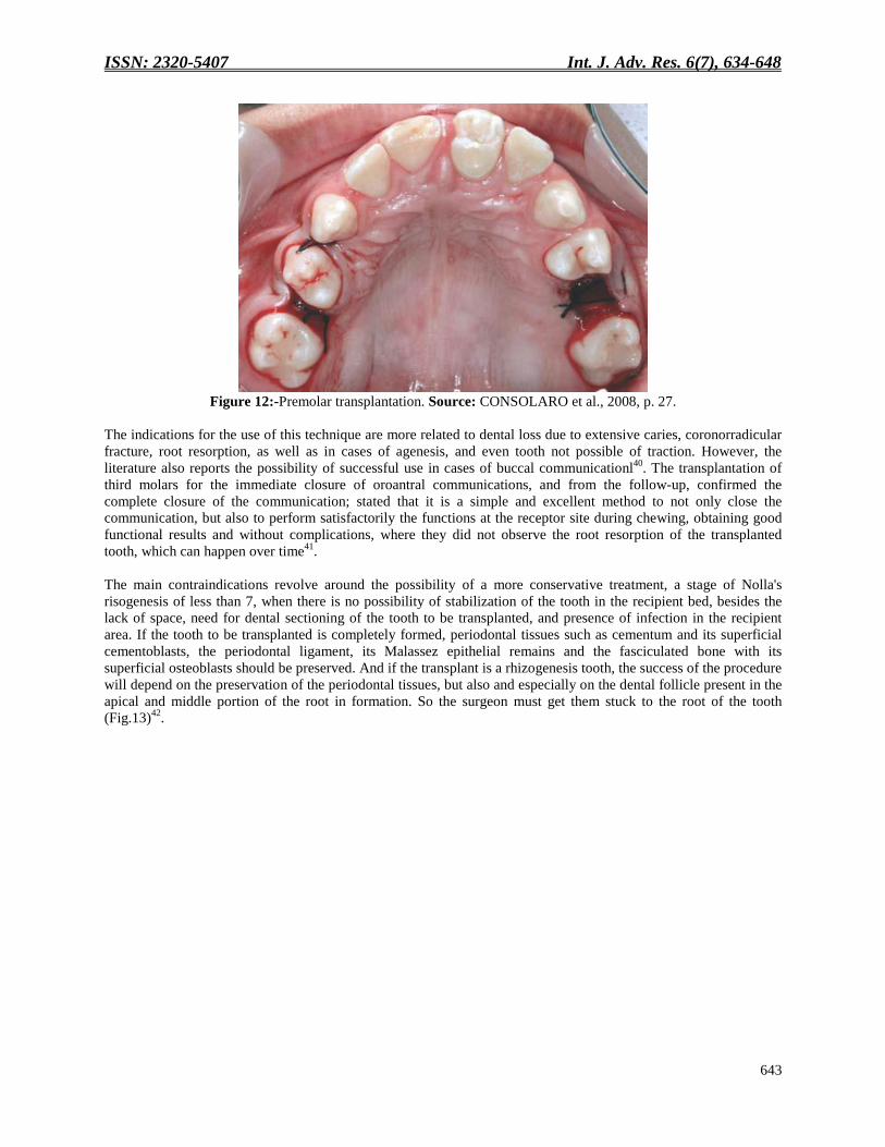
Figure 12:-Premolar transplantation. Source: CONSOLARO et al., 2008, p. 27.
The indications for the use of this technique are more related to dental loss due to extensive caries, coronorradicular
fracture, root resorption, as well as in cases of agenesis, and even tooth not possible of traction. However, the
literature also reports the possibility of successful use in cases of buccal communicationl40
. The transplantation of
third molars for the immediate closure of oroantral communications, and from the follow-up, confirmed the
complete closure of the communication; stated that it is a simple and excellent method to not only close the
communication, but also to perform satisfactorily the functions at the receptor site during chewing, obtaining good
functional results and without complications, where they did not observe the root resorption of the transplanted
tooth, which can happen over time41
.
The main contraindications revolve around the possibility of a more conservative treatment, a stage of Nolla's
risogenesis of less than 7, when there is no possibility of stabilization of the tooth in the recipient bed, besides the
lack of space, need for dental sectioning of the tooth to be transplanted, and presence of infection in the recipient
area. If the tooth to be transplanted is completely formed, periodontal tissues such as cementum and its superficial
cementoblasts, the periodontal ligament, its Malassez epithelial remains and the fasciculated bone with its
superficial osteoblasts should be preserved. And if the transplant is a rhizogenesis tooth, the success of the procedure
will depend on the preservation of the periodontal tissues, but also and especially on the dental follicle present in the
apical and middle portion of the root in formation. So the surgeon must get them stuck to the root of the tooth
(Fig.13)42
.

ISSN: 2320-5407 Int. J. Adv. Res. 6(7), 634-648
644
Figure 13:-Periodontal tissues attached. Source: CONSOLARO et al., 2008, p. 25.
Teeth that are in development have newly formed, fibrous embryonic tissues, facilitating the revascularization
process because it presents good permeability to body fluids, mediators and the invasion of endothelial buds at the
site of transplantation. This initial phase may be called plasma embedding. In fully formed teeth, the tissues are
mature, fibrous and denser than the embryonic or newly formed tissues, which means that the fluids can not
permeate the intimacy of the mature tissues only by capillarity and humidification, resulting in necrosis in the tooth
transplantation, requiring the endodontic treatment of this43
.
Tongue of Flap:-
Tongue defects can be used to reconstruct the oral cavity in cleft palate surgeries, intraoral reconstructions after
resection of pathological lesions or traumatic avulsions44
. It has also been reported as a lining tissue for branch,
angle, body and symphysis reconstruction of the mandible, buccal floor, hard and soft palate, maxillary alveolar
region, closure of buconasal and bucosinusal fistulas, and reconstruction of jugal mucosa, in addition to lower lip45
.
Several factors contribute to the successful use of this flap, such as the rich vascular supply of the lingual artery and
its terminal branches promoting good nutrition, adequate flap elevation, establishment of contact between the
borders of the flap and adjacent tissue without too much pressure on the recipient bed and tension-free pedicle46
. The
flap incision design differs depending on the location and size of the fistula, which can be done with partial or total
thickness, with anterior or posterior base (Fig. 14, 15, 16 and 17), taking care that the pedicle does not have a thinner
thickness than the flap and can be anchored by the suture of the bony palate, reducing the chances of detachment,
and a new surgical intervention, usually after three weeks, is made to loosen the pedicle47
.

ISSN: 2320-5407 Int. J. Adv. Res. 6(7), 634-648
645
Figure 14. Marking of the previous base flap.
Figure 15. Incision of the flap.
Figure 16. Flap positioned near the fistula. Source: MAHAJAN et al., 2014, p. 211-212.
Figure 17. Lateral flap from region to posterior base. Source: RAHPEYMA; KHAJEHAHMADI, 2015, p. 175.
In some cases, there is difficulty in intubation for general anesthesia of the second surgical intervention, for pedicle
rupture, due to the placement of the flap and tongue, and the professional should be ready to perform the procedure
under local anesthesia if necessary48
. The main trans and postoperative complication observed in some cases is
bleeding and dehiscence of the flap; and some disadvantages during the healing phase consist of the sensory and
palate decrease of the donor area, discomfort when moving the tongue during speech and swallowing, returning to a
state of normality after healing, but the lingual remnant even under hypertrophic compensation does not return to its
original size, becoming asymmetrical, and although it does not limit mobility, it may interfere with speech, thus it
must be clarified in advance to the patient or person49
.
The anterior base lingual flap is a safe and reliable technique that presents good and consistent results50
. However,
can being a difficult technique with speech and swallowing errors is not currently the flap of choice for closure of
oroantral fistulas, being more than when there is an insufficiency of tissues that favor the performance of a
technique51
.

ISSN: 2320-5407 Int. J. Adv. Res. 6(7), 634-648
646
Final considerations:-
It can be concluded from this study that:-
Oroantral communication is a condition that frequently causes maxillary sinusitis, due to the free passage of
microorganisms, liquids and food debris to the sinus; it is then necessary to establish immediate treatment whenever
possible. In cases of small communication, compressive sutures should be performed to maintain blood clotting in
order to favor healing, and in cases of greater communication, one must use the techniques in the flap, depending on
the characteristics of each case. vary from the need to use a simpler and easier to handle flap such as vestibular flap,
palatal flap rotation or a combination of these, to the need to perform a more complex such as the rotation of the
Bichat fat ball. The professional who performs dental extractions must be able to treat the complication, otherwise
he should consider referral to a specialist when during the study of the case the communication risk is perceived.
Conflicts of interest:-
The authors declare that there are no conflicts of interest.
References:- 1. Abuabara A, Cortez AL, Passeri LA, De Moraes M, Moreira RW. Evaluation of different treatments for
oroantral/oronasal communications: experience of 112 cases. Int. J. Oral Maxillofac. 2006; 35(2): 155-158.
2. Abrahams JJ, Berger SB. Oral-maxillary sinus fistula (oroantral fistula): clinical features and findings on
multiplanar CT. AJR Am. J. Roentgenol., 1995; 165(5): 1273-1276.
3. Anavi Y, Gal G, Silfen R, Calderon S. Palatal rotation-advancement flap for delayed repair of oroantral fistula:
a retrospective evaluation of 63 cases. Oral Surg. Oral Med. Oral Pathol. Oral Radiol. Endod. 2003; 96(5): 527-
534.
4. Baumann A, Ewers R. Application of the buccal fat pad in oral reconstruction. J. Oral Maxillofac. Surg. 2000;
58(4): 389–392.
5. Bernardino Júnior R, Sousa GC, Lizardo FB, Bontempo DB, Guimarães PP, Macedo JH. Corpo adiposo da
bochecha: um caso de variação anatômica. Biosci. J., 2008; 24(4): 108-113.
6. Borgonovo AE, Berardinelli FV, Favale M, Maiorana C. Surgical options in oroantral fistula treatment. The
Open Dentistry Journal. 2012; 6(1): 94-98.
7. Bränemark PI, Adell R, Albrektsson T, Lekholm U, Lindstrom J, Rockler B. An experimental and clinical study
of osseointegrated implants penetrating the nasal and maxillary sinus. J. Oral Maxillofac. Surg. 1984; 42(8):
497-505.
8. Candamourty R, Jain MK, Sankar K, Babu MRR. Double-layered closure of oroantral fistula using buccal fat
pad and buccal advancement flap. J. Nat. Sci. Biol. Med. 2012; 3(2): 203–205.
9. Consolaro A, Pinheiro TN, Intra JBG, Roldi A. Transplantes dentários autógenos: uma solução para casos
ortodônticos e uma casuística brasileira. R. Dental Press Ortodon. Ortop. Facial. 2008; 13(2): 23-28.
10. Cuffari L, Palumbo M. Transplante de germe do terceiro molar. Jornal Bras. Odontol. Clín. 1997; 1(2): 23-27.
11. Dean A, Alamillos F, García-López A, Sánchez J, Peñalba M. The buccal fat pad in oral reconstruction. Head
Neck. 2001; 23(5): 383-388.
12. Dym H, Wolf JC. Oroantral communication. Oral Maxillofac. Surg. Clin. North. Am. 2012; 24(2): 239-247.
13. Egyedi P. Utilization of the buccal fat pad for closure of oro-antral and/or oro-nasal communications. J.
Maxillofac. Surg. 1977; 5(4): 241-244.
14. Ericson S, Finne K, Persson G. A clinical–radiographic review of treated oro-antral communications. Int. J.
Oral Surg. 1973; 2(5): 185-195.
15. Freitas TMC, Farias JG, Mendonça RG, Alves MF, Ramos Junior RP, Câncio AV. Fístulas oroantrais:
diagnóstico e propostas de tratamento. Rev. Bras. Otorrinolaringol. 2003; 69(6): 838-844.
16. Güven O. A clinical study on oroantral fistula. J. Craniomaxillofac. Surg. 1998; 26(4): 267-271.
17. Haas R, Watzak G, Baron M, Tepper G, Mailath G, Watzek G. A preliminary study of monocortical boné grafts
for oroantral fistula closure. Oral Surg. Oral Med. Oral Pathol. Oral Radiol. Endod. 2003; 96(3): 263-266.
18. Hanazawa Y, Itoh K, Mabashi T, Sato K. Closure of oroantral communications using a pedicled buccal fat pad
graft. J. oral maxillofac. surg. 1995; 53(7): 771-775.
19. Kitagawa Y, Sano K, Nakamura M, Ogasawara T. Use of third molar transplantation for closure of the oroantral
communication after tooth extraction: A report of 2 cases. Oral Surg. Oral Med. Oral Pathol. Oral Radiol.
Endod. 2003; 95(4): 409-415.
20. Klopp CT, Schurter M. The surgical treatment of cancer of the soft palate and tonsil. Cancer.1956; 9(6): 1239-
1243.

ISSN: 2320-5407 Int. J. Adv. Res. 6(7), 634-648
647
21. Magro Filho O, Garbin JREA, Ribeiro JRPD, Felipetti FA. Fechamento de fístula buco-sinusal usando tecido
adiposo bucal. Rev. Odontol. Bras. Central. 2010; 19(50): 275-279.
22. Mahajan RK, Chhajlani R, Ghildiyal HC. Role of tongue flap in palatal fistula repair: A series of 41 cases.
Indian J. Plast. Surg. 2014; 47(2): 210-215.
23. Martin-Granizo R, Naval L, Costas A, Goizueta C, Rodriguez F, Monje F, Muñoz M, Diaz F. Use of buccal fat
pad to repair intraoral defects: review of 30 cases. Br. J. Oral Maxillofac. Surg. 1997; 35(2): 81-84.
24. Meirelles RC, Neves-Pinto RM. Fístula oroantral e retalho mucoso geniano - revisão de 25 casos. Rev. Bras.
Otorrinolaringol. 2008; 74(1): 85-90.
25. Meyer E, Liebenberg SJR, Fagan JJ. Buccal fat pad – a simple, underutilised flap. S. Afr. J. Surg. 2012; 50(2):
47-49.
26. Miranda RF, Amorim RH, Rettore Junior R. Tratamento de comunicações buco-sinusais pós-exodontia. Revista
do CROMG. 1999; 5(1): 60-63.
27. Novais Junior MIL, Almeida MSC, Costa CHM, Sousa Filho LF, Mendes Júnior OR. Uso do Corpo Adiposo de
Bichat para fechamento de comunicação oroantral. Odontol. Clín.-Cient. 2015; 14(3): 719-723.
28. Neder A. Use of buccal fat pad for grafts. Oral surg. Oral Med. Oral Pathol. 1983; 55(4): 349-350.
29. Neves EM, Guimarães RES, Bartels VP, Ferreira AC, Pedroso SL. Fechamento de fístula oro-antral com
gordura de Bichat. Rev. Bras. Cir. Craniomaxilofac. 2009; 12(3): 125-18.
30. Peixoto AC, Melo AR, Santos TS. Transplante dentário: atualização da literatura e relato de caso. Rev. Cir.
Traumatol. Buco-Maxilo-Fac. 2013; 13(2): 75-80.
31. Prado R, Salim MAA, Gadioli B, Almeida TM. Tratamento de fístula buco-sinusal: revisão de literatura e relato
de caso clínico. Rev. bras. Odontol. 2008; 65 (1): 101-105.
32. Rahpeyma A, Khajehahmadi S. Posteriorly based lateral tongue flap for reconstruction of large palatal-alveolar
fistulas in cleft patients. Ann. Maxillofac. Surg. 2015; 5(2): 174-178.
33. Rapidis AD, Alexandridis CA, Eleftheriadis E, Angelopoulos AP. The use of the buccal fat pad for
reconstruction of oral defects: review of the literature and report of 15 cases. J. Oral Maxillofac. Surg. 2000;
58(2): 158-163.
34. Salins PC, Kishore SK. Anteriorly based palatal flap for closure of large oroantral fistula. Oral Surg., Oral Med.
Oral pathol. Radiol. Endod. 1996; 82(3): 253-256.
35. Siegel EB, Bechtold W, Sherman PM, Stoopack JC. Pedicle tongue flap for closure of an oroantral defect after
partial maxillectomy. J. Oral Surgery. 1977; 35(9): 746-748.
36. Matos JDM, Santos IKS, Perreira ALC, Oliveira, AJAG, Vasconcelos BCG, Pita Neto IC. Vasconcelos JEL.
The use of the rich fibrin in platelets and leukocytes as alternative treatment for lifting the maxillary sinus - A
literature review. Int. J. of Dev. Res. 2017; 7(6): 13436-13441.
37. Silveira VM, Netto BA, Côsso MG, Fonseca LC. A utilização da tomografia computadorizada na avaliação da
comunicação bucosinusal. Arq. bras. odontol. 2008; 4(1): 24-27.
38. Tideman H, Bosanquet A, Scott J. Use of the buccal fat pad as a pedicled graft. J. Oral Maxillofac. Surg. 1986;
44(6): 435-440.
39. Watzak G, Tepper G, Zechner W, Monov G, Busenlechner D, Watzek G. Bony press-fit closure of oro-antral
fistulas: a technique for pre-sinus lift repair and secondary closure. J. Oral Maxillofac. Surg. 2005; 63(9): 1288-
1294.
40. Yalçin S, Öncü B, Emes Y, Atalay B, Aktas I. Surgical Treatment of Oroantral Fistulas: A Clinical Study of 23
Cases. J. Oral Maxillofac. Surg. 2011; 69(1): 333-339.
41. Santos IKS, Matos
JDM, Barros JD, Medeiros CR, Cariri TFA, Vasconcelos
BCG, Vardiero VA, Vasconcelos
JEL. Bichectomy as an alternative treatment for facial harmonization - Case Report. Int J Inf Res Rev. 2017;
05(11): 1495-1502.
42. Lima JFM, Matos JDM, Santos JPS, Oliveira AJAG, Vasconcelos JEL, Zogheib LV, Castro DS. Maxillary
sinus lift surgery techniques: A literature review. Int. J. of Adv. Res. 2017; 5(8): 832-844.
43. Santos IKS, Matos
JDM, Júnior
CUFS, Oliveira AJAG, Franco
JMPL, Vasconcelos
BCG; Vasconcelos EMGM,
Gomes TN, Vardiero VA, Vasconcelos JEL. Surgery of the maxillary sinus with piezocirurgic system use: Case
report. Int J Inf Res Rev. 2017; 4(6): 4258-4260.Pereira ALC, Matos JDM, Cavalcante-Pereira N, Franco
JMPL, Vasconcelos BCG, Vardiero VA, Gomes TN, Vasconcelos, J. E. L. Maxillary Sinus lift and use of L-
PRF with a delayed installation of implants–Case report. Int. J. of Dev. Res. 2017; 7(6): 13174-13177.
44. Silveira RL, Santos MESM, Takahashi A, Bourguignon Filho AM, Heitz C. Tratamento de fístula bucosinusal
através de retalho palatino. Rev. Cir. Traumatol. Buco-Maxilo-fac. 2008; 8(1): 29-34.

ISSN: 2320-5407 Int. J. Adv. Res. 6(7), 634-648
648
45. Figueiredo PS, Lima CF, Santana VS, Matos JDM, Franca RJ, Figueiredo RS, Oliveira AJAG, Vasconcelos
JEL, Neto ICP. Surgical Treatment of the Non-Surgical Jaw Condyle Fracture Version: A Literature Review.
Int. J. Oral. Health. Med. Res. 2017;4(4):1-8.
46. Santos IKS, Matos JDM, Franco JMPL, Pereira NC, Pita Neto IC, Vasconcelos JEL. Mucocele In Blandin-
Nuhn Glands: Case Report And Integrative Review. Int. Arc. of Med. 2016; 9(392):1-7.
47. Silva SAB, Matos JDM, Nascimento JA, Maia SES, Lima RVM, Vasconcelos RBA, Bezerra AL, Oliveira
AJAG, Vasconcelos BCG, Zogheib LV, Lima JFM, Castro DSM, Gomes TN, Vasconcelos JEL, Vardiero VA.
Removal of radical cyst in previous region of maxila: Case report and integrative review. Int. J. of Adv. Res.
2018; 6(1):70-87.
48. Santos IKS, Matos JDM, Pereira NC, Franco JMPL, Vardiero VA, Vasconcelos JEL. Supernumerary Tooth
Removal Using Nitrous Oxide - Case Report. Int. J. of Inf. Res. And Rev. 2017; 4(5): 4164-4168.
49. Nascimento JA, Matos JDM, Meneses RO, Araujo JBGN, Oliveira AJAG, Vasconcelos BCG, Gomes TN,
Vardiero VA, Vasconcelos JEL, Araujo Junior DN. Removal Of Ectopic Canine Of The Nasal Fossa: Case
Report. Int. J. of Adv. Res. 2017; 5(8):1253-1257.
50. Santos IKS, Matos JDM, Perreira ALC, Oliveira AJAG, Franco JMPL, Vardiero VA, Vasconcelos JEL.
Removal Of Ectopic Canine In Region Of Mandible - Case Report. J. Med. Bio Sci. Res. 2017; 3(3):24-31.