Embed Size (px)
Citation preview

Universidade de Aveiro Departamento de Biologia
2013
José Fernando Seabra Babo
Screening molecular de Hepatozoon em anfíbios. Molecular screening of Hepatozoon in amphibian hosts.
Dissertação apresentada à Universidade de Aveiro para
cumprimento dos requisitos necessários à obtenção do grau de
Mestre em Biologia Aplicada, realizada sob a orientação científica
do Doutor D. James Harris, investigador do CIBIO-UP (Centro de
Investigação em Biodiversidade e Recursos Genéticos da
Universidade do Porto) e do Departamento de Biologia da
Faculdade de Ciências da Universidade do Porto e co-orientação
do Professor Doutor Amadeu Mortágua Velho da Maia Soares,
Professor Catedrático do Departamento de Biologia da
Universidade de Aveiro.
Universidade de Aveiro Departamento de Biologia
2013
José Fernando
Seabra Babo
Screening molecular de Hepatozoon em anfíbios. Molecular screening of Hepatozoon in amphibian hosts.

DECLARAÇÃO
Declaro que este relatório é integralmente da minha autoria, estando devidamente
referenciadas as fontes e obras consultadas, bem como identificadas de modo claro as
citações dessas obras. Não contém, por isso, qualquer tipo de plágio quer de textos
publicados, qualquer que seja o meio dessa publicação, incluindo meios eletrónicos, quer
de trabalhos académicos.

‘Não são as espécies mais fortes que sobrevivem
nem as mais inteligentes, e sim as mais suscetíveis
a mudanças.’
Charles Darwin

o júri
presidente Professor Doutor António José Arsénia Nogueira professor associado C/ Agregação da Universidade de Aveiro
Doutora Maria João Veloso da Costa Ramos Pereira bolseira de Pós-Doutoramento da Universidade de Aveiro
Doutor David James Harris investigador do CIBIO- Centro de Investigação em Biodiversidade e Genética

agradecimentos
Ao meu orientador James, por todo o tempo despendido comigo e pela ajuda
que me deu na realização deste trabalho. Foi um prazer trabalhar e apreender
consigo.
Ao Professor Amadeu Soares pela oportunidade que me deu em ir para o CIBIO
realizar a tese.
Ao João Maia por me ter ajudado nos primeiros passos deste trabalho e estar
sempre presente para ajudar, e por todo o conhecimento que me transmitiu.
A todas as pessoas do laboratório que tornavam o dia mais divertido e um ótimo
local de trabalho.
Um agradecimento especial a um amigo, Nuno Taser que me acompanha à sete
anos.
Aos meus amigos, Américo, André, Mimi, Neves, Zorion e muitos outros pelos
momentos de diversão passados durante o ano, e por me deixarem dormir na
sua casa quando precisava.
À minha namorada me conheceu na parte final deste projeto e me aturou e
apoiou sempre.
À minha tia Bina por todo o apoio que me deu.
À minha irmã pela ajuda com os esquemas e imagens, o lado artístico da família
ficou claramente com ela.
À minha mãe e a minha avó por todos os sacrifícios que fazem e fizeram para
que eu pudesse estudar e tirar um curso superior. Sem elas não era quem sou
hoje.

palavras-chave
resumo
Apicomplexa; parasita; Hepatozoon; anfíbio; Ichthyopthirius multifiliis; Dactylosoma ranarum; screening molecular; PCR.
A diversidade de parasitas tem sido pouco estudada devido a vários fatores, como o tamanho de algumas espécies, a localização dentro do hospedeiro, e o foco em grupos com fortes interesses antropogénicos ou com impacto econômico significativo. Apicomplexa é um grupo grande e diverso de parasitas unicelulares e com uma ampla distribuição e é o agente patogénico com mais sucesso conhecido pelo homem. Dentro do grupo alguns gêneros têm recebido pouca atenção, como caso de Hepatozoon. Estes são organismos pouco estudados que requerem mais amostragem. Técnicas de rastreio molecular, como PCR, são rápidas e fáceis de usar, e permitem a deteção eficiente desses parasitas. Elas também permitem a sequenciação produzindo dados para análises filogenéticas. Contudo mais dados são necessários para determinar corretamente a taxonomia e as relações filogenéticas do grupo. O objetivo deste trabalho foi detetar a diversidade de parasitas através de screening de amostras de tecido e sangue de diferentes espécies de anfíbios, utilizando primers específicos e reconstruir as relações filogenéticas das sequências produzidas. Várias espécies de anfíbios da Península Ibérica, Ilhas do Mediterrâneo, região Macaronésia e Marrocos foram analisadas usando primers específicos para parasitas e esfregaços de sangue foram observados. Parasitas foram encontrados em vários slides, sendo possível a identificação de pelo menos dois Hepatozoon, no entanto os primers não foram capazes de detetar as infeções. Apenas dois parasitas foram amplificados atráves de primers Hep, um parasita protozoário, Ichthyopthirius multifiliis, identificado numa amostra de Bufo calamita de Portugal, e um parasita Apicomplexa, Dactylosoma ranarum, em Pelophylax saharicus de Marrocos. Os primers utilizados não parecem ser úteis com anfíbios e novos primers precisam ser desenvolvidos para corretamente identificar a identidade dos parasitas observados nos slides.

keywords
abstract
Apicomplexa; parasite; Hepatozoon; amphibian; Ichthyopthirius multifiliis; Dactylosoma ranarum; molecular screening; PCR. Parasite diversity has been poorly studied due to several factors, like the size of some species, location within the host, and the focus on groups with strong anthropogenic interests or with significant economic impact. Apicomplexa is a vast and diverse group of unicellular parasites with wide distribution and the most successful pathogen known to man. Within the group some genus have received little attention, like the case of Hepatozoon. They are poorly studied organisms requiring more sampling. Molecular screening techniques like PCR are quick and easy to use, and allow efficient detection of these parasites. It also allows sequencing which produce data for phylogenetic analyses. Nonetheless more data is necessary to correctly establish taxonomy and phylogenetic relations of the group. The aim of this work was to assess the diversity of parasites trough screening of tissue and blood samples from different amphibian species using specific parasite primers and reconstruct the phylogenetic relationships of the sequences produced. Several species of amphibians from Iberian Peninsula, Mediterranean Islands, Macaronesia region and Morocco were analyzed using specific parasite primers and blood smears were observed. Parasites were observed in several slides, with at least two Hepatozoon being identified, however primers fail to detect them. Only two parasites were amplified, a protozoan parasite, Ichthyopthirius multifiliis, identified in a Bufo calamita sample from Portugal, and a Apicomplexa parasite, Dactylosoma ranarum, in Pelophylax saharicus from Morocco.
The primers used seem not to be useful with amphibians and new primers need
to be developed to correctly assess the identity of the parasites observed In the smears.

i
Index
Index…………………………………………………………………………………..I
List of Figures………………………………………………………………………...II
List of Tables………………………………………………………………………...IV
1 Chapter one - Introduction………………………………………………………….5
1.1 Parasitism…………………………………………………………………….6
1.2 Apicomplexa………………………………………………………………….7
1.3 Amphibian hosts……………………………………………………………19
1.4 Parasite detection techniques…………………………………………….23
1.5 Objectives…………………………………………………………………...29
1.6 Organization of the thesis………………………………………………….30
1.7 Taxonomy…………………………………………………………………...31
2 Chapter two - Materials and Methods……………………………………..……32
2.1 Sample collection…………………………………………………………..33
3 Chapter three - Putative Ichthyophthirius identified in the amphibian Bufo
calamita through molecular screening.…………………………………….……………….40
4 Chapter four - Screening for Apicomplexan parasites in amphibians…...…...48
5 Chapter five – General discussion………….……………………………………53
6 Chapter six - Concluding remarks and future possibilities…………………….57
6.1 Concluding remarks………………………………………………………..58
6.2 Future possibilities………………………………………………………….60
7 References………………………………………………………………………….61
Appendix 1…………………………………………………………………………….71
Appendix 2…………………………………………………………………………….72

ii
List of Figures
Figure 1.1 – Ultrastructural characteristics of the Apicomplexa. The image at the
bottom corresponds to an electron microscopy photograph of a Toxoplasma gondii. C -
conoid; CC - cortical cisternal layer; DG - dense granule; G – Golgi Complex; M -
microneme; N - nucleus; R - rhoptry. The parasite and vacuolar network (VN) are
enclosed by a vacuole membrane (VM). Scale bar, 1 μm. From:Kaasch and Joiner
(2000). The image at the top is a schematic of the principal cellular components of
apicomplexans. From: Šlapeta and Morin-Adeline
(2011)…………………………………………………………………………………………..10
Figure 1.2 – Life cycle of Apicomplexan parasites. The Center circle represents a
generic Apicomplexan life cycle. The outer circle represent the specific life cycle of
Plasmodium falciparum. The middle circle represents the life cycle of Toxoplasma
gondii. The bradyzoite form (∗) is responsible for reactivation of latent infection and is
an obligatory stage between tachyzoites and gametes. From: Morrissette and Sibley
(2002)…………………………………………………………………………………………11
Figure 1.3 – Hypothetical tree of Apicomplexa groups and their relationships. The width
and number on the branches refers to the named species and thus, the known
diversity. From: Šlapeta and Morin-Adeline (2011)…………….....................................13
Figure 1.4 – Bayesian tree representing Hepatozoon phylogenetic relations. Based on
562 bp 18S rRNA gene sequences. Bayesian posterior probabilities and ML bootstrap
values are given above and below the nodes respectively. + is indicated when both
values are 100%. The branches of JN181157, AF130361 and AF297085 were
shortened by 50%. From: Maia et al. (2012b)……………………………………………..18
Figure 1.5 – Diagram of the cycle of Hepatozoon sipedon. 1 Gamonts in erythrocytes of
the snake host are ingested by mosquitoes and are released in the gut. 2
Microgamonts and macrogamonts associate in syzygy in a parasitophorous vacuole in
a fat body cell of the mosquito haemocoel. 3 Gamonts undergo gametogenesis by 4
days post-feeding, after which one of the microgametes fertïlizes the macrogamete. 4
Resulting zygote forms an immature oocyst. 5 Nucleus of oocyst divides during the
initial stages of sporoblast development at 20 days post-feeding. 6 Oocyst, mature at
28 days post-feeding, contains an average of 600 sporocysts. 7 Each sporocyst
contains eight sporozoites. 8 Sporozoites are released into the gut of a frog when an
infected mosquito is ingested. 9 Dizoic cysts form in frog hepatocytes at 7 days post-
infection. 10 Cystozoites are released into the gut of a snake when an infected frog is
ingested. 11 Mature macromeronts are present in snake hepatocytes and other cells of
visceral organs after 15 days post-feeding. 12 Macromerozoites released from these

iii
macromeronts invade the bloodstream of the snake and reinfect hepatocytes and other
cells of visceral organs at 16 days post-feeding. 13 Micromeronts are mature after 30
days post-feeding. 14 Micromerozoites released from micromeronts infect erythrocytes
of the snake host, forming gamonts which are infective to mosquitoes during
subsequent feedings. From: Smith (1998)…………………………………………………22
Figure 1.6 – Representation of the life cycle of Hepatozoon catesbianae in his hosts. A.
Merozoites released from hepatic meronts enter erythrocytes. B. Merozoites transform
into gamonts. C. Mosquitoes feeding on infected frogs ingest erythrocytic gamonts. D.
Gamonts escape from erythrocytes in gut of mosquito and enter Malpighian tubules. E.
Micro- and macrogamonts come to lie within a common parasitophorous vacuole in
tubule cells. F. Gametogenesis ensues with formation of two biflagellate microgametes,
one of which fertilizes the macrogamete. G. The zygote expands into a spherical
oocyst. H. Oocysts undergo segmentation to form sporoblasts. I. Sporoblasts transform
into sporocysts. J. Each sporocyst contains four sporozoites. K. Frogs are infected by
ingesting mosquitoes containing sporocysts. L. Sporozoites enter hepatic parenchymal
cells where they develop into meronts. From: Desser et al. (1995)……………………..23
Figure 1.7 – Diagram representing the organization of nuclear genes of ribosomal
subunits. Scheme by José Babo……………………………………………………………29
Figure 2.1 – Geographic distribution of the species used as samples………………….33
Figure 2.2 –Photos of the species used during this thesis. A –Bufo bufo; B –
Amietophrynus mauritanicus; C –Discoglossus sardus; D –Bufo calamita; E –Hyla
meridionalis; F –Hyla sarda; G –Pelobates cultripes; H –Pelophylax perezi; I –
Pelophylax saharicus; J –Pleurodeles waltl; K –Bufotes balearicus; L –Bufotes
boulengerie. Photos E and I were taken by Daniele Salvi, Photos A, C, D, F, G, H, J, K,
L were taken by Matt Wilson, Photo B was taken by Pierre-Yves Vaucher……………35
Figure 3.1 – ML tree of the apparent Ichthyophthirius from a Bufo calamita, and closest
available comparative sequences from GenBank. Support for the Bayesian and for ML
analysis are given above and below the nodes, respectively. The branch of
Paramecium tetrawelia was shortened 75%.................................................................43
Figure 2.1 –Images of positive Hepatozoon infections in A) A. mauritanicus, and B) P.
saharicus, both of which failed to amplify using the screening protocol employed, C) a
positive infection of presumed Dactylosoma ranarum in Pelophylax saharicus, and D) a
typical negative sample. The scale bar corresponds to 20μm…………………………...50

iv
List of Tables
Table 2.1 –List of amphibians host species used in this thesis…..……………………..34
Table 2.2 –Details of the PCR primers used in this thesis……………………………….36
Table 2.3 –Reagents used, and respective concentrations for the Hep, HEMO and CR
primers, using invitrogen Taq DNA Polymerase…………………………………………..36
Table 2.4 – PCR protocols for the Hep, HEMO and CR primers using invitrogen Taq
DNA Polymerase……………………………………………………………………………...37
Table 2.5 – Reagents used, and respective concentrations for the Hep, HEMO and CR
primers, using Bioline MyTaqTM DNA Polymerase………………………………………..38
Table 2.6 – PCR protocols for the Hep, HEMO and CR primers using Bioline MyTaqTM
DNA Polymerase……………………………………………………………………………...38
Table 4.1 –Amphibian hosts species screened for parasites, the number tested using
alternative source material (tissue or blood), and the number examined under the
microscope on slides…………………………………………………………………………48

5
1 Chapter one
General Introduction, Objectives

6
1.1 Parasitism
The relations established between species are normally defined by the effect of the
interaction on each of the species. This relation can be beneficial for both species
(Mutualism), beneficial for one of the species but with no harm to the other
(Commensalism), beneficial for one of the species at the expense of the other (Parasitism,
Predation), one of the species can be inhibited with no effect on the other (Amensalism),
and finally both species can be inhibited (Competitive interactions).
In this thesis we are going to focus our attention on the relations of parasitism.
Therefore it is important to understand what a parasitic relationship means. If we go to the
origin of the word parasite, it derives from the Medieval French word parasite, which
comes from the Latin parasites, the latinisation of the Greek παράσιτος (parasitos), παρά
(para), "beside, by"+ σῖτος (sitos), "wheat". The word παράσιτος means “one who eats at
the table of another”. The literal interpretation leads to the conclusion that a large part of
living creatures are parasites (Poulin & Morand 2000). Bringing these to the context of this
thesis we could interpret parasitism as one living creature that feeds from another living
one without causing death to the latter, or else it would be predation.
Even though defining parasitism can be difficult and can lead to several different
classifications such as organism that lives in or on another living organism obtaining from
it part or all of its organic nutrient, and commonly exhibiting some degree of adaptive
structural modification (Bush et al. 2001). Therefore more strict definitions should be
adopted such as, a close host-parasite relation with the latter passing a large part of its life
history in or on his host. Even with the adoption of stricter definitions, the number and
diversity of parasites is huge (Poulin & Morand 2000). We can consider parasites within
taxa including bacteria, virus, fungi, algae, metazoans.
After classifying what parasitism is another problem emerges: How to classify the
types of parasites? This can be a hard task, and again several answers could be given.
We can divide them accordingly to where they can be found, endoparasites and
ectoparasites. Therefore the ones living inside the host body are endoparasites, and the
ones who live outside are ectoparasites. We can also classify them according to their size,
microparasites and macroparasites. Microparasites as the name says are microscopic,
while the macroparasites are bigger and can be seen without using the microscope. Both
micro and macroparasites can be either endo or ectoparasites. In this thesis we aim to
concentrate in a group of parasites that lives in the blood cells of their host, and therefore
are endoparasites and microparasites.

7
The size of parasites is one of many reasons why describing their diversity is so
difficult; some species can have very small sizes. Another reason is the primarily focus on
species considered valuable to man such as those with agricultural, veterinary or medical
interest (Poulin & Morand 2000; Morrison 2009). Some parasites occur with low
prevalence values, so inadequate sample efforts can also lead to an underestimation of
diversity values (Poulin & Morand 2000). Also, parasite identification has been traditionally
done with the use of the microscope. However, when identification is solely based on
morphological characteristics, this can lead to reports of distinct parasite species as a
single one, and vice-versa. In other words, many different parasites may morphological
appear very similar and be mistaken for a single species, known as “cryptic species”.
Even though this problem is starting to be overcome through the use of molecular
techniques, we can expect that the number of parasitic species described only represents
a fraction of the real diversity of this group (Adl et al. 2007; Morrison 2009). Another
problem is we still have not identified all free-living organisms in the world, and
considering that each of these organisms are potential hosts for at least one species of
parasite, we can expect that the diversity of parasites to be much higher than is currently
recognized (Poulin & Morand 2000).
1.2 Apicomplexa
The phylum Apicomplexa, also known as Sporozoa, comprehends a huge and
diverse group of unicellular protozoans with a wide environmental distribution. Most of
them are obligate intracellular parasites, and probably the most successful pathogens
known to man (Sato 2011). The morphological shape depends on the genus and lifecycle
stage and these parasites are typically quite host specific. They are known to parasitize a
large number of organisms, virtually all vertebrates, including humans, and marine and
terrestrial invertebrates (Frölich et al. 2012). Six thousand species are described, which
only represent a tiny fraction of the real number existing, estimated at 1.2 to 10 million
(Adl et al. 2007). This number can be very inaccurate, since it is believed that all animal
species host at least one of these parasites (Morrison & Ellis 1997). Not all biodiversity
has been described, and therefore with each new host identified, a potential new parasite
can be discovered (Poulin & Morand 2000).
The group can be found in humans and domestic animals, and is responsible for
several diseases with both medical and veterinary significance (Massimine et al. 2005).
Even though many of them are not pathogenic to their host, it is estimated that they cause

8
the deaths of 1 million people every year and agricultural losses of over US $1 billion per
year (Beck et al. 2009).
One of the most notorious parasitizing humans is the genus Plasmodium, the
agent responsible for malaria. It is estimated that the genus include about 172 species,
with 89 occurring in reptiles, 32 in birds and 51 in mammals, of which 4 cause malaria in
humans (Paul et al. 2003). The benign tertian malaria caused by Plasmodium vivax, is the
most widely distributed human malaria, with an estimated 70-80 million cases per year
(Cui et al. 2005), while Plasmodium falciparum is responsible for 3 to 5% of deaths
worldwide each year (Black et al. 2005), and is the most studied species in the genus.
Another important pathogenic to man is Toxoplasma gondii, an opportunistic parasite
present in 30% of the humans. The parasite represents little or no harm to healthy
individuals, but can be fatal in those with compromised immune systems, like AIDS or
cancer patients, and is dangerous for pregnant women (Hill & Dubey 2002; Massimine et
al. 2005). This pathogen is widely distributed across the globe, although its prevalence
varies with region (Hill & Dubey 2002). Cryptosporidium are widespread intestinal
pathogens and cause a disease called cryptosporidiosis (Beck et al. 2009). This disease
results in sickness and severe diarrhea, and in risk groups, like young children, the elderly
and immunosuppressed individuals, the disease can be fatal (de la Parte-Pérez et al.
2005). Other species infecting humans include Babesia, Cyclospora and Sarcocystis
(Leander 2003).
Apicomplexan parasites also cause huge agricultural losses, with Eimeria spp.
being responsible for losses over US $1.5 billion losses (Sharman et al. 2010). Eimeria
spp., which causes the disease coccidiosis has a huge economical importance for the
poultry industry (Beck et al. 2009; Frölich et al. 2012). Also Theileria, a tick-transmitted
Apicomplexan parasite, is known to infect livestock and causes important economic
losses (Beck et al. 2009). Neospora caninum is another pathogen of animals, responsible
for important losses in cattle. As the name suggest, the parasite was primarily associated
with dogs. Its cyst-forming parasite causing neuromuscular disorders in dogs and, a huge
cause of abortion and neonatal mortality in cattle (Dubey et al. 2007). Other relevant
parasites include Babesia, Besnoitia, Cryptosporidium, Sarcocystis, and Toxoplasma
(Muller & Hemphill 2013).
These parasites present very different transmission modes. Plasmodium and
Theileria for instance are vector-borne; Eimeria, Toxoplasma and Cryptosporidium form
highly resistant cyst that can be transmitted through contaminated materials, like food or
water (de la Parte-Pérez et al. 2005; Beck et al. 2009). Allied to this, the resistance to

9
most known drugs and the small number of existent vaccines makes very difficult to
prevent these diseases (Frölich et al. 2012).
Even with this important medical, veterinary and economical component many
other groups along with the Apicomplexa are very poorly studied. Although some
characteristics are easily studied such as life-cycles patterns, cyst organization, and
ultrastructure and host, the difficulty in finding and identifying these unicellular
endoparasites complicates the description of them (Morrison 2009).
1.2.1 General biology and life cycle
The name of the group Apicomplexa was determined by unique internal structures
that the organisms of the group possess (Levine 1973) (Figure 1.1). These structures
were only possible to observe after the invention of Electron Microscopy. The name of the
group derives from the presence of an apical complex on at least one of the life stage (Adl
et al. 2005). This complex is found normally in the infective stages at the front end,
displacing the nucleus and mitochondria towards the back (Aikawa et al. 1978). It is
responsible for recognizing, attaching and invading host cells (Smith & Desser 1997;
Walker et al. 2011; Frölich et al. 2012). The complex consists of a cytoskeleton
comprising a closed conoid and at least one polar ring, associated with secretory
organelles, rhoptries and micronemes (Adl et al. 2005; Walker et al. 2011). It also contains
Apicoplasts, a non-photosynthetic relict plastid (Walker et al. 2011), dense granules on
the posterior part, an endosymbiotic derived organelle mitochondrion, and the
acidocalcinomes. Apicomplexans also possess tubular mitochondrial cristae, micropores
and a pellicle with three membranous layers subtended by microtubules, which place
them within the Alveolates (Smith & Desser 1997). The number of and shape of rhoptries,
micronemes and dense granules vary according to the group (Leander 2008). Further, the
conoid is not present in the hematozoan group.
The life cycle is complex and can vary within the group (Morrissette & Sibley
2002). Sexual and asexual reproduction is present and several hosts can be involved
during this process. Having infected the host, the parasites invade the cells and divide
until the host cell is lysed and new parasites are released. Extracellular division normally
does not occur, therefore when released, these parasites need to invade new cells in
order for the cycle to continue (Morrissette & Sibley 2002).

10
Fig
ure
1.1
– U
ltra
str
uctu
ral cha
racte
ristics o
f th
e A
pic
om
ple
xa
.
Th
e im
ag
e a
t th
e b
ott
on
co
rresp
ond
s t
o a
n e
lectr
on
mic
rosco
py p
ho
tog
rap
h o
f a
Toxo
pla
sm
a g
ond
ii. C
- c
on
oid
; C
C -
cort
ical cis
tern
al la
ye
r; D
G -
de
nse
gra
nu
le; G
– G
olg
i C
om
ple
x;
M -
mic
ron
em
e;
N -
nu
cle
us;
R -
rhoptry. The parasite and vacuolar network (VN) are enclosed by a vacuole m
embrane (VM). Scale bar, 1 μm.
Fro
m:K
aa
sch
an
d J
oin
er
(20
00
).
Th
e im
age
at th
e to
p is a
sch
em
atic o
f th
e p
rincip
al ce
llula
r co
mp
on
en
ts o
f a
pic
om
ple
xa
ns. F
rom
: Šlapeta and Morin
-Ad
elin
e (
201
1).

11
The life history normally consists at three distinct steps of development:
gametogony (sexual), sporogony (asexual) and merogony (asexual) (Leander 2003). A
sexual reproduction phase occurs, gametogony, where the fusion of gametes originates a
diploid zygote. This zygote invades a cell and rapidly originates haploid offspring through
meiosis. This process is called sporogony. After that, the haploid progeny, sporozoites,
leave the host cell to invade others specific cells. They use a variety of molecular tools,
including surface adhesions such as parasite surface proteins and binding antigens to
enter host cells (Baum et al. 2008). Upon establishment in a new host cell, sporozoites
typically produce merozoites, although pathway may differ depending on the species
(Figure 1.2). During this process, merogony, merozoites multiply, invading new cells and
giving origin to new merozoites. These merozoites can develop into gametocytes,
producing gametes so the cycle may continue. A scheme of a generic, Toxoplasma, and
Plasmodium life cycle is presented in Figure 1.2. Some aspects of the life cycle may be
different depending on the species (Adl et al. 2005; Frölich et al. 2012).
Figure 1.2 – Life cycle of Apicomplexan parasites. The Center circle represents a generic Apicomplexan life cycle. The outer circle represent the specific life cycle of Plasmodium falciparum. The middle circle represents the life cycle of Toxoplasma gondii. The bradyzoite form (∗) is responsible for reactivation of latent infection and is an obligatory stage between tachyzoites and gametes. From: Morrissette and Sibley (2002).

12
1.2.2 Phylogeny
The phylogeny of the Apicomplexa, and the groups within, is a controversial
subject, since in recent years establishes of relationships have been altered and
redefined. This chapter will present the current knowledge about Apicomplexa phylogeny,
followed by the phylogeny within the phylum.
Apicomplexa belongs to the monophyletic group Alveolata, along with
Dinoflagellates, Ciliates, and some minor lineages (Fast et al. 2002; Leander & Keeling
2004). Within the Alveolata, Apicomplexa and Dinoflagellates are more closely related to
one another than either is to Ciliates. Despite all the morphological differences between
Apicomplexa and Dinoflagellates, molecular tools support this phylogeny, and together
they form the Myzozoa (Escalante & Ayala 1995; Fast et al. 2002; Leander & Keeling
2004). The large number of morphological differences could be explained by the lack of
morphological information of intermediate lineages that are now being detected by
molecular tools (Leander 2003). Molecular phylogenetic analyses of several protein genes
have shown that the closest sister lineage of Dinoflagellates are the Perkinsids, mollusks
and microeukaryotes parasites (Bushek et al. 2002; Saldarriaga et al. 2003; Leander &
Keeling 2004). Perkinsids are not specifically related to Colpodellids and Chromerids, in
fact phylogenetic analyses of small subunit rRNA sequences, have shown that these two
groups are sister groups to Apicomplexa (Kuvardina et al. 2002; Leander 2003; Moore et
al. 2008). Colpodellids were suggested to be the earliest divergent sister group to the
Apicomplexa (Leander et al. 2003). The same conclusion was reached by Siddall et al.
(2001), using SSU rDNA to perform a phylogenetic analysis. Colpodellids are small
predatory flagellates that possess an apical complex used to consume algae and other
protists. Although the group presents an apical complex, it lacks the parasitic life style
typical of the Apicomplexa. This brings controversy into the phylogeny of the group, with
some authors considering that they belong with the Apicomplexa, while others consider
them a separate group (Adl et al. 2005; Walker et al. 2011). The phylum Chromerida was
also reported as a sister group of the Apicomplexa (Moore et al. 2008). This was
supported by the analyses of nuclear LSU rDNA and SSU rDNA sequences, and analyses
of the plastid rDNA (Moore et al. 2008). Despite being a photosynthetic alveolate, like
many Dinoflagellates, the photosynthetic plastid of this group is related most closely to the
apicoplast of Apicomplexa (Moore et al. 2008). Therefore Apicomplexa are currently
thought to be closer related to Chromerids than to Dinoflagellates, and this supports the
idea that the apicoplast is a trace of what remains of a red-algal derived chloroplast
(Waller & McFadden 2005; Janouškovec et al. 2010; Sato 2011).

13
The phylogeny within the Apicomplexa group was primarily estimated using
morphological characteristics. The use of molecular tools to support morphological traits
is in its infancy (Leander 2003). It has been postulated that before using any
morphological trait to establish taxonomy, the association with that group must be first
confirmed with clade analyses (Morrison 2009). The lack of support for morphological
traits used in the past, in conjunction with inappropriate taxon sampling and misuse of
genetic analysis tools are some reasons for earlier conflicting estimates of the phylogeny
of Apicomplexa (Kopecna et al. 2006; Morrison 2009). However, an attempt to improve
classification criteria is ongoing (Adl et al. 2007; Imam 2009). This could lead to changes
in the historically recognized groups, Coccidian, Cryptosporidia, Gregarines,
Haemosporinids, and Piroplasms (Barta et al. 2012). Figure 1.3 demonstrates a
hypothetical tree of the Apicomplexa.
Coccidians in conjunction with Haemosporinids and Piroplams form a clade. The
Coccidia group is a very diverse one, with many life cycles presented. Barta et al. (2012)
identify the tissue coccidia (Eimeriorina: Sarcocystidae), the enteric coccidia (Eimeriorina:
Eimeriidae), the adeleorinid coccidia (Adeleorina: Adeleidae), and the hemogregarines
(Adeleorina: various families). They can present monoxenous life cycle parasitizing
Figure 1.3 – Hypothetical tree of Apicomplexa groups and their relationships. The width and number on the branches refers to the named species and thus, the known diversity. From: Šlapeta and Morin-Adeline (2011).

14
vertebrate and invertebrate hosts (Eimeriidae and adeleorinid coccidian respectively).
They can also have heteroxenous life cycle (hemogregarines and Sarcocystidae), using
vertebrates as intermediate and invertebrates as definitive hosts. The mode of
transmission between hosts is usually a predator-prey relationship, infective stages are
produced within the prey and life cycle only completes within the predator. Like many
other Apicomplexa groups phylogeny may be poorly estimated, due to lack of enough
data and limited sample, with many organisms being overlooked, in favour of veterinary
important species. The remaining two groups, Haemosporinids and Piroplams are
considered sister clades and together form a monophyletic class called hematozoa (syn.
Aconoidasida) (Escalante & Ayala 1995; Adl et al. 2005). Both possess heteroxenous life
cycles, parasitizing vertebrate (sexual reproduction stages) and invertebrate (asexual
reproduction stages) hosts. The order Piroplasmida contains the genera Babesia and
Theileria, while the order Haemospororida contains the most medical and veterinary
significant genera Plasmodium, Haemoproteus and Leucocytozoon. An endosymbiotic
marine protist with uncertain classification since its discovery in the 19th century is the
newest addition to the Apicomplexa and Hematozoa: Nephromyces are ubiquitous
nonhereditary symbionts, transmitted horizontally to new hosts. This relation has been
established using rDNA and morphological traits (Saffo et al. 2010). Cryptosporidia are
intracellular monoxenous parasites that infect vertebrates, including humans. They have a
direct life cycle, with intracellular but extracytoplasmatic development. The morphology
and life cycle are typically Coccidian, and Cryptosporidium was considered a member of
coccidia, until phylogenetic evidence showed its closer affinity with gregarines (Zhu et al.
2000; Leander et al. 2003; Leander & Keeling 2004). The group possibly evolved from
Gregarines, however the position within the Apicomplexa remains uncertain (Rueckert et
al. 2011). Some fundamental differences to Coccidians are evident: the lack of a plastid;
the presence of an acristate, ribosome-studded mitochondrion posterior to the nucleus;
and plant-like polyamine biosynthesis by decarboxylation of arginine rather than ornithine.
Another big difference is the resistance to anticoccidial drugs, which would not be
expected if Cryptosporidia belonged with Coccidia. The distinction of the two groups
explains why this resistance was observed (Zhu et al. 2000). The last group is the
Gregarines, which inhabit different body spaces within the marine and terrestrial
invertebrate hosts. They possess monoxenous life cycle and can present extracellular
feeding stages. Gregarines are considered to be product of the first lineage that diverges
within the Apicomplexa. It is the less well known group within the Apicomplexa. The lack
of information on this group makes unclear whether Cryptosporidium should be nested

15
within Gregarines, although the majority of phylogenetic studies are based on the small-
subunit ribosomal DNA sequences from a few species (Leander 2003; Rueckert &
Leander 2008), which may explain this lack of resolution.
The phylogeny and taxonomy within this ancient lineage therefore still remains
uncertain. Apicomplexa probably diverge from Dinoflagellates 700-900 Million years ago
(Escalante & Ayala 1995; Douzery et al. 2004). Despite several revisions, controversy
remains within the group. The recent placement of Nephromyces reveals that not all
apicomplexans present parasitic life style. This show how much is still to be discovered
about this particularly diverse group of organisms.
1.2.3 Hepatozoon
Hepatozoon species (Apicomplexa: Adeleida) are hemogregarines with
heteroxenous life cycle known to parasitize most groups of tetrapod vertebrates as
intermediate hosts and a large number of blood sucking invertebrates (ticks, mites, lice,
fleas, reduviids and dipterans) as definitive hosts (Smith 1996; Harkness et al. 2009).
About 336 species of Hepatozoon have been reported (Smith 1996). They can be found in
the visceral organs and blood cells of the hosts (Criado-Fornelio et al. 2007). The way of
transmission is trough arthropods vectors (Smith 1996). Other routes are known to occur,
like vertical transmission in the case of H. canis (Murata et al. 1993) and prey predator
transmission when the intermediate vertebrate host ingests the definitive host containing
Hepatozoon oocysts. Species within the genus share some basic characteristics in their
life cycle such as: an asexual stage, sporogony, occurs in a haematophagous invertebrate
definitive host; the merogony and the sexual stage, gamontogony, occurs in the vertebrate
host (Baneth et al. 2003). There are some doubts about the taxonomic placement of the
genus Hepatozoon. Originally assigned within the family Haemogregarinidae (Lèger
1911), the genus was then elevated to family level by Wenyon (1926), just to be assigned
as a genus again by Levine (1988). To fully assess specific status, morphological
characters, life cycle patterns and host specificity is required (Mathew et al. 2000; Perkins
& Keller 2001). Nevertheless, much of this information is not collected, and lack of
information about the sporogonic development exist (Baneth et al. 2003; Moço et al.
2012). Thus, same species can be encountered with different names just because they
are found in another location or different host (Smith et al. 1999).
Both Haemogregarina spp., Hepatozoon spp., and Karyolysus spp., present
similarities in the intraerythrocytic gamonts, and thus were all placed within the family the
large family Haemogregarinidae (Baneth et al. 2003). However, differences in the vector

16
choice of these genus justify the separation into different families (Baneth et al. 2003).
Parasites transmitted by the bite of leeches and found in cells of cold-blooded vertebrates
represent the family Haemogregarinidae (Haemogregarina, Cyrilia and Desseria spp.). If
the vectors are ticks or mites and they can be found parasitizing cold blooded vertebrates
they should represent the family Karyolysidae (Karyolysus and Hemolivia spp.). If the host
in infected by resilient oocysts with numerous sporocysts by the ingestion of an
invertebrate host, characteristic of Hepatozoon spp., it should be placed within the family
Hepatozoidae. The hypothesis that the genus Hepatozoon should raised to a family level
was supported by Barta et al. (2012). In their study based on 18S rDNA, they suggest that
the genus should be raised to family level, or, as already proposed by Smith and Desser
(1997), using morphological characters, divided into at least two genera, making
Hepatozoon paraphyletic. The passage from a monophyletic to a paraphyletic group was
also supported by Mathew et al. (2000). In their study they report three different
associations, one where Hepatozoon aegypti and Hepatozoon gracilis were clustered
together, both species uses mosquitoes as vectors; the second one presents Hepatozoon
americanum and Hepatozoon canis, again the two parasites use the same type of vector,
ixodid ticks; the third one presents Hepatozoon lygosomarum in a clade together with
representatives of Haemogregarina, Cyrilia, Desseria, Karyolysus, and Hemolivia.
Nevertheless this was achieved using morphological characters, which are known to be
homoplastic. Barta et al. (2012) reported 4 different clades for Hepatozoon spp., including
one sequence of Hemolivia mariae. The clades present high degree of host-parasite
association of various species with their definitive hosts. The association with the
vertebrate host could be there could be lower. First the most basal clade includes species
of Hepatozoon canis, Hepatozoon americanum, Hepatozoon ursi, Hepatozoon felis, and
an unnamed Hepatozoon sp. from the pine marten. Thus these Hepatozoon species
typically use carnivores and ixodid ticks as hosts. The next diverged clade consisting of an
unnamed Hepatozoon species and the Hemolivia mariae. The host in this clade are
reptiles (brown water python and Australia sleepy lizard respectively), and Amblyomma
species (ticks). The third group presented comprised Hepatozoon species infecting
marsupial mammals, and using Ixodes species (ticks) as definitive host. The final clade is
the most derived one, it present Hepatozoon species using a variety of amphibians,
rodents and reptiles as intermediate host, and several arthropods as vectors. Similar
phylogenetic trees have been achieved in other studies (Figure 1.4) (Harris et al. 2011;
Pinto et al. 2012). However Harris et al. (2013a) results contrary the ones of Barta et al.
(2012), and suggest that Hemolivia should be not included with Hepatozoon. The small

17
size of the sequence used by Barta et al. (2012) could compromise estimates of
phylogenetic relationships. More details are necessary to correctly infer phylogeny within
Hepatozoon. Although parasites found in reptiles and snakes do not form a monophyletic
group, amphibians seems to (Figure 1.4). Within the hemogregarines, Hepatozoon
species possess the most complex life cycles. The heteroxenous life cycle probably
evolved from a monoxenous ancestral. However, it is not establish if they evolve to two
host life cycle and from that to three host, or the reverse occurred. In the final comments
of their work, Barta et al. (2012) states the need to gather more data about life cycle data
additional sequences from other species, to better understand their evolutionary history.
The first report of a bat infected with a Hepatozoon species was made by Pinto et
al. (2012). A large number of bats species have an insectivorous diet, so prey-predator
patterns are possible. However some species also predate on small vertebrates which
can potentiate the opportunity for transmission. The prey-predator transmission route has
been reported in several studies, especially in carnivores and snakes (Baneth et al. 2003;
Allen et al. 2011; Baneth 2011; Tomé et al. 2012; Viana et al. 2012). Baneth (2011)
suggests that Hepatozoon americanum infection in dogs may have two transmission
routes.
Hepatozoon americanum is responsible for American canine hepatozoonosis
disease, the parasite can be transmitted by the Gulf Coast Tick (Amblyomma maculatum)
or by predation and ingestion of parasite cystozoite forms from mammal host tissues.
Other transmissions pathways are possible in Hepatozoon species (Murata et al. 1993;
Baneth 2011; Hornok et al. 2013). Vertical transmission of H. canis has been proved,
Murata et al. (1993) kept naturally infected pregnant females in a free controlled
environment until the birth occur. They were able to observe meronts and gamonts in the
progeny few days after the birth.
Amphibians are part of many Hepatozoon species life cycles, either as unique
intermediate host, or as a first intermediate host later ingested by a second one (Smith et
al. 1994; Kim et al. 1998; Smith et al. 1999; Viana et al. 2012). The number of stages of
life cycle within the amphibian host may differ according to the Hepatozoon species. In a
three host pattern transmission, sporogony occurs in the haemocoel of the definitive host
(Smith 1996). After that, the first intermediate host (many times an amphibian) ingests a
mosquito and development of cysts occurs in the liver and lung tissues (Smith 1998;
Viana et al. 2012). The second intermediate host consumes the parasitized paratenic host
and cystozoites are released giving origin to meronts. Paratenic hosts are hosts that are
not necessary for the development of a particular species of parasite, however they could

18
Figure 1.4 – Bayesian tree representing Hepatozoon phylogenetic relations. Based on 562 bp 18S rRNA gene
sequences. Bayesian posterior probabilities and ML bootstrap values are given above and below the nodes respectively. + is indicated when both values are 100%. The branches of JN181157, AF130361 and AF297085 were shortened by 50%. From: Maia et al. (2012b).

19
be used to maintain the life cycle of that particular parasite. Merozoites are then released
into the blood stream and infect erythrocytes giving origin to gamonts. These gamonts are
ingested by the mosquito when he feeds from the blood of the second intermediate host.
This case is reported in caimans and snakes (Smith 1998; Sloboda et al. 2008; Viana et
al. 2012). The difference to two host life cycles is the absence of a cystic stage. The
sporocysts ingested by the amphibian release sporozoites that directly give origin to
meronts. Meronts give origin to merozoites and the process proceeds identical to the
previous described (Desser et al. 1995; Kim et al. 1998). Only one round of hepatic or
erythrocytic merogony is reported in frogs (Kim et al. 1998; Smith et al. 2000). Mature
gamonts resulting of invade erythrocytes by merozoites appear after some weeks time
(Harkness et al. 2009). Normally each erythrocyte only presents one gametocyte,
however more than one can also be found (Jovani et al. 2004).
The level of pathogenicity of Hepatozoon is still not clear. Mortality in mosquitoes
that feed on blood of infected hosts has been reported (Ball et al. 1967; Smith 1996;
Harkness et al. 2009). Three mosquito species (Culex. tarsalis, Anopheles albimanus, and
Aedes sierrensis) that fed on blood from indigo snakes (Drymarchon corais) infected with
Hepatozoon rarefaciens, presented considerable mortality (Ball et al. 1967). Two other
species, Culex territans and Culex pipiens, also presented the same result when fed from
garter snakes (Thamnophis sirtalis) infected with Hepatozoon sipedon (Smith 1996).
These results were reported with mosquitoes that fed on frogs as well (Harkness et al.
2009). However in the last case the cause of death was not confirmed. Information
regarding Hepatozoon species is still very poor is some areas, with most of the efforts
concentrated in medical and veterinary important species. There is a need to gather much
more data regarding life cycles, sequences from different species and genes, to be
possible to correctly establish taxonomy and phylogenetic relation of the group.
1.3 Amphibian hosts
Amphibians are the most threatened class of vertebrates worldwide (Koprivnikar et
al. 2012; Li et al. 2013). According to IUCN data nearly 41% of amphibian species are
threatened with extinction, that is classified in the Red List as ‘vulnerable’, ‘endangered’ or
‘critically endangered’ (Li et al. 2013). Habitat loss, pollution, invasive species, and various
pathogens such as Ranavirus, the chytrid fungus Batrachochytrium dendrobatidis, and
protistan parasites are some of the reasons for global extinctions and mass mortalities
(Aisien et al. 2011; Koprivnikar et al. 2012; Landsberg et al. 2013; Li et al. 2013).

20
Amphibians can be either definitive hosts or intermediate hosts for their parasites.
Definitive when they are the only vertebrate host used by the parasite in its life cycle,
intermediate when they are used as the first vertebrate host and are the vehicle of
transmission to the vertebrate that going to be the definitive host. Macroparasites and
microparasites require different resources from the host.
Many studies have been carried out on amphibian parasites. For example, a study
conducted with Leptodactylus melanonotus in México, reported 20 taxa of helminths
infecting this species (7digeneans and 13 nematodes). This raises the number of
helminths parasitizing L. melanonotus to 36 (Mata-López et al. 2012). Recently
trematodes parasites, known to play important ecological roles in the aquatic environment,
have been investigated as sources of pathology and mortality in amphibians (Orlofske et
al. 2013; Preston et al. 2013). The most commonly found echinostome trematode
Echinostoma trivolvis causes negative effects in growth and development, and depending
on the development stage and age of larval can be fatal (Szuroczki & Richardson 2012;
Orlofske et al. 2013). Another trematode parasite, Ribeiroia ondatrae, is responsible for
malformations such as missing limbs or extra limbs (Lunde & Johnson 2012). In Benin
several species of trematodes (five species) were also reported in amphibians, as well as
eight nematodes species, three monogeneans, and two cestodes (Aisien et al. 2011).
These parasites can be found in several different organs and body cavities. Hosts with
different ecology should present different parasites. Usually nematodes are found in
relatively terrestrial amphibian species, while on the other hand trematodes are normally
found in ranids, tree frogs, and aquatic amphibians (Koprivnikar et al. 2012).
Virus are also important pathogens for amphibians, and an important one
responsible for population decline is the Frog virus 3, a species of the genus Ranavirus,
that infects both larval and adult amphibians (Landsberg et al. 2013). Funguses are also a
parasite of amphibians. A pathogenic and perhaps the most famous one is the chytrid
fungus, Batrachochytrium dendrobatidism. It is responsible for a fatal and infectious
disease called chytridiomycosis (Li et al. 2013). The fungus infects the amphibians
causing electrolyte imbalance through disruption of cutaneous osmoregulatory functions
which can result in death (Li et al. 2013).
Occupying both an aquatic and terrestrial environment, amphibians are exposed to
a huge variety of vectors and consequently they are very susceptible to acquire blood
parasites (Barta & Desser 1984). Amphibians are known to accommodate a variety of
blood parasites such as Apicomplexans, filarial nematodes, hemoflagellates, bacteria, and
viruses (McKenzie & Starks 2008).

21
In Australia two Myxosporean Parasites, Cystodiscus axonis and Cystodiscus
australis, have been reported in seven Australian frogs species, of which four are
endangered species (Hartigan et al. 2012a; Hartigan et al. 2012b). Cystodiscus parasites
are associated with inflammation of the nervous tissue and hepatic disease.
Infections with different species of Trypanosoma, have been reported in several
species of amphibians around the world (Barta & Desser 1984; Žičkus 2002; Readel &
Goldberg 2009; Gupta et al. 2012). In Costa Rica four Trypanosoma parasites have been
identified in frogs, two at the species level (Trypansoma loricatum and Trypanosoma
chattoni) (McKenzie & Starks 2008). The same authors with frogs from Uganda identified
besides Trypanosoma sp., a Hepatzoon sp., and a microfilariae of undetermined
classification. A nematode microfilariae (Foleyellides striatus) was also identified in frogs
from Costa Rica, and in the same study two Apicomplexans were reported (Hepatozoon
sp. and Lankesterella sp.) (McKenzie & Starks 2008).
Several species of hemogregarinas have been discovered from the family
Bufonidae. In India, a report of a Hepatozoon sp. in blood from a Bufo melanostictus, lead
the authors to try to classify the parasite within the ones discovered in the Bufonidae
family. However the lack of similarity gave rise to a novel Hepatozoon species, H.
gangwarii n. sp. (Gupta et al. 2012).
Within tetrapods, Apicomplexa are very common blood parasites, and one genus
is often identified, Hepatozoon, a major part of this thesis. Hepatozoon are known to
parasitize blood cells of several organisms including amphibians. Within this genus only
42 species are described in anurans, and from those only 2 have complete life cycles
described (Boulianne et al. 2007).. Hepatozoon caimanis, a parasite of Caiman yacare
and Caiman latirostris, seems to have anurans as intermediate host in the Pantanal region
(Viana et al. 2012). This Hepatozoon uses anurans as paratenic hosts, and the
transmission occurs when the crocodilians predate and eat these amphibians. Similar
transmissions occur with other parasite species. Hepatozoon sipedon was reported to
infect the Northern leopard frog (Rana pipiens), which is used as an intermediate host in
the transmission route to the Northern water snake (Nerodia sipedon sipedon) (Smith et
al. 1994). This species, as many others that use amphibians as intermediate hosts, has
three hosts during its life cycle. Cystic development takes part in an anuran host and
merogonic development occurs in snakes (Figure 1.5).
From frogs of the genus Rana seventeen species of Hepatozoon have already
been described (Smith et al. 2000). Among them the two species that have fully described
life cycles, Hepatozoon catesbianae and Hepatozoon clamatae.These two species of

22
Hepatozoon have been reported in frogs of Nova Scotia and other locations (Boulianne et
al. 2007). These authors observe higher affinity of Hepatozoon clamatae for green frogs
than for bullfrogs. The opposite was observed with Hepatozoon catesbianae, were
bullfrogs showed a higher affinity. The life cycle of these two species is very similar both in
the mosquito vector and vertebrate host (Kim et al. 1998) In comparison with the life cycle
of Hepatozoon sipedon, Hepatozoon catesbianae only has two hosts during the life cycle
(Figure 1.6). This species has a direct life cycle without a cystic stage (Smith et al. 1999).
Figure 1.5 – Diagram of the life cycle of Hepatozoon sipedon. 1 Gamonts in erythrocytes of the snake host are ingested by mosquitoes and are released in the gut. 2 Microgamonts and macrogamonts associate in
syzygy in a parasitophorous vacuole in a fat body cell of the mosquito haemocoel. 3 Gamonts undergo gametogenesis by 4 days post-feeding, after which one of the microgametes fertïlizes the macrogamete. 4 Resulting zygote forms an immature oocyst. 5 Nucleus of oocyst divides during the initial stages of sporoblast development at 20 days post-feeding. 6 Oocyst, mature at 28 days post-feeding, contains an average of 600 sporocysts. 7 Each sporocyst contains eight sporozoites. 8 Sporozoites are released into the gut of a frog when an infected mosquito is ingested. 9 Dizoic cysts form in frog hepatocytes at 7 days post-infection. 10 Cystozoites are released into the gut of a snake when an infected frog is ingested. 11 Mature macromeronts are present in snake hepatocytes and other cells of visceral organs after 15 days post-feeding. 12 Macromerozoites released from these macromeronts invade the bloodstream of the snake and reinfect hepatocytes and other cells of visceral organs at 16 days post-feeding. 13 Micromeronts are mature after 30 days post-feeding. 14 Micromerozoites released from micromeronts infect erythrocytes of the snake host, forming gamonts which are infective to mosquitoes during subsequent feedings. From: Smith (1998).

23
1.4 Parasite detection techniques
Parasitologists search for the most accurate method for detecting and identifying
parasites never stops, and nowadays there are several different tools at their disposal to
do this work. We can divide these tools into classical diagnostics techniques and nucleic
acid-based diagnostics. In the classical diagnostics techniques we can place microscopy,
Figure 1.6 – Representation of the life cycle of Hepatozoon catesbianae in his hosts. A. Merozoites released from hepatic meronts enter erythtocytes. B. Merozoites transform into gamonts. C. Mosquitoes feeding on infected frogs ingest erythrocytic gamonts. D. Gamonts escape from erythrocytes in gut of mosquito and enter Malpighian tubules. E. Micro- and macrogamonts come to lie within a common parasitophorous vacuole in tubule cells. F. Gametogenesis ensues with formation of two biflagellate microgametes, one of which fertilizes the macrogamete. G. The zygote expands into a spherical oocyst. H. Oocysts undergo segmentation to form sporoblasts. I. Sporoblasts transform into sporocysts. J. Each sporocyst contains four sporozoites. K. Frogs are infected by ingesting mosquitoes containing sporocysts. L. Sporozoites enter hepatic parenchymal cells where they develop into meronts. From: Desser et al. (1995).

24
and serology-based assays (Immunodiagnosis – antibody detection, Antigen detection). In
the nucleic acid-based diagnostics are multilocus enzyme electrophoresis, southern blot
technique, PCR, and LAMP (loop mediated isothermal amplification).
Microscopy is a classical diagnostic technique of common use that was the only
tool available to parasitologists in the past that allowed the detection and characterization
of microparasites (Ndao 2009). The use of this tool was only possible due to the work of
the Dutch scientist Antony van Leeuwenhoek, that turned the microscope from a novelty
to a scientific tool (de Waal 2012). It allows to diagnose infection in various host samples
(Ndao 2009).
The microscope was such an important tool, that the data allowing the first
taxonomic assignments and phylogeny reconstruction for parasites were based on this
technique. With the evolution of this tool (e.g. Electron microscopy), the knowledge about
parasites has also evolved, and more accurate data has been produced. The discovery of
many intracellular structures, impossible to see with the optical microscope, allowed better
reconstruction of the phylogeny of many parasites and also a better taxonomic
arrangement. In the case of haemosporidians the microscope has been used as a tool to
describe several aspects such as life history strategies, vertebrate hosts, and aspects of
ecology for more than 100 years (Valkiūnas et al. 2008). One of the simplest applications
of this technique consists in the examination of smears in a slide. In the case of
Protozoans in the circulatory system (Hepatozoon, and many other Apicomplexans) blood
smears are prepared. The negative aspect of this method is that usually the preparation
and examination of the samples is time-consuming, labour intensive, and the correct
diagnosis is dependent of experienced and qualified staff (Ndao 2009; de Waal 2012).
Microscopy has several attractive aspects, offering advantages over other methods. With
microscopy it is possible to quantify the intensity of the infection, easily identify mix
infections (different types of parasites within the same host), differentiate between the
distinct developmental stages of the life cycle, and determine which tissue or cell the
parasite is occupying. Nonetheless, the technique has limitations that can lead to wrong
taxonomic placements, failure to detect infections, especially when infection levels are low
and the difficulties of using morphological characters to reconstruct phylogenies (Richard
et al. 2002; Morrison 2009).
In a study conducted by Richard et al. (2002), they compared the efficiency of PCR
and microscopy for detection of avian haemosporidians. Their results showed that PCR is
more accurate detecting the presence of these parasites; they also reported that for
screening a large number of samples, PCR was faster, cheaper, and more reliable than

25
microscopic screening. Other studies reported the same superiority of PCR tests over
microscopy (Jarvi et al. 2002; Durrant et al. 2006). These results were not achieved by
Valkiūnas et al. (2008), who state that PCR and microscopy methods underestimate
roughly the same amount of infections in haemosporidian parasites, except in the case of
low infection levels. They also argue that the results presented by microscopy can be
influenced by the protocol used.
The search for more accurate methods led parasitologists to the serology-based
assays, that are more sensitive than microscopy and can be used to indirectly detect
infections (Ndao 2009). This method can be used when biological samples or tissue
specimens are unavailable or the parasites occur at very low densities.
Serological techniques are divided into two groups, the antibody-detection assays
and the antigen-detection assays. Common tests used to detect the presence of
antibodies in response to a specific protozoan in the antibody-detection groups are: the
complement fixation test (CFT); the immunodiffusion (ID); the indirect haemagglutination
(IHA); the latex agglutination (LA); the indirect or direct immunofluorescent antibody test
(IFAT or DFAt); the radio-immunoassay (RAI); and the enzyme-linked immunosorbent
assay (ELISA). These tests have some negative aspects such as false positives that can
result from cross reactions between closely related parasites. Another cause of false
positives is the long persistence of the antibodies, even after elimination of the parasite.
This means that not all positive results are resulting from an infected host (de Waal 2012).
Possible solutions for this problem are the antigen-detection assays. These tests, instead
of detecting the host antibodies, aim to specifically detect the parasite antigens. However,
these tests also have negative aspects. There are no standardized protocols and reagents
for these tests, which result in variation in the results between laboratories. Furthermore,
cross reactions continue to be presented (de Waal 2012). Both groups of tests have been
improving and can be found commercially.
A study using three different methods, microscopy, PCR and a serology based
method (IFAT) was performed by Karagenc et al. (2006). They were trying to detect
Hepatozoon spp., and at the same time determine which method was the most efficient.
They were able to identify Hepatozoon canis and the method that was presented to be the
most efficient detecting the parasite was the serologic method (36.8%), followed by PCR
(25.8%) and finally microscopy (10.6%).
The nucleic acid-based diagnostics techniques, or also called molecular tools, are
compared to microscopy, much more recent. The use of these tolls has recently become
standard in field surveys of parasites detection (Beck et al. 2009). As any other technique,

26
they present positive aspects but also some negative ones. PCR is by far the most
common of these techniques, but other techniques like Multilocus enzyme
electrophoresis, Southern blot technique and LAMP are also used (Ndao 2009; de Waal
2012). These methods have improved sensitivity and specificity over the classical ones
(Ndao 2009).
Multilocus enzyme electrophoresis consists in characterizing organisms by the
relative mobilities under electrophoresis of a large number of intracellular enzymes. The
method has been used in some studies with Trypanosoma and Apicomplexa parasites
(Shirley 1975; Barnabé et al. 2000). The disuse of this method compared to PCR comes
from the many disadvantages it possess such as, failed identifications, the impossibility of
knowing the degree of relationship between different phenotypes, and because it is a time
consuming and expensive method (de Waal 2012).
Southern blot technique uses restrictions enzymes to digest DNA fragments.
These fragments, after being separated by electrophoresis, are transferred onto
membrane filters and hybridized with complementary labelled probes. The method has
already been applied to several protozoans. Nonetheless the design of proper probes
capable of hybridizing and digest the DNA fragments is a limitation to the technique (de
Waal 2012).
LAMP has also been used in protozoans with some good results (Karanis &
Ongerth 2009). The method consists of using six different primers, purposely designed to
recognize eight independent regions of the target gene. Amplification only occurs if all the
primers bind and form a product. To amplify the target DNA a robust polymerase (BST) is
used, followed by an autocycling strand displacement mechanism at a constant
temperature (60-65ºC). Contrary to PCR, there is no need to perform variations in
temperature and DNA extraction is not necessary (Karanis & Ongerth 2009; de Waal
2012).
In 1983 the classical method of PCR was developed. This method is still by far the
most commonly used. There are many reasons for the popularity of this technique; it is
simple to use, fast, does not require much time, is sensitive and cheaper than the
conventional methods (Richard et al. 2002; Su et al. 2010). This technique consists of
denaturation by heating of the double-stranded genomic DNA template. After that a
decrease in the temperature allows the set of primers to hybridize (anneal) to their
complementary sequences. The next step is the extension of the template DNA in both
directions from the primer sites by enzymatic catalysis with a thermostable DNA
polymerase (Taq) and results in double-stranded products. A third step of higher

27
temperature denatures the DNA, and the cycle is then repeated normally 30 to 40 times.
The presence or not of amplifications of the expected sizes is interpreted as an infected
sample or not, respectively. The original PCR may be modified to further increase
sensitivity and specificity, such as the nested PCR, RT-PCR (real-time PCR), and
multiplexed PCR (Beck et al. 2009; Ndao 2009; de Waal 2012). These modifications were
used to try to overcome some of the limitations of the classical PCR.
Microscopy is able to easily detect mixed infections, and quantify the intensity of a
given infection. This is not as easy in PCR, and is one of the limitations of the method.
Each set of primers (depending in their specificity) detects a set of parasites in the PCR
reaction. To overcome this limitation detecting mixed infection, multiplex PCR has been
used. Combined use of numerous specific primer sets into a single PCR assay allows the
detection of different parasites in the same reaction (Zarlenga & Higgins 2001). Real-time
PCR has been used to try to solve the second limitation of the method. This variant of
PCR, besides reducing the problems of cross-contamination, allows the quantification of
infection intensity (Gasser 2006). The method has been used with different parasites,
including Hepatozoon spp.(Criado-Fornelio et al. 2007). PCR being a sensitive technique
is susceptible to cross-contamination and can produce false positives. If the primers
chosen have low specificity or the infecting parasites were very closely related, the other
parasites may also be amplified, which may be wrongly interpreted. The specificity of the
reaction is very important to avoid amplifications of other organisms that can be in the
host. The specificity is controlled by many factors, such as the design of the primers,
buffer and cycling conditions, particularly primer annealing temperature. Therefore
choosing or designing appropriate primers is crucial. This choice will be influenced by the
questions to be addressed. Genes possess different evolution rates, and consequently the
targeted region must contain an adequate sequence variability to allow the identification to
the taxonomic level required. On the other hand, highly conserved primers may amplify
the host. Sequencing all PCR products helps reduce this problem.
The possibility of generating sequences data for phylogenetic and epidemiological
studies of parasites makes PCR a very attractive method (Valkiūnas et al. 2008). The
sequences produced are the reflection of the region targeted. The targeted regions for
Apicomplexan parasites can be several, including mitochondrial genomes, nuclear
genomes, and apicoplast genomes (Hikosaka et al. 2013). All suffer and accumulate
mutations over time, and the rate of these mutations varies according to the region
(Gasser 1999).

28
Mitochondrial DNA is commonly used to study population genetics,
phylogeography, speciation, systematic, and genetic variation in many species and
genera (Geller et al. 2013). The choice for these genes is due to particularly
characteristics that they possess, making them attractive to perform phylogenetic
analyses. These characteristics include high copy number, conserved sites, and lack of
recombination since they are normally haploid and only inherited from the mother (Gasser
1999). However, the mitochondrial genome of Apicomplexa parasites has some
characteristics that make it hard to work with. Although they are greatly reduce,
sometimes possessing only three protein-coding genes (Cytochrome c oxidase subunits I
and III, and cytochrome b), this group of parasites presents high variability in the genome
structure, and also presents NUMTs (transfer of homologous mtDNA to nuclear DNA),
and rRNA fragments (Hikosaka et al. 2010; Hikosaka et al. 2013). Apicoplast genomes
seem to present high variability in size and structure. Complete sequences for these
genomes are available from 9 Plasmodium species, Babesia bovis, Theileria parva,
Eimeria tenella and Toxoplasma gondii (Hikosaka et al. 2013). In the case of nuclear
genomes, the sequences available report to species with medical or veterinary
importance, with complete sequences available from seven Plasmodium species, two
Cryptosporidium species, Babesia bovis, Theileria parva, Theileria annulata, Eimeria
tenella and Toxoplasma gondii (Walker et al. 2011; Hikosaka et al. 2013). Even with the
increasing number of complete genomes the production of new nuclear markers has not
been easy. Ribosomal RNA genes display several attractive characteristics to be used in
molecular studies such as the high number of copies in the genome; they have conserved
and variable regions, great characteristics for primer design and a good source of
phylogenetic data respectively; and they possess a high number of transcripts in the cell
(Perkins et al. 2011). rRNA is a good provider of functional genetic markers, and these
genes, especially 18S rRNA (small subunit ribosomal RNA), are the most commonly used
for phylogenetic reconstructions of protists (Adl et al. 2007). These genes consist of
tandemly arrayed sequence repeats, normally encountered in clusters, that can be found
in across the genome (Gasser 1999). The 18S rRNA is consider a good marker to be
used for reconstructing phylogenetic relationships among apicomplexans, and also
protists in general (Adl et al. 2007; Perkins et al. 2011). Nevertheless, Perkins and Keller
(2001) alerts to two possible setbacks: firstly 18S rRNA genes of Apicomplexa parasites
possess several insertions/deletions that can lead to difficulties in ascertaining alignment;
secondly several species of Apicomplexa parasites possess specific arrangements of
these genes, they can present variable number of copies dispersed among many

29
chromosomes, that are expressed at different stages of the life cycle (Perkins & Keller
2001). Furthermore, this gene is generally quite slowly evolving and so may not be
informative at distinguishing between more closely related parasites.
Other markers have been applied to parasites studies. Internal transcribed spacers
(ITS) offer accurate species markers (Gasser 1999). These genes (ITS1 and ITS2) are
highly variable non-transcribed regions, and normally have faster rates of evolution
(Zarlenga & Higgins 2001; Perkins et al. 2011). However, these are the same gene cluster
as 18S rRNA, and so the difficulties of using a single marker remain. A demonstrative
figure is presented (Figure 1.7).
1.5 Objectives
Improving the current knowledge on distribution, diversity and evolution of
parasites is necessary. Apicomplexan parasites have been successfully amplified from
various hosts in CIBIO, including Hepatozoon species, using 18S rRNA specific primers
(Harris et al. 2012; Maia et al. 2012b; Tomé et al. 2012; Harris et al. 2013a). This allows
the understanding and estimating phylogenetic relationships of these parasites.
Apicomplexans diversity is huge and apart of certain groups with medical, veterinary and
economic interest is still poorly studied. The phylogenetic relations of the group are also
poorly studied. Reptiles, mammals and birds are some of the vertebrates hosts studied for
these parasites. In this study we focus on amphibians as hosts, perhaps the less explored
vertebrate group.
Amphibians can be used by these parasites either as unique intermediate host, or
as a first intermediate host later ingested by a second one. Therefore a wide diversity of
parasites is expected. In the case of Hepatozoon are the lineages found in amphibians
used as a first intermediate host closely related to those found in the predator? Or if the
parasite only uses amphibians as vertebrate hosts we can expect to observe a strong
coevolution. Many questions can be raised, for example, can we find the same parasite
lineages infecting different species or are they host-specific? Can we find mixed
Figure 1.7 – Diagram representing the organization of nuclear genes of ribosomal subunits. Scheme by José Babo.

30
infections, and different lineages in the same amphibian species? What is the intensity of
the different lineages, and what are the most common ones?
Considering all the different questions that can be made the objectives of this
thesis were:
1. Screen tissue and blood samples from different amphibian species
using specific parasite primers;
2. Assess the diversity of these parasites, specifically Hepatozoon;
3. Conduct microscopic surveys on available blood smears from these
hosts and compare detection with molecular methods;
4. Reconstruct the phylogenetic relationships of the sequences produced.
1.6 Organization of the thesis
Chapter 1 – General Introduction, Objectives
Introductory chapter with past studies and relevant information to contextualize the work
developed, as well as the general objectives and aims of this work.
Chapter 2 – Materials and Methods
Description of the techniques used during this work. Details of every step will be given,
procedures for sample collection to sequence analysis, passing trough DNA extraction
and blood smears.
Chapter 3 – Screening for Apicomplexan parasites in amphibians
This chapter presents the finding of a Dactylosoma ranarum in the amphibian Pelophylax
perezi using molecular methods, and the finding of several hemoparasites through
microscopic examination of blood smears.
Chapter 4 – Putative Ichthyophthirius identified in the amphibian Bufo calamita
through molecular screening.
This chapter presents the finding of an Ichthyophthirius in the amphibian Bufo calamita.
Chapter 5 - General Discussion
A general discussion of the results.

31
Chapter 6 – Final Remarks
This chapter presents the main conclusion of the study and ideas for future work. Apendix 1 Apendix 2
1.7 Taxonomy
Amphibian taxonomy around the regions targeted in this thesis has been suffering
some changes, and are in a state of flux. Therefore is this thesis we follow Beukema et al.
(2013), with the exception of one species, Epidalea calamita, which is going to be call
Bufo calamita to correspond to the same taxonomy used for the publication of one of the
works this thesis produced.

32
2 Chapter two
MATERIALS AND METHODS

33
2.1 Sample collection
A net was used to make the captures. After capturing the animals, a toe clip was
taken and the animal was released in the same place it was captured. Tadpoles were also
collected. In this case the entire animal was collected. The tissue samples were preserved
in 96% ethanol. If the cut in the toe resulted in bleeding, the blood was used to prepare
blood smears for microscopic observation and also stored in Whatman filter paper; for
detailed information see Appendix 1. GPS coordinates of the sampling location and the
date of capture were registered. Photographs were taken of each individual. Some
supplementary information (such as sex, age and size) was recorded when possible and
the presence of ectoparasites (e.g., ticks and mites) was checked and recorded. When
present, these were also collected. Host and parasite DNA extraction is possible from
both blood in Whatman filter paper and tissue samples.
The total number of samples used in this thesis was 353, from which 230 were
tissue samples, 72 blood samples and 51 slides. These samples are from a total of 269
individuals. The samples were collected prior to the beginning of this thesis. They include
12 species (Figure 2.2, Table 2.1), from locations spread throughout a vast area, including
the Iberian Peninsula, Mediterranean Islands, Macaronesia region and Morocco (Figure
2.1).
Figure 2.1 – Geographic distribution of the species used as samples.

34
Table 2.1 – List of amphibian host species used in this thesis.
Species Number of
samples
Bufo bufo 5
Amietophrynus mauritanicus 38
Discoglossus sardus 1
Bufo calamita 66
Hyla meridionalis 11
Hyla sarda 2
Pelobates cultripes 45
Pelophylax perezi 24
Pelophylax saharicus 82
Pleurodeles waltl 12
Bufotes balearicus 5
Bufotes boulengeri 11
Total 302
2.2 DNA extraction, PCR and Electrophoresis
The extracted DNA used for this thesis comes from two biological sources, blood and
tissue. The tissue was preserved in 96% ethanol and the blood was preserved in
Whatman filter paper and stored in the freezer at -20ºC. The DNA was extracted
employing the high salt extraction method (Sambrook et al. 1989). First a small portion of
tissue is cut is very small pieces, or a small portion of paper in the case of blood samples;
and collected into an eppendorf tube. Proteinase K (8 µl at 25 mg/ml) and lysis buffer (600
µl of solution composed of: 0,5M tris; 0,1M EDTA; 2% SDS; pH 8,0) are added to the
eppendorf; this allows the digestion of the tissue and releases the cellular contents. Heat
is also applied to help the reaction occur (typically 56º C). Ammonium acetate (300 µl of
solution composed of: 7M; pH 8,0) is used to precipitate the proteins, the sample is then
subjected to centrifugation (for 15 minutes at 14000 rpm at 0º C), and the supernatant
collected to a new eppendorf tube. The next step is to add ice cold isopropanol (600 µl),
used to precipitate the DNA into a pellet during centrifugation (for 25 minutes at 14000
rpm at 0º C). After, the DNA pellet is washed with ice cold 70% ethanol (1000 µl and
centrifuged for 15 minutes at 14000 rpm at 0º C) and left at room temperature, to allow the

35
Figure 2.2 –Photos of the species used during this thesis. A –Bufo bufo; B –Amietophrynus mauritanicus; C –Discoglossus
sardus; D –Bufo calamita; E –Hyla meridionalis; F –Hyla sarda; G –Pelobates cultripes; H –Pelophylax perezi; I –Pelophylax
saharicus; J –Pleurodeles waltl; K –Bufotes balearicus; L –Bufotes boulengerie. Photos E and I were taken by Daniele Salvi,
Photos A, C, D, F, G, H, J, K, L were taken by Matt Wilson, Photo B was taken by Pierre-Yves Vaucher.
ethanol to evaporate. The last step is to add ultrapure water (50 µl) to hydrate the DNA.
The full details for the method can be found in Appendix 2.
Several primer sets that were previously used for detection of multiple parasites
from host tissue samples, with satisfactory results at CIBIO producing sequences
matching apicomplexan parasites,(Harris et al. 2011; Maia et al. 2011; Harris et al. 2012;
Maia et al. 2012b; Tomé et al. 2012) were tested in amphibian hosts. The primers used in
this thesis for parasite screening were the pairs HepF300/HepR900 (Ujvari et al. 2004)
and HEMO1/HEMO2 (Perkins & Keller 2001), designed to target the 18S rRNA gene
region (Table 2). The Hep primers result in amplification product of about 600bp long,
while the HEMO primers amplify a fragment with about 900 bp long. Another pair of
primers were also used during this thesis, the CR-1 and CR-2 (Ellis et al. 1998). These
primers were design to amplify a fragment with around 900 bp of the 28S rRNA gene of
Coccidia (Table 2.2).
E F G H
I J K L
A B C D

36
Conditions and protocols relative to PCRs are described in Table 2.3, Table 2.4,
Table 2.5 and Table 2.6 for each pair of primers. PCRs were performed on a Biometra
TProfessional Standard gradient Thermocycler. Negative and positive controls were run
with each reaction. A positive control is a sample previously known to be infected and
negative control is the mixture of all reagents except the DNA. This is used to ensure that
the PCR reaction was successful and that no contaminations occurred. The PCR reagents
were all prepared and distributed in equal proportions by the reaction wells prior to the
adding of DNA. The DNA was added to each well separately. Two models of Taq were
used due to a changing of supply in the laboratory.
Table 2.2 – Details of the PCR primers used in this thesis.
Name Gene Sequence (5'→3') Reference
Hep300F 18S rRNA
GTTTCTGACCTATCAGCTTTCGACG Ujvari et al., 2004
Hep900R CAAATCTAAGAATTTCACCTCTGAC
HEMO1 18S rRNA
TATTGGTTTTAAGAACTAATTTTATGATTG Perkins & Keller 2001 HEMO2 CTTCTCCTTCCTTTAAGTGATAAGGTTCAC
CR1 28S rRNA
CTGAAATTGCTGAAAAGGAA Ellis et al., 1998
CR2 CCAGCTACTAGATGGTTCGA
Table 2.3 – Reagents used, and respective concentrations for the Hep, HEMO and CR primers, using invitrogen Taq DNA Polymerase.
The PCR products were run in in 2% agarose, containing 1 μl of GelRed Nucleic
Acid Stain (10,000x in water, BIOTIUM) per 50 μl of agarose solution. The gel is placed
Reagent Concentration
Hep HEMO CR
Water To 20 μl To 20 μl To 20 μl
Buffer 1 X 1 X 1 X
MgCl2 1.5 mM 3.75 mM 3.75 mM
dNTPs 0.125 mM each 0.2 mM each 0.2 mM each
BSA 0.4 mg/μl 0.4 mg/μl 0.4 mg/μl
Primer forward 0.6 μM 0.5 μM 1 μM
Primer reverse 0.6 μM 0.5 μM 1 μM
Taq 1 U 0.5 U 0.5 U
DNA 2 μl 2 μl 2 μl

37
into a mold and, after it solidified, was loaded. To load the gel a mixture of 2 μl of PCR
product and 2 μl of methylene blue solution was used for each well (the mixture was no
longer necessary when the PCR reaction was conducted using Bioline MyTaqTM DNA
Polymerase). A reference ladder was also loaded, to allow verification of the size of the
amplicons. Then the gel was placed in the electrophoresis equipment and run at 250
volts. Once the electrophoresis was completed, the results were checked using an
ultraviolet transilluminator and a photograph of each gel was taken.
Table 2.4 – PCR protocols for the Hep, HEMO and CR primers using invitrogen Taq DNA Polymerase.
Hep Hemo
CR
Step Function ºC Time Cycles ºC Time Cycles ºC Time Cycles
Initial Denature Denature 94 3’ 1 94 3’ 1 94 7’ 1
Thermal cycling
Denature 94 30’’
35
94 30’’
35
94 1’
35 Anneal 60 30’’ 48 30’’ 55 30’’
Extend 72 1’ 72 1’ 72 1’
Final Extend Extend 72 10’ 1 72 10’ 1 72 10’ 1
Hold Hold 10 ∞ 1 10 ∞ 1 10 ∞ 1
2.3 Sequencing and Data Analysis
The positive PCR products were sent to a private company (Macrogen Inc.), to be
purified and sequenced. Then the resulting chromatograms were checked manually and
assembled in BioEdit (Hall 1995). After that the sequences were blasted to identify closest
matches in GenBank. Two sequences matched with parasites.
Two methods were used to perform phylogenetic analysis. Maximum Likelihood
(ML), and Bayesian Inference (BI). Maximum Likelihood (ML) was performed using
PhyML 3.0 (Guindon et al. 2010), and Bayesian analysis was implemented using Mr.
Bayes 3.1 (Huelsenbeck & Ronquist 2001). The best fitting models of nucleotide
substitution for each alignment were chosen according to the Akaike Information Criteria
carried out in jModeltest 0.1.1 (Posada 2008).

38
Table 2.5 – Reagents used, and respective concentrations for the Hep, HEMO and CR primers, using Bioline MyTaq
TM DNA Polymerase.
Reagent Concentration
Hep HEMO CR
Water To 20 μl To 20 μl To 20 μl
Buffer 5 X 5 X 5 X
Primer forward 0.6 μM 0.5 μM 1 μM
Primer reverse 0.6 μM 0.5 μM 1 μM
Taq 1 U 0.5 U 0.5 U
DNA 2 μl 2 μl 2 μl
Table 2.6 – PCR protocols for the Hep, HEMO and CR primers using Bioline MyTaq
TM DNA Polymerase.
Hep Hemo
CR
Step Function ºC Time Cycles ºC Time Cycles ºC Time Cycles
Initial Denature Denature 95 3’ 1 95 3’ 1 95 7’ 1
Thermal cycling
Denature 95 30’’
35
95 30’’
35
95 1’
35 Anneal 60 30’’ 48 30’’ 55 30’’
Extend 72 1’ 72 1’ 72 1’
Final Extend Extend 72 10’ 1 72 10’ 1 72 10’ 1
Hold Hold 10 ∞ 1 10 ∞ 1 10 ∞ 1
2.4 Blood Smears
Whenever natural bleeding occurs blood smears were prepared. A drop of blood is
placed in a slide and with help from another slide (called the “spreader”) is spread. The
“spreader” should be placed at a 45° angle and backing into the drop. When the drop is
reached the blood should spread by capillarity across the edge which is then pushed
across the other slide. The slide is then left to be air-dried. Information about the sample
should be written at the end of the slide, preferably with a pencil. The “spreader” will
become the next slide to receive a smear, thus serving two purposes. Each slide can
accommodate more than one smear with proper identification.

39
The next step is fixing the smear, for that 100% methanol is used. Recipients that
can store slides in individual slots so they do not touch each other should be used. The
slides are immersed in 100% methanol for around 2 minutes. After being fixed, the slide
needs to be stained. The solution used for staining contains Giemsa and distilled water
with a pH around 7.0. The slides are immersed for a period of 50-55 minutes, followed by
a 2 minute wash with distilled water. Finally the smear is left to dry at room temperature
and is ready to be stored.

40
3 Chapter three
Screening for Apicomplexan parasites in amphibians
Article submitted 2013 to Herpetozoa

41
Screening for Apicomplexan parasites in amphibians
J. Seabra-Babo1, J.P. Maia2,3 D. James Harris2,3
1Departamento de Biologia, Universidade de Aveiro, Campus Universitário de Santiago,
3810-193 Aveiro, Portugal.
2CIBIO-UP, Centro de Investigação em Biodiversidade e Recursos Genéticos da
Universidade do Porto, Campus Agrário de Vairão, 4485-661 Vairão, Portugal
3Departamento de Biologia, Faculdade de Ciências, Universidade do Porto, Rua do
Campo Alegre FC4, 4169-007 Porto, Portugal
Author for correspondence:
Introduction
Parasites are ubiquitous, but also a poorly known component of biodiversity, with
estimates of 0.1% of species described for some groups (Morrison 2009), while other
scientists simply note “we have no credible way of estimating how many parasitic
protozoa … exist” (Dobson et al. 2008). Yet parasites have dual interests for conservation
biologists, both for their impact on hosts with parasite-driven declines in wildlife becoming
increasingly common (Pedersen & Fenton 2007), but also because this relationship with
the hosts increases their risk of co-extinction, with the loss of hosts causing a cascading
extinction effect. Indeed, models suggest co-extinction may be the most common form of
biodiversity loss (Dunn et al. 2009). Given the difficulties in alpha-taxonomy of most
groups, and the lack of parasitologists (Šlapeta 2013), molecular screening, much like the
common “DNA barcoding” approach, may be an extremely valuable first assessment for
some parasites. Clearly, integrative approaches combining morphological and molecular
data would be preferable given time (Will et al. 2005), but molecular screening has many
advantages, besides being relatively quick and easy. For a start, sequence data is
invaluable for placing parasites in a phylogenetic framework. Furthermore, many times
host samples are collected for other purposes, such as host genetic assessments, but
these same samples can be reused in parasite screening studies.

42
Apicomplexan blood parasites are a prime example of a group where screening
can be useful. Assessments of these have in the past focused on groups with strong
anthropogenic interests due to health reasons, such as Plasmodium, or groups with
significant economic impact. Others, such as Hepatozoon, despite being the most
common blood parasite of reptiles (Telford 2009) gained little attention. Molecular
screening however, using primers specific for a section of the 18S rRNA gene, has greatly
clarified phylogeny (e.g. Barta et al. 2012; Harris et al. 2012) identified infections in new
host orders (e.g. Pinto et al. 2012) and indicated that predator-prey trophic pathways may
be widespread in some cases, such as between lizards and snakes (Tomé et al. 2013). At
the same time, detection of other parasites such as Stramenopiles (Maia et al. 2012a),
gives further valuable information. Despite this, screening with specific primers can be
misleading if some parasites are not detected (Zehtindjiev et al. 2012). It is therefore
necessary to test this aspect in different host groups.
Amphibians as a whole have suffered global declines, and parasites are a key
driving factor (Beebee & Griffiths 2005). Although the role of the fungus Batrachochytrium
dendrobatidis is widely accepted (e.g. Daszak et al. 2003), screening for other parasites is
needed. For example, Hepatozoon were first recorded in one of the three amphibian
orders, Gymnophiona, only recently (Harris et al. in press). Various Hepatozoon have
been identified in amphibians using microscopy (e.g. Stenberg & Bowerman 2010), and
they were detected during screening of an introduced population of frogs, Pelophylax
perezi, from the Azores islands (Harris et al. 2013c). On the other hand, a screening of
Bufo calamita from the Iberian Peninsula did not detect Apicomplexan parasites, only a
putative Ichthyopthirius multifiliis, an important ciliate pathogen of fish (Harris et al.
2013b).
The aim of this study was to screen a large number of amphibians from Europe
and North Africa, using samples that had been primarily collected for studies of the host.
Because of this, in most cases blood smears to assess prevalence under the microscope
were not available. However, the same technique had proved effective in amphibians from
the Azores, and in reptiles from this region (e.g. Maia et al. 2012b). Thus we had prior
reasons to expect the method to be effective. Since it is known that these primers can
detect other organisms, all positive samples were sequenced. When few positives were
identified (see results), additional samples were collected specifically for both molecular
screening and microscopy. These were then compared to assess the efficiency of
detection using molecular screening.

43
Materials and Methods
Tissue samples (toe clips) were collected from 136 amphibians belonging to 6
species, from various localities in the Iberian Peninsula, the Balearic islands and Morocco
and stored in 96% ethanol (Table 1). The taxonomy of many amphibians in this region is
in a state of flux, but here we follow Beukema et al. (2013). For a smaller number of
specimens, blood drops stored on Whatman paper (56 specimens, 4 species) and blood
smears (51 specimens, 4 species) were also available (indicated in Table 4.1). Blood
smears were stained with Giemsa as described elsewhere (e.g., Telford, 2009) and
examined using an Olympus CX41 microscope with an in-built digital camera (SC30)
(Olympus, Hamburg, Germany). Several photomicrographs per slide were taken at 400×
magnification and stitched using cell^B software (basic image-acquisition and archiving
software, Olympus, Münster, Germany). When no parasites were identified after circa 10
minutes of examination, the slides were considered negative. When parasites were
identified, even in very low numbers, slides were scored as positive. For some examples,
intensity of infection was estimated based on numbers of parasites per 3,000 cells.
Table 4.1 –Amphibian host species screened for parasites, the number tested using alternative
source material (tissue or blood), and the number examined under the microscope on slides.
DNA was extracted using standard High Salt methods (Sambrook et al. 1989).
Detection of blood parasites was made using PCR reactions with the primers HepF300
and HepR900 (Ujvari et al. 2004), which were designed to amplify Hepatozoon parasites.
Conditions of the PCR are detailed in Harris et al. (2011). A limited subset (47) of samples
was also tested with the Hemo1 and Hemo2 primers (Perkins & Keller 2001). Although
Species Tissue Blood Slides
Pelobates cultripes 45 - -
Pelophylax saharicus 52 30 26
Hyla meridionalis - 11 7
Amietophrynus
mauritanicus 30 8 10
Bufotes boulengeri 4 7 8
Bufo bufo 5 - -
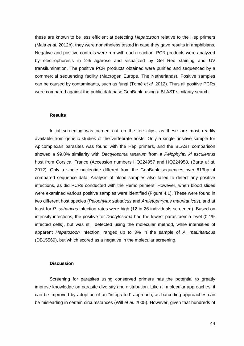
44
these are known to be less efficient at detecting Hepatozoon relative to the Hep primers
(Maia et al. 2012b), they were nonetheless tested in case they gave results in amphibians.
Negative and positive controls were run with each reaction. PCR products were analyzed
by electrophoresis in 2% agarose and visualized by Gel Red staining and UV
transilumination. The positive PCR products obtained were purified and sequenced by a
commercial sequencing facility (Macrogen Europe, The Netherlands). Positive samples
can be caused by contaminants, such as fungi (Tomé et al. 2012). Thus all positive PCRs
were compared against the public database GenBank, using a BLAST similarity search.
Results
Initial screening was carried out on the toe clips, as these are most readily
available from genetic studies of the vertebrate hosts. Only a single positive sample for
Apicomplexan parasites was found with the Hep primers, and the BLAST comparison
showed a 99.8% similarity with Dactylosoma ranarum from a Pelophylax kl esculentus
host from Corsica, France (Accession numbers HQ224957 and HQ224958, (Barta et al.
2012). Only a single nucleotide differed from the GenBank sequences over 613bp of
compared sequence data. Analysis of blood samples also failed to detect any positive
infections, as did PCRs conducted with the Hemo primers. However, when blood slides
were examined various positive samples were identified (Figure 4.1). These were found in
two different host species (Pelophylax saharicus and Amietophrynus mauritanicus), and at
least for P. saharicus infection rates were high (12 in 26 individuals screened). Based on
intensity infections, the positive for Dactylosoma had the lowest parasitaemia level (0.1%
infected cells), but was still detected using the molecular method, while intensities of
apparent Hepatozoon infection, ranged up to 3% in the sample of A. mauritanicus
(DB15569), but which scored as a negative in the molecular screening.
Discussion
Screening for parasites using conserved primers has the potential to greatly
improve knowledge on parasite diversity and distribution. Like all molecular approaches, it
can be improved by adoption of an “integrated” approach, as barcoding approaches can
be misleading in certain circumstances (Will et al. 2005). However, given that hundreds of

45
samples can be quickly assessed to give a rapid first overview of parasite prevalence, and
at the same time can sometimes detect unexpected forms, screening is likely to become
more and more common in the future. Given this, it becomes essential to know which
primers can be used in different circumstances.
For most studies of Hepatozoon, the Hep primers (Ujvari et al. 2004) have proven
to be efficient at detecting not only divergent Hepatozoon lineages (e.g. Harris et al.
2012), but also various other parasites. In amphibians they have detected at least two
unrelated lineages, from the frog host Pelophylax perezi and from caecilians of the genus
Grandisonia (Harris et al. 2013c, in press respectively). Most studies have indicated that
identification efficiency was at least as high, or even higher, when compared to
assessment of blood smears (e.g. O’Dwyer et al. 2013). Yet in this study they failed to
amplify any Hepatozoon, which were clearly identified in at least two of the species
screened. One possibility is that toe clips are not ideal sources of material for studies of
Figure 2.1 –Images of positive Hepatozoon infections in A) A. mauritanicus, and B) P.
saharicus, both of which failed to amplify using the screening protocol employed, C) a positive
infection of presumed Dactylosoma ranarum in Pelophylax saharicus, and D) a typical negative
sample. The scale bar corresponds to 20μm.

46
these parasites in amphibians. However, we also did not detect any parasites in blood
drops stored in Whatman paper, which implies that the source of the host sample was not
the issue. Rather the primers used failed to amplify Hepatozoon lineages in these hosts.
This is unexpected, given that they have amplified Hepatozoon from P. perezi in the
Azores islands, but not P. perezi or the related P. saharicus in the Iberian Peninsula and
N. Africa. On the other hand, it is clear that many different Hepatozoon lineages can be
found in the same intermediate host species (e.g. Tomé et al. 2013). It is possible that
some common Hepatozoon are not detected with these primers, while other lineages
which occasionally occur are amplified. Parasites can sometimes be overlooked, in both
molecular screening and under the microscope, when parasitaemia levels are very low.
This might explain why we detected Dactylosoma but not Hepatozoon, if parasitaemia
levels were much lower in the latter. However, our assessment of parasitaemia levels
indicates the opposite, with a very low level for Dactylosoma (just 0.1% of erythrocytes
infected, an order of magnitude lower than some infection rates with Hepatozoon). The
identity of the Hepatozoon identified under the microscope remains unclear. Forty two
Hepatozoon have been associated with amphibian hosts (Smith 1996), and since in much
of the earlier literature the hosts were not identified below the generic level, previous
identifications of Hepatozoon from toads and frogs from North Africa may well correspond
to P. saharicus and A. mauritanicus.
The finding of Dactylosoma in one sample of P. saharicus increases the list of
parasites that have now been detected using molecular screening with these primers. The
very high similarity with the sample from GenBank indicates it is very likely to be D.
ranarum, and implies low intermediate host specificity for this parasite. Indeed, D.
ranarum is thought to occur in amphibians from Africa and North and South America, Asia
and Europe (Barta 1991). Genetic data from hosts from other regions will be invaluable in
assessing this further.
To conclude, although these primers have been used in screening various
mammal and reptile hosts for Hepatozoon, they do not appear to be useful for
amphibians. However, they did detect Dactylosoma, and may be useful for screening for
these parasites. Hepatozoon were detected in P. saharicus and A. mauritanicus in
Morocco using microscopy. New primers need to be developed to amplify in these
amphibian parasites, so that they can be appropriately identified and placed in a
phylogenetic framework.
47
Acknowledgements
This work forms part of the MSc thesis of J.S-B., supervised by DJH. JPMCM is
supported by a Fundação para a Ciência e a Tecnologia (FCT) PhD grant
(SFRH/BD/74305/2010) and co-financed by FSE and POPH and EU. DJH is partially
supported by FEDER through the COMPETE program and the Project “Genomics and
Evolutionary Biology” cofinanced by North Portugal Regional Operational Programme
2007/2013 (ON.2 – O Novo Norte), under the National Strategic Reference Framework
(NSRF), through the European Regional Development Fund (ERDF). Fieldwork in North
Africa was supported by the Percy Sladen fund (to DJH) and by the Chicago
Herpetological Society (to JPMCM). The authors are grateful to their many colleagues
who contributed samples for this study.
References
The bibliography used in this chapter can be found in the References chapter of
this thesis

48
4 Chapter four
Putative Ichthyophthirius identified in the amphibian Bufo calamita through
molecular screening.
Article published in 2013 in BULLETIN OF THE EUROPEAN ASSOCIATION
OF FISH PATHOLOGISTS 33 (1), 24-27)

49
Note
Putative Ichthyophthirius identified in the amphibian Bufo calamita through
molecular screening.
D. J. Harris1 2, J. Seabra-Babo4, J. Tavares4 and J. P. M. C. Maia1 2 3
1CIBIO-UP, Centro de Investigação em Biodiversidade e Recursos Genéticos da
Universidade do Porto, Campus Agrário de Vairão, 4485-661 Vairão, Portugal;
2Departamento de Biologia, Faculdade de Ciências, Universidade do Porto, Rua
do Campo Alegre FC4, 4169-007 Porto, Portugal;
3Institut de Biologia Evolutiva (CSIC-UPF). Passeig Marítim de la Barceloneta, 37-
49. 08003 Barcelona. Spain;
4Departamento de Biologia, Universidade de Aveiro, Campus Universitário de
Santiago, 3810-193 Aveiro, Portugal.
Abstract
The protozoan parasite Ichthyopthirius multifiliis is an important pathogen of many
fish species. During molecular screening of 18S rRNA gene, we identified an apparent
Ichthyopthirius from the amphibian Bufo calamita, to our knowledge the first such
detection reported from a wild amphibian. This has important implications for parasite
control approaches in aquaculture.
Protistans are one of the most important groups of pathogens of fish (Scholz
1999). Of these, one of the most pathogenic protozoan parasites of freshwater fish is the
ciliate Ichthyophthirius multifiliis, which causes Ichthyopthiriasis, or “white spot disease”
(Dickerson 2006). This is a major problem in aquaculture, compounded by its
cosmopolitan distribution, having been reported from all regions where fish are cultivated.
Furthermore, it affects a wide range of fish including carp, trout, eel, catfish and
ornamental fish (Scholz 1999). Water temperature appears to be critical for disease
outbreaks, which are more common when fish are stressed and water temperature rises
(Dickerson 2006). One important, unanswered question is how does an obligate parasite
survive between outbreaks? It has been hypothesized that it is most likely that the
parasite is maintained through low level infections (Dickerson 2006).

50
Although identification of parasites was traditionally based on morphological
characters, molecular techniques are a powerful tool to identify various parasite groups in
freshwater environments. Not only can larger numbers of samples be screened quickly,
but molecular data can also give information regarding the diversity of parasites and their
phylogenetic relationships.
During routine screening of the amphibian Bufo calamita from the Iberian
Peninsula a standard protocol was followed. DNA was extracted from toe-clips using
standard high-salt methods (Sambrook et al. 1989). Primers used were HepF300 and
HepR900 (Ujvari et al. 2004), that target a part of the 18S rRNA gene. They are known to
amplify Apicomplexan parasites (Harris et al. 2012) as well as various other groups
including Stramenopiles (Maia et al. 2012a) and some fungi (Tomé et al. 2012) .PCR
conditions consisted of 35 cycles of 94ºC (30 seconds), 60ºC (30 seconds) and 72ºC (1
minute) – see Harris et al. (2011)for more details. Negative and positive controls were run
with each reaction. All positive PCRs were sequenced by a commercial service
(Macrogen Inc.). Resulting chromatograms were checked manually and assembled in
BioEdit (Hall 1995). BLAST was used to identify closest matches in GenBank.
Out of the 56 samples of B. calamita screened (10 from Castelo Branco, 5 from
Mindelo and 19 from Aveiro in Portugal, and 22 from Trabazos in Spain), two positives
(DB16921 and DB16925 from Trabazos) matched ciliates in the BLAST search (GenBank
Accession numbers KC512767 and KC512768 respectively). The first gave the closest
match with Hausmanniella discoidea (Accession number EU039900, Dunthorne et al.,
2008). The match was 98% with 7 identified differences over 531bp. Hausmanniella are
widespread protozoans, often identified in environmental assessments (e.g. Bartosŏva
and Tirjakova (2008)). However, the second ciliate gave a closest match with I. multifiliis
(Accession number U17354, Wright and Lynn (1995)). To confirm this assessment, two
different phylogenetic analyses were carried out (Maximum Likelihood and Bayesian
Inference) using the most closely related sequences available on GenBank. Maximum
Likelihood (ML) analysis included random sequence addition (100 replicate heuristic
searches), and support for nodes was estimated using the bootstrap technique
(Felsenstein 1985) with 500 replicates, using PhyML 3.0 (Guindon et al. 2010). The AIC
criteria carried out in jModeltest 0.1.1 (Posada 2008) were used to choose the model of
evolution employed (TrN+G). Bayesian analysis was implemented using Mr. Bayes 3.1
(Huelsenbeck & Ronquist 2001) with parameters estimated as part of the analysis. The
analysis was run for 5x10⁶ generations, saving one tree each 1000 generations. The log-
likelihood values of the sample point were plotted against the generation time and all the

51
trees prior to reaching stationary were discarded. Remaining trees were combined in a
50% majority consensus tree. Following , Paramecium tetraurelia (X03772) was
designated as an outgroup (Figure 3.1).
As expected, the ciliate identified from B. calamita was sister taxa to I. multifiliis with
high support. Sister taxa to this was Ophryoglena catenula (U17355, Wright and Lynn
(1995)), then two uncultured clones (EF586162 and EF586110,Dopheide et al. (2008)),
and then Tetrahymena paravorax (EF070253,Chantangsi et al. (2007)), relationships
similar to those previously proposed (Wright & Lynn 1995).
This result is important for several reasons. First, I. multifiliis is currently
considered to exclusively parasitise freshwater fish, although tadpole stages of the marsh
frog Limnodynastes peronii were successfully infected experimentally (Gleeson 1999).
Our results indicate that the parasite DNA could be detected from the clipped toe of a wild
amphibian. This does not per se demonstrate infection - during the tomont stage of the life
cycle of I. multifiliis it produces a sticky capsule that can attach to various substrates.
From this capsule theronts are released which can survive for 2-4 days while actively
searching for a host. Whether detection indicates an infection or not, clearly needs further
investigation. Secondly the sequence shows limited differentiation from the published I.
Figure 3.1 – ML tree of the apparent Ichthyophthirius from a Bufo calamita, and closest available comparative sequences from GenBank. Support for the Bayesian and for ML analysis are given above and below the nodes, respectively. The branch of Paramecium tetrawelia was shortened 75%.

52
multifiliis sequence, with 12 differences over 544 bp. It has previously been proposed that
multiple strains or even cryptic species may occur within I. multifiliis (Nigrelli et al.
1976).and our results seem to be further evidence for this with the two sequences
showing some divergence whilst clearly belonging to the same clade.
Given the importance of I. multifiliis as a parasite of freshwater fish, it becomes
imperative to ascertain if other amphibian species can also host similar parasites, and
whether these can cross infect between amphibians and fish. If amphibians are acting as
sinks for these parasites, it will be important to take this into account when trying to control
disease outbreaks. Further molecular screening of additional fish and amphibian species,
either using general primers as in this study or ones more specific for Ichthyopthirius, will
be essential to provide further information on this possibility.
JPMCM is supported by a Fundação para a Ciência e a Tecnologia (FCT) PhD
grant (SFRH/ BD/74305/2010) and co-financed by FSE and POPH and EU. Thanks to the
anonymous reviewer for their useful comments on an earlier version.
References
The bibliography used in this chapter can be found in the References chapter of
this thesis.

53
5 Chapter five
General discussion

54
General Discussion
In this chapter I will present a general discussion of the work performed. As the
two previous chapters present the papers produced during this thesis, this chapter will be
dedicated to an overall evaluation of the results. Possible explanations for the results will
be suggested, and an attempt to clarify the reasons for choosing molecular methods as
the main tool.
In the beginning of the work for this thesis we proposed to screen several tissue
samples of amphibians, with the objective of detecting parasites with a focus on
Hepatozoon species. For that 230 samples from 8 species were subjected to PCR using
Hep primers (Ujvari et al. 2004). The samples came from several locations, this would
have permitted to analyse differences between lineages of parasites detected, and also
what lineages were most common and differences between intensities. A smaller subset
of samples (47) were also tested with the Hemo1 and Hemo2 primers (Perkins & Keller
2001) and CR1 and CR2 primers (Ellis et al. 1998).
The primary use of Hep primers (Ujvari et al. 2004) come from the previous
success they demonstrated, detecting Hepatozoon in several other studies (Harris et al.
2012; Maia et al. 2012b; Tomé et al. 2012; Harris et al. 2013c). Hemo primers, although
less efficient than Hep for other groups of vertebrates (Maia et al. 2012b), were tested in
case they gave results in amphibians. The use of CR primers was justified by the
possibility of detecting other groups of parasites. The entire sample set tested with Hemo
and CR primers was negative. Nonetheless PCR reactions with Hep primers produced 27
positives. All these positives were sequenced, and since contaminants or other false
positives can be the cause of them, sequences were compared against the public
database GenBank, using a BLAST. Blast search results in only two parasites with the
majority of sequences corresponding to fungi. Fungi have been reported in other studies
with these primers (Tomé et al. 2012). The two more interesting parasites were a putative
Ichthyopthirius multifiliis detected in Bufo calamita from Castelo Branco, Portugal, and a
Dactylosoma ranarum in a Pelophylax saharicus from Morocco.
Although for fewer samples, other source of material for DNA extraction was
available, primarily blood drops from individuals that presented natural bleeding upon the
collection of the toe clip. Even though toe clips proved efficient in other studies (Harris et
al. 2013c), as different sources can influence the result of the PCR (McKenzie & Starks
2008), these samples were also tested. A total of 72 samples were available from 8
species. Again Hep primers were used, and a small number of samples (23) were tested

55
with Hemo and CR primers. All blood samples failed to detect any positive infections,
implying that the source of the material was not the problem.
With such results there were only two possible explanations: either all the
amphibian samples were free of Hepatozoon infections; or the primers fail to detect these
infections, perhaps because of genetic differences to Hepatozoon parasites found in other
vertebrate groups. Several blood smears stained with Giemsa were collected, and the
answers to the possibilities mention above reside in the observation of these smears. Fifty
one slides were available from 4 species and all were examined using using an Olympus
CX41 microscope with an in-built digital camera (SC30) (Olympus, Hamburg, Germany).
The slides were observed for 10 minutes and after, if no parasite was found they were
consider parasite free, however if parasites were observed, even in very low numbers the
slides were considered positive for infection. When positive the intensity of infection was
estimated based on numbers of parasites per 3,000 cells. From 51 slides 13 were
positive. The species with positive score were Amietophrynus mauritanicus, (10%, 1/ 10),
and Pelophylax saharicus, (46%, 12/26). In two slides hemogregarines were clearly
identified (DB15569 from Amietophrynus mauritanicus, DB14709 from Pelophylax
saharicus).
Despite the differences in sample size between the two species, an analysis to the
ecologic necessities of these species can give some clues about the differences in
prevalence. Regarding habitat, Amietophrynus mauritanicus can be encountered near
both permanent or temporary water bodies, while Pelophylax saharicus is more restricted
to permanent water bodies (Le Berre 1989; Chillasse et al. 2002). Differences are also
found regarding the feeding habits of these two species, A. mauritanicus feeds primarily of
insects, while P. saharicus feeds from insects, fishes and even other frogs (Doumergue
1901; Chillasse et al. 2002). The question is: Can these differences in feeding habits
explain the difference in prevalence numbers? Transmission by mosquito bite and trophic
transmission is known to occur in frogs (Ferguson & Smith 2012). Therefore, frogs can be
infected while serving as a meal for an infected mosquito, or while feeding from infected
preys. Taking this into account, the higher numbers presented by P. saharicus 40%
compared to A. mauritanicus, 10%, can be the reflection of infections transmitted by
insects plus the feeding of infected fishes and frogs. The apparent higher exposition to
potential parasites hosts can suggest that P. saharicus can be more susceptible to
parasite infections.
Although P. saharicus presented higher prevalence, that does not mean higher
level of pathogenicity. The impact of these parasites to their hosts can be significant,

56
however is still not determined in many species, and may vary according to the species.
Some parasite species can be tolerated with low level infections and cause severe illness
when high levels of infection is presented without being fatal for the host, while other are
highly pathogenic and result in death (Baneth 2011). Hepatozoon species that infect
amphibians form a separate clade, therefore can present unique characteristics and
implications for their hosts. Further investigation is required to assess how these parasites
are affecting amphibians and the implications of these associations.
The positive results using microscopy suggest that the primers used fail to detect
parasites. Although microscopy of gamonts is not adequate to identify the parasites to a
specific level, two identified as Hepatozoon sp. and several without classification, it
demonstrate a higher identification efficiency over PCR for these primers, at least for
amphibians samples.
This difficulty identifying hemogregarines based on morphological characters is the
main reason why the use of molecular tools has been increasing. The use of
morphological characters that are many times homoplastic in the past created a huge
controversy surrounding knowledge about parasite diversity and distribution. The lack of
information about some groups and the use of these characters to establish taxonomy
and phylogenetic relations have originated wrong taxonomic placements and phylogenies.
Although the use of molecular tools has been improving our knowledge about parasites, it
also sometimes creates conflicts with taxonomy accepted using traditional tools (Barta
2001). Molecular tools allow to process quickly large quantities of samples, and the ability
to distinguish individuals that possess similar morphological characters. However as seen
in this thesis to use molecular tools such as PCR some precautions have to be taken, and
selecting appropriated primers is fundamental.
Simply saying that the primers fail to detect Hepatozoon that were clearly visible
under the microscope is not the only explanation possible. Hep primers are known to
amplify several lineages of Hepatozoon even within the same intermediate host (Tomé et
al. 2013). Given that they were not able to detect infections in this thesis, it is possible that
the lineages identified using microscopy are different lineages from the previously
reported. The current information is not enough to establish if: either they are distant
lineages or uncommon ones, which make the primers used unable to detect them. These
hypotheses are interesting from an evolutionary perspective, and deserve attention for
future researchers. New primers and testing different genes is required to clarify these
possibilities.

57
6 Chapter six
Concluding remarks and future possibilities

58
6.1 Concluding remarks
The work developed for this thesis was an attempt to gather knowledge about
parasites from amphibians, particularly Hepatozoon. It was intended to be a large
molecular survey of the diversity and distribution of parasites in amphibians from the
Mediterranean region and North Africa. Not a single Hepatozoon infection was detected,
and only two key parasites were detected, what was unexpected since the primers used
presented good results with other samples (Harris et al. 2012; Maia et al. 2012b; Tomé et
al. 2012; Harris et al. 2013c). Nonetheless microscopy was able to detect several
infections with at least two apparently being Hepatozoon.
In the first study 56 individuals of the species Bufo calamita were screened for the
presence of parasites using specific primers, known to work with other vertebrates (Harris
et al. 2011; Maia et al. 2012b; Tomé et al. 2012). Two infections were detected in two
different individuals. Both infections appear to be ciliates, probably Hausmanniella
discoidea and Ichthyophthirius multifiliis. The first is often identified in environmental
assessments, however the second is considered to exclusively parasitize freshwater fish.
This was the first report of this parasite in a wild amphibian host. However, detection itself
may not reflect infection, since these parasites during the tomont stage of the life cycle
produce a sticky capsule that can attach to various substrates. The phylogenetic
relationships of this sequence revealed a similarity to those previously proposed for
Ichthyophthirius multifiliis (Wright & Lynn 1995), although limited differentiation from the
published sequence was showed. This suggests that multiples strains or even cryptic
species may occur within I. multifiliis, supporting the theory of Nigrelli et al. (1976).
Ichthyophthirius multifiliis is considered an obligate parasite and questions about
how the parasite survives between outbreaks are still unanswered. There has been
hypothesized that low level infections is used to maintain the parasite. However with a
finding like these further questions rise. Are amphibians acting as reservoirs for these
parasites between outbreaks? Can cross infections between amphibians and fish occur?
And what other species of amphibians are parasitized by this organism?
Before trying to answer these questions the infection should be confirmed. With
this, future work should aim at screening both fish and amphibians with these or more
specific primers. More variable markers than the one used (18S rRNA) would be
particularly valuable. The finding of this parasite in an amphibian sample should also be
taken into account when planning for disease outbreak control.
For the second study tissue and blood samples were used as well as blood
smears. 136 tissue samples from six different species of amphibians were used, 56 blood

59
drops stored in Whatman filter paper from 4 different species and 51 blood smears from 4
species also. The samples were tested with parasite specific primers and the blood slides
were examined using an Olympus CX41 microscope with an in-built digital camera.
Samples were scored as negative when no parasites were observed under the
microscope and positive when parasites were observed even in very low numbers. An
apicomplexan parasite however was detected, Dactylosoma ranarum from a Pelophylax
saharicus tissue sample. The parasite found show very high similarity with a comparable
sample from GenBank, which was reported in a Pelophylax kl esculentus, implying that
the parasite presents low intermediate host specificity. Concerning the blood smears
several slides were scored positive and two were identified as Hepatozoon. This implies
that the primers fail to detect these infections. This suggest that either these primers are
not efficient with amphibian samples or at least some of the Hepatozoon lineages of
amphibians are quite distant to the ones found in reptiles, such lizards and snakes, and so
did not amplify. Therefore new primers need to be developed to detect Hepatozoon in
amphibians. The finding of Dactylosoma using Hep primers increases the list of parasites
that have now been detected with them.
Hep primers fail to detect Hepatozoon infections in this thesis, nonetheless they
were able to detect an Apicomplexa parasite and proved efficient in other studies (Harris
et al. 2012; Maia et al. 2012b; Tomé et al. 2012; Harris et al. 2013c). As in other studies
(Harris et al. 2012; Maia et al. 2012a; Tomé et al. 2012), detection of other organisms also
occurred. This suggests that Hep primers are less specific than previously thought, which
could cause incorrect infection estimates. Therefore it is very important to validate the
PCR products amplification through the use of sequencing, comparing with published
sequences (e.g. BLAST), and performing phylogenetic analyses. Molecular screening
through PCR is a very useful tool with many advantages, nonetheless it also has
limitations as shown in this study. However, these limitations can be overcome by
optimization of the technique and development of more specific assays, and it should be
complemented with the use of traditional techniques, such as microscopy.
In general, the results obtained may lead to thinking that the used primers are not
effective for screening for hemoparasites in amphibians. However, the Hep primers allow
the detection of several different organisms, in this thesis two ciliates and an
apicomplexan. Unlike other studies (Richard et al. 2002; Durrant et al. 2006), microscopy
has proven more efficient than PCR in this thesis. This can be explained by the already
mentioned inefficiency of the primers. New primers need to be developed for routine
screen of Hepatozoon parasites in amphibians. Other methods of DNA extraction can also

60
be considered as a possible factor influencing the inefficacy of the PCR technique. Using
commercial kits or other sources of tissue can influence the outcome of the PCR.
Nonetheless these are just speculative assumptions, since the amplification of
Hepatozoon from infected amphibians was possible using commercial kit to extract DNA
from P. perezi from the Azores (Harris et al. 2013c). Trying to assess the diversity of
Hepatozoon in amphibians is required to increase the knowledge of this group. It also
would provide valuable information for phylogenetic and distribution studies. Much work is
yet to be done.
6.2 Future possibilities
The knowledge about parasite diversity is still poor. Improving the knowledge
about the Hepatozoon group will help to increase the information available to
parasitologists and will allow more accurate phylogenetic reconstructions. However more
investigation is necessary to improve this lack of information. Combining information about
different groups of hosts (snakes, lizards, amphibians, and mammals) will enhance the
overall view of the group. Also the use of samples from different location as well as from
different seasonal periods, will allow testing hypothesis about the development and
ecology of hemoparasites. Implementing new methods, like real-time PCR, which is not
only more accurate that the conventional PCR, but also allows to estimate the level of
parasitaemia, evaluate and compare differences among the type of tissue and DNA
extraction technique, factors that can influence the results.
Meanwhile, developing new primers and continue routinely screening of
amphibians parasites, both through microscopy and molecular tools, is required. More
sampling from different species and different regions are crucial. There is need to sample
a much broader range of taxa, not focusing the efforts solely on parasites of medical and
veterinary importance. Developing new protocols which use alternatives markers to the
18SrRNA, and generating multiple sequences from different genes is necessary to
improve the estimate of phylogenetic relationships and overall diversity.
Also very important is identifying the hematophagous invertebrate hosts to assess
complete life cycles for the species within the Hepatozoon group. Therefore collecting
ectoparasites is a necessity. In CIBIO a vast collection of these invertebrates has been
assemble over the years, ticks, mites, leeches and others. It is necessary to start
processing these samples, and continue to collect them during field work. Summing up,
future research should focus on improved sampling and optimization of the molecular
methods for routine parasite screen in amphibians

61
7 References

62
Adl, S.M., Leander, B.S., Simpson, A.G., Archibald, J.M., Anderson, O.R., Bass, D. et al. (2007). Diversity, nomenclature, and taxonomy of protists. Systematic biology, 56, 684-689.
Adl, S.M., Simpson, A.G., Farmer, M.A., Andersen, R.A., Anderson, O.R., Barta, J.R. et al. (2005). The new higher level classification of eukaryotes with emphasis on the taxonomy of protists. Journal of Eukaryotic Microbiology, 52, 399-451.
Aikawa, M., Miller, L., Johnson, J. & Rabbege, J. (1978). Erythrocyte entry by malarial parasites. A moving junction between erythrocyte and parasite. The Journal of Cell Biology, 77, 72-82.
Aisien, M.S.O., Nago, S.G.A. & Rodel, M.O. (2011). Parasitic infections of amphibians in the Pendjari Biosphere Reserve, Benin. Afr. Zool., 46, 340-349.
Allen, K.E., Yabsley, M.J., Johnson, E.M., Reichard, M.V., Panciera, R.J., Ewing, S.A. et al. (2011). Novel Hepatozoon in Vertebrates From the Southern United States. The Journal of parasitology, 97, 648-653.
Ball, G.H., Chao, J. & Telford, S.R., Jr. (1967). The life history of Hepatozoon rarefaciens (Sambon and Seligmann, 1907) from Drymarchon corais (Colubridae), and its experimental transfer to Constrictor constrictor (Boidae). The Journal of parasitology, 53, 897-909.
Baneth, G. (2011). Perspectives on canine and feline hepatozoonosis. Veterinary parasitology, 181, 3-11.
Baneth, G., Mathew, J.S., Shkap, V., Macintire, D.K., Barta, J.R. & Ewing, S.A. (2003). Canine hepatozoonosis: two disease syndromes caused by separate Hepatozoon spp. Trends in parasitology, 19, 27-31.
Barnabé, C., Brisse, S. & Tibayrenc, M. (2000). Population structure and genetic typing of Trypanosoma cruzi, the agent of Chagas disease: a multilocus enzyme electrophoresis approach. Parasitology, 120, 513-526.
Barta, J.R. (1991). The Dactylosomatidae. Advances in Parasitology, 30, 1-37. Barta, J.R. (2001). Molecular approaches for inferring evolutionary relationships among
protistan parasites. Veterinary parasitology, 101, 175-186. Barta, J.R. & Desser, S.S. (1984). Blood parasites of amphibians from Algonquin Park,
Ontario. J. Wildl. Dis., 20, 180-189. Barta, J.R., Ogedengbe, J.D., Martin, D.S. & Smith, T.G. (2012). Phylogenetic Position of
the Adeleorinid Coccidia (Myzozoa, Apicomplexa, Coccidia, Eucoccidiorida, Adeleorina) Inferred Using 18S rDNA Sequences. Journal of Eukaryotic Microbiology, 59, 171-180.
Bartosŏva, P. & Tirjakova, E. (2008). Diversity and ecology of ciliates (Alveolata: Ciliophora) living in the bark and decaying wood mass in Slovakia. Acta protozoologica, 47, 173-187.
Baum, J., Gilberger, T.-W., Frischknecht, F. & Meissner, M. (2008). Host-cell invasion by malaria parasites: insights from Plasmodium and Toxoplasma. Trends in parasitology, 24, 557-563.
Beck, H.P., Blake, D., Darde, M.L., Felger, I., Pedraza-Diaz, S., Regidor-Cerrillo, J. et al. (2009). Molecular approaches to diversity of populations of apicomplexan parasites. International journal for parasitology, 39, 175-189.
Beebee, T.J.C. & Griffiths, R.A. (2005). The amphibian decline crisis: A watershed for conservation biology? Biological Conservation, 125, 271-285.
Beukema, W., De Pous, P., Donaire-Barroso, D., Bogaert, S., Garcia-Porta, J., Escoriza, D. et al. (2013). Review of the systematics, distribution, biogeography and natural history of Moroccan amphibians. Zootaxa, 3661, 1-60.
Black, C.G., Wu, T., Wang, L., Topolska, A.E. & Coppel, R.L. (2005). MSP8 is a non-essential merozoite surface protein in Plasmodium falciparum. Molecular and Biochemical Parasitology, 144, 27-35.

63
Boulianne, B., Evans, R.C. & Smith, T.G. (2007). Phylogenetic analysis of Hepatozoon species (Apicomplexa: Adeleorina) infecting frogs of Nova Scotia, Canada, determined by ITS-1 sequences. The Journal of parasitology, 93, 1435-1441.
Bush, A.O., Fernández, J.C., Esch, G.W. & Seed, J.R. (2001). Parasitism: The Diversity and Ecology of Animal Parasites. Cambridge University Press.
Bushek, D., Dungan, C.F. & Lewitus, A.J. (2002). Serological Affinities of the Oyster Pathogen Perkinsus marinus (Apicomplexa) with Some Dinoflagellates (Dinophyceae). Journal of Eukaryotic Microbiology, 49, 11-16.
Chantangsi, C., Lynn, D.H., Brandl, M.T., Cole, J.C., Hetrick, N. & Ikonomi, P. (2007). Barcoding ciliates: a comprehensive study of 75 isolates of the genus Tetrahymena. International Journal of Systematic and Evolutionary Microbiology, 57, 2412-2425.
Chillasse, L., Dakki, M. & Th+evenot, M. (2002). Régimes alimentaires de deux espèces de Bufonidae (Bufo bufo spinosus et Bufo mauritanicus) au lac Aguelmam Azegza (Maroc). International Society for the Study and Conservation of Amphibians, Paris, FRANCE.
Criado-Fornelio, A., Buling, A., Cunha-Filho, N.A., Ruas, J.L., Farias, N.A., Rey-Valeiron, C. et al. (2007). Development and evaluation of a quantitative PCR assay for detection of Hepatozoon sp. Veterinary parasitology, 150, 352-356.
Cui, L., Fan, Q., Hu, Y., Karamycheva, S.A., Quackenbush, J., Khuntirat, B. et al. (2005). Gene discovery in Plasmodium vivax through sequencing of ESTs from mixed blood stages. Molecular and Biochemical Parasitology, 144, 1-9.
Daszak, P., Cunningham, A.A. & Hyatt, A.D. (2003). Infectious disease and amphibian population declines. Diversity and Distributions, 9, 141-150.
de la Parte-Pérez, M.A., Bruzual, E., Brito, A. & Hurtado, M.d.P. (2005). Cryptosporidium spp. y Criptosporidiosis. Revista de la Sociedad Venezolana de Microbiología, 25, 06-14.
de Waal, T. (2012). Advances in diagnosis of protozoan diseases. Veterinary parasitology, 189, 65-74.
Desser, S.S., Hong, H. & Martin, D.S. (1995). The life history, ultrastructure, and experimental transmission of Hepatozoon catesbianae n. comb., an apicomplexan parasite of the bullfrog, Rana catesbeiana and the mosquito, Culex territans in Algonquin Park, Ontario. The Journal of parasitology, 81, 212-222.
Dickerson, H.W. (2006). Ichthyopthirius multifiliis and Cryptocaryon irritans (Phylum Ciliophora). In: Fish Disease and Disorders (ed. P.T.K. Woo, E). CAB International UK.
Dobson, A., Lafferty, K.D., Kuris, A.M., Hechinger, R.F. & Jetz, W. (2008). Homage to Linnaeus: How many parasites? How many hosts? Proceedings of the National Academy of Sciences, 105, 11482-11489.
Dopheide, A., Lear, G., Stott, R. & Lewis, G. (2008). Molecular characterization of ciliate diversity in stream biofilms. Applied and Environmental Microbiology, 74, 1740-1747.
Doumergue, F. (1901). Essai sur la faune erpétologique de l'Oranie : avec des tableaux analytiques et des notions pour la détermination de tous les reptiles & batraciens du Maroc, de l'Algérie et de la Tunisie / F. Doumergue. L. Fouque, Oran :.
Douzery, E.J.P., Snell, E.A., Bapteste, E., Delsuc, F. & Philippe, H. (2004). The timing of eukaryotic evolution: Does a relaxed molecular clock reconcile proteins and fossils? Proceedings of the National Academy of Sciences, 101, 15386-15391.
Dubey, J.P., Schares, G. & Ortega-Mora, L.M. (2007). Epidemiology and Control of Neosporosis and Neospora caninum. Clinical Microbiology Reviews, 20, 323-367.
Dunn, R.R., Harris, N.C., Colwell, R.K., Koh, L.P. & Sodhi, N.S. (2009). The sixth mass coextinction: are most endangered species parasites and mutualists? Proceedings of the Royal Society B: Biological Sciences, 276, 3037-3045.

64
Durrant, K.L., Beadell, J.S., Ishtiaq, F., Graves, G.R., Olson, S.L., Gering, E. et al. (2006). Avian Haematozoa in South America: A Comparison of Temperate and Tropical Zones. Ornithological Monographs, 60, 98-111.
Ellis, J.T., Amoyal, G., C. Ryce, C., Harper, P.A.W., Clough, K.A., Homan, W.L. et al. (1998). Comparison of the large subunit ribosomal DNA of Neospora and Toxoplasma and development of a new genetic marker for their differentiation based on the D2 domain. Molecular and Cellular Probes, 1-13.
Escalante, A.A. & Ayala, F.J. (1995). Evolutionary origin of Plasmodium and other Apicomplexa based on rRNA genes. Proceedings of the National Academy of Sciences, 92, 5793-5797.
Fast, N.M., Xue, L., Bingham, S. & Keeling, P.J. (2002). Re-examining alveolate evolution using multiple protein molecular phylogenies. Journal of Eukaryotic Microbiology, 49, 30-37.
Felsenstein, J. (1985). Confidence limits on phylogenies: an approach using the bootstrap.
Ferguson, L.V. & Smith, T.G. (2012). Reciprocal Trophic Interactions and Transmission of Blood Parasites between Mosquitoes and Frogs. Insects, 3, 410-423.
Frölich, S., Entzeroth, R. & Wallach, M. (2012). Comparison of Protective Immune Responses to Apicomplexan Parasites. Journal of Parasitology Research, volume 2012.
Gasser, R.B. (1999). PCR-based technology in veterinary parasitology. Veterinary parasitology, 84, 229-258.
Gasser, R.B. (2006). Molecular tools—advances, opportunities and prospects. Veterinary parasitology, 136, 69-89.
Geller, J., Meyer, C., Parker, M. & Hawk, H. (2013). Redesign of PCR primers for mitochondrial cytochrome c oxidase subunit I for marine invertebrates and application in all-taxa biotic surveys. Molecular Ecology Resources, 13, 851-861.
Gleeson, D.J. (1999). Experimental infection of striped marshfrog tadpoles (Limnodynastes peronii) by Ichthyophthirius multifiliis. The Journal of parasitology, 85, 568-570.
Guindon, S., Dufayard, J.-F., Lefort, V., Anisimova, M., Hordijk, W. & Gascuel, O. (2010). New Algorithms and Methods to Estimate Maximum-Likelihood Phylogenies: Assessing the Performance of PhyML 3.0. Systematic biology, 59, 307-321.
Gupta, D.K., Gupta, N. & Gangwar, R. (2012). Infectivity of Bufo melanostictus (Amphibia: Bufonidae) to Two New Species of Haematozoan Parasites from Rohilkhand, India. Proceedings of the Zoological Society, 65, 22-32.
Hall, T.A. (1995). BioEdit: a user-friendly biological sequence alignment editor and analysis program for Windows 95/98/NT. Nucleic Acids Symposium Series, 41, 95-98.
Harkness, L.M., Drohan, A.E., Dickson, C.M. & Smith, T.G. (2009). Experimental Transmission of Hepatozoon clamatae (Apicomplexa: Adeleida) to the Wood Frog, Rana sylvatica, and to the Mosquito Culex pipiens. The Journal of parasitology, 96, 434-436.
Harris, D.J., Graciá, E., Jorge, F., Maia, J.P.M.C., Perera, A., Carretero, M.A. et al. (2013a). Molecular Detection of Hemolivia (Apicomplexa: Haemogregarinidae) from Ticks of North African Testudo graeca (Testudines: Testudinidae) and an Estimation of Their Phylogenetic Relationships Using 18S rRNA Sequences. Comparative Parasitology, 80, 292-296.
Harris, D.J., Maia, J., J. P. M. C. & Perera, A. (2011). Molecular characterization of Hepatozoon species in reptiles from the Seychelles. The Journal of parasitology, 97, 106-110.

65
Harris, D.J., Maia, J., J. P. M. C. & Perera, A. (2012). Molecular Survey of Apicomplexa in Podarcis Wall Lizards Detects Hepatozoon, Sarcocystis and Eimeria species. The Journal of parasitology, 98, 592-597.
Harris, D.J., Seabra-Babo, J., Tavares, J. & Maia, J. (2013b). Putative Ichthyophthirius identified in the amphibian Bufo calamita through molecular screening. Bull. Eur. Assoc. Fish Pathol., 33, 24-27.
Harris, D.J., Spigonardi, M.P., Maia, J.P. & Cunha, R.T. (2013c). Molecular survey of parasites in introduced Pelophylax perezi (Ranidae) water frogs in the Azores. Acta Parasitologica, 58.
Hartigan, A., Dhand, N.K., Rose, K., Slapeta, J. & Phalen, D.N. (2012a). Comparative Pathology and Ecological Implications of Two Myxosporean Parasites in Native Australian Frogs and the Invasive Cane Toad. PloS one, 7.
Hartigan, A., Peacock, L., Rosenwax, A., Phalen, D.N. & Slapeta, J. (2012b). Emerging myxosporean parasites of Australian frogs take a ride with fresh fruit transport. Parasites & Vectors, 5.
Hikosaka, K., Kita, K. & Tanabe, K. (2013). Diversity of mitochondrial genome structure in the phylum Apicomplexa. Molecular and Biochemical Parasitology, 188, 26-33.
Hikosaka, K., Watanabe, Y., Tsuji, N., Kita, K., Kishine, H., Arisue, N. et al. (2010). Divergence of the mitochondrial genome structure in the apicomplexan parasites, Babesia and Theileria. Molecular biology and evolution, 27, 1107-1116.
Hill, D. & Dubey, J.P. (2002). Toxoplasma gondii: transmission, diagnosis and prevention. Clinical Microbiology and Infection, 8, 634-640.
Hornok, S., Tánczos, B., Fernández de Mera, I.G., de la Fuente, J., Hofmann-Lehmann, R. & Farkas, R. (2013). High prevalence of Hepatozoon-infection among shepherd dogs in a region considered to be free of Rhipicephalus sanguineus. Veterinary parasitology, 196, 189-193.
Huelsenbeck, J.P. & Ronquist, F. (2001). MRBAYES: Bayesian inference of phylogeny. Bioinformatics, 17, 754-755.
Imam, T.S. (2009). The complexities in the classification of protozoa: A challenge to parasitologists. Bayero Journal of Pure and Applied Sciences, 2, 159-164.
Janouškovec, J., Horák, A., Oborník, M., Lukeš, J. & Keeling, P.J. (2010). A common red algal origin of the apicomplexan, dinoflagellate, and heterokont plastids. Proceedings of the National Academy of Sciences.
Jarvi, S.I., Schultz, J.J. & Atkinson, C.T. (2002). PCR Diagnostics underestimate the prevalence of avian malaria (Plasmodium relictum) in experimentally infected passerines. The Journal of parasitology, 88, 153-158.
Jovani, R., Amo, L., Arriero, E., Krone, O., Marzal, A., Shurulinkov, P. et al. (2004). Double gametocyte infections in apicomplexan parasites of birds and reptiles. Parasitology research, 94, 155-157.
Kaasch, A.J. & Joiner, K.A. (2000). Protein-targeting determinants in the secretory pathway of apicomplexan parasites. Current Opinion in Microbiology, 3, 422-428.
Karagenc, T.I., Pasa, S., Kirli, G., Hosgor, M., Bilgic, H.B., Ozon, Y.H. et al. (2006). A parasitological, molecular and serological survey of Hepatozoon canis infection in dogs around the Aegean coast of Turkey. Veterinary parasitology, 135, 113-119.
Karanis, P. & Ongerth, J. (2009). LAMP – a powerful and flexible tool for monitoring microbial pathogens. Trends in parasitology, 25, 498-499.
Kim, B., Smith, T.G. & Desser, S.S. (1998). The life history and host specificity of Hepatozoon clamatae (Apicomplexa: Adeleorina) and ITS-1 nucleotide sequence variation of Hepatozoon species of frogs and mosquitoes from Ontario. The Journal of parasitology, 84, 789-797.
Kopecna, J., Jirku, M., Obornik, M., Tokarev, Y.S., Lukes, J. & Modry, D. (2006). Phylogenetic analysis of coccidian parasites from invertebrates: search for missing links. Protist, 157, 173-183.

66
Koprivnikar, J., Marcogliese, D.J., Rohr, J.R., Orlofske, S.A., Raffel, T.R. & Johnson, P.T.J. (2012). Macroparasite Infections of Amphibians: What Can They Tell Us? EcoHealth, 9, 342-360.
Kuvardina, O.N., Leander, B.S., Aleshin, V.V., Myl’Nikov, A.P., Keeling, P.J. & Simdyanov, T.G. (2002). The Phylogeny of Colpodellids (Alveolata) Using Small Subunit rRNA Gene Sequences Suggests They are the Free-living Sister Group to Apicomplexans. Journal of Eukaryotic Microbiology, 49, 498-504.
Landsberg, J.H., Kiryu, Y., Tabuchi, M., Waltzek, T.B., Enge, K.M., Reintjes-Tolen, S. et al. (2013). Co-infection by alveolate parasites and frog virus 3-like ranavirus during an amphibian larval mortality event in Florida, USA. Diseases of aquatic organisms, 105, 89-99.
Le Berre, C. (1989). Faune du Sahara 1. Poissons-Amphibiens-Reptiles. Raymond Chabaud–Lechevallier, Paris.
Leander, B.S. (2003). Phylogeny of gregarines (Apicomplexa) as inferred from small-subunit rDNA and beta-tubulin. International Journal of Systematic and Evolutionary Microbiology, 53, 345-354.
Leander, B.S. (2008). Marine gregarines: evolutionary prelude to the apicomplexan radiation? Trends in parasitology, 24, 60-67.
Leander, B.S. & Keeling, P.J. (2004). Early Evolutionary History of Dinoflagellates and Apicomplexans (Alveolata) as Inferred from Hsp90 and Actin Phylogenies1. Journal of Phycology, 40, 341-350.
Leander, B.S., Kuvardina, O.N., Aleshin, V.V., Mylnikov, A.P. & Keeling, P.J. (2003). Molecular phylogeny and surface morphology of Colpodella edax (Alveolata): insights into the phagotrophic ancestry of apicomplexans. Journal of Eukaryotic Microbiology, 50, 334-340.
Lèger, L. (1911). Caryospora simplex, coccidie monospore`e et la classification des coccides. Archiv fu¨r Protistenkunde, 22, 71-88.
Levine, N.D. (1973). Protozoan parasites of domestic animals and of man. Burgess. Levine, N.D. (1988). The protozoan phylum Apicomplexa. 1 (1988). Taylor & Francis
Group. Li, Y.M., Cohen, J.M. & Rohr, J.R. (2013). Review and synthesis of the effects of climate
change on amphibians. Integrative Zoology, 8, 145-161. Lunde, K.B. & Johnson, P.T.J. (2012). A Practical Guide for the Study of Malformed
Amphibians and Their Causes. Journal of Herpetology, 46, 429-441. Maia, J.P., Gomez-Diaz, E. & Harris, D.J. (2012a). Apicomplexa primers amplify
Proteromonas (Stramenopiles, Slopalinida, Proteromonadidae) in tissue and blood samples from lizards. Acta Parasitologica, 57, 337-341.
Maia, J.P., Harris, D.J. & Perera, A. (2011). Molecular survey of Hepatozoon species in lizards from North Africa. The Journal of parasitology, 97, 513-517.
Maia, J.P., Perera, A. & Harris, D.J. (2012b). Molecular survey and microscopic examination of Hepatozoon Miller, 1908 (Apicomplexa: Adeleorina) in lacertid lizards from the western Mediterranean. Folia Parasitologica, 59, 241-248.
Massimine, K.M., Doan, L.T., Atreya, C.A., Stedman, T.T., Anderson, K.S., Joiner, K.A. et al. (2005). Toxoplasma gondii is capable of exogenous folate transport. A likely expansion of the BT1 family of transmembrane proteins. Molecular and Biochemical Parasitology, 144, 44-54.
Mata-López, R., León-Règagnon, V. & García-Prieto, L. (2012). Helminth Infracommunity Structure of Leptodactylus melanonotus (Anura) in Tres Palos, Guerrero, and Other Records for This Host Species in Mexico. The Journal of parasitology, 99, 564-569.
Mathew, J.S., Van Den Bussche, R.A., Ewing, S.A., Malayer, J.R., Latha, B.R. & Panciera, R.J. (2000). Phylogenetic relationships of Hepatozzon (Apicomplexa:
67
Adeleorina) based on molecular, morphological, and life-cycle characters. The Journal of parasitology, 86, 366-372.
McKenzie, V.J. & Starks, H.A. (2008). Blood Parasites of Two Costa Rican Amphibians with Comments on Detection and Microfilaria Density Associated with Adult Filarial Worm Intensity. The Journal of parasitology, 94, 824-829.
Moço, T., Silva, R., Madeira, N., dos Santos Paduan, K., Rubini, A., Leal, D. et al. (2012). Morphological, morphometric, and molecular characterization of Hepatozoon spp. (Apicomplexa, Hepatozoidae) from naturally infected Caudisona durissa terrifica (Serpentes, Viperidae). Parasitology research, 110, 1393-1401.
Moore, R.B., Oborník, M., Janouškovec, J., Chrudimský, T., Vancová, M., Green, D.H. et al. (2008). A photosynthetic alveolate closely related to apicomplexan parasites. Nature, 451, 959-963.
Morrison, D.A. (2009). Evolution of the Apicomplexa: where are we now? Trends in parasitology, 25, 375-382.
Morrison, D.A. & Ellis, J.T. (1997). Effects of nucleotide sequence alignment on phylogeny estimation: a case study of 18S rDNAs of apicomplexa. Molecular biology and evolution, 14, 428-441.
Morrissette, N.S. & Sibley, L.D. (2002). Cytoskeleton of apicomplexan parasites. Microbiology and Molecular Biology Reviews, 66, 21-38.
Muller, J. & Hemphill, A. (2013). In vitro culture systems for the study of apicomplexan parasites in farm animals. International journal for parasitology, 43, 115-124.
Murata, T., Inoue, M., Tateyama, S., Taura, Y. & Nakama, S. (1993). Vertical transmission of Hepatozoon canis in dogs. The Journal of Veterinary Medical Science, 55, 867-868.
Ndao, M. (2009). Diagnosis of parasitic diseases: old and new approaches. Interdisciplinary perspectives on infectious diseases, 2009, 278246.
Nigrelli, R.F., Pokorny, K.S. & Ruggieri, G.D. (1976). Notes on Ichthyophthirius multifiliis, a ciliate parasitic on freshwater fishes, with some remarks on possible physiological races and species. . Transactions of the American Microscopical Society 95, 607-613.
O’Dwyer, L.H., Moço, T.C., Paduan, K.d.S., Spenassatto, C., da Silva, R.J. & Ribolla, P.E.M. (2013). Description of three new species of Hepatozoon (Apicomplexa, Hepatozoidae) from Rattlesnakes (Crotalus durissus terrificus) based on molecular, morphometric and morphologic characters. Experimental Parasitology, 135, 200-207.
Orlofske, S.A., Belden, L.K. & Hopkins, W.A. (2013). Larval wood frog (Rana =Lithobates sylvatica) development and physiology following infection with the trematode parasite, Echinostoma trivolvis. Comparative Biochemistry and Physiology a-Molecular & Integrative Physiology, 164, 529-536.
Paul, R.E.L., Ariey, F. & Robert, V. (2003). The evolutionary ecology of Plasmodium. Ecology Letters, 6, 866-880.
Pedersen, A.B. & Fenton, A. (2007). Emphasizing the ecology in parasite community ecology. Trends in ecology & evolution, 22, 133-139.
Perkins, S.L. & Keller, A.K. (2001). Phylogeny of nuclear small subunit rRNA genes of hemogregarines amplified with specific primers. The Journal of parasitology, 870-876.
Perkins, S.L., Martinsen, E.S. & Falk, B.G. (2011). Do molecules matter more than morphology? Promises and pitfalls in parasites. Parasitology, 138, 1664-1674.
Pinto, C.M., Helgen, K.M., Fleischer, R.C. & Perkins, S.L. (2012). Hepatozoon Parasites (Apicomplexa: Adeleorina) in Bats. The Journal of parasitology, 99, 722-724.
Posada, D. (2008). jModelTest: phylogenetic model averaging. Mol Biol Evol. 2008 Jul;25(7):1253-6. doi: 10.1093/molbev/msn083. Epub 2008 Apr 8.

68
Poulin, R. & Morand, S. (2000). The diversity of parasites. The Quarterly Review of Biology, 75, 277-293.
Preston, D.L., Orlofske, S.A., Lambden, J.P. & Johnson, P.T.J. (2013). Biomass and productivity of trematode parasites in pond ecosystems. Journal of Animal Ecology, 82, 509-517.
Readel, A.M. & Goldberg, T.L. (2009). Blood Parasites of Frogs From an Equatorial African Montane Forest in Western Uganda. The Journal of parasitology, 96, 448-450.
Richard, F.A., Sehgal, R.N.M., Jones, H.I. & Smith, T.B. (2002). A Comparative Analysis of PCR-Based Detection Methods for Avian Malaria. The Journal of parasitology, 88, 819-822.
Rueckert, S. & Leander, B.S. (2008). Morphology and phylogenetic position of two novel marine gregarines (Apicomplexa, Eugregarinorida) from the intestines of North-eastern Pacific ascidians. Zoologica Scripta, 37, 637-645.
Rueckert, S., Simdyanov, T.G., Aleoshin, V.V. & Leander, B.S. (2011). Identification of a Divergent Environmental DNA Sequence Clade Using the Phylogeny of Gregarine Parasites (Apicomplexa) from Crustacean Hosts. PloS one, 6, e18163.
Saffo, M.B., McCoy, A.M., Rieken, C. & Slamovits, C.H. (2010). Nephromyces, a beneficial apicomplexan symbiont in marine animals. Proceedings of the National Academy of Sciences, 107, 16190-16195.
Saldarriaga, J.F., McEwan, M.L., Fast, N.M., Taylor, F.J.R. & Keeling, P.J. (2003). Multiple protein phylogenies show that Oxyrrhis marina and Perkinsus marinus are early branches of the dinoflagellate lineage. International Journal of Systematic and Evolutionary Microbiology, 53, 355-365.
Sambrook, J., Fritsch, E.F. & Maniatis, T. (1989). Molecular Cloning: A Laboratory Manual. Cold Spring Harbour Press, New York.
Sato, S. (2011). The apicomplexan plastid and its evolution. Cellular and Molecular Life Sciences, 68, 1285-1296.
Scholz, T. (1999). Parasites in cultured and feral fish. Veterinary parasitology, 84, 317-335.
Sharman, P.A., Smith, N.C., Wallach, M.G. & Katrib, M. (2010). Chasing the golden egg: vaccination against poultry coccidiosis. Parasite Immunology, 32, 590-598.
Shirley, M.W. (1975). Enzyme variation in Eimeria species of the chicken. Parasitology, 71, 369-376.
Siddall, M.E., Reece, K.S., Nerad, T.A. & Burreson, E.M. (2001). Molecular Determination of the Phylogenetic Position of a Species in the Genus Colpodella (Alveolata). American Museum Novitates, 1-12.
Šlapeta, J. (2013). Ten simple rules for describing a new (parasite) species. International Journal for Parasitology: Parasites and Wildlife, 2, 152-154.
Šlapeta, J. & Morin-Adeline, V. (2011). Apicomplexa Levine 1970. Sporozoa Leucart 1879. Version 18 May 2011. http://tolweb.org/Apicomplexa/2446/2011.05.18 in The Tree of Life Web Project, http://tolweb.org/.
Sloboda, M., Kamler, M., Bulantova, J., Votypka, J. & Modry, D. (2008). Rodents as intermediate hosts of Hepatozoon ayorgbor (Apicomplexa: Adeleina: Hepatozoidae) from the African ball python, Python regius? Folia Parasitologica, 55, 13-16.
Smith, T. & Desser, S. (1997). Phylogenetic analysis of the genus Hepatozoon Miller, 1908 (Apicomplexa: Adeleorina). Syst Parasitol, 36, 213-221.
Smith, T.G. (1996). The genus Hepatozoon (Apicomplexa: Adeleina). The Journal of parasitology, 82, 565-585.
Smith, T.G. (1998). Life histories, ultrastructure, molecular biology and phylogenetic relationships of Hepatozoon species, Phylum Apicomplexa: Suborder adeleorina. Ottawa: National Library of Canada Biblioth ue nationale du Canada.

69
Smith, T.G., Desser, S.S. & Martin, D.S. (1994). The development of Hepatozoon sipedon sp. nov. (Apicomplexa: Adeleina: Hepatozoidae) in its natural host, the Northern water snake (Nerodia sipedon sipedon), in the culicine vectors Culex pipiens and C. territans, and in an intermediate host, the Northern leopard frog (Rana pipiens). Parasitology research, 80, 559-568.
Smith, T.G., Kim, B. & Desser, S.S. (1999). Phylogenetic relationships among Hepatozoon species from snakes, frogs and mosquitoes of Ontario, Canada, determined by ITS-1 nucleotide sequences and life-cycle, morphological and developmental characteristics. International journal for parasitology, 29, 293-304.
Smith, T.G., Kim, B., Hong, H. & Desser, S.S. (2000). Intraerythrocytic development of species of hepatozoon infecting ranid frogs: evidence for convergence of life cycle characteristics among apicomplexans. The Journal of parasitology, 86, 451-458.
Stenberg, P.L. & Bowerman, W.J. (2010). First report of Hepatozoon sp. in the Oregon Spotted Frog, Rana pretiosa. J. Wildl. Dis., 46, 956-960.
Su, C., Shwab, E.K., Zhou, P., Zhu, X.Q. & Dubey, J.P. (2010). Moving towards an integrated approach to molecular detection and identification of Toxoplasma gondii. Parasitology, 137, 1-11.
Szuroczki, D. & Richardson, J.M.L. (2012). The Behavioral Response of Larval Amphibians (Ranidae) to Threats from Predators and Parasites. PloS one, 7.
Telford, S.R. (2009). Hemoparasites of the Reptilia: Color Atlas and Text. Taylor & Francis.
Tomé, B., Maia, J.P. & Harris, D.J. (2013). Molecular Assessment of Apicomplexan Parasites in the Snake Psammophis from North Africa: Do Multiple Parasite Lineages Reflect the Final Vertebrate Host Diet? The Journal of parasitology, 99, 883-887.
Tomé, B., Maia, J.P.M.C. & Harris, D.J. (2012). Hepatozoon Infection Prevalence in Four Snake Genera: Influence of Diet, Prey Parasitemia Levels, or Parasite Type? The Journal of parasitology, 98, 913-917.
Ujvari, B., Madsen, T. & Olsson, M. (2004). High prevalence of Hepatozoon spp. (Apicomplexa, Hepatozoidae) infection in water pythons (Liasis fuscus) from tropical Australia. The Journal of parasitology, 670-672.
Valkiūnas, G., Iezhova, T.A., Križanauskienė, A., Palinauskas, V., Sehgal, R.N.M. & Bensch, S. (2008). A Comparative Analysis of Microscopy and PCR-Based Detection Methods for Blood Parasites. The Journal of parasitology, 94, 1395-1401.
Viana, L.A., Soares, P., Silva, J.E., Paiva, F. & Coutinho, M.E. (2012). Anurans as paratenic hosts in the transmission of Hepatozoon caimani to caimans Caiman yacare and Caiman latirostris. Parasitology research, 110, 883-886.
Walker, G., Dorrell, R.G., Schlacht, A. & Dacks, J.B. (2011). Eukaryotic systematics: a user's guide for cell biologists and parasitologists. Parasitology, 138, 1638-1663.
Waller, R.F. & McFadden, G.I. (2005). The apicoplast: a review of the derived plastid of apicomplexan parasites. Current Issues in Molecular Biology, 7, 57-79.
Wenyon, C.M. (1926). Protozoology: A Manual for Medical Men, Veterinarians and Zoologists. Ballière, Tindall and Cassel Ltd., London, U.K.
Will, K.W., Mishler, B.D. & Wheeler, Q.D. (2005). The Perils of DNA Barcoding and the Need for Integrative Taxonomy. Systematic biology, 54, 844-851.
Wright, A.D. & Lynn, D.H. (1995). Phylogeny of the fish parasite Ichthyophthirius and its relatives Ophryoglena and Tetrahymena (Ciliophora, Hymenostomatia) inferred from 18S ribosomal RNA sequences. Molecular biology and evolution, 12, 285-290.
Zarlenga, D.S. & Higgins, J. (2001). PCR as a diagnostic and quantitative technique in veterinary parasitology. Veterinary parasitology, 101, 215-230.

70
Zehtindjiev, P., Križanauskienė, A., Bensch, S., Palinauskas, V., Asghar, M., Dimitrov, D. et al. (2012). A New Morphologically Distinct Avian Malaria Parasite That Fails Detection By Established Polymerase Chain Reaction–Based Protocols for Amplification of the Cytochrome B Gene. The Journal of parasitology, 98, 657-665.
Zhu, G., Keithly, J.S. & Philippe, H. (2000). What is the phylogenetic position of Cryptosporidium? International Journal of Systematic and Evolutionary Microbiology, 4, 1673-1681.
Žičkus, T. (2002). The First Data on the Fauna and Distribution of Blood Parasites of Amphibians in Lithuania. Acta Zoologica Lituanica, 12, 197-202.

71
Appendix 1
Extracting DNA from blood samples stored in Whatman filter paper is possible. For
this the samples should be stored like in the picture presented and the following steps
need to be done.
1. Identify the paper with sample information (location, date), preferably using
a pencil;
2. In each division place a some drops of blood;
3. Let the blood air dry;
4. Store the disk in a zip-lock plastic bag with some silica gel to prevent
damage by humidity. Push all the air out of the bag;
5. Preferably one disk should be store by bag, but if the space is limited
multiple disks can be stored in the same bag. For this blood drops need to
be already dry and a blank, clean filter paper disk is placed between papers
with blood drops;
6. The bags containing disks can be at ambient temperature until returning to
the lab, and then they should be stored in a freezer at -20ºC.
7. To extract the DNA remove the disks from the freezer and allow them to
reach room temperature.
Draw by José Babo

72
Appendix 2
We can divide the High-salt method of extraction in 3 steps. First the preparation of
the material, second the addition of the reagents and finally preparation of DNA for
analysis. These three steps will be explained in detail.
1. First the bench should be cleaned with ethanol;
2. A glass also cleaned with ethanol should be marked in divisions in equal
number of samples to be extracted;
3. Mark the eppendorfs with the sample code;
4. Light up a lamp and prepare a cup with ethanol for the dissection material;
5. Use fire for sterilization of the material;
6. Cut the tissue in small pieces and put it in the corresponded eppendorf.
Sterilize the material between each sample;
After all these preparations, the next phase is the addition of the reagents.
1. First add 600 µl of Lysis buffer (0,5M tris; 0,1M EDTA; 2% SDS; pH 8,0) to
each eppendorf;
2. Using a pipette add 8 µl at 25 mg/ml of proteinase K, mix well with the
vortex and incubate overnight at 56ºC;
3. Put the tubes in the freezer for 30 minutes (for the solution to cool);
4. Add 300µl of ammonium acetate (7M; pH 8,0) to each tube, agitate and
centrifuge for 15 minutes at 14000 rpm at 0ºC. The precipitation of proteins
will form a white pellet at the bottom of the tube (if precipitated proteins
remain in the supernatant, add 100 µl of ammonium acetate and centrifuge
again);
5. Label new eppendorfs with the corresponding codes and transfer the
supernatant into these, add 600 µl of ice-cold isopropanol. This step will
allow the precipitation of DNA;
6. Leave the tubes in the freezer for 3hours to overnight;
7. Centrifuge for 10-30 minutes at 14000 rpm at 0ºC, put the eppendorfs in
the centrifuge with the opening lid turned to the centre, since the DNA
pellet will form in the other side. Discard the supernatant;
Finally the DNA is ready to be prepared to for analysis.
1. Pipette 1000 µl of ice-cold 70% ethanol to the tubes and mix well tapping
the end of the tube until the DNA pellet is released;
2. Centrifuge for 15 minutes at 14000 rpm at 0ºC and discard the supernatant;

73
3. Leave the eppendorfs with the lid open at room temperature or in the
incubator for the ethanol to completely evaporate;
4. When the ethanol is completely evaporated add 50 to 200 µl of ultra-pure
water (or other DNA hydration solution) and leave to hydrate for at least 2
hours (preferably overnight) at room temperature with agitation.