Embed Size (px)
Citation preview

i
MEG DA SILVA FERNANDES
Enterococcus spp. E Bacillus cereus ISOLADOS DO PROCESSAMENTO DE RICOTA: PATOGENICIDADE, FORMAÇÃO DE BIOFILMES MULTIESPÉCIE E
DETECÇÃO DE AUTOINDUTORES AI-2
Enterococcus spp. AND Bacillus cereus ISOLATED FROM RICOTTA
PROCESSING: PATHOGENICITY, MULTI-SPECIES BIOFILM FORMATION
AND DETECTION OF THE AUTOINDUCER AI-2
Campinas
2014

ii

iii
UNIVERSIDADE ESTADUAL DE CAMPINAS
FACULDADE DE ENGENHARIA DE ALIMENTOS
MEG DA SILVA FERNANDES
Enterococcus spp. E Bacillus cereus ISOLADOS DO PROCESSAMENTO DE
RICOTA: PATOGENICIDADE, FORMAÇÃO DE BIOFILMES MULTIESPÉCIE E
DETECÇÃO DE AUTOINDUTORES AI-2
Enterococcus spp. AND Bacillus cereus ISOLATED FROM RICOTTA PROCESSING:
PATHOGENICITY, MULTI-SPECIES BIOFILM FORMATION AND DETECTION OF THE
AUTOINDUCER AI-2
Tese apresentada à Faculdade de Engenharia de Alimentos da Universidade Estadual de Campinas como parte dos requisitos exigidos para a obtenção do título de Doutora em Tecnologia de Alimentos.
Thesis presented to the Faculty of Food
Engineering of the University of Campinas in partial fulfillment of the requirements for the degree of Doctor in Food Technology.
Orientador: Prof. Dr. Arnaldo Yoshiteru Kuaye Co-orientadora: Profa. Dra. Dirce Yorika Kabuki Este exemplar corresponde à versão final da tese defendida pela aluna Meg da Silva Fernandes e orientada pelo prof. Dr. Arnaldo Yoshiteru Kuaye. _____________________________________ Prof. Dr. Arnaldo Yoshiteru Kuaye
Campinas
2014

iv
FICHA CATALOGRÁFICA Universidade Estadual de Campinas
Biblioteca da Faculdade de Engenharia de Alimentos Claudia Aparecida Romano - CRB 8/5816
Informações para Biblioteca Digital Título em outro idioma: Enterococcus spp. and Bacillus cereus isolated from ricotta processing: pathogenicity, multi-species biofilm formation and detection of the autoinducer AI-2 Palavras-chave em inglês: Enterococcus Bacillus cereus Listeria monocytogenes Biofilm Quorum sensing Área de concentração: Tecnologia de Alimentos Titulação: Doutora em Tecnologia de Alimentos Banca examinadora: Arnaldo Yoshiteru Kuaye [Orientador] Ernani Porto Maria Amelia de Jesus Piton Maristela da Silva do Nascimento Sergio Bertelli Pflanzer Junior Data de defesa: 10-11-2014 Programa de Pós-Graduação: Tecnologia de Alimentos

v
BANCA EXAMINADORA
_____________________________________________ Prof. Dr. Arnaldo Yoshiteru Kuaye
Orientador
_____________________________________________ Prof. Dr. Ernani Porto
Escola Superior de Agricultura “Luiz Queiroz” Titular
_____________________________________________
Profa. Dra. Maristela da Silva do Nascimento Instituto de Tecnologia de Alimentos
Titular
_____________________________________________ Profa. Dra. Maria Amelia de Jesus Piton
Paulínia – SP Titular
_____________________________________________
Prof. Dr. Sergio Bertelli Pflanzer Universidade Estadual de Campinas
Titular
_____________________________________________ Profa. Dra. Walkíria Hanada Viotto
Universidade Estadual de Campinas Suplente
_____________________________________________
Prof. Dr. Benício Alves de Abreu Filho Universidade Estadual de Maringá
Suplente
_____________________________________________ Prof. Dr. Marcelo Cristianinni
Universidade Estadual de Campinas Suplente

vi
RESUMO
Enterococcus faecium e Enterococcus faecalis são espécies de patógenos oportunistas que infectam principalmente imunocomprometidos. Estas espécies são encontradas em produtos lácteos e possuem capacidade de formar biofilme em superfícies que contatam com os alimentos. A sua remoção é muito dependente dos procedimentos de higienização. Os Enterococcus spp. utilizam o sistema de comunicação célula-célula (quorum sensing) para a formação de biofilmes. A formação de biofilme mono e multiespécie, a eficácia dos procedimentos de higienização no controle destes biofilmes e a produção de moléculas sinalizadoras de quorum sensing por cepas de E. faecalis, E. faecium, Bacillus cereus e Listeria monocytogenes foram avaliadas. Os ensaios foram realizados com cupons de aço inoxidável e variando-se a temperatura (7, 25 e 39 °C) e o tempo (0, 1, 2, 4, 6 e 8 dias). Após 1 e 8 dias de contato nas temperaturas de 25 e 39 °C, os cupons foram submetidos a diferentes processos de higienização. Os sanitizantes testados foram: hipoclorito de sódio (0,2%), ácido peracético (0,2%), quaternário de amônio (3,0%) e biguanida (1,0%). A detecção das moléculas sinalizadoras de quorum sensing AI-2 foi realizada através da avaliação do gene luxS e de ensaio biológico de bioluminescência. Nenhum dos micro-organismos avaliados foi capaz de formar biofilmes a 7 °C. Enterococcus sp. foram capazes de formar biofilmes, com contagens acima de 8 log ufc/cm2 para as temperaturas de 25 e 39 °C após 8 dias de contato. Em cultivo multiespécie, a temperatura 25 °C favoreceu o desenvolvimento do biofilme de L. monocytogenes (contagens acima de 6 log ufc/cm2). Por sua vez, a 39 °C observou-se o efeito negativo no desenvolvimento do biofilme de L. monocytogenes em cultivo misto, com redução significativa nas contagens ao longo do tempo (valores abaixo de 0,4 log ufc/cm2). As contagens de B. cereus, para ambas as temperaturas em diferentes tempos de exposição situaram-se abaixo de 4,1 log ufc/cm2. Em contrapartida, a contagem de esporos de B. cereus evoluiu ao longo do tempo, atingindo contagens em torno de 4,6 log ufc/cm2. A limpeza com tensoativo aniônico complementada por outra etapa (limpeza ácida, limpeza ácida + sanitização ou sanitização) foi capaz de remover os biofilmes mono e multiespécie em todas as condições testadas. O ácido peracético foi o sanitizante mais eficiente e a biguanida o menos eficiente. Todas as cepas de Enterococcus spp. e B. cereus apresentaram o gene luxS e induziram o fenômeno de bioluminescência em Vibrio harveyi BB170, indicando a presença de autoindutores AI-2. Palavras-chaves: Enterococcus sp., Bacillus cereus; Listeria monocytogenes; ricota; biofilme; quorum sensing.

vii
ABSTRACT Enterococcus faecium and Enteroccus faecalis are opportunistic pathogens species that infect mainly immunocompromised individuals. These species are found in dairy products and are capable of forming biofilms on surfaces that contact with food. Their removal is highly dependent on the cleaning procedures. It is known that enterococci use the cell-cell communication (quorum sensing) to biofilm formation. The formation of mono- and multi-species biofilm, the effectiveness of sanitization procedures to control these biofilms and the production of signaling molecules of quorum sensing (AI-2) by strains of E. faecalis, E. faecium, Bacillus cereus and Listeria monocytogenes were evaluated in this work. The biofilms were grown on stainless steel coupons at various incubation temperatures (7, 25 and 39 °C) and times (0, 1, 2, 4, 6 and 8 days). After 1 and 8 days of contact at 25 and 39 °C, the coupons were subjected to different sanitation procedures: anionic tensioactive cleaning, acid-anionic tensioactive cleaning, sanitization, anionic tensioactive cleaning + sanitization, acidic- anionic tensioactive cleaning + sanitization and chlorinated alkaline cleaning. The sanitizers tested were: sodium hypochlorite (0.2%), peracetic acid (0.2%), quaternary ammonium (3%), and biguanide (1%). The detection of AI-2 molecules was performed by evaluating the luxS gene and biological bioluminescence assay. None of the microorganisms evaluated was able to form biofilms at 7 °C. Enterococcus sp. were able to form biofilms, with counts above 8 log CFU/cm2 for the temperatures of 25 and 39 °C after 8 days of contact. In multi-species culture, the temperature of 25 °C favored the development of L. monocytogenes biofilms (counts above 6 log CFU/cm2). On the other hand, at 39 °C it was observed a negative effect in the development of L. monocytogenes biofilms in mixed culture, with a significant reduction in counts over time (values below 0.4 log CFU/cm2). The counts of B. cereus, for both temperatures at different exposure times were below 4.1 log CFU/cm2. In contrast, the spore counts of B. cereus evolved over time, reaching scores of around 4.6 log CFU/cm2. The anionic tensioactive cleaning complemented by an aditional step (acid cleaning, acid cleaning + sanitization or sanitization) was able to remove mono- and multi-species biofilms in all tested conditions. The peracetic acid was the most effective sanitizer and the less efficient was biguanide. All strains of Enterococcus spp. and B. cereus showed the luxS gene and induced the phenomenon of bioluminescence in Vibrio harveyi BB170, indicating the presence of AI-2 autoinducers. Keywords: Enterococcus sp., Bacillus cereus; Listeria monocytogenes; ricotta; biofilm; quorum sensing.

viii
SUMMARY
INTRODUÇÃO GERAL ..................................................................................... 1 Referências bibliográficas ............................................................................... 4
OBJETIVOS ....................................................................................................... 7 Objetivo Geral ................................................................................................. 7 Objetivos específicos ...................................................................................... 7
REVISÃO BIBLIOGRÁFICA .............................................................................. 9 1.1 Ricota ........................................................................................................ 9 1.2 O gênero Enterococcus spp.. ................................................................. 10 1.3 Identificação molecular de Enterococcus spp. ........................................ 11 1.4 Patogenicidade de Enterococcus spp. .................................................... 12 1.5 Resistência de Enterococcus spp. aos agentes antimicrobianos ........... 13 1.6 Enterococcus spp. e a formação de biofilmes ........................................ 15
1.6.1 Biofilmes multiespécie ...................................................................... 17 1.6.1.1 Listeria monocytogenes ................................................................ 17 1.6.1.2 Bacillus cereus .............................................................................. 19
1.7 Influência das etapas de higienização no controle de biofilmes ............. 20 1.7.1 Principais sanitizantes ...................................................................... 21 1.7.1.1 Hipoclorito de sódio ....................................................................... 21 1.7.1.2 Ácido peracético ............................................................................ 22 1.7.1.3 Compostos de amônio quaternário ............................................... 23 1.7.1.4 Biguanida ...................................................................................... 24
1.8 Quorum sensing (QS) e a relação com a formação de biofilmes ............ 25 Referências bibliográficas ................................................................................ 27 ARTIGO 1 ........................................................................................................ 41 Dissemination of Enterococcus faecalis and Enterococcus faecium in a ricotta processing plant and evaluation of pathogenic and antibiotic resistance profiles41 Abstract ............................................................................................................ 43 1 Introduction .................................................................................................... 44 2 Material and Methods .................................................................................... 45
2.1 Sampling ................................................................................................. 45 2.2 Isolation and identification of Enterococcus spp. .................................... 48 2.3 Virulence factors (gelatinase and β-hemolysin) ...................................... 49
2.3.1 Gelatinase test ................................................................................. 49 2.3.2 Hemolytic activity.............................................................................. 50
2.4 Identification of virulence genes by PCR and multiplex PCR .................. 50 2.5 Antimicrobial susceptibility testing .......................................................... 50 2.6 Discriminatory identification of Enterococcus cultures ............................ 51
2.6.1 Amplification of the Intergenic Spacer Region by PCR (ITS-PCR) ... 51 2.6.2 Sequencing the ITS region ............................................................... 51
3 Results and discussion .................................................................................. 51 3.1 Occurrence of Enterococcus spp. in ricotta processing line .................... 51 3.2 Phenotypic and genotypic virulence factors ............................................ 54

ix
3.3 Antibiotic resistance ................................................................................ 62 3.4 Discriminatory identification of Enterococcus cultures by intergenic region analysis ......................................................................................................... 64
4 Conclusion ..................................................................................................... 68 Acknowledgements .......................................................................................... 69 References ....................................................................................................... 69 ARTIGO 2 ........................................................................................................ 73 Enterotoxigenic profile, antimicrobial susceptibility, and biofilm formation of Bacillus cereus isolated from ricotta processing ............................................................ 73 Abstract ............................................................................................................ 75 1 Introduction .................................................................................................... 76 2 Materials and Methods .................................................................................. 77
2.1 Sample collection .................................................................................... 77 2.2 Isolation and identification of Bacillus cereus .......................................... 80 2.3 Detection of the enterotoxin genes NHE and HBL by PCR ..................... 80 2.4 Determination of hemolytic activity ......................................................... 81 2.5 Antimicrobial susceptibility testing .......................................................... 81 2.6 Evaluation of biofilm formation ................................................................ 81 2.7 Scanning Electron Microscopy ................................................................ 83
3 Results and discussion .................................................................................. 83 3.1 Occurrence of Bacillus cereus in the ricotta processing facility ............... 83 3.2 The presence of the enterotoxin genes NHE and HBL ........................... 85 3.3 Hemolytic activity .................................................................................... 88 3.4 Antimicrobial susceptibility ...................................................................... 88 3.5 Evaluation of biofilm formation ................................................................ 90
4 Conclusion ..................................................................................................... 95 Acknowledgements .......................................................................................... 95 References ....................................................................................................... 95 ARTIGO 3 ...................................................................................................... 101 Biofilms of Enterococcus faecalis and Enterococcus faecium isolated from the processing of ricotta and the control of these pathogens through cleaning and sanitization procedures .................................................................................. 101 1 Introduction .................................................................................................. 104 2 Material and Methods .................................................................................. 105
2.1 Evaluation of biofilm formation .............................................................. 105 2.2 Evaluation of the sanitation steps in the removal of biofilms ................. 107 2.3 Statistical analysis of results ................................................................. 109 2.4 Scanning Electron Microscopy (SEM)................................................... 110
3 Results and discussion ................................................................................ 110 3.1 Assessment of biofilm formation ........................................................... 110 3.2 Assessment of the sanitation steps in the removal of biofilms .............. 116
4 Conclusion ................................................................................................... 122 Acknowledgments .......................................................................................... 122 References ..................................................................................................... 123

x
ARTIGO 4 ...................................................................................................... 127 Formação de biofilme multiespécie e quorum sensing em cepas de Enterococcus faecium, Enterococcus faecalis e Bacillus cereus isolados do processamento de ricota .............................................................................................................. 127 Resumo .......................................................................................................... 129 1 Introdução ................................................................................................... 130 2 Material e Métodos ...................................................................................... 132
2.1 Cepas bacterianas ................................................................................ 132 2.2 Avaliação da formação de biofilme ....................................................... 133 2.3 Microscopia eletrônica de varredura (MEV) .......................................... 135 2.4 Pesquisa de quorum sensing ................................................................ 136
2.4.1 Detecção do gene luxS .................................................................. 136 2.4.2 Detecção de moléculas sinalizadoras de quorum sensing AI-2 ..... 137
3 Resultados e discussão............................................................................... 138 3.1 Formação de biofilmes .......................................................................... 138 3.2 Avaliação do sistema quorum sensing.................................................. 143
4 Conclusão ................................................................................................... 146 Agradecimentos ............................................................................................. 147 Referências .................................................................................................... 147 ARTIGO 5 ...................................................................................................... 153 Influência dos procedimentos de higienização na remoção de biofilme multiespécie de Enterococcus faecium, Enterococcus faecalis e Bacillus cereus isolados do processamento de ricota ................................................................................ 153 Resumo .......................................................................................................... 155 1 Introdução ................................................................................................... 156 2 Material e métodos ...................................................................................... 157
2.1 Formação de biofilme multiespécie ...................................................... 157 2.2 Avaliação das etapas de higienização na remoção de biofilme multiespécie .................................................................................................................... 158 2.3 Microscopia Eletrônica de Varredura (MEV) ......................................... 161
3 Resultados e discussão............................................................................... 161 4 Conclusão ................................................................................................... 168 Agradecimentos ............................................................................................. 169 Referências .................................................................................................... 169 ARTIGO 6 ...................................................................................................... 173 Behavior of Listeria monocytogenes in a multi-species biofilm with Enterococcus faecalis and Enterococcus faecium and control through sanitation procedures173 Abstract .......................................................................................................... 175 1 Introduction .................................................................................................. 176 2 Materials and Methods ................................................................................ 178
2.1 Evaluation of biofilm formation .............................................................. 178 2.2 Evaluation of the sanitation steps in the removal of multi-species biofilm180 2.3 Statistical analysis of results ................................................................. 183 2.4 Scanning Electron Microscopy .............................................................. 183

xi
3 Results and discussion ................................................................................ 184 3.1 Assessment of L. monocytogenes biofilm formation in multi-species culture with Enterococcus spp. ............................................................................... 184
3.1.1 Effect of temperature, exposure time and multi-species culture ..... 184 3.1.2 Effect of pH .................................................................................... 188
3.2 Assessment of sanitization procedures for removing biofilms ............... 189 4 Conclusion ................................................................................................... 193 Acknowledgments .......................................................................................... 194 References ..................................................................................................... 194 CONCLUSÕES GERAIS ............................................................................... 199

xii

xiii
À minha mãe, pelo amor incondicional
Ao meu marido, pela presença constante
Dedico.

xiv

xv
AGRADECIMENTOS
À Deus, que me acompanha em todos os momentos, me dando forças para encarar todos os desafios da vida. À Monja Coen Sensei, que me despertou para o verdadeiro sentindo da vida. Às pessoas mais importantes da minha vida: minha mãe Gilza, meu irmão Darci, meus avós Maria, Ivanir e Airton (In memoriam), tia Fátima e meus queridos primos Fran, Duda, Mateus e Mariana. Vocês são meus constantes exemplos de amor, carinho e dedicação. Obrigada pelo incentivo e por estarem sempre por perto de alguma forma. Amo vocês! Ao Chico, que começou sendo meu namorado, tonou-se meu “namorido” e hoje é meu marido e que em todas estas fases foi o meu melhor amigo. Agradeço pela imensa ajuda que me ofereceu na elaboração deste trabalho. Agradeço especialmente pelo carinho, incentivo, apoio e amor que me deu em todos os momentos, bons ou ruins, desta jornada. Sem você, eu não teria conseguido realizar este trabalho de forma tão bonita. Ao meu orientador Prof. Dr. Arnaldo e a minha co-orientadora Profa. Dra. Dirce, pela confiança que depositaram em mim, pela disponibilidade em me ajudar em todos os momentos que precisei e principalmente pelo carinho, respeito e ética que demostraram ao longo destes anos de trabalho e amizade. Levarei estes exemplos para o resto da minha vida. Aos meus irmãos de coração: Ritinha, Gustavo, Gê, Márcio, Gabriel e Willian pela presença constante em minha vida. Amigos para todas as horas, nas melhores horas e nas piores também...sou imensamente grata por ter tido a oportunidade de conhecê-los. Ainda que a distância já esteja nos separando, carrego vocês do lado esquerdo do peito, para toda a vida ... “Dos amores humanos, o menos egoísta, o mais puro e desinteressado é o amor da amizade”. Amo vocês! À minha segunda família: Tô, seu Zé, Ciça e Fê que representam em muitos momentos a minha família do sul. Agradeço todos os dias por tê-los como minha família também. Aos amigos Márcio e Wellington, Gê e Ritinha por terem me acolhido com tanto carinho em suas casas nestes momentos de vindas à Campinas, com destaque para as jantas do Márcio, que são um espetáculo. Aos meus eternos amigos do sul: Di, Dê, Lê, Mi, Tati, Bebel, Daniel, Carina e Verê (In memoriam) que mesmo distantes, foram muito importantes para a realização deste trabalho, pois me deram carinho, amor, incentivo, apoio e ombro amigo.

xvi
À Fundação de Amparo à Pesquisa do Estado de São Paulo (Fapesp - Processo n°2010/10507-7) por ter acreditado no meu trabalho e pelo auxílio financeiro. Ao Laboratório de Higiene pela acolhida de 6 anos. Não encontro palavras para descrever todos os sentimentos que me vem a cabeça neste momento. Foram tantos acontecimentos, tantas conquistas, tanto trabalho, tantas análises, mas tanto amor por tudo que aprendi...agradeço de coração pelas queridas companheiras de jornada: Lu, Maria Amelia, Marcilia, Graci, Diana e Ana. Obrigada pela ajuda na parte prática das análises, pela paciência em me ensinarem a trabalhar com biofilmes, biologia molecular, quorum sensing...cada uma me ajudou de forma muito importante para que este trabalho fosse realizado. Mas, especialmente obrigada pelos bons momentos vividos. À Leila, Sandra e Adriana que me ajudaram muito como auxiliares de laboratório, mas especialmente por terem sempre me tratado com carinho e respeito. Ao Laboratório de Óleos e Gorduras do DTA, por todo o carinho, pelos ótimos momentos juntos, pelos cafés, festinhas, confraternizações e especialmente aos grandes amigos que fiz por lá: Rita (que já vem de longa data...), Gabriel, Gustavo, Willian (agora da área de leites) e prof. Daniel. Agradeço pela acolhida em vários momentos, que me fez sentir parte daquele laboratório, ainda que óleos e higiene não “se misturem”, segundo o prof. Daniel. Eu, como sou da área de higiene, não concordo com esta expressão, obviamente...rs. Ao Laboratório de Cereais pelo ótimos momentos juntos, pela amizade, pelas gargalhadas sem fim, pelos mates, cafés da manhã e feijoadas, pelas “missas” de todas as terças-feiras...nossa, tanta coisa boa aconteceu. Obrigada queridos amigos Gê, Márcio, Tatá, Larinha, Fer, Alê (embora seja do lab. de embalagens, considera-se do lab. de cereais, assim como eu...rs). Ao Márcio especialmente, por ter realizado as análises físico-químicas do meu trabalho. Aos meus queridos estagiários Isa, João e Ana. Sem vocês grande parte deste trabalho não teria se realizado, ou pelo menos não seria tão bem realizado como foi com a ajuda de vocês. Aos amigos Marcília e Wellington, que estiveram do meu lado em um dos momentos que mais precisei, quando perdi a minha melhor amiga. Talvez nem vocês saibam disso, mas a estadia de vocês na minha casa em Campinas foi muito importante para que eu não deixasse a “peteca cair”.

xvii
Ao Prof. Dr. Marcio José da Silva, pela grande ajuda com as análises moleculares. Sobretudo, vê-lo ao meu lado da bancada fazendo as análises, despendendo do seu precioso tempo comigo, me fez admirá-lo também como pessoa. À profa. Dra. Luciana Esper, amiga particular, pela grande ajuda que prestou na elaboração deste trabalho, desde as longas conversas a respeito do trabalho, auxílio no desenho dos primers e no desenvolvimento prático até as correções finais deste trabalho. Além dos ótimos momentos juntas de congresso, cervejinhas e boas risadas. À amiga Carla Carraro pela doação dos sanitizantes, mas especialmente pelo carinho com que me tratou em todos os momentos que nos encontrávamos pelos corredores do DTA. Ao Leandro de Souza e a Profa. Dra. Janaina Rigonato pela grande ajuda com as análises moleculares. Ao prof. Dr. Geraldo Renato de Paula pelo auxílio no desenho dos primers utilizados neste trabalho. Às empresas Start Química e Sandet® pela doação dos detergentes e sanitizantes utilizados para a realização deste trabalho. Ao prof. Dr. Luiz Antônio Viotto por ter despendido do seu tempo para me ajudar a definir como avaliar as etapas de higienização na remoção dos biofilmes deste trabalho. À empresa SOORO por ter fornecido o soro de leite utilizado na realização deste trabalho. Aos membros da banca examinadora, pelas valiosas sugestões na conclusão deste trabalho. Aos colegas do Departamento de Tecnologia de Alimentos. A todos os amigos que conquistei aqui em Campinas e que de alguma forma contribuíram para a realização deste trabalho. À Faepex, pelo auxílio financeiro de projeto e os auxílios viagens para idas à congressos nacionais e internacionais. À profa. Dra. Maristela da Silva Nascimento pela doação da cultura de B. cereus ATCC 14579.

xviii
Ao Departamento de Tecnologia de Alimentos, espcialmente à Marlene e Tânia, sempre tão prestativas e carinhosas,
Muitíssimo obrigada!

1
INTRODUÇÃO GERAL
A ricota é um queijo fresco de origem italiana, obtido pela precipitação das
proteínas do soro do queijo, através da acidificação associada ao calor. A
elaboração da ricota constitui uma alternativa para o aproveitamento do soro, que
é considerado um resíduo na indústria de laticínios (RIBEIRO et al., 2005).
A ricota é considerada um produto leve e saudável, devido ao seu baixo
teor de gordura, alto valor proteico, ausência ou baixo teor de sal e por ser de fácil
digestão, razão pela qual é amplamente consumida (CERESER et al., 2011). A
estimativa da Associação Brasileira das Indústrias de Queijos (ABIQ) é de que
15.500 ton. de ricota serão produzidas no Brasil em 2014.
Devido a estas características, a ricota é consumida inclusive por pessoas
que se encontram debilitadas e até mesmo hospitalizadas. O consumo de ricota
por estas pessoas, em especial, gera uma grande preocupação em saúde pública,
uma vez que a ricota possui características favoráveis de pH, umidade, bem como
condições de processamento que propiciam a multiplicação das bactérias do
gênero Enterococcus, que por sua vez, não são reconhecidas como seguras
(GRAS) por serem consideradas de natureza “ambígua” (FOULQUIÉ MORENO et
al., 2006).
Um número considerável de linhagens de Enterococcus spp. apresentam
propriedades bioquímicas e biotecnológicas interessantes, como atividade
proteolítica, lipolítica, esterolítica e utilização do citrato, relevantes para o seu
desempenho tecnológico. Desta forma, podem ser utilizadas como culturas
iniciadoras em produtos fermentados ou como probióticos. Além disso, algumas
linhagens são capazes de produzir bacteriocinas contra micro-organismos
patogênicos e, desta forma, apresentam um grande potencial na preservação de
alimentos (BELGACEM et al., 2010; BHARDWAJ et al., 2010; FOULQUIÉ
MORENO et al., 2006).
Por outro lado, são considerados patógenos nosocomiais, que causam
diversos tipos de infecções principalmente nos seres humanos

2
imunocomprometidos. Além disso, a presença de genes de virulência e a
resistência a antibióticos têm sido relatadas em enterococos isolados de alimentos
(BARBOSA et al., 2009; CARIOLATO et al., 2008; GOMES et al., 2008;
KASIMOGLU-DOGRU et al., 2010).
Uma das grandes preocupações da presença dos enterococos nos
alimentos está relacionada aos genes de virulência, que podem ser facilmente
trocados entre as cepas (EATON e GASSON, 2001). E mais preocupante ainda, é
que a capacidade de transferência de informações genéticas, pelo processo de
conjugação, pode ocorrer no trato gastrointestinal de humanos a partir do
consumo de alimentos contaminados com enterococos (ÇITAK et al., 2004;
GELSOMINO et al., 2001).
Os enterococos, quando não são adicionados intencionalmente nos
alimentos, podem estar presentes acidentalmente no produto final devido à
contaminação durante o processamento ou recontaminação após tratamento
térmico, aumentando, desta maneira, o número de bactérias consumidas.
Portanto, o aumento da severidade das infecções nosocomiais causadas por
cepas de enterococos multirresistentes a antimicrobianos e a falta de
conhecimento sobre seus fatores de virulência geram preocupação na presença
intencional e/ou acidental deste micro-organismo em alimentos (FRANZ et al.,
1999; FRANZ et al., 2003; GIRAFFA et al., 1997; GIRAFFA, 2002).
Além disso, falhas nos procedimentos de higienização permitem que os
Enterococcus spp. formem biofilmes em superfícies abióticas no ambiente de
processamento de alimentos (GELSOMINO et al., 2002; JAHAN e HOLLEY, 2014;
SUZZI et al., 2000; TEMELLI et al., 2006). Estes biofilmes constituem um grande
foco de contaminação comprometendo a qualidade e segurança dos alimentos
(FORSYTHE, 2013). É importante ressaltar que nas indústrias de laticínios, onde
há uma diversidade de produção, os biofilmes possivelmente serão compostos por
diferentes espécies microbianas e podem apresentar comportamentos variados.
Neste contexto, destacam-se os Enterococcus spp., Bacillus cereus e Listeria

3
monocytogenes, micro-organismos estes frequentemente encontrados em leite e
produtos lácteos.
Uma das principais estratégias utilizadas no controle de biofilmes é a
higienização. Os procedimentos de higienização consistem no uso combinado de
detergentes e sanitizantes (FORSYTHE, 2013). Os sanitizantes mais utilizados na
indústria de alimentos são o hipoclorito de sódio, compostos de amônio
quaternário e ácido peracético (ANDRADE et al., 2008). Estes sanitizantes são
muitas vezes submetidos a avaliações laboratoriais, como o teste de suspensão
que utiliza apenas suspensões microbianas e não considera a formação de
exopolissacarídeos, fundamental para a adesão. Porém, os micro-organismos que
se encontram no interior dos biofilmes são protegidos da ação de sanitizantes,
comprometendo a segurança dos alimentos. Portanto, a inativação e remoção das
células microbianas capazes de formar biofilmes merecem uma maior atenção
(PENG et al., 2002).
Como os biofilmes são constituídos de agregados de células, eles se
tornam um ambiente propício para a comunicação célula-célula, denominado
quorum sensing. Esta comunicação pode desempenhar um papel tanto na ligação
de células quanto no desprendimento de biofilmes (BASSLER, 2002; DONLAN,
2002). Entretanto, o mecanismo de quorum sensing e a correlação com a
formação ou não de biofilmes ainda não estão completamente elucidados e
poucos estudos foram feitos com Enterococcus sp., B. cereus e L.
monocytogenes, particularmente em biofilmes multiespécie.
O conhecimento dos mecanismos de sobrevivência e de interação de
diferentes micro-organismos em um biofilme misto e a sua relação com o sistema
quorum sensing podem auxiliar no desenvolvimento de medidas de controle e de
eliminação destes biofilmes no ambiente de processamento de alimentos,
aumentando, desta forma, a segurança dos alimentos.

4
Referências bibliográficas ANDRADE, N. J., PINTO, C. L. O., ROSADO, M. S. (2008). Controle da higienização na indústria de alimentos. In: Andrade, N. J. (Ed.), Higiene na Indústria de Alimentos – Avaliação e controle da adesão e formação de biofilmes bacterianos. Varela: São Paulo, pp. 412. BARBOSA, J., FERREIRA, V., TEIXEIRA, P. (2009). Antibiotic susceptibility of enterococci isolated from traditional fermented meat products. Food Microbiology, 26, 527–532. BASSLER, B. L. (2002). Small talk. Cell–to-cell communication in bacteria. Cell, 109, 421-424. BELGACEM, Z. B., ABRIOUEL, H., OMAR, N. B., LUCAS, R., MARTINEZ-CANAMERO, M., GALVEZ, A., MANAI, M. (2010). Antimicrobial activity, safety aspects, and some technological properties of bacteriocinogenic Enterococcus faecium from artisanal Tunisian fermented meat. Food Control, 21, 462–470. BHARDWAJ, A., GUPTA, H., KAPILA, S., KAUR, G., VIJ, S., MALIK, R. K. (2010). Safety assessment and evaluation of probiotic potential of bacteriocinogenic Enterococcus faecium KH 24 strain under in vitro and in vivo conditions. International Journal of Food Microbiology, 141, 156–164. CARIOLATO, D., ANDRIGHETTO, C., LOMBARDI, A. (2008). Occurrence of virulence factors and antibiotic resistances in Enterococcus faecalis and Enterococcus faecium collected from dairy and human samples in North Italy. Food Control, 19, 886-892. CERESER, N. D., ROSSI JÚNIOR, O. D., MARCHI, P. G. F., SOUZA, V., CARDOZO, M. V., MARTINELI, T. M. (2011). Avaliação da qualidade microbiológica da ricota comercializada em supermercados do estado de São Paulo. Ciência Animal Brasileira, 12(1), 149-155. ÇITAK, S., YUCEL, N., ORHAN, S. (2004). Antibiotic resistance and incidence of Enterococcus species in turkish white cheese. International Journal of Dairy Technology, 57, 27-31. DONLAN, R. M. (2002). Biofilms: Microbial Life on Surfaces. Emerging Infectious Diseases, 8(9), 881-890. EATON, T. J., GASSON, M. (2001). Molecular screening of Enterococcus virulence determinants and potential for genetic exchange between food and medical isolates. Applied Environmental Microbiology, 67, 1628-1635.

5
FORSYTHE, S. J. (2013). Microbiologia da Segurança de Alimentos, 2ª ed., Artemed: Porto Alegre, pp. 607. FOULQUIÉ MORENO, M.R., SARANTINOPOULOS, P., TSAKALIDOU, E., DE VUYST, L. (2006). The role and application of enterococci food and health. International Journal of Food Microbiology, 106, 1-24. FRANZ, C. M. A. P., HOLZAPFEL, W. H.; STILES, M. E. (1999). Enterococci at the crossroads of food safety? International Journal of Food Microbiology, 47, 1-24. FRANZ, C. M. A. P., STILES, M. E., SCHLEIFER, K. H., HOLZAPFEL, W. H. (2003). Enterococci in foods - a conundrum for food safety. International Journal of Food Microbiology, 88, 105-122. GELSOMINO, R., VANCANNEYT, S. C., CONDON, S., SWINGS, J., COGAN, T.M. (2001). Enterococcal diversity in the environment of an Irish Cheddar-type cheesemaking factory. International Journal of Food Microbiology, 71, 177-188. GELSOMINO, R., VANCANNEYT, S. C., COGAN, T. M., CONDON, S., SWINGS, J. (2002). Source of enterococci in a farmhouse raw-milk cheese. Applied Environmental Microbiology, 68, 3560-3565. GIRAFFA, G.; CARMINATI, D., NEVIANI, E. (1997). Enterococci isolated from dairy products: a review of risk and potential technological use. Journal of Food Protection, 60, 732-738. GIRAFFA, G. (2002). Enterococci from foods. FEMS Microbiology Reviews, 26, 163-171. GOMES, B. C., ESTEVES, C. T., PALAZZO, I. C. V., DARINI, A. L. C., FELIS, G. E., SECHI, L. A., FRANCO, B. D. G. M., MARTINIS, E. C. P. (2008). Prevalence and characterization of Enterococcus spp. isolated from Brazilian foods. Food Microbiology, 25, 668-675. JAHAN, M., HOLLEY, R. A. (2014). Incidence of virulence factors in enterococci from raw and fermented meat and biofilm forming capacity at 25 °C and 37 °C. International Journal of Food Microbiology, 170, 65-69. KASIMOGLU-DOGRU, A., GENCAY, Y. E., AYAZ, N. D. (2010). Prevalence and antibiotic resistance profiles of Enterococcus species in chicken at slaughter level; absence of vanA and vanB genes in E. faecalis and E. faecium. Research in Veterinary Science, 89, 153-158.

6
PENG, J. S., TSAI, W. C., CHOU, C. C. (2002). Inactivation and removal of Bacillus cereus by sanitizer and detergent. International Journal of Food Microbiology, 77, 11-18. RIBEIRO, A. C., MARQUES, S. C., SODRÉ, A. F., ABREU, L. R., PICCOLI, R. H. (2005). Controle microbiológico da vida de prateleira de ricota cremosa. Ciência e Agrotecnologia, 29, 113-117. SUZZI, G., CARUSO, M., GARDINI, F., LOMBARDINI, A., VANNINI, L., GUERZONI, M. E., ANDRIGHETTO, C., LANORTE, M. T. (2000). A survey of enterococci isolated from artisanal Italian goat’s cheese (semicotto caprino). Journal of Applied Microbiology, 89, 267-274. TEMELLI, S., ANAR, S., SEN, C., AKYUVA, P. (2006). Determination of microbiological contamination sources during Turkish white cheese production. Food Control, 17, 856-861.

7
OBJETIVOS
Objetivo Geral
Este trabalho teve como objetivo geral avaliar a patogenicidade de cepas de
E. faecium e E. faecalis isoladas da linha de processamento de ricota, a formação
de biofilmes multiespécie com B. cereus e L. monocytogenes, e a produção de
moléculas AI-2 sinalizadoras de quorum sensing.
Objetivos específicos
Identificar as possíveis fontes de contaminação na linha de processamento
de ricota;
Determinar o potencial de patogenicidade das cepas de E. faecium, E.
faecalis e B. cereus isoladas no processamento de ricota;
Avaliar a formação de biofilmes monoespécie de E. faecium e E. faecalis e
multiespécie de E. faecium e E. faecalis com L. monocytogenes ou B. cereus.
Verificar a resistência dos biofilmes mono e multiespécies a diferentes
procedimentos de higienização.
Pesquisar a produção de moléculas AI-2 sinalizadoras de quorum sensing
por cepas de E. faecium, E. faecalis, B. cereus e L. monocytogenes.

8

9
CAPÍTULO 1
REVISÃO BIBLIOGRÁFICA
1.1 Ricota
O nome ricota é derivado da palavra em latim “recocta”, que significa
recozido, ou cozido duas vezes. A ricota é um produto de origem italiana, mais
popular na região sul do país, produzida de várias formas e com leite de vários
mamíferos, como o de vaca, cabra e búfala (KOSIKOWSKI e MISTRY, 1999).
De acordo com o Regulamento de Inspeção Industrial e Sanitária de
Produtos de Origem Animal (RIISPOA), artigo 610, a ricota é definida como o
produto obtido da albumina de soro de queijos, adicionado de leite até 20% do seu
volume, tratado convenientemente, e tendo o máximo de três dias de fabricação.
O RIISPOA também estabelece que este queijo deve apresentar formato
cilíndrico, peso de 300 g a 1 kg, crosta rugosa, não-formada ou pouco nítida,
consistência mole, não-pastosa e friável, textura fechada ou com alguns buracos
mecânicos, cor branca ou branco-creme, odor e sabor próprios (BRASIL, 1997).
A ricota possui alto valor proteico (10 a 14%), baixo teor de gordura (4 a
5%), ausência ou baixo teor de sal (0 a 1%), pH entre 4,9 a 6,1 e umidade entre
70 a 73% (ESPER, et al., 2011). Devido a estas características, é classificada
segundo a Portaria 146 de 07 de março de 1996 do Ministério da Agricultura,
Pecuária e Abastecimento (MAPA) como um queijo magro de muita alta umidade
(BRASIL, 1996). Por esta razão, a ricota se torna muito suscetível à contaminação
microbiana e mesmo sendo armazenada sob refrigeração, apresenta uma vida de
prateleira muito limitada.
A principal matéria-prima para a fabricação de ricota é o soro de queijo, e
por esta razão também é conhecida como “queijo albumina”. A ricota é constituída
basicamente de albumina e de lactoglobulina, principais proteínas do soro. Estas
proteínas são facilmente desnaturadas e precipitadas pelo calor, sob influência da

10
acidificação, o que constitui o princípio básico da fabricação da ricota (RIBEIRO et
al., 2005).
A fabricação de ricota é realizada através da adição de 10-20% (v/v) de leite
ao soro a uma temperatura de 60-65 ºC. Opcionalmente, adiciona-se sal 0,1%
(p/v). Após atingir a temperatura entre 85-90 ºC, adiciona-se o agente acidificante
(ácido lático, acido acético, acido cítrico ou fermento) ocorrendo, desta forma, a
precipitação e a ascensão (arraste de outros elementos diluídos como caseína e
gordura) das proteínas. A massa é então colocada em formas e levada para a
câmara fria onde fica por um período de 6 a 24 horas. Normalmente, 1 kg de ricota
pode ser obtido a partir de 15-20 litros de soro (KOSIKOWSKI e MISTRY,1999).
1.2 O gênero Enterococcus spp.
O gênero Enterococcus (anteriormente estreptococos “fecal” ou grupo D de
Lancefield) pertence ao grupo das bactérias ácido-lácticas, Gram-positivo, não
esporogênico, anaeróbico facultativo, catalase negativo e oxidase negativo. A sua
morfologia é de cocos, que ocorrem predominantemente aos pares ou pequenas
cadeias (FOULQUIÉ MORENO et al., 2006; SILVA et al., 2007).
São micro-organismos onipresentes que fazem parte da microbiota do trato
intestinal de humanos e animais e podem estar presentes no solo, na superfície
das águas, em plantas e diversos alimentos (GIRAFFA, 2002).
Os enterococos são mesófilos e sua temperatura ótima de crescimento é de
35 °C. Entretanto, crescem em ampla faixa de temperatura (entre 10 e 45 °C),
podendo sobreviver durante 30 minutos a 60 °C, e de resistir às condições de
pasteurização rápida. Alguns enterococos também são relativamente resistentes
ao congelamento. Crescem em meios contendo elevadas concentrações de
cloreto de sódio (NaCl) e 40% de sais biliares (FRANZ et al., 1999; PERRI, 2010;
SILVA et al., 2007).
Atualmente, o gênero Enterococcus possui 28 espécies identificadas
(FOULQUIÉ MORENO et al., 2006), sendo E. faecium e E. faecalis as espécies
predominantes encontradas nos alimentos. Alguns estudos relatam a presença

11
deste micro-organismo em diversos tipos de queijos, com contagens de até 107
UFC/g (CARVALHO et al., 2005; FERNANDES, 2010; GELSOMINO et al., 2001;
GOMES et al., 2008; ORTIGOSA et al., 2008).
A prevalência de enterococos em queijos pode estar associada às práticas
de higiene deficientes durante a ordenha do leite e/ou processamento dos
mesmos. Além disso, os aparelhos de coleta do leite, água, equipamentos,
utensílios, ambientes de processamento e manipuladores podem ser fontes de
contaminação de Enterococcus, que por sua vez poderão estar presentes no
produto final (FERNANDES, 2010; GELSOMINO et al., 2002; SUZZI et al., 2000;
TEMELLI et al., 2006).
1.3 Identificação molecular de Enterococcus spp.
A identificação das espécies de Enterococcus sp. através de determinações
bioquímicas além de demandar muito tempo de análise são subjetivas, uma vez
que as características de metabolismo das diferentes espécies são muito
similares. Por estas razões, as técnicas moleculares estão cada vez mais sendo
utilizadas na identificação e diferenciação das espécies de Enterococcus, por
serem métodos mais confiáveis e rápidos (ALVES et al., 2004).
Dentre as técnicas mais utilizadas na diferenciação de espécies do gênero
Enterococcus estão as técnicas de amplificação de genes específicos, como o ddl
(DUTKA-MALEN et al., 1995), assim como a amplificação da região intergênica
espaçadora dos genes do rRNA 16S e 23S (ITS) (ALVES et al., 2004).
A amplificação do gene ddl (D-alanina, D-Alanina ligase) através da técnica
de Reação da Polimerase em Cadeia (PCR) é muito utilizada para a diferenciação
das espécies de E. faecium e E. faecalis que apresentam variabilidade e
fragmentos de diferentes tamanhos para este gene (EATON e GASSON, 2001).
A região ITS 16S-23S é uma boa candidata para a diferenciação entre as
espécies bacterianas. Sequências da região ITS apresentam uma baixa variação
intra espécies ao passo que apresentam alta divergência interespécies (TUNG et
al., 2008). O gênero Enterococcus é caracterizado pela alta variabilidade da região

12
ITS, sendo que os isolados possuem regiões com diferentes dimensões,
permitindo a diferenciação dos Enterococcus ao nível de espécie (ALVES et al.,
2004).
1.4 Patogenicidade de Enterococcus spp.
Nos últimos anos, Enterococcus spp. tem sido considerado um importante
patógeno oportunista e nosocomial, sendo responsável pela terceira maior causa
de bacteremia nos Estados Unidos e a quarta na Europa (OGIER e SERROR,
2008). E. faecalis é responsável pela maioria das infecções em humanos (mais de
80%), enquanto que E. faecium é a segunda em números e apresenta uma
frequência muito menor (abaixo de 20%), porém é altamente significante devido à
sua alta taxa de resistência a múltiplos agentes antimicrobianos (FRANZ et al.,
2003; OGIER e SERROR, 2008).
A patogenicidade dos enterococos pode ser atribuída à sua capacidade de
sobreviver a ambientes estressantes, de obter genes de virulência de outros
micro-organismos patogênicos e de ser resistente aos antibióticos. Estas
características podem estar relacionadas, em parte, pela habilidade natural destas
bactérias em adquirir plasmídeos, os quais são responsáveis pela transferência de
genes de virulência e de resistência antimicrobiana e por permitirem a
sobrevivência dos enterococos em ambientes não usuais e/ou estressantes, como
o ambiente hospitalar (FRANZ e HOLZAPFEL, 2006; GIRAFFA, 2002; MUNDY et
al., 2000).
A patogenicidade causada pelas bactérias do gênero Enterococcus ocorre
devido à capacidade de produzirem substâncias que podem causar algum tipo de
dano ao hospedeiro, denominadas fatores de virulência (MUNDY et al., 2000). Os
fatores de virulência (Tabela 1) seguem uma sequência de eventos, que são
determinados pela colonização, invasão do tecido e mecanismos de defesa do
hospedeiro, ocasionando a infecção (FRANZ e HOLZAPFEL, 2006).

13
Tabela 1: Alguns fatores de virulência encontrados em Enterococcus spp. e as
possíveis associações com o estágio de virulência
Fator de virulência Possível associação com o estágio de virulência
Adesina de colágeno (ace)
Intercedem na ligação ao colágeno, fibronectina e laminina da matriz extracelular do hospedeiro, promovendo a aderência da bactéria às células do hospedeiro.
Adesina de parede celular (efa)
Adesinas que promovem o desenvolvimento de endocardites
Citolisina (cyl) Toxina que causa ruptura da membrana dos glóbulos vermelhos e diversas outras células humanas
cylM
Modificação pós-translacional de citolisina
cylB
Transporte de citolisina
cylA
Ativação da citolisina
Gelatinase (gelE) Protease, capaz de hidrolisar colágeno, gelatina, hemoglobina e outros pequenos peptídeos biologicamente ativos
Proteína de superfície (esp)
Formação de biofilmes Favorece a colonização e persistência de E. faecalis no local da infecção
Substância de agregação (agg)
Adesão de células eucarióticas/promoção de colonização Adesão da matriz proteica extracelular Invasão das células eucarióticas Aumento da sobrevivência das células imunes Facilita a transferência de plasmídeos
Vancomicina (vanB) Apresenta níveis variáveis de resistência induzida à vancomicina
Fonte: adaptado de EATON e GASSON (2001); FRANZ e HOLZAPFEL (2006); OGIER e SERROR (2008).
1.5 Resistência de Enterococcus spp. aos agentes antimicrobianos
Os antibióticos têm sido muito utilizados nas fazendas, seja para fins
terapêuticos, visando principalmente à cura de mastites, ou ainda incorporados à
alimentação animal como suplemento dietético.
Entretanto, o uso indiscriminado de antibióticos com dosagens inadequadas
conduz à presença de resíduos de antibióticos nos alimentos de origem animal e o

14
surgimento de micro-organismos patogênicos resistentes a diferentes agentes
antimicrobianos, o que representa um sério problema para a área econômica e de
saúde pública (CERQUEIRA, 2003; NASCIMENTO et al., 2001).
Dentro deste contexto, pesquisas de bactérias do gênero Enterococcus são
de grande relevância, pois estes micro-organismos são frequentemente isolados
de mastites e de alimentos. Além disso, a maioria das cepas possui um perfil de
resistência a diversos agentes antimicrobianos utilizados na área clínica e
veterinária (BARBOSA et al., 2009; CARIOLATO et al., 2008; KASIMOGLU-
DOGRU et al., 2010; VIANNI e LÁZARO, 2003).
Os enterococos ainda podem estar presentes no ambiente hospitalar, local
onde os antibióticos são muito utilizados, o que favorece a sobrevivência e
disseminação de micro-organismos resistentes. Nesse caso, o aumento da
resistência de enterococos, entre os seres humanos, também pode estar
relacionado ao uso indiscriminado de antimicrobianos (VANCANNEYT et al.,
2002).
A resistência dos enterococos aos agentes antimicrobianos pode ser
intrínseca, mediada por genes localizados no cromossomo, que é uma
característica presente em quase todas as cepas de enterococos ou pode ser
adquirida, mediada por genes localizados em plasmídeos ou transposons
(BARBOSA et al., 2009).
O potencial genético do micro-organismo (acumulação de mutações do
DNA gerando resistência ou transferência de genes de resistência) e a seleção de
cepas resistentes pelo uso de antibióticos em doses terapêuticas são
considerados fatores importantes para a resistência adquirida dos enterococos
(KLARE et al., 2003). Nesse sentido, a grande preocupação é que os genes de
resistência aos antibióticos podem ser transferidos, por intermédio de feromônios,
plasmídeos conjugativos ou transposons, para outros enterococos e até mesmo
para patógenos mais virulentos (BARBOSA et al., 2009).
Enterococcus spp. apresentam resistência intrínseca a diversos agentes
antimicrobianos como penicilinas, cefalosporinas, lincosamidas, beta lactâmicos e

15
baixos níveis de aminoglicosídeos. A resistência adquirida está relacionada ao
cloranfenicol, eritromicina, tetraciclina, rifampicina, ampicilina e glicopeptídeos
(BARBOSA et al., 2009).
Em particular, os enterococos resistentes à vancomicina (VRE)
representam um grande problema no tratamento de infecções clínicas humanas,
uma vez que esta droga é utilizada como tratamento de último recurso para
enterococos resistentes a múltiplos antibióticos (OGIER e SERROR, 2008).
1.6 Enterococcus spp. e a formação de biofilmes
Os procedimentos de higienização ineficazes permitem que os micro-
organismos aderidos aos equipamentos e superfícies transformem-se em
potencial fonte de contaminação. Dentre as bactérias capazes de aderir às
superfícies utilizadas no processamento de alimentos, com possível formação de
biofilme, estão as do gênero Enterococcus (FIGUEIREDO, 2000; GELSOMINO et
al., 2002; ROSADO, 2009).
Os micro-organismos, sob condições favoráveis, tais como temperatura e
presença de substratos, aderem e interagem com as superfícies e iniciam a
multiplicação celular (OLIVEIRA et al., 2006). Quando a massa celular é suficiente
para agregar nutrientes, resíduos e outros micro-organismos, está formado o
biofilme. Biofilmes são, portanto, uma comunidade complexa e estruturada de
micro-organismos, envoltos por uma matriz extracelular de polissacarídeos (EPS),
aderidos entre si e/ou a uma superfície ou interface (COSTERTON et al., 1995).
O mecanismo de adesão microbiana e consequente formação de biofilmes
é um processo dinâmico envolvendo uma série de etapas. A primeira é
denominada como reversível, pois nesta etapa o micro-organismo ainda está
fracamente aderido à superfície por meio de forças de van der Waals e interações
eletrostáticas, e desta forma, pode ser removido facilmente através da simples
lavagem (WIRTANEN e SALO, 2005).
A segunda é considerada como irreversível, pois nesta etapa ocorre a
adesão física da célula à superfície por material extracelular de natureza

16
polissacarídica ou protéica produzido pelo micro-organismo, denominado de
exopolissacarídeo (EPS). O EPS forma um complexo com o material da superfície
e/ou ligações específicas de receptores localizados no pili, fímbrias e flagelos dos
micro-organismos, através de interações como dipolo-dipolo, pontes de
hidrogênio, ligações iônicas e covalentes, etc. Este mecanismo favorece
condições de aderência ao peptideoglicano das bactérias Gram positivas e da
membrana externa das Gram negativas, auxiliando no processo de formação do
biofilme. A remoção de células aderidas irreversivelmente é difícil e requer
aplicação de uma forte força mecânica ou interrupção química da força de
aderência pela aplicação de enzimas, detergentes, surfactantes, desinfetantes
e/ou por calor (SINDE e CARBALLO, 2000).
Após a adesão irreversível dos micro-organismos, inicia-se o processo de
maturação do biofilme, onde as bactérias aderidas começam a crescer e a se
multiplicar, utilizando os nutrientes presentes no ambiente próximo a elas, levando
à formação de micro colônias, as quais aumentam, e formam camadas de células
que irão cobrir toda a superfície. Simultaneamente, as bactérias aderidas
produzem mais EPS auxiliando na ancoragem de outras células e protegendo a
colônia da flutuação do ambiente (CHARACKLIS e MARSHALL, 1990).
Por fim, ocorre o desprendimento espontâneo de bactérias presentes no
biofilme através do processo de erosão ou de perda de massa. O processo de
erosão é a contínua perda de células individuais e/ou pequenas porções, tornando
a superfície do biofilme lisa, ao passo que a perda de massa consiste na perda
rápida de grandes porções do biofilme, levando consequentemente a uma maior
rugosidade da estrutura. Ambos os processos são gerados pelo desenvolvimento
de estresse na estrutura de biofilmes (CHARACKLIS, 1990; DALTON et al., 1996;
PICIOREANU et al., 2000).
O desprendimento de células bacterianas do biofilme pode levar à
contaminação do alimento que está sendo processado ou então à colonização de
outras regiões, dando origem a um novo biofilme em uma nova superfície
(NORWOOD e GILMOUR, 1999).

17
Devido à alta densidade populacional, o biofilme proporciona um ambiente
ideal para o mecanismo de transferência de plasmídeos. Uma vez que os
plasmídeos carregam os genes promotores de resistência a antimicrobianos, a
associação de bactérias em biofilmes facilita o mecanismo de seleção, através das
trocas genéticas, tornando-as resistentes contra os agentes antimicrobianos
(DONLAN, 2002). Levando-se em consideração as características de
patogenicidade que os Enterococcus spp. apresentam, torna-se de grande
relevância o estudo destes micro-organismos no processo de adesão e formação
de biofilmes.
1.6.1 Biofilmes multiespécie
Nas indústrias de laticínios, onde há uma diversidade de matérias-primas e
produtos, possivelmente os biofilmes serão compostos por diferentes espécies
microbianas. Desta forma, os produtos do metabolismo de uma espécie auxiliam o
crescimento das demais e a adesão de uma espécie pode fornecer substâncias
que promovem a ligação de outras ou ainda pode ocorrer a interação entre EPS
produzidos por diferentes espécies, promovendo o aumento da estabilidade do
biofilme (ESPER, 2010). Inversamente, a competição pelos nutrientes e o acúmulo
de metabólitos tóxicos produzidos pelas espécies competidoras poderão limitar a
diversidade de espécies em um biofilme (NIKOLAEV e PLAKUNOV, 2007).
Os biofilmes podem constituir um foco de contaminação para os alimentos,
acarretando na deterioração dos produtos além de causar surtos de origem
alimentar (SIMÕES et al., 2010). Dentre estes patógenos, destacam-se Listeria
monocytogenes, Bacillus cereus e Enterococcus spp., que são frequentemente
encontrados em leite e em produtos lácteos.
1.6.1.1 Listeria monocytogenes
Listeria monocytogenes é uma bactéria Gram-positiva, não formadora de
esporos, anaeróbio facultativo. É móvel por meio de flagelo e multiplica-se em
ampla faixa de temperatura (0 a 42 °C). O gênero Listeria é dividido em seis

18
espécies, dentre as quais L. monocytogenes é a que causa maior preocupação
com relação às enfermidades causadas por alimentos (FORSYTHE, 2013).
L. monocytogenes é considerado um dos principais patógenos de origem
alimentar, responsável pela listeriose, a qual é relacionada a uma alta taxa de
letalidade em seres humanos. A listeriose pode causar gastrenterite em pessoas
saudáveis, aborto em mulheres grávidas, septicemia e infecção do sistema
nervoso central (meningite e encefalite) em neonatos, idosos e pacientes
imunocomprometidos (PILCHOVÁ et al., 2014).
L. monocytogenes é frequentemente encontrada em produtos lácteos,
devido a sua capacidade de sobreviver em condições adversas, tais como alta
concentração de sal, baixa temperatura e baixo pH (KABUKI et al., 2004;
WARRINER e NAMVAR, 2009).
L. monocytogenes está entre os patógenos capazes de aderir ao aço
inoxidável, produzir exopolissacarídeo e consequentemente formar biofilmes. As
áreas de refrigeração da indústria de alimentos tornam-se locais que favorecem a
multiplicação, a adesão e a formação de biofilme de L. monocytogenes, uma vez
que esta bactéria é capaz de se multiplicar em temperatura de refrigeração. Além
disso, as indústrias de laticínios geram muitos resíduos de alto valor nutricional
que aliados a um ambiente úmido favorecem a formação de biofilmes de L.
monocytogenes (TRAVAGIN, 2010).
Alguns estudos já relataram a capacidade desta bactéria formar biofilme
monoespécie (CHATURONGKASUMRIT et al., 2011; NORWOOD e GILMOUR,
1999; OLIVEIRA et al., 2010; SKOVAGER et al., 2013) e também na presença de
outros micro-organismos, como Staphylococcus xylosus, Pseudomonas fragi,
Pseudomonas putida (HASSAN et al., 2004; NORWOOD e GILMOUR, 2001) e
Salmonella entérica (KOSTAKI et al., 2012). Contudo, a avaliação da interação de
L. monocytogenes e Enterococcus sp. em um único biofilme ainda não foi
relatada.

19
1.6.1.2 Bacillus cereus
Bacillus cereus é um bastonete Gram-positivo, aeróbio facultativo, móvel
por meio de flagelos peritríquios, formador de esporos termorresistentes, que
podem ser centrais ou subterminais (FRANCO e LANDGRAF, 2002). São
geralmente mesófilos, com temperatura ótima de crescimento variando de 25 a 37
ºC. Contudo, cepas específicas de B. cereus são psicrotróficas e se multiplicam
em temperaturas de 4 ºC, e outras são termofílicas, podendo se multiplicar a 75
ºC. Multiplicam-se na faixa de pH entre 4,3 a 9,3 (FRANCO e LANDGRAF, 2002).
B. cereus é reconhecido como o quarto maior agente causador de doenças
transmitidas por alimentos (TRAN et al., 2011). Ele é responsável por dois tipos de
doença, a “síndrome emética” e a “síndrome diarreica”, esta última, caracterizada
pela ocorrência de dor abdominal e diarreia aquosa de 8 a 16 horas após ingestão
do alimento (OUOBA et al., 2008), geralmente relacionada ao consumo de
alimentos proteicos, como a ricota (ANKOLEKAR et al., 2009).
A síndrome diarreica é atribuída à presença de enterotoxinas, um grupo de
proteínas que inclui duas toxinas termo lábeis, a hemolisina BL (HBL) e a não
hemolítica (NHE). O complexo HBL, localizada no operon hbl, consiste em três
proteínas denominadas B, L1 e L2, codificadas pelos genes hblA, hblD e hblC,
respectivamente (CARLIN et al., 2010). O componente B é responsável pela
ligação às células e os componentes L1 e L2 pela lise celular. Estes três
componentes são necessários para intensificar ao máximo as atividades
hemolítica, citotóxica e dermonecrótica (BEECHER et al., 1995). O complexo NHE
é composto por três proteínas NheA, NheB e NheC, codificada pelo operon
nheABC (GRANUM et al., 1999). Nesta enterotoxina, o componente responsável
pela ligação às células alvo é o NheB. Também requer todos os componentes
para a sua máxima atividade citotóxica e/ou enterotóxica (MCKILLIP, 2000).
B. cereus sobrevive a várias condições ambientais, devido a sua
capacidade de formar esporos, que são resistentes a diferentes temperaturas,
ampla faixa de pH, desidratação, irradiação e agentes químicos (GRANUN e

20
LUND, 1997). Desta forma, contribui para a sua sobrevivência durante as etapas
de fabricação de produtos lácteos.
Algumas linhagens de B. cereus são capazes de formar biofilmes em
plantas de processamento de laticínios (MALEK et al., 2012; PEÑA et al., 2014;
SALUSTIANO et al., 2010; SHI e ZHU, 2009; WIJMAN et al., 2007). Os biofilmes
de B. cereus podem se originar de células vegetativas ou de esporos, sendo que
os esporos aderem com mais facilidade na superfície de aço inoxidável, em
função das suas propriedades hidrofóbicas (RYU e BEUCHAT, 2005). Os esporos
aderidos se tornam mais resistente à higienização podendo recontaminar o
alimento processado. Com o retorno das condições ambientais favoráveis, os
esporos podem facilmente converter-se em células vegetativas através do
processo de germinação (ELHARIRY, 2011).
1.7 Influência das etapas de higienização no controle de biofilmes
Uma das principais estratégias utilizadas no controle de biofilmes é a
higienização, cujos procedimentos consistem no uso combinado de detergentes e
sanitizantes. Os detergentes, embora diminuam a carga bacteriana das
superfícies, tem como função principal a remoção de resíduos orgânicos e
minerais, enquanto a sanitização visa à redução dos micro-organismos
deteriorantes e a eliminação dos patogênicos a níveis seguros (FORSHYTE, 2013;
PENG et al., 2002).
Os produtos químicos normalmente usados para a limpeza são tensoativos
ou produtos alcalinos, com o objetivo de suspender e dissolver resíduos de
alimentos, através da diminuição da tensão superficial, emulsionamento de
gorduras e desnaturação de proteínas (SIMÕES et al., 2010). Na indústria de
laticínios é comum o uso periódico de detergente ácido, como ácido nítrico ou
fosfórico, para auxiliar na remoção de incrustrações minerais ou de alimentos com
elevado teor de resíduo mineral, como a “pedra de leite”.
Além disso, um procedimento de limpeza eficaz deve romper ou dissolver a
matriz de EPS associada aos biofilmes, de modo que os sanitizantes consigam

21
destruir as células vivas do biofilme. Entretanto, somente a aplicação de
detergente não é capaz de remover totalmente o biofilme, e os micro-organismos
remanescentes podem aderir a outras superfícies e formarem novos biofilmes.
Portanto, etapas posteriores complementares, tais como a sanitização são
indispensáveis visando a redução dos micro-organismos para níveis aceitáveis
(SIMÕES et al., 2010).
O hipoclorito de sódio, compostos de amônio quaternário e ácido peracético
estão entre os sanitizantes mais utilizados na indústria de alimentos (ANDRADE et
al., 2008). Estes sanitizantes são muitas vezes submetidos a avaliações
laboratoriais, como o teste de suspensão, que utiliza apenas suspensões
microbianas e não considera a formação de exopolissacarídeos, fundamental para
a adesão. No entanto, os micro-organismos aderidos apresentam uma maior
resistência à ação dos sanitizantes. Portanto, a inativação e remoção das células
microbianas, especialmente as que são capazes de aderir e formar biofilmes
merece uma maior atenção (PENG et al., 2002). Além disso, é importante
destacar os biofilmes de esporos bacterianos, uma vez que eles podem resistir
ainda mais aos agentes químicos usados no procedimento de higienização. A
capa do esporo apresenta uma estrutura rígida e se caracteriza pelo alto conteúdo
do aminoácido cistina. Este aminoácido não é passível de oxidação, dificultando a
ação do agente químico (MORAES et al., 1997).
1.7.1 Principais sanitizantes
1.7.1.1 Hipoclorito de sódio
O cloro, sob a forma de hipoclorito de sódio, é o composto mais utilizado
para garantir a qualidade microbiológica e aumentar a segurança de alimentos
processados (NASCIMENTO et al., 2005). Na indústria de alimentos, o cloro é
utilizado no processo de desinfecção de superfícies de alimentos, tubulações,
equipamentos e ambientes (MACÊDO e JORDÃO, 1999).
A ação bactericida do cloro, está vinculada ao ácido hipocloroso, um ácido
fraco que em valores de pH inferiores a 6,0 está predominantemente na forma não

22
dissociada. O ácido hipocloroso tem a capacidade de atravessar a membrana
celular, oxidar os grupos sulfidrilas de certas enzimas que participam da via
glicolítica, e, desta forma, eliminar a célula.
Este sanitizante apresenta várias vantagens, como baixo custo, rápida
ação, efetivo contra vários micro-organismos, incluindo os esporos, efetivos em
baixas concentrações além de ser de fácil aplicação. Por outro lado, o hipoclorito
de sódio perde eficiência quando reage com a matéria orgânica e deve ser
armazenado de maneira adequada, pois o contato da luz decompõe os produtos
clorados e a temperatura elevada provoca sua volatilização (ANDRADE et al.,
2008).
Alguns estudos já relataram a eficiência do hipoclorito de sódio na remoção
de biofilmes de alguns micro-organismos patogênicos, como Staphylococcus
aureus (TOTÉ et al., 2010), E. faecalis (GIARDINO et al., 2007; SPRATT et al.,
2001) e L. monocytogenes (CABEÇA et al., 2006).
1.7.1.2 Ácido peracético
O ácido peracético é um forte agente oxidante, sendo que o seu potencial
de oxidação é maior que o do cloro. Este sanitizante é disponível comercialmente
sob a forma de uma mistura em equilíbrio de ácido peracético, peróxido de
hidrogênio, ácido acético e água conforme mostrado pela equação:
CH3CO2H + H2O2→CH3CO3H + H2O
onde:
CH3CO2H = ácido acético; CH3CO3H = ácido peracético; H2O2 = peróxido de
hidrogênio; H2O = água.
O mecanismo de ação do ácido peracético não está completamente
elucidado. Provavelmente o poder de desinfecção é devido ao seu alto poder
oxidante, o qual promove a oxidação dos componentes celulares. O ácido
peracético age sobre a membrana citoplasmástica dos micro-organismos,

23
desativando as funções fisiológicas, como por exemplo, a barreira osmótica,
impedindo, desta forma, a atividade celular (KITIS, 2004).
O ácido peracético apresenta excelente ação sanitizante contra bactérias
Gram-positivas, Gram-negativas, fungos filamentosos, leveduras, vírus e esporos.
Age em baixas temperaturas, permanece ativo em presença de matéria orgânica e
não é afetado pela dureza da água (ANDRADE et al., 2008).
Alguns estudos tem descrito o ácido peracético como o sanitizante mais
efetivo contra biofilmes (ANDRADE et al., 1998; BELTRAME et al., 2012; FATEMI
e FRANK, 1999; HOLAH et al., 1990; IBUSQUISA et al., 2011; MARQUES et al.,
2007). Em contrapartida, alguns estudos já demosntraram que o ácido peracético
foi menos eficiente que outros sanitizantes testados contra biofilmes (KRÓLASIK
et al., 2010; ROSSONI e GAYLARDE, 2000)
Dentre as desvantagens deste sanitizante, destacam-se: ser irritante à pele,
necessitar de manuseio especializado pois os vapores são irritantes, possuir baixa
estabilidade à estocagem, ser incompatível com ferro, cobre e alumínio, além do
composto concentrado apresentar um odor pungente de vinagre (ANDRADE e
MACÊDO, 1996).
1.7.1.3 Compostos de amônio quaternário
Os compostos de amônio quaternário são classificados como tensoativos
catiônicos, que possuem em sua estrutura um átomo de nitrogênio ligado
covalentemente a quatro grupos alquil ou aril, o que resulta na formação de carga
positiva no átomo de nitrogênio. A modificação dos radicais dá origem a derivados
que apresentam atividade germicida, dentre eles o cloreto benzalcônio, que possui
alta solubilidade em água é o mais utilizado na indústria de alimentos (ANDRADE
e MACÊDO, 1996).
Estes compostos interferem nas propriedades de permeabilidade da
membrana celular, levando ao extravasamento de metabólitos, e ainda interferem
no metabolismo de proteínas, causando a desnaturação proteica e inibição
enzimática (ANDRADE et al., 2008).

24
Os compostos de amônio quaternário são efetivos contra bactérias Gram-
positivas, Gram-negativas, fungos filamentosos e leveduras. Entretanto, são pouco
efetivos contra esporos (WESSELS e INGMER, 2013). Alguns estudos verificaram
que estes compostos foram eficientes na remoção de biofilmes de micro-
organismos, como B. cereus (LEE et al., 2010; PENG et al., 2002), P. aeruginosa,
E. coli, Salmonella Typhimurium, S. aureus e E. hirae (LEE et al., 2005).
Os quaternários de amônio são geralmente utilizados para sanitização de
pisos, paredes e equipamentos e no controle microbiológico de ar de ambientes
de processamento na indústria de alimentos (ANDRADE et al., 2008).
Dentre as vantagens dos compostos de amônio quaternário, destacam-se:
boa penetração, baixa corrosividade, toxidade leve, inodoros, estáveis a ampla
faixa de pH e estáveis durante o armazenamento. Por outro lado, necessitam de
enxágue quando são empregados em superfícies que entram em contato com
alimentos, por deixarem resíduos. Além disso, apresentam atividade reduzida na
presença de cálcio, magnésio e ferro, além de causar sabores estranhos em
laticínios (MARTINS e KUAYE, 1996).
1.7.1.4 Biguanida
A biguanida é conhecida como PHMB - cloridrato de polihexametileno
biguanida - um polímero sintético que possui caráter catiônico (SANTOS e
FERNANDES, 2010).
O PHMB tem um amplo espectro de atividade antimicrobiana contra
bactérias Gram-negativas, Gram-positivas, alguns fungos e protozoários. O
mecanismo de ação começa com uma rápida atração do PHMB catiônico na
superfície bacteriana negativamente carregada provocando uma falha no
mecanismo de defesa da célula e a ruptura da parede da célula. O PHMB então é
atraído para a membrana citoplasmática, onde causa a perda de substâncias de
baixo peso molecular, tais como íons de potássio, cálcio e a inibição de enzimas
responsáveis pela união da membrana, tais como o ATPase. A grande ruptura
subsequente da membrana citoplasmática pode então levar à perda de

25
substâncias macromoleculares e à precipitação das substâncias celulares
(SANTOS e FERNANDES, 2010; WESSELS e INGMER, 2013).
O PHMB vem sendo estudado há décadas como um composto ativo em
formulações de desinfetantes para indústrias alimentícias, possuindo uma
excelente atividade no controle de micro-organismos patogênicos, tais como,
Escherichia coli, Staphylococcus aureus e Pseudomonas aeruginosas bem como
endosporos de bactérias termorresistentes (Bacillus sp.). A biguanida polimérica é
utilizada principalmente na desinfecção de equipamentos, pisos, paredes, em
sistemas abertos ou CIP - Clean-In-Place - (SANTOS e FERNANDES, 2010).
UEDA e KUWABARA (2007) verificaram que a biguanida foi capaz de
eliminar os biofilmes de E. coli e Salmonella Enteritidis, mas não o biofilme de S.
aureus. Além disso, o estudo de CABEÇA et al. (2006) verificou que a biguanida
foi o sanitizante menos eficiente na remoção de biofilme de L. monocytogenes.
Dentre as vantagens da biguanida, encontram-se o seu amplo espectro de
ação contra micro-organismos mesmo na presença de matéria orgânica, como o
leite. Além disso, apresentam baixa toxicidade em mamíferos, baixa corrosividade
e não formam espuma (SANTOS e FERNANDES, 2010).
1.8 Quorum sensing (QS) e a relação com a formação de biofilmes
O fenômeno de quorum sensing (sensor de quorum) corresponde a um
processo de comunicação intra e interespécies microbianas, medido por sinais
químicos extracelulares, denominados moléculas sinalizadoras ou autoindutoras
(AI). Estas moléculas são produzidas pelas bactérias durante a sua multiplicação e
são liberadas no ambiente. O acúmulo extracelular desses sinais denota a
presença de população relativamente densa e faz com que as bactérias
apresentem um comportamento coordenado (GRIFFITHS, 2005; READING e
SPERANDIO, 2006). Este mecanismo permite que as células controlem muitas de
suas funções, tais como expressão de genes de virulência, transferência de
plasmídeos, produção de toxinas, formação de biofilmes, produção de

26
exopolissacarídeos, esporulação, dentre outros (NAKAYAMA et al., 2006; SIFRI et
al., 2002; SIMÕES et al., 2010; ZHU e MEKALANOS, 2003).
Este fenômeno também permite que as bactérias organizem respostas
defensivas contra hospedeiros eucarióticos além de favorecer o acesso a
nutrientes ou a nichos ambientais mais favoráveis e de aperfeiçoar a capacidade
das bactérias de se diferenciarem em formas mais bem adaptadas a sobreviverem
em ambientes hostis, onde as condições de crescimento são restritas (VIANA,
2006).
As bactérias utilizam várias “linguagens”, dependendo se a comunicação
ocorre entre a própria espécie ou entre diferentes espécies (GRIFFITHS, 2005).
As acil homoserinas lactonas (sinalização tipo LuxR/AI1) e os oligopeptídeos são
as principais moléculas autoindutoras envolvidas na comunicação intraespécie em
bactérias Gram-negativas e Gram-postivas, respectivamente.
A molécula de furanosil borato diester (sinalização tipo LuxS/AI2) está
envolvida na comunicação interespécie (entre micro-organismos Gram-positivos e
Gram-negativos), denominada sistema “universal” de comunicação. O gene luxS
codifica a enzima S-ribosilhomocisteinase. Esta enzima participa do metabolismo
da S-adenosilmetionina (SAM) e atua na produção de autoindutores-2 (AI-2). Esse
processo ocorre através de três etapas enzimáticas (SCHAUDER et al., 2001):
1) Após várias reações de metiltransferase, a SAM é transformada em S-adenosil
homocisteína (SAH);
2) SAH sofre uma reação catalizada pela enzima Pfs gerando adenina e S-ribosil
homocisteína (SRH).
3) LuxS converte SRH em homocisteína e 4,5-diidroxi-2,3-pentanediona (DPD).
Este último composto é muito instável e reage espontaneamente com a água para
formar diferentes furanonas, conhecidas coletivamente como AI-2.
A enzima Fsr também está envolvida na produção de AI-2. Em E. faecalis, o
sistema Fsr é responsável pela modulação da expressão do fator de virulência

27
gelatinase (codificado pelo gene gelE), bem como na formação de biofilmes
(HANCOCK e PEREGO, 2004; PILLAI et al., 2004).
Os biofilmes por serem constituídos de agregados de células, se tornam um
ambiente propício para a comunicação célula-célula, denominado quorum sensing.
Esta comunicação pode desempenhar um papel tanto na ligação de células
quanto no desprendimento de biofilmes (BASSLER, 2002; DONLAN, 2002).
Para muitos patógenos bacterianos, a produção de biofilme é regulada pelo
sistema quorum sensing (AUGER et al., 2006; HARDIE e HEURLIER, 2008;
READING e SPERANDIO, 2006; ZHU e MEKALANOS, 2003). SHAO et al. (2012)
e AUGER et al. (2006) constataram que o AI-2 desempenha um papel importante
no aumento da formação de biofilme de E. faecalis e B. cereus, respectivamente.
Para L. monocytogenes, os dados da literatura têm mostrado o efeito
inverso do LuxS/AI-2 na formação do biofilme (BELVAL et al., 2006; SELA et al.,
2006). Segundo BELVAL et al. (2006), a eliminação do LuxS levou a formação de
um biofilme mais denso de L. monocytogenes.
O estudo de AUGER et al. (2006) também constatou que o aumento do
nível de AI-2 no meio resultou numa diminuição da densidade do biofilme de B.
cereus. Este resultado indica que a presença de AI-2 também pode provocar a
liberação de uma grande proporção das células do biofilme.
Esta comprovado que o tipo de bactéria, concentração de células e
nutrientes disponíveis no meio afetam o sistema QS (SURETTE e BASLER,
1998). Entretanto, até o momento, não há muito estudos que mostrem a relação
do sistema QS na formação de um biofilme multiespécie. Portanto, estudos neste
sentido devem ser conduzidos para elucidar os mecanismos de QS na
participação de biofilmes.
Referências bibliográficas
ALVES, P. I., MARTINS, M. P., SEMEDO, T., FIGUEIREDO, MARQUES, J. J. F., TENREIRO, R., CRESPO, M. T. B., 2004. Comparison of phenotypic and

28
genotypic taxonomic methods for the identification of dairy enterococci. Antonie van Leeuwenhoek, 85, 237-252. ANDRADE, N. J., BRIDGEMAN, T. A., ZOTTOLA, E. A. (1998). Bacteriocidal Activity of Sanitizers against Enterococcus faecium Attached to Stainless Steel as Determined by Plate Count and Impedance Methods. Journal of Food Protection, 7, 833-838. ANDRADE, N. J., MACÊDO, J. A. B. (1996). Higienização na indústria de alimentos. Varela: São Paulo, pp.182. ANDRADE, N. J., PINTO, C. L. O., ROSADO, M. S. (2008). Controle da higienização na indústria de alimentos. In: Andrade, N. J. (Ed.), Higiene na Indústria de Alimentos – Avaliação e controle da adesão e formação de biofilmes bacterianos. Varela: São Paulo, pp. 412. ANKOLEKAR, C., RAHMATI, T., LABBÉ, R. G. (2009). Detection of toxigenic Bacillus cereus and Bacillus thuringiensis spores in U.S. rice. International Journal of Food Microbiology, 128, 460-466. AUGER, S., KRIN, E., AYMERICH, S., GOHAR, M. (2006). Autoinducer 2 affects biofilm formation by Bacillus cereus. Applied Environmental and Microbiology, 72, 937–941. BARBOSA, J., FERREIRA, V., TEIXEIRA, P. (2009). Antibiotic susceptibility of enterococci isolated from traditional fermented meat products. Food Microbiology, 26, 527–532. BASSLER, B. L. (2002). Small talk. Cell–to-cell communication in bacteria. Cell. 109, 421-424. BEECHER, D. J., SCHOENI, J. L., WONG, A. C. (1995). Enterotoxic activity of Hemolysin BL from Bacillus cereus. Infection and Immunity, 63, 4423-4428. BELTRAME, C. A., KUBIAK, G. B., LERIN, L. A., ROTTAVA, I., MOSSI, A. J., OLIVEIRA, D., CANSIAN, R. L., TREICHEL, H., TONIAZZO, G. (2012). Influence of different sanitizers on food contaminant bacteria: effect of exposure temperature, contact time, and product concentration. Ciência e Tecnologia de Alimentos, 32(2), 228-233. BELVAL, S. C., GAL, L., MARGIEWES, S., GARMYN, D., PIVETEAU, P., GUZZO, J. (2006). Assessment of the Roles of LuxS, S-Ribosyl Homocysteine, and Autoinducer 2 in Cell Attachment during Biofilm Formation by Listeria

29
monocytogenes EGD-e. Applied and Environmental Microbiology, 72(4), 2644–2650. BRASIL (1997). Regulamento da Inspeção Industrial e Sanitária de produtos de Origem Animal – RIISPOA. Título VIII: Inspeção industrial e sanitária do leite e derivados. Ministério da Agricultura, Pecuária e Abastecimento. Disponível em: http://www.agricultura.gov.br. BRASIL (1996). Portaria n° 146, de 07 de março de 1996. Aprova regulamentos técnicos de identidade e qualidade dos produtos lácteos. Ministério da Agricultura, Abastecimento e Reforma Agrária. Disponível em: http://www.agricultura.gov.br. CABEÇA, T. K., PIZZOLITTO, A. C., PIZZOLITTO, E. L. (2006). Assessment of action of disinfectants against Listeria monocytogenes biofilms. Alimentos e Nutrição, 17(2), 121-125. CARIOLATO, D., ANDRIGHETTO, C., LOMBARDI, A. (2008). Occurrence of virulence factors and antibiotic resistances in Enterococcus faecalis and Enterococcus faecium collected from dairy and human samples in North Italy. Food Control, 19, 886-892. CARLIN, F., BRILLARD, J., BROUSSOLLE, V., CLAVEL, T., DUPORT, C., JOBIN, M., GUINEBRETIÈRE, M., AUGER, S., SOROKINE, A., NGUYEN-THÉ, C. (2010). Adaptation of Bacillus cereus, an ubiquitous worldwide-distributed foodborne pathogen, to a changing environment. Food Research International, 43, 1885-1894. CARVALHO, J. D. G.; BRUNO, L. M.; NASSU, R. T.; LIMA, C. P.; VASCONCELOS, N. M.; KUAYE, A. Y. (2005). Bactérias ácido lácticas isoladas de queijo de Coalho artesanais comercializados em Fortaleza, CE. Revista do Instituto Cândido Tostes, 60(345), 221-224. CHARACKLIS, W. G., MARSHALL, K. C. (1990). Biofilms. John Wiley e Sons, Inc.: New York, USA, pp. 2552-2555. CHARACKLIS, W. G. (1990). Biofilm processes. In: CHARACKLIS, W. G., MARSHALL, K. C. (Eds.), Biofilms. John Wiley e Sons, Inc.: New York, USA, pp. 195–231. CHATURONGKASUMRIT, Y., TAKAHASHI, H., KEERATIPIBUL, S., KUDA , T., KIMURA, B. (2011). The effect of polyesterurethane belt surface roughness on Listeria monocytogenes biofilm formation and its cleaning efficiency. Food Control, 22, 1893-1899.

30
CERQUEIRA, M. M. O. P. (2003). Detecção de resíduos de antibióticos em leite – Testes disponíveis e considerações. In: BRITO, J. R. F. (Ed.), Diagnóstico da qualidade do leite, impacto para a indústria e a questão dos resíduos de antibióticos. Embrapa Gado de Leite: Juiz de Fora, MG, pp. 77-87. COSTERTON, J. W., LEWANDOWSKI, Z., CALDWELL, D. E., KORBER, D. R., LAPPIN-SOCOTT, H. M. (1995). Microbial Biofilms. Annual Review of Microbiology, 49, 711-745. DALTON, H. M.; GOODMAN, A. E.; MARSHALL, K. C. (1996). Diversity in surface colonization behavior in marine bacteria. Journal of Industrial Microbiology, 17, 228-234. DONLAN, R. M. (2002). Biofilms: Microbial Life on Surfaces. Emerging Infectious Diseases, 8(9), 881-890. DUTKA-MALEN, S., EVERS, S., COURVALIN, P. (1995). Detection of Glycopeptide Resistance Genotypes and Identification to the Species Level of Clinically Relevant Enterococci by PCR. Journal of Clinical Microbiology, 33(1), 24-27. EATON, T. J., GASSON, M. (2001). Molecular screening of Enterococcus virulence determinants and potential for genetic exchange between food and medical isolates. Applied Environmental Microbiology, 67, 1628-1635. ELHARIRY, H. M. (2011). Attachment strength and biofilm forming ability of Bacillus cereus on green-leafy vegetables: Cabbage and lettuce. Food Microbiology, 28, 1266-1274.
ESPER, L. M. R. (2010). Enterobacter sakazakii (Cronobacter spp) e Bacillus cereus: Quorum sensing, formação de biofilme e ação de sanitizantes. Campinas – SP, 104p. Tese (Doutorado em Tecnologia de Alimentos) – Departamento de Tecnologia de Alimentos, Universidade Estadual de Campinas. FATEMI, P., FRANK, J. F. (1999). Inactivation of Listeria monocytogenes/Pseudomonas Biofilms by Peracid Sanitizers. Journal of Food Protection, 7, 761-765. FERNANDES, M. S. (2010). Avaliação de riscos e de pontos críticos de contaminação por Enterococcus spp. e Bacillus cereus no processamento de ricota. Campinas – SP, 152p. Dissertação (Mestrado em Tecnologia de Alimentos) - Faculdade de Engenharia de Alimentos, Universidade Estadual de Campinas.

31
FIGUEIREDO, H. M. (2000). Adesão bacteriana em modelo de circuito de processamento de leite. Viçosa – MG, 85p. Tese (Doutorado em Ciência e Tecnologia de Alimentos) – Departamento de Tecnologia de alimentos, Universidade Federal de Viçosa. FORSYTHE, S. J. (2013). Microbiologia da Segurança de Alimentos, 2ª ed., Artemed: Porto Alegre, pp. 607. FOULQUIÉ MORENO, M.R., SARANTINOPOULOS, P., TSAKALIDOU, E., DE VUYST, L. (2006). The role and application of enterococci food and health. International Journal of Food Microbiology, 106, 1-24. FRANCO, B. D. G. M., LANDGRAF, M. (2002). Microbiologia de Alimentos. Atheneu: São Paulo, pp.182. FRANZ, C. M. A. P., HOLZAPFEL, W. H.; STILES, M. E. (1999). Enterococci at the crossroads of food safety? International Journal of Food Microbiology, 47, 1-24. FRANZ, C. M. A. P., STILES, M. E., SCHLEIFER, K. H., HOLZAPFEL, W. H. (2003). Enterococci in foods - a conundrum for food safety. International Journal of Food Microbiology, 88, 105-122. FRANZ, C. M. A. P., HOLZAPFEL, W. H. (2006). Enterococci. In: Motarjemi, Y., Adams, M. (Eds.), Emerging foodborne pathogens. CRC Press: New York, pp 557-613. GELSOMINO, R., VANCANNEYT, S. C., CONDON, S., SWINGS, J., COGAN, T.M. (2001). Enterococcal diversity in the environment of an Irish Cheddar-type cheesemaking factory. International Journal of Food Microbiology, 71, 177-188. GELSOMINO, R., VANCANNEYT, S. C., COGAN, T. M., CONDON, S., SWINGS, J. (2002). Source of enterococci in a farmhouse raw-milk cheese. Applied Environmental Microbiology, 68, 3560-3565. GIARDINO, L., AMBU, E., SAVOLDI, E., RIMONDINI, R., CASSANELLI, C., DEBBIA, E. A. (2007). Comparative Evaluation of Antimicrobial Efficacy of Sodium Hypochlorite, MTAD, and Tetraclean Against Enterococcus faecalis Biofilm. Journal of Endodontics, 33(7), 852-855. GIRAFFA, G. (2002). Enterococci from foods. FEMS Microbiology Reviews, 26, 163-171.

32
GOMES, B. C., ESTEVES, C. T., PALAZZO, I. C. V., DARINI, A. L. C., FELIS, G. E., SECHI, L. A., FRANCO, B. D. G. M., MARTINIS, E. C. P. (2008). Prevalence and characterization of Enterococcus spp. isolated from Brazilian foods. Food Microbiology, 25, 668-675. GRANUN, P., E., LUND, T. (1997). Bacillus cereus and its food poisoning toxins. FEMS Microbiology Letters, 157, 223-228. GRANUM, P. E., O’ SULLIVAN, K., LUND, T. (1999). The sequence of the non hemolytic enterotoxin operon from Bacillus cereus. FEMS Microbiology Letters, 177(2), 225- 229. GRIFFITHS, M. (2005). Quorum sensing. In: Griffiths, M. (Ed.), Understanding pathogen behaviour: virulence, stress response and resistance. Woodhead. Pub: Cambridge, England, pp.580-640. HANCOCK, L. E., PEREGO, M. (2004). The Enterococcus faecalis fsr two-component system controls biofilm development through production of gelatinase. Journal of Bacteriology, 186, 5629-5639. HASSAN, A. N., BIRT, D. M., FRANK, J. F. (2004). Behavior of Listeria monocytogenes in a Pseudomonas putida Biofilm on a Condensate-Forming Surface. Journal of Food Protection, 2, 322-327. HARDIE K. R., HEURLIER, K. (2008). Establishing bacterial communities by ‘word of mouth’: LuxS and autoinducer 2 in biofilm development. Nature reviews Microbiology, 6, 635-643. HOLAH, J. T., HIGGS, C., ROBINSON, S., WORTHINGTON, D., SPENCELEY, H. (1990). A conductance-based surface disinfection test food hygiene. Letters in Applied Microbiology, 11, 255-259. IBUSQUISA, P. A., HERRERA, J. J. R., CABO, M. L. (2011). Resistance to benzalkonium chloride, peracetic acid and nisin during formation of mature biofilms by Listeria monocytogenes. Food Microbiology, 28(3), 418-428. KABUKI, D. Y., KUAYE, A. K., WIEDMANN, M., BOOR, K. J. (2004). Molecular Subtyping and Tracking of Listeria monocytogenes in Latin-Style Fresh-Cheese Processing Plants. Journal of Dairy Science, 87, 2803–2812. KASIMOGLU-DOGRU, A., GENCAY, Y. E., AYAZ, N. D. (2010). Prevalence and antibiotic resistance profiles of Enterococcus species in chicken at slaughter level; absence of vanA and vanB genes in E. faecalis and E. faecium. Research in Veterinary Science, 89, 153-158.

33
KITIS, M. (2004). Disinfection of wastewater with peracetic acid: a review. Environmental International, 30, 47-55. KLARE, I., Konstabel, C., Badstübner D., Werner, G., Witte, W. (2003). Occurrence and spread of antibiotic resistances in Enterococcus faecium. International Journal of Food Microbiology, 88, 269-290. KOSIKOWSKI, F. V., MISTRY, V. V. (1999). Soft Italian Cheese-Mozzarella and Ricotta. Cheese and Fremented Milk Foods: Volume I: Origins and Principles. 3ed. F.V. Kosikowski L.L.C: Virginia, 1(11), pp.174-179. KOSTAKI, M., CHORIANOPOULOS, N., BRAXOU, E., NYCHAS, G. J., GIAOURISC, E. (2012). Differential Biofilm Formation and Chemical Disinfection Resistance of Sessile Cells of Listeria monocytogenes Strains under Monospecies and Dual-Species (with Salmonella enterica) Conditions. Applied and Environmental Microbiology, 78(8), 2586-2595. KRÓLASIK, J., ZAKOWSKA, Z., KREPSKA, M., KLIMEK, L. (2010). Resistance of bacterial biofilms formed on stainless steel surface to disinfecting agent. Polish Journal of Microbiology, 59(4), 281-287. LEE, M. J., KIM, Y. S., CHO, Y. H., PARK, H. K., PARK, B.K., LEE, K. H., KANG, G. J., JEON, D. H., PARK, K. H., HA, S. D. (2005). Evaluation of efficacy of sanitizers and detergent marketed in KOREA. Korean Journal of Food Science and Technology, 37, 671-677. LEE, M. J., HA, J. H., KIM, Y. S., RYU, J. H., Ha, S. D. (2010). Reduction of Bacillus cereus Contamination in Biofilms on Stainless Steel Surfaces by Application of Sanitizers and Commercial Detergent. Journal of the Korean Society for Applied Biological Chemistry, 53(1), 89-93. MACÊDO, T. C. S.; JORDÃO, C. P. (1999). Formação de Trihalometanos em soluções sanificantes utilizadas no processo de desinfecção de indústrias de alimentação. Revista do Instituto de Laticínios Cândido Tostes, 54(309), 216-230. MALEK, F., MOUSSA-BOUDJEMÂA, B., KHAOUANI-YOUSFI, F., KALAI, A., KIHEL, M. (2012). Microflora of biofilm on Algerian dairy processing lines: An approach to improve microbial quality of pasteurized milk. African Journal of Microbiology Research, 6, 3836-3844. MARQUES, S. C., REZENDE, J. G. O. S., ALVES, L. A. F., SILVA, B. C., ALVES, E., ABREU, L. R., PICCOLI, R. H. (2007). Formation of biofilms by Staphylococcus

34
aureus on stainless steel and glass surfaces and its resistance to some seleted chemical sanitizers. Brazilian Journal of Microbiology, 38, 538-543. MARTINS, E. C.; KUAYE, A. Y. (1996). Emprego do ácido peracético e outros sanitizantes na indústria de alimentos. Revista Higiene Alimentar, 10(43), 5-8. MCKILLIP, J. L. (2000). Prevalence and expression of enterotoxins in Bacillus cereus and other Bacillus spp., a literature review. Antonie van Leeuwenhoek, 77, 393-399. MORAES, M. S. V., ANDRADE, N. J., CHAVES, J. B. P., PASSOS, F. J. V., GOMIDE, L. A. M. (1997). Isolamento de esporos de equipamentos de abatedouros avícolas e avaliação de sua resistência a sanificantes químicos. Ciências e Tecnologia de Alimentos, 17(3), 1-12.
MUNDY, L. M., SAHM, D. F., GILMORE, M. (2000). Relationships between enterococcal virulence and antimicrobial resistance. Clinical Microbiology Reviews, 13, 513-522. NAKAYAMA, J., CHEN, S., OYAMA, N., NISHIGUCHI, K., AZAB, E. A., TANAKA, E., KARIYAMA, R., SONOMOTO, K. (2006). Revised model for Enterococcus faecalis fsr quorum-sensing system: small open reading frame, fsrD, encodes GBAP propeptide corresponding to staphylococcal AgrD. Journal of Bacteriology, 188, 8321-8326. NASCIMENTO, G. G. F.; MAESTRO, V.; CAMPOS, M. S. P. (2001). Ocorrência de resíduos de antibióticos no leite comercializado em Piracicaba, SP. Revista de Nutrição, 14(2), 119-124. NASCIMENTO, L. C.; LIMA, L. C. O.; VALLE, R. H. P.; VEIGA, S. M. O. M.; FIORINI, J. E. (2005). Uso de derivados clorados, ozônio e ultra-som na sanificação de água e alimentos. Revista Higiene Alimentar, 19, 48-57. NIKOLAEV, Y. A., PLAKUNOV, V. K. (2007). Biofilm - “City of microbes” or an analogue of multicellular organisms? Microbiology, 76, 125-138. NORWOOD, D. E., GILMOUR, A. (1999). Adherence of Listeria monocytogenes strains to stainless steel coupons. Journal of Applied Microbiology, 86, 576–582. NORWOOD, D. E., GILMOUR, A. (2001). The differential adherence capabilities of two Listeria monocytogenesstrains in monoculture and multispecies biofilms as a function of temperature. Letters in Applied Microbiology, 33(4), 320-324.

35
OGGIER, J. C., SERROR, P. (2008). Safety assessment of dairy microorganisms: The Enterococcus genus. International Journal of Food Microbiology, 126, 291-301. OLIVEIRA, M. M. M., BRUGNERA, D. F., ALVES, E. PICCOLI, R. H. (2010). Biofilm formation by listeria monocytogenes on stainless steel surface and biotransfer potential. Brazilian Journal of Microbiology, 41, 97-106. OLIVEIRA, L. A. T.; FRANCO, R. M.; CARVALHO, J. C. A. P.; FILHO, E. S. A.; GONÇALVES, P. M. R. (2006). Biofilme na indústria de alimentos. Higiene Alimentar, 20(141), 33-35. ORTIGOSA, M.; IRIGOYEN, A.; URDIN, M.; GARCÍA, S.; IBAÑEZ, F. C.; TORRE, P. (2008). Sources of enterococci in Idiazábal-type cheese. International Journal of Food Microbiology, 125, 146–152. OUOBA, L. I. I., THORSEN, L., VARNAM, A. H. (2008). Enterotoxins and emetic toxins production by Bacillus cereus and other species of Bacillus isolated from Soumbala and Bikalga, African alkaline fermented food condiments. International Journal of Food Microbiology, 124, 224-230. PEÑA, W. E. L., ANDRADE, N. J., SOARES, N. F. F., ALVARENGA, V. O., RODRIGUES JUNIOR, S., GRANATO, D., ZUNIGA, A. D. G., SANT’ANA, A. S. (2014). Modelling Bacillus cereus adhesion on stainless steel surface as affected by temperature, pH and time. International Dairy Journal, 34, 153-158. PENG, J. S., TSAI, W. C., CHOU, C. C. (2002). Inactivation and removal of Bacillus cereus by sanitizer and detergent. International Journal of Food Microbiology, 77, 11-18. PERRI, J. M. (2010). Bactérias do gênero Enterococcus em queijo de coalho influência do processamento na seleção microbiana, perfil tecnológico e implicações em segurança de alimentos. Campinas – SP, 93 p. Dissertação (Mestrado em Tecnologia de Alimentos) - Faculdade de Engenharia de Alimentos, Universidade Estadual de Campinas. Campinas – SP. PICIOREANU, C.; MARK, C. M.; VAN LOOSDRECHT; HEIJNEN, J. J. (2000). Two-Dimensional Model of Biofilm Detachment Caused by Internal Stress from Liquid Flow. Biotechnology and Bioengineering, 72(2), 205-218. PILCHOVÁ, T., HERNOULD, M., PRÉVOST, H., DEMNEROVÁ, K., PAZLAROVÁ, J., TRESSE, O. (2014). Influence of food processing environments on structure initiation of static biofilm of Listeria monocytogenes Food Control, 35, 366-372.
36
PILLAI, S. K., SAKOULAS, G., ELIOPOULOS, G. M., MOELLERING, R. C., MURRAY, B. E., INOUYE, R. T. (2004). Effects of glucose on fsr-mediated biofilm formation in Enterococcus faecalis. Journal of Infectious Diseases, 190, 967−970. READING, N. C., SPERANDIO, V., 2006. Quorum sensing: the many languages of bacteria. FEMS Microbiology Letters, 254, 1-11. ROSADO, M. S. (2009). Biofilme de Enterococcus faecium em superfície de aço inoxidável: caracterização tecnológica, modelagem e controle por agentes sanitizantes. Campinas - SP, 98p. Dissertação (Mestre em Tecnologia de Alimentos). Faculdade de Engenharia de Alimentos, Universidade Estadual de Campinas. ROSSONI, E. M. M., GAYLARDE, C. C. (2000). Comparison of sodium hypochlorite and peracetic acid as sanitising agents for stainless steel food processing surfaces using epifluorescence microscopy. International Journal of Food Microbiology, 61, 81-85. RYU, J. H., BEUCHAT, L. R. (2005). Biofilm formation and sporulation by Bacillus cereus on a stainless steel surface and subsequent resistance of vegetative cells and spores to chlorine, chlorine dioxide, and a peroxyacetic acid-based sanitizer. Journal of Food Protection, 68, 2614-2622. SALUSTIANO, V. C., ANDRADE, N. J ., RIBEIRO JUNIOR, J. I., FERNANDES, P. E., LOPES, J. P., BERNARDES, P. C., PORTUGAL, J. G. (2010). Controlling Bacillus cereus adherence to stainless steel with different cleaning and sanitizing procedures used in dairy plants. Arquivo Brasileiro de Medicina Veterinária e Zootecnia, 62, 1478-1483. SANTOS, P. D., FERNANDES, P. H. S. (2010). Utilização de Cloridrato de Polihexametileno Biguanida (PHMB) na desinfecção de indústrias cervejeiras. Revista TECCEN, 3(1), 59-67. SCHAUDER, S., SHOKAT, K., SURETTE, M. G., BASSLER, B. L. (2001). The LuxS family of bacterial autoinducers: biosynthesis of a novel quorumsensing signal molecule. Molecular Microbiology, 41, 463−476. SELA, S., FRANK, S., BELAUSOV, E., PINTO, R. (2006). A Mutation in the luxS Gene Influences Listeria monocytogenes Biofilm Formation. Applied and Environmental Microbiology, 72(8), 5653–5658. SHAO, C., SHANG, W., YANG, Z., SUN, Z., LI, Y., GUO, J., WANG, X., ZOU, D.Y., WANG, S., LEI, H., CUI, Q., YIN, Z., LI, X., WEI, X., LIU, W., HE, X., JIANG,

37
Z., DU, S., LIAO, X., HUANG, L., WANG, Y., YUAN, J. (2012). LuxS-Dependent AI‑2 Regulates Versatile Functions in Enterococcus faecalis V583. Journal of Proteome Research, 11, 4465−4475. SHI, X., ZHU, X. (2009). Biofilm formation and food safety in food industries. Trends in Food Science & Technology, 20, 407-413. SIFRI, C. D., MYLONAKIS, E., SINGH, K. V., QIN, X., GARSIN, D. A., MURRAY, B. E., AUSUBEL, F. M., CALDERWOOD, S. B. (2002). Virulence effect of Enterococcus faecalis protease genes and the quorum-sensing locus fsr in Caenorhabditis elegans and mice. Infection and Immunity, 70, 5647-5650. SILVA, N., JUNQUEIRA, V. C. A., SILVEIRA, N. F. A., TANIWAKI, M. H., SANTOS, R. F. S, GOMES, R. A. R. (2007). Manual de métodos de análise microbiológica de alimentos. 3ed. Varela: São Paulo, pp.552. SIMÕES, M., SIMÕES, L., VIEIRA, M., 2010. A review of current and emergent biofilm control strategies. LWT - Food Science and Technology, 43, 573-583. SINDE, E., CARBALLO, J. (2000). Attachment of Salmonella spp. and Listeria monocytogenes to Stainless Steel, Rubber and Polytetrafluorethylene: the Influence of Free Energy and the Effect of Commercial Sanitizers. Food Microbiology, 17, 439-447. SKOVAGER, A., LARSEN, M. H., CASTRO-MEJIA, J.L., HECKER, M., ALBRECHT, D., GERTH, U., ARNEBORG, N., INGMER, H. (2013). Initial adhesion of Listeria monocytogenes to fine polished stainless steel under flow conditions is determined by prior growth conditions. International Journal of Food Microbiology, 65 (1), 35-42. SPRATT, D. A., PRATTEN, J., WILSON, M., GULABIVALA, K. (2001). An in vitro evaluation of the antimicrobial efficacy of irrigants on biofilms of root canal isolates. International Endodontic Journal, 34(4), 300-307. SURETTE, M. G.; BASSLAER, B. L. (1998). Quorum sensing in Escherichia coli and Salmonella Typhimurium. Proceedings of the National Academy of Sciences of the United States of America, 995(12), 7046-7050. SUZZI, G., CARUSO, M., GARDINI, F., LOMBARDINI, A., VANNINI, L., GUERZONI, M. E., ANDRIGHETTO, C., LANORTE, M. T. (2000). A survey of enterococci isolated from artisanal Italian goat’s cheese (semicotto caprino). Journal of Applied Microbiology, 89, 267-274.

38
TEMELLI, S., ANAR, S., SEN, C., AKYUVA, P. (2006). Determination of microbiological contamination sources during Turkish white cheese production. Food Control, 17, 856-861. TOTÉ, K., HOREMANS, T., VANDEN BERGHE, D., MAES, L., COS, P. (2010). Inhibitory effect of biocides on the viable masses and matrices of Staphylococcus aureus and Pseudomonas aeruginosa biofilms. Applied and Environmental Microbiology, 76(10), 3135-3142. TRAN, S. L., GUILLEMET, E., NGO-CAMUS, M., CLYBOUW, C., PUHAR, A., MORIS, A., GOHAR, M., LERECLUS, D., RAMARAO, N. (2011). Haemolysin II is a Bacillus cereus virulence factor that induces apoptosis of macrophages. Cell Microbiology, 13, 92-108. TRAVAGIN, B. N. F. S. (2010). Estudo da formação de biofilmes de Listeria monocytogenes frente a diferentes condições encontradas em laticínios. Piracicaba – SP, 98p. Dissertação (Mestrado em Ciência e Tecnologia de Alimentos) - Escola Superior de Agricultura “Luiz de Queiroz”, Universidade de São Paulo. TUNG, S. K., TENG, L. J., VANEECHOUTTE, M., CHEN, H. M. CHANG, T. C. (2008). Identification of species of Abiotrophia, Enterococcus, Granulicatella and Streptococcus by sequence analysis of the ribosomal 16S–23S intergenic spacer region. Journal of Medical Microbiology, 56, 504–513. UEDA, S., KUWABARA, Y. (2007). Susceptibility of biofilm Escherichia coli, Salmonella Enteritidis and Staphylococcus aureus to detergents and sanitizers. Biocontrol Science, 12(4), 149-153. VANCANNEYT, M., LOMBARDI, A., ANDRIGHETTO, C., KNIJFF, E., TORRIANI, S., BJORKROTH, K.J., FRANZ, C.M., FOULQUIE MORENO, M.R., REVETS, H., DE VUYST, L., SWINGS, J., ERSTERS, K., DELLAGLIO, F., HOLZAPFEL, W.H. (2002). Intraspecies genomic groups in Enterococcus faecium and their correlation with origin and pathogenicity. Applied and Environmental Microbiology, 68, 1381-1391. VIANA, E. S. (2006). Moléculas sinalizadoras de Quorum Sensing em biofilmes formados por bactérias psicrotróficas isoladas de leite. Viçosa – MG, 176p. Tese (Doutorado em Microbiologia Agrícola) – Departamento de Engenharia Agrícola, Universidade Federal de Viçosa. VIANNI, M. C. E.; LÁZARO, N. S. (2003). Perfil de susceptibilidade a antimicrobianos em amostras de cocos Gram-positivos, catalase negativos,

39
isoladas de mastite subclínica bubalina. Pesquisa Veterinária Brasileira, 23(2), 47-51. WARRINER, K., NAMVAR, A. (2009). Why is the hysteria with Listeria? Trends in Food Science and Technology, 20, 245–254. WESSELS, S., INGMER, H. (2013). Modes of action of three disinfectant active substances: A review. Regulatory toxicology and Pharmacology, 67, 456-467. WIJMAN, J. G. E., DE LEEUW, P. P. L. A., MOEZELAAR, R., ZWIETERING, M. H., ABEE, T. (2007). Air-Liquid Interface Biofilms of Bacillus cereus: Formation, Sporulation, and Dispersion. Applied and Environmental Microbiology, 73, 1481-1488. WIRTANEN, G., SALO, S. (2005). Biofilm Risks. In: LELIEVELD, H. L. M., MOSTERT, M. A., HOLAH, J. T. (Eds.), Handbook of Hygiene Control in the Food Industry. Woodhead. Pub: Cambridge, England, pp. 46-68. ZHU, J., MEKALANOS J. (2003). Quorum sensing-dependent biofilms enhance colonization in Vibrio cholerae. Developmental Cell, 5, 647-656.

40

41
CAPÍTULO 2
ARTIGO 1
Artigo submetido a revista “Food Control”.
Dissemination of Enterococcus faecalis and Enterococcus faecium in a
ricotta processing plant and evaluation of pathogenic and antibiotic
resistance profiles
Meg da Silva Fernandes; Graciela Fujimoto; Leandro Pio de Souza; Dirce Yorika
Kabuki; Márcio José da Silva; Arnaldo Yoshiteru Kuaye
Nota: As seções 2.1, 2.2 e 3.1 deste artigo fazem parte dos resultados da
dissertação de mestrado da aluna, os quais não foram publicados anteriormente.

42
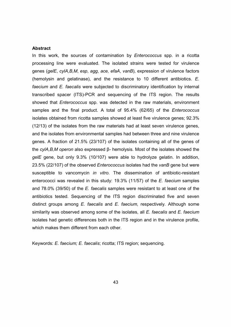
43
Abstract
In this work, the sources of contamination by Enterococcus spp. in a ricotta
processing line were evaluated. The isolated strains were tested for virulence
genes (gelE, cylA,B,M, esp, agg, ace, efaA, vanB), expression of virulence factors
(hemolysin and gelatinase), and the resistance to 10 different antibiotics. E.
faecium and E. faecalis were subjected to discriminatory identification by internal
transcribed spacer (ITS)-PCR and sequencing of the ITS region. The results
showed that Enterococcus spp. was detected in the raw materials, environment
samples and the final product. A total of 95.4% (62/65) of the Enterococcus
isolates obtained from ricotta samples showed at least five virulence genes; 92.3%
(12/13) of the isolates from the raw materials had at least seven virulence genes,
and the isolates from environmental samples had between three and nine virulence
genes. A fraction of 21.5% (23/107) of the isolates containing all of the genes of
the cylA,B,M operon also expressed β- hemolysis. Most of the isolates showed the
gelE gene, but only 9.3% (10/107) were able to hydrolyze gelatin. In addition,
23.5% (22/107) of the observed Enterococcus isolates had the vanB gene but were
susceptible to vancomycin in vitro. The dissemination of antibiotic-resistant
enterococci was revealed in this study: 19.3% (11/57) of the E. faecium samples
and 78.0% (39/50) of the E. faecalis samples were resistant to at least one of the
antibiotics tested. Sequencing of the ITS region discriminated five and seven
distinct groups among E. faecalis and E. faecium, respectively. Although some
similarity was observed among some of the isolates, all E. faecalis and E. faecium
isolates had genetic differences both in the ITS region and in the virulence profile,
which makes them different from each other.
Keywords: E. faecium; E. faecalis; ricotta; ITS region; sequencing.

44
1 Introduction
Ricotta is a fresh Italian cheese obtained through the precipitation of cheese
whey proteins by acidification associated with heat. It is considered a light and
healthy product due to its low fat and salt content and high protein content and
easy digestibility. For this, ricotta is widely consumed, even by individuals who are
debilitated or hospitalized (Ribeiro, Marques, Sodré, Abreu, & Piccoli, 2005).
The consumption of ricotta by debilitated and/or hospitalized individuals, in
particular, causes a great public health concern because ricotta shows pH values
and moisture that favor the multiplication of Enterococcus spp. Unlike most lactic
acid bacteria, the Enterococcus genus is not generally recognized as safe (GRAS).
This genus is considered an important nosocomial pathogen that causes various
types of infections, especially in immunocompromised individuals (Foulquié
Moreno, Sarantinopoulos, Tsakalidou, & De Vuyst, 2006; Oggier & Serror, 2008).
The pathogenicity of enterococci bacteria is attributed to their natural ability to
acquire plasmids that transfer virulence and antimicrobial resistance genes, and
allow enterococci to survive in unusual and / or stressful environments (Franz &
Holzapfel, 2006; Giraffa, 2002).
E. faecalis is the species of Enterococcus that is responsible for most
human infections (over 80%), whereas E. faecium is the second most common
source of infection. Although it has a much lower frequency (below 20%), E.
faecium infections are still highly significant due to its high level of resistance to
multiple antimicrobial agents (Franz, Stiles, Schleifer, & Holzapfel, 2003; Oggier &
Serror, 2008). These species have been shown to be the two most predominant
species of bacteria in several different types of cheese (Carvalho, Bruno, Nassu,
Lima, Vasconcelos, & Kuaye 2005; Gelsomino, Vancanneyt, Condon, Swings, &
Cogan, 2001; Gomes et al., 2008).
The prevalence of Enterococcus in cheese is associated with the ability of
these bacteria to resist to high temperatures, high salt concentrations, and a wide
pH range. Moreover, their prevalence is also associated with poor hygiene
practices during milking and/or processing. Environmental contamination (e.g.,

45
equipment, water, air, or handlers) can also contribute significantly to the presence
of enterococci in the final product (Gelsomino, Vancanneyt, Cogan, Condon, &
Swings, 2002; Suzzi et al., 2000; Temelli, Anar, Sen, & Akyuva, 2006). The
presence of these microorganisms in food is worrying because of their ability to
transfer genetic information through the conjugation process, which can occur in
the gastrointestinal tract of humans who ingest food contaminated with enterococci
(Çitak, Yucel, & Orhan, 2004; Gelsomino, Vancanneyt, Condon, Swings, & Cogan,
2001).
Due to the high prevalence of Enterococcus in the processing environment
of several types of cheese and its potential severity for humans, the present study
aimed to evaluate sources of Enterococcus spp. contamination in a ricotta
processing line in southeastern Brazil and to check the pathogenicity and antibiotic
resistance profile of these bacteria.
2 Material and Methods
2.1 Sampling
A total of 126 samples were collected on three different days at the ricotta
processing line at a dairy plant located in the southern of Minas Gerais State,
Brazil (Table 1).
Table 1. Number of samples collected in the dairy manufacturing Sample 1st
collection 2nd collection
3rd collection
Total samples analyzed
Raw milk 1 1 1 3 Pasteurized milk 1 1 1 3 Cheese whey 3 3 3 9 Water 1 1 1 3
Ricotta before packing 5 5 5 15 Packed ricotta 5 5 5 15 Environmental surfaces 20 20 20 60 Environment air 6 6 6 18 Total 42 42 42 126

46
Samples of raw and pasteurized milk (10 aliquots of 100 ml of each type of
milk) were randomly collected throughout the production period of the day,
resulting in 1000 ml per sampling unit. From each batch of the ricotta process,
1,000 ml of cheese whey were obtained from different points of the whey reception
tank. During each visit, five units of ricotta before packaging (exposed to room
temperature for 2 h) and five units (300 g each) of packaged ricotta (stored at 7 °C
for 21 days) from the same batch were also obtained.
Samples of the environmental surfaces at the ricotta processing line were
collected after the sanitization procedures and were collected from 20 locations for
each collection, as shown in Fig. 1.

47
Filtration
Pasteurization
Cheese production
Cheese whey
Ricotta production
Acidity reduction
Heating (65 °C)
Addition of milk (20%) and dye
Heating (90°C)
Cooling (7 °C)
Packaging
Milk reception
Addition of acidifier agent (0,05%)
Stirring
Precipitation
Salting
Molding
Storage (7 °C)
Wait at room temperature (5h)
1. Milk reception tank
8. Milk collection bucket
9. Thermometer
Air sample: Beginning: 9am
Middle: 11am End: 3pm
2. Plastic connecting tube 3. Whey collection box 4. Ricotta processing tank (whey exit pipe) 5. Support of the whey entrance hose 6. Broom 7. Whey sewage drain
10. Stirrer
11. 12. Shovel and pot for collecting curds 13. Curds retention sieve
14. Mold 15. Press 16. Retention mesh 17. Wall 18. Molding countertop
19. Storage box
20. Packaging countertop
Air sample: 11am
Air sample: 1pm
Air sample: 1pm
Fig. 1. Sampling points in the ricotta processing line.

48
The collection of the environmental and air samples was performed
according to Fernandes, Fujimoto, Schneid, Kabuki, & Kuaye (2014).
2.2 Isolation and identification of Enterococcus spp.
The samples were diluted 1:10 in 0.1% peptone water (Difco, Becton,
Dickinson and Company, Sparks, MD, USA), plated on Kennel Fecal (KF)
Streptococcus Agar (Difco, Becton, Dickinson and Company) supplemented with
1% 2,3,5-triphenyl-tetrazolium chloride – TTC (Vetec Química, Rio de Janeiro,
Brazil), and incubated at 45 ºC for 48 h (Hartman, Deibel, & Sieverding, 2001).
Enterococcus were identified at the genus level using conventional phenotypic
methods, including Gram staining, catalase production, growth at 10 ºC and 45 ºC,
growth in Brain Heart Infusion (BHI) broth (Difco, Becton, Dickinson and Company)
containing 6.5% NaCl, growth in BHI broth (Difco, Becton, Dickinson and
Company) at pH 9.6 and growth on bile-esculin Agar (Acumedia Manufactures,
Maryland, USA), according to APHA methodology (Hartman, Deibel, & Sieverding,
2001).
Genetic identification at the species level was performed using polymerase
chain reaction (PCR) (Dutka-Malen, Evers, & Courvalin, 1995) in 120 isolates.
Genomic DNA from pure cultures was extracted according to the method described
in Furrer, Candrian, Hoefelein, & Luethy (1991). The primers used for the
amplification of the ddlE. faecium and ddlE. faecalis genes (Invitrogen, Life Technologies,
Carlsbad, USA) are shown in Table 2.
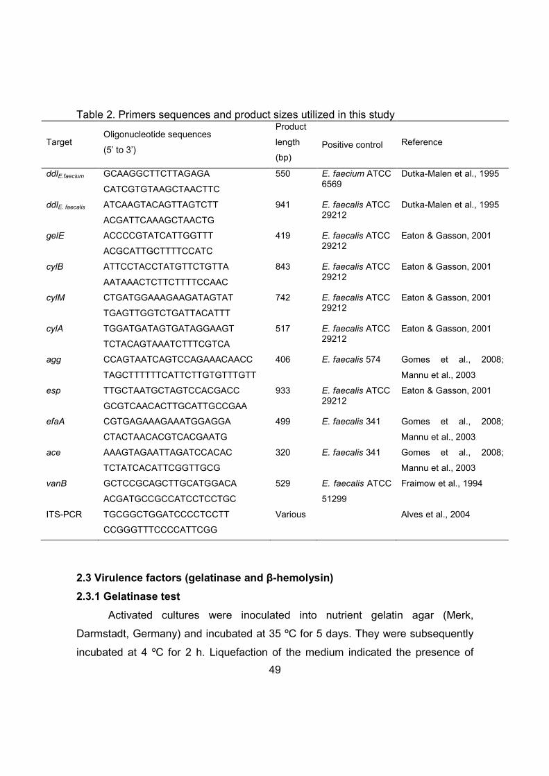
49
Table 2. Primers sequences and product sizes utilized in this study
Target Oligonucleotide sequences
(5’ to 3’)
Product
length
(bp)
Positive control Reference
ddlE.faecium GCAAGGCTTCTTAGAGA
CATCGTGTAAGCTAACTTC
550 E. faecium ATCC 6569
Dutka-Malen et al., 1995
ddlE. faecalis ATCAAGTACAGTTAGTCTT
ACGATTCAAAGCTAACTG
941 E. faecalis ATCC 29212
Dutka-Malen et al., 1995
gelE ACCCCGTATCATTGGTTT
ACGCATTGCTTTTCCATC
419 E. faecalis ATCC 29212
Eaton & Gasson, 2001
cylB ATTCCTACCTATGTTCTGTTA
AATAAACTCTTCTTTTCCAAC
843 E. faecalis ATCC 29212
Eaton & Gasson, 2001
cylM CTGATGGAAAGAAGATAGTAT
TGAGTTGGTCTGATTACATTT
742 E. faecalis ATCC 29212
Eaton & Gasson, 2001
cylA TGGATGATAGTGATAGGAAGT
TCTACAGTAAATCTTTCGTCA
517 E. faecalis ATCC 29212
Eaton & Gasson, 2001
agg CCAGTAATCAGTCCAGAAACAACC
TAGCTTTTTTCATTCTTGTGTTTGTT
406 E. faecalis 574 Gomes et al., 2008;
Mannu et al., 2003
esp TTGCTAATGCTAGTCCACGACC
GCGTCAACACTTGCATTGCCGAA
933 E. faecalis ATCC 29212
Eaton & Gasson, 2001
efaA CGTGAGAAAGAAATGGAGGA
CTACTAACACGTCACGAATG
499 E. faecalis 341 Gomes et al., 2008;
Mannu et al., 2003
ace AAAGTAGAATTAGATCCACAC
TCTATCACATTCGGTTGCG
320 E. faecalis 341 Gomes et al., 2008;
Mannu et al., 2003
vanB GCTCCGCAGCTTGCATGGACA
ACGATGCCGCCATCCTCCTGC
529 E. faecalis ATCC
51299
Fraimow et al., 1994
ITS-PCR TGCGGCTGGATCCCCTCCTT
CCGGGTTTCCCCATTCGG
Various Alves et al., 2004
2.3 Virulence factors (gelatinase and β-hemolysin)
2.3.1 Gelatinase test
Activated cultures were inoculated into nutrient gelatin agar (Merk,
Darmstadt, Germany) and incubated at 35 ºC for 5 days. They were subsequently
incubated at 4 ºC for 2 h. Liquefaction of the medium indicated the presence of

50
gelatinase activity. The strain B. cereus NCTC 1143 was used as a positive control
(Lopes, Simões, Tenreiro, Marques, & Crespo, 2006 - adapted).
2.3.2 Hemolytic activity
Hemolysin activity was determined using BHI Agar (Difco, Becton, Dickinson
and Company) containing 5% defibrinized horse blood (BBV, São Paulo, Brazil)
and incubation at 37 ºC for 48 h. Zones of clearing around the colonies indicated β-
hemolysin production. The strain Staphylococcus aureus ATCC 27154 was used
as a positive control (Eaton & Gasson, 2001).
2.4 Identification of virulence genes by PCR and multiplex PCR
The expression of the gelE, cylB, cylA, and cylM genes was analyzed
simultaneously using multiplex PCR according to the methods described in Gomes
et al., 2008 and Vankerckhoven et al., 2004. The primers (Invitrogen, Life
Technologies) used for the amplification of virulence genes are shown in Table 2.
For the esp, agg, ace, and efaA genes, the reactions were performed according to
the protocol outlined in Eaton & Gasson (2001) and Mannu et al. (2003). For the
vanB gene, the amplification reaction was performed according to the method of
Fraimow, Jungkind, Lander, Delso, & Dean (1994). Electrophoresis of the PCR
products was performed on 1.5% agarose gels, which were stained with ethidium
bromide. Visualization of the PCR products was performed using a UV
transilluminator.
2.5 Antimicrobial susceptibility testing
E. faecalis and E. faecium were tested using the standard disk diffusion
method recommended by the National Committee for Clinical Laboratory
Standards (NCCLS, 2003). The following antimicrobials (BioRad Laboratories,
Marnes-la-Coquette, France) commonly used in the treatment of clinical infections
were evaluated at the following doses: ampicillin (10 µg); chloramphenicol (30 µg);
erythromycin (15 µg); streptomycin (300 µg); gentamicin (120 µg); norfloxacin (10

51
µg); rifampicin (5 µg); teicoplanin (30 µg); tetracycline (30 µg); and vancomycin (30
µg).
2.6 Discriminatory identification of Enterococcus cultures
To identify potential contamination sources during ricotta processing, 57 E.
faecium cultures and 50 E. faecalis cultures were subjected to discriminatory
identification to differentiate genetic profiles of each strain obtained.
2.6.1 Amplification of the Intergenic Spacer Region by PCR (ITS-PCR)
The amplification of the ITS region of 16S-23S rDNA was performed using
PCR according to Alves et al. (2004). The primers (Invitrogen, Life Technologies)
used in this study for the amplification of the ITS region are shown in Table 2.
2.6.2 Sequencing the ITS region
The amplicons generated by ITS-PCR were also subjected to sequencing in
an automatic capillary sequencer (ABI PRISM 3700 DNA Analyzer – Applied
Biosystems – HITACHI). The sequences obtained were compared with the
sequences available in Genbank. The multiple ITS sequences were aligned using
Clustal W software integrated with MEGA5 software (Kumar, Nei, Dudley, &
Tamura, 2008). The phylogenetic tree was constructed using the neighbor-joining
(Saitou & Nei, 1987), maximum-parsimony (Fitch, 1971), and maximum-likelihood
(Felsenstein, 1981) algorithms present in these software programs.
3 Results and discussion
3.1 Occurrence of Enterococcus spp. in ricotta processing line
Enterococcus spp. was observed in raw materials, surface samples, and
ricotta. It is interesting to highlight the variation in the levels of contamination that
were observed throughout the collection period (Table 3). High Enterococcus spp.
counts were found in raw milk in all the three collections. However, the
Enterococcus counts observed in the pasteurized milk sample were less than 2 log

52
CFU/ml, thus indicating that the heat treatment used by the factory (74.5 °C for 15 -
20 sec) is able to decrease Enterococcus spp. counts in milk.
Table 3. Counts of Enterococcus spp. in samples collected in a ricotta processing line Sample 1st
collection 2nd collection
3rd collection
Raw milka 5.70 4.56 6.28 Pasteurized milka <2 <2 <2 Cheese whey 1a <2 <2 3.78 Cheese whey 2a <2 <2 3.93 Cheese whey 3a <2 <2 <2 Ricotta processing tankb 2.85 <1 2.11 Whey collection boxb < 1 <1 2.77 Wallb 4.36 <1 7.53 Molding countertopb <1 2.3 <1 Packaging countertopb <1 <1 2.83 Whey sewage drainb 6.08 <1 6.48 Moldc 5.73 4.78 3.04 Retention meshc 5.20 <1 6.62 Broomc 8.26 <1 <1 Air sampled 3 3 - Ricotta before packing 1e 2.43 <2 <2 Ricotta before packing 2e <2 <2 <2 Ricotta before packing 3e <2 <2 <2 Ricotta before packing 4e <2 <2 <2 Ricotta before packing 5e <2 <2 <2 Packed ricotta 1e 4.48 3.53 5.56 Packed ricotta 2e 4.18 3.90 5.15 Packed ricotta 3e 5.38 3.63 7.08 Packed ricotta 4e 4.41 4.30 5.92 Packed ricotta 5e 5.36 3.62 4.45 a Units are log CFU/ml b Units are log CFU/cm2
c Units are log CFU per unit d Units are log CFU per plate e Units are log CFU/g.
Enterococcus spp. counts as high as 3 log CFU/ml were observed in cheese
whey, a fact that results from its composition being rich in nutrients, its high water
activity, and its pH (6.0), which is conducive to the multiplication of

53
microorganisms. In addition, the whey obtained from the ricotta processing tank
was at an average temperature of 39 °C.
Among the surfaces in contact with the ricotta, it can be highlighted the mold as
a source of Enterococcus spp. which exhibited persistent contamination in all the
three collections and reaching counts up to 5.73 log CFU per sample unit (Table
3). This profile is worrisome because after molding no heat treatment occurs, and
the ricotta is maintained for 5h at room temperature before cold room storage (7
°C). These results reveal the need to conduct a more effective cleaning and
sanitizing procedures in cheese molds.
The evaluation of a wall contamination – near the molding process - revealed
this contributing source, with counts of 7.53 log CFU/cm2 to samples in the third
collection.
Samples from other surfaces, such as the curd-retention sieve, the stirrer, the
shovel, the pot, and the thermometer, all exhibited counts less than 1 log CFU per
sample unit in each of the three collections, which is to be expected because of
their prolonged exposure to a chlorinated solution.
Counts higher than 2 log CFU/cm2 were observed on the surfaces of stainless
steel equipment (e.g., countertops and the ricotta processing tank). The presence
of Enterococcus spp. on these surfaces even after performed standard sanitization
procedures indicates the presence of biofilms (Andrade, Ajão, & Zottola, 1998;
Gelsomino, Vancanneyt, Cogan, Condon, & Swings, 2002; Temelli, Anar, Sen, &
Akyuva, 2006) and highlights the need for more efficient cleaning and sanitization
programs.
Enterococcus spp. counts in the 15 ricotta samples remained below 2.5 log
CFU/g before they were packaged. However, all 15 of the final product samples
analyzed after 21 days of storage at 7 °C were contaminated by these
microorganisms - counts ranging from 3.53 log CFU/g to 7.08 log CFU/g (Table 3).
The presence of Enterococcus in the ricotta processing line may be attributed
to the contamination of raw materials (Table 3). These bacteria have the ability to
survive and multiply during all of the processing stages under different conditions,

54
including a wide temperature range (cooking temperature 90 °C and refrigerated
storage at 7 °C) (Carvalho, Bruno, Nassu, Lima, Vasconcelos, & Kuaye 2005;
Giraffa, 2003), variable pH and salt content (Franz, Stiles, Schleifer, & Holzapfel,
2003; Giraffa, 2002). In fact, these conditions may favor and select the most
resistant bacteria that reach the final product. It is not discarded, the contamination
due to biofilm formed on food contact surfaces (mold, countertops, retention
mesh). Another critical point was the long exposure time (5 h) of the ricotta to room
temperature after the heat treatment (90 ºC).
Of the 120 isolates - confirmed as Enterococcus genus by biochemical tests
- 47.5% (57/120) were identified as E. faecium and 41.7% (50/120) as E. faecalis
by PCR. 10.8% (13/120) of the isolates were negative for both ddlE.faecium and
ddlE.faecalis identification genes, although they exhibited the characteristic results for
this genus in phenotypic tests, which indicates the presence of other Enterococcus
species.
E. faecalis was the predominant species observed in the raw materials
(100%, 13/13), and E. faecium was the predominant species in the ricotta (66%,
43/65). E. faecium (48.3%, 14/29) and E. faecalis (51.7%, 15/29) were observed at
similar rates in samples from the ricotta processing environment. The prevalence
of these two species has been reported for different types of cheese in one other
brazilian study (Gomes et al., 2008) and in studies from several other countries
(Cariolato, Andrighetto, & Lombardi, 2008; Çitak, Yucel, & Orhan, 2004;
Gelsomino, Vancanneyt, Cogan, Condon, & Swings, 2002; Suzzi et al., 2000).
3.2 Phenotypic and genotypic virulence factors
Fig. 2 shows that six virulence genes (efaA, ace, agg, esp, cylB, and cylM)
were more frequent among E. faecalis isolates than among E. faecium isolates.
The gelE and vanB genes were more frequent among E. faecium isolates than
among E. faecalis isolates. The presence of the cylA gene was observed in all of
the E. faecium and E. faecalis strains. Previous studies have reported a greater
association of virulence genes with E. faecalis strains than with E. faecium strains

55
isolated from food (Barbosa, Gibbs, & Teixeira, 2010; Gomes et al., 2008; Mannu
et al., 2003; Martín-Platero, Valdivia, Maqueda, & Martínez-Bueno, 2009).
However, in the present study, a wide dissemination of virulence genes was
observed in both species (Fig. 2, Tables 4 and 5).
Fig. 2. Prevalence of virulence genes in E. faecium and E. faecalis isolated from a
ricotta processing line.
The gene encoding the collagen binding protein, ace, is frequently associated
with E. faecalis strains (Jahan & Holley, 2014; Mannu et al., 2003). However, in the
present study, 84.2% of the E. faecium strains had this gene. At least one other
brazilian study has also revealed the presence of the ace gene among E. faecium
strains obtained from food (Gomes et al., 2008). The efaA gene encoding the
endocarditis antigen was observed to be widely disseminated among the E.
faecium (84.2%) and E. faecalis (100%) species. The agg gene encoding the
aggregation substance appears to be involved in the process of conjugation
between the Enterococcus species, promoting cell aggregation and facilitating the
Virulence genes
% o
f is
olat
es

56
transfer of plasmids, which can transport virulence and antimicrobial resistance
genes. Although the agg gene is usually associated with E. faecalis, in the present
study, this gene was identified both in E. faecium (36.8%) and in E. faecalis (58%),
as reported in the study by Barbosa, Gibbs, & Teixeira (2010). The esp gene was
observed in 54.4% of E. faecium, which is in contrast to the findings of other
studies, where this gene was associated only with E. faecalis strains (Barbosa,
Gibbs, & Teixeira, 2010; Gomes et al., 2008; Martín-Platero, Valdivia, Maqueda, &
Martínez-Bueno, 2009). The esp gene, which encodes the surface protein, has the
ability to adhere to and form biofilms on abiotic surfaces, which makes it difficult to
eliminate potentially virulent Enterococcus bacteria (Eaton & Gasson, 2001; Franz
& Holzapfel, 2006).
Several very worrisome results were identified in the current study. For
example, 95.4% (62/65) of the Enterococcus isolates obtained from ricotta samples
had at least five virulence genes. In addition, 92.3% (12/13) of the isolates from the
raw materials had at least seven virulence genes, and the isolates from
environmental samples had between three and nine virulence genes. Five isolates
(three from environmental samples and two obtained from ricotta prior to
packaging) had all of the virulence genes examined. None of the 107 Enterococcus
isolates were completely free from all virulence genes (Tables 4 and 5).
The results also showed that 36.8% (21/57) of the E. faecium isolates and
14% (7/50) of the E. faecalis isolates produced β-hemolysis. In addition, 29.8%
(17/57) of the E. faecium isolates and 50% (25/50) of the E. faecalis isolates
showed α-hemolysis, and 33.4% (19/57) of the E. faecium isolates and 36%
(18/50) of the E. faecalis isolates were γ-hemolytic (Tables 4 and 5). The presence
of β-hemolytic activity in human and horse erythrocytes is usually related to the
presence of the operon genes of cylA,B,M cytolysin (Franz & Holzapfel, 2006).
Thus, it was found that 21.5% (23/107) of the isolates had all of the genes from the
cylA,B,M operon and were able to express β-hemolysis (Tables 4 and 5). Of these,
15 isolates were from ricotta samples, indicating that consumers might be exposed
to a potentially dangerous product. Potential interactions between these strains

57
and susceptible hosts (such as immunocompromised individuals and pregnant
women) are certainly a reason for concern.
All the E. faecium and E. faecalis isolates that did not showed one or two
genes from the cylA,B,M operon were α-hemolytic or not able to express this
virulence factor, except for three isolates (084, 105, 106 - Tables 4 and 5), which
did not have the gene cylB+ and were β-hemolytic. This may result from the
presence of the cylA gene, which is an essential subunit for hemolysin activity (Han
et al., 2011). Approximately one-third of the isolates (35/107) had the cylA gene but
did not exhibit hemolytic activity. For these cases, the apparent absence of the
virulence factor may result from low levels of gene expression or from the
inactivation of the gene expression product (Eaton & Gasson, 2001). It was also
observed that some isolates with the operon cylA,B,M genes were not β-hemolytic
and that most of the E. faecium and E. faecalis isolates had the gelE gene, but only
9.3% (10/107) of these isolates were able to hydrolyze gelatin (Tables 4 and 5).
This may be due to the presence of silent genes, genetic mutations, or the
absence of subunits required for activity of the cyl/gel genes (Han et al., 2011).

58
Table 4. Phenotypic and genotypic characteristics found in E.faecalis
Profile Sample (isolate number)
Collection Operon cylABM
Hemolytic activitya
gelE Gelatinase vanB Vancomycin-
resistant
Other antibiotics resistance
Other virulence
genes 1 PR3 (011) 1
cylABM+ α gelE+ Negative vanB- Sensible - efaA+, ace+
PR3 (012) 1 PR3 (015) 1 PR4 (020) 1 PR9 (039) 2 PR9 (040) 2
2 PR7 (031) RPT (084) RPT (085)
2 1 1
cylAM+ γ β γ
gelE+ Negative vanB- Sensible - esp+, efaA+,
ace+
3 PR7 (032) PR8 (038)
2 2 2 3
cylABM+
γ α
gelE+
Negative Positive Negative Positive
vanB- Sensible - agg+, esp+, efaA+, ace+ PR9 (042)
RPT (115) α β
4 PR7 (033) 2 cylABM+ α gelE+ Negative vanB- Sensible -
agg+, efaA+, ace+ PR8 (035)
5 PR8 (036) 2
cylABM+
α
gelE+
Negative Negative Negative Negative Positive Negative
vanB- Sensible - esp+, efaA+,
ace+
PR10 (047) 2 α PR12 (055) RM (078) CW (081) RPT (087)
3 3 3 1
γ γ γ γ
6 PR8 (037) 2 cylAM+ α gelE- Negative vanB- Sensible -
agg+, esp+, efaA+, ace+
7 PR10 (045) 2 cylABM+ α gelE+ Negative vanB- Sensible
Erythromycin, streptomycin, tetracycline
agg+, esp+, efaA+, ace+ PR10 (046)
8 PR11 (048)
3 cylABM+
β β γ γ
gelE+ Negative vanB- Sensible Tetracycline agg+, esp+, efaA+, ace+
PR13 (058) PR13 (061) PR13 (062)
9 RM (068) 1
cylABM+ α gelE+
Negative Negative Negative Negative
vanB- Sensible Rifampicin esp+, efaA+,
ace+
RM (069) 1 RM (070) 1 RM (071) 1 RPT (116) 3 Positive
10 RM (072) 1
cylABM+
α
gelE+ Negative vanB- Sensible Rifampicin agg+, esp+, efaA+, ace+
RM (075) 2 α RM (079) 3 γ RPT (117) RPT (118)
3 3
α β

59
Continuation...
Profile Sample (isolate number)
Collection Operon cylABM
Hemolytic activitya
gelE Gelatinase vanB Vancomycin-
resistant
Other antibiotics resistance
Other virulence
genes 11 RM (073) 2
cylABM+
β β α
gelE+
Positive vanB- Sensible
Erythromycin, rifampicin
agg+, esp+, efaA+, ace+
RM (074) RM (076)
2 Negative Negative
12 RM (077) 3 cylB+ α gelE+ Negative vanB- Sensible -
agg+, esp+, efaA+, ace+
13 W (092) 1 cylABM+ γ gelE+ Negative vanB+ Sensible -
agg+, esp+, efaA+, ace+
14 WSD (096) 1 cylABM+ γ gelE+ Positive vanB+ Sensible -
esp+, efaA+, ace+
15 WCB (110) 3 cylAM+ γ gelE+ Negative vanB- Sensible -
agg+, efaA+, ace+
16 WCB (111) 3 cylABM+ γ gelE+
Negative vanB- Sensible
Rifampicin Tetracycline
agg+, efaA+, ace+ WCB (113) Positive
17 WCB (112) 3 cylAM+ γ gelE+ Negative vanB- Sensible Tetracycline
agg+, efaA+, ace+ WCB (114)
18 PC (130) 3 cylAM+ γ gelE+ Negative vanB+ Sensible Tetracycline
agg+, esp+, efaA+, ace+
PR: packaged ricotta; RPT: ricotta processing tank; RM: raw milk; CW: Cheese whey; W: wall; WSD: whey sewage drain; WCB: Whey collection box;
PC: Packaging countertop aβ: hemolytic activity; α: partial hemolytic activity, γ: no hemolytic activity.

60
Table 5. Phenotypic and genotypic characteristics found in E.faecium
Profile Sample (isolate number)
Collection Operon cylABM
Hemolytic activitya
gelE Gelatinase vanB Vancomycin-
resistant Other antibiotics
resistance Other virulence
genes
1 PR1 (001) PR3 (014)
1 cylABM+ β α
gelE+ Negative vanB+ Sensible - efaA+, ace+
2 PR1 (002) MC (103) MC (104)
1 2 2
cylABM+ β gelE+ Negative Negative Positive
vanB+ Sensible - esp+, ace+
3
PR1 (003) PR14 (064)
1 cylABM+ β gelE+ Negative vanB+ Sensible - esp+, efaA+, ace+
M (089) 4 PR1 (004) 1 cylABM+ β gelE+ Negative vanB- Sensible - efaA+
5 PR1 (005) 1
cylABM+ β gelE+ Negative
vanB+ Sensible - - PR2 (006) MC (107)
1 3
Negative Positive
6 PR2 (007) 1 cylABM+ β gelE+ Negative vanB- Sensible - ace+
7
PR2 (009) PR3 (013) PR4 (016)
1 1 1
cylABM+
α β β
gelE+ Negative vanB- Sensible - efaA+, ace+ PR4 (018) 1 α PR4 (019) PR5 (021)
1 1
α β
PR6 (026) 2 α 8 PR5 (022) 1
cylABM+ γ gelE+ Negative vanB- Sensible Rifampicin efaA+, ace+ PR8 (034) 2
9 PR5 (023) 1 cylABM+ γ gelE+ Negative vanB- Sensible Rifampicin agg+, efaA+, ace+
10
PR5 (024) PR5 (025)
1 1
cylABM+
β α
gelE+ Negative vanB- Sensible - esp+, efaA+, ace+
PR11 (051)
3 α
PR12 (054)
3 γ
PR12 (056)
3 γ
PR14 (063)
3 α
11
PR6 (027) 2
cylABM+
α
gelE+ Negative vanB- Sensible - agg+, esp+, efaA+,
ace+
PR6 (028) 2 γ PR11 (050)
3 α
PR11 (052)
3 α

61
Continuation...
Profile Sample (isolate number)
Collection Operon cylABM
Hemolytic activitya
gelE Gelatinase vanB Vancomycin-
resistant Other antibiotics
resistance Other virulence
genes
12 PR6 (029) 2 cylAM+ γ gelE+ Negative vanB- Sensible - efaA+, ace+
13
PR7 (030)
2 cylABM+
γ
gelE+ Negative vanB- Sensible - agg+, efaA+, ace+
PR9 (041) α PR10 (043)
α
PR10 (044)
α
14 PR11 (049)
3 cylAM+ α gelE+ Negative vanB- Sensible - esp+, efaA+, ace+
15
PR12 (053)
3 cylABM+ γ gelE+ Negative vanB- Sensible Tetracycline agg+, esp+, efaA+,
ace+ PR12 (057)
16 PR13 (059)
3 cylABM+ γ gelE+ Positive vanB- Sensible Tetracycline efaA+, ace+
17 PR14 (065)
3 cylAM+ α gelE+ Negative vanB+ Sensible - agg+, esp+, efaA+,
ace+
18
RBP (082)
1 cylABM+
β β γ γ
gelE+ Negative vanB+ Sensible - agg+, esp+, efaA+,
ace+ RBP (083) B (098) B (099)
19 MC (105) 2 cylAM+ β gelE+ Negative vanB+ Sensible - efaA+, ace+ 20 MC (106) 2 cylAM+ β gelE+ Negative vanB+ Sensible - -
21 EA (109) 2 cylAM+ γ gelE+ Negative vanB- Sensible Erythromycin,
rifampicin -
22 M (119)
3 cylAM+ γ gelE+ Negative vanB- Sensible Erythromycin agg+, esp+, efaA+ M (120)
23 M (121) 3 cylAM+ γ gelE+ Negative vanB- Sensible Erythromycin,
rifampicin agg+, esp+, efaA+
24 M (122) 3 cylAM+ γ gelE+ Negative vanB- Sensible Erythromycin,
rifampicin esp+, efaA+
25 PC (131) 3 cylAM+ α gelE+ Negative vanB+ Sensible - esp+, efaA+, ace+
PR: packaged ricotta; MC: Molding countertop; M: mold; RBP: Ricotta before packing; B: broom; EA: Environment air; PC: Packaging countertop. aβ: hemolytic activity; α: partial hemolytic activity, γ: no hemolytic activity.

62
3.3 Antibiotic resistance
In Fig. 3 it is shown that different antimicrobial resistance profiles were
observed among the species, with 19.3% (11/57) of E. faecium and 78%
(39/50) of E. faecalis being resistant to at least one of the tested antibiotics.
However, all of the isolates were sensitive to ampicillin, gentamicin, teicoplanin,
and vancomycin.
Fig. 3. Antibiotic resistance of E. faecium and E. faecalis isolated from a ricotta
processing line.
S: Susceptible; I: Intermediary; R: Resistant.
AMP: ampicillin; CLO: chloramphenicol; ERI: erythromycin; GEN: gentamicin;
NOR: norfloxacin; RA: rifampicin; STREP: streptomycin; TEC: teicoplanin; TET:
tetracycline; VAN: vancomycin.
The results demonstrated that 18.7% (20/107) of the isolates were
resistant to rifampicin, 14% (15/107) to tetracycline, 9.3% (10/107) to
erythromycin, 1.9% (2/107) to high concentrations of streptomycin, and 0.9%
(1/107) to chloramphenicol. Because chloramphenicol is prohibited for
Antibiotics
% o
f is
olat
es

63
veterinary use in Brazil (Brasil, 2003), the resistance of Enterococcus to this
antibiotic was not expected. It is possible that the observed antibiotic resistance
results from the illegal use of this drug.
All isolates from raw milk samples were classified as intermediate or
resistant to the antibiotics erythromycin and rifampin. These antibiotics are
typically used in veterinary medicine for the treatment of mastitis. Therefore, it is
possible that the observed resistance is associated with the excessive and
perhaps inadequate use of these antibiotics in farms, which represents a
serious public health problem. Only 23.1% (15/65) and 17% (5/29) of the
isolates from ricotta samples and processing environment, respectively, were
sensitive to all of the antibiotics tested. The remaining isolates from these
samples were classified as intermediate or resistant to one, two, or three
antibiotics.
The dissemination of antibiotic-resistant Enterococci (ARE) in food has
been frequently reported (Barbosa, Ferreira, & Teixeira 2009; Cariolato,
Andrighetto, & Lombardi, 2008; Kasimoglu-Dogru, Gencay, & Ayaz, 2010). The
ubiquitous nature of Enterococcus and their resistance to adverse
environmental conditions provide this microorganism with the ability to colonize
a variety of different habitats, thus facilitating its dissemination through the food
chain. Therefore, the presence of ARE in food is a reason for growing concern.
Vancomycin-resistant enterococci (VRE) represent a particularly
significant problem in the treatment of clinical human infections because this
drug is used as a last resort for treating enterococci that are resistant to multiple
other antibiotics (Oggier & Serror, 2008). The present results revealed that
23.5% (22/107) of the observed Enterococcus isolates (eight from packaged
ricotta, two from ricotta before packaging, and 11 from environmental samples)
had the vanB gene but were susceptible to vancomycin in vitro (Table 4).
However, it is important to note that the in vitro tests for antimicrobial resistance
to antibiotics do not always reflect the in vivo situation (Oggier & Serror, 2008).
Once again, the current results suggest that consumers might be at risk of
exposure to a product that is potentially dangerous.

64
3.4 Discriminatory identification of Enterococcus cultures by intergenic
region analysis
The 16S-23S intergenic region was amplified using PCR from 107
Enterococcus isolates. Four different clusters could be identified, and 27.1%
(29/107) of the isolates belonged to cluster I, 7.5% (8/107) to cluster II, 64.5%
(69/107) to cluster III and 0.9% (1/107) to cluster IV. Clusters I, II, and III had
three major bands that ranged in size from 390 to 650 bp. Cluster IV had four
main bands with sizes between 500 and 700 bp. Both Enterococcus species
had similar cluster profiles (I, II, and III). In addition, sequencing analysis of the
intergenic region of E. faecium and E. faecalis was performed. Because these
two species are genetically very similar, the 16S-23S rDNA (genes coding for
rRNA) intergenic region was evaluated, which is referred to as the ITS region. It
has been suggested that this segment of DNA would be under minimal selective
pressure compared to the selective pressure on rRNA genes, a fact that could
therefore allow for species identification based on the enhanced variability
between species within a genus (Tyrrell, Bethune, Willey, & Low, 1997), which
could aid in identifying the possible sources of contamination in ricotta
processing.
Figures 4A and 4B show the phylogenetic trees of E. faecalis and E.
faecium, respectively. Because the sequencing was performed in a very small
region, phenotypic and genotypic data were used to complement the
sequencing analysis (Table 4 – E. faecalis and Table 5 – E. faecium).
Sequencing of ITS region discriminated five distinct groups among E.
faecalis (Fig. 4A). Although some similarity was observed among some of the
isolates, most of the E. faecalis isolates had genetic differences both in the ITS
region and in the virulence profile, which makes them different from each other
(Table 4). Only strains 012 and 015 (from packaged ricotta in collection one)
and strains 061 and 062 (from packaged ricotta in collection three) had genetic
similarities regarding the ITS and the same virulence profile; thus, they may be
considered as the same strain.
Regarding the genotypic and phenotypic virulence characteristics, 9
different profiles were identified within the first group. Groups two, three, and

65
four are composed of only one isolate each. Group five had only one virulence
profile. According to the phylogenetic tree (Fig. 4 A), in group one, genetically
similar strains were often isolated from different sources. For example, strains
040 and 074 were isolated from packaged ricotta and raw milk, respectively.
Strains 116 and 130 were isolated from the ricotta processing tank and the
packaging countertop, respectively. Strains 068 and 075 were isolated from raw
milk but were obtained in different collections (one and two, respectively). The
same was observed for the strains 042 e 055 (both from packaged ricotta,
collection two and three, respectively), and 071 and 079 (both from raw milk,
collections one and three, respectively). Although all the isolates were similar
based on the ITS region sequencing, they had different virulence profiles.
In Fig. 4B it is shown seven different groups identified among E. faecium
isolates. Although similarities were observed between some of the isolates, all
E. faecium isolates had genetic differences in their ITS regions and virulence
profiles, which demonstrates that they are different from each other. Only the
strains 053 and 057 (from packaged ricotta, collection three), and strains 002
and 103 (from packaged ricotta and molding countertop, respectively) had
genetic similarities regarding the ITS and the same virulence profile; thus, they
may be considered as the same strain. The similarity between strains 002 and
103 indicates the molding countertop as a possible source of contamination for
the ricotta.
Isolates 044 (from packaged ricotta from the second collection) and 054
(from packaged ricotta from the third collection), 050 and 051 (from packaged
ricotta from the third collection, but isolated from different packages) were very
similar genetically, although they showed differences in their phenotypic
(hemolytic activity) and virulence genes profiles. Isolates 014 (from packaged
ricotta from the first collection) and 099 (broom) showed similarity in ITS region,
but they had different phenotypic (hemolytic activity) and virulence genes
profiles (Table 5). A similar result was observed for samples 007 (from
packaged ricotta from the first collection) and 120 (from the mold), 005 (from
packaged ricotta from the first collection) and 105 (from the ricotta processing
countertop), 003 (from packaged ricotta from the first collection) and 122 (from

66
the mold), 019 (from packaged ricotta from the first collection) and 089 (from the
mold), and 060 (from packaged ricotta from the third collection) and 082 (from
ricotta before packaging). Samples 009 and 043 are from ricotta, but they were
obtained in collections one and two, respectively. The analysis of these results
shows a similarity between the isolates obtained from raw materials, the
environment, and the final product.
A

67
Fig. 4. Phylogenetic trees based on the ITS sequences of E. faecalis (A) and E. faecium (B) isolated from a ricotta processing line. * Profile related to Table 4. ** Profile related to Table 5.
B

68
These results demonstrate that both E. faecalis and E. faecium are able
to survive ricotta processing and persist in the final product. In addition, some
isolates that showed genetic similarities were obtained on different collection
days, it highlights the resistance of this microorganism over time in the ricotta
factory.
The results also revealed a great deal of genetic diversity among E.
faecalis and E. faecium isolates from the ricotta processing line. The most
frequent differences between these strains involved data regarding their
virulence genes. It is important to note that a large number of the virulence
genes evaluated are plasmidial, and therefore, genetic exchange may have
occurred (Foulquié Moreno, Sarantinopoulos, Tsakalidou, & De Vuyst, 2006).
Thus, some strains that might be from the same generation may have acquired
several virulence genes throughout the process that distinguish them from
others of the same origin. This may partially explain the great variation among
the strains, although these strains were often isolated from the same source
and had similar genetic profiles. The tracking for E. faecalis and E. faecium was
performed in a small factory that processes 10,000 to 12,000 liters of milk from
approximately 100 producers each day, which consisted of several small milk
producers from various locations, most of which likely did not have adequate
training in good agricultural practices. This set of actions may be partially
responsible for the genetic variability of the isolates and for the presence of
enterococci with expressive pathogenic profiles, especially regarding their
antibiotic resistance profiles.
4 Conclusion
This work revealed the disseminated and persistent contamination of E.
faecium and E. faecalis in ricotta processing environment. These species
presented virulence characteristics and resistance to antibiotics used in human
clinical practice. A variable genetic profile was found among E. faecalis and E.
faecium isolates and their ability to survive in stress condition during process
was evidenced.

69
By these relevant data, although no regulation is established for
Enterococcus in milk and high moisture cheese, the control of this
microorganism with effective monitoring program and good manufacturing
practices are required to reduce the contamination in dairy industries.
Acknowledgements
This work was supported by the Fundação de Amparo à Pesquisa do
Estado de São Paulo (FAPESP), Process no. 2010/10507-7, and partially
supported by Fundação de Apoio ao Ensino, à Pesquisa e à Extensão
(FAEPEX), UNICAMP.
References
Alves, P. I., Martins, M. P., Semedo, T., Figueiredo, Marques, J. J. F., Tenreiro, R., & Crespo, M. T. B. (2004). Comparison of phenotypic and genotypic taxonomic methods for the identification of dairy enterococci. Antonie van Leeuwenhoek, 85, 237-252. Andrade, N. J., Ajão, D. B., & Zottola, E. A. (1998). Growth and adherence on stainless steel by Enterococcus faecium cells. Journal of Food Protection, 61, 1454-1458. Barbosa, J., Ferreira, V., & Teixeira, P. (2009). Antibiotic susceptibility of enterococci isolated from traditional fermented meat products. Food Microbiology, 26, 527–532. Barbosa, J., Gibbs, P. A., & Teixeira, P. (2010). Virulence factors among enterococci isolated from traditional fermented meat products produced in the North of Portugal. Food Control, 21, 651-656. Brasil (2003). Ministério da Agricultura, Pecuária e Abastecimento. Instrução Normativa n°9, de 27 de junho de 2003. In: Proíbe a fabricação, a manipulação, o fracionamento, a comercialização, a importação e o uso dos princípios ativos cloranfenicol, nitrofuranos e os produtos que contenham estes princípios ativos, para uso veterinário e suscetível de emprego na alimentação de todos os animais e insetos. Available in: http://www.agricultura.gov.br. Cariolato, D., Andrighetto, C., & Lombardi, A. (2008). Occurrence of virulence factors and antibiotic resistances in Enterococcus faecalis and Enterococcus faecium collected from dairy and human samples in North Italy. Food Control, 19, 886-892.

70
Carvalho, J. D. G., Bruno, L. M., Nassu, R. T., Lima, C. P., Vasconcelos, N. M., & Kuaye, A. Y. (2005). Bactérias ácido láticas isoladas de queijo de Coalho artesanais comercializados em Fortaleza, CE. Revista do Instituto de Laticínios Cândido Tostes, 60, 221-224. Çitak, S., Yucel, N., & Orhan, S. (2004). Antibiotic resistance and incidence of Enterococcus species in Turkish white cheese. International Journal of Dairy Technology, 57, 27-31. Dutka-Malen, S., Evers, S., & Courvalin, P. (1995). Detection of glycopeptide resistance genotypes and identification to the species level of clinically relevant enterococci by PCR. Journal of Clinical Microbiology, 33, 24-27. Eaton, T. J., & Gasson, M. (2001). Molecular screening of Enterococcus virulence determinants and potential for genetic exchange between food and medical isolates. Applied and Environmental Microbiology, 67, 1628-1635. Felsenstein, J. (1981). Evolutionary trees from DNA sequences: a maximum likelihood approach. Journal of Molecular Evolution, 17, 368–376. Fernandes, M. S., Fujimoto, G., Schneid, I., Kabuki, D. Y., & Kuaye, A. Y. (2014). Enterotoxigenic profile, antimicrobial susceptibility, and biofilm formation of Bacillus cereus isolated from ricotta processing. International Dairy Journal, 38, 16-23. Fitch, W. M. (1971). Toward defining the course of evolution: minimum change for a specific tree topology. Systematic Zoology, 20, 406-416. Franz, C. M.A. P., Stiles, M. E., Schleifer, K. H., & Holzapfel, W. H. (2003). Enterococci in foods - a conundrum for food safety. International Journal of Food Microbiology, 88, 105-122. Franz, C. M. A. P., & Holzapfel, W. H. (2006). Enterococci. In Y. Motarjemi, & M. Adams (Eds.), Emerging foodborne pathogens (pp. 557-613). New York: CRC Press. Foulquié Moreno, M. R., Sarantinopoulos, P., Tsakalidou, E., & De Vuyst, L. (2006). The role and application of enterococci food and health. International Journal of Food Microbiology, 106, 1-24. Fraimow, H. S., Jungkind, D. L., Lander, D. W., Delso, D. R., & Dean, J. L. (1994). Urinary tract infection with an Enterococcus faecalis isolate that requires vancomycin for growth. Annals of Internal Medicine, 121, 22-26. Furrer, B., Candrian, U., Hoefelein, C. H., & Luethy, J. (1991). Detection and identification of Listeria monocytogenes in cooked sausage products and in milk

71
by in vitro amplification of hemolysis gene fragments. Journal of Applied Bacteriology, 70, 372-379. Gelsomino, R., Vancanneyt, S. C., Condon, S., Swings, J., & Cogan, T. M. (2001). Enterococcal diversity in the environment of an Irish Cheddar-type cheese making factory. International Journal of Food Microbiology, 71, 177-188. Gelsomino, R., Vancanneyt, S. C., Cogan, T. M., Condon, S., & Swings, J. (2002). Source of enterococci in a farmhouse raw-milk cheese. Applied and Environmental Microbiology, 68, 3560-3565. Giraffa, G. (2002). Enterococci from foods. FEMS Microbiology Reviews, 26, 163-171. Giraffa, G. (2003). Functionality of enterococci in dairy products. International Journal of Food Microbiology, 88, 215-222.
Gomes, B. C., Esteves, C. T., Palazzo, I. C. V., Darini, A. L. C., Felis, G. E., Sechi, L. A., Franco, B. D. G. M., & Martinis, E. C. P. (2008). Prevalence and characterization of Enterococcus spp. isolated from Brazilian foods. Food Microbiology, 25, 668-675. Han, D., Unno, T., Jang, J., Lim, K., Lee, S. N., Ko, A. G. P., Sadowsky, M. J., & Hor-Gil, H. (2011). The occurrence of virulence traits among high-level aminoglycosides resistant Enterococcus isolates obtained from feces of humans, animals, and birds in South Korea. International Journal of Food Microbiology, 144, 387–392. Hartman, P. A., Deibel, R. H., & Sieverding, L. M. (2001). Enterococci. In F. P. Downes, & K. Ito (Eds.), Compendium of Methods for the Microbiological Examination of Foods (pp. 83-87). Washington: American Public Health Association. Jahan, M., & Holley, R. A. (2014). Incidence of virulence factors in enterococci from raw and fermented meat and biofilm forming capacity at 25 °C and 37 °C. International Journal of Food Microbiology, 170, 65-69. Kasimoglu-Dogru, A., Gencay, Y. E., & Ayaz, N. D. (2010). Prevalence and antibiotic resistance profiles of Enterococcus species in chicken at slaughter level; absence of vanA and vanB genes in E. faecalis and E. faecium. Research in Veterinary Science, 89, 153-158. Kumar, S., Nei, M., Dudley, J., & Tamura, K. (2008). MEGA: A biologist- centric software for evolutionary analysis of DNA and protein sequences. Briefings in Bioinformatics, 9, 299-306. Lopes, M. F. S., Simões, A. P., Tenreiro, R., Marques, J. J. F., & Crespo, M. T. B. (2006). Activity and expression of a virulence factor, gelatinase, in dairy enterococci. International Journal of Food Microbiology, 112, 208-214.

72
Mannu, L., Paba, A., Daga, E., Comunian, R., Zanetti, S., Dupre, I., & Sechi, L. A. (2003). Comparison of the incidence of virulence determinants and antibiotic resistance between Enterococcus faecium strains of dairy, animal and clinical origin. International Journal of Food Microbiology, 88, 291-304.
Martín-Platero, A. M., Valdivia, E., Maqueda, M., & Martínez-Bueno, M. (2009). Characterization and safety evaluation of enterococci isolated from Spanish goats' milk cheeses. International Journal of Food Microbiology, 132, 24-32. NCCLS - National Committee for Clinical Laboratory Standards (2003). Performance standards for antimicrobial disk susceptibility tests, (8th ed.) Approved standard. Villanova, Pennsylvania: National Committee for Clinical Laboratory Standards.
Oggier, J. C., & Serror, P. (2008). Safety assessment of dairy microorganisms: The Enterococcus genus. International Journal of Food Microbiology,126, 291-301.
Ribeiro, A. C., Marques, S. C., Sodré, A. F., Abreu, L. R., & Piccoli, R. H. (2005). Controle microbiológico da vida de prateleira de ricota cremosa. Ciência e Agrotecnologia, 29, 113-117.
Saitou, N., & Nei, M. (1987). The neighbor-joining method: a new method for reconstructing phylogenetic trees. Molecular Biology and Evolution, 4, 406-425. Suzzi, G., Caruso, M., Gardini, F., Lombardini, A., Vannini, L., Guerzoni, M. E., Andrighetto, C., & Lanorte, M. T. (2000). A survey of enterococci isolated from artisanal Italian goat’s cheese (semicotto caprino). Journal of Applied Microbiology, 89, 267-274. Temelli, S., Anar, S., Sen, C., & Akyuva, P. (2006). Determination of microbiological contamination sources during Turkish white cheese production. Food Control, 17, 856-861. Tyrrell, G. J., Bethune, R. N., Willey, B., & Low, D. E. (1997). Species Identification of Enterococci via Intergenic Ribosomal PCR. Journal of Clinical Microbiology, 35, 1054-1060. Vankerckhoven, V., Autgaerden, T. V., Vael, C., Lammens, C., Chapelle, S., Rossi, R., Jabes, D., & Goossens, H. (2004). Development of a Multiplex PCR for the Detection of asa1, gelE, cylA, esp, and hyl Genes in Enterococci and Survey for Virulence Determinants among European Hospital Isolates of Enterococcus faecium. Journal of Clinical Microbiology, 42, 4473–4479.

73
CAPÍTULO 3
ARTIGO 2
Artigo publicado na revista “International Dairy Journal”
(Int. Dairy J., 38, 16-23, 2014).
Enterotoxigenic profile, antimicrobial susceptibility, and biofilm
formation of Bacillus cereus isolated from ricotta processing
Meg da Silva Fernandes; Graciela Fujimoto; Isabela Schneid;
Dirce Yorika Kabuki; Arnaldo Yoshiteru Kuaye
Nota: As seções 2.1, 2.2 e 3.1 deste artigo fazem parte dos resultados da
dissertação de mestrado da aluna, os quais não foram publicados
anteriormente.

74

75
Abstract
The sources of Bacillus cereus contamination in a ricotta processing plant were
evaluated. In addition, the enterotoxigenic potential, antimicrobial susceptibility,
and biofilm formation of the isolates were verified. B. cereus was detected in the
raw materials, environmental samples (mold, press, storage box, packaging
table, and whey sewage drain), and in the product. From a total of 42 B. cereus
isolates, 38.1% and 92.9% were positive for the three genes of the HBL
(hemolysin BL) and NHE (non-hemolytic enterotoxin) complex, respectively.
The isolates were 100% resistant to the antibiotics ampicillin, penicillin, and
trimethoprim, and 9.5% were resistant to erythromycin. B. cereus formed a
biofilm after 4 d at 25 °C and after 1 d at 39 °C, with counts of log 5.0 and log
5.1 cfu cm-2, respectively. During biofilm maturation, an increase in the number
of B. cereus spores occurred. However, the biofilm was not formed over 8 d of
incubation at 7 °C as B. cereus isolates in this study were not psychrotrophic.

76
1 Introduction
Ricotta cheese is a fresh cheese of Italian origin that is obtained by
precipitating the proteins from cheese whey through acidification combined with
heat. Because of its nutritional, physicochemical, and biochemical
characteristics, ricotta cheese is conducive to microbial multiplication. In this
context, Bacillus cereus is known for its ability to multiply in a wide range of
temperatures and for being a potential contaminant of milk and environments in
the dairy industries. Additionally, B. cereus produces heat-resistant spores, thus
contributing to its survival during the manufacture of dairy products (Rajkowski
& Bennett, 2003).
Bacillus cereus is recognized as the fourth major causative agent of
foodborne diseases (Tran et al., 2011). It is responsible for two types of
disease, i.e., "emetic syndrome" and "diarrheal syndrome". The latter is
characterized by abdominal pain and aqueous diarrhea 8 to 16 h after
consumption (Ouoba, Thorsen, & Varnam, 2008) and is often related to the
consumption of high-protein foods such as ricotta. Diarrheal syndrome is
attributed to the presence of enterotoxins, a group of proteins that includes two
heat labile toxins, hemolysin BL (HBL) and non-hemolytic (NHE). The HBL
complex, which is encoded by the hbl operon, consists of three proteins called
B, L1, and L2, encoded by the genes hblA, hblD, and hblC, respectively (Carlin
et al., 2010). Component B is responsible for the binding of the complex to the
cells, and the L1 and L2 components are responsible for cell lysis. These three
components are required to maximize the hemolytic, cytotoxic, and
dermonecrotic activities of HBL (Beecher, Schoeni, & Wong, 1995). NHE is
composed of three proteins, NheA, NheB, and NheC, which are encoded by the
nheABC operon. In this enterotoxin, NheB is the component responsible for
binding to the target cells, and all three components are required for its
maximum cytotoxic and/or enterotoxic activity (McKillip, 2000).
Combined with its ability to produce toxins, it has been reported that
some strains of B. cereus are resistant to antibiotics (Ankolekar, Rahmati, &
Labbé, 2009; Chaves, Pires, & Vivoni, 2011). Bacterial resistance to antibiotics
is a serious problem from public health and clinical perspectives. There is

77
evidence that the indiscriminate treatment of animals with antibiotics can make
their products and derivatives a source for antibiotic resistance in humans
(Banerjee & Sarkar, 2004). Therefore, it is important to evaluate the resistance
of foodborne B. cereus to a variety of antibiotics.
In addition to these features of pathogenicity, some strains of B. cereus
are also able to form biofilms in dairy processing plants (Malek, Moussa-
Boudjemâa, Khaouani-Yousfi, Kalai, & Kihel, 2012; Wijman, De Leeuw,
Moezelaar, Zwietering, & Abee, 2007). Biofilms of B. cereus may originate from
vegetative cells or spores, as the spores adhere more easily to the stainless
steel surfaces, due to their hydrophobic properties (Ryu & Beuchat, 2005).
Spores are resistant to sanitation and will recontaminate the processed food.
With the return of favorable environmental conditions, the spores can easily be
converted into vegetative cells through the process of germination (Elhariry,
2011).
To date, most studies that have assessed the biofilm formation of B.
cereus (Bernardes et al., 2010; Kumari & Sarkar, 2014, Pagedar & Singh, 2012;
Peña et al., 2014) simulated the conditions in the dairy industry and have used
milk as the main raw material. To the best of the authors’ knowledge, this study
is the first to evaluate the biofilm forming ability of B. cereus by simulating the
conditions in a ricotta processing facility, where the main raw material is cheese
whey. We believe that these differences influence the ability of this
microorganism to form biofilms. Consequently, the aim of this study was to
investigate the enterotoxigenic profile, antimicrobial susceptibility, and biofilm
formation of B. cereus isolated in a ricotta processing facility.
2 Materials and Methods
2.1 Sample collection
A total of 126 samples were obtained on three different days at the ricotta
processing facility in a dairy plant located at the South of Minas Gerais, Brazil
(Table 1).
Samples of raw and pasteurized milk (10 aliquots of 100 mL of each milk)
were randomly collected throughout the production period of the day, resulting

78
in 1000 mL per sampling unit of raw and pasteurized milk. From each batch of
the ricotta process, 1000 mL of cheese whey were taken from various points in
the whey reception tank. On each visit, five units of ricotta before packaging
(exposed to room temperature for 2 h) and five units (300 g each) of packaged
ricotta (stored at 7 °C for 21 d) from the same batch were obtained.
Table 1. Number of samples collected in the dairy facility Sample 1st
collection 2nd
collection 3rd
collection Total number of
samples Raw milk 1 1 1 3 Pasteurized milk 1 1 1 3 Cheese whey 3 3 3 9 Water 1 1 1 3
Ricotta before packing 5 5 5 15 Packed ricotta 5 5 5 15 Environmental surfaces 20 20 20 60 Air sample 6 6 6 18 Total 42 42 42 126
Samples were obtained from the environmental surfaces of the ricotta
processing facility after the cleaning and sanitation procedures of the plant were
performed, and samples were collected from 20 places for each collection, as
shown in Fig. 1.
The contact sponge method was used, and the sponges were previously
moistened in 20 mL of peptone water (0.1% peptone, Difco, Becton, Dickinson
and Company, Sparks, MD, USA) plus 0.5% neutralizing sodium thiosulfate
(Merck SA Indústrias Químicas, Rio de Janeiro, Brazil) (Evancho, Sveum,
Moberg, & Frank, 2001). The sampling of each site was carried out in a
standardized 100 cm-2 area. For the milk reception tank, ricotta processing tank,
and plastic connecting tube (used to transport the cheese whey from the
holding cheese processing tank to the ricotta processing tank), the swab
contact method was used instead due to the difficulty of using the sponge. For
this purpose, the area analyzed was 80 cm-2 for the plastic connecting tube, 180
cm-2 for the milk reception tank (milk exit pipe), and 162 cm-2 for the ricotta
processing tank (whey exit pipe) (Evancho et al., 2001). Particular samples,
such as pot, shovel, stirrer, thermometer, mold, press, sieve, retention mesh

79
and broom, were examined according to the recommendation of American
Public Health Association (APHA, 2001), using cfu per sample unit as a
reference. Additionally, the rinse solution method was used to sample the
broom used to clean the ricotta processing tank and to sample the retention
mesh used in the molding of the ricotta (Evancho et al., 2001).
Filtration
Pasteurization
Cheese production
Cheese whey
Ricotta production
Acidity reduction
Heating (65 °C)
Addition of milk (20%) and dye
Heating (90°C)
Cooling (7 °C)
Packaging
Milk reception
Addition of acidifier agent (0,05%)
Stirring
Precipitation
Salting
Molding
Storage (7 °C)
Wait at room temperature (5h)
1. Milk reception tank
8. Milk collection bucket
9. Thermometer
Air sample: Beginning: 9am
Middle: 11am End: 3pm
2. Plastic connecting tube 3. Whey collection box 4. Ricotta processing tank (whey exit pipe) 5. Support of the whey entrance hose 6. Broom 7. Whey sewage drain
10. Stirrer
11. 12. Shovel and pot for collecting curds 13. Curds retention sieve
14. Mold 15. Press 16. Retention mesh 17. Wall 18. Molding table
19. Storage box
20. Packaging table
Air sample: 11am
Air sample: 1pm
Air sample: 1pm
Fig. 1. Sampling points in the ricotta processing line.

80
Air samples were collected in the production area (at the beginning,
middle, and end of the ricotta production process), in the waiting room (where
the ricotta is exposed to room temperature), in cold storage, and in the
packaging room; in total, 6 samples were obtained for each collection. Air
sampling was performed using the sedimentation method, according to APHA
(Evancho et al., 2001).
The samples were transported in refrigerated thermal boxes, and
analyses were performed less than 24 h after collection, except for the
packaged ricotta samples (evaluated after 21 d stored at 7 °C).
2.2 Isolation and identification of Bacillus cereus
The samples were diluted 1:10 in 0.1% peptone water (Difco, Becton,
Dickinson and Company), plated on Mannitol Yolk Polymyxin Agar (MYP)
(Difco, Becton, Dickinson and Company) and incubated at 30 °C for 24 h
(Bennett & Belay, 2001). The identification of B. cereus was performed in
accordance with APHA (Bennett & Belay, 2001), using Gram stain, catalase
reaction, anaerobic fermentation of glucose, decomposition of tyrosine, motility
test, rhizoid growth, and the detection of toxin crystals; this final test was carried
out according to the methodology described by Sharif and Alaeddinoglu (1998).
2.3 Detection of the enterotoxin genes NHE and HBL by PCR
A total of 42 isolates from environmental samples, raw material and
ricotta were evaluated for the presence of the hblA, hblC, and hblD genes,
which encode the HBL enterotoxins, and the nheA, nheB, and nheC genes,
which encode the NHE enterotoxins NHE, using the PCR technique. The
primers (Invitrogen, Life Technologies, Carlsbad, CA, USA) used for the
amplification of genes were described by Hansen and Henrichsen (2001). The
extraction and amplification of the DNA were carried out according to Martins,
Kabuki, and Kuaye (2009). The B. cereus NCTC 1143 strain was used as a
positive control.
Electrophoresis of the PCR products was performed in a 1.5% agarose
gel (Invitrogen, Life Technologies) stained with ethidium bromide (MO Bio

81
Laboratories, Carlsbad, CA, USA), and the products were viewed using a UV
transilluminator.
2.4 Determination of hemolytic activity
Hemolytic activity was measured on Trypticase Soy Agar (TSA) plates
(Difco, Becton, Dickinson and Company) supplemented with 5% horse blood.
The plates were incubated at 30° C for 24 to 48 h (Prüβ, Dietrich, Nibler,
Martlbauer, & Scherer, 1999). The hemolytic activity of the isolates was
classified as α (partial), β (total), D (discontinuous β-hemolysis) or non-
hemolytic.
2.5 Antimicrobial susceptibility testing
Antimicrobial susceptibility was determined for the 42 isolates of B.
cereus using the agar disc-diffusion method, as recommended by the National
Committee for Clinical Laboratory Standards (NCCLS, 2003). Six antibiotics
(BioRad Laboratories, Marnes-la-Coquette, Hauts-de-Seine, France) often used
in clinical and veterinary areas were evaluated: ampicillin (10 µg), penicillin (10
µg), gentamicin (10 µg), tetracycline (30 µg), trimethoprim (10 µg), and
erythromycin (15 µg). The interpretation of the inhibition zones was carried out
according to Banerjee and Sarkar (2004).
2.6 Evaluation of biofilm formation
Biofilm formation was evaluated using AISI 304#4 stainless steel
coupons (0.366 µm roughness, square, 10 mm x 10 mm x 1 mm). Before use,
the coupons were washed with a neutral detergent (sodium alkylbenzene
sulfonate), rinsed with distilled water, immersed in 70% (v/v) ethanol for 1 h at
room temperature, rinsed again with distilled water and sterilized at 121 °C for
15 min (Parizzi, Andrade, Soares, Silva, & Monteiro, 2004).
The culture medium used for the formation of the biofilm consisted of a
mixture of 80% whey powder (SOORO®, Soro concentrado indústria de
produtos lácteos LTDA, Marechal Cândido Rondon, Paraná, Brazil)
reconstituted in sterile ultra-pure water (final concentration of 6.5% soluble

82
solids) and 20% whole UHT milk (SHEFA®, Usina de beneficiamento
agropecuária Tuiuti LTDA, Amparo, São Paulo, Brazil).
Four cultures of B. cereus isolated from the ricotta processing
environment (mold, whey sewage drain, packaging table, and storage box) that
showed a prominent virulence profile, including the presence of the HBL and
NHE enterotoxins genes and multiple resistances to the antibiotics tested, were
selected for the formation of biofilm. Each culture was separately inoculated in
BHI broth (Difco, Becton, Dickinson and Company) at 35 °C for 24 h. Then, 1
mL of each activated culture was combined and vortexed (2800 rpm) for 1 min.
Serial decimal dilutions were performed until the suspension reached a
concentration of approximately 1x10² cfu of Bacillus cereus per mL.
The culture medium was homogenized and transferred to a sterile
container, where the final inoculum was approximately 1-5x10² cfu mL-1. Four
stainless steel coupons were aseptically immersed, such that they would not
touch the borders of the container. The containers were incubated at 7 °C, 25
°C, and 39 °C, and the analyses were carried for 0, 1, 2, 4, 6, and 8 d. After
every 48 h of incubation, the coupons were immersed in a new container
inoculated with the same culture medium that was initially used to maintain the
concentration of the medium. After each inoculation, the count of the culture
medium was determined on Plate Count Agar (PCA) (Difco, Becton, Dickinson
and Company), and the plates were incubated at 32 °C for 48 h (Morton, 2001).
The pH of the culture medium was determined prior to the initial
inoculation, and all subsequent inoculations, at each temperature analyzed.
The evaluation of biofilm formation and adhesion was performed using
the plate count technique. At each time and temperature of contact, two
coupons were removed from the culture medium and separately transferred into
tubes containing 10 mL of peptone water. The coupons were immersed for 1
min to remove the planktonic cells. Then, each coupon was immersed in 5 mL
of the same solution and vortexed (2800 rpm) for 2 min to remove the sessile
cells (Andrade, Ajão, & Zottola, 1998). The resulting solution was serially diluted
in peptone water and plated onto BHI agar (Difco, Becton, Dickinson and
Company), and the plates were incubated at 35 °C for 48 h. For the aerobic

83
mesophilic spore count, the coupons were subjected to heat shock at 80±1 °C
for 12 min, followed by plating on supplemented (0.1% soluble starch) PCA and
incubation at 32 °C for 48 h (Frank & Yousef, 2004). The experiment was
repeated three times.
All investigated variables were subjected to analysis of variance
(ANOVA), and the results were compared using Tukey’s test. Statistical
significance was defined as p<0.05. The statistical analyses were performed
using the STATISTICA 7.0 software (Statsoft, Tulsa, USA).
2.7 Scanning Electron Microscopy
The coupons were prepared according to the protocol proposed by Lou,
Song, Hong, Wang, and Lin (2013). The coupons were observed using a
Scanning Electron Microscope (JEOL, model JSM – 5800LV – JEOL, Tokyo,
Japan).
3 Results and discussion
3.1 Occurrence of Bacillus cereus in the ricotta processing facility
The presence of B. cereus was observed in the raw materials, on surface
samples, and in product. It is interesting to highlight the variation in the
contamination throughout the collection period (Table 2). In the first collection,
there was a higher B. cereus count in the pasteurized milk than in the raw milk.
The presence of B. cereus in the sample can be attributed to spores present in
raw materials that were activated during pasteurization. Subsequently, these
spores germinated and multiplied in pasteurized milk (Watanuki & Gallo, 2008).
A B. cereus count of up to 3 log cfu mL-1 in the cheese whey was also
observed, a situation that is justified by its composition being rich in nutrients,
having a high water activity and having a pH (6.0) that was conducive to the
multiplication of microorganisms. Additionally, the whey taken from the ricotta
processing tank was at an average temperature of 39 °C.
Among the surfaces that came in contact with the ricotta, the mold can be
highlighted as a potential source of B. cereus, as it exhibited persistent
contamination in all three collections and contained counts of up to 7.23 log cfu

84
per sample unit (Table 2). This observation is worrisome, as the molding
occurred after heat treatment, and the ricotta was stored in a cold room at 7 °C
only after spending 5 h at room temperature. The results show unsatisfactory
hygienic conditions, as the sampling was carried out after the cleaning and
sanitizing procedures had been performed. Due to such high counts on this
surface, the mold can be considered to be one of the sources of ricotta
contamination.
Table 2. Counts of B. cereus in samples collected in the ricotta processing line Sample 1st
collection 2nd collection
3rd collection
Raw milk (log cfu mL-1) <2 < 2 4.15 Pasteurized milk (log cfu mL-1) 3.08 < 2 < 2 Cheese whey 1 (log cfu mL-1) 2.43 < 2 < 2 Cheese whey 2 (log cfu mL-1) <2 < 2 < 2 Cheese whey 3 (log cfu mL-1) 3.00 < 2 < 2 Packaging table (log cfu cm-2) 1.60 0 0 Storage box (log cfu cm-2) 1.68 0 0 Mold (log cfu per unit) 7.23 3.30 6.57 Press (log cfu per unit) 0 3.00 0 Whey sewage drain (log cfu cm-2) 2.41 3.60 0 Ricotta before packing 1 (log cfu g-1) < 2 < 2 4.00 Ricotta before packing 2(log cfu g-1) 3.78 < 2 3.98 Ricotta before packing 3(log cfu g-1) 2.86 2.30 < 2 Ricotta before packing 4 (log cfu g-1) 3.70 < 2 < 2 Ricotta before packing 5 (log cfu g-1) < 2 < 2 3.83 Packed ricotta 1 (log cfu g-1) < 2 < 2 < 2 Packed ricotta 2 (log cfu g-1) 3.85 < 2 < 2 Packed ricotta 3 (log cfu g-1) < 2 < 2 < 2 Packed ricotta 4 (log cfu g-1) < 2 < 2 < 2 Packed ricotta 5 (log cfu g-1) < 2 < 2 < 2
Samples from other surfaces, such as the curds retention sieve, stirrer,
shovel, pot, and thermometer, showed counts less than 1 cfu per sample unit in
all collections, which are justified due to their long exposure to a chlorinated
solution.
Ricotta is classified as a high moisture cheese according to the RDC
12/2001 resolution of ANVISA (Brasil, 2001); however, this resolution does not
set maximum levels for B. cereus. One of the few legal references for this

85
microbiological parameter was established for pasteurized chilled dairy
desserts, for which the maximum limit of B. cereus is 2 log cfu g-1.
In the ricotta samples, it was observed that 46.7% (7/15) of the ricotta
samples before packing displayed B. cereus scores above 2 log cfu g-1 (Table
2). In the molding, the ricotta is exposed to room temperature for a period of 5 h
(precooling) and is then stored in a cold room at 7 °C; the following day, the
ricotta is unmolded and packed. It is known that heat treatment of raw materials
(milk and cheese whey) contributes to the reduction in vegetative cell counts of
B. cereus. However, the heat resistant spores, from the raw milk and from
biofilms formed on the contact surfaces, may survive the heat shock. The
favorable environment (time and temperature) may have favored the
germination and multiplication of the vegetative cells after the precipitation and
molding processes.
For the 15 samples of packed ricotta (stored at 7 °C for 21 d), only 6.7%
(1/15), the packed ricotta 2 from the first collection, showed a count greater than
2 log cfu g-1. The vast majority of the packed samples (14/15) showed lower B.
cereus scores than the samples taken before packing. This behavior is probably
due to the competing microbiota (Enterococcus) that withstood the heat
treatment and is better adapted to the environment, resulting in the inhibition of
B. cereus growth. In the same samples of packed ricotta that contained low
counts of B. cereus, high counts of E. faecium and E. faecalis were detected
(data not shown). Some studies have reported that E. faecium and E. faecalis
strains of food origin may produce a variety of bacteriocins, which are active
against foodborne pathogens such as B. cereus (Hajikhani, Beyatli, & Aslim,
2007; Valenzuela et al., 2009).
3.2 The presence of the enterotoxin genes NHE and HBL
The genes encoding the HBL and NHE enterotoxins were widely
detected among the B. cereus isolates. For the HBL complex, the following
results were obtained: 38.1% (16/42) of the isolates were positive for all genes
(hblA, hblC, and hblD), 47.6% (20/42) were positive for one or two genes, and
14.3% (6/42) were negative for all genes.

86
For the NHE complex, 92.9% (39/42) of isolates were positive for all
genes (nheA, nheB, and nheC) and 7.1% (3/42) were positive for at least two
genes. As noted in previous studies (Chaves, Pires, & Vivoni, 2011;
Guinebretiére, Broussole, & The, 2002; Molva, Sudagidan, & Okuklu, 2009), B.
cereus isolated from food more frequently encodes genes from the NHE
complex.
PCR analysis is used as a routine method to detect the enterotoxin
genes in a large variety of strains. However, lower levels of sensitivity are
observed due to the high degree of molecular heterogeneity in the genes
encoding the components of the HBL and NHE complexes, and this
heterogeneity may lead to false PCR negatives (Guinebretiére, Broussole, &
The, 2002; Molva, Sudagidan, & Okuklu, 2009) and thus underestimate the
presence of these genes.
Among the 42 B. cereus isolates, 8 different toxigenic profiles (Table 3)
from the 6 genes of the HBL and NHE complexes were identified. In this work,
the predominance of profile I among the B. cereus isolates was observed
(35.7%), in which all of the genes were positive by PCR test. From this group,
two isolates were collected from ricotta samples. Additionally, all isolates
presented at least three of the genes investigated.
Isolates collected from the ricotta samples had the same toxigenic
profiles as isolates collected from the environmental samples, such as the mold,
press, storage box, packaging table, and whey sewage drain (Table 3). These
isolates were characterized by the presence of 3 to 6 virulence genes,
suggesting that these environmental samples are possible sources of
contamination for the ricotta.
The HBL and NHE enterotoxins are considered the main virulence factor
of B. cereus, related to the diarrheal syndrome. Because HBL and NHE are
tripartite toxins, in both cases, the three components are needed to generate
the active toxin (Beecher & MacMillan, 1991; Lund & Granun, 1996). Thus, our
results are a cause for concern, as most of the B. cereus isolates (39/42) had at
least one of the sets of genes encoding one of the analyzed enterotoxins and
were, therefore, potentially toxigenic.

87
Table 3. Toxigenic profile of B. cereus strains isolated from a ricotta processing line
Sample Number of isolates (%)
Profile HBL complex NHE complex
hblA hblD hblC nheA nheB nheC Mold, packaging table, ricotta before packing,
packed ricotta, whey sewage drain 15 (35.7%) I + + + + + +
Cheese whey, ricotta before packing, packaging table, storage box, whey sewage
drain
6 (14.3%) II - + + + + +
Pasteurized milk, ricotta before packing, mold
4 (9.5%) III. - + - + + +
Ricotta before packing, storage box, whey sewage drain, mold, press
8 (19%) IV - - + + + +
Raw milk, pasteurized milk, cheese whey, ricotta before packing, whey sewage drain,
press
6 (14.3%) V - - - + + +
Mold 1 (2.4%) VI + + + + + - Mold 1 (2.4%) VII - - + + - +
Packed ricotta 1 (2.4%) VIII - - + - + +

88
The heterogeneity among the strains can increase the chances of a
persistent environmental contamination reaching the final product (Martins, Kabuki,
Kuaye, 2009). In the case of ricotta processing, factors such as a very moist
production room, slow cooling, high moisture of the ricotta, and pH can encourage
the multiplication of foodborne pathogens and the production of enterotoxins by B.
cereus.
3.3 Hemolytic activity
β-hemolytic activity on blood agar was observed in all of the B. cereus
isolates, except one (from the mold), which was only positive for the hblC gene;
this result reinforces the theory that all three proteins are necessary for maximum
hemolytic activity (Beecher & MacMillan, 1991).
On the other hand, 6 isolates that did not present any of the genes encoding
the HBL enterotoxin, exhibited β-hemolysis. Similar results were also reported in
the literature (Chaves, Pires, & Vivoni, 2011; Molva, Sudagidan, & Okuklu, 2009),
and the authors of these papers dispute the relationship between hemolytic activity
and the production of HBL enterotoxin. Oda et al. (2010) reported that B. cereus is
able to produce an enzyme, sphingomyelinase, which has hemolytic activity. Prüß,
Dietrich, Nibler, Martlbauer, and Scherer (1999) also mention other hemolytic
factors that are produced by B. cereus, such as cereolysin, cereolysin AB or
cereolysin-hemolytic. Perhaps because of this fact, some isolates showed
hemolytic activity, regardless of whether they possessed the genes encoding the
hemolysin BL.
3.4 Antimicrobial susceptibility
The present work acquired an even greater importance when the multiple
antibiotic resistance of the isolates of B. cereus (Table 4) was verified. All isolates
(42/42) were resistant to ampicillin, penicillin, and trimethoprim, and 9.5% (4/42)
were resistant to erythromycin. All isolates (42/42) were sensitive to gentamicin
and tetracycline.

89
The B. cereus isolates resistant to erythromycin were collected from raw and
pasteurized milk, ricotta before packing, and the mold. The B. cereus isolates that
were resistant to the other examined antibiotics were collected from environmental
samples, raw material, and final product.
Table 4. Antimicrobial resistance profile of B. cereus strains isolated from the ricotta processing line
Antibiotic Concentration
(µg/disc) Number of isolates (%)
Sensitive Intermediary Resistant Ampicillin 10 0 0 42 (100%) Penicillin 10 0 0 42 (100%)
Erythromycin 15 38 (90.5%) 0 4 (9.5%) Tetracycline 30 42 (100%) 0 0 Gentamicin 10 42 (100%) 0 0
Trimethoprim 10 0 0 42 (100%)
The results found in our study were similar to the results of Roy, Moktan,
and Sarkar (2007), who also found 100% of the B. cereus isolates were resistant to
ampicillin, penicillin, and trimethoprim. Ampicillin and penicillin inhibit cell wall
synthesis, and trimethoprim inhibits folic acid synthesis. Some studies (Ankolekar,
Rahmati, & Labbé, 2009; Banerjee & Sarkar, 2004; Chaves, Pires, & Vivoni, 2011)
have reported the multi-antibiotic resistance of B. cereus isolated from food.
Taking into account that these antibiotics are frequently used in veterinary
medicine and that most of the B. cereus isolates resistant to the antibiotics were
from samples of raw material, such as raw milk, it is possible that the inappropriate
use of antibiotics in rural properties is the cause for the observed resistance. The
resistance to antibiotics represents a serious public health problem.
The isolation of a large number of multi-antibiotic resistant B. cereus strains
from food is worrying. Even though the use of antibiotics is not a general rule for
the treatment of gastroenteritis, they are a common therapeutic measure adopted
to combat acute necrotizing gastritis caused by B. cereus, mainly in
immunocompromised patients (Roy, Moktan, & Sarkar, 2007).

90
3.5 Evaluation of biofilm formation
The formation of B. cereus biofilm was studied at 7, 25 and 39 °C for 0, 1, 2,
4, 6, and 8 d. A mixture of 80% whey and 20% whole milk was used as the culture
medium. These conditions were in accordance with the Brazilian Legislation
(RIISPOA, article 610 - Brasil, 1952) in order to simulate the raw materials used in
the manufacture of ricotta. The inoculum added to the culture medium (1-5x10² cfu
mL-1) was determined from the average count of this microorganism in the whey
from the processing plant evaluated in this study (Table 2). The choice of
temperatures was justified by the conditions found in the processing plant from
which the B. cereus cultures were isolated: 7 °C is the refrigeration temperature, 25
°C is the average room temperature, and 39 °C is the average temperature of the
whey in the ricotta processing tank. The times were established with the purpose of
evaluating biofilm behavior for a period of approximately one week. The period of
48 h for replacement of culture medium in containers was determined according to
the current legislation on the quality of raw milk, which establishes this as the
maximum time between milking and receiving the milk in the establishment where
it will be processed (Brasil, 2002).
According to Fig. 2 A, temperature and time of incubation of the coupons in
the culture medium influenced the adhesion and biofilm formation of B. cereus, as
significant differences were observed between the different conditions (p<0.05). At
25 °C, day 4 statistically differed from the others (p<0.05) and showed the highest
average of B. cereus counts (5.1 log cfu cm-2). Day 8 did not statistically differ from
days 1, 2 and 6 (p≥0.05). At the temperature of 39 °C, day 1 showed the highest
average B. cereus counts (5.0 log cfu cm-2) and was significantly different (p <0.05)
from the others. Days 4, 6, and 8 did not statistically differ from each other
(p≥0.05). When the incubation times at different temperatures (25 and 39 °C) were
examined, only day 2 had close averages of counts that were not significantly
different from each other (p≥0.05). For all other days, the incubation temperatures
significantly influenced (p<0.05) the adhesion of B. cereus to surfaces.

91
The value of 5 log cfu cm-2 was used as the threshold for the formation of
the biofilm, as was initially proposed by Ronner and Wong (1993). From this value,
it can be concluded that B. cereus was able to form a biofilm after 4 d at 25 °C and
after 1 d at 39 °C (Fig. 2 A). At 7 °C, the microorganism was unable to form a
biofilm during the 8 d period. After 4 d of contact at 25 °C (Fig. 3 A and Fig. 3 B),
B. cereus adhered to the surface and was enclosed in the whey protein network
that constitutes a complex structure on the surface of the stainless steel.
After all days of incubation at 7 °C, the number of adhered cells was less
than 1 log cfu cm-2. These results indicate that the strains used in this work are
probably not psychrotrophic. Although there are psychrotrophic lineages, the
Bacillus genus is classified as mesophilic (Rajkowski & Bennett, 2003), and 7 °C is
therefore not considered favorable for the multiplication of most strains.

92
Time (days)
Time (days)
Fig. 2. Average counts of Bacillus cereus vegetative cells (A) and spores (B) (log
cfu cm-2) adhering to the stainless steel surfaces over a period of 8 days at three
incubation temperatures.
log
CF
U c
m-2
lo
g C
FU
cm
-2
A
B

93
Fig. 3. Scanning electron microscopy of the Bacillus cereus biofilm formed on
stainless steel coupons. (A) - (B) 4 d of contact at 25 °C.
After 1 d of incubation at 25 °C, the counts reached 3.6 log cfu cm-2, which
demonstrates the adherence of the B. cereus cells to the stainless steel surface.
After 4 d, the scores increased to 5.1 log cfu cm-2. It is likely that, in the first couple
of days, B. cereus went through a process of adaptation in the culture medium,
during which it adjusted its metabolic activities, and after 4 d, the adhered cells
resulted in the formation of a biofilm. After 6 d, the counts decreased by
approximately 1 log cycle.
At 39 °C, the average of counts of B. cereus after 1 d already met the
characteristics of a biofilm. This temperature is within the optimal range for the
multiplication of B. cereus, which possibly influenced the adhesion of this
microorganism to the surface. However, during the remaining days of incubation,
there was a reduction in the vegetative cell counts.
Several factors should be taken into consideration for the adhesion and
biofilm formation of B. cereus, including the strains that were used and the initial
population that was inoculated (Penã et al., 2014). Additionally, the composition of
the culture medium, especially the milk proteins (Dat, Hamanaka, Tanaka, &
Uchino, 2012; Parkar, Flint, Palmer, & Brooks, 2001), and the environmental
conditions to which they were exposed, including temperature, pH, moisture, and
A 3000x B 6000x
10u 1u

94
oxygen level (Peña et al., 2014; Ryu & Beuchat, 2005; Wijman, De Leeuw,
Moezelaar, Zwietering, & Abee, 2007), should be considered.
The pH of the culture medium was evaluated for each of the different
incubation times and temperatures. The pH of the medium prior to inoculation of
the microorganism was 6.4. At 7 °C, the pH remained 6.8 during the 8 d period. At
25 and 39 °C, after 2 d of incubation, the pH dropped to 4.6 subsequent to each
change of culture medium (the culture medium being changed every two days) and
returned to this pH value for the remainder of the incubation. The periodic
exposure to a low pH may have negatively affected the multiplication of the
microorganism and induced spore formation. Thus, it may have interfered in the
growth of the biofilm (Karunakaran & Biggs, 2011; Peña et al., 2014).
It is worth mentioning that, in this study, a significant increase (p <0.05) in
the spore counts of B. cereus was observed throughout biofilm formation (Fig. 2 B)
at temperatures of 25 and 39 °C. The constant supply of nutrients (replacement of
culture medium every 48 h) may have favored this increase. Although B. cereus
presents a high sporulation capacity under certain nutrient-limiting conditions may
reduce biofilm sporulation in some strains (Lindsay et al., 2006).
The B. cereus spores usually adhere to surfaces at a greater rate than the
vegetative cells, due to their higher hydrophobicity (Pagedar & Singh, 2012;
Parkar, Flint, Palmer, & Brooks, 2001) and their morphology (spores possess
appendage-like structures which may contribute to biofilm formation) (Ankolekar &
Labbé, 2010). Due to the environmental conditions that favor the sporulation of B.
cereus and the characteristics of its spores, it was expected that the adhesion of
spores would be greater than the vegetative cells over time. The biggest concern is
that, later, the spores can be released into the food production environment and
may cause the recontamination of products, thus affecting food safety (Wijman, De
Leeuw, Moezelaar, Zwietering, & Abee, 2007). This condition is alarming when the
B. cereus strains found in this work are considered, as these strains contained the
genes encoding the NHE and HBL enterotoxins and were resistant to multiple
antibiotics.

95
4 Conclusion
In this work, the presence of B. cereus in the raw material, in the processing
environment, and in the final product was detected in a ricotta processing facility.
The presence of enterotoxin-encoding B. cereus strains that were resistant to
several antibiotics commonly used in clinical areas was verified and reveals the
importance of quality control during processing and before consumption of ricotta.
During biofilm maturation, an increase in the B. cereus spore count
occurred. The results of this work suggest that the mechanisms involved in biofilm
formation of B. cereus, as well as in the sporulation of the microorganism over
time, can be related to the contact time and temperature, pH, and the composition
of the culture medium (cheese whey and whole milk).
Brazilian legislation has not established microbiological parameters for B.
cereus in milk and high moisture cheese. However, due to its resistance to
pasteurization, ability to form biofilms, and persistent presence during ricotta
processing, the control of this microorganism especially in ricotta processing plants
is extremely important.
Acknowledgements
This work was supported by the Fundação de Amparo à Pesquisa do
Estado de São Paulo (FAPESP) – Process n°2010/10507-7. The authors are
indebted to SOORO for providing the cheese whey and “Espaço da Escrita da
Unicamp” for translating this manuscript.
References
Andrade, N. J., Ajão, D. B., & Zottola, E. A. (1998). Growth and adherence on stainless steel by Enterococcus faecium cells. Journal of Food Protection, 61, 1454-1458. Ankolekar, C., & Labbé, R. G. (2010). Physical characteristics of spores of food-associated isolates of the Bacillus cereus group. Applied and Environmental Microbiology, 76, 982–984.

96
Ankolekar, C., Rahmati, T., & Labbé, R. G. (2009). Detection of toxigenic Bacillus cereus and Bacillus thuringiensis spores in U.S. rice. International Journal of Food Microbiology, 128, 460-466. APHA (2001) F. P. Downes, & K. Ito (Eds.), Compendium of Methods for the Microbiological Examination of Foods (4th ed.). (pp.676). Washington, DC, USA: American Public Health Association. Banerjee, M., & Sarkar, P. K. (2004). Antibiotic resistance and susceptibility to some food preservative measures of spoilage and pathogenic micro-organisms from spices. Food Microbiology, 21, 335–342. Beecher, D. J., & Macmillan, J. D. (1991). Characterization of the components of Hemolysin BL from Bacillus cereus. Infection and Immunity, 59, 1778-1784. Beecher, D. J., Schoeni, J. L., & Wong, A. C. (1995). Enterotoxic activity of Hemolysin BL from Bacillus cereus. Infection and Immunity, 63, 4423-4428. Bennett, R. W., Belay, N. (2001). Bacillus cereus. In: F. P. Downes, & K. Ito (Eds.), Compendium of Methods for the Microbiological Examination of Foods (4th ed.). (pp. 311-316) Washington, DC, USA: American Public Health Association. Bernardes, P. C., Andrade, N. J., Ferreira, S. O., Sá, J. P. N., Araújo, E. A., Delatorre, D. M. Z., & Luiz, L. M. P. (2010). Assessment of hydrophobicity and roughness of stainless steel adhered by an isolate of Bacillus cereus from a dairy plant. Brazilian Journal of Microbiology, 41, 984-992. BRASIL. (1952). Ministério da Agricultura, Pecuária e Abastecimento. Decreto n°30691, de 29 de março de 1952. In Regulamento de Inspeção Industrial e Sanitária de produtos de Origem Animal – RIISPOA. Avaiable in: http://www.agricultura.gov.br. BRASIL. (2001). Ministério da Saúde - Agência Nacional de Vigilância Sanitária. Resolução RDC n°12, de 02 de janeiro de 2001. In Aprova regulamento técnico sobre padrões microbiológicos para alimentos. Avaiable in: http://portal.anvisa.gov.br/wps/portal/anvisa/home. BRASIL. (2002). Ministério da Agricultura, Pecuária e Abastecimento. Instrução Normativa nº 51, de 18 de setembro de 2002. In Regulamento Técnico da coleta de leite cru refrigerado e seu transporte a granel. Avaiable in: http://www.agricultura.gov.br.

97
Carlin, F., Brillard, J., Broussolle, V., Clavel, T., Duport, C., Jobin, M., Guinebretière, M., Auger, S., Sorokine, A., & Nguyen-Thé, C. (2010). Adaptation of Bacillus cereus, an ubiquitous worldwide-distributed foodborne pathogen, to a changing environment. Food Research International, 43, 1885-1894. Chaves, J. Q., Pires, E. S., & Vivoni, A. M. (2011). Genetic diversity, antimicrobial resistance and toxigenic profiles of Bacillus cereus isolated from food in Brazil over three decades. International Journal of Food Microbiology, 147, 12–16. Dat, N. M., Hamanaka, D., Tanaka, F., & Uchino, T. (2012). Control of milk pH reduces biofilm formation of Bacillus licheniformis and Lactobacillus paracasei on stainless steel. Food Control, 23, 215-220. Elhariry, H. M. (2011). Attachment strength and biofilm forming ability of Bacillus cereus on green-leafy vegetables: Cabbage and lettuce. Food Microbiology, 28, 1266-1274. Evancho, G. M., Sveum, W. H., Moberg, L. J., & Frank, J. F. (2001). Microbiological Monitoring of the Food Processing Environment. In: F. P. Downes, & K. Ito (Eds.), Compendium of methods for the microbiological examination of foods (4th ed.). (pp. 25-36) Washington, DC, USA: American Public Health Association. Frank, J. F., & Yousef, A. E. (2004). Tests for groups of microorganisms. In H. Wehr, & J. F. Frank (Eds.), Standard Methods for the Examination of Dairy Products (17th ed). (pp. 227-248) Washington, DC, USA: American Public Health Association. Guinebretière, M. H., Broussole, V., & The, C. N. (2002). Enterotoxigenic profiles of food-poisoning and food-borne Bacillus cereus strain. Journal of Clinical Microbiology, 40, 3053-3056. Hajikhani, R., Beyatli, Y., & Aslim, B. (2007). Antimicrobial activity of enterococci strains isolated from white cheese. International Journal of Dairy Technology, 60, 105-108. Hansen, B. M., & Hendriksen, N. B. (2001). Detection of enterotoxin Bacillus cereus and Bacillus thruringiensis strains by PCR analysis. Applied and Environmental Microbiology, 67, 185-189. Karunakaran, E., & Biggs, C. A. (2011). Mechanisms of Bacillus cereus biofilm formation: an investigation of the physicochemical characteristics of cell surfaces and extracellular proteins. Applied Microbiology and Biotechnology, 89, 1161-1175.

98
Kumari, S., & Sarkar, P. K. (2014). In vitro model study for biofilm formation by Bacillus cereus in dairy chilling tanks and optimization of clean-in-place (CIP) regimes using response surface methodology. Food Control, 36, 153-158. Lindsay, D., Brözel, V. S., & Von Holy. (2006). Biofilm-spore response in Bacillus cereus and Bacillus subtilis during nutrient limitation. Jourrnal of Food Protection, 69, 1168-1172. Lou, Z., Song, X., Hong, Y., Wang H., & Lin, Y. (2013). Separation and enrichment of burdock leaf components and their inhibition activity on biofilm formation of E. coli. Food Control, 32, 270-274. Lund, T., & Granum, P. E. (1996). Characterization of a non-haemolytic enterotoxin complex from Bacillus cereus isolated after a foodborne outbreak. FEMS Microbiology Letters, 141, 151-156. Malek, F., Moussa-Boudjemâa, B., Khaouani-Yousfi, F., Kalai, A., & Kihel, M. (2012). Microflora of biofilm on Algerian dairy processing lines: An approach to improve microbial quality of pasteurized milk. African Journal of Microbiology Research, 6, 3836-3844. Martins, I. M., Kabuki, D. Y., & Kuaye, A. K. (2009). Determination and characterization of pathogens found in dairy products. Revista Instituto Adolfo Lutz, 68, 359-365. McKillip, J. L. (2000). Prevalence and expression of enterotoxins in Bacillus cereus and other Bacillus spp., a literature review. Antonie van Leeuwenhoek, 77, 393-399. Molva, C., Sudagidan, M., & Okuklu, B. (2009). Extracellular enzyme production and enterotoxigenic gene profiles of Bacillus cereus and Bacillus thuringiensis strains isolated from cheese in Turkey. Food Control, 20, 829-834. Morton, R. D. (2001). Aerobic Plate Count. In: F. P. Downes, & K. Ito (Eds.), Compendium of methods for the microbiological examination of foods (4th ed.). (pp. 63-67) Washington, DC, USA: American Public Health Association. National Committee for Clinical Laboratory Standards (2003). Performance standards for antimicrobial disk susceptibility tests, (8th ed.) Approved standard. Villanova, Pennsylvania: National Committee for Clinical Laboratory Standards.

99
Oda, M., Takahashi, M., Matsuno, T., Uoo, K., Nagahama, M., & Sakurai, J. (2010). Hemolysis induced by Bacillus cereus sphingomyelinase. Biochimica et Biophysica Acta, 1798, 1073–1080. Ouoba, L. I. I., Thorsen, L., & Varnam, A. H. (2008). Enterotoxins and emetic toxins production by Bacillus cereus and other species of Bacillus isolated from Soumbala and Bikalga, African alkaline fermented food condiments. International Journal of Food Microbiology, 124, 224-230. Pagedar, A., & Singh, J. (2012). Influence of physiological cell stages on biofilm formation by Bacillus cereus of dairy origin. International Dairy Journal, 23, 30-35. Parizzi, S. Q. F., Andrade, N. J., Soares, N. F. F., Silva, C. A. S., & Monteiro, E. A. M. (2004). Bacterial adherence to different inert surfaces evaluated by epifluorescence microscopy and plate count method. Brazilian Archives of Biology and Technology, 47, 77-83. Parkar, S. G., Flint, S. H., Palmer, J. S., & Brooks. J. D. (2001). Factors influencing attachment of thermophilic bacilli to stainless steel. Journal Applied Microbiology, 90, 901-908. Peña, W. E. L., Andrade, N. J., Soares, N. F. F., Alvarenga, V. O., Rodrigues Junior, S., Granato, D., Zuniga, A. D. G., & Sant’Ana, A. S. (2014). Modelling Bacillus cereus adhesion on stainless steel surface as affected by temperature, pH and time. International Dairy Journal 34, 153-158. Prüß B. M., Dietrich, R., Nibler, B., Martlbauer, E., & Scherer, S. (1999). The Hemolytic Enterotoxin HBL Is Broadly Distributed among Species of the Bacillus cereus Group. Applied and Environmental Microbiology, 65, 5436–5442. Rajkowski k.T., & Bennett, R.W. (2003). Bacillus cereus. In M. D. Miliotis, & J. W. Bier (Eds.), International Handbook of Foodborne Pathogens (pp. 27–39). New York, NY, USA: Marcel Dekker, Inc. Ronner, A.B., & Wong, A. C. L. (1993). Biofilm development and sanitizer inactivation of Listeria monocytogenes and Salmonella typhimurium on stainless steel and bunan rubber. Journal of Food Protection, 56, p.750-758. Roy, A., Moktan, B., & Sarkar, P. K. (2007). Characteristics of Bacillus cereus isolates from legume-based Indian fermented foods. Food Control, 18, 1555-1564. Ryu, J. H., & Beuchat, L. R. (2005). Biofilm formation and sporulation by Bacillus cereus on a stainless steel surface and subsequent resistance of vegetative cells

100
and spores to chlorine, chlorine dioxide, and a peroxyacetic acid-based sanitizer. Journal of Food Protection, 68, 2614-2622. Sharif, F. A., & Alaeddinoglu, G. A. (1998). Rapid and simple method for staining of the crystal protein of Bacillus thuringensis. Journal of Industrial Microbiology, 3, 227-229. Tran, S. L., Guillemet, E., Ngo-Camus. M., Clybouw, C., Puhar, A., Moris, A., Gohar, M., Lereclus, D., & Ramarao, N. (2011). Haemolysin II is a Bacillus cereus virulence factor that induces apoptosis of macrophages. Cell Microbiology, 13, 92-108. Valenzuela, A., S., Omar, N., Abriouel, H., López, R. L., Veljovic, K., Canamero, M. M., Topisirovic, M. K. L., & Gálvez, A. (2009). Virulence factors, antibiotic resistance, and bacteriocins in enterococci from artisan foods of animal origin. Food Control, 20, 381–385. Watanuki, M. M., Gallo, C. R. (2008). Detecção de Bacillus cereus em leite e avaliação da germinação dos esporos após tratamento térmico. Revista Instituto Adolfo Lutz, 67, 202-207. Wijman, J. G. E., De Leeuw, P. P. L. A., Moezelaar, R., Zwietering, M. H., & Abee, T. (2007). Air-Liquid Interface Biofilms of Bacillus cereus: Formation, Sporulation, and Dispersion. Applied and Environmental Microbiology, 73, 1481-1488.

101
CAPÍTULO 4
ARTIGO 3
Artigo submetido a revista “International Journal of Food Microbiology”.
Biofilms of Enterococcus faecalis and Enterococcus faecium
isolated from the processing of ricotta and the control of these
pathogens through cleaning and sanitization procedures
Meg da Silva Fernandes; Dirce Yorika Kabuki;
Arnaldo Yoshiteru Kuaye

102

103
Abstract
The biofilm formation of E. faecalis and E. faecium isolated from the processing of
ricotta on stainless steel coupons was evaluated, and the effect of cleaning and
sanitization procedures in the control of these biofilms was determined. The
formation of biofilms was observed while varying the incubation temperature (7, 25
and 39 °C) and time (0, 1, 2, 4, 6 and 8 days). At 7 °C, the counts of E. faecalis
and E. faecium were below 2 log CFU/cm2. For the temperatures of 25 and 39 °C,
after 1 day, the counts of E. faecalis and E. faecium were 5.75 and 6.07 log
CFU/cm2, respectively, which is characteristic of biofilm formation. The tested
sanitation procedures a) acid-anionic tensioactive cleaning, b) anionic tensioactive
cleaning + sanitizer and c) acid-anionic tensioactive cleaning + sanitizer were
effective in removing the biofilms, reducing the counts to levels below 0.4 log
CFU/cm2. The sanitizer biguanide was the least effective, and peracetic acid was
the most effective. These studies revealed the ability of enterococci to form biofilms
and the importance of the cleaning step and the type of sanitizer used in
sanitization processes for the effective removal of biofilms.
Keywords: E. faecalis; E. faecium; ricotta; biofilm; cleaning; sanitization.

104
1 Introduction
Enterococcus faecalis and Enterococcus faecium are species with high
incidence in dairy products due to their ability to resist high temperatures, high salt
concentrations and a wide range of pH values (Giraffa, 2003). The prevalence of
these microorganisms in cheese may also be associated with inadequate hygiene
practices during milking and/or cheese processing. Failures in the sanitization
procedures allow Enterococcus to form biofilms on abiotic surfaces in the food
processing environment (Andrade et al., 1998; Gelsomino et al., 2002; Jahan and
Holley, 2014; Suzzi et al., 2000; Temelli et al., 2006).
Biofilms are considered a complex and structured community of
microorganisms surrounded by an extracellular matrix of polysaccharides adhered
to each other and/or to a surface or interface (Costerton et al., 1995). These
biofilms are a major cause of food contamination, compromising food quality and
safety (Forsythe, 2013). The main strategy to prevent biofilm formation is to clean
and disinfect regularly before bacteria attach firmly to food preparation surfaces
(Peng et al., 2002; Simões et al., 2006).
The most used sanitizers in the food industry are sodium hypochlorite,
quaternary ammonium compounds, and peracetic acid (Andrade et al., 2008).
These sanitizers are often subjected to laboratory tests (suspension and dilution of
use tests) that use only microbial suspensions and do not consider the formation of
exopolysaccharides (EPS), which are essential for adhesion. Therefore, the
inactivation and removal of microbial cells, especially those that are able to adhere
and form biofilms, deserves further attention (Peng et al., 2002).
The control of biofilms in the food industry is important in regard to biofilms
formed by E. faecalis and E. faecium. These microorganisms are considered
nosocomial pathogens that cause several types of infections, mainly in
immunocompromised humans. Additionally, the presence of several virulence
genes and resistance to various antibiotics have been frequently reported in
enterococci isolated from food (Barbosa et al., 2009; Cariolato et al., 2008; Gomes
et al., 2008; Kasimoglu-Dogru et al., 2010; Riboldi et al., 2009).

105
To date, most of the studies evaluating Enterococcus biofilm formation
(Andrade et al., 1998; Jahan and Halley, 2014) simulated the conditions of the
dairy industry, using milk as the main raw material. This is the first study to
evaluate the ability of E. faecalis and E. faecium to form biofilms simulating the
conditions of ricotta processing, where whey is the main raw material. We believe
that this difference may influence the ability of these microorganisms to form
biofilms, thus affecting their removal. Consequently, the aim of this study was to
evaluate the formation of biofilms of E. faecalis and E. faecium isolated from the
processing of ricotta on stainless steel coupons and to evaluate the effect of the
cleaning and sanitization procedures in the control of these biofilms.
2 Material and Methods
2.1 Evaluation of biofilm formation
Two species were used to create and examine biofilm formation: i) biofilm
composed of four strains of E. faecalis; and ii) biofilm composed of four strains of
E. faecium. All strains were isolated from the processing environment of the ricotta
producer (Table 1) and have been previously characterized (data not shown).
These strains were selected for possessing virulence genes, including the esp and
gelE genes that are associated with biofilm formation (Hancock and Perego, 2004;
Toledo-Arana et al., 2001). Moreover, some strains are resistant to antibiotics.

106
Table 1. Characteristics of the E. faecalis and E. faecium strains used for the formation of biofilms
Species Location of isolation + Virulence genes Antibiotic resistance
E. faecalis Ricotta processing tank cylABM, gelE, esp, efaA, ace
-
E. faecalis Whey collection box cylABM, gelE, agg, efaA, ace, esp
Tetracyclin and Rifampicin
E. faecalis Ricotta processing tank cylABM, gelE, vanB, agg, esp, efaA, ace
Rifampicin
E. faecalis Packaging countertop cylAM, gelE, vanB, agg, esp, efaA, ace
Tetracyclin
E. faecium Mold cylABM, gelE, vanB, esp, efaA, ace
-
E. faecium Molding countertop cylABM, gelE, vanB, esp, ace
-
E. faecium Air sample cylAM, gelE, esp Erythromycin and Rifampicin
E. faecium Mold cylAM, gelE, esp, efaA Erythromycin and Rifampicin
Biofilm formation was evaluated using AISI 304#4 stainless steel coupons
(0.366 µm roughness, square, 10 mm x 10 mm x 1 mm). Before use, the coupons
were washed with a neutral detergent (sodium alkylbenzene sulfonate), rinsed with
distilled water, immersed in 70% (v/v) ethanol for 1 h at room temperature, rinsed
again with distilled water and sterilized at 121 °C for 15 min (Parizzi et al., 2004).
The culture medium used for the formation of the biofilm consisted of a mixture
of 80% whey powder (SOORO®, Soro concentrado indústria de produtos lácteos
LTDA, Marechal Cândido Rondon, Paraná, Brazil) reconstituted in sterile ultra-pure
water (final concentration of 6.5% soluble solids) and 20% whole UHT milk
(SHEFA®, Usina de beneficiamento agropecuária Tuiuti LTDA, Amparo, São
Paulo, Brazil).
For each experiment, each culture was separately inoculated in Brain Heart
Infusion (BHI) broth (Difco, Becton, Dickinson and Company, Sparks, MD, USA) at
35 °C for 24 h. Then, 1 ml of each activated culture was combined and vortexed
(2800 rpm) for 1 min. Serial decimal dilutions were performed until the suspension
reached a concentration of approximately 1x10⁴ CFU of microorganisms per ml.

107
The culture medium was homogenized and transferred to a sterile container,
in which the final inoculum was approximately 1-5x10² CFU/ml. Four stainless steel
coupons were aseptically immersed, such that they would not touch the borders of
the container. The containers were incubated at 7 °C, 25 °C and 39 °C, and the
analyses were performed for 0, 1, 2, 4, 6 and 8 days. After every 48 h of
incubation, the coupons were immersed in a new container inoculated with the
same culture medium that was initially used to maintain the concentration of the
medium. After each inoculation, the count of the culture medium was determined
on Plate Count Agar (PCA) (Difco, Becton, Dickinson and Company), and the
plates were incubated at 32 °C for 48 h (Morton, 2001).
The pH of the culture medium was determined prior to the initial inoculation
and following all subsequent inoculations at each temperature analyzed.
The evaluation of the adhesion and biofilm formation was performed using
the plate count technique. At each time and temperature of contact, two coupons
were removed from the culture medium and separately transferred into tubes
containing 10 ml of peptone water. The coupons were immersed for 1 min to
remove the planktonic cells. Then, each coupon was immersed in 5 ml of the same
solution and vortexed (2800 rpm) for 2 min to remove the sessile cells (Andrade et
al., 1998). The resulting solution was serially diluted in peptone water and plated
onto BHI agar (Difco, Becton, Dickinson and Company), and the plates were
incubated at 35 °C for 48 h. The experiment was repeated three times.
2.2 Evaluation of the sanitation steps in the removal of biofilms
The biofilms of E. faecalis and E. faecium were produced under the same
conditions described in Section 2.1. After 1 and 8 days of formation at 25 and 39
°C, the coupons were removed from the culture medium and immediately
subjected to the different sanitation procedures, according to the steps outlined in
Table 2.
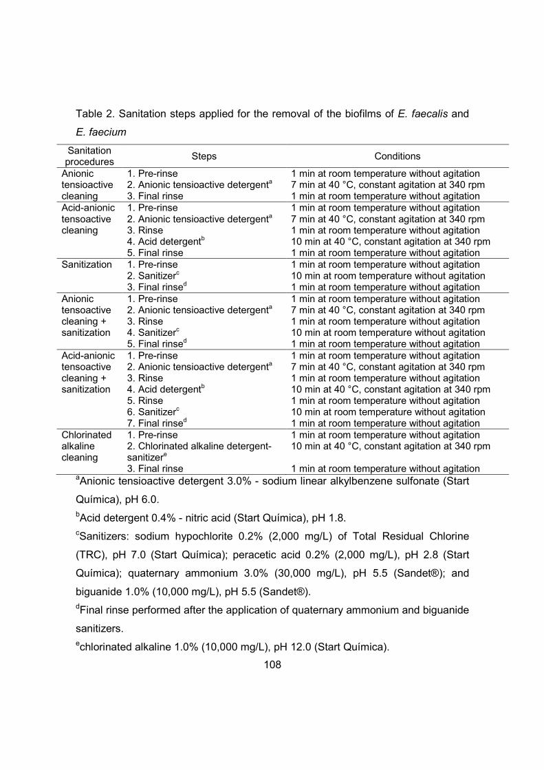
108
Table 2. Sanitation steps applied for the removal of the biofilms of E. faecalis and
E. faecium
Sanitation procedures
Steps Conditions
Anionic tensioactive cleaning
1. Pre-rinse 2. Anionic tensioactive detergenta 3. Final rinse
1 min at room temperature without agitation 7 min at 40 °C, constant agitation at 340 rpm 1 min at room temperature without agitation
Acid-anionic tensoactive cleaning
1. Pre-rinse 2. Anionic tensioactive detergenta 3. Rinse 4. Acid detergentb 5. Final rinse
1 min at room temperature without agitation 7 min at 40 °C, constant agitation at 340 rpm 1 min at room temperature without agitation 10 min at 40 °C, constant agitation at 340 rpm 1 min at room temperature without agitation
Sanitization 1. Pre-rinse 2. Sanitizerc 3. Final rinsed
1 min at room temperature without agitation 10 min at room temperature without agitation 1 min at room temperature without agitation
Anionic tensoactive cleaning + sanitization
1. Pre-rinse 2. Anionic tensioactive detergenta 3. Rinse 4. Sanitizerc 5. Final rinsed
1 min at room temperature without agitation 7 min at 40 °C, constant agitation at 340 rpm 1 min at room temperature without agitation 10 min at room temperature without agitation 1 min at room temperature without agitation
Acid-anionic tensoactive cleaning + sanitization
1. Pre-rinse 2. Anionic tensioactive detergenta 3. Rinse 4. Acid detergentb 5. Rinse 6. Sanitizerc 7. Final rinsed
1 min at room temperature without agitation 7 min at 40 °C, constant agitation at 340 rpm 1 min at room temperature without agitation 10 min at 40 °C, constant agitation at 340 rpm 1 min at room temperature without agitation 10 min at room temperature without agitation 1 min at room temperature without agitation
Chlorinated alkaline cleaning
1. Pre-rinse 2. Chlorinated alkaline detergent-sanitizere 3. Final rinse
1 min at room temperature without agitation 10 min at 40 °C, constant agitation at 340 rpm 1 min at room temperature without agitation
aAnionic tensioactive detergent 3.0% - sodium linear alkylbenzene sulfonate (Start
Química), pH 6.0. bAcid detergent 0.4% - nitric acid (Start Química), pH 1.8. cSanitizers: sodium hypochlorite 0.2% (2,000 mg/L) of Total Residual Chlorine
(TRC), pH 7.0 (Start Química); peracetic acid 0.2% (2,000 mg/L), pH 2.8 (Start
Química); quaternary ammonium 3.0% (30,000 mg/L), pH 5.5 (Sandet®); and
biguanide 1.0% (10,000 mg/L), pH 5.5 (Sandet®).
dFinal rinse performed after the application of quaternary ammonium and biguanide
sanitizers. echlorinated alkaline 1.0% (10,000 mg/L), pH 12.0 (Start Química).

109
The detergents were prepared according to the manufacturer’s instructions.
The sodium hypochlorite solution was prepared and analyzed according to the
methodology of the American Public Health Association (APHA) (Eaton
and Franson, 2005). The other sanitizers and the chlorinated alkaline product were
prepared according to the manufacturer’s instructions. All solutions were prepared
in sterile flasks, and then, 10 ml of the solution was transferred into sterile test
tubes. The pre-rinse, rinse and final rinse steps were performed in sterile test tubes
containing 10 ml of sterile ultra-pure water. All solutions were prepared at the time
of testing.
After performing the sanitation steps (Table 2), each coupon was transferred
to 5 ml of 0.1% peptone water supplemented with a neutralizing solution of 1%
sodium thiosulfate (for sodium hypochlorite, peracetic acid, acid detergent, and
chlorinated alkaline detergent) and Letheen broth (Difco, Becton, Dickinson and
Company) (for quaternary ammonium and biguanide) and vortexed (2800 rpm) for
two minutes to remove the sessile cells (Andrade et al., 1998). The resulting
solution was serially diluted in peptone water and plated onto BHI agar (Difco,
Becton, Dickinson and Company), and the plates were incubated at 35 °C for 48 h.
For the control coupons, the same procedures described for the determination of
adhered cells (section 2.1) were performed. Three replicates were performed for
each treatment, and in each replicate, 2 coupons were evaluated for each
sanitation step and 2 coupons were evaluated for the control. The control coupons
did not receive cleaning agents or sanitizers, and their counts were used to
calculate the number of decimal reductions due to the sanitation steps.
2.3 Statistical analysis of results
All investigated variables were subjected to an analysis of variance
(ANOVA), and the results were compared using Tukey’s test. Statistical
significance was defined as p<0.05. The statistical analyses were performed using
the STATISTICA 7.0 software (Statsoft, Tulsa, USA).

110
2.4 Scanning Electron Microscopy (SEM)
SEM was performed for the visualization of biofilms and to determine the
effect of the steps of sanitation on biofilm removal.
After 1 and 8 days of contact at 25 and 39 °C, the coupons were immersed
in 10 ml of 0.1% peptone water for 1 minute without agitation for the removal of
planktonic cells. Then, each coupon was immersed in 2 ml of 0.1 M phosphate
buffer solution supplemented with 2% glutaraldehyde (Sigma-Aldrich, St. Louis,
Mo., USA) for 3 h. The coupons were washed twice in a 0.1 M phosphate buffer
solution and subjected to dehydration with ethanol p.a. (Dinâmica Química
Contemporânea Ltda., Diadema, SP) as described by Lou et al. (2013). The
coupons were transferred to a Critical Point Dryer (Balzers, model CPD 030 -
Balzers Union AG, Balzers, Liechtenstein) with an injection of carbon dioxide for
complete removal of the alcohol. After drying, the coupons were coated with a thin
layer of gold for 180 seconds using a Sputter Coater (Balzers, model SCD 050 -
Balzers Union AG, Balzers, Liechtenstein). The coupons were then observed
under a Scanning Electron Microscope (JEOL, model JSM - 5800LV - JEOL,
Tokyo, Japan).
To observe the effect of the sanitation steps on biofilm removal, the coupons
were subjected to the sanitation steps (as described in section 2.2) after 1 and 8
days of contact at 25 and 39 °C, and then, the same steps described above were
performed.
3 Results and discussion
3.1 Assessment of biofilm formation
The formation of E. faecalis and E. faecium biofilms were evaluated at 7, 25 and
39 °C for 0, 1, 2, 4, 6 and 8 days. A mixture of 80% whey and 20% whole milk was
used as the culture medium. These conditions were in accordance with the
Brazilian Regulation (RIISPOA, article 610 - Brasil, 1952) to simulate the raw
materials used in manufacturing ricotta. The inoculum in the culture medium (1-
5x10² CFU/ml) was determined from the average count of this microorganism in
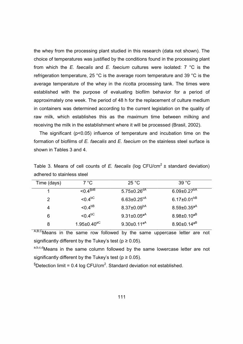
111
the whey from the processing plant studied in this research (data not shown). The
choice of temperatures was justified by the conditions found in the processing plant
from which the E. faecalis and E. faecium cultures were isolated: 7 °C is the
refrigeration temperature, 25 °C is the average room temperature and 39 °C is the
average temperature of the whey in the ricotta processing tank. The times were
established with the purpose of evaluating biofilm behavior for a period of
approximately one week. The period of 48 h for the replacement of culture medium
in containers was determined according to the current legislation on the quality of
raw milk, which establishes this as the maximum time between milking and
receiving the milk in the establishment where it will be processed (Brasil, 2002).
The significant (p<0.05) influence of temperature and incubation time on the
formation of biofilms of E. faecalis and E. faecium on the stainless steel surface is
shown in Tables 3 and 4.
Table 3. Means of cell counts of E. faecalis (log CFU/cm2 ± standard deviation)
adhered to stainless steel
Time (days) 7 °C 25 °C 39 °C
1 <0.4§bB 5.75±0.26dA 6.09±0.27bA
2 <0.4bC 6.63±0.25cA 6.17±0.01bB
4 <0.4bB 8.37±0.09bA 8.59±0.35ªA
6 <0.4bC 9.31±0.05ªA 8.98±0.10ªB
8 1.95±0.40aC 9.30±0.11ªA 8.90±0.14ªB A,B,CMeans in the same row followed by the same uppercase letter are not
significantly different by the Tukey’s test (p ≥ 0.05). a,b,c,dMeans in the same column followed by the same lowercase letter are not
significantly different by the Tukey’s test (p ≥ 0.05). §Detection limit = 0.4 log CFU/cm2. Standard deviation not established.

112
Table 4. Means of cell counts of E. faecium (log CFU/cm2 ± standard deviation)
adhered to stainless steel
Time (days) 7 °C 25 °C 39 °C
1 <0.4§B 5.96±0.16cA 6.07±0.06cA
2 <0.4B 5.91±0.03cA 6.03±0.08 cA
4 <0.4C 6.97±0.24bB 7.40±0.13bA
6 <0.4B 7.86±0.27aA 7.54±0.04 bA
8 <0.4C 7.84±0.06aB 8.03±0.07aA A,B,CMeans in the same row followed by the same uppercase letter are not
significantly different by the Tukey’s test (p ≥ 0.05). a,b,cMeans in the same column followed by the same lowercase letter are not
significantly different by the Tukey’s test (p ≥ 0.05). §Detection limit = 0.4 log10 CFU/cm2. Standard deviation not established.
Using a value of 5 CFU/cm2 as the threshold for the formation of the biofilm
(Ronner and Wong, 1993), the ability of E. faecalis and E. faecium to form a biofilm
after 1 day at 25 °C and 39 °C can be observed. At 7 °C, the microorganisms were
unable to form a biofilm during the 8 day period.
At 7 °C, the counts of E. faecalis and E. faecium differed significantly
(p<0.05) from the other temperatures for all days evaluated. At 25 and 39 °C, there
were significant differences (p<0.05) in the counts of E. faecalis after 2, 6, and 8
days and in those of E. faecium after 4 and 8 days.
There were no differences (p≥0.05) in the counts of E. faecalis after 1 day of
contact at 25 and 39 °C; the counts were 5.75 and 6.09 CFU/cm2, respectively.
After 8 days of contact, the counts increased by approximately 3.6 and 3.0 log at
25 and 39 °C, respectively. Thus, the biofilm formation was slightly better at 25 °C
than at 39 °C (p<0.05).
The counts of E. faecium did not differ after 1 day of contact at 25 and 39 °C
(p≥0.05) and were 5.96 and 6.07 CFU/cm2, respectively. After 8 days of contact,

113
the counts increased nearly 2.0 log for both temperatures. However, the biofilm
formation was slightly better at 39 °C than at 25 °C (p<0.05).
Due to the structure of their membrane (contents of fatty acids and lipids),
Enterococcus spp. have the ability to multiply in a wide temperature range (5 - 50
°C), with an optimum temperature of 42.7 °C (Fisher and Phillips, 2009). In this
study, the temperatures of 25 and 39 °C were both favorable for biofilm formation
of E. faecalis and E. faecium.
Although the temperature of 7 °C was not optimal for their multiplication,
some strains of Enterococcus spp. were able to grow over time at refrigeration
temperatures. After 8 days of contact, the count for E. faecalis was 1.95 CFU/cm2,
which, although not characteristic of a biofilm, indicated high adhesion of the
microorganism to the stainless steel surface that could evolve over time to a
mature biofilm. The counts for E. faecalis over 8 days of contact increased by at
least more than one log compared to E. faecium for all temperatures assessed. It is
clear from these results that E. faecalis has a greater ability than E. faecium to
form biofilms.
The influence of the pH of the culture medium on biofilm formation was
assessed for the different times and temperatures of incubation. The pH of the
medium before the inoculation of the microorganism was 6.9. At 7 °C, the pH was
7.0 and remained constant throughout the eight days of contact. For the
temperatures of 25 and 39 °C, after two days of incubation, the pH was 4.6 and
remained at this value throughout the 8 days of incubation because the culture
medium was changed every two days. Due to its membrane durability and
impermeability to acid and alkali (Fisher and Phillips, 2009), Enterococcus spp.
have the ability to multiply in a wide pH range (4.0 to 9.0). Throughout the 8 days
of incubation, the pH of the culture medium remained within the multiplication
range of Enterococcus, which possibly influenced the multiplication and adhesion
of the microorganism to the stainless steel.
Therefore, the conditions evaluated in this study, such as time, temperature
and pH, together with the nutrients available in the culture medium (which was

114
renewed every 48 h), influenced the adhesion and biofilm formation of E. faecium
and E. faecalis to the stainless steel surface. Other factors that may contribute to
biofilm formation of Enterococcus are their morphology (presence of pili, which
promote adhesion to other bacterial cells and inorganic particles) and the
production of EPS, which are responsible for cell-cell and cell-surface adhesion
(Forsythe, 2013; Franz et al., 2011).
According to Meira et al. (2012), the maturation of microbial biofilms usually
occurs between 3 and 6 days after the initial adhesion, and only after 10 days there
is an increase in the population density with the production and deposition of EPS.
However, in this study, the adhesion of the microorganism to the surface can be
observed (Fig. 1A) after 1 day of contact. After 8 days of contact, a dense matrix
composed of bacteria, EPS and culture medium components can be observed,
characterizing the formation of a mature biofilm (Fig. 1B). The EPS has a global
protective effect on microorganisms of the biofilm against adverse conditions, such
as the use of sanitizers (Bae et al., 2012; Simões et al., 2010), thus compromising
food safety.
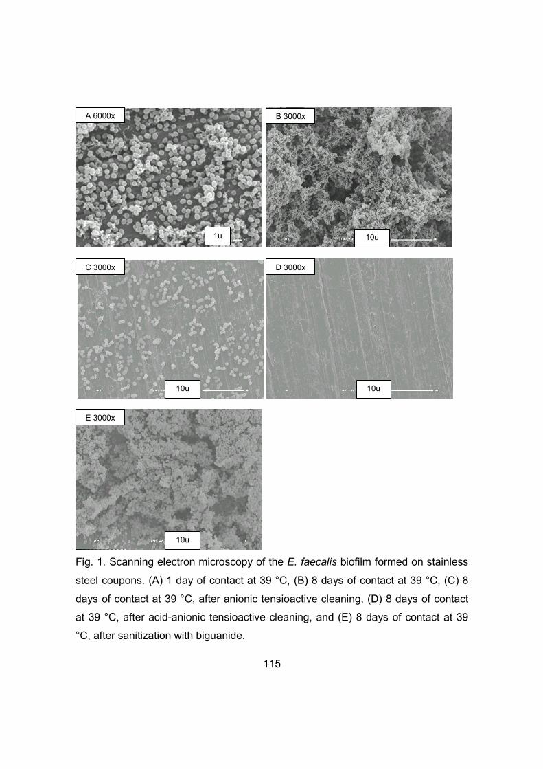
115
Fig. 1. Scanning electron microscopy of the E. faecalis biofilm formed on stainless
steel coupons. (A) 1 day of contact at 39 °C, (B) 8 days of contact at 39 °C, (C) 8
days of contact at 39 °C, after anionic tensioactive cleaning, (D) 8 days of contact
at 39 °C, after acid-anionic tensioactive cleaning, and (E) 8 days of contact at 39
°C, after sanitization with biguanide.
A 6000x
1u
B 3000x
10u
C 3000x
10u
D 3000x
10u
10u
E 3000x

116
3.2 Assessment of the sanitation steps in the removal of biofilms
Different sanitation steps for the removal of biofilms of E. faecalis and E.
faecium formed after 1 and 8 days of contact at 25 and 39 °C were evaluated: 1)
Anionic tensioactive cleaning, 2) acid-anionic tensioactive cleaning, 3) sanitization,
4) anionic tensioactive cleaning + sanitization, 5) acidic- anionic tensioactive
cleaning + sanitization and 6) chlorinated alkaline cleaning (Table 2).
The temperatures of 25 and 39 °C were selected because they allowed the
formation of E. faecium and E. faecalis biofilms. Days 1 and 8 of contact time were
selected because they showed the lowest (1 day) and highest (8 days)
microorganism counts (Tables 3 and 4).
According to Tables 5 and 6, there is a significant influence (p<0.05) of both
the temperature and the sanitation steps on the counts of E. faecium and E.
faecalis.

117
Table 5. Means* of the counts (log CFU/cm2) ± standard deviation of E. faecalis
after the different steps of sanitation after 8 days of contact at 25 and 39 °C
Types of sanitation 25 °C 39 °C
Control 8.00±0.57aA 8.23±0.13aA
Anionic tensioactive cleaning <0.4§cB 4.30±0.69cA
Acid- anionic tensioactive cleaning <0.4c <0.4d
Sanitization
Sodium hypochlorite
<0.4cB
6.67±0.18bA
Peracetic acid <0.4c <0.4d
Quaternary ammonium 3.93±0.64bA 3.74±0.24cA
Biguanide 7.97±0.66aA 8.18±0.22aA
Anionic tensioactive cleaning +
sanitization**
<0.4c <0.4 d
Acid-anionic tensioactive cleaning +
sanitization**
<0.4c <0.4 d
Chlorinated alkaline cleaning <0.4c <0.4 d *Means of three measurements.
A,BMeans in the same row followed by the same uppercase letter are not
significantly different by the Tukey’s test (p ≥ 0.05).
a,b,c,dMeans in the same column followed by the same lowercase letter are not
significantly different by the Tukey’s test (p ≥ 0.05). **Step performed with all sanitizers (sodium hypochlorite, peracetic acid, quaternary
ammonium and biguanide) in independents tests. §Detection limit = 0.4 log CFU/cm2. Standard deviation not established.

118
Table 6. Means* of the counts (log10 CFU/cm2) ± standard deviation of E. faecium
after the different steps of sanitation after 8 days of contact at 25 and 39 °C
Types of sanitation 25 °C 39 °C
Control 7.24±0.10ªB 7.61±0.16ªA
Anionic tensioactive cleaning <0.4§cB 3.33±0.13cA
Acid- anionic tensioactive cleaning <0.4c <0.4d
Sanitization
Sodium hypochlorite
4.10±0.04bB
6.53±0.39bA
Peracetic acid <0.4c <0.4d
Quaternary ammonium <0.4c <0.4d
Biguanide 6.75±0.49ªA 7.08±0.19ª,bA
Anionic tensioactive cleaning +
sanitization**
<0.4c <0.4d
Acid-anionic tensioactive cleaning +
sanitization **
<0.4c <0.4d
Chlorinated alkaline cleaning <0.4c <0.4d *Means of three measurements.
A,BMeans in the same row followed by the same uppercase letter are not
significantly different by the Tukey’s test (p ≥ 0.05). a,b,c,dMeans in the same column followed by the same lowercase letter are not
significantly different by the Tukey’s test (p ≥ 0.05). **Step performed with all sanitizers (sodium hypochlorite, peracetic acid, quaternary
ammonium and biguanide) in independents tests. §Detection limit = 0.4 log CFU/cm2. Standard deviation not established.
In biofilms formed at 39 °C after 1 day of contact (6.41±0.07 and 6.24±0.29
log CFU/cm2 for E. faecalis and E. faecium, respectively), sanitization with
biguanide reduced the counts of E. faecalis and E. faecium by 3.65 and 2.32 log
CFU/cm2, respectively. However, biguanide was not able to completely remove the
biofilms. All other sanitizers tested, as well as the other types of sanitation, reduced

119
the counts of the two microorganisms to levels below the limit of detection (<0.4 log
CFU/cm2). After 1 day of contact at 25 °C, the counts of E. faecalis and E. faecium
were 5.40±0.38 and 5.78±0.07 log CFU/cm2, respectively. In this case, all types of
sanitation reduced the counts of the two microorganisms to values <0.4 log
CFU/cm2. After 1 day of contact, the biofilm is not yet mature, and there is still no
well-developed EPS structure. These conditions facilitate the action of detergents
and sanitizers on the biofilm, which is more easily removed (Srey et al., 2013).
After 8 days of contact at 39 °C, step 1 - anionic tensioactive cleaning -
reduced (p<0.05) the E. faecalis counts by 3.93 log/cm2 and the E. faecium counts
by 4.28 log/cm2, although without completely removing the biofilms. Regarding the
temperature of 25 °C after 8 days of contact, the anionic tensioactive cleaning
reduced both microorganisms to counts <0.4 log CFU/cm2.
In this study, the anionic tensioactive detergent removed 4.28 log cycles of
the E. faecium biofilm and 3.93 log cycles of the E. faecalis biofilm. As observed in
Fig. 1C, the anionic tensioactive cleaning step removed the EPS matrix and the
components of the culture medium (fats, proteins and carbohydrates). Thus, cells
that were weakly adhered or wrapped in this matrix were released, leading to a
decrease in the E. faecalis and E. faecium counts after applying the anionic
tensioactive detergent. An agitation system (340 rpm), which simulates the
application of mechanical energy on the surface to help the cleaning process
(Forsythe, 2013), was coupled to the use of anionic tensioactive detergent.
Furthermore, the anionic tensioactive detergent was used at a temperature of 40
°C. However, the presence of bacteria adhered to the surface was still observed
(Fig. 1C). These data indicate that the use of anionic tensioactive cleaning alone is
not effective in the complete removal of biofilms of E. faecalis and E. faecium, and
remaining microorganisms may bind to other surfaces and form new biofilms.
Therefore, additional subsequent steps, such as acid cleaning and sanitization, are
indispensable for the reduction of microorganisms to acceptable levels (Srey et al.,
2013). In this study, the acid- anionic tensioactive cleaning sequence was able to
remove the biofilm and reduce the counts to levels below the limit of detection

120
(<0.4 log CFU/cm2). The use of the acid detergent under agitation (340 rpm)
contributed to the release of the remaining cells strongly adhered to the stainless
steel surface (Fig. 1D). The acid detergent may have contributed favorably to the
removal process by acting on the solubility of minerals in the biofilm-surface
interaction.
The anionic tensioactive cleaning + sanitization, acid- anionic tensioactive
cleaning + sanitization and the chlorinated alkaline cleaning steps reduced the
counts of the two microorganisms to levels <0.4 log CFU/cm2. The importance of
the cleaning steps in the removal of biofilms was evident, and in particular, the
anionic tensioactive cleaning complemented by another step (acid cleaning, acid
cleaning + sanitization or sanitization) promotes the complete removal of the
biofilms of both microorganisms from the stainless steel surface. Thus, the action
of the detergent was necessary for the removal of this nutrient and the EPS matrix,
facilitating the action of the acid detergent and/or sanitizers on the elimination of
cells.
However, when the sanitation step was tested directly on the biofilm, there
were differences (p<0.05) between the sanitizers, depending on the species and
on the temperature at which the biofilm was formed. For example, after 8 days of
contact at 25 °C, the treatments with peracetic acid and sodium hypochlorite were
the most effective in reducing the E. faecalis biofilms on the stainless steel
coupons, with counts <0.4 log CFU/cm2. After application of the quaternary
ammonium, the biofilms of E. faecalis decreased by 4.07 log CFU/cm2. After
application of the biguanide, the counts of E. faecalis did not differ (p≥0.05) from
the counts of the negative control, decreasing by only 0.03 log CFU/cm2. At 39 °C,
all sanitizers were significantly different from each other (p<0.05). Only the
treatment with peracetic acid was effective in reducing the E. faecalis biofilm on the
stainless steel coupons (<0.4 log CFU/cm2). The quaternary ammonium and
sodium hypochlorite reduced the biofilm by 4.49 and 1.56 log CFU/cm2,
respectively. The biguanide sanitizer was the least effective in removing the biofilm
of E. faecalis, with a reduction of only 0.05 log CFU/cm2. This sanitizer had virtually

121
no effect on the biofilms of E. faecalis (Fig. 1E), and a dense matrix of bacteria,
culture medium components and EPS was observed.
For the biofilms of E. faecium, after 8 days of contact at 25 °C, the
processes of sanitization with peracetic acid and quaternary ammonium were the
most effective on the stainless steel coupons, with counts <0.4 log CFU/cm2. The
reduction in the E. faecium counts after application of sodium hypochlorite was
3.14 log CFU/cm2. After application of biguanide, the counts of E. faecium did not
differ (p≥0.05) from the counts of the negative control, with a reduction of the
biofilm of only 0.49 log CFU/cm2. At 39°C, the treatments with peracetic acid and
quaternary ammonium were the most effective in reducing the E. faecium biofilm
on stainless steel coupons (<0.4 log CFU/cm2). There was no significant difference
(p≥0.05) in the counts of E. faecium after the use of sodium hypochlorite and
biguanide, which reduced the biofilm by 1.08 and 0.53 log CFU/cm2, respectively.
Since the formation of the E. faecalis biofilm was slight better at 25 °C than
at 39 °C, after 8 days of contact, it was expected that the sanitizers would have
greater difficulty in removing the biofilm at 25 °C. However, the sodium
hypochlorite was more effective at removing the biofilm formed at 25 °C than that
formed at 39 °C. The other sanitizers showed similar effectiveness for both
temperatures. In addition, given that the formation of the E. faecium biofilm was
slight better at 39 °C than at 25 °C, after 8 days of contact, it was expected that the
sanitizers would have greater difficulty in removing the biofilm at 39 °C; however,
this was observed only for sodium hypochlorite. The other sanitizers showed
similar effectiveness at both temperatures.
The E. faecalis isolates, aside from having a greater ability to form biofilms
(section 3.2), showed resistance to a greater number of sanitizers (sodium
hypochlorite, quaternary ammonium and biguanide) than E. faecium (resistant to
sodium hypochlorite and biguanide).
The resistance of biofilms formed after 8 days of contact to the sanitizers
may be associated with the presence of the EPS matrix and culture medium
components, which would act as a physical barrier or through chemical reactions

122
(Andrade et al., 2008). In this study, peracetic acid was able to remove all biofilms,
even in the presence of the EPS matrix and organic matter from the culture
medium, because it is a strong oxidizing agent and does not interact with organic
matter residues (Andrade et al., 2008).
4 Conclusion
E. faecalis and E. faecium were able to form biofilms on stainless steel
surfaces at the temperatures of 25 to 39 °C over 8 days. Although the
microorganisms did not form biofilms at 7 °C over the period of 8 days, the
adhesion of E. faecalis at 7 °C could be observed from the 8th day of contact.
The temperature and time of biofilm formation and the sanitation steps
influenced the removal of the biofilms. The steps of acid-anionic tensioactive
cleaning, anionic tensioactive cleaning + sanitization and acid-anionic tensioactive
cleaning + sanitization were effective in the removal of the biofilms. The peracetic
acid was the most efficient sanitizer, capable of removing all biofilms of E. faecium
and E. faecalis under all conditions tested. In contrast, the biguanide sanitizer was
the least effective because it was unable to remove any of the biofilms. The anionic
tensioactive cleaning step is extremely important for success in removing E.
faecium and E. faecalis biofilms.
The possible presence of these bacteria in environments or on surfaces
under harsh conditions of temperature and time, such as those tested here, should
be avoided during the processing of ricotta to not compromise food safety.
Acknowledgments
This work was supported by the Fundação de Amparo à Pesquisa do
Estado de São Paulo (FAPESP) – Process n°2010/10507-7. The authors are
indebted to SOORO for providing the cheese whey, to Start Química and Sandet®
for donating the detergents and sanitizers and to prof. Dr. Viotto (UNICAMP) for
help in the testing of the sanitation steps in the removal of the biofilms in this study.

123
References
Andrade, N. J., Pinto, C. L. O., Rosado, M. S., 2008. Controle da higienização na indústria de alimentos. In: Andrade, N. J. (Ed.), Higiene na Indústria de Alimentos – Avaliação e controle da adesão e formação de biofilmes bacterianos. Varela, São Paulo, pp. 412. Andrade, N. J., Ajão, D. B., Zottola, E. A., 1998. Growth and adherence on stainless steel by Enterococcus faecium cells. Journal of Food Protection 61, 1454-1458. Bae, Y. M., Baek, S. Y., Lee, S. Y., 2012. Resistance of pathogenic bacteria on the surface of stainless steel depending on attachment form and efficacy of chemical sanitizers. International Journal of Food Microbiology 153, 465-473. Barbosa, J., Ferreira, V., Teixeira, P., 2009. Antibiotic susceptibility of enterococci isolated from traditional fermented meat products. Food Microbiology 26, 527–532. Brasil, 1952. Ministério da Agricultura, Pecuária e Abastecimento. Decreto n° 30691, de 29 de março de 1952. In: Regulamento de Inspeção Industrial e Sanitária de Produtos de Origem Animal – RIISPOA. Available in: http://www.agricultura.gov.br. Brasil, 2002. Ministério da Agricultura, Pecuária e Abastecimento. Instrução Normativa nº 51, de 18 de setembro de 2002. In: Regulamento Técnico da coleta de leite cru refrigerado e seu transporte a granel. Available in: http://www.agricultura.gov.br. Cariolato, D., Andrighetto, C., Lombardi, A., 2008. Occurrence of virulence factors and antibiotic resistances in Enterococcus faecalis and Enterococcus faecium collected from dairy and human samples in North Italy. Food Control 19, 886-892. Costerton, J. W., Lewandowski, Z., Caldwell, D. E., Korber, D. R., Lappin-Socott, H. M., 1995. Microbial Biofilms. Annual Review of Microbiology 49, 711-745. Eaton, A. D., Franson, M. A. H., 2005. Standard Methods of the Examination of Water and Wastewater, 21th ed., American Public Health Association, Washington, DC, pp. 55. Fisher, K., Phillips, C., 2009. The ecology, epidemiology and virulence of Enterococcus. Microbiology 155, 1749-1757. Forsythe, S. J., 2013. Microbiologia da Segurança de Alimentos, second ed., Artemed, Porto Alegre.

124
Franz, C. M., Huch, M., Abriouel, H., Holzapfel, W., Gálvez, A., 2011. Enterococci as probiotics and their implications in food safety. International Journal of Food Microbiology 151, 125-140. Gelsomino, R., Vancanneyt, S. C., Cogan, T. M., Condon, S., Swings, J., 2002. Source of enterococci in a farmhouse raw-milk cheese. Applied and Environmental Microbiology 68, 3560-3565. Giraffa, G., 2003. Functionality of enterococci in dairy products. International Journal of Food Microbiology 88, 215– 222. Gomes, B. C., Esteves, C. T., Palazzo, I. C. V., Darini, A. L. C., Felis, G. E, Sechi, L. A., Franco, B. D. G. M., Martins, E. C. P., 2008. Prevalence and characterization of Enterococcus spp. isolated from Brazilian foods. Food Microbiology 25, 668– 675. Hancock, L. E., Perego, M., 2004. The Enterococcus faecalis fsr two-component system controls biofilm development through production of gelatinase. Journal of Bacteriology 186, 5629-5639. Jahan, M., Holley, R. A., 2014. Incidence of virulence factors in enterococci from raw and fermented meat and biofilm forming capacity at 25 °C and 37 °C. International Journal of Food Microbiology 170, 65-69. Kasimoglu-Dogru, A., Gencay, Y. E., Ayaz, N. D., 2010. Prevalence and antibiotic resistance profiles of Enterococcus species in chicken at slaughter level; absence of vanA and vanB genes in E. faecalis and E. faecium. Research in Veterinary Science 89, 153–158. Lou, Z., Song, X., Hong, Y., Wang H., Lin, Y., 2013. Separation and enrichment of burdock leaf components and their inhibition activity on biofilm formation of E. coli. Food Control 32, 270-274. Meira, Q. G. S., Barbosa, I. M., Athayde, A. J. A. A., Siqueira-Junior, J. P., Souza, E. L., 2012. Influence of temperature and surface kind on biofilm formation by Staphylococcus aureus from food-contact surfaces and sensitivity to sanitizers. Food Control 25, 469-475. Morton, R. D., 2001. Aerobic Plate Count. In: Downes, F. P., Ito, K. (Eds.), Compendium of methods for the microbiological examination of foods, 4th ed., American Public Health Association, Washington, DC, pp. 63-67.

125
Parizzi, S. Q. F., Andrade, N. J., Silva, A. S., Soares, N. F. F. S., Silva, A. M., 2004. Bacterial Adherence to Different Inert Surfaces Evaluated by Epifluorescence Microscopy and Plate Count Method. Brazilian Archives of Biology and Technology 47, 77-83. Peng, J. S., Tsai, W. C., Chou, C. C., 2002. Inactivation and removal of Bacillus cereus by sanitizer and detergent. International Journal of Food Microbiology 77, 11-18. Riboldi, G. P., Frazzon, J., D’Azevedo, P. A., Frazzon, A. P. G., 2009. Antimicrobial resistance profile of Enterococcus spp. isolated from food in southern Brazil. Brazilian Journal of Microbiology 40, 125-128. Ronner, A. B., Wong, A. C. L., 1993. Biofilm development and sanitizer inactivation of Listeria monocytogenes and Salmonella typhimurium on stainless steel and bunan rubber. Journal of Food Protection 56, 750-758. Simões, M., Simões, L. C., Machado, I., Pereira, M. O., Vieira, M. J., 2006. Control of flow-generated biofilms using surfactants – evidence of resistance and recovery. Food and Bioproducts Processing 84, 338–345. Simões, M., Simões, L., Vieira, M., 2010. A review of current and emergent biofilm control strategies. LWT - Food Science and Technology 43, 573-583. Srey, S., Jahid, I. K., Ha, S. D., 2013. Biofilm formation in food industries: A food safety concern. Food Control 31, 572-585. Suzzi, G., Caruso, M., Gardini, F., Lombardini, A., Vannini, L., Guerzoni, M. E., Andrighetto, C., Lanorte M. T., 2000. A survey of enterococci isolated from artisanal Italian goat’s cheese (semicotto caprino). Journal of Applied Microbiology 89, 267-274. Temelli, S., Anar, S., Sen, C., Akyuva, P., 2006. Determination of microbiological contamination sources during Turkish white cheese production. Food Control 17, 856-861. Toledo-Arana, A., Valle, J., Solano, C., Arrizubieta, M. J., Cucarella, C., Lamata, M., Amorena, B., Leiva, J., Penade´s, J. R., Lasa, I., 2001. The Enterococcal Surface Protein, Esp, is involved in Enterococcus faecalis biofilm formation. Applied and Environmental Microbiology 67, 4538-4545.

126

127
CAPÍTULO 5
ARTIGO 4
Este artigo será submetido a revista “Food Microbiology”.
Formação de biofilme multiespécie e quorum sensing em cepas
de Enterococcus faecium, Enterococcus faecalis e Bacillus
cereus isolados do processamento de ricota
Meg da Silva Fernandes, Dirce Yorika Kabuki,
Luciana Maria Ramires Esper, Arnaldo Yoshiteru Kuaye

128

129
Resumo
A formação de biofilme multiespécie e a capacidade de produção de moléculas
sinalizadoras de quorum sensing por cepas de E. faecalis, E. faecium e B. cereus
(células vegetativas e esporos) isoladas do processamento de ricota foram
avaliadas. Os ensaios de formação de biofilme em superfícies de cupons de aço
inoxidável, superfície esta amplamente utilizada nas indústrias de alimentos, foram
realizados variando-se a temperatura (7, 25 e 39 °C) e o tempo (0, 1, 2, 4, 6 e 8
dias). A exposição dos micro-organismos testes a temperatura de 7 °C, resultou
em contagens abaixo de 1,4 log UFC/cm2, valor este que não caracteriza a
formação de biofilme. Para as temperaturas de 25 e 39 °C, após 1 dia de
exposição, as contagens de Enterococcus sp. foram de 5,27 e 5,87 log ufc/cm2,
respectivamente – caracterizando a formação de biofilmes. As contagens de B.
cereus, para ambas as temperaturas em diferentes tempos de exposição,
situaram-se abaixo de 4,1 log ufc/cm2. A contagem de esporos de B. cereus
evoluiu ao longo do tempo, atingindo contagens em torno de 4,6 log ufc/cm2. Este
é o primeiro estudo a avaliar a interação entre estes diferentes gêneros e espécies
em um único biofilme, utilizando como meio de cultivo, o soro de leite, principal
matéria prima na fabricação da ricota. Destaca-se também que todas as cepas
avaliadas, além de apresentarem o gene luxS, também apresentaram capacidade
de induzir o fenômeno de bioluminescência em Vibrio harveyi BB170, indicando a
presença de autoindutores AI-2.
Palavras chave: E. faecalis, E. faecium, B. cereus, biofilme, quorum sensing.

130
1 Introdução
As superfícies de equipamentos utilizadas para a manipulação,
processamento e armazenamento de alimentos são reconhecidas como uma das
principais fontes de contaminação microbiana. Este fato é de grande preocupação
para a indústria de alimentos, principalmente pela capacidade que os micro-
organismos possuem de aderir e formar biofilmes nestas superfícies (Wong,
1998).
Os biofilmes são considerados uma comunidade complexa e estruturada de
micro-organismos, envoltos por uma matriz extracelular de polissacarídeos,
aderidos entre si e/ou a uma superfície ou interface (Costerton et al., 1995). Estes
biofilmes reduzem a eficiência dos sanitizantes, trazem prejuízos econômicos para
a indústria, além de constituírem um foco de contaminação para os alimentos
(Simões et al., 2010).
Na indústria de laticínios, os biofilmes podem ser uma fonte de
contaminação persistente, causando deterioração dos alimentos além de
causarem doenças transmitidas por alimentos, acarretando possíveis problemas
de saúde pública (Simões et al., 2010). Dentre estes patógenos, destacam-se B.
cereus e Enterococcus spp. que são frequentemente encontrados em leite e em
produtos lácteos, como por exemplo a ricota (Cossedu et al., 1997; De Santis et
al., 2008; Fadda et al., 2012; Fernandes et al., 2014; Gomes et al., 2008).
Bacillus cereus é capaz de produzir enterotoxinas que são responsáveis
pela ocorrência de dois tipos de doenças: a “síndrome emética” e a “síndrome
diarreica”. Algumas cepas também são resistentes a vários antibióticos (Ankolekar
et al., 2009; Chaves et al., 2011; Granum e Lund, 1997). Além disso, B. cereus
produzem esporos termo resistentes, que são capazes de aderir à superfície
assim como as células vegetativas. O processo de esporulação em um biofilme
torna-o ainda mais resistente à ação de detergentes e sanitizantes (Elhariry,
2011).
E. faecium e E. faecalis são considerados patógenos nosocomiais, que
causam diversos tipos de infecções principalmente nos seres humanos

131
imunocomprometidos. Além disso, Enterococcus isolados de alimentos tem sido
frequentemente associado à presença de genes de virulência e resistência a
antibióticos (Barbosa et al., 2009; Cariolato et al., 2008; Gomes et al., 2008;
Kasimoglu-Dogru et al., 2010).
Os biofilmes mais comuns na natureza são compostos por duas ou mais
espécies. Desta forma, os produtos do metabolismo de uma espécie auxiliam o
crescimento das demais e a adesão de uma espécie pode fornecer substâncias
que promovem a ligação de outras. Inversamente, a competição pelos nutrientes e
o acúmulo de metabólitos tóxicos produzidos pelas espécies colonizadoras
poderão limitar a diversidade de espécies em um biofilme (Nikolaev e Plakunov,
2007). Nas indústrias de laticínios, onde há uma diversidade de matérias-primas e
produtos, possivelmente os biofilmes apresentarão uma composição heterogênea
e comportamento variado. Alguns estudos já relataram a presença de B. cereus,
E. faecalis e E. faecium em plantas de processamento de queijos e também a
capacidade destes micro-organismos formarem biofilmes (Andrade et al., 1998;
Fernandes et al., 2014; Jahan e Holley, 2014; Kumari e Sarkar, 2014; Peña et al.,
2014).
Como os biofilmes são constituídos de agregados de células, eles se
tornam um ambiente propício para a comunicação célula-célula, denominado
quorum sensing (Bassler, 2002). O fenômeno quorum sensing corresponde a um
processo de comunicação intra e interespécies microbianas e é mediado por
sinais químicos extracelulares, denominados moléculas sinalizadoras ou
autoindutoras (AI). Estas moléculas são produzidas pelas bactérias durante a sua
multiplicação e são liberadas no ambiente. O acúmulo extracelular desses sinais
denota a presença de população relativamente densa e faz com que as bactérias
apresentem um comportamento coordenado (Griffiths, 2005; Reading e Sperandio,
2006). Este mecanismo permite que as células controlem muitas de suas funções,
tais como expressão de genes de virulência, transferência de plasmídeos,
produção de toxinas, formação de biofilmes, produção de exopolissacarídeos,

132
esporulação, dentre outros (Bassler, 2002; Nakayama et al., 2006; Sifri et al.,
2002; Simões et al., 2010; Zhu e Mekalanos, 2003).
Muitas bactérias, incluindo vários patógenos clinicamente importantes,
podem utilizar uma molécula - autoindutor AI-2 - para a comunicação intercelular.
A molécula AI-2 é um furanosil borato diester, obtida a partir de S-
adenosilmetionina e enzimas específicas como LuxS estão envolvidas neste
processo. LuxS é o responsável pela catalização na etapa final da biossíntese do
AI-2.
O objetivo deste estudo foi avaliar o efeito do tempo e da temperatura na
formação de biofilme multiespécie de E. faecium, E. faecalis e B. cereus, próximos
às condições reais em um indústria de lácteos (superfície de aço inoxidável e soro
de leite), além de pesquisar a capacidade de produção de moléculas sinalizadoras
de quorum sensing.
2 Material e Métodos
2.1 Cepas bacterianas
Para avaliação de biofilmes multiespécie, foram utilizadas 4 cepas de E.
faecium, 4 cepas de E. faecalis e 4 cepas de B. cereus, todas isoladas do
ambiente de processamento de uma indústria de ricota (Tabela 1). Estas cepas
foram selecionadas por apresentarem destacado perfil de virulência (presença de
genes de virulência e resistência a diferentes antibióticos) (Fernandes et al.,
2014). Todas as cepas do gênero Enterococcus apresentam os genes de
virulência esp e gelE que estão associados a formação de biofilmes (Hancock e
Perego, 2004; Toledo-Arana et al., 2001).

133
Tabela 1. Características das culturas utilizadas na formação do biofilme
multiespécie
Espécie (n° da cepa)
Local de isolamento Genes de virulência Resistência a antibióticos
E. faecium (89) Forma cylABM, gelE, vanB, esp, efaA, ace
-
E. faecium (104) Bancada de enformagem cylABM, gelE, vanB, esp, ace - E. faecium (109) Ar ambiente cylAM, gelE, esp Eritromicina
e Rifampicina E. faecium (122) Forma cylAM, gelE, esp, efaA Eritromicina
e Rifampicina E. faecalis (87) Tanque de processamento cylABM, gelE, esp, efaA, ace - E. faecalis (113) Caixa de recolhimento do
soro cylABM, gelE, agg, efaA, ace Tetraciclina
Rifampicina E. faecalis (118) Tanque de processamento cylABM, gelE, vanB, agg, esp,
efaA, ace Rifampicina
E. faecalis (130) Bancada de embalagem cylAM, gelE, vanB, agg, esp, efaA, ace
Tetraciclina
B. cereus (164) Bancada de embalagem hblA, hblC, hblD, nheA, nheB, nheC
Ampicilina, Penicilina e Trimetropim
B. cereus (167) Caixa de armazenamento hblC, hblD, nheA, nheB, nheC
Ampicilina, Penicilina e Trimetropim
B. cereus (174) Ralo de escoamento do soro
hblA, hblC, hblD, nheA, nheB, nheC
Ampicilina, Penicilina e Trimetropim
B. cereus (176) Forma hblA, hblC, hblD, nheA, nheB, nheC
Ampicilina, Penicilina e Trimetropim
2.2 Avaliação da formação de biofilme
A formação de biofilme bacteriano foi avaliada em cupons de aço inoxidável
AISI 304, acabamento número 4 (quadrados com dimensões de 10 mm x 10 mm x
1 mm). Antes de cada ensaio os cupons foram lavados com cetergente neutro
líquido, enxaguados com água destilada, imersos em álcool 70% (v/v) por 1h a
temperatura ambiente, enxaguados novamente com água destilada e esterilizados
a 121 °C por 15 min (Parizzi et al., 2004).
O meio de cultura utilizado para a formação de biofilme consistiu de uma
mistura de 80% de soro de leite em pó (SOORO®, Soro concentrado indústria de
produtos lácteos LTDA, Marechal Cândido Rondon, Paraná, Brasil) reconstituido

134
em água ultra pura estéril (concentração final de 6,5% de sólidos solúveis) e 20%
de leite integral UHT (SHEFA®, Usina de beneficiamento agropecuária Tuiuti
LTDA, Amparo, São Paulo, Brasil).
Cada cultura foi ativada separadamente em caldo Brain Heart Infusion - BHI
(Difco, Becton, Dickinson and Company, Sparks, MD, USA) a 35°C por 24h. Após,
1 ml de cada cultura ativada foi misturada em um tubo estéril e submetida a vortex
(2800 rpm) por 1 min. Diluições decimais seriadas foram realizadas até a obtenção
de suspensão de aproximadamente 1x10⁴ ufc de micro-organismos por ml.
O meio de cultivo foi homogeneizado e distribuído em frascos estéreis com
capacidade de 80 ml, sendo que em cada frasco a concentração do inóculo final
foi de aproximadamente 1x10² UFC/ml. Quatro cupons de aço inoxidável foram
imersos assepticamente de modo que ficassem suspensos no recipiente. Estes
recipientes foram incubados nas temperaturas de 7, 25 e 39 °C e as análises
realizadas nos tempos 0, 1, 2, 4, 6 e 8 dias. Após 48 horas de incubação, os
cupons foram imersos em um novo meio de cultivo inoculado com a mesma
suspensão de micro-organismos utilizada inicialmente, a fim de manter o
fornecimento constante de nutrientes. Depois de cada inoculação, a contagem do
meio de cultivo foi determinada em placas de Plate Count Agar – PCA (Difco,
Becton, Dickinson and Company), que foram incubadas a 32 °C por 48 h (Morton,
2001).
O pH do meio de cultivo foi determinado antes da inoculação da suspensão
de micro-organismos e após cada inoculação subsequente para cada temperatura
analisada.
A avaliação da adesão e formação de biofilme foi realizada pela técnica de
contagem em placas. A cada tempo e temperatura de contato, dois cupons foram
retirados do meio de cultivo e transferidos separadamente para tubos contendo 10
ml de água peptonada (0,1% peptona, Difco, Becton, Dickinson and Company)
onde ficaram imersos por 1 minuto em repouso para retirada das células
planctônicas. Em seguida, cada cupom foi imerso em 5 ml da mesma solução e
submetidos a vortex (2800 rpm) por dois minutos para remoção das células

135
sésseis (Andrade et al., 1998). A partir da solução resultante, foram realizadas as
diluições subsequentes seguidas do plaqueamento em superfície em ágar
Mannitol Yolk Polimixin – MYP – (Difco, Becton, Dickinson and Company) para a
enumeração de B. cereus. A contagem de Enterococcus spp. foi realizada pela
técnica de plaqueamento em profundidade em ágar Kenner Fecal – KF – (Difco,
Becton, Dickinson and Company) suplementado com 1% de cloreto 2,3,5 -
trifeniltetrazólio – TTC – (Vetec Química, Rio de Janeiro, Brasil). As placas foram
incubadas a 35 °C por 48 h. Para a contagem total de esporos aeróbios mesófilos,
os cupons foram submetidos a um choque térmico de 80±1 °C por 12 minutos,
seguidos de plaqueamento em PCA (Difco, Becton, Dickinson and Company)
suplementado com 0,1% de amido solúvel e incubação a 32 °C por 48 horas
(Frank e Yousef, 2004). O experimento foi repetido três vezes.
Paralelamente, foram realizadas as contagens de Enterococcus sp. e B.
cereus do meio de cultivo após 1, 2, 4, 6 e 8 dias nas temperaturas de 7, 25 e 39
°C. A cada tempo e temperatura, 1 ml do meio de cultivo foi diluído seriadamente
em 9 ml de água peptonada 0,1%. Em seguida, alíquotas de 1ml foram
plaqueadas em ágar KF (Difco, Becton, Dickinson and Company) suplementado
com 1% de TTC (Vetec Química, Rio de Janeiro, Brasil) para a contagem de
Enterococcus spp. e 0,1 ml em ágar MYP (Difco, Becton, Dickinson and Company)
para a contagem de B. cereus. As placas foram incubadas a 35 °C por 48 h.
Todas as variáveis estudadas foram submetidas a Análise de Variância
(ANOVA) e os resultados comparados através do Teste de Tukey. A significância
estatística foi definida como p<0,05. Os testes foram realizados no software
STATISTICA versão 7.0 (Statsoft, Tulsa, USA).
2.3 Microscopia eletrônica de varredura (MEV)
Após 1 e 8 dias de contato a 25 e 39 °C, os cupons foram imersos em 10 ml
de água peptonada 0,1% por 1 min sem agitação para a remoção das células
planctônicas. Após, cada cupom foi imerso em 2 ml de solução tampão fosfato 0,1
M suplementada com 2% de gluteraldeído (Sigma-Aldrich, St. Louis, Mo., USA)

136
por 3 h. Os cupons foram lavados duas vezes em uma solução de tampão fosfato
0,1 M e submetidos desidratação com etanol p.a. (Dinâmica Química
Contemporânea Ltda., Diadema, SP) como descrito por Lou et al. (2013). Os
cupons foram transferidos para o Critical Point Dryer (Balzers, model CPD 030 -
Balzers Union AG, Balzers, Liechtenstein) com uma injecção de dióxido de
carbono para a completa remoção do álcool. Após a secagem, os cupons foram
revestidos com uma fina camada de ouro por 180 segundos usando o Sputter
Coater (Balzers, model SCD 050 - Balzers Union AG, Balzers, Liechtenstein).
Após, os cupons foram observados em microscópio eletrônico de varredura
(JEOL, model JSM - 5800LV - JEOL, Tokyo, Japan).
2.4 Pesquisa de quorum sensing
2.4.1 Detecção do gene luxS
Todas as culturas utilizadas na formação do biofilme multiespécie foram
avaliadas quanto à presença do gene luxS. Os primers (Invitrogen, Life
Technologies, Carlsbad, USA) utilizados para a amplificação do gene luxS estão
demosntrados na Tabela 2. Os primers foram desenhados com base nas
sequências dos genomas de E. faecalis e E. faecium que se encontram
depositadas no GenBank do National Center for Biotechnology Information – NCBI
– no formato Fasta, para a identificação do gene luxS.
Tabela 2. Sequência de primers utilizados para a detecção do gene luxS
Micro-organismo
Sequência de oligonucleotídeos (5’ para 3’)
Tamanho do produto
(pb)
Referência
luxS E. faecalis CACCATATGTTCGCCTTGCT ATAAAAACCAGTGCGGCAAC
203 Este estudo
luxS E. faecium GAGCACTTGACTGCCGAACT GCCACATTGTGTTTCATTGC
198 Este estudo
luxS B. cereus CCCTTTCACAGGCAGTTTTC GATCATACGATTGTAAAGGCACC
450 Esper, 2010

137
O DNA genômico das culturas puras foi extraído de acordo com o método
descrito por Furrer et al. (1991). A mistura de PCR continha 0,2 µl da enzima Taq
polimerase (1U/µl), 2,5 µl de tampão 10 X (200 mM Tris-HCl- pH 8,0, 500 mM
KCl), 1,5 µl de MgCl2 50mM, 0,5 µl de dNTPs 10 mM, 1 µl (12,5 mM) de cada um
dos primers, 1 ul do DNA extraído e água Mili-Q esterilizada, totalizando 25 µl. As
condições da reação no termociclador foram: ciclo inicial de 94 ºC por 3 min, 35
ciclos de 94 ºC por 30 seg, 60 ºC por 1 min, 72 ºC por 1,5 min, e etapa final de
extensão de 72 ºC por 7 min e depois refrigerada para 4 ºC. A eletroforese dos
produtos da PCR foi realizada em gel de agarose (Invitrogen, Life Technologies,
EUA) a 1,5% e os produtos visualizados através de transiluminador UV.
2.4.2 Detecção de moléculas sinalizadoras de quorum sensing AI-2
As culturas utilizadas na formação do biofilme multiespécie foram incubadas
em caldo Luria-Bertani (LB) (Difco, Becton, Dickinson and Company) acrescido de
0,5% de glicose, sob agitação constante de 175 rpm por 24 h a 30 °C. Em
seguida, as culturas foram diluídas 1:100 em caldo LB (acrescido de 0,5% de
glicose) e incubadas novamente nas mesmas condições descritas, por 6 horas.
Alíquotas de 1 ml do crescimento bacteriano foram transferidas para tubos
Eppendorf® e centrifugadas (centrífuga Eppendorf® 5415D, Hamburg, Germany)
a 15000xg por 5 minutos. Os sobrenadantes foram filtrados em membrana com
0,22 µm de poro (Millex® GV, Merck Millipore Ltd., Co. Cork, Ireland) As amostras
foram submetidas ao teste imediatamente após a filtração.
O biossensor Vibrio harveyi BB170 foi ativado em meio Autoinducer
Bioassay (AB) (Surette e Bassler, 1998) e incubado a 30 °C por 16 h sob agitação
constante de 175 rpm. Em seguida, a cultura foi diluída 1:5000 em meio AB. Como
controles positivos foram utilizadas as cepas de Salmonella Typhimurium ATCC
14028 e Escherichia coli O157:H7. Como controles negativos foram utilizados os
meios de cultura LB e AB estéreis (Taga e Xavier, 2011).
Para a detecção de AI-2, 10 µl de sobrenadante filtrado das cepas e 90 µl
da diluição de Vibrio harveyi BB170 foram depositados nas cavidades das placas

138
de microtítulo (Greiner Bio-One GmbH, Frickenhausen, Germany). As placas
foram incubadas a 30 °C, sob agitação constante de 300 rpm e a cada 15 minutos,
durante pelo menos 5 horas, a atividade de bioluminescência foi quantificada, em
relative light units (RLU) através de luminômetro (FLUOstar OPTIMA, BMG
Labtech, Offenburg, Germany). Todos os ensaios foram realizados em triplicata e
os resultados apresentados corresponderam à indução de bioluminescência, a
qual foi determinada pela seguinte equação (Taga e Xavier, 2011):
Eq. 1:
Indução da bioluminescência = RLU da amostra/RLU controle negativo.
3 Resultados e discussão
3.1 Formação de biofilmes
A significativa (p<0,05) influência da temperatura e do tempo de incubação
na formação de biofilmes multiespécies na superfície do aço inoxidável é mostrada
nas Tabelas 3 e 4.
Tabela 3. Médias das contagens de Enterococcus spp. (log ufc/cm2±desvio
padrão) aderidas em aço inoxidável para as temperaturas de 7, 25 e 39 °C em
cultivo misto com B. cereus
Dia Temperaturas
7 °C 25 °C 39 °C
1 <0,4§b 5,27±0,49aC 5,87±0,07aC
2 <0,4c 6,34±0,40aB 5,26±0,34bC
4 <0,4c 8,35±0,14aA 7,04±0,70bB
6 <0,4c 8,75±0,04aA 7,65±0,45bA,B
8 <0,4c 9,12±0,37aA 8,30±0,25bA a,bMédias na mesma linha acompanhadas de mesma letra minúscula não são significativamente diferentes pelo teste de Tukey (p>0,05). A,B,CMédias na mesma coluna acompanhadas de mesma letra maiúscula não são significativamente diferentes pelo teste de Tukey (p≥0,05). §Limite de detecção do método = 0,4 log ufc/cm2. Desvio padrão não estabelecido.

139
Tabela 4. Médias das contagens de células vegetativas de B. cereus (log
ufc/cm2±desvio padrão) aderidas em aço inoxidável para as temperaturas de 7, 25
e 39 °C em cultivo misto com Enterococcus spp.
Dia Temperaturas
7 °C 25 °C 39 °C
1 <1,4§b 3,14±0,54aB 3,55±0,55aA
2 <1,4b 2,99±0,48aB <1,4bB
4 <1,4b 4,05±0,05aA <1,4bB
6 <1,4 <1,4C <1,4B
8 <1,4 <1,4C <1,4B a,bMédias na mesma linha acompanhadas de mesma letra minúscula não são significativamente diferentes pelo teste de Tukey (p>0,05). A,B,CMédias na mesma coluna acompanhadas de mesma letra maiúscula não são significativamente diferentes pelo teste de Tukey (p≥0,05). §Limite de detecção do método = 1,4 log ufc/cm2. Desvio padrão não estabelecido.
A exposição dos micro-organismos testes à temperatura de 7 °C, não
mostrou o desenvolvimento de biofilme durante o período de 8 dias de contato,
sugerindo que as cepas utilizadas neste estudo não são psicrotróficas.
Nos ensaios a 25 °C e 39 °C, Enterococcus spp. (E. faecalis e E. faecium)
apresentaram capacidade de formar biofilmes após 1 dia (Tabela 3). As contagens
de Enterococcus spp., após 1 dia, não diferiram entre si (p≥0,05) para as
temperaturas de 25 e 39 °C e foram de 5,27 e 5,87 log ufc/cm2, respectivamente.
Após 8 dias de contato, as contagens nos biofilmes formados a 25 e 39 °C,
aumentaram em torno de 4,0 e 3,0 ciclos log, respectivamente. Entretanto, a
formação do biofilme foi maior a 25 °C do que a 39 °C (p<0,05), sendo a
temperatura de 25 °C considerada mais favorável para a formação de biofilme de
Enterococcus spp. em cultivo misto (multiespécie). Estes dados são importantes,
pois 25 °C é a média da temperatura ambiente e 39 °C é a temperatura média em
que o soro de queijo se apresentava no tanque de processamento de ricota
durante as visitas à industria, demonstrando que nestas temperaturas, que são

140
frequentes na indústria de alimentos, há a possibilidade de formação de biofilmes
nos equipamentos utilizados.
Os biofilmes de B. cereus a 25 e 39 °C, no entanto, apresentaram
contagens baixas, entre 2,99 a 4,05 log ufc/cm2 (Tabela 4), não características de
biofilme. As contagens de B. cereus, após 1 dia de contato nas temperaturas de
25 e 39 °C (p≥0,05) foram de 3,14 e 3,55 log ufc/cm2, respectivamente. Após 4
dias, as contagens aumentaram em torno de 1 ciclo log para a temperatura de 25
°C, enquanto que a 39 °C, as contagens de B. cereus decaíram para valores
abaixo do limite de detecção do método (<1,4 log ufc/cm2) a partir do 2º dia de
incubação.
Independente da espécie microbiana ou superfície analisada postula-se que
o processo de adesão ocorra com a máxima intensidade quando as bactérias são
mantidas na faixa de temperatura ótima de crescimento (Meira et al., 2012).
Assim, Enterococcus spp. e B. cereus por serem mesófilos (Fisher e Phillips,
2009; Rajkowski e Bennett, 2003) teriam boas condições de crescimento a 25 e 39
°C. Entretanto, em cultivos mistos, para ambas as temperaturas e diferentes
períodos, observou-se uma maior contagem (p<0,05) de Enterococcus spp. em
relação ao B. cereus. Este resultado revelou que os Enterococcus spp.
apresentaram maior capacidade de formar biofilme além de possivelmente
exercerem um efeito antagonista frente ao B. cereus.
Em cultivos mistos observou-se ainda, ao longo do tempo de incubação a
25 e 39 °C, a ocorrência de um aumento significativo (p<0,05) da contagem de
esporos de B. cereus (Tabela 5), que pode ser atribuído a exposição do B. cereus
às condições desfavoráveis de pH, temperatura e presença de micro-organismos
competidores que influenciaram na sua esporulação. Além disso, o fornecimento
constante de nutrientes (substituição do meio de cultivo a cada 48 h) pode ter
favorecido este aumento (Fernandes et al., 2014; Lindsay et al., 2006). Os
esporos, devido às suas características morfológicas e de hidrofobicidade, aderem
mais facilmente à superfície do que as células vegetativas (Ankolekar e Labbé,
2010; Pagedar e Singh, 2012; Parkar et al., 2001).

141
Tabela 5. Médias das contagens de esporos de B. cereus (log ufc/cm2±desvio
padrão) aderidas em aço inoxidável para as temperaturas de 7, 25 e 39 °C em
cultivo misto com Enterococcus spp.
Dia Temperaturas
7 °C 25 °C 39 °C
1 <0,4§b 2,04±0,11aB 2,32±0,31aD
2 <0,4b 2,41±0,25aB 2,73±0,21aC,D
4 <0,4b 3,48±0,27aA 3,35±0,15aB,C
6 <0,4b 4,12±0,33aA 3,59±0,26aB
8 <0,4b 4,12±0,37aA 4,59±0,21aA a,bMédias na mesma linha acompanhadas de mesma letra minúscula não são significativamente diferentes pelo teste de Tukey (p>0,05). A,B,C,DMédias na mesma coluna acompanhadas de mesma letra maiúscula não são significativamente diferentes pelo teste de Tukey (p≥0,05). §Limite de detecção do método = 0,4 log ufc/cm2. Desvio padrão não estabelecido.
Avaliando as contagens do meio de cultivo (2 log ufc/ml) onde os cupons
foram imersos e comparando com as contagens de células sésseis dos cupons,
verifica-se que a 7 °C, a contagem de Enterococcus spp. no meio de cultivo estava
em torno de 3,5 log ufc/ml e no cupom estava abaixo do limite de detecção do
método (<0,4 ufc/cm2). Portanto, Enterococcus spp. foram capazes de se
multiplicar no meio de cultivo, mas não de aderir ao aço inoxidável. Em
contrapartida, B. cereus não foi capaz de sobreviver no meio de cultivo, com
contagens abaixo do limite de detecção do método (<1 log ufc/ml). A 25 °C,
Enterococcus sp. e B. cereus estavam presentes no meio de cultivo com elevadas
contagens (acima de 6 log ufc/ml), mas as contagens de Enterococcus spp.
aderidos ao cupom de aço inoxidável eram em torno de 2 ciclos log superiores às
de B. cereus. Neste caso, a diminuição da população de B. cereus no cupom
provavelmente não foi resultado da morte celular e sim de um desprendimento das
células que ficaram presentes no meio de cultivo (Ryu e Beuchat, 2005). A 39 °C,
as contagens de Enterococcus spp. permaneceram acima de 8 log ufc/ml no meio

142
de cultivo, enquanto que B. cereus apresentaram contagens acima de 6 log ufc/ml
apenas no primeiro dia. A partir do segundo dia as contagens de células
vegetativas de B. cereus decaíram para valores abaixo do limite de detecção do
método (<2 log ufc/ml). Neste caso, mesmo a uma temperatura favorável, o
desenvolvimento e sobrevivência de B. cereus em ambiente multiespécie foi
afetado pela microbiota concorrente - os enterococos.
O estudo prévio (Fernandes et al., 2014) com as mesmas culturas de B.
cereus utilizadas no presente estudo, mostrou que estas culturas, quando em
cultivo simples (monoespécie), apresentaram contagens superiores a 5 log ufc/cm2
após 4 dias de contato a 25 °C e após 1 dia de contato a 39 °C. Portanto, no
presente estudo, pode-se verificar a forte influência da seleção dos E. faecalis e E.
faecium, com suas características de tolerância ao calor e sobrevivência em
situações adversas no ambiente (Giraffa, 2002), em competição multiespécie.
Além disso, alguns estudos têm relatado que E. faecium e E. faecalis isolados de
alimentos podem produzir uma variedade de bacteriocinas, que são ativas contra
patógenos de origem alimentar, como B. cereus (Hajikhani et al., 2007; Valenzuela
et al., 2009).
O desenvolvimento dos biofilmes também pode ser influenciado pelo pH do
meio de cultivo. Antes do contato do micro-organismo com a superfície abiótica, o
pH do meio era de 7,0. Nos ensaios a temperatura de 7 °C, pouca variação do pH
foi observada, com valores de 6,9 do inicio até o 8° dia de contato. Para as
temperaturas de 25 e 39 °C, após trocas períodicas do meio de cultivo a cada dois
dias, o pH declinava de 7,0 para 4,6. Estas condições são favoráveis à
multiplicação e ao desenvolvimento dos biofilmes de Enterococcus spp. no aço
inoxidável. Em contrapartida, para B. cereus, a exposição periódica a um baixo pH
pode ter afetado negativamente a multiplicação e induzido a formação de esporos.
Desta forma, o pH pode ter interferido no crescimento do biofilme (Karunakaran e
Biggs, 2011; Peña et al., 2014).
O desenvolvimento de biofilmes multiespécie depende das características
particulares de cada micro-organismo. Na Fig. 1 A pode-se observar uma

143
quantidade pequena de B. cereus aderidos à superfície após 4 dias de contato.
Por sua vez, neste mesmo cupom (Fig. 1 B) a matriz composta por Enterococcus
sp., EPS e constituintes do meio de cultivo, caracterizam-se pela formação de um
biofilme maduro. A presença desta matriz exerce um efeito protetor sobre os
micro-organismos do biofilme contra condições adversas, como o contato com
sanitizantes (Bae et al., 2012; Simões et al., 2010).
Observou-se em particular a capacidade do B. cereus em se adaptar às
condições adversas do meio ambiente (metabólitos dos enterococos e variações
de pH) através da esporulação crescente e formação de biofilmes na forma de
esporos.
Fig. 1. Microscopia eletrônica de varredura de biofilme multiespécie de Enterococcus spp. e B. cereus formado em cupons de aço inoxidável a 25 °C por 4 dias.
3.2 Avaliação do sistema quorum sensing
A possível comunicação célula a célula entre Enterococcus spp. e B. cereus
foi evidenciada pela presença da enzima LuxS e pela reação positiva de
bioluminescência.
Todas as cepas de B. cereus, E. faecalis e E. faecium apresentaram o gene
luxS, o que demonstra a possibilidade de produção do autoindutor AI-2 (Xavier e
Bassler, 2003). O autoindutor AI-2 é sintetizado pela enzima LuxS, que está
envolvida no metabolismo da S-adenosil-metionina (SAM), esta converte S-ribosil
A 3000x
10u
B 6000x
1u
144
homocisteína em homocisteína e 4,5-dihidróxi-2,3-pentanodiona (DPD). DPD é um
composto muito instável que reage com a água e cicliza para formar vários
furanonas, uma das quais é considerada a precursora de AI-2 (Schauder et al.,
2001).
Os resultados indicativos da presença de moléculas sinalizadoras de
quorum sensing AI-2 através da detecção da presença do gene luxS foram
corroborados pela confirmação da capacidade de indução do fenômeno de
bioluminescência em Vibrio harveyi BB170. Índices relativos de atividade de luz
(RLU) normatizados e variando de 0,89 a 1,59 foram obtidos. De acordo com a
Fig. 2, destacam-se as cepas de E. faecalis n° 113 e B. cereus n° 174, que
mostraram maior atividade de indução que outras amostras testadas.
Ao calcular a atividade de AI-2, levou-se em consideração o fato de que,
além da possível presença de moléculas de AI-2 exógeno – produzido pelas cepas
testes – existia também a produção de AI-2 endógeno – produzido pelo V. harveyi.
O AI-2 endógeno não é alvo do estudo e o mesmo pode causar erro de
interpretação, uma vez que após o período de algumas horas, com o aumento da
população de V. harveyi BB170, ocorre o acúmulo de AI-2 endógeno, induzindo
novamente a bioluminescência (Piton, 2012). Portanto, ao calcular a atividade de
AI-2, a luz foi medida no instante imediatamente anterior ao início da produção de
luz pelo V. harveyi (Taga, Xavier, 2011).

145
Amostras
Fig. 2. Indução de bioluminescência em amostras de Vibrio harveyi BB170 para detecção de autoindutor AI-2 de sobrenadantes de cepas de E. faecalis (n° 87, 113, 118, 130), E. faecium (n°89, 104, 109, 122) e B. cereus (n° 164, 167, 174, 176). aIndução da bioluminescência= relative light units da amostra/ relative light units do controle negativo.
O AI-2 desempenha um papel importante no aumento da formação de
biofilme de E. faecalis (Shao et al., 2012) e B. cereus (Auger et al., 2006). O
estudo de Auger et al. (2006) verificou também que o aumento do nível de AI-2 no
meio resulta numa diminuição da densidade do biofilme de B. cereus. Este
resultado indica que a presença de AI-2 também pode provocar a liberação de
uma grande proporção das células do biofilme. Embora não tenha sido
comprovado o efeito do AI-2 na formação do biofilme no presente estudo, esta
poderia ser uma das possíveis explicações do B. cereus estar presente no meio
de cultivo, mas não ter aderido à superfície.
Para muitos patógenos bacterianos, a produção de biofilme é regulada pelo
sistema quorum sensing (QS) (Auger et al., 2006; Hardie e Heurlier, 2008;
Reading e Sperandio, 2006; Zhu e Mekalanos, 2003). Dois sistemas de QS, Cyls e
Fsr, têm sido bastante estudados para E. faecalis. Entretanto, até o momento,
Indu
ção
de
bio
lum
ine
scê
nci
aa

146
apenas um trabalho (Shao et al., 2012) estudou o papel do LuxS/AI-2 na formação
de biofilme de E. faecalis. Além disso, não há estudos que mostrem a relação do
sistema QS na formação de um biofilme multiespécie. Está comprovado que o tipo
de bactéria, concentração de células e nutrientes disponíveis no meio afetam o
sistema QS (Surette e Basler, 1998).
No presente estudo, embora alguns indicativos como a presença do gene
luxS e a capacidade de induzir o fenômeno da bioluminescência tenham sido
evidenciados, não é conclusiva a correlação entre o quorum sensing e a formação
de biofilmes multiespécie. No entanto, foi dado o primeiro passo neste sentido,
uma vez que as bactérias isoladas do ambiente de processamento de ricota e que
são capazes de formar biofilme apresentaram o gene luxS e foram capazes de
induzir o fenômeno da bioluminescência. Além disso, quando em cultivo misto, B.
cereus não foi capaz de formar biofilme e fica a dúvida se o sistema quorum
sensing mais uma vez poderia estar envolvido nesta relação. Portanto, fica
registrada a importância destes dados e a sugestão da sua utilização em futuros
trabalhos.
4 Conclusão
Em cultivo misto apenas as cepas de Enterococcus spp. foram capazes de
formar biofilmes em superfície de aço inoxidável nas temperaturas de 25 e 39 °C
ao longo de 8 dias, ao contrário de B. cereus que sofreu redução ao longo do
tempo. A exposição à temperatura de 7 °C mostrou-se inadequada para a
formação de biofilme multiespécie.
Os resultados encontrados neste trabalho sugerem que os mecanismos
envolvidos na formação de biofilme multiespécie bem como na esporulação do
micro-organismo ao longo do tempo, pode estar relacionado com o tempo de
contato, temperatura e pH do meio de cultivo.
A confirmação da presença do gene luxS associada a resposta positiva do
fenômeno de bioluminescência com Vibrio harveyi BB170, em todos os isolados

147
estudados, corrobora com a hipótese da presença do autoindutor AI-2 e existência
do fenômeno de quorum sensing.
Agradecimentos
Este trabalho foi apoiado pela Fundação de Amparo à Pesquisa do Estado
de São Paulo (FAPESP) – Processo n°2010/10507-7. Os autores agradecem a
empresa SOORO por ter fornecido o soro de leite utilizado na realização deste
trabalho. Os autores também agradecem ao prof. Dr. Geraldo Renato de Paula
pelo auxílio no desenho dos primers e a profa. Dra. Maristela da Silva Nascimento
pela doação da cultura de B. cereus ATCC 14579.
Referências
Andrade, N.J., Ajão, D.B., Zottola, E.A., 1998. Growth and adherence on stainless steel by Enterococcus faecium cells. J. Food Protect. 61, 1454-1458. Ankolekar, C., Rahmati, T., Labbé, R.G., 2009. Detection of toxigenic Bacillus cereus and Bacillus thuringiensis spores in U.S. rice. Int. J. Food Microbiol. 128, 460-466. Ankolekar, C., Labbé, R.G., 2010. Physical characteristics of spores of food-associated isolates of the Bacillus cereus group. Appl. Environ. Microbiol. 76, 982–984. Auger, S., Krin, E., Aymerich, S., Gohar, M., 2006. Autoinducer 2 affects biofilm formation by Bacillus cereus. Appl. Environ. Microbiol. 72, 937–941. Bae, Y.M., Baek, S.Y., Lee, S.Y., 2012. Resistance of pathogenic bacteria on the surface of stainless steel depending on attachment form and efficacy of chemical sanitizers. Int. J. Food Microbiol. 153, 465-473. Barbosa, J., Ferreira, V., Teixeira, P., 2009. Antibiotic susceptibility of enterococci isolated from traditional fermented meat products. Food Microbiol. 26, 527–532. Bassler, B.L., 2002. Small talk. Cell–to-cell communication in bacteria. Cell. 109, 421-424.

148
Brasil, 2002. Ministério da Agricultura, Pecuária e Abastecimento. Instrução Normativa nº 51, de 18 de setembro de 2002, in: Regulamento Técnico da coleta de leite cru refrigerado e seu transporte a granel. Available in: http://www.agricultura.gov.br. Cariolato, D., Andrighetto, C., Lombardi, A., 2008. Occurrence of virulence factors and antibiotic resistances in Enterococcus faecalis and Enterococcus faecium collected from dairy and human samples in North Italy. Food Control 19, 886-892. Chaves, J.Q., Pires, E.S., Vivoni, A.M., 2011. Genetic diversity, antimicrobial resistance and toxigenic profiles of Bacillus cereus isolated from food in Brazil over three decades. Int. J. Food Microbiol. 147, 12–16. Cossedu, A.M., Santi, E.P.L., Mazzette, R., Fresi, A., Lai, G., 1997. Ricotta bovina fresca confezionata: caratteristiche microbiologiche di interesse igienico-sanitario. Latte. 7, 76-81. Costerton, J.W., Lewandowski, Z., Caldwell, D. E., Korber, D. R., Lappin-Socott, H. M., 1995. Microbial Biofilms. Annu. Rev. Microbiol. 49, 711-745. De Santis, E. P. L., Foddai, A., Virdis, S., Marongiu, P., Pilo, A. L., Scarano, C. Toxin gene pattern in Bacillus cereus group strains isolated from sheep ricotta cheese. Vet. Res. Commun. 32, 323-336. Elhariry, H.M., 2011. Attachment strength and biofilm forming ability of Bacillus cereus on green-leafy vegetables: Cabbage and lettuce. Food Microbiol. 28, 1266-1274. Esper, L. M. R., 2010. Enterobacter sakazakii (Cronobacter spp) e Bacillus cereus: Quorum sensing, formação de biofilme e ação de sanitizantes. Campinas – SP, 104p. Tese (Doutorado em Tecnologia de Alimentos) – Departamento de Tecnologia de Alimentos, Universidade Estadual de Campinas. Fadda, A., Delogu, A., Mura, E., Noli, A. C., Porqueddu, G., Rossi, M. L., Terrosu, G., 2012. Presenza di Bacillus cereus, Escherichia coli e Enterobacteriaceae in ricotta fresca e salata: controlli ufficiali nel periodo 2009 – 2012. Ital. J. Food Safety, 5, 43-45. Fernandes, M.S., Fujimoto, G., Schneid, I., Kabuki, D.Y., Kuaye, A.Y., 2014. Enterotoxigenic profile, antimicrobial susceptibility, and biofilm formation of Bacillus cereus isolated from ricotta processing. Int. Dairy J. 38, 16-23. Fisher, K., Phillips, C., 2009. The ecology, epidemiology and virulence of Enterococcus. Microbiol. 155, 1749-1757.

149
Frank, J.F., Yousef, A.E., 2004. Tests for groups of microorganisms, in: Wehr, H., Frank, J.F. (Eds.), Standard Methods for the Examination of Dairy Products. American Public Health Association. Washington, pp. 227-248. Furrer, B., Candrian, U., Hoefelein, C.H., Luethy, J., 1991. Detection and identification of Listeria monocytogenes in cooked sausage products and in milk by in vitro amplification of hemolysis gene fragments. J. Appl. Bacteriol. 70, 372-379. Giraffa, G., 2002. Enterococci from foods. FEMS Microbiol. Rev. 26, 163-171. Gomes, B.C., Esteves, C.T., Palazzo, I.C.V., Darini, A.L.C., Felis, G.E, Sechi, L.A., Franco, B.D.G.M., Martins, E.C.P., 2008. Prevalence and characterization of Enterococcus spp. isolated from Brazilian foods. Food Microbiol. 25, 668– 675. Granun, P.E., Lund, T., 1997. Bacillus cereus and its food poisoning toxins. FEMS Microbio. Lett. 157, 223-228. Griffiths, M., 2005. Quorum sensing, in: Griffiths, M. (Ed.), Understanding pathogen behaviour: virulence, stress response and resistance. Woodhead, pp.580-640. Hajikhani, R., Beyatli, Y., Aslim, B., 2007. Antimicrobial activity of enterococci strains isolated from white cheese. Int. J. Dairy Technol. 60, 105-108. Hancock, L.E., Perego, M., 2004. The Enterococcus faecalis fsr two-component system controls biofilm development through production of gelatinase. J. Bacteriol. 186, 5629-5639. Hardie, K.R., Heurlier, K., 2008. Establishing bacterial communities by ‘word of mouth’: LuxS and autoinducer 2 in biofilm development. Nat. Rev. Microb. 6, 635-643. Jahan, M., Holley, R.A., 2014. Incidence of virulence factors in enterococci from raw and fermented meat and biofilm forming capacity at 25 °C and 37 °C. Int. J. Food Microbiol. 170, 65-69. Karunakaran, E., Biggs, C.A., 2011. Mechanisms of Bacillus cereus biofilm formation: an investigation of the physicochemical characteristics of cell surfaces and extracellular proteins. Appl. Microbiol. Biotechnol. 89, 1161-1175. Kasimoglu-Dogru, A., Gencay, Y.E., Ayaz, N.D., 2010. Prevalence and antibiotic resistance profiles of Enterococcus species in chicken at slaughter level; absence of vanA and vanB genes in E. faecalis and E. faecium. Res. Vet. Sci. 89, 153–158.

150
Kumari, S., Sarkar, P. K., 2014. In vitro model study for biofilm formation by Bacillus cereus in dairy chilling tanks and optimization of clean-in-place (CIP) regimes using response surface methodology. Food Control, 36, 153-158. Lindsay, D., Brözel, V.S., Von Holy., 2006. Biofilm-spore response in Bacillus cereus and Bacillus subtilis during nutrient limitation. J. Food Protect., 69, 1168-1172. Lou, Z., Song, X., Hong, Y., Wang H., Lin, Y., 2013. Separation and enrichment of burdock leaf components and their inhibition activity on biofilm formation of E. coli. Food Control. 32, 270-274. Meira, Q.G.S., Barbosa, I.M., Athayde, A.J.A.A., Siqueira-Junior, J.P., Souza, E.L., 2012. Influence of temperature and surface kind on biofilm formation by Staphylococcus aureus from food-contact surfaces and sensitivity to sanitizers. Food Control. 25, 469-475. Morton, R.D., 2001. Aerobic Plate Count, in: Downes, F.P., Ito, K. (Eds.), Compendium of methods for the microbiological examination of foods. American Public Health Association, Washington, pp. 63-67. Nakayama, J.S., Chen, N., Oyama, K., Nishiguchi, E.A., Azab, E., Tanaka, R., Kariyama, Sonomoto, K., 2006. Revised model for Enterococcus faecalis fsr quorum-sensing system: small open reading frame, fsrD, encodes GBAP propeptide corresponding to staphylococcal AgrD J. Bacteriol. 188, 8321-8326. Nikolaev, Y.A., Plakunov, V.K., 2007. Biofilm - “City of microbes” or an analogue of multicellular organisms? Microbiol. 76, 125-138. Pagedar, A., Singh, J., 2012. Influence of physiological cell stages on biofilm formation by Bacillus cereus of dairy origin. Int. Dairy J. 23, 30-35. Parizzi, S.Q.F., Andrade, N.J., Silva, A.S., Soares, N.F.F.S., Silva, A.M., 2004. Bacterial Adherence to Different Inert Surfaces Evaluated by Epifluorescence Microscopy and Plate Count Method. Braz. Arch. Biol. Technol. 47, 77-83. Parkar, S.G., Flint, S.H., Palmer, J.S., Brooks. J.D., 2001. Factors influencing attachment of thermophilic bacilli to stainless steel. J. Appl. Microbiol. 90, 901-908. Peña, W.E.L., Andrade, N.J., Soares, N.F.F., Alvarenga, V.O., Rodrigues Junior, S., Granato, D., Zuniga, A.D.G., Sant’Ana, A.S., 2014. Modelling Bacillus cereus adhesion on stainless steel surface as affected by temperature, pH and time. Int. Dairy J. 34, 153-158.

151
Pillai, S.K., Sakoulas, G., Eliopoulos, G.M., Moellering, R.C., Murray, B.E., Inouye, R.T., 2004. Effects of glucose on fsr-mediated biofilm formation in Enterococcus faecalis. J. Infect. Dis. 190, 967−970. Rajkowski K.T., Bennett, R.W., 2003. Bacillus cereus, in: Miliotis, M.D., Bier, J.W. (Eds.), International Handbook of Foodborne Pathogens, Marcel Dekker, Inc., New York, pp. 27–39. Reading, N.C., Sperandio, V., 2006. Quorum sensing: the many languages of bacteria. FEMS Microbiol. Lett. 254, 1-11. Ryu, J.H., Beuchat, L.R., 2005. Biofilm formation and sporulation by Bacillus cereus on a stainless steel surface and subsequent resistance of vegetative cells and spores to chlorine, chlorine dioxide, and a peroxyacetic acid-based sanitizer. J. Food Protect. 68, 2614-2622. Schauder, S., Shokat, K., Surette, M.G., Bassler, B.L., 2001. The LuxS family of bacterial autoinducers: biosynthesis of a novel quorumsensing signal molecule. Mol. Microbiol. 41, 463−476. Shao, C., Shang, W., Yang, Z., Sun, Z., Li, Y., Guo, J., Wang, X., Zou, D.Y., Wang, S., Lei, H., Cui, Q., Yin, Z., Li, X., Wei, X., Liu, W., He, X., Jiang, Z., Du, S., Liao, X., Huang, L., Wang, Y., Yuan, J., 2012. LuxS-Dependent AI‑2 Regulates Versatile Functions in Enterococcus faecalis V583. J. Prot. Res. 11, 4465−4475. Sifri, C.D., Mylonakis, E., Singh, K.V., Qin, X., Garsin, D.A., Murray, B.E., Ausubel, F.M., Calderwood, S.B., 2002. Virulence effect of Enterococcus faecalis protease genes and the quorum-sensing locus fsr in Caenorhabditis elegans and mice. Infect. Immun. 70, 5647-5650. Simões, M., Simões, L., Vieira, M., 2010. A review of current and emergent biofilm control strategies. LWT - Food Sci. Technol. 43, 573-583. Surette, M.G., Bassler, B.L., 1998. Quorum sensing in Escherichia coli and Salmonella typhimurium. Proceedings of the National Academy of Sciences of the United States of America. 995, 7046-50. Taga, M.E., Xavier, K.B., 2011. Methods for Analysis of Bacterial Autoinducer-2 Production. Curr. Protoc. Microbiol. 23, 1-15. Toledo-Arana, A., Valle, J., Solano, C., Arrizubieta, M.J., Cucarella, C., Lamata, M., Amorena, B., Leiva, J., Penade´s, J.R., Lasa, I., 2001. The Enterococcal Surface Protein, Esp, is involved in Enterococcus faecalis biofilm formation. Appl. Environ. Microbiol. 67, 4538-4545.

152
Valenzuela, A.S., Omar, N., Abriouel, H., López, R.L., Veljovic, K., Canamero, M.M., Topisirovic, M.K.L., Gálvez, A., 2009. Virulence factors, antibiotic resistance, and bacteriocins in enterococci from artisan foods of animal origin. Food Control. 20, 381–385. Xavier, K. B., Bassler, B. L. (2003). LuxS quorum sensing: more than just a numbers game. Curr. Opin. Microbiol. 6, 191–197. Wong, A.C.L., 1998. Biofilm in food processing environments. J. Dairy Sci. 81, 2765–2770. Zhu, J., Mekalanos J., 2003. Quorum sensing-dependent biofilms enhance colonization in Vibrio cholerae. Develop. Cell. 5, 647-656.

153
CAPÍTULO 6
ARTIGO 5
Este artigo será submetido a revista “Food Control”.
Influência dos procedimentos de higienização na remoção de
biofilme multiespécie de Enterococcus faecium, Enterococcus
faecalis e Bacillus cereus isolados do processamento de ricota
Meg da Silva Fernandes; Ana Carolina Alvares;
Dirce Yorika Kabuki; Arnaldo Yoshiteru Kuaye

154

155
Resumo
A eficácia dos procedimentos de limpeza e sanitização na remoção de biofilmes
multiespécie de E. faecalis, E. faecium e B. cereus formados nas temperaturas de
25 e 39 °C e tempos de 1 e 8 dias foi avaliada. Os procedimentos de higienização:
a) limpeza com tensoativo aniônico e detergente ácido, b) limpeza com tensoativo
aniônico + sanitizante e c) limpeza com tensoativo aniônico e detergente ácido +
sanitizante, foram eficazes na remoção dos biofilmes reduzindo as contagens a
níveis inferiores a 0,4 log cfu/cm2. Nos biofilmes de 1 dia a 25 e 39 °C, todos os
procedimentos de higienização foram eficientes na remoção do biofilme
multiespécie, com exceção do biofilme de 1 dia a 39 °C, no qual a biguanida
removeu apenas 1,38 ciclos log cfu/cm2 das contagens de Enterococcus spp. A
aplicação do detergente tensoativo aniônico nos biofilmes de 8 dias, promoveu a
remoção de aproximadamente 5 ciclos log cfu/cm2 de Enterococcus spp. além da
remoção total de esporos de B. cereus (<0,4 log ufc/cm2). O ácido peracético foi o
sanitizante com melhor desempenho reduzindo as contagens de Enterococcus
sp., células vegetativas e esporos de B. cereus para valores inferiores a 0,4
ufc/cm2 em todas as condições avaliadas. Por sua vez, a biguanida foi o
sanitizante menos eficiente, com exceção do ensaio com biofilme de 8 dias a 25
°C, onde o hipoclorito de sódio foi o menos eficiente. Esta pesquisa revelou a
importância da etapa de limpeza e do tipo de agente sanitizante nos processos de
higienização para efetiva remoção dos biofilmes multiespécie.
Palavras chave: E. faecalis, E. faecium, B. cereus, biofilme multiespécie,
higienização.
156
1 Introdução
Os biofilmes são considerados uma comunidade complexa e estruturada de
micro-organismos, envoltos por uma matriz extracelular de polissacarídeos (EPS),
aderidos entre si e/ou a uma superfície ou interface (Costerton, Lewandowski,
Caldwell, Korber, & Lappin-Socott, 1995). Em um biofilme, os micro-organismos
passam a ter capacidade de sobreviver em ambientes hostis, sendo a sua
fisiologia e o seu comportamento significativamente diferentes do que as células
planctônicas (Simões, Simões, & Vieira, 2010).
Na indústria de laticínios, os biofilmes são compostos predominantemente
por micro-organismos, EPS e resíduos de leite, principalmente proteínas e fosfato
de cálcio. A formação de biofilmes neste tipo de indústria pode levar a sérios
prejuízos econômicos, reduzir a eficiência dos sanitizantes, além de constituírem
um foco de contaminação para os alimentos (Simões, Simões, & Vieira, 2010).
Uma das principais estratégias utilizadas no controle de biofilmes é a
higienização. Os procedimentos de higienização consistem no uso combinado de
detergentes e sanitizantes, no qual os detergentes, embora diminuam a carga
bacteriana das superfícies, tem como função principal a remoção de resíduos
orgânicos e minerais, enquanto a sanitização visa à redução dos micro-
organismos deterioradores e a eliminação dos patogênicos a níveis seguros
(Forsythe, 2013). Porém, os micro-organismos que se encontram no interior dos
biofilmes, devido à estrutura de EPS, são protegidos da remoção quando expostos
ao escoamento de líquidos e a alta turbulência, além de serem protegidos da ação
de sanitizantes (Simões, Simões, & Vieira, 2010). Normalmente as indústrias de
alimentos realizam uma avaliação laboratorial dos sanitizantes a serem utilizados.
No entanto, na maioria das vezes, apenas suspensões microbianas são levadas
em conta, não considerando a formação de exopolissacarídeos, fundamental para
a adesão (Peng, Tsai, & Chou, 2002).
Os biofilmes, muitas vezes, são constituídos de micro-organismos
patogênicos, que por sua vez podem contaminar os alimentos e vir a causar
doenças. Dentre estes patógenos, destacam-se os Enterococcus spp. e B. cereus,

157
por serem contaminantes potenciais do leite e do ambiente de indústrias de
laticínios (Giraffa, 2003; Rajkowski & Bennett, 2003). B. cereus apresenta uma
característica peculiar de produzir esporos resistentes ao calor (Granum & Lund,
1997). Enterococcus spp. também são capazes de sobreviver a temperaturas
elevadas, contribuindo, desta forma, para sua sobrevivência durante as etapas de
fabricação de produtos lácteos. Estes, por sua vez, se não forem removidos de
forma eficiente, podem promover a formação dos biofilmes bacterianos (Bower,
McGuire, & Daeschel, 1996).
A patogenicidade de Enterococcus spp. pode ser atribuída à sua
capacidade de obter genes de virulência de outros micro-organismos patogênicos
e de ser resistente a antibióticos, característica essas que podem estar
relacionadas, em parte, à habilidade natural destas bactérias em adquirir
plasmídeos, os quais são responsáveis pela transferência de genes de virulência e
de resistência (Eaton & Gasson, 2001).
Bacillus cereus está associado com a ocorrência de intoxicação alimentar
por produzir enterotoxinas, além de ser resistente a vários antibióticos (Ankolekar,
Rahmati, & Labbé, 2009; Chaves, Pires, & Vivoni, 2011; Granum & Lund, 1997).
Além disso, B. cereus produzem esporos termo resistentes, que são capazes de
aderir à superfície assim como as células vegetativas. O processo de esporulação
em um biofilme torna-o ainda mais resistente à ação de detergentes e sanitizantes
(Elhariry, 2011).
Portanto, o objetivo deste trabalho foi avaliar a eficácia dos procedimentos
de limpeza e sanitização na remoção de biofilmes multiespécie de E. faecalis, E.
faecium e B. cereus.
2 Material e métodos
2.1 Formação de biofilme multiespécie
Para avaliação de biofilmes multiespécie, foram utilizadas 4 cepas de E.
faecium, 4 cepas de E. faecalis e 4 cepas de B. cereus, todas isoladas do
ambiente de processamento de uma indústria de ricota. Estas cepas foram
158
selecionadas por apresentarem destacado perfil de virulência (presença de genes
de virulência, resistência a diferentes antibióticos) (Fernandes, Fujimoto, Schneid,
Kabuki, & Kuaye, 2014). Além disso, as cepas de Enterococcus spp. apresentam
os genes de virulência esp e gelE que estão associados a formação de biofilmes
(Hancock & Perego, 2004; Toledo-Arana et al., 2001).
A formação de biofilme bacteriano foi avaliada em cupons de aço inoxidável
AISI 304, acabamento número 4 (quadrados com dimensões de 10 mm x 10 mm x
1 mm). Antes de cada ensaio os cupons foram lavados com detergente netro
líquido, enxaguados com água destilada, imersos em álcool 70% (v/v) por 1h a
temperatura ambiente, enxaguados novamente com água destilada e esterilizados
a 121 °C por 15 min (Parizzi, Andrade, Silva, Soares, & Silva 2004).
O meio de cultura utilizado para a formação de biofilme consistiu de uma
mistura de 80% de soro de leite em pó (SOORO®, Soro concentrado indústria de
produtos lácteos LTDA, Marechal Cândido Rondon, Paraná, Brasil) reconstituido
em água ultra pura estéril (concentração final de 6,5% de sólidos solúveis) e 20%
de leite integral UHT (SHEFA®, Usina de beneficiamento agropecuária Tuiuti
LTDA, Amparo, São Paulo, Brasil).
A formação do biofilme foi realizada de acordo com o protocolo descrito por
Fernandes, Fujimoto, Schneid, Kabuki, & Kuaye (2014).
2.2 Avaliação das etapas de higienização na remoção de biofilme
multiespécie
Após 1 e 8 dias de contato nas temperaturas de 25 e 39 °C, os cupons
foram retirados do meio de cultivo e imediatamente submetidos aos diferentes
processos de higienização, conforme as etapas descritas na Tabela 1.

159
Tabela 1. Etapas de higienização aplicadas para a remoção dos biofilmes de E.
faecium e E. faecalis
Tipo de
higienização
Etapas Condições
Limpeza com tensoativo aniônico
1. Pré-enxágue 2. Detergente tensoativo aniônico 3. Enxágue final
1 min a temperatura ambiente sem agitação 7 min a 40 °C, agitação constante de 340 rpm 1 min a temperatura ambiente sem agitação
Limpeza com tensoativo aniônico e detergente ácido
1. Pré-enxágue 2. Detergente tensoativo aniônico 3. Enxágue 4. Detergente ácido 5. Enxágue final
1 min a temperatura ambiente sem agitação 7 min a 40 °C, agitação constante de 340 rpm 1 min a temperatura ambiente sem agitação 10 min a 40 °C, agitação constante de 340 rpm 1 min a temperatura ambiente sem agitação
Sanitização 1. Pré-enxágue 2. Sanitizante 3. Enxágue final*
1 min a temperatura ambiente sem agitação 10 min a temperatura ambiente sem agitação 1 min a temperatura ambiente sem agitação
Limpeza com tensoativo aniônico + sanitização
1. Pré-enxágue 2. Detergente tensoativo aniônico 3. Enxágue 4. Sanitizante 5. Enxágue final*
1 min a temperatura ambiente sem agitação 7 min a 40 °C, agitação constante de 340 rpm 1 min a temperatura ambiente sem agitação 10 min a temperatura ambiente sem agitação 1 min a temperatura ambiente sem agitação
Limpeza com tensoativo aniônico e detergente ácido + sanitização
1. Pré-enxágue 2. Detergente tensoativo aniônico 3. Enxágue 4. Detergente ácido 5. Enxágue 6. Sanitizante 7. Enxágue final*
1 min a temperatura ambiente sem agitação 7 min a 40 °C, agitação constante de 340 rpm 1 min a temperatura ambiente sem agitação 10 min a 40 °C, agitação constante de 340 rpm 1 min a temperatura ambiente sem agitação 10 min a temperatura ambiente sem agitação 1 min a temperatura ambiente sem agitação
Limpeza alcalina-clorada
1. Pré-enxágue 2. Detergente alcalino-clorado 3. Enxágue final
1 min a temperatura ambiente sem agitação 10 min a 40 °C, agitação constante de 340 rpm 1 min a temperatura ambiente sem agitação
*enxágue final realizado após a aplicação dos sanitizantes quaternário de amônio
e biguanida.
Para as etapas de limpeza, foram utilizados: i) detergente tensoativo
aniônico a base de linear alquilbenzeno sulfonato de sódio (Start Química, Lima &
Pergher Ind. Com. e Rep. LTDA, Uberlândia, MG) a 3,0%, pH 6,0 e ii) detergente
ácido a base de ácido nítrico (Start Química, Lima & Pergher Ind. Com. e Rep.
LTDA) a 0,4%, pH 1,8. Os detergentes foram preparados de acordo com as
indicações do fabricante.

160
Para as etapas de sanitização, foram utilizadas as soluções de: a)
hipoclorito de sódio a 0,2% (2,000 mg/L) de Cloro Residual Total (CRT), pH 7,0
(Start Química, Lima & Pergher Ind. Com. e Rep. LTDA), b) ácido peracético a
0,2% (2,000 mg/L), pH 2,8 (Start Química, Lima & Pergher Ind. Com. e Rep.
LTDA), c) quaternário de amônio a 3,0% (30,000 mg/L), pH 5,5 (Sandet® Química
LTDA, São José do Rio Preto, SP) e d) biguanida a 1,0% (10,000 mg/L), pH 5,5
(Sandet® Química LTDA). Além disso, foi avaliada a eficiência de um produto: e)
alcalino-clorado a 1,0% (10,000 mg/L), pH 12,0 (Start Química, Lima & Pergher
Ind. Com. e Rep. LTDA). A solução clorada foi preparada e analisada de acordo
com a metodologia da American Public Health Association – APHA – (Eaton &
Franson, 2005). Os demais sanitizantes e o produto alcalino-clorado foram
preparados de acordo com as indicações dos fabricantes. Todas as soluções
foram preparadas em frascos estéreis e após foram transferidos 10 mL da solução
em tubos de ensaio estéreis. As etapas de pré-enxague, enxágue e enxágue final
foram realizadas em tubos de ensaio estéreis contendo 10 mL de água ultra pura
estéril. Todas as soluções foram preparadas no momento da realização dos
testes.
Após a realização das etapas de higienização (Tabela 1), cada cupom foi
transferido para 5 mL de água peptonada 0,1% acrescida de solução neutralizante
de tiossulfato de sódio a 1% (para hipoclorito de sódio, ácido peracético,
detergente ácido, detergente alcalino-clorado) e caldo Letheen (Difco, Becton,
Dickinson and Company) (para quaternário de amônio e biguanida) e submetidos
a vórtex (2800 rpm) por dois minutos para remoção das células sésseis (Andrade,
Ajão, & Zottola 1998). A partir da solução resultante, foram realizadas as diluições
subsequentes seguidas do plaqueamento em superfieice em ágar Mannitol Yolk
Polimixin – MYP – (Difco, Becton, Dickinson and Company) para a enumeração de
B. cereus. A contagem de Enterococcus spp. foi realizada pela técnica de
plaqueamento em profundidade em ágar Kennel Fecal – KF – (Difco, Becton,
Dickinson and Company) suplementado com 1% de cloreto 2,3,5 - trifeniltetrazólio
– TTC – (Vetec Química, Rio de Janeiro, Brasil). As placas foram incubadas a 35

161
°C por 48 h. Para a contagem total de esporos aeróbios mesófilos, os cupons
foram submetidos a um choque térmico de 80±1 °C por 12 minutos, seguidos de
plaqueamento em profundidade em Plate Count Agar – PCA – (Difco, Becton,
Dickinson and Company) suplementado com 0,1% de amido solúvel e incubação a
32 °C por 48 horas (Frank & Yousef, 2004). Para os cupons controle, foram
realizados os mesmos procedimentos descritos para determinação de células
aderidas (Fernandes, Fujimoto, Schneid, Kabuki, & Kuaye, 2014). Foram
realizadas três repetições para cada tratamento, sendo que em cada repetição
foram avaliados 2 cupons para cada etapa de higienização e 2 cupons para
controle. Os cupons controle não receberam agentes de limpeza nem sanitizante e
suas contagens foram utilizadas para o cálculo do número de reduções decimais
devido aos processos de higienização.
Todas as variáveis estudadas foram submetidas a Análise de Variância
(ANOVA) e os resultados comparados através do Teste de Tukey. A significância
estatística foi definida como p<0,05. Os testes foram realizados no software
STATISTICA versão 7.0 (Statsoft, Tulsa, USA).
2.3 Microscopia Eletrônica de Varredura (MEV)
Para observar o efeito das etapas de higienização na remoção dos
biofilmes, os cupons foram submetidos às etapas de sanitização (como descrito na
secção 2.2) após 1 e 8 dias de contacto a 25 e 39 ° C.
Os cupons foram preparados de acordo com o protocolo proposto por Lou,
Song, Hong, Wang, & Lin (2013). Os cupons foram observados em Microscópio
Eletrônico de Varredura (JEOL, model JSM – 5800LV – JEOL, Tokyo, Japan).
3 Resultados e discussão
Os biofilmes de 1 e 8 dias a 25 e 39 °C foram selecionados para avaliação
dos diferentes procedimentos de higienização (Tabela 1) na remoção do biofilme
multiespécie de E. faecalis, E. faecium e B. cereus por terem apresentado as

162
menores e as maiores contagens dos micro-organismos em estudo prévios (dados
não mostrados).
No biofilme de 1 dia a 25 °C, as contagens de Enterococcus sp. e células
vegetativas de B. cereus foram de 5,11 e 3,62 log ufc/cm2, respectivamente. Nesta
condição, todos os procedimentos de higienização foram eficientes na remoção do
biofilme multiespécie, com contagens residuais nas superfícies de aço inoxidável
abaixo do limite de detecção do método (<0,4 ufc/cm2).
Após 1 dia de contato a 39 °C, as contagens de Enterococcus sp. e células
vegetativas de B. cereus nos biofilmes foram de 5,76 e 3,67 ufc/cm2,
respectivamente. Este biofilme em particular mostrou-se mais resistente a
aplicação do sanitizante biguanida, com redução de apenas 1,38 ciclos log ufc/cm2
nas contagens de Enterococcus sp. Os demais procedimentos de higienização
promoveram a redução das contagens para níveis não detectáveis.
Além disso, para ambas (25 e 39 °C) temperaturas de exposição, foi
possível observar a esporulação de B. cereus a partir do primeiro dia de contato,
com contagens de 2,04 e 2,32 log ufc/cm2, respectivamente. Nesta condição,
todos os procedimentos de higienização foram eficientes na eliminação dos
esporos, com contagens abaixo do limite de detecção do método (<0,4 ufc/cm2).
Após 1 dia de contato, o biofilme ainda não está maduro e ainda não há
uma estrutura de exopolissacarídeo (EPS) bem desenvolvida (Fig. 1 A). Estas
condições facilitam a ação dos detergentes e sanitizantes sobre o biofilme, que
são removidos com mais facilidade (Srey, Jahid, & Ha, 2013).

163
Fig 1. Microscopia eletrônica de varredura do biofilme multiespécie de
Enterococcus faecium, Enterococcus faecalis e Bacillus cereus formado em
cupom de aço inoxidável. (A) 1 dia a 39 °C, (B) 8 dias a 39 °C, após limpeza
alcalina, (C) 8 dias a 39 °C, após limpeza alcalina-ácida e (D) 8 dias a 39 °C.
As etapas de limpeza com tensoativo aniônico e sanitização com hipoclorito
de sódio, quaternário de amônio e biguanida não foram eficientes na remoção total
do biofilme multiespécie formado após 8 dias de contato para as temperaturas de
25 e 39 °C (Tabelas 2 e 3).
A 3000x
10u
B 3000x
C 3000x D 3000x
10u
10u 10u

164
Tabela 2. Média* das contagens (log ufc/cm2) ± desvio padrão de Enterococcus
spp. em cultivo misto com B. cereus após 8 dias de contato a 25 e 39 °C, após as
diferentes etapas de higienização
Etapa de higienização 25 °C 39 °C
Controle negativo 8,44±0,05ªA 7,75±0,07ªB
Limpeza com tensoativo aniônico 2,90±0,09dA 2,69±0,20dA
Limpeza com tensoativo aniônico e
detergente ácido
<0,4§e <0,4e
Sanitização
Hipoclorito de sódio
5,41±0.19bB
7,13±0,21bA
Ácido peracético <0,4e <0,4e
Quaternário de amônio 4,44±0,28cA 3,29±0,21cB
Biguanida 8,19±0,34ªA 7,62±0,33ª,bA
Limpeza com tensoativo aniônico +
sanitização**
<0,4e <0,4e
Limpeza com tensoativo aniônico e
detergente ácido + sanitização**
<0,4e <0,4e
Limpeza alcalina clorada <0,4e <0,4e *Média de três repetições.
a,b,c,d,eMédias na mesma coluna acompanhadas de mesma letra minúscula não
são significativamente diferentes pelo teste de Tukey (p ≥ 0,05).
A,BMédias na mesma linha acompanhadas de mesma letra maiúscula não são
significativamente diferentes pelo teste de Tukey (p ≥ 0,05). **Etapa realizada com todos os sanitizantes (hipoclorito de sódio, ácido peracético,
quaternário de amônio e biguanida) em testes independentes. §Limite de detecção do método = 0,4 log ufc/cm2. Desvio padrão não estabelecido.

165
Tabela 3. Média* das contagens (log ufc/cm2) ± desvio padrão de esporos de B.
cereus em cultivo misto com Enterococcus spp. após 8 dias de contato a 25 e 39
°C, após as diferentes etapas de higienização
Etapa de higienização 25 °C 39 °C
Controle negativo 4,12±0,37ªB 4,59±0,21ªA
Limpeza com tensoativo aniônico <0,4§c <0,4d
Limpeza com tensoativo aniônico e
detergente ácido
<0,4c <0,4d
Sanitização
Hipoclorito de sódio
2,01±0,28bB
2,83±0,17bA
Ácido peracético <0,4c <0,4d
Quaternário de amônio <0,4c <0,4d
Biguanida <0,4cB 2,63±0,43cA
Limpeza com tensoativo aniônico +
sanitização**
<0,4c <0,4d
Limpeza com tensoativo aniônico e
detergente ácido + sanitização**
<0,4c <0,4d
Limpeza alcalina clorada <0,4c <0,4d *Média de três repetições.
a,b,c,dMédias na mesma coluna acompanhadas de mesma letra minúscula não são
significativamente diferentes pelo teste de Tukey (p ≥ 0,05).
A,BMédias na mesma linha acompanhadas de mesma letra maiúscula não são
significativamente diferentes pelo teste de Tukey (p ≥ 0,05). **Etapa realizada com todos os sanitizantes (hipoclorito de sódio, ácido peracético,
quaternário de amônio e biguanida) em testes independentes. §Limite de detecção do método = 0,4 log ufc/cm2. Desvio padrão não estabelecido.
Os biofilmes de 8 dias a 25 °C apresentaram contagens de 8,44 e 4,12 log
ufc/cm2 de Enterococcus sp. e esporos de B. cereus, respectivamente. A limpeza
com tensoativo aniônico não removeu completamente o biofilme multiespécie, mas

166
reduziu as contagens de Enterococcus sp. para 2,90 ufc/cm2. Já os esporos de B.
cereus foram completamente removidos com a aplicação do detergente tensoativo
aniônico.
As contagens de Enterococcus sp. e esporos de B. cereus em biofilmes de
8 dias a 39 °C foi de 7,75 e 4,59 log ufc/cm2, respectivamente. A limpeza com
tensoativo aniônico promoveu a redução de Enterococcus sp. para 2,69 log
ufc/cm2, e eliminou os esporos de B. cereus (contagem <0,4 log ufc/cm2). Neste
estudo, o detergente tensoativo aniônico foi capaz de eliminar grande parte do
biofilme multiespécie. Isso ocorreu porque o detergente tensoativo aniônico foi
capaz de remover os constituintes do meio de cultivo (gorduras, proteínas e
carboidratos) bem como a matriz de EPS (Fig. 1 B). Além da ação química do
detergente, a ação térmica (uso da temperatura a 40 °C) e a ação mecânica
(sistema de agitação a 340 rpm) também participaram na eficiência do processo
de limpeza (Forsythe, 2013). Desta forma, as células que estavam fracamente
aderidas ou envoltas nesta matriz foram liberadas, ocorrendo à diminuição das
contagens dos micro-organismos após a aplicação do detergente tensoativo
aniônico (Fisher & Phillips, 2009).
Outro resultado marcante observado neste estudo foi a eficácia da limpeza
com tensoativo aniônico e detergente ácido em remover completamente o biofilme
multiespécie (contagens <0,4 log ufc/cm2). Este bom desempenho do detergente
ácido sob agitação (340 rpm), possivelmente se deve à sua capacidade de
solubilizar os minerais envolvidos na interação biofilme-superfície ou de romper
ligações, promovendo a liberação das células remanescentes fortemente aderidas
à superfície do aço inoxidável (Fig. 1 C).
Os procedimentos de limpeza com tensoativo aniônico + sanitização,
limpeza com tensoativo aniônico e detergente ácido + sanitização e limpeza
alcalina-clorada reduziram as contagens dos micro-organismos para contagens
<0,4 log ufc/cm2. A importância das etapas de limpeza na remoção dos biofilmes
ficou evidenciada e, em particular observou-se que a limpeza com tensoativo
aniônico complementada por outra etapa (limpeza ácida, limpeza ácida +

167
sanitização ou sanitização) promoveu a remoção total dos biofilmes da superfície
de aço inoxidável.
Entretanto, quando se avaliou a eficácia somente da aplicação simples da
etapa de sanitização diretamente sobre o biofilme, foi possível observar diferenças
(p<0,05) entre os sanitizantes dependendo da espécie microbiana e da
temperatura em que o biofilme foi formado (Tabelas 2 e 3).
Nos biofilmes de 8 dias a 25 °C, apenas o tratamento com ácido peracético
foi eficaz na redução de Enterococcus sp. e esporos de B. cereus sobre os cupons
de aço inoxidável, com contagens inferiores a 0,4 log ufc/cm2. A aplicação de
quaternário de amônio e de hipoclorito de sódio reduziu a população do biofilme
de Enterococcus spp. para 4,44 e 5,41 log ufc/cm2, respectivamente; enquanto
que a biguanida reduziu apenas 0,25 ciclos log ufc/cm2 deste biofilme. Já para os
esporos de B. cereus, todos os sanitizantes testados removeram completamente o
biofilme, com exceção do hipoclorito de sódio, que restringiu a remoção de 2,11
ciclos log deste biofilme. Estatisticamente, a ordem de eficácia dos sanitizantes
frente aos Enterococcus sp. foi: ácido peracético > quaternário de amônio >
hipoclorito de sódio > biguanida. Já para os esporos de B. cereus, a ordem de
eficiência dos sanitizantes foi: ácido peracético = quaternário de amônio =
biguanida > hipoclorito de sódio.
Nos ensaios de 8 dias a 39 °C, o tratamento com ácido peracético foi o
único capaz de remover completamente o biofilme de Enterococcus sp. e esporos
de B. cereus. O quaternário de amônio removeu 4,46 ciclos log ufc/cm2 do biofilme
de Enterococcus sp. enquanto que o hipoclorito de sódio e a biguanida removeram
apenas 0,62 e 0,13 ciclos log da população deste biofilme, respectivamente. Para
os biofilmes de esporos de B. cereus, tanto o hipoclorito de sódio quanto a
biguanida removeram cerca de 2 ciclos log da população. Neste caso, o
quaternário de amônio foi capaz de remover completamente o biofilme.
Estatisticamente, a ordem de eficácia dos sanitizantes frente aos Enterococcus sp.
foi: ácido peracético > quaternário de amônio> hipoclorito de sódio = biguanida. Já

168
para os esporos de B. cereus, a ordem de eficiência dos sanitizantes foi: ácido
peracético = quaternário de amônio > hipoclorito de sódio = biguanida.
De modo geral, a aplicação isolada de sanitizantes não foi suficiente para
remover o biofilme multiespécie. Isso ocorreu porque após 8 dias, o biofilme
apresentou uma matriz bem estruturada de EPS (Fig. 1 D) e os sanitizantes não
conseguiram penetrar nesta matriz e atingir as células vivas do biofilme (Simões,
Simões, & Vieira, 2010).
Para o biofilme de Enterococcus sp., a biguanida mostrou-se a menos
eficiente; enquanto que para os esporos de B. cereus, o hipoclorito de sódio foi o
menos eficiente. Neste estudo, o ácido peracético foi capaz de remover todos os
biofilmes, mesmo após 8 dias de contato, na presença da matriz de EPS e matéria
orgânica, que pode ser justificado pela característica de ser um forte agente
oxidante e não interagir com resíduos de matéria orgânica (Andrade, Pinto, &
Rosado, 2008).
A eficiência dos sanitizantes foi aumentada por um tratamento anterior com
detergente. Portanto, a ação detergente foi necessária para a remoção desta
matriz de nutrientes e EPS, facilitando a ação do detergente ácido e/ou
sanitizantes na eliminação das células. Entretanto, somente a aplicação da
limpeza com tensoativo aniônico não foi eficaz na remoção total do biofilme
multiespécie e os micro-organismos remanescentes podem aderir a outras
superfícies e formarem novos biofilmes. Portanto, etapas posteriores
complementares, tais como a limpeza ácida e sanitização são indispensáveis
visando à redução dos micro-organismos para níveis aceitáveis.
4 Conclusão
A temperatura e o tempo de formação dos biofilmes assim como as etapas
de higienização influenciaram na remoção dos biofilmes. A remoção dos biofilmes
de 39 °C foi mais difícil do que dos biofilmes de 25 °C.

169
As etapas de limpeza com tensoativo aniônico e detergente ácido, limpeza
com tensoativo aniônico + sanitização e limpeza com tensoativo aniônico e
detergente ácido + sanitização foram eficazes na remoção dos biofilmes.
O ácido peracético foi o sanitizante mais eficiente, removendo os biofilmes
em todas as condições testadas. Em contrapartida, a biguanida foi o sanitizante
menos eficiente na remoção da maioria dos biofilmes. A eficiência dos sanitizantes
foi incrementada quando se utilizou um tratamento de limpeza anterior, com
destaque para o uso de detergente tensoativo aniônico na remoção do biofilme
multiespécie.
Agradecimentos
Este trabalho foi apoiado pela Fundação de Amparo à Pesquisa do Estado
de São Paulo (FAPESP) – Processo n°2010/10507-7. Os autores agradecem a
empresa SOORO pelo fornecimento do soro de leite, as empresas Start Química e
Sandet® pela doação dos detergents e sanitizantes e ao prof. Dr. Luiz Antônio
Viotto pela ajuda na etapa de avaliação das etapas de higienização na remoção
dos biofilmes deste trabalho.
Referências
Andrade, N. J., Ajão, D. B., & Zottola, E. A. (1998). Growth and adherence on stainless steel by Enterococcus faecium cells. Journal of Food Protection, 61, 1454-1458. Andrade, N. J., Pinto, C. L. O., & Rosado, M. S. (2008). Controle da higienização na indústria de alimentos. In: N. J. Andrade (Ed.), Higiene na Indústria de Alimentos – Avaliação e controle da adesão e formação de biofilmes bacterianos. (pp. 412), São Paulo: Varela. Ankolekar, C., Rahmati, T., & Labbé, R. G. (2009). Detection of toxigenic Bacillus cereus and Bacillus thuringiensis spores in U.S. rice. International Journal of Food Microbiology, 128, 460-466.

170
Bower, C. K., McGuire, J., & Daeschel, M. A. (1996). The adhesion and detachment of bacteria and spores on food-contact surfaces. Trends in Food Science & Technology, 7, 152-157. Chaves, J. Q., Pires, E. S., & Vivoni, A. M. (2011). Genetic diversity, antimicrobial resistance and toxigenic profiles of Bacillus cereus isolated from food in Brazil over three decades. International Journal of Food Microbiology, 147, 12–16. Costerton, J. W., Lewandowski, Z., Caldwell, D. E., Korber, D. R., & Lappin-Socott, H. M. (1995). Microbial Biofilms. Annual Reviews of Microbiology, 49, 711-745.
Eaton, A. D., & Franson, M. A. H. (2005). Standard Methods of the Examination of Water and Wastewater. (21th ed.). Washington: American Public Health Association.
Eaton, T. J., Gasson, M. (2001). Molecular screening of Enterococcus virulence determinants and potential for genetic exchange between food and medical isolates. Applied and Enviroment Microbiology, 67, 1628-1635.
Elhariry, H. M. (2011). Attachment strength and biofilm forming ability of Bacillus cereus on green-leafy vegetables: Cabbage and lettuce. Food Microbiology, 28, 1266-1274. Fernandes, M. S., Fujimoto, G., Schneid, I., Kabuki, D. Y., & Kuaye, A. Y. (2014). Enterotoxigenic profile, antimicrobial susceptibility, and biofilm formation of Bacillus cereus isolated from ricotta processing. International of Dairy Journal, 38, 16-23. Fisher, K., & Phillips, C. (2009). The ecology, epidemiology and virulence of Enterococcus. Microbiology, 155, 1749-1757.
Forsythe, S. J. (2013). Microbiologia da Segurança de Alimentos. (2nd ed.). Porto Alegre: Artemed. Frank, J. F., & Yousef, A. E. (2004). Tests for groups of microorganisms. In H. Wehr, & J. F. Frank (Eds.), Standard Methods for the Examination of Dairy Products (pp. 227-248). Washington: American Public Health Association. Giraffa, G. (2003) Functionality of enterococci in dairy products. International Journal of Food Microbiology, 88, 215– 222. Granun, P. E., & Lund, T. (1997). Bacillus cereus and its food poisoning toxins. FEMS Microbiology Letters, 157, 223-228.

171
Hancock, L. E., & Perego, M. (2004). The Enterococcus faecalis fsr two-component system controls biofilm development through production of gelatinase. Journal of Bacteriology, 186, 5629-5639. Lou, Z., Song, X., Hong, Y., Wang H., & Lin, Y. (2013). Separation and enrichment of burdock leaf components and their inhibition activity on biofilm formation of E. coli. Food Control, 32, 270-274. Parizzi, S. Q. F., Andrade, N. J., Silva, A. S., Soares, N. F. F. S., & Silva, A. M. (2004). Bacterial Adherence to Different Inert Surfaces Evaluated by Epifluorescence Microscopy and Plate Count Method. Brazilian Archives of Biology and Technology, 47, 77-83. Peng, J. S., Tsai, W. C., & Chou, C. C. (2002). Inactivation and removal of Bacillus cereus by sanitizer and detergent. International Journal of Food Microbiology, 77, 11-18. Rajkowski, K. T., & Bennett, R. W. (2003). Bacillus cereus. In M. D., Miliotis, & J. W. Bier (Eds.), International Handbook of Foodborne Pathogens (pp. 27–39). New York: Marcel Dekker, Inc. Simões, M., Simões, L., & Vieira, M. (2010). A review of current and emergent biofilm control strategies. LWT - Food Science and Technology, 43, 573-583. Srey, S., Jahid, I. K., & Ha, S. D. (2013). Biofilm formation in food industries: A food safety concern. Food Control, 31, 572-585. Toledo-Arana, A., Valle, J., Solano, C., Arrizubieta, M.J., Cucarella, C., Lamata, M., Amorena, B., Leiva, J., Penade´s, J.R., & Lasa, I. (2001). The Enterococcal Surface Protein, Esp, is involved in Enterococcus faecalis biofilm formation. Applied and Environmental Microbiology, 67, 4538-4545.

172

173
CAPÍTULO 7
ARTIGO 6
Artigo submetido a revista “International Journal of Food Microbiology”.
Behavior of Listeria monocytogenes in a multi-species biofilm with Enterococcus faecalis and Enterococcus faecium and control
through sanitation procedures
Meg da Silva Fernandes; Dirce Yorika Kabuki;
Arnaldo Yoshiteru Kuaye

174

175
Abstract
The formation of multi-species biofilms (mixed culture) by Enterococcus faecalis,
Enterococcus faecium and Listeria monocytogenes and the effectiveness of
sanitation procedures for the control of these biofilms were evaluated. The biofilms
were grown on stainless steel coupons at various incubation temperatures (7, 25
and 39 °C) and times (0, 1, 2, 4, 6 and 8 days). In all tests, at 7 °C, the microbial
counts were below 0.4 log CFU/cm2 and not characteristic of biofilms.
Enterococcus spp. formed biofilms with counts above 8 log CFU/cm2 at 25 and 39
°C after 8 days of growth. However, L. monocytogenes biofilms were significantly
affected by the presence of Enterococcus spp. and by temperature. At 25 °C, the
development of L. monocytogenes biofilms was favored in multi-species cultures,
with counts above 6 log CFU/cm2 after 8 days of growth, values much higher (2 log
units) than those observed for mono-species cultures. In contrast, at 39 °C, a
negative effect was observed for L. monocytogenes biofilm development in mixed
cultures, with a significant reduction in counts over time and values below 0.4 log
CFU/cm2 starting at day 4. Anionic tensioactive cleaning complemented with
another step (acid cleaning, acid cleaning + sanitization or sanitization) removed
the multi-species biofilms under all conditions tested. Peracetic acid was the most
effective sanitizer, removing the multi-species biofilms under all tested conditions.
In contrast, biguanide was the least effective sanitizer, failing to remove biofilms
under all the test conditions.
Keywords: E. faecalis, E. faecium, L. monocytogenes, biofilm, sanitization, quorum
sensing.

176
1 Introduction
Biofilms are defined as a complex and structured community of
microorganisms surrounded by an extracellular matrix of polysaccharides and
adhering together and/or to a surface or interface (Costerton et al., 1995). Biofilms
increase the resistance of cells to environmental stresses, reducing the
effectiveness of sanitizers and causing economic losses in the food industry (Bae
et al., 2012; Simões et al., 2010).
Biofilms may be a source of food contamination, resulting in product
deterioration and foodborne pathogen outbreaks (Simões et al., 2010). Among
these pathogens are Listeria monocytogenes and Enterococcus spp., which are
often found in dairy products due to their ability to survive in such adverse
conditions as high salt concentration, low temperature and low pH (Giraffa, 2003;
Warriner and Namvar, 2009). Enterococcus spp. are also able to survive at higher
temperatures, such as those used for pasteurization (Giraffa, 2003).
L. monocytogenes is considered one of the major foodborne pathogens and
is responsible for listeriosis, which is associated with high lethality in humans.
Moreover, L. monocytogenes is associated with spontaneous abortion in pregnant
women, sepsis and infection of the central nervous system (meningitis and
encephalitis) in newborns, in the elderly and in immunocompromised patients and
with gastroenteritis in healthy persons (Pilchová et al., 2014).
E. faecium and E. faecalis are considered nosocomial pathogens that cause
various types of infections mainly in immunocompromised humans. Enterococcus
spp. isolated from food have frequently been associated with the presence of
virulence genes and resistance to various antibiotics (Barbosa et al., 2009;
Cariolato et al., 2008; Gomes et al., 2008; Kasimoglu-Dogru et al., 2010). Some
specific strains of Enterococcus can inhibit the growth of other pathogenic bacteria,
such as L. monocytogenes, due to the production of bacteriocins, which act as
natural food preservatives (Belgacem et al., 2010; Bhardwaj et al., 2010; Foulquié
Moreno et al., 2006).

177
In the dairy industry, where there is a diversity of raw materials and
products, biofilms are likely composed of different microbial species. In multi-
species biofilms, the metabolic products of one species can assist in the growth of
others, and the adhesion of one species can provide substances that promote the
binding of other species. However, competition for nutrients and accumulation of
toxic metabolites produced by colonizing species can limit the species diversity in a
biofilm (Nikolaev and Plakunov, 2007). Previous studies have reported the
presence of L. monocytogenes, E. faecalis and E. faecium in cheese processing
plants (Gelsomino et al., 2001; Jacquet et al., 1993; Kabuki et al., 2004; Ortigosa
et al., 2008; Waak et al., 2002) and have shown the ability of these
microorganisms to form biofilms (Andrade et al., 1998; Blackman and Frank, 1996;
Di Bonaventura et al., 2008; Jahan and Holley, 2014). However, the present work
is the first study to evaluate the interaction of these different species in a single
biofilm under conditions that simulate ricotta cheese processing, where whey is the
main raw material.
A typical strategy to prevent biofilm formation is to clean and disinfect
surfaces regularly before bacteria can become firmly attached (Peng et al., 2002;
Simões et al., 2006). Sodium hypochlorite, quaternary ammonium compounds and
peracetic acid are among the most widely used sanitizers in the food industry
(Andrade et al., 2008). These sanitizers are often subjected to laboratory
evaluations such as the suspension test, which uses microbial suspensions only
and does not consider the formation of exopolysaccharides (EPS), which are
essential for adhesion. Therefore, inactivation and removal of microbial cells,
especially those that can adhere and form biofilms, deserve greater attention
(Peng et al., 2002).
The objective of this study was to evaluate the effect of time and
temperature on the formation of mono-specie (L. monocytogenes) and multi-
species (L. monocytogenes, E. faecalis, and E. faecium) biofilms. The ability of
cleaning and sanitizing procedures to control these biofilms was also evaluated.

178
2 Materials and Methods
2.1 Evaluation of biofilm formation
Two different types of biofilms were formed: (i) a multi-species biofilm
composed of 4 strains of E. faecalis, 4 strains of E. faecium and 4 strains of L.
monocytogenes; and (ii) a mono-species biofilm composed of the same 4 strains of
L. monocytogenes used in the multi-species biofilm.
All strains of E. faecalis and E. faecium were isolated from the processing
environment of the ricotta (Table 1) and were previously characterized (data not
shown). These strains have virulence genes, including the esp and gelE genes that
are associated with biofilm formation (Hancock and Perego, 2004; Toledo-Arana et
al., 2001). Moreover some strains are resistant to antibiotics. The strains of L.
monocytogenes were isolated from ricotta (Esper et al., 2011) and belong to the
collection of cultures from the Laboratory of Higyene, Unicamp.
Table 1. Characteristics of the E. faecalis and E. faecium strains used for the
formation of biofilms
Species Location of isolation + Virulence genes Antibiotic resistance
E. faecalis Ricotta processing tank
cylABM, gelE, esp, efaA, ace
-
E. faecalis Whey collection box cylABM, gelE, agg, efaA, ace, esp
Tetracyclin and Rifampicin
E. faecalis Ricotta processing tank
cylABM, gelE, vanB, agg, esp, efaA, ace
Rifampicin
E. faecalis Packaging countertop cylAM, gelE, vanB, agg, esp, efaA, ace
Tetracyclin
E. faecium Mold cylABM, gelE, vanB, esp, efaA, ace
-
E. faecium Molding countertop cylABM, gelE, vanB, esp, ace
-
E. faecium Air sample cylAM, gelE, esp Erythromycin and Rifampicin
E. faecium Mold cylAM, gelE, esp, efaA Erythromycin and Rifampicin

179
Each culture was separately inoculated in Brain Heart Infusion (BHI) broth
(Difco, Becton, Dickinson and Company, Sparks, MD, USA) at 35 °C for 24 h.
Then, 1 ml of each activated culture was combined and vortexed (2800 rpm) for 1
min. Serial decimal dilutions were performed until the suspension reached a
concentration of approximately 1x10⁴ CFU of microorganisms per ml.
Biofilm formation was evaluated using AISI 304#4 stainless steel coupons
(0.366 µm roughness, 10 mm x 10 mm x 1 mm). Before use, the coupons were
washed with a neutral detergent (sodium alkylbenzene sulfonate), rinsed with
distilled water, immersed in 70% (v/v) ethanol for 1 h at room temperature, rinsed
again with distilled water and sterilized at 121 °C for 15 min (Parizzi et al., 2004).
The culture medium used for the formation of the biofilm consisted of a mixture
of 80% whey powder (SOORO®, Soro concentrado indústria de produtos lácteos
LTDA, Marechal Cândido Rondon, Paraná, Brazil) reconstituted in sterile ultra-pure
water (final concentration of 6.5% soluble solids) and 20% whole UHT (Ultra High
Temperature) milk (SHEFA®, Usina de beneficiamento agropecuária Tuiuti LTDA,
Amparo, São Paulo, Brazil).
The culture medium was homogenized and transferred to a sterile container,
in which the final inoculum was approximately 1-5x10² CFU/ml. Four stainless steel
coupons were aseptically immersed, such that they would not touch the borders of
the container. The containers were incubated at 7 °C, 25 °C and 39 °C, and the
analyses were performed for 0, 1, 2, 4, 6 and 8 days. After every 48 h of
incubation, the coupons were immersed in a new container inoculated with the
same culture medium that was initially used to maintain the concentration of the
medium. After each inoculation, the count of the culture medium was determined
on Plate Count Agar (PCA) (Difco, Becton, Dickinson and Company), and the
plates were incubated at 32 °C for 48 h (Morton, 2001).
The pH of the culture medium was determined prior to the initial inoculation
and following all subsequent inoculations at each temperature analyzed.
The evaluation of the adhesion and biofilm formation was performed
according to the protocol proposed by Fernandes et al. (2014). For the biofilm of

180
type (i), the resulting solution was serially diluted in 0.1% peptone water and plated
onto Kenner Fecal (KF) agar (Difco, Becton, Dickinson and Company)
supplemented with 1% 2,3,5-triphenyl-tetrazolium chloride – TTC (Vetec Química,
Rio de Janeiro, Brazil) for couting Enterococcus spp. and Oxford agar (OXA)
supplemented with Modified Oxford Antimicrobic Supplement (Difco, Becton,
Dickinson and Company) for couting L. monocytogenes. For the biofilm of type (ii),
after each procedure the counts of adhered cells to the surface were performed by
plating onto BHI agar (Difco, Becton, Dickinson and Company). The plates were
incubated at 35 °C for 48 h. Each experiment was repeated three times.
Enterococcus spp. and L. monocytogenes CFU counts in culture medium
after 1, 2, 4, 6 and 8 days at 7, 25 and 39 °C were conducted in parallel. For each
time and temperature, 1 ml of culture medium was homogenized in 9 ml of 0.1%
peptone in water, and then successive dilutions were performed. Next, 1 ml
aliquots were plated on KF agar (Difco, Becton, Dickinson and Company)
supplemented with 1% TTC (Vetec Química, Rio de Janeiro, Brazil) for
Enterococcus spp. counting, while 0.1 ml aliquots were plated on OXA agar
supplemented with Modified Oxford Antimicrobic Supplement (Difco, Becton,
Dickinson and Company) for L. monocytogenes counting. The plates were
incubated at 35 °C for 48 h.
2.2 Evaluation of the sanitation steps in the removal of multi-species biofilm
The multi-species biofilm was produced under the same conditions
described in Section 2.1. After 1 and 8 days of formation at 25 and 39 °C, the
coupons were removed from the culture medium and immediately subjected to the
different sanitation procedures, according to the steps outlined in Table 2.

181
Table 2. Sanitation steps applied for the removal of the multi-species biofilms
Type of
sanitation Steps Conditions
Anionic tensioactive cleaning
1. Pre-rinse 2. Anionic tensioactive detergent 3. Final rinse
1 min at room temperature without agitation 7 min at 40 °C, constant agitation at 340 rpm 1 min at room temperature without agitation
Acid-anionic tensioactive cleaning
1. Pre-rinse 2. Anionic tensioactive detergent 3. Rinse 4. Acid detergent 5. Final rinse
1 min at room temperature without agitation 7 min at 40 °C, constant agitation at 340 rpm 1 min at room temperature without agitation 10 min at 40 °C, constant agitation at 340 rpm 1 min at room temperature without agitation
Sanitization 1. Pre-rinse 2. Sanitizer 3. Final rinsea
1 min at room temperature without agitation 10 min at room temperature without agitation 1 min at room temperature without agitation
Anionic tensioactive cleaning + sanitization
1. Pre-rinse 2. Anionic tensioactive detergent 3. Rinse 4. Sanitizer 5. Final rinsea
1 min at room temperature without agitation 7 min at 40 °C, constant agitation at 340 rpm 1 min at room temperature without agitation 10 min at room temperature without agitation 1 min at room temperature without agitation
Acid-anionic tensioactive cleaning + sanitization
1. Pre-rinse 2. Anionic tensioactive detergent 3. Rinse 4. Acid detergent 5. Rinse 6. Sanitizer 7. Final rinsea
1 min at room temperature without agitation 7 min at 40 °C, constant agitation at 340 rpm 1 min at room temperature without agitation 10 min at 40 °C, constant agitation at 340 rpm 1 min at room temperature without agitation 10 min at room temperature without agitation 1 min at room temperature without agitation
Chlorinated alkaline cleaning
1. Pre-rinse 2. Chlorinated alkaline detergent-sanitizer 3. Final rinse
1 min at room temperature without agitation 10 min at 40 °C, constant agitation at 340 rpm 1 min at room temperature without agitation
aFinal rinse performed after the application of quaternary ammonium and biguanide
sanitizers.
For the cleaning steps, the following reagents were used: i) anionic
tensioactive detergent based on sodium linear alkylbenzene sulfonate (Start
Química, Lima & Pergher Ind. Com. e Rep. LTDA, Uberlândia, MG) at 3.0%, pH
6.0, and ii) acid detergent based on nitric acid (Start Química, Lima & Pergher Ind.
Com. e Rep. LTDA) at 0.4%, pH 1.8. The detergents were prepared according to
the manufacturer’s instructions.
182
For the sanitation steps, the following solutions were used: a) sodium
hypochlorite at 0.2% (2,000 mg/L) of Total Residual Chlorine (TRC), pH 7.0 (Start
Química, Lima & Pergher Ind. Com. e Rep. LTDA), b) 0.2% (2,000 mg/L) peracetic
acid, pH 2.8 (Start Química, Lima & Pergher Ind. Com. e Rep. LTDA), c) 3.0%
(30,000 mg/L) quaternary ammonium, pH 5.5 (Sandet® Química LTDA, São José
do Rio Preto, SP), and d) 1.0% (10,000 mg/L) biguanide, pH 5.5 (Sandet®
Química LTDA). In addition, the efficiency of product e) 1.0% (10,000 mg/L)
chlorinated alkaline, pH 12.0 (Start Química, Lima & Pergher Ind. Com. e Rep.
LTDA), was evaluated. The sodium hypochlorite solution was prepared and
analyzed according to the methodology of the American Public Health Association
(APHA) (Eaton and Franson, 2005). The other sanitizers and the chlorinated
alkaline product were prepared according to the manufacturer’s instructions. All
solutions were prepared in sterile flasks, and then, 10 ml of the solution were
transferred into sterile test tubes. The pre-rinse, rinse and final rinse steps were
performed in sterile test tubes containing 10 ml of sterile ultra-pure water. All
solutions were prepared at the time of testing.
After performing the sanitation steps (Table 2), each coupon was transferred
to 5 ml of 0.1% peptone water supplemented with a neutralizing solution of 1%
sodium thiosulfate (for sodium hypochlorite, peracetic acid, acid detergent, and
chlorinated alkaline detergent) or Letheen broth (Difco, Becton, Dickinson and
Company) (for quaternary ammonium and biguanide) and vortexed (2800 rpm) for
two minutes to remove the sessile cells (Andrade et al., 1998). The resulting
solution was serially diluted in peptone water and plated onto KF agar (Difco,
Becton, Dickinson and Company) for the enumeration of Enterococcus sp. and
OXA agar (Difco, Becton, Dickinson and Company) for the enumeration of L.
monocytogenes and the plates were incubated at 35 °C for 48 h. For the control
coupons, the same procedures described for the determination of adhered cells
(section 2.1) were performed. Three replicates were performed for each treatment,
and in each replicate, 2 coupons were evaluated for each sanitization step and 2
coupons were evaluated for the control. The control coupons did not receive

183
cleaning agents or sanitizers, and their counts were used to calculate the number
of decimal reductions due to the sanitation steps.
2.3 Statistical analysis of results
All investigated variables were subjected to an analysis of variance
(ANOVA), and the results were compared using Tukey’s test. Statistical
significance was defined as p<0.05. The statistical analyses were performed using
the STATISTICA 7.0 software (Statsoft, Tulsa, USA).
2.4 Scanning Electron Microscopy
The coupons were prepared according to the protocol proposed by Lou et
al., 2013. After 8 days of contact at 39 °C, the coupons were immersed in 10 ml of
0.1% peptone water for 1 minute without agitation for the removal of planktonic
cells. Then, each coupon was immersed in 2 ml of 0.1 M phosphate buffer solution
supplemented with 2% glutaraldehyde (Sigma-Aldrich, St. Louis, Mo., USA) for 3 h.
The coupons were washed twice in a 0.1 M phosphate buffer solution and
subjected to dehydration with ethanol p.a. (Dinâmica Química Contemporânea
Ltda., Diadema, SP). The coupons were transferred to a Critical Point Dryer
(Balzers, model CPD 030 - Balzers Union AG, Balzers, Liechtenstein) with an
injection of carbon dioxide for complete removal of the alcohol. After drying, the
coupons were coated with a thin layer of gold for 180 seconds using a Sputter
Coater (Balzers, model SCD 050 - Balzers Union AG, Balzers, Liechtenstein). The
coupons were then observed under a Scanning Electron Microscope (JEOL, model
JSM - 5800LV - JEOL, Tokyo, Japan).
To observe the effect of the sanitation steps on biofilm removal, the coupons
were subjected to the sanitation steps (as described in section 2.2) after 8 days of
contact at 39 °C, and then, the same steps described above were performed.

184
3 Results and discussion
3.1 Assessment of L. monocytogenes biofilm formation in multi-species
culture with Enterococcus spp.
3.1.1 Effect of temperature, exposure time and multi-species culture
An initial finding of our study was the significant (p<0.05) effect of the
incubation temperature with the abiotic surface on multi-species cultures with L.
monocytogenes. At 7 °C, despite each studied species showing counts in the
culture medium of approximately 4 log CFU/ml (Table 3), there was no biofilm
formation on stainless steel surfaces during the 8 day incubation period. In
contrast, after 1 day of exposure at 25 and 39 °C, biofilms formed with different
profiles and counts that reached values above 3.70 log CFU/cm2 for L.
monocytogenes and above 5.5 log CFU/cm2 for E. faecalis and E. faecium (Fig. 1).

185
Days
Fig. 1. Enterococcus spp. and L. monocytogenes multi-species biofilm and L.
monocytogenes mono-species biofilm counts after 8 days at 25 and 39 °C.
A: 25 °C L. monocytogenes; B: 25 °C Enterococcus spp.; C: 25 °C L.
monocytogenes mono-species; D: 39 °C L. monocytogenes; E: 39 °C
Enterococcus spp.; F: 39 °C L. monocytogenes mono-species.
The counts of Enterococcus spp. after 1 day of contact were greater
(p<0.05) at 39 °C than at 25 °C, with counts of 6.99 and 5.53 log CFU/cm2,
respectively. After 8 days of contact, the counts further increased by 3.5 and 1.3
log units at 25 and 39 °C, respectively (Fig. 1). However, biofilm formation was
greater at 25 °C than at 39 °C (p<0.05).
Biofilm formation by L. monocytogenes was significantly affected by
temperature and by mono- or multi-species culture conditions. Incubation of multi-
species cultures with Enterococcus spp. at 25 °C resulted in biofilms with the
highest counts (Fig. 1, curve A). The counts of L. monocytogenes and
Enterococcus spp. biofilms after 8 days of contact reached values of approximately
A
B
C
E
F
D
Log
CF
U/c
m2

186
6.4 and 8.7 log CFU/cm2, respectively. Under identical conditions, the multi-species
culture medium had values of 7.0 and 8.6 log CFU/ml (Table 3).
Table 3. Mean of the counts of multi-species biofilm (CFU/mL) ± standard deviation
in the culture medium after 8 days at 7, 25 and 39 °C
Day 7 °C 25 °C 39 °C Listeria sp. Enterococcus sp. Listeria sp. Enterococcus sp. Listeria sp. Enterococcus sp.
1 3,48±0.20 3,82±0.17 7,64±0.11 8,58±0.04 5,60±0.19 8,41±0.15
2 3,96±0.10 3,95±0.10 7,59±0.06 8,61±0.09 5,00±0.22 8,41±0.16
4 3,70±0.02 3,70±0.12 7.58±0.15 8,70±0.13 5,30±0.05 8,74±0.03
6 3,70±0.18 4,13±0.08 7,48±0.23 8,61±0.06 5,70±0.08 8,46±0.09
8 3,48±0.21 3,76±0.15 7,00±0.17 8,64±0.19 5,54±0.12 8,72±0.06
The effect of temperature on L. monocytogenes biofilm development was
also observed in mono-species culture. According to Fig. 1 (curves C and F),
higher counts (p<0.05) were observed at 25 °C than at 39 °C. On day 8 at 25 °C,
the L. monocytogenes count remained at approximately 4.1 log CFU/cm2, while at
39 °C the corresponding count was 2.8 log CFU/cm2.
As shown in Fig. 1, at 25 °C there was a significant increase (p<0.05) in the
count of L. monocytogenes in multi-species culture (Fig. 1, curve A) compared to
the corresponding profile in mono-species culture (Fig. 1, curve C). This behavior
is certainly associated with exopolysaccharide (EPS) products of Enterococcus
spp. metabolism (Fig. 2A) that promote the adhesion of L. monocytogenes to the
surface. Specifically, an adverse effect of the multi-species culture was observed
on the L. monocytogenes population. Competition with Enterococcus spp. at all
incubation temperatures resulted in lower L. monocytogenes counts in multi-
species culture than in mono-species culture.

187
Fig. 2. Scanning electron microscopy of the multi-species biofilm formed on
stainless steel coupons. (A) 8 days of contact at 39 °C, (B) 8 days of contact at 39
°C, after anionic tensioactive cleaning, (C) 8 days of contact at 39 °C, after acid-
anionic tensioactive cleaning, and (D) 8 days of contact at 39 °C, after sanitization
with biguanide.
This finding is disturbing, all 3 species are commonly found in the dairy
industry and have been reported as persistent forms of contamination. In addition,
certain strains of Enterococcus spp. and L. monocytogenes, such as those used in
this study, show a pathogenic profile (Esper et al., 2011), making these results
even more alarming.
Exposure to temperatures of 39 °C prompted a very different situation. The
counts of L. monocytogenes in multi-species biofilms, after 1 day of contact were
higher (p<0.05) at 39 °C than at 25 °C, with counts of 4.82 and 3.77 log CFU/cm2,
respectively. The exposure of the abiotic surface to mono-species culture for 8
10u 10u
10u 10u
D 3000x C 2000x
B 3000x A 3000x

188
days generated L. monocytogenes biofilms with counts of 3.0 log CFU/cm2, while
the corresponding counts in multi-species culture were below the detection limit
(Fig. 1, curves D and F). Conditions favoring Enterococcus in the competition for
nutrients and even for the production and accumulation of toxic metabolites may
have limited the presence of L. monocytogenes in the biofilm (Nikolaev and
Plakunov, 2007).
The adverse effect of the 39 °C temperature on the formation and
maintenance of the L. monocytogenes biofilm may be, at least in part, related to
reduced flagellar expression by L. monocytogenes at these higher temperatures.
Flagellar motility is known to affect binding and biofilm formation (Lemon et al.,
2007). In addition, may be associated with possible competition among the
different species that promoted the selection of E. faecalis and E. faecium due to
their characteristic heat tolerance and survival in adverse environmental conditions
(Giraffa, 2002). In addition, some studies have reported that E. faecium and E.
faecalis strains originating in food products may produce a variety of bacteriocins,
which are active against foodborne pathogens such as L. monocytogenes
(Ahmadova et al., 2013; Hajikhani et al., 2007; Valenzuela et al., 2009).
3.1.2 Effect of pH
The development of biofilms can be affected by the pH of the culture
medium. Before the contact of the microorganism with the abiotic surface, the pH
of the medium was 7.0. In the tests at 7 °C, there was little change in pH, with its
value remaining at 6.9 from the start of the experiment until the 8th day of contact.
At 25 and 39 °C, after periodic replacement of the culture medium every 2 days,
the pH declined from 7.0 to 4.5. Enterococcus spp. can multiply over a wide pH
range (4.0 to 9.0). During the 8 days of incubation, the pH of the culture medium
stayed within the multiplication range of Enterococcus, possibly affecting the
multiplication and adhesion of the microorganism to the stainless steel surface.
However, this pH is at the limit for Listeria multiplication, which is 4.4 (Pearson and

189
Marth, 1990). Therefore, it is likely that the low pH favored the selection of
Enterococcus spp. over L. monocytogenes.
3.2 Assessment of sanitization procedures for removing biofilms
The effects of different sanitization procedures on the removal of multi-
species biofilms containing E. faecalis, E. faecium and L. monocytogenes were
analyzed after 1 and 8 days of contact at 25 and 39 °C. The following methods
were evaluated: 1) anionic tensioactive cleaning; 2) acid-anionic tensioactive
cleaning; 3) sanitization; 4) anionic tensioactive cleaning + sanitization; 5) acid-
anionic tensioactive cleaning + sanitization; and 6) chlorinated alkaline cleaning
(Table 1).
The 1 and 8 day biofilms at 25 and 39 °C were selected because they
presented both the lowest and the highest counts of the microorganisms under
study (Fig. 1).
In the biofilm formed after 1 day at 25 °C, the counts of Enterococcus spp.
and L. monocytogenes were 5.67 and 4.34 CFU/cm2, respectively. Under these
conditions, all of the tested sanitization protocols were effective for removing the
multi-species biofilm, leaving counts below the detection limit (< 0.4 CFU/cm2).
After 1 day of contact at 39 °C, the counts of Enterococcus spp. and L.
monocytogenes were 6.68 and 4.13 CFU/cm2, respectively. Except for sanitization
with biguanide, which decreased the CFU/cm2 counts of Enterococcus spp. by 4.62
log units, all other procedures reduced the microorganism counts to undetectable
levels. After 1 day of contact, the biofilm is not yet mature, and a well-developed
EPS structure is not yet developed. The immaturity of the biofilms facilitates the
action of detergents and sanitizers so that the biofilms are more easily removed
(Srey et al., 2013).
As shown in Fig. 3, anionic tensioactive cleaning and sanitization with
sodium hypochlorite, quaternary ammonium and biguanide were all ineffective for
the complete removal of the multi-species biofilm after 8 days of contact at 25 or 39
°C.

190
Type of sanitation
Fig. 3. Counts of Enterococcus spp. and L. monocytogenes before and after
hygienization by anionic tensioactive cleaning and sanitization in 8 day multi-
species biofilms at 25 and 39 °C.
CC: Control coupons; AT: anionic tensioactive cleaning; SH: sodium hypochlorite;
PA: peracetic acid; QA: quaternary ammonium; Bi: biguanide.
Biofilms grown for 8 days at 25 °C achieved cell counts of 8.61 and 6.2
CFU/cm2 for Enterococcus spp. and for L. monocytogenes, respectively. Anionic
tensioactive cleaning reduced the Enterococcus spp. and L. monocytogenes
counts to 1.55 and 2.41 CFU/cm2, respectively, but did not completely remove the
multi-species biofilm. The count of Enterococcus spp. in 8 day biofilms at 39 °C
was 8.58 CFU/cm2, and anionic tensioactive cleaning reduced this count to 4.61
CFU/cm2 (Fig. 2B).
The anionic tensioactive cleaning step is capable of removing the EPS
matrix and components of the culture medium (fats, proteins and carbohydrates). A
temperature of 40 °C in addition to a stirring system (340 rpm) that simulated the
application of mechanical energy to the surface were used in combination with the
Log
CF
U/c
m2

191
anionic tensioactive detergent, thus aiding the cleaning process (Forsythe, 2013).
Consequently, cells whose adhesion or encasement in the biofilm matrix was weak
were released, leading to decreased microorganism counts after application of the
anionic tensioactive detergent. However, anionic tensioactive cleaning was the
only method that was ineffective for the complete removal of the multi-species
biofilm; the remaining microorganisms can adhere to other surfaces and form new
biofilms. Therefore, subsequent steps, such as acid cleaning and sanitization, are
indispensable for the reduction of microorganisms to acceptable levels (Srey et al.,
2013).
In this study, acid-anionic tensioactive cleaning was capable of removing the
biofilm (Fig. 2C) and reducing the bacterial counts to undetectable levels (<0.4 log
CFU/cm2). The application of acid detergent with stirring (340 rpm) contributed to
the release of the remaining cells that strongly adhered to the stainless steel
surface. The acid detergent may have contributed favorably to the removal process
by dissolving minerals that take part in the biofilm-surface interaction.
The anionic tensioactive cleaning + sanitization, acid-anionic tensioactive
cleaning + sanitization and chlorinated alkaline cleaning procedures reduced the
counts of the two bacterial genera to counts <0.4 log CFU/cm2. The importance of
the cleaning steps for the removal of biofilms was evident, and the anionic
tensioactive cleaning complemented by another step (acid cleaning, acid cleaning
+ sanitization or sanitization) was particularly effective for the complete removal of
biofilms of both microorganisms from the stainless steel surface. Therefore, the
action of the anionic tensioactive detergent was necessary for removal of the
matrix of nutrients and EPS, facilitating the action of the acid detergent and/or
sanitizer in the elimination of the cells.
However, when assessing the sanitization step directly on the biofilm, there
were differences (p<0.05) among the sanitizers that depended on the species and
the temperature at which the biofilm was formed (Fig. 3). For example, on 8 day
biofilms at 25 °C, only treatment with peracetic acid was effective for reducing
Enterococcus spp. on the stainless steel coupons to counts <0.4 log CFU/cm2. On

192
such 8 day biofilms, the effectiveness of sodium hypochlorite and quaternary
ammonium did not differ (p>0.05); they decreased Enterococcus spp. counts by
2.68 and 3.34 log units, respectively. After treatment with biguanide, the
Enterococcus spp. count was 7.52 log CFU/cm2, representing a reduction of only
1.09 log units. Statistically, the order of sanitizer effectiveness against
Enterococcus spp. was peracetic acid > sodium hypochlorite = quaternary
ammonium > biguanide.
After 8 days of contact at 25 °C, the treatments with peracetic acid or
quaternary ammonium efficiently eliminated L. monocytogenes cells from the multi-
species biofilm (counts <1.4 log CFU/cm2). After sanitization with sodium
hypochlorite and biguanide, the L. monocytogenes counts were 3.21 and 5.65 log
CFU/cm2, respectively. Statistically, the order of sanitizer effectiveness against L.
monocytogenes was peracetic acid = quaternary ammonium > sodium hypochlorite
> biguanide.
At 39 °C, all the sanitizers showed significant differences in their
effectiveness (p<0.05). The order of effectiveness of the sanitizers against
Enterococcus spp. was peracetic acid > quaternary ammonium > sodium
hypochlorite > biguanide. Only treatment with peracetic acid effectively removed
the Enterococcus spp. biofilm from the stainless steel coupons (<0.4 log CFU/cm2).
Sodium hypochlorite and quaternary ammonium reduced the biofilm by 3.08 and
3.82 log units, respectively. After use of biguanide, the Enterococcus spp. count
did not significantly differ (p≥0.05) from the count of the negative control, and the
biofilm was reduced by only 0.58 log units (Fig. 2D).
In the multi-species biofilm, the Enterococcus spp. count after 8 days of
contact was greater (p<0.05) at 25 °C than at 39 °C. Consequently, it was
expected that sanitizers would have more difficulty removing biofilms formed at 25
°C. This expectation was confirmed for sodium hypochlorite and quaternary
ammonium. The peracetic acid showed the same effectiveness at both
temperatures and was the most effective treatment under all conditions tested.
Cleaning with biguanide showed a similar effect on the reduction of Enterococcus

193
spp. (p>0.05) at both temperatures, and it was considered the least effective
treatment under all conditions tested.
The resistance of the microorganisms in the 8 day multi-species biofilm to
different sanitizers may be partially explained by the capacity of cells in the biofilm
to adapt to unfavorable growth conditions and to produce some defense
mechanisms, such as the synthesis of EPS (Charalambia-Eirini et al., 2011). The
presence of EPS, along with the components of the culture medium (proteins, fats
and carbohydrates), represents a physical or chemical barrier that protects cells
from the action of the sanitizers (Andrade et al., 2008; Cruz and Fletcher, 2012;
Forsythe, 2013). In the present study, peracetic acid was able to remove all
biofilms, even after 8 days of growth and in the presence of the EPS matrix and
organic matter from the culture medium. Its effectiveness is likely attributable to its
status as a strong oxidizing agent that does not interact with organic residues
(Andrade et al., 2008).
Inadequate cleaning and sanitization of certain parts of food processing
equipment that are inaccessible to the sanitizer (Charalambia-Eirini et al., 2011)
can lead to accumulation of E. faecalis, E. faecium and L. monocytogenes on
surfaces and therefore increase the difficulty of removing these pathogens.
4 Conclusion
In this study, the ability of E. faecalis and E. faecium strains to form biofilms
on stainless steel surfaces at 25 and 39 °C over an 8 day period was evaluated
along with the influence of these strains on L. monocytogenes biofilms.
Over the course of 8 days at 25 °C, the density of L. monocytogenes in the
multi-species biofilm was higher than the density in the L. monocytogenes mono-
specie biofilm. In contrast, in multi-species culture at 39 °C, the opposite effect was
observed; the biofilms that formed were less dense than those formed in mono-
species culture, eventually reaching values below the detection limit.
The temperature and time for biofilm formation and the sanitization method
affected the effectiveness of biofilm removal. The methods of acid- anionic

194
tensioactive cleaning, anionic tensioactive cleaning + sanitization and acid-anionic
tensioactive cleaning + sanitization were effective for removing biofilms. Peracetic
acid was the most effective sanitizer and was capable of removing biofilms under
all conditions tested. In contrast, biguanide was the least effective sanitizer and
was unable to remove any of the biofilms. The anionic tensioactive cleaning step
was important for successful removal of multi-species biofilms, provided it is
followed by an acid cleaning or sanitization.
Acknowledgments
This work was supported by the Fundação de Amparo à Pesquisa do
Estado de São Paulo (FAPESP) – Process no. 2010/10507-7. The authors are
indebted to SOORO for providing the cheese whey, to Start Química and Sandet®
for donating detergents and sanitizers, and to prof. Dr. Luiz Antônio Viotto for help
in the evaluation of the sanitation steps for biofilm removal.
References
Ahmadova, A., Todorov, S. D., Choiset, Y., Rabesona, H., Zadi, T. M., Kuliyev, A., Franco, B. D. G. M., Chobert, J. M., Haertlé, T., 2013. Evaluation of antimicrobial activity, probiotic properties and safety of wild strain Enterococcus faecium AQ71 isolated from Azerbaijani Motal cheese. Food Control 30, 631-641. Andrade, N. J., Pinto, C. L. O., Rosado, M. S., 2008. Controle da higienização na indústria de alimentos. In: Andrade, N. J. (Ed.), Higiene na Indústria de Alimentos – Avaliação e controle da adesão e formação de biofilmes bacterianos. Varela, São Paulo, pp. 412. Andrade, N. J., Ajão, D. B., Zottola, E. A., 1998. Growth and adherence on stainless steel by Enterococcus faecium cells. Journal of Food Protection 61, 1454-1458. Bae, Y. M., Baek, S. Y., Lee, S. Y., 2012. Resistance of pathogenic bacteria on the surface of stainless steel depending on attachment form and efficacy of chemical sanitizers. International Journal of Food Microbiology 153, 465-473.

195
Barbosa, J., Ferreira, V., Teixeira, P., 2009. Antibiotic susceptibility of enterococci isolated from traditional fermented meat products. Food Microbiology 26, 527–532. Belgacem, Z. B., Abriouel, H., Omar, N. B., Lucas, R., Martinez-Canamero, M., Galvez, A., Manai, M., 2010. Antimicrobial activity, safety aspects, and some technological properties of bacteriocinogenic Enterococcus faecium from artisanal Tunisian fermented meat. Food Control 21, 462–470. Bhardwaj, A., Gupta, H., Kapila, S., Kaur, G., Vij, S., Malik, R. K., 2010. Safety assessment and evaluation of probiotic potential of bacteriocinogenic Enterococcus faecium KH 24 strain under in vitro and in vivo conditions. International Journal of Food Microbiology 141, 156–164. Blackman, I. C., Frank, J. F., 1996. Growth of Listeria monocytogenes as a Biofilm on Various Food-Processing Surfaces. Journal of Food Protection 8, 795-800. Cariolato, D., Andrighetto, C., Lombardi, A., 2008. Occurrence of virulence factors and antibiotic resistances in Enterococcus faecalis and Enterococcus faecium collected from dairy and human samples in North Italy. Food Control 19, 886-892. Charalambia-Eirini A., Belessi, A. S., Gounadaki, A. N. Psomas, P. N. Skandamis, 2011. Efficiency of different sanitization methods on Listeria monocytogenes biofilms formed under various environmental conditions, International Journal of Food Microbiology 145, Supplement 1, S46-S52. Costerton, J. W., Lewandowski, Z., Caldwell, D. E., Korber, D. R., Lappin-Socott, H. M., 1995. Microbial Biofilms. Annual Review of Microbiology 49, 711-745. Cruz, C. D., Fletcher, G. C., 2012. Assessing manufacturers' recommended concentrations of commercial sanitizers on inactivation of Listeria monocytogenes Food Control 26, 194-199. Di Bonaventura, G., Piccolomini, R., Paludi, D., D’Orio, V., Vergara, A., Conter, M., Ianieri, A., 2008. Influence of temperature on biofilm formation by Listeria monocytogenes on various food-contact surfaces: relationship with motility and cell surface hydrophobicity. Journal of Applied Microbiology 104, 1552-1561.
Eaton, A. D., Franson, M. A. H., 2005. Standard Methods of the Examination of Water and Wastewater, 21th ed., American Public Health Association, Washington, DC, pp. 55. Esper, L. M. R., Kabuki, D. Y., Kuaye, A. Y., 2011. Qualidade microbiológica de ricotas comerciais e os riscos associados à presença de Listeria monocytogenes. Revista do Instituto Adolfo Lutz 70, 554-559.
196
Fernandes, M. S., Fujimoto, G., Schneid, I., Kabuki, D. Y., Kuaye, A. Y., 2014. Enterotoxigenic profile, antimicrobial susceptibility, and biofilm formation of Bacillus cereus isolated from ricotta processing. International Dairy Journal 38, 16-23. Forsythe, S. J., 2013. Microbiologia da Segurança de Alimentos, second ed., Artemed, Porto Alegre. Foulquié Moreno, M. R., Sarantinopoulos, P., Tsakalidou, E., De Vuyst, L., 2006. The role and application of enterococci food and health. International Journal of Food Microbiology 106, 1-24. Gelsomino, R., Vancanneyt, S. C., Condon, S., Swings, J., Cogan, T. M., 2001. Enterococcal diversity in the environment of an Irish Cheddar-type cheesemaking factory. International Journal of Food Microbiology 71, 177-188. Giraffa, G., 2002. Enterococci from foods. FEMS Microbiology Reviews 26, 163-171. Giraffa, G., 2003. Functionality of enterococci in dairy products. International Journal of Food Microbiology 88, 215– 222. Gomes, B. C., Esteves, C. T., Palazzo, I. C. V., Darini, A. L. C., Felis, G. E, Sechi, L. A., Franco, B. D. G. M., Martins, E. C. P., 2008. Prevalence and characterization of Enterococcus spp. isolated from Brazilian foods. Food Microbiology 25, 668– 675. Hajikhani, R., Beyatli, Y., Aslim, B., 2007. Antimicrobial activity of enterococci strains isolated from white cheese. International Journal of Dairy Technology 60, 105-108. Hancock, L. E., Perego, M., 2004. The Enterococcus faecalis fsr two-component system controls biofilm development through production of gelatinase. Journal of Bacteriology 186, 5629-5639. Jacquet, C., Rocourt, J., Reynaud, A., 1993. Study of Listeria monocytogenes contamination in a dairy plant and characterization of the strains isolated. Internatinal Journal of Food Microbiology 20, 13-22. Jahan, M., Holley, R. A., 2014. Incidence of virulence factors in enterococci from raw and fermented meat and biofilm forming capacity at 25 °C and 37 °C. International Journal of Food Microbiology 170, 65-69.

197
Kabuki, D. Y., Kuaye, A. K., Wiedmann, M., Boor, K. J., 2004. Molecular Subtyping and Tracking of Listeria monocytogenes in Latin-Style Fresh-Cheese Processing Plants. Journal of Dairy Science 87, 2803–2812. Lemon, K. P., Higgins, D. E., Kolter, R., 2007. Flagellar Motility Is Critical for Listeria monocytogenes Biofilm Formation. Journal of Bacteriology 189, 4418-4424. Lou, Z., Song, X., Hong, Y., Wang H., Lin, Y., 2013. Separation and enrichment of burdock leaf components and their inhibition activity on biofilm formation of E. coli. Food Control 32, 270-274. Morton, R. D., 2001. Aerobic Plate Count. In: Downes, F. P., Ito, K. (Eds.), Compendium of methods for the microbiological examination of foods, 4th ed., American Public Health Association, Washington, DC, pp. 63-67. Nikolaev, Y. A., Plakunov, V. K., 2007. Biofilm - “City of microbes” or an analogue of multicellular organisms? Microbiology 76, 125-138. Ortigosa, M., Irigoyen, A., Urdin, M., García, S., Ibañez, F. C., Torre, P., 2008. Sources of enterococci in Idiazábal-type cheese. International Journal of Food Microbiology 125, 146-152. Parizzi, S. Q. F., Andrade, N. J., Silva, A. S., Soares, N. F. F. S., Silva, A. M., 2004. Bacterial Adherence to Different Inert Surfaces Evaluated by Epifluorescence Microscopy and Plate Count Method. Brazilian Archives of Biology and Technology 47, 77-83. Pearson, L. J., Marth, E. H., 1990. Listeria monocytogenes - threat to a safe supply: a review. Journal of Dairy Science 73, 912-928. Peng, J. S., Tsai, W. C., Chou, C. C., 2002. Inactivation and removal of Bacillus cereus by sanitizer and detergent. International Journal of Food Microbiology 77, 11-18. Pilchová, T., Hernould, M., Prévost, H., Demnerová, K., Pazlarová, J., Tresse, O., 2014. Influence of food processing environments on structure initiation of static biofilm of Listeria monocytogenes. Food Control 35, 366-372. Simões, M., Simões, L. C., Machado, I., Pereira, M. O., Vieira, M. J., 2006. Control of flow-generated biofilms using surfactants – evidence of resistance and recovery. Food and Bioproducts Processing 84, 338–345.

198
Simões, M., Simões, L., Vieira, M., 2010. A review of current and emergent biofilm control strategies. LWT - Food Science and Technology 43, 573-583. Srey, S., Jahid, I. K., Ha, S. D., 2013. Biofilm formation in food industries: A food safety concern. Food Control 31, 572-585. Toledo-Arana, A., Valle, J., Solano, C., Arrizubieta, M. J., Cucarella, C., Lamata, M., Amorena, B., Leiva, J., Penade´s, J. R., Lasa, I., 2001. The Enterococcal Surface Protein, Esp, is involved in Enterococcus faecalis biofilm formation. Applied and Environmental Microbiology 67, 4538-4545. Valenzuela, A.S., Omar, N., Abriouel, H., López, R.L., Veljovic, K., Canamero, M.M., Topisirovic, M.K.L., Gálvez, A., 2009. Virulence factors, antibiotic resistance, and bacteriocins in enterococci from artisan foods of animal origin. Food Control 20, 381–385. Waak, E., Tham, W., Tham, M. L. D., 2002. Prevalence and Fingerprinting of Listeria monocytogenes Strains Isolated from Raw Whole Milk in Farm Bulk Tanks and in Dairy Plant Receiving Tanks. Applied and Environmental Microbiology 68, 3366–3370. Warriner, K., Namvar, A., 2009. Why is the hysteria with Listeria? Trends in Food Science and Technology 20, 245–254.

199
CONCLUSÕES GERAIS
• Os resultados mostraram a extensa variação genética dos isolados de E.
faecalis e E. faecium e a sua capacidade de sobreviver ao processamento
de ricota chegando até o produto final bem como a sua persistência ao
longo das coletas na indústria.
• Os genes de virulência foram amplamente difundidos entre as cepas de E.
faecium, E. faecalis e B. cereus que também apresentaram-se resistentes a
antibióticos utilizados na área clínica, revelando a importância do cuidado
no controle do processamento e do consumo de ricota .
• Cepas de B. cereus que apresentaram os genes que codificam a
enterotoxina HBL exibiram β-hemólise; assim como cepas de Enterococcus
spp., que apresentaram todos os genes do operon cylA,B,M e foram
capazes de expressar β-hemólise. Algumas destas cepas eram
provenientes de amostras de ricota, indicando a exposição do consumidor a
um produto que pode ser potencialmente perigoso.
• A 7 °C, nenhum dos micro-organismos (mono e multiespécie) foram
capazes de formar biofilmes. Entretanto, foi possível verificar uma adesão
de E. faecalis (monoespécie) a 7 °C a partir do 8º dia de contato.
• B. cereus formou biofilme após 4 dias de contato a 25 °C e após 1 dia a 39
°C. Durante a maturação do biofilme, observou-se um aumento na
contagem de esporos de B. cereus. E. faecalis e E. faecium foram capazes
de formar biofilmes em superfície de aço inoxidável nas temperaturas de 25
e 39 °C ao longo de 8 dias.

200
• Em cultivo misto, apenas as cepas de E. faecalis e E. faecium foram
capazes de formar biofilmes maduros em superfície de aço inoxidável nas
temperaturas de 25 e 39 °C ao longo de 8 dias, ao contrário de B. cereus
que sofreu redução ao longo do tempo.
• Ao longo de 8 dias a 25 °C os biofilmes multiespécie de L. monocytogenes
se apresentaram mais densos em comparação com aqueles do cultivo
monoespécie. Em contrapartida, a exposição a 39 °C em cultivo
multiespécie, observou-se efeito contrário, com a formação de biofilmes
menos densos que os do cultivo monoespécie, e ao longo do tempo
atingindo valores abaixo do limite de detecção.
• Os resultados encontrados neste trabalho sugerem que os mecanismos
envolvidos na formação de biofilmes mono e multiespécie bem como na
esporulação de B. cereus ao longo do tempo, pode estar relacionado com o
tempo de contato e temperatura, pH e a composição do meio de cultura (de
soro de leite e leite integral).
• As etapas de limpeza alcalina-ácida, limpeza alcalina + sanitização e
limpeza alcalina-ácida + sanitização foram eficazes na remoção de todos os
biofilmes. O ácido peracético foi o sanitizante mais eficiente, capaz de
remover todos os biofilmes em todas as condições avaliadas. Em
contrapartida, a biguanida foi o sanitizante menos eficiente, já que não foi
capaz de remover nenhum dos biofilmes. Destaca-se a importância da
etapa de limpeza alcalina para se obter sucesso na remoção dos biofilmes
mono e multiespécie.
• A confirmação da presença do gene luxS associada a resposta positiva do
fenômeno de bioluminescência com Vibrio harveyi BB170, em todos os

201
isolados estudados, corrobora com a hipótese da presença do autoindutor
AI-2 e existência do fenômeno de quorum sensing em cultivos mistos.