Embed Size (px)
Citation preview
i
SIMONE APPENZELLER
O SISTEMA NERVOSO CENTRAL NO LÚPUS
ERITEMATOSO SISTÊMICO:
ANÁLISES CLÍNICA E DE RESSONÂNCIA MAGNÉTICA
Universidade Estadual de Campinas/SP
2006
iii
SIMONE APPENZELLER
O SISTEMA NERVOSO CENTRAL NO LÚPUS
ERITEMATOSO SISTÊMICO:
ANÁLISES CLÍNICA E DE RESSONÂNCIA MAGNÉTICA
Tese de Doutorado apresentada ao Curso
de Pós-Graduação em Clínica Médica da
Faculdade de Ciências Médicas da
Universidade Estadual de Campinas para
obtenção do título de doutor em Clínica
Médica, na área de concentração em
Clínica Médica
ORIENTADORA: PROF. DRA. LÍLIAN TEREZA LAVRAS COSTALLAT
CO- ORIENTADOR: PROF. DR. FERNANDO CENDES
Campinas/SP
2006
Apoio: FAPESP
iv
FICHA CATALOGRÁFICA ELABORADA PELA
BIBLIOTECA DA FACULDADE DE CIÊNCIAS MÉDICAS DA UNICAMP
Bibliotecário: Sandra Lúcia Pereira – CRB-8ª / 6044
Título em ingles: Central nervous system involvement in systemic lupus erythematosus: clinical and
magnetic resonance imaging analysis.
Keywords:
• Functional analysis
• Atrophy
• Corticosteroids
Área de concentração: Clínica Médica
Titulação: Doutorado em Clínica Médica
Banca examinadora:
Profª Drª. Lílian Tereza Lavras Costallat
Profª Drª Eloísa Silva Dutra de Oliveira Bonfá
Profª Drª Emilia Inoue Sato
Profª Drª Sandra Regina Muchinechi Fernandes
Profº Drº Manoel Barros Bertolo
Appenzeller, Simone Ap48s O sistema nervoso central no lúpus eritematoso sistêmico: análises
clínica e de ressonância magnética. / Simone Appenzeller. Campinas, SP : [s.n.], 2006.
Orientadores : Lílian Tereza Lavras Costallat; Fernando Cendes Tese ( Doutorado ) Universidade Estadual de Campinas. Faculdade
de Ciências Médicas. 1. Análise funcional. 2. Atrofia. 3. Corticosteróides. I.
Costallat, Lílian Tereza Lavras. II. Cendes, Fernando. III. Universidade Estadual de Campinas. Faculdade de Ciências Médicas. IV. Título.
v
BANCA EXAMINADORA DA TESE DE DOUTORADO
Orientadora: Prof. Dra. Lílian Tereza Lavras Costallat
Membros:
1. Prof. Dra. Eloísa Silva Dutra de Oliveira Bonfá
2. Prof. Dr. Emilio Inoue Sato
3. Prof. Dra. Sandra Regina Machinechi Fernandes
4. Prof. Dr. Manoel Barros Bertolo
5. Prof. Dra. Lílian Tereza Lavras Costallat
Curso de Pós-Graduação em Clínica Médica, área de concentração em Clínica Médica da
Faculdade de Ciências Médicas da Universidade Estadual de Campinas.
Data: 12 / 08 / 2006
vii
DEDICATÓRIA
Aos meus pais que sempre me incentivaram e nunca mediram
esforços para meus estudos.
A minha irmã por compartilhar todos os momentos bons e difíceis.
Ao Carlos pela compreensão e apoio em todos os momentos.
ix
AGRADECIMENTOS
À Prof Dra Lilian Tereza Lavras Costallat, uma professora admirada e
respeitada, com o qual tive o grande privilégio de conviver durante todos estes anos. Pelo
exemplo de liderança, sabedoria e pela grande oportunidade que me concedeu. Por ter sido
minha orientadora desde a iniciação científica na graduação.
Ao Prof Dr Fernando Cendes, o qual tive a felicidade de tê-lo como meu co-
orientador e amigo, acrescentando seus conhecimentos, paciência e amizade. Agradeço
ainda pela confiança que depositou em mim desde o início deste trabalho e por transmitir o
caminho da competência e do sucesso, incentivando e mostrando a todos aqueles que tem o
privilégio de estar ao seu lado como se faz pesquisa e como ser um excelente professor e
orientador.
Aos Professores Dr Li Min Li, Dra Andréia Vasconcellos Faria e Dra Maria
Augusta Montenegro pelas instimáveis contribuições e grande prazer de tê-los como co-
autores de vários artigos realizados durante o período de meu doutorado.
Às minhas amigas Aline e Giselle pelo empenho e ajuda na realização desta
trabalho.
A todos os colegas e amigos do LNI e da ressonância, em especial Eliane,
Sérgio, Cris, Bianca, Fabrício, Pablo, Fabrício, Heloísa, Maurício, Carol, André e Anelyssa
pelo convíveo científico e pessoal, mesmo em horários pouco convencionais.
Aos amigos André, Sérgio Lúcio, Pablo e Fabrício pela inestimável ajuda na
confecção da tese.
Aos docentes da Disciplina de Reumatologia pelo incentivo, em especial ao
Prof Dr Samara.
Aos pacientes e voluntários, por tornarem esta pesquisa realidade.
À Fundação de Amparo à Pesquisa do Estado de São Paulo pelo financiamneto
desta pesquisa.
xi
O exemplo nobre torna fáceis os feitos mais
difíceis.
(J. W. Goethe)
xiii
SUMÁRIO
PÁG.
RESUMO................................................................................................................... xix
ABSTRACT............................................................................................................... xxiii
1. INTRODUÇÃO. E REVISÃO BIBLIOGRÁFICA........................................... 27
1.1. Epidemiologia.................................................................................................. 29
1.2. Critérios classificatórios de LES..................................................................... 29
1.3. Manifestações neuropsiquiátricas no LES....................................................... 31
1.3.1. Histórico................................................................................................. 31
1.3.2. Classificação.......................................................................................... 32
1.3.3. Importância clínica das manifestações do SNC no LES........................ 36
1.3.4. Etiopatogenia do comprometimento do SNC no LES........................... 37
1.4. Métodos de neuroimagem para Avaliação do Comprometimento do SNC.... 38
1.4.1. Métodos de avaliação estrutural............................................................. 39
1.4.2.. Métodos de avaliação funcional............................................................ 44
1.4.3. Outros métodos de neuroimagem.......................................................... 49
2. OBJETIVOS.......................................................................................................... 51
2.1. Objetivo geral da tese...................................................................................... 53
2.2. Objetivos específicos de cada artigo................................................................ 53
3. PACIENTES E MÉTODOS................................................................................. 57
3.1. Metodologia Comum a todos os trabalhos...................................................... 59
3.1.1. Seleção da casuística.............................................................................. 59
3.1.2. Critérios de inclusão............................................................................... 59
3.1.3. Critérios de exclusão.............................................................................. 59
3.1.4. Aspectos éticos....................................................................................... 60
3.1.5. Análise clínico-laboratorial.................................................................... 60
3.2. Metodologia aplicada a artigos específicos..................................................... 61
xiv
3.2.1. Investigação clínica................................................................................ 62
3.2.2. Investigação com técnicas de Neuroimagem......................................... 63
3.2.3. Análise estatística................................................................................... 69
3.3. Apresentação e análise dos dados.................................................................... 70
4. RESULTADOS (Artigos)...................................................................................... 71
4.1. Neurolupus………………………………..…………………………………. 73
4.2. Central nervous system manifestations in systemic lupus erythematosus…... 77
4.3. Magnetic resonance spectroscopy in the evaluation of central nervous system manifestations of systemic lupus erythematosus………………........
100
4.4. Epileptic seizures in systemic lupus erythematosus………………………… 106
4.5. Clinical implications of migraine in systemic lupus erythematosus: relation to cumulative organ damage………………………………………………...
112
4.6. Acute psychosis in systemic lupus erythematosus……………….…………. 120
4.7. Cerebral venous thrombosis: influence of risk factors and imaging findings on prognosis……………………………………………………………........
138
4.8. Cerebral and corpus callosum atrophy in systemic lupus erythematosus…………………………………………………………..........
147
4.9. Longitudinal analysis of gray and white matter loss in patients with systemic lupus erythematosus………………………………………….........
155
4.10. Hippocampal atrophy in Systemic lupus erythematosus…………………... 182
4.11. Voxel-based morphometry of brain SPECT can detect the presence of active central nervous system involvement in systemic lupus erythematosus…………………………………………………………........
203
4.12. Evidence of reversible axonal dysfunction in systemic lupus erythematosus: a proton MRS study……………………………………….
210
4.13. Increased choline/creatinine ratio on MRS may predict appearance of white matter lesions in systemic lupus erythematosus……………………
219
5. DISCUSSÃO.......................................................................................................... 235
6. CONCLUSÕES..................................................................................................... 247
7. REFERÊNCIAS BIBLIOGRÁFICAS................................................................ 251
xv
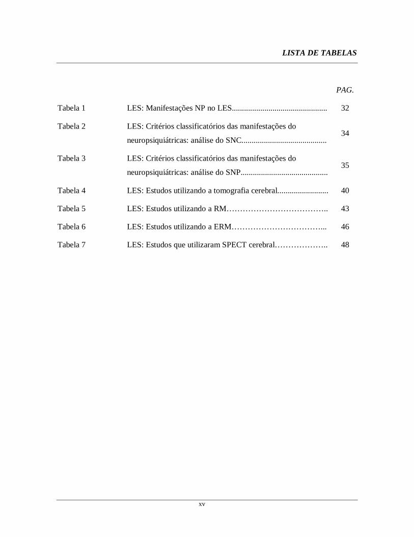
LISTA DE TABELAS
PAG.
Tabela 1 LES: Manifestações NP no LES............................................... 32
Tabela 2 LES: Critérios classificatórios das manifestações do
neuropsiquiátricas: análise do SNC.......................................... 34
Tabela 3 LES: Critérios classificatórios das manifestações do
neuropsiquiátricas: análise do SNP........................................... 35
Tabela 4 LES: Estudos utilizando a tomografia cerebral......................... 40
Tabela 5 LES: Estudos utilizando a RM……………………………….. 43
Tabela 6 LES: Estudos utilizando a ERM……………………………... 46
Tabela 7 LES: Estudos que utilizaram SPECT cerebral……………….. 48
xvii
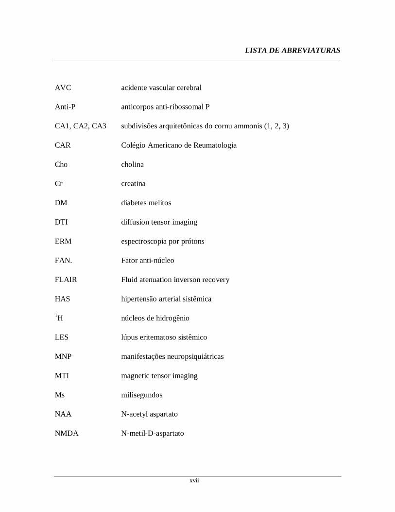
LISTA DE ABREVIATURAS
AVC acidente vascular cerebral
Anti-P anticorpos anti-ribossomal P
CA1, CA2, CA3 subdivisões arquitetônicas do cornu ammonis (1, 2, 3)
CAR Colégio Americano de Reumatologia
Cho cholina
Cr creatina
DM diabetes melitos
DTI diffusion tensor imaging
ERM espectroscopia por prótons
FAN. Fator anti-núcleo
FLAIR Fluid atenuation inverson recovery
HAS hipertensão arterial sistêmica
1H núcleos de hidrogênio
LES lúpus eritematoso sistêmico
MNP manifestações neuropsiquiátricas
MTI magnetic tensor imaging
Ms milisegundos
NAA N-acetyl aspartato
NMDA N-metil-D-aspartato

xviii
NP neuropsiquiátrico
PET tomografia por emissão de pósitrons
Ppm partículas por milhão
RM ressonância magnética
SAAF síndrome do anticorpo antifosfolípide
SB substância branca
SNA sistema nervoso autonômico
SNC sistema nervoso central
SNP sistema nervoso periférico
SPECT Single Photon Emission Computer Tomography
TC tomografia computadorizada cerebral
TVC trombose venosa central
UNICAMP Universidade Estadual de Campinas
VBM Morfometria baseada em voxels

xix
RESUMO
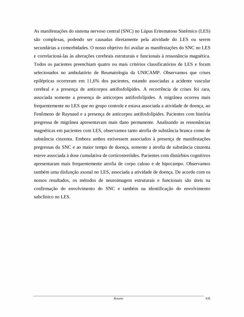
Resumo xxi
As manifestações do sistema nervoso central (SNC) no Lúpus Eritematoso Sistêmico (LES)
são complexas, podendo ser causadas diretamente pela atividade do LES ou serem
secundárias a comorbidades. O nosso objetivo foi avaliar as manifestações do SNC no LES
e correlacioná-las às alterações cerebrais estruturais e funcionais à ressonância magnética.
Todos os pacientes preenchiam quatro ou mais critérios classificatórios de LES e foram
selecionados no ambulatório de Reumatologia da UNICAMP. Observamos que crises
epilépticas ocorreram em 11,6% dos pacientes, estando associadas a acidente vascular
cerebral e a presença de anticorpos antifosfolípides. A recorrência de crises foi rara,
associada somente a presença de anticorpos antifosfolípides. A migrânea ocorreu mais
frequentemente no LES que no grupo controle e estava associada a atividade de doença, ao
Fenômeno de Raynaud e a presença de anticorpos antifosfolípides. Pacientes com história
pregressa de migrânea apresentavam mais dano permanente. Analisando as ressonâncias
magnéticas em pacientes com LES, observamos tanto atrofia de substância branca como de
substância cinzenta. Embora ambos estivessem associados à presença de manifestações
pregressas do SNC e ao maior tempo de doença, somente a atrofia de substância cinzenta
esteve associada à dose cumulativa de corticosteróides. Pacientes com distúrbios cognitivos
apresentaram mais frequentemente atrofia de corpo caloso e de hipocampo. Observamos
também uma disfunção axonal no LES, associada a atividade de doença. De acordo com os
nossos resultados, os métodos de neuroimagem estruturais e funcionais são úteis na
confirmação do envolvimento do SNC e também na identificação do envolvimento
subclínico no LES.
xxiii
ABSTRACT
Abstract xxv
Central nervous system (CNS) manifestations in systemic lupus erythematosus (SLE) are
complex. They may be directly caused by SLE disease activity or may be secondary to
comorbities. Our objective was to determine CNS manifestations in SLE patients and to
determine structural and functional neuroimaging abnormalities associated with its
occurrence. Patients with four or more classification criteria for SLE, followed at the
Rheumatology Unit of the State University of Campnas were included. We observed 11.6%
of epileptic seizures in SLE patients. The occurrence of epileptic seizures was associated
with the presence of stroke and antiphospholipid antibodies. Recurrence of seizures was
rare and associated only with the presence of antiphospholipid antibodies. Migraine was
more frequently observed in SLE patients than controls and was associated with disease
activity, Raynaud’s phenomenon and antiphospholipid antibodies. Pacients with past
history of migraine had more frequently organ damage. We observed white and gray matter
atrophy in SLE patients. Although both were associated with disease duration and past
history of CNS involvement, only gray matter atrophy was associated with the total
corticosteroid dose. Patients with cognitive impairment had more frequently corpus
callosum and hippocampal atrophy. A transient axonal dysfunction, secondary to disease
activity and not to CNS involvement, was observed in SLE. Our results suggest that
structural and functional neuroimaging methods are useful in confirming CNS involvement,
but also identify subclinical involvement in SLE patients.

27
1. INTRODUÇÃO E REVISÃO DA LITERATURA
Introdução e Revisão da Literatura 29
O Lúpus Eritematoso Sistêmico (LES) é uma doença do tecido conjuntivo com
manifestações clínicas diversas, caracterizada por períodos de remissão e exacerbação, com
participação intensa do sistema imunológico (Dubois e Tuffanelli, 1964).
1.1. EPIDEMIOLOGIA
A prevalência de LES é de aproximadamente 0,1% na população geral (Siegel e
Lee, 1973; Petri, 2002). Quanto às diferentes raças, observa-se a freqüência de 1 para cada
250 mulheres negras nos Estados Unidos da América; 22,4 para cada 100.000 asiáticos e
10,3 para cada 100.000 caucasianos (Fessel, 1974; Hopkinson et al., 1994; Alarcon, 2001;
Petri, 2002). Apresenta-se, entretanto, como uma rara patologia entre os negros africanos
(Molina et al., 1997; Molokhia et al., 2001). No Brasil observa-se uma freqüência maior
entre os caucasóides, principalmente na região sudeste do país (Chahade et al., 1995).
Apesar de surgir geralmente na segunda e terceira década de vida, o LES pode
se manifestar em qualquer idade, inclusive na primeira infância (Dubois e Tuffanelli, 1964;
Siegel e Lee, 1973; Petri, 2002). Nas crianças, a relação entre sexo feminino e masculino é
de 1,4 a 5,8:1; nos adultos varia de 8:1 a 13:1; nos indivíduos de idade mais avançada, esta
relação é de 2:1 (Marini et al., 1999; Costallat et al., 2002).
1.2. CRITÉRIOS CLASSIFICATÓRIOS DE LES
Não existem critérios definitivos para o diagnóstico do LES. O Colégio
Americano de Reumatologia definiu critérios classificatórios de LES, segundo o qual são
Introdução e Revisão da Literatura 30
necessários no mínimo quatro critérios clínicos e/ou laboratoriais entre onze (Tan et al.,
1982), após cuidadosa investigação e exclusão de doenças infecciosas, neoplásicas, entre
outras.
Os critérios considram as seguintes manifestações:
• “Rash” malar
• Lesão discóide
• Fotossensibilidade
• Úlceras da mucosa oral
• Artrite não-deformante
• Serosite (pleurite, pericardite).
• Doença renal (proteinúria persistente, cilindrúria).
• Envolvimento do sistema nervoso central (convulsão e psicose)
• Alterações hematológicas (anemia, leucopenia ou plaquetopenia).
• Alterações imunológicas: células LE, anti-DNA, anti-Sm ou VDRL falso-
positivo.
• Fator anti-núcleo (FAN)
Uma proposta de modificação destes critérios foi feita por Hochberg (1997),
excluindo as células LE e substituindo o VDRL falso-positivo pela presença do anticorpo
anticardiolipina.

Introdução e Revisão da Literatura 31
1.3. MANIFESTAÇÕES NEUROPSIQUIÁTRICAS (NP) NO LES
As manifestações NP no LES são complexas e podem ser definidas como
manifestações neurológicas do sistema nervoso central (SNC), periférico (SNP) e
autonômico (SNA) e de síndromes psiquiátricas observadas em pacientes com LES. Podem
ser causadas diretamente pela atividade do LES, serem secundárias a comorbidades como
hipertensão arterial sistêmica (HAS), diabetes mellitos (DM), uremia e infecção. Poderiam
ocorrer também ou ainda serem patologias primariamente distintas e concomitantes em
pacientes com LES, sendo consideradas associações fortutas. Por definição, para ser
considerada primariamente decorrente do LES, outras possíveis causas necessitam ser
cuidadosamente excluídas (Hanly, 2005; Hanly e Harrison, 2005).
1.3.1. Histórico
A primeira menção à doença “ lupus” ocorreu no século X por Hebernus of
Tours na biografia de St Martin (Estes e Christian, 1971; Smith e Cyr, 1988). Porém, a
primeira descrição do quadro clínico do Lúpus Eritematoso foi feita vez por Biett em 1828
(Skinner, 1949; Smith e Cyr, 1988), sendo que Kaposi observou a sua natureza sistêmica,
descrevendo alguns pacientes com lesões viscerais (Kaposi, 1875). Osler (1904), enfatiza o
acometimento sistêmico, alertando sobre a possibilidade de alterações viscerais sem
concomitância com as lesões cutâneas; descreveu também a instabilidade do quadro clínico
e suas fases alternadas de agudização e remissão dos sintomas. A partir de 1945 surgem os
primeros estudos de grandes casuísticas descrevendo as principais manifestações clínicas e
do SNC (Daly, 1945; Clark e Bailey, 1956; Dubois e Tuffanelli, 1964; Klippel e Zvaifler,
1975; Sergent et al., 1975; Feinglass et al., 1976; Ellis and Verity, 1979; Adelmann et al.,
1986; Kaell et al., 1986).
Introdução e Revisão da Literatura 32
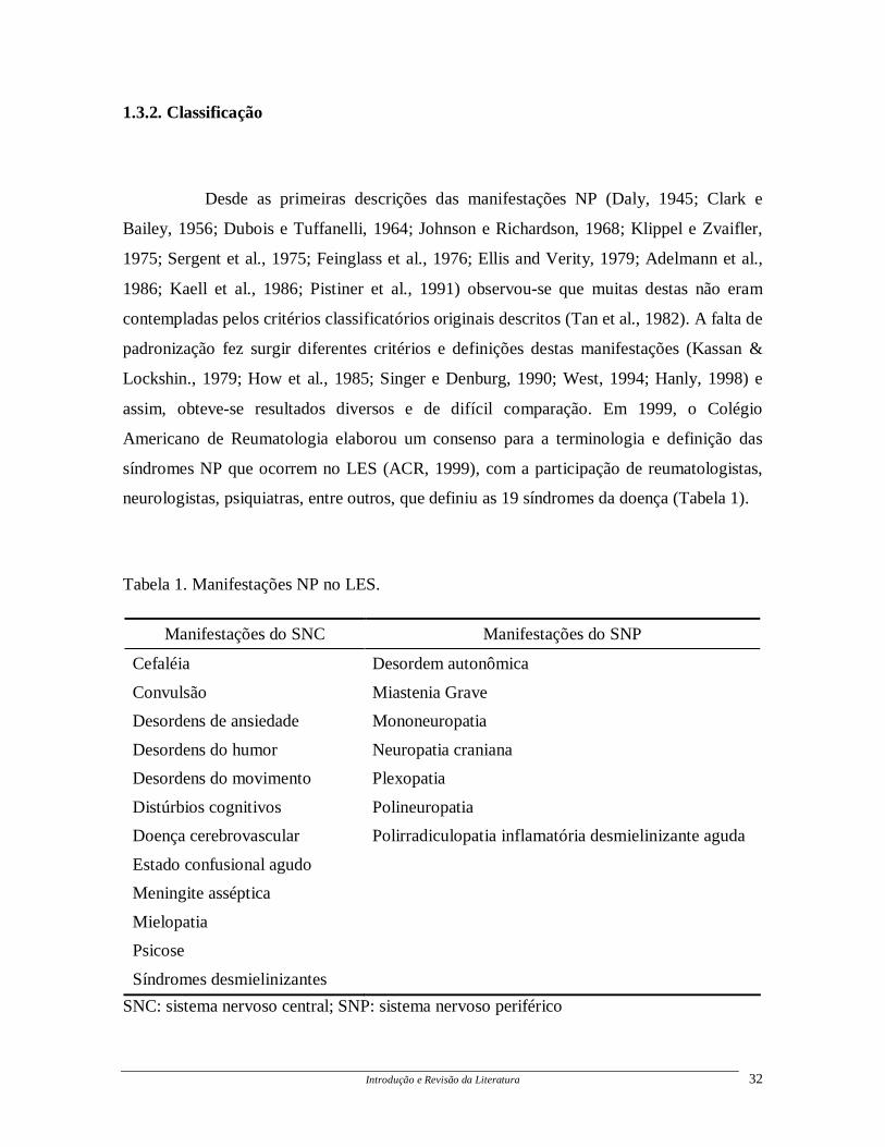
1.3.2. Classificação
Desde as primeiras descrições das manifestações NP (Daly, 1945; Clark e
Bailey, 1956; Dubois e Tuffanelli, 1964; Johnson e Richardson, 1968; Klippel e Zvaifler,
1975; Sergent et al., 1975; Feinglass et al., 1976; Ellis and Verity, 1979; Adelmann et al.,
1986; Kaell et al., 1986; Pistiner et al., 1991) observou-se que muitas destas não eram
contempladas pelos critérios classificatórios originais descritos (Tan et al., 1982). A falta de
padronização fez surgir diferentes critérios e definições destas manifestações (Kassan &
Lockshin., 1979; How et al., 1985; Singer e Denburg, 1990; West, 1994; Hanly, 1998) e
assim, obteve-se resultados diversos e de difícil comparação. Em 1999, o Colégio
Americano de Reumatologia elaborou um consenso para a terminologia e definição das
síndromes NP que ocorrem no LES (ACR, 1999), com a participação de reumatologistas,
neurologistas, psiquiatras, entre outros, que definiu as 19 síndromes da doença (Tabela 1).
Tabela 1. Manifestações NP no LES.
Manifestações do SNC Manifestações do SNP
Cefaléia Desordem autonômica
Convulsão Miastenia Grave
Desordens de ansiedade Mononeuropatia
Desordens do humor Neuropatia craniana
Desordens do movimento Plexopatia
Distúrbios cognitivos Polineuropatia
Doença cerebrovascular Polirradiculopatia inflamatória desmielinizante aguda
Estado confusional agudo
Meningite asséptica
Mielopatia
Psicose
Síndromes desmielinizantes
SNC: sistema nervoso central; SNP: sistema nervoso periférico
Introdução e Revisão da Literatura 33
Posteriormente, estes critérios foram validados, apresentando uma
especificidade de 46% (Ainiala et al., 2001). Porém, este mesmo estudo demonstrou que,
excluíndo-se cefaléia, ansiedade, depressão leve, distúrbio cognitivo leve e polineuropatia
sem confirmação por eletroneuromigrafia, a especificidade aumenta para 93%. Portanto,
apesar desta classificação ser atualmente a mais aceita, há ainda limitações que podem ser
futuramente modificadas (Hanly, 2004).
A avaliação de cada uma destas manifestações envolve uma série de testes
neurofisiológicos (Omdal et al, 1989; Omdal et al., 1991; Omdal et al., 1993; Omdal et al.,
1996; Costallat et al., 1997), técnicas laboratoriais (Blustein et al., 1981; Bluestein e
Woods, 1982; Bluestein e Zvaifler, 1983; Gharavi et al., 1987; Bonfa et al., 1987; Robbins
et al., 1988; Temesvari et al., 1983; Costallat et al., 1990; Denburg et al., 1994) e de
neuroimagem, incluindo a tomografia computadorizada cerebral (TC) (Gonzales-Scarano et
al., 1979; Carette et al., 1982; Kaell et al., 1986; Yang et al., 1993; Zanardi et al., 2001;
Omdal et al.,1989) e a ressonância magnética (RM) (Miller et al, 1992; Stimmler et al.,
1993; Jarek et al., 1994; McCune et al., 1998; Kozora et al., 1998), quando necessários.
Vários estudos utilizaram estes critérios para descrever a freqüência ou
prevalência das manifestações NP no LES, sejam do SNC (Tabela 2) ou SNP (Tabela 3).
Apesar de ainda ser observada uma grande variabilidade entre as freqüências, o uso desta
mesma classificação permite supor que estas diferenças possam ser devidas ao número de
pacientes incluídos e à diferenças loco-regionais (Hanly, 2005).
Intr
oduç
ão e
Rev
isão
da
Lite
ratu
ra
34
Tab
ela
2. L
ES
: Cri
téri
os c
lass
ific
atór
ios
das
man
ifes
taçõ
es n
euro
psiq
uiát
rica
s: a
nális
e do
sis
tem
a ne
rvos
o ce
ntra
l
Ans
.: an
sied
ade;
Cef
.: C
efal
éia;
con
v.:
Con
vuls
ivas
; D
es.
Mov
.: D
esor
dens
do
mov
imen
to;
DC
: di
stúr
bios
cog
niti
vos;
DC
V:
doen
ça c
éreb
ro
vasc
ular
; E
CA
: es
tado
con
fusi
onal
agu
do ;
MA
: M
enin
gite
ass
épti
ca;
NA
: nã
o av
alia
do;
No:
núm
ero;
pac
.: P
acie
ntes
; P
sic.
: P
sico
se;
Pre
v.:
Pre
valê
ncia
; Mie
l.: M
ielo
patia
; S. D
esm
.: sí
ndro
me
Des
mie
lini
zant
e.
*ref
erid
o pe
lo p
acie
nte
Aut
ores
/ A
no
No
de
pac.
Pre
v.
NP
(%
)
Cef
. (%
)
Cri
ses
conv
. (%
)
Ans
. (%
) H
umor
(%
)
Des
. m
ov.
(%)
DC
(%
) D
CV
(%)
EC
A
(%)
MA
(%
) M
iel.
(%)
Psi
c.
(%)
S.
Des
m.
(%)
Ain
iala
et a
l.,
2001
46
91
54
9
13
44
2 80
15
7
2 0
0 2
Cos
talla
t et a
l.,
2001
52
7 34
3*
7,
4 0,
8*
3,0*
0,
8 1,
3*
2,5
3 0,
4 1
5,3
0,2
Mok
et a
l.,
2001
51
8 19
4
28
1,5
6 2
NA
19
14
1
8 11
1,
5
Bre
y et
al,
20
02
128
80
57
16
24
23,0
1
79
2 0
0 5,
0 6,
5 0
Alf
reta
et a
l.,
2003
61
72
21
11
6
27
0 52
24
0
0 0
0 3
San
na e
t al.,
20
03
323
57,3
24
8,
3 7,
4 16
,7
1,2
10,8
17
,6
3,7
0 1,
2 7,
7 0,
9
Han
ly e
t al.,
20
04
111
37
24,4
2,
4 2,
4 9,
8 0
7,3
9,8
7,0
2,4
0 7,
3 2,
4
Mik
dash
i et a
l.,
2004
13
0 56
,9
NA
7,
6 0
NA
0
27,3
25
,7
0 0
6,1
15,1
0
Han
ly e
t al.,
20
05
53
31
9,4
0
0 0
1,9
0 3,
8 0
0 0
1,9
Shi
moj
ima
et a
l.,
2005
25
10
0 12
36
0
0 0
12
24
0 0
4 32
0
Rob
ert e
t al.,
20
06
50
78
55,6
20
,5
0 0
23,1
18
16
,2
16,2
0
0 16
,2
0
Intr
oduç
ão e
Rev
isão
da
Lite
ratu
ra
35
Tab
ela
3. L
ES
: Cri
téri
os c
lass
ific
atór
ios
das
man
ifes
taçõ
es n
euro
psiq
uiát
rica
s: a
nális
e do
sis
tem
a ne
rvos
o pe
rifé
rico
NA
: não
ava
liado
; No:
núm
ero
Aut
ores
/
Ano
N
o de
pa
cien
tes
Des
orde
ns
auto
nôm
icas
(%
)
Mia
sten
ia
Gra
ve
(%)
Mon
oneu
ropa
tia
(%)
Neu
ropa
tia
cran
iana
(%
)
Ple
xopa
tia
(%)
Pol
ineu
ropa
tia
(%)
Pol
irad
icul
opat
ia
infl
amat
ória
de
smie
lini
zant
e ag
uda
(%)
Ain
iala
et a
l.,
2001
46
0
2 0
7 0
28
2
Cos
talla
t et a
l.,
2001
52
7 0
0,2
1,3
1,5
0 4
0,2
Mok
et a
l.,
2001
51
8 0
0 1,
5 3
0 1
0
Bre
y et
al.,
2002
12
8 0
0 8
2 0
22
0
Alf
reta
et a
l.,
2003
61
3
0 0
4 0
13
0
San
na e
t al.,
2003
32
3 0
1,5
1,8
1,5
0 2,
8 0,
6
Han
ly e
t al.,
2004
11
1 0
0 0
4,9
0 4,
9 0
Mik
dash
i et a
l,
2004
13
0 0
0 0
0 0
18,2
0
Han
ly e
t al.,
2005
53
0
0 0
0 0
0 0
Shi
moj
ima
et a
l.,
2005
25
0
0 0
0 0
12
0
Rob
ert e
t al.,
2006
50
N
A
0 7,
9 0
0 0
0
Introdução e Revisão da Literatura 36
Os sintomas NP podem se apresentar isoladamente ou em conjunto, ocorrendo
em episódios únicos durante a fase de exacerbação da doença, associados ou não a outros
sinais de atividade do LES. Ocorrem em qualquer tempo da doença, podendo ser o seu
primeiro sinal clínico (McCune e Golbus, 1988; Costallat et al., 1990; Pistiner et al., 1991).
Os principais problemas diagnósticos consistem na distinção entre as alterações
neurológicas causadas pelo LES, com anormalidades imunológicas tendo papel
preponderante, e eventos secundários, como complicações da HAS, distúrbios metabólicos,
distúrbios de coagulação, infecção grave e corticoterapia (How et al., 1985; Hanly, 2004),
que podem ocorrer em até 41% dos pacientes (Hanly et al., 2004).
Portanto, como não existe diagnóstico definitivo para o acometimento NP, em
especial do SNC, outras causas como as infecciosas, metabólicas ou por drogas devem
sempre ser exaustivamente excluídas.
1.3.3. Importância clínica das manifestações do SNC no LES
A importância das manifestações do SNC no LES pode ser determinada
analisando a influência na mortalidade, qualidade de vida e índice de dano permanente
(Feng et al., 1973; Cheatum et al., 1973; Sergent et al., 1975; Lee et al., 1977; Ginzler et al.,
1982; Sibley et al., 1992; Kovacs et al., 1993; Carlomagno et al., 2000; Jonson et al., 2002;
Mikdashi e Handwerger, 2004).
Apesar de não haver consenso em diferentes estudos quanto a uma maior
mortalidade nos pacientes com manifestações do SNC (Swaak et al., 1989; Jonsonn et al.,
1989; Jonson et al., 2002), já foi observado que estes pacientes apresentam um aumento dos
escores de incapacidade (Jonson et al., 2002), maior fadiga (Mikdashi e Handwerger, 2004)
e uma pior qualidade de vida (Hanly et al., 2004). Em um estudo para determinar o índice
de dano em pacientes com manifestações do SNC observou-se que a presença de doença
ativa na instalação e a presença de anticorpos antifosfolípides eram fatores preditivos para
maior dano permanente em pacientes com LES (Mikdashi e Handwerger, 2004).
Introdução e Revisão da Literatura 37
Poucos estudos analisaram o comprometimento do SNC de forma longitudinal
(Hanly et al., 1994; Hay et al., 1994; Carlomagno et al., 2000; Karassa et al., 2000;
Waterloo et al., 2002). Em pacientes que foram internados devido ao comprometimento do
SNC e seguidos por dois anos, observou-se uma boa evolução em 69% e uma estabilização
do quadro em 19% dos casos, sendo que o número de manifestações NP prévias e a
presença da síndrome do anticorpo antifosfolípide indicaram pior prognóstico (Karassa et
al., 2000). Em relação ao distúrbio cognitivo, também foi observado que a maioria dos
pacientes apresenta flutuações da cognição, não evoluíndo, portanto para demência (Hanly
et al., 1994; Hay et al., 1994; Carlomagno et al., 2000; Waterloo et al., 2002).
1.3.4. Etiopatogenia do comprometimento do SNC no LES
Estudos anatomopatológicos de cérebros de pacientes com LES, com e sem
comprometimento do SNC, evidenciaram predominantemente comprometimento
microvascular, com poucos sinais de vasculite (Johnson e Richardson, 1968; Ellis and
Verity, 1979; Zvaifler and Bluestein, 1982; Hanly, 1992; Abbott et al.; 2003). Embora
alguns destes estudos tenham demonstrado um comprometimento microvascular,
aparentemente estes achados não justificam a maioria das manifestações do SNC no LES.
Portanto, a etiopatogenia do SNC no LES parece ser multifatorial, envolvendo, além do
comprometimento da pequena circulação, a produção de autoanticorpos e o processo
inflamatório (Hanly 2005; Hanly e Harrison, 2005).
A presença de anticorpos contra neurônios, ribossomos e fosfolípides já foram
associados às manifestações do SNC no LES. Em modelo animal foi demonstrado que
anticorpos antineuronais induzem déficits de memória, convulsões e alterações
neuropatológicas (Kobiler e Allweis, 1976; Morris et al., 1986). Um aumento de anticorpos
antineuronais foi observado por Hanson et al (1992), embora nenhuma manifestação clínica
específica estivesse associada a este achado. Em pacientes com manifestações do SNC
observou-se um aumento dos receptores N-metil-D-aspartato (NMDA), NR2a e NR2b, o
Introdução e Revisão da Literatura 38
que parece ter uma consequência funcional que leva a lesão neuronal (Lipton e Rosenberg,
1994). Foi demonstrado que anticorpos anti NR2 estão associados a déficit de memória
(Akbarian et al., 1996) e psicose (Teh e Isenberg, 1998). Os anticorpos anti-ribosomal P
(anti P) apresentam uma prevalência de 13-20% no LES, dependendo do grupo étnico
estudado (Arnett et al., 1996), e estão associados a psicose e depressão (Bonfa et al., 1987;
Tzioufas et al., 2000; Gerli et al., 2002). Os anticorpos antifosfolípides estão relacionados
primariamente a manifestações focais, porém já foram descritas associações com
convulsão, coréia, mielite transversa e disfunção cognitiva (Love e Santoro, 1990; Menon
et al., 1999; Chapman et al.,1999; Hanly, 2003; Hanly, 2005).
Vários estudos têm analisado o papel dos processos inflamatórios no LES
(Hirohata e Miyamoto, 1990; Shiozawa et al., 1992; Jara et al., 1998; Trysberg et al., 2000;
Faber-Elmann et al., 2002; Schenatto et al., 2006). Interleucinas (Hirohata e Miyamoto,
1990; Jara et al., 1998; Trysberg et al., 2000), fator de necrose tumoral (Shiozawa et al.,
1992), metaloproteinases (Faber-Elmann et al., 2002; Ainiala et al., 2004) e S 100 beta
(Schenatto et al., 2006) parecem estar associados às manifestações do SNC no LES e aos
achados à RM.
1.4. MÉTODOS DE NEUROIMAGEM PARA AVALIAÇÃO DO
COMPROMETIMENTO DO SNC
Os métodos de neuroimagem são utilizados para estabelecer a presença de
anormalidades cerebrais difusas e/ou focais. Podem ser classificados em métodos de
avaliação estrutural e funcional.
Introdução e Revisão da Literatura 39
1.4.1 Métodos de avaliação estrutural
Os métodos de análise estrutural visam determinar alterações morfológicas e a
sensibilidade depende do método utilizado.
Tomografia computadorizada
A tomografia computadorizada tem a vantagem de ser disponível na maioria
dos serviços de médio porte e tem um custo operacional relativamente baixo. A tomografia
computadorizada tem como princípio o uso de raio-X para gerar o contraste na imagem, o
qual resulta da diferença dos coeficientes de atenuação entre duas estruturas adjacentes, na
ordem de alguns pontos percentuais. Os coeficientes de atenuação estão relacionados com
as densidades dos elétrons, que são proporcionais aos números anatômicos dos elementos
constituintes dos compostos químicos. Portanto, a gordura, que é rica em carbono, é mais
transparente do que a água, uma vez que o oxigênio tem um número anatômico maior que o
carbono.
A tomografia computadorizada pode detectar grande parte dos tumores,
malformações arteriovenosas e malformações cerebrais extensas, acidentes vasculares,
lesões infecciosas e é sensível para detecções de lesões calcificadas. Entretanto, é pouco
sensível para detectar lesões na base do crânio e pequenas lesões corticais, de um modo
geral.
No LES a tomografia computadorizada tem sua validade para detectar ainda
lesões cerebrais focais agudas e na hidrocefalia, sendo, porém, pouco sensível na doença
cerebral difusa ou na identificação de alterações na substância branca (Gonzalez-Scarano et
al., 1979; Vermess et al., 1983; Kaell et al.,1986; Waterloo et al., 1999; Zanardi et al.,
2001). Vários estudos têm utilizado a tomografia computadorizada de crânio como método
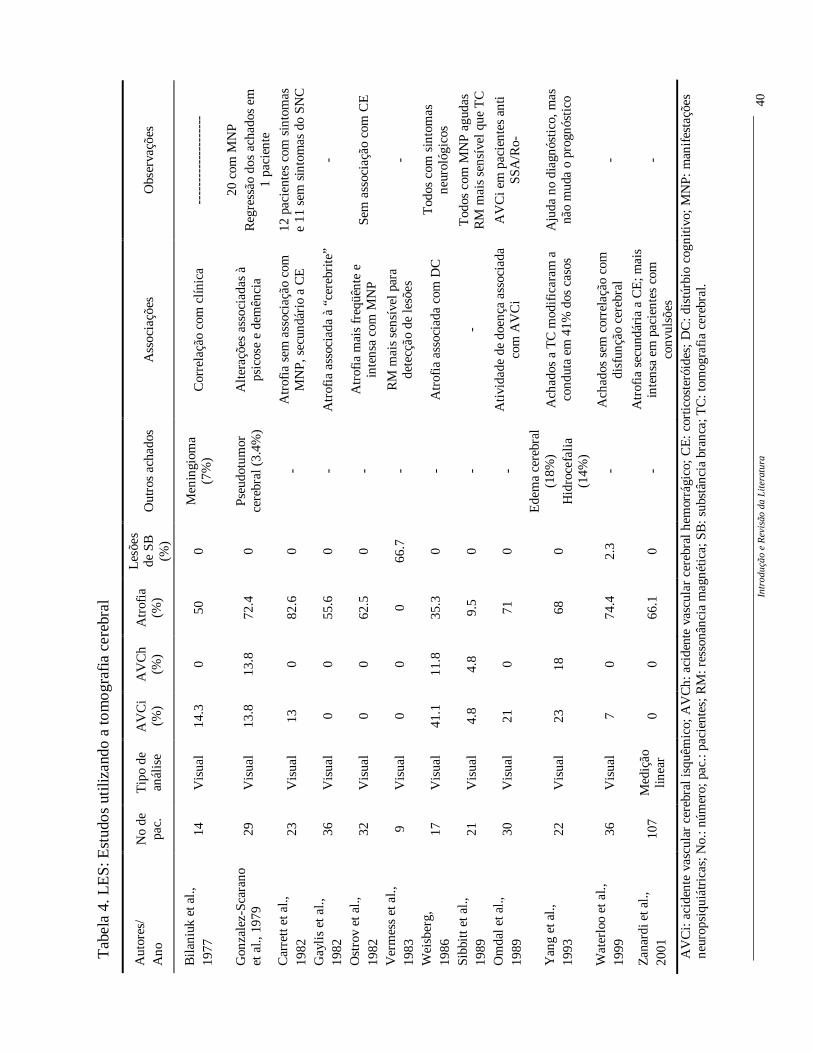
de investigação no comprometimento do SNC no LES (Tabela 4).
Intr
oduç
ão e
Rev
isão
da
Lite
ratu
ra
40
Tab
ela
4. L
ES
: Est
udos
uti
lizan
do a
tom
ogra
fia
cere
bral
Aut
ores
/ A
no
No
de
pac.
T
ipo
de
anál
ise
AV
Ci
(%)
AV
Ch
(%)
Atr
ofia
(%
)
Les
ões
de S
B
(%)
Out
ros
acha
dos
Ass
ocia
ções
O
bser
vaçõ
es
Bil
aniu
k et
al.,
19
77
14
Vis
ual
14.3
0
50
0 M
enin
giom
a (7
%)
Cor
rela
ção
com
clín
ica
----
----
----
----
----
Gon
zale
z-Sc
aran
o et
al.,
197
9 29
V
isua
l 13
.8
13.8
72
.4
0 Ps
eudo
tum
or
cere
bral
(3.
4%)
Alt
eraç
ões
asso
ciad
as à
ps
icos
e e
dem
ênci
a
20 c
om M
NP
Reg
ress
ão d
os a
chad
os e
m
1 pa
cien
te
Car
rett
et a
l.,
1982
23
V
isua
l 13
0
82.6
0
- A
trof
ia s
em a
ssoc
iaçã
o co
m
MN
P, s
ecun
dári
o a
CE
12
pac
ient
es c
om s
into
mas
e
11 s
em s
into
mas
do
SNC
Gay
lis
et a
l.,
1982
36
V
isua
l 0
0 55
.6
0 -
Atr
ofia
ass
ocia
da à
“ce
rebr
ite”
-
Ost
rov
et a
l.,
1982
32
V
isua
l 0
0 62
.5
0 -
Atr
ofia
mai
s fr
eqüê
nte
e in
tens
a co
m M
NP
Sem
ass
ocia
ção
com
CE
Ver
mes
s et
al.,
19
83
9 V
isua
l 0
0 0
66.7
-
RM
mai
s se
nsív
el p
ara
dete
cção
de
lesõ
es
-
Wei
sber
g,
1986
17
V
isua
l 41
.1
11.8
35
.3
0 -
Atr
ofia
ass
ocia
da c
om D
C
Tod
os c
om s
into
mas
ne
urol
ógic
os
Sibb
itt e
t al.,
19
89
21
Vis
ual
4.8
4.8
9.5
0 -
- T
odos
com
MN
P ag
udas
R
M m
ais
sens
ível
que
TC
Om
dal e
t al.,
19
89
30
Vis
ual
21
0 71
0
- A
tivi
dade
de
doen
ça a
ssoc
iada
co
m A
VC
i A
VC
i em
pac
ient
es a
nti
SSA
/Ro-
Yan
g et
al.,
19
93
22
Vis
ual
23
18
68
0
Ede
ma
cere
bral
(1
8%)
Hid
roce
fali
a (1
4%)
Ach
ados
a T
C m
odif
icar
am a
co
ndut
a em
41%
dos
cas
os
Aju
da n
o di
agnó
stic
o, m
as
não
mud
a o
prog
nóst
ico
Wat
erlo
o et
al.,
19
99
36
Vis
ual
7 0
74.4
2.
3 -
Ach
ados
sem
cor
rela
ção
com
di
sfun
ção
cere
bral
-
Zan
ardi
et a
l.,
2001
10
7 M
ediç
ão
linea
r 0
0 66
.1
0 -
Atr
ofia
sec
undá
ria
a C
E; m
ais
inte
nsa
em p
acie
ntes
com
co
nvul
sões
-
AV
Ci:
aci
dent
e va
scul
ar c
ereb
ral i
squê
mic
o; A
VC
h: a
cide
nte
vasc
ular
cer
ebra
l hem
orrá
gico
; C
E:
cort
icos
teró
ides
; D
C: d
istú
rbio
cog
nitiv
o; M
NP:
man
ifes
taçõ
es
neur
opsi
quiá
tric
as; N
o.: n
úmer
o; p
ac.:
paci
ente
s; R
M: r
esso
nânc
ia m
agné
tica;
SB
: sub
stân
cia
bran
ca; T
C: t
omog
rafi
a ce
rebr
al.
Introdução e Revisão da Literatura 41
Ressonância Magnética
A RM utiliza como princípio, a propriedade dos núcleos de hidrogênio (1H) em
emitir um sinal eletromagnético em resposta a um pulso de radiofreqüência. Alterações
sutis no conteúdo de água do tecido resultam em variações neste sinal do próton 1H. Após
ser captada, a diferença de intensidade neste sinal em vários pontos do espaço (que reflete a
estrutura molecular dos tecidos) é transformada em uma imagem em preto e branco através
de processos de computação gráfica. As características únicas desse método possibilitam a
modificação da intensidade de sinal relativa dos tecidos através da alteração de parâmetros
operacionais específicos. Além disso, as imagens podem ser obtidas em qualquer plano,
minimizando dificuldades técnicas relacionadas à posição do paciente.
O exame de RM é complexo, constituindo-se de diferentes técnicas e
seqüências de pulso. As seqüências de pulso podem de uma maneira simplificada, ser
divididas em seqüências ponderadas em T1 e T2. O sinal obtido nas seqüências ponderadas
em T1 é resultante da liberação de energia que ocorre quando a interação entre os núcleos
de 1H excitados com o meio molecular regional (relaxamento spin-lattice). As seqüências
ponderadas em T1 permitem o estudo do sinal proveniente do parênquima cerebral,
enfatizando, assim, a morfologia. As seqüências ponderadas em T2 (T2, densidade de
prótons, e Fluid atenuation inverson recovery (FLAIR)) baseiam-se na aquisição do sinal
proveniente da interação entre os núcleos 1H excitados e outros núcleos em diferentes
estados de energia (relaxamento spin-spin). Estas seqüências possuem alta sensibilidade na
detecção de alterações patológicas, que determinam aumento do conteúdo local de água
e/ou alterações intersticiais, tais como gliose, desmielinização, edema e infiltração tumoral,
por exemplo. As imagens ponderadas em T1 e T2 podem ser obtidas utilizando-se
diferentes técnicas de parâmetros que influenciam as características das imagens obtidas e o
tempo de duração do exame.
Introdução e Revisão da Literatura 42
Nas técnicas de processamento as imagens obtidas podem ser manipuladas em
uma estação de trabalho (Workstation) para atender a diversos propósitos. O processamento
das imagens obtidas pela RM permite quantificar e qualificar as alterações encontradas, de
modo que, ao determinarmos a sua relação biológica com variáveis clínicas, obtem-se
resultados objetivos e reproduzíveis. Estes resultados podem ser utilizadas no seguimento
destes pacientes, quando a comparação com imagens obtidas posteriormente, se tornar
necessária.
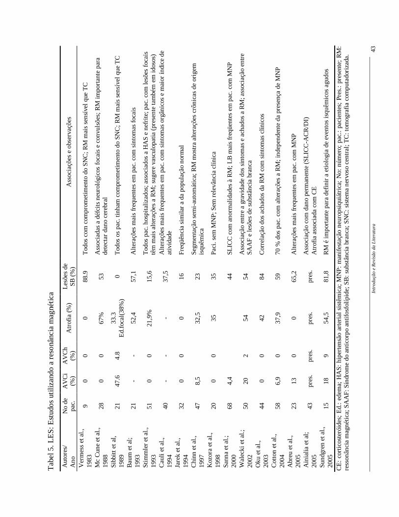
Na RM de pacientes com LES podem ser observadas atrofia cerebral localizada
ou difusa e lesões em substância branca. A atrofia cerebral é vista na RM em 33 a 67% dos
pacientes com LES, sendo fatores causais, entre outros, a longa duração da doença, o
infarto cerebral prévio, a idade mais avançada dos pacientes e o uso de corticosteróides
(Walecki et al., 2002; Kozora et al., 1998). Os padrões de lesão de substância branca
descritas no LES são áreas puntiformes de hipersinal em imagens T2 e FLAIR, localizadas
na região periventricular ou subcortical. As áreas focais hiperintensas também podem
envolver córtex, núcleos da base e tronco cerebral. Outras alterações observadas em RM de
pacientes com LES incluem infarto, hemorragia e atrofia cerebral (Walecki et al., 2002).
No entanto, o freqüente aparecimento de áreas de hipersinal na substância branca e sua
possível associação com atividade da doença, bem como sua associação com distúrbios
cognitivos são assunto ainda controversos, dificultando a correlação entre as manifestações
clínicas e os achados de neuroimagem (Walecki et al., 2002).
A RM é, portanto, o exame mais sensível para se detectar as alterações
cerebrais, tanto no LES como em outras doenças difusas do tecido conjuntivo. Alguns
estudos têm utilizado a RM para avaliação do comprometimento do SNC no LES (Tabela
5).
Intr
oduç
ão e
Rev
isão
da
Lite
ratu
ra
43
Tab
el 5
. LE
S: E
stud
os u
tiliz
ando
a r
eson
ânci
a m
agné
tica
Aut
ores
/ A
no
No
de
pac.
A
VC
i (%
) A
VC
h (%
) A
trof
ia (
%)
Les
ões
de
SB (
%)
Ass
ocia
ções
e o
bser
vaçõ
es
Ver
mes
s et
al.,
19
83
9 0
0 0
88.9
T
odos
com
com
prom
etim
ento
do
SNC
; RM
mai
s se
nsív
el q
ue T
C
Mc
Cun
e et
al.,
19
88
28
0 0
67%
53
A
ssoc
iada
s a
défc
its
neur
ológ
icos
foc
ais
e co
nvul
sões
; RM
impo
rtan
te p
ara
dete
ctar
dan
o ce
rebr
al
Sibb
itt e
t al,
19
89
21
47.6
4.
8 33
.3
Ed.
foca
l(38
%)
0 T
odos
os
pac.
tinh
am c
ompr
omet
imen
to d
o SN
C; R
M m
ais
sens
ível
que
TC
Bau
m e
t al;
19
93
21
- -
52,4
57
,1
Alt
eraç
ões
mai
s fr
eque
ntes
em
pac
. com
sin
tom
as f
ocai
s
Stim
mle
r et
al.,
19
93
51
0 0
21,9
%
15,6
T
odos
pac
. hos
pita
lizad
os; a
ssoc
iado
s a
HA
S e
nefr
ite;
pac
. com
lesõ
es f
ocai
s tê
m m
ais
alte
raçõ
es a
RM
; sug
ere
vasc
ulop
atia
(pr
esen
te ta
mbé
m e
m id
osos
) C
auli
et a
l.,
1994
40
-
- -
37,5
A
lter
açõe
s m
ais
freq
uent
es e
m p
ac. c
om s
into
mas
org
ânic
os e
mai
or ín
dice
de
ativ
idad
e Ja
rek
et a
l.,
1994
32
0
0 0
16
Freq
üênc
ia s
imila
r a
da p
opul
ação
nor
mal
Chi
nn e
t al.,
19
97
47
8,5
32
,5
23
Segm
enta
ção
sem
i-au
tom
átic
a; R
M m
ostr
a al
tera
ções
crô
nica
s de
ori
gem
is
quêm
ica
Koz
ora
et a
l.,
1998
20
0
0 35
35
Pa
ci. s
em M
NP;
Sem
rel
evân
cia
clín
ica
Sann
a et
al.;
20
00
68
4,4
44
SLIC
C c
om a
norm
alid
ades
à R
M; L
B m
ais
freq
üent
es e
m p
ac. c
om M
NP
Wal
ecki
et a
l.;
2002
50
20
2
54
54
Ass
ocia
ção
entr
e a
grav
idad
e do
s si
ntom
as e
ach
ados
a R
M; a
ssoc
iaçã
o en
tre
SAA
F e
lesõ
es d
e su
bstâ
ncia
bra
nca
Oku
et a
l.,
2003
44
0
0 42
84
C
orre
laçã
o do
s ac
hado
s da
RM
com
sin
tom
as c
línic
os
Cot
ton
et a
l.,
2004
58
6,
9 0
37,9
59
70
% d
os p
ac. c
om a
lter
açõe
s a
RM
; ind
epen
dent
e da
pre
senç
a de
MN
P
Abr
eu e
t al.,
20
05
23
13
0 0
65,2
A
lter
açõe
s m
ais
freq
uent
es e
m p
ac. c
om M
NP
Ain
ialia
et a
l;
2005
43
pr
es.
pres
. pr
es.
pres
. A
ssoc
iaçã
o co
m d
ano
perm
anen
te (
SLIC
C-A
CR
/DI)
A
trof
ia a
ssoc
iada
com
CE
Su
ndgr
en e
t al.,
20
05
15
18
9 54
,5
81,8
R
M é
impo
rtan
te p
ara
defi
nir
a et
iolo
gia
de e
vent
os is
quêm
icos
agu
dos
CE
: co
rtic
oste
róid
es;
Ed.
: ed
ema;
HA
S: h
iper
tens
ão a
rter
ial s
istê
mic
a; M
NP:
man
ifes
taçã
o ne
urop
siqu
iátr
ica;
No:
núm
ero;
pac
.: pa
cien
tes;
Pre
s.:
pres
ente
; R
M:
ress
onân
cia
mag
néti
ca; S
AA
F: S
índr
ome
do a
ntic
orpo
ant
ifos
folí
pide
; SB
: sub
stân
cia
bran
ca; S
NC
: sis
tem
a ne
rvos
o ce
ntra
l; T
C: t
omog
rafi
a co
mpu
tado
riza
da.
Introdução e Revisão da Literatura 44
1.4.2. Métodos de avaliação funcional
Espectroscopia por prótons
A espectroscopia por prótons (ERM) permite a quantificação não invasiva in
vivo de alguns compostos químicos de importância biológica que estão presentes em
concentrações muito menores que a água nos tecidos. O sinal de ERM proveniente de 1H é
inerentemente mais forte do que qualquer outro núcleo. Quase todos os metabólitos de alta
concentração contém núcleos de 1H, que em princípio, podem ser utilizados para identificá-
los na ERM.
Espectros de prótons com supressão de água do cérebro humano utilizando um
tempo de echo entre 136 e 272 milisegundos (ms) revelam três ressonâncias principais:
(1) uma em 3,2 partículas por milhão (ppm), que se origina das tetrametil-
aminas, sobretudo as colinas, ricas em fosfolípides (Cho), marcadores, em certas
circunstâncias, da quebra de mielina;
(2) uma em 3,0 ppm, que se origina primariamente da creatina e fosfocreatina
(Cr);
(3) uma em 2,0 ppm que se origina em grupos N-acetil, principalmente N-
acetilaspartato (NAA), marcador de integridade neuronal;
Várias evidências indicam que o NAA pode ser usado como um marcador
neuronal, já que é encontrado exclusivamente em neurônios e processos neuronais (Birken
Introdução e Revisão da Literatura 45
et al., 1991; Moffett et al., 1991; Simmons et al., 1999). Em espectros do cérebro humano
in vivo, como nas doenças neurodegenerativas (Van der Knaap et al., 1992), acidentes
vasculares (Graham et al., 1992; Duijin et al., 1992) e tumores (Arnold et al., 1992; Preul et
al., 1996) observa-se uma diminuição da NAA em relação a Cr. Quando reduções relativas
do sinal do NAA ocorrem, devido à degeneração axonal e/ou neuronal, alterações
irreversíveis são esperadas. Entretanto, existem observações de redução reversível do NAA
em várias doenças, demonstrando que uma disfunção neuronal ou uma mudança relativa do
volume neuronal, pode provocar redução do NAA (Destefano et al., 1995; Destefano et al.,
1995; Davie et al., 1994). A habilidade de quantificar perda ou dano neuronal é uma das
aplicações da ERM na investigação de doenças que acometem o SNC. Mudanças na
intensidade de ressonância da Cho provavelmente resultam da elevação das concentrações
de equilíbrio da fosfocolina e da glicerofosfocolina. Estes fosfolípides de membrana são
liberados no meio extracelular durante a destruição da mielina. Portanto a intensidade da
ressonância da Cho aumenta na presença de lesões dismelinizantes agudas (Arnold et al.,
1992). A concentração total de creatina é relativamente constante no tecido cerebral.
Portanto é plausível a idéia de se utilizar a creatina como um padrão interno para
normalizar a intensidade da ressonância de sinal (devido à falta de homogenieade do campo
magnético e do campo utilizado).
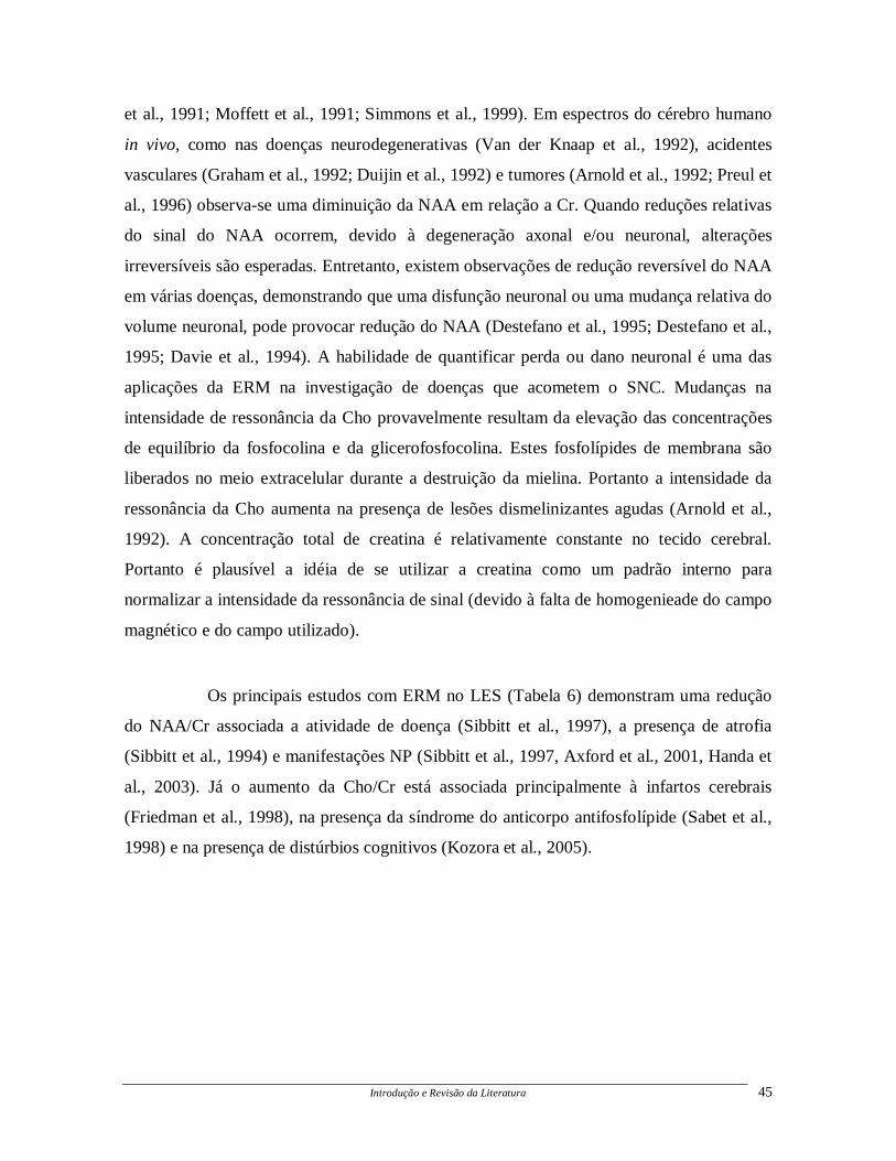
Os principais estudos com ERM no LES (Tabela 6) demonstram uma redução
do NAA/Cr associada a atividade de doença (Sibbitt et al., 1997), a presença de atrofia
(Sibbitt et al., 1994) e manifestações NP (Sibbitt et al., 1997, Axford et al., 2001, Handa et
al., 2003). Já o aumento da Cho/Cr está associada principalmente à infartos cerebrais
(Friedman et al., 1998), na presença da síndrome do anticorpo antifosfolípide (Sabet et al.,
1998) e na presença de distúrbios cognitivos (Kozora et al., 2005).
Intr
oduç
ão e
Rev
isão
da
Lite
ratu
ra
46
Tab
ela
6. L
ES
: est
udos
uti
lizan
do a
esp
ectr
osco
pia
por
prót
ons
Cho
: cho
lina;
Cr:
cre
atin
a; E
RM
: esp
ectr
osco
pia
por
ress
onân
cia
mag
néti
ca; m
I: m
ioin
usit
ol; M
NP:
man
ifes
taçõ
es n
euro
psiq
uiát
rica
s; m
s: m
ilise
gund
os; N
AA
: N
-ace
tyl a
spar
tato
; RM
: res
sonâ
ncia
mag
néti
ca; S
AA
F: s
índr
ome
do a
ntic
orpo
ant
ifos
folí
pide
; SB
: sub
stân
cia
bran
ca; S
LIC
C: S
LIC
C/A
CR
-DI;
SPE
CT
: sin
gle
phot
on e
mis
sion
com
pute
r to
mog
raph
y.
Aut
or/
Ano
L
ocal
izaç
ão d
a E
RM
N
o de
pa
cien
tes
Alt
eraç
ões
da E
RM
A
ssoc
iaçõ
es c
líni
cas
Sibb
itt e
t al.,
19
94
SB, s
uper
ior
aos
vent
rícu
los
nos
dois
he
mis
féri
os
21
� NA
A/C
r M
ais
inte
nso
na p
rese
nça
de a
trof
ia
Dav
ie e
t al.,
19
95
Les
ões
de S
B (
5 pa
cien
tes)
e S
B n
orm
al (
7 pa
cien
tes)
13
� NA
A/C
r Se
m c
orre
laçã
o co
m M
NP;
� NA
A/C
r na
s le
sões
B
rook
s et
al.,
19
97
Les
ões
e SB
nor
mal
nas
reg
iões
per
iven
tric
ular
e
occi
pita
l e s
ubst
ânci
a ci
nzen
ta o
ccip
ital
14
� NA
A/C
r E
m to
das
as a
reas
est
udad
as; m
ais
inte
nso
nas
lesõ
es
Chi
nn e
t al.,
19
97
SB f
ront
al e
par
ieto
-occ
ipit
al
47
� NA
A/C
r
� Cho
/Cr
Ass
ocia
ção
com
cor
tico
ster
óide
s
Sibb
itt e
t al.,
19
97
SB n
orm
al p
arie
tal
36
� NA
A/C
r A
tivi
dade
da
doen
ça e
MN
P
Sabe
t et a
l.,
1998
SB
pro
fund
a oc
cipi
to-p
arie
tal
43
� NA
A/C
r
� Cho
/Cr
com
SA
AF
Frie
dman
et a
l.,
1998
SB
nor
mal
occ
ipit
o-pa
riet
al
42
� NA
A/C
r
� Cho
/Cr
� NA
A/C
r em
lesõ
es f
ocai
s
� Cho
/Cr
com
infa
rtos
cer
ebra
is
Bro
oks
et a
l.,
1999
Les
ões
e SB
nor
mal
nas
reg
iões
pe
rive
ntri
cula
res
e oc
cipi
tais
e s
ubst
ânci
a ci
nzen
ta o
ccip
ital
12
� Cho
/Cr
Dis
túrb
io c
ogni
tivo
SL
ICC
Lim
et a
l.,
2000
G
angl
ios
basa
is e
SB
per
iven
tric
ular
26
� NA
A/C
r em
gan
glio
s da
bas
e
� Cho
/Cr
peri
vent
ricu
lar
MN
P m
aior
es; s
em a
ssoc
iaçã
o co
m a
chad
os
à R
M
Axf
ord
et a
l.,
2001
Su
bstâ
ncia
bra
nca
pari
etal
nor
mal
9
� NA
A,
� mI
� Cho
� NA
A: M
NP
mai
ores
� mI
na s
ubst
ânci
a br
anca
nor
mal
� Cho
: MN
P m
enor
es
Han
da e
t al.,
20
03
SB p
arie
to-o
ccip
ital
e fr
onta
l nor
mal
20
� NA
A/C
r M
NP
Cas
telli
no e
t al.,
20
05
Áre
as h
ipop
erfu
ndid
as e
nor
mop
erfu
ndid
as n
o SP
EC
T
8
� NA
A/ C
ho e
m a
reas
hi
pope
rfun
dida
s
� Cho
/Cr
em á
reas
no
rmop
erfu
ndid
as
Les
ões
de s
ubst
ânci
a br
anca
; Apa
reci
men
to
de le
sões
de
subs
tânc
ia b
ranc
a em
áre
as
hipo
perf
undi
das
com
� NA
A/ C
ho
Koz
ora
et a
l.,
2005
SB
nor
mal
fro
ntal
e p
eriv
entr
icul
ar
7
� Cho
/Cr
� na
pres
ença
de
dist
úrbi
os c
ogni
tivos
Introdução e Revisão da Literatura 47
Single photon emission computed tomography (SPECT)
No SPECT observa-se alterações na função cerebral e na barreira hemato-
encefálica e não de estruturas cerebrais como na RM. A distribuição do contraste é
proporcional ao fluxo sanguíneo na hora da injeção e ao metabolismo cerebral. O SPECT
pode ser um método sensível, mostrando alterações no fluxo cerebral dos pacientes com
sintomas NP. Pacientes com manifestações difusas NP tem áreas simétricas e disseminadas
de hipoperfusão e aqueles com manifestações focais não tem áreas tão disseminadas
(Rogers et al., 1992; Rubbert et al., 1993; Szer et al., 1993; Emmi et al., 1993; Colamussi et
al., 1995; Kodama et al., 1995; Kovacs et al.,1995; Russo et al., 1998; Nossent et al., 1991;
Lin et al., 1997; Huang et al., 2002; Liu et al., 2003; Oku et al., 2003; Handa et al., 2003;
Sundgreen et al., 2005; Zhang et al., 2005).
Estas alterações de perfusão são mais comuns em regiões parietal, frontal,
temporal e gânglios da base, ou seja, aquelas supridas pela artéria cerebral média (Rubbert
et al., 1993; Szer et al., 1993; Emmi et al., 1993; Kovacs et al.,1995; Colamussi et al., 1995;
Kodama et al., 1995; Lin et al., 1997; Russo et al., 1998; Nossent et al., 1991). As
alterações, tanto difusas quanto focais, muitas vezes ocorrem sem alterações da tomografia
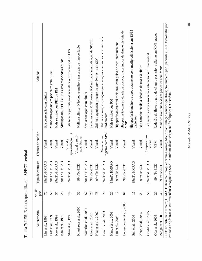
computadorizada ou RM, principalmente nas manifestações do SNC leve (Tabela 7).
Intr
oduç
ão e
Rev
isão
da
Lite
ratu
ra
48
Tab
ela
7: L
ES
: Est
udos
que
uti
lizar
am S
PE
CT
cer
ebra
l
EC
D: E
tilc
iste
inat
o dí
mer
o; H
PMA
O: H
exam
etil
prop
ileno
amin
a ox
ima;
MN
P: m
anif
esta
ção
neur
opsi
quiá
tric
a; N
o: n
úmer
o; p
ac.:
paci
ente
s; P
ET
: tom
ogra
fia
por
emis
são
de p
ósitr
ons;
RM
: res
sonâ
ncia
mag
néti
ca; S
AA
F: s
índr
ome
do a
ntic
orpo
ant
ifos
folí
pide
; Tc:
tecn
ésio
Aut
ores
/Ano
N
o. d
e pa
c.
Tip
o de
con
tras
te
Téc
nica
de
anál
ise
Ach
ados
Lin
et a
l., 1
998
72
99m
Tc-
HM
PAO
V
isua
l B
oa c
orre
laçã
o co
m c
línic
a
Las
s et
al,
1989
50
99
mT
c-H
MPA
O
Vis
ual
Mai
or a
lter
ação
na
em p
acie
ntes
com
SA
AF
Kao
et a
l., 1
999
37
99m
Tc-
HM
PAO
V
isua
l M
ais
sens
ível
que
PE
T o
u R
M
Kao
et a
l., 1
999
25
99m
Tc-
HM
PAO
V
isua
l A
lter
ação
no
SPE
CT
e P
ET
est
ão a
ssoc
iada
s a
MN
P
Shen
et a
l., 1
999
109
99m
Tc-
HM
PAO
V
isua
l e
apre
sent
ação
3D
E
sta
técn
ica
perm
ite a
valia
r m
elho
r o
flux
o ce
rebr
al n
o L
ES
Kik
ukaw
a et
al.,
200
0 32
99
mT
c-E
CD
V
isua
l e s
emi-
quan
titat
iva
Mel
hora
clín
ica;
Não
hou
ve m
elho
ra n
as á
reas
de
hipo
perf
usão
Wat
erlo
o et
al.,
200
1 52
99
mT
c-H
MPA
O
Vis
ual
Sem
ass
ocia
ção
com
clí
nica
Che
n et
al.,
200
2 20
99
mT
c-E
CD
V
isua
l Pa
cien
tes
com
MN
P le
ves
e R
M n
orm
ais:
sem
indi
caçã
o de
SPE
CT
Hua
ng e
t al.,
200
2 78
99
mT
c-E
CD
V
isua
l Ú
til n
o di
agnó
stic
o pr
ecoc
e do
env
olvi
men
to d
o SN
C
Bor
elli
et a
l., 2
003
20
99m
Tc-
HM
PAO
V
isua
l e c
o-re
gist
ro c
om S
PM
Úti
l par
a co
-reg
istr
o, s
uger
e qu
e al
tera
ções
ana
tôm
icas
oco
rrem
mai
s ta
rdia
men
te
Han
da e
t al.,
200
3 20
99
mT
c H
MPA
O
Vis
ual
Mai
s se
nsív
el q
ue R
M
Liu
et a
l., 2
003
12
99m
Tc-
EC
D
Vis
ual
Hip
oper
fusã
o ce
rebr
al m
elho
rou
com
pul
so d
e m
etil
pred
inis
olon
a
Lop
es-L
ongo
et a
l., 2
003
67
99m
Tc-
EC
D
Vis
ual
Hip
oper
fusã
o co
m a
tivi
dade
de
doen
ça, m
aior
índi
ce d
e da
no e
his
tóri
a de
M
NP
Sun
et a
l., 2
004
15
99m
Tc-
HM
PAO
V
isua
l H
ipop
erfu
são
mel
horo
u ap
ós tr
atam
ento
com
met
ilpre
dini
solo
na e
m 1
3/15
pa
cien
tes
Abr
eu e
t al.,
200
5 23
99
mT
c-E
CD
V
isua
l N
ão c
orre
laci
onad
o a
acha
dos
de R
M e
a c
línic
a
Om
dal e
t al.,
200
5 56
99
mT
c-H
MPA
O
Com
para
tivo
-vi
sual
Fa
diga
não
est
ava
asso
ciad
a a
alte
raçã
o no
flu
xo c
ereb
ral
Oda
et a
l., 2
005
20
99m
Tc
EC
D
VB
M
Red
ução
do
flux
o no
gir
o do
cín
gulo
pos
teri
or e
tála
mo
em M
NP
grav
es
Zan
gh e
t al.,
200
5 43
99
mT
c-E
CD
V
isua
l M
ais
sens
ível
que
RM
em
pac
ient
es c
om M
NP
Introdução e Revisão da Literatura 49
1.4.3. Outros métodos de imagem
Outros métodos de imagem estruturais (magnetic transfer imaging, diffusion
tensor imaging) e funcionais (ressonância magnética funcional, tomografia de emissão de
pósitrons) têm sido utilizados para avaliação do comprometiento do SNC no LES, porém
não foram aqui descritos por não terem sido utilizados no presente trabalhos.

51
2. OBJETIVOS
Objetivos 53
2.1. OBJETIVO GERAL DA TESE
O objetivo geral foi avaliar as manifestações do SNC no LES e correlacioná-las
a alterações cerebrais estruturais e funcionais através de técnicas de neuroimagem.
2.2. OBJETIVOS ESPECÍFICOS DE CADA ARTIGO
Artigo 1: Neurolupus.
Revisão sobre o histórico do comprometimento do SNC no LES.
Artigo 2: Central nervous system manifestations in systemic lupus erythematosus
Revisão sobre as manifestações do SNC no LES, incluíndo classificação,
etiologia, investigação e tratamento.
Artigo 3: Magnetic resonance spectroscopy in the evaluation of central nervous
system manifestations of systemic lupus erythematosus.
Revisão da literatura sobre a utilização da espectroscopia por RM na avaliação
das manifestações do SNC no LES.
Artigo 4: Epileptic seizures in systemic lupus erythematosus.
Determinar a freqüência de epilepsia no LES e fatores de risco associados a sua
ocorrência.
Artigo 5: Clinical implications of migraine in systemic lupus erythematosus:
relation to cumulative organ damage.
Avaliar a importância clínica da migrânea no LES e sua relação com o dano
permanente.
Objetivos 54
Artigo 6: Acute psychosis in systemic lupus erythematosus.
Determinar a freqüência de psicose aguda no LES e fatores de risco associados.
Determinar variáveis clínicas que diferenciem psicose aguda daquela induzida
por corticosteróides.
Artigo 7: Cerebral venous thrombosis: influence of r isk factors and imaging
findings on prognosis.
Determinar achados clínicos, de neuroimagem e de prognóstico associados a
ocorrencia de trombose venosa central de diferentes etiologias.
Artigo 8: Cerebral and corpus callosum atrophy in systemic lupus erythematosus.
Determinar o volume cerebral e do corpo caloso em pacientes com LES e
fatores clínicos, laboratoriais e de tratamento associados.
Artigo 9: Longitudinal analysis of gray and white matter loss in patients with
systemic lupus erythematosus.
Determinar a presença e a progressão de atrofia de substância branca e cinzenta
através da análise de morfometria baseada em voxels de pacientes com LES.
Artigo 10: Hippocampal atrophy in systemic lupus erythematosus.
Determinar a freqüência e progressão da atrofia hipocampal em pacientes com
LES e fatores associados.
Artigo 11: Voxel-based morphometry of brain SPECT can detect the presence of
active central nervous system involvement in systemic lupus
erythematosus.
Avaliar se a análise do SPECT cerebral pela técnica de VBM é sensível para
detectar alterações funcionais em pacientes com comprometimento do SNC no
LES.

Objetivos 55
Artigo 12: Evidence of reversible axonal dysfunction in systemic lupus
erythematosus: a proton MRS study.
Avaliar a presença de disfunção axonal no LES.
Artigo 13: Increased choline/creatine ratio on MRS may predict appearance of
white matter lesions in systemic lupus erythematosus.
Determinar se o aumento da relação cholina/creatina na ERM pode predizer o
aparecimento de lesões de substância branca no LES.
57
3. PACIENTES E MÉTODOS
Pacientes e Métodos 59
Todos os artigos que compõe esta tese apresentam metodologia semelhante no
que concerne à seleção de pacientes, critérios de inclusão e exclusão, aspectos éticos,
análise clínica e laboratorial e metodologia aplicada a artigos específicos (atividade de
doença, índice de dano, métodos de neuroimagem). Excetua-se o artigo #7 que trata de
trombose venosa central não somente no LES, mas de diferentes etiologias.
3.1. METODOLOGIA COMUM À TODOS OS TRABALHOS
3.1.1. Seleção da casuística
Os pacientes que participaram do estudo clínico e de RM foram selecionados no
ambulatório de LES da Reumatologia da UNICAMP.
3.1.2. Critérios de inclusão
Foram incluídos pacientes com diagnóstico de LES segundo os critérios
estabelecidos pelo Colégio Americano de Reumatologia (Tan et al., 1982).
3.1.3. Critérios de exclusão
Foram excluídos os pacientes que:
1. Pacientes com investigação incompleta.
2. Pacientes que perderam seguimento.
3. Pacientes com prontuários incompletos.

Pacientes e Métodos 60
Alguns outros critérios de inclusão e exclusão são pertinentes à diferentes
artigos e estes estão adequadamente detalhados nos respectivos trabalhos (artigos #5, #7,
#8-#13).
3.1.4. Aspectos Éticos
Todos os diferentes estudos foram aprovados pelo Comitê de Ética em Pesquisa
da Faculdade de Ciências Médicas da Universidade Estadual de Campinas (UNICAMP).
No caso dos estudos prospectivos com grupo controle, todos os pacientes e voluntários
participantes foram devidamente esclarecidos quanto às finalidades da pesquisa, e
assinaram, previamente, os formulários de consentimento informado.
3.1.5. Análise clínico-laboratorial
Em todos os estudos, as variáveis clínicas, laboratoriais e o uso de
imunossupressores foram analisadas em dois períodos: ao diagnóstico do LES e durante o
seguimento destes pacientes, através da revisão dos prontuários dos pacientes: presença de
adinamia; emagrecimento (> 4 kg); febre (�
37,8o C); artrite (não erosiva em duas ou mais
articulações periféricas, vista pelo médico); necrose asséptica (documentada por radiografia
simples, cintilografia ou ressonância nuclear magnética); deformidades articulares
(geralmente redutíveis, vistas pelo médico); eritema malar (eritema fixo sobre as
eminências malares e/ou pregas naso-labiais); lesões discóides (placas eritematosas com
descamação,podendo ocorrer atrofia nas lesões antigas); alopécia; úlcera oral e/ou nasal
(ulceração oral e/ou em nasofaringe, geralmente dolorosa, observadas por médico);
fotossensibilidade (“rash” cutâneo resultado da exposição à luz solar, relatado na história
clínica ou observada por médico); nefrite (definida pela presença de proteinúria maior que
0,5 g/l em 24 horas, aumento progressivo de creatinina sérica ou ainda alterações
histopatológicas quando compatíveis com nefrite lúpica, segundo critérios da Organização
Mundial de Saúde); HAS: pressão sistólica maior que 130 mmHg e/ou pressão diastólica
maior que 90 mmHg; síndrome nefrótica (proteinúria maior que 3 g/l em 24 horas); serosite
(presença de pleurite, pericardite ou ambas documentada no exame clínico e por imagem);
Pacientes e Métodos 61
outras manifestações pulmonares como hipertensão pulmonar, pneumonite e hemorragia
pulmonar; outras manifestações cardíacas como miocardite, endocardite própria do LES e
infarto do miocárdio; miopatia (revelada por fraqueza muscular, alterações enzimáticas,
alterações da biópsia muscular e /ou da eletromiografia).
Foram considerados também o envolvimento intestinal, hepático, e do sistema
retículo-endotelial, presença de tromboembolismo pulmonar e alterações oculares e a
presença do fenômeno de Raynaud.
Todos os exames laboratoriais e autoanticorpos foram realizados seguindo
técnicas de rotina utilizadas no Laboratório de Patologia Clinica e no Laboratório de
Investigação em Alergia e Imunologia. As alterações hematológicas quando induzidas por
drogas ou infeções foram excluídas. Foram considerados: leucopenia (�
4000
células/mm3); linfopenia (�
1500 cels/mm3); anemia hemolítica (Coombs direto positivo,
aumento de bilirubina indireta, anemia aguda); trombocitopenia (�
100000 cels/mm3);
FAN (por imunofluorescência indireta, positivo em títulos maiores que 1:40); anticorpo
anti-DNA (por imunofluorescência indireta com Crithidia luciliae como substrato);
anticorpo anti Sm (por imunodifusão dupla); Anticorpo anti cardiolipina (por método
imunoenzimático) (Harris et al., 1987); anticoagulante lúpico (por TTPA e Russel) (Brandt
et al., 1995). Pacientes com seguimento no ambulatório antes de 1999 tiveram a dosagem
do anticorpo antifosfolípide e anticardiolipina realizados após esta data.
A terapêutica atual e pregressa foi analisada através da revisão dos prontuários.
Doses de diferentes corticosteróides foram convertidas para doses equivalentes de
prednisona. Dose total de prednisona foi calculada através da somatória das doses diárias,
através de um programa especificamente desenvolvida para esta finalidade.
3.2. METODOLOGIA APLICADA A ARTIGOS ESPECÍFICOS
Além da metodologia comum a todos os artigos, metodologias específicas,
tanto clínicas como de neuroimagens, foram aplicados a diferentes artigos, a saber:
Pacientes e Métodos 62
3.2.1; Investigação clínica:
3.2.1.1. Avaliação das manifestações NP
- retrospectiva (artigos #4-#7, #8-#13)
- prospectiva (artigos #8-#13)
As manifestações neuropsiquiátricas foram analisadas retrospectivamente,
através da revisão dos prontuários, conforme as orientaçõs do Colégio Americano de
Reumatologia de 1999 (ACR, 1999).
Nos estudos prospectivos os seguintes testes foram aplicados aos pacientes com
objetivo de determinar a presença de manifestação NP:
• Distúrbios cognitivos:
o Minimental (Folstein et al., 1975): Neste teste foram verificados a orientação, a
atenção e o registro, a atenção e o cálculo, a memória imediata, a linguagem, a
praxia e a fluência verbal. Para cada item foi dado um determinado número de
pontos, somados ao final. Considerou-se como normal um escore de 28±2, como
depressão um total de 19±3, e como demência um total de 10±3.
o Memória lógica (Spranoel, 1992): Para a avaliação da memória lógica foram
lidos para o paciente dois textos, cada um com 23 idéias, que deveriam ser
recontados e para cada idéia corretamente memorizada era aplicado um ponto.
Os pontos foram somados e anotados.
o Teste de atenção (Spranoel, 1992): Também foi testada a atenção do paciente
tendo este que repetir certos números em ordem direta e inversa. Os números
variaram de 3 a 9 algarismos e os pontos foram dados de acordo com o número
de algarismos. Os pontos foram somados e anotados.
Pacientes e Métodos 63
• Depressão: Escala de depressão de Beck (Beck e Beamesderfer, 1974; Beck et al.,
1974)
• Ansiedade: Hospital anxiety and depression scale (HAD/CAGE) (Hermann, 1997)
• Manifestações psiquiátricas: Brief Psychiatric Rating Scale (BPRS) (Overall e Beller,
1984)
3.2.1.2. Atividade de doença
- SLEDAI (artigos #6, #8-#13)
- MEX-SLEDAI (artigo #5)
A atividade de doença do LES foi avaliada através do SLEDAI (Bombardier et
al., 1992). O MEX-SLEDAI (Guzman et al., 1992) por não incluir cefaléia como atividade
de doença foi utilizado no trabalho sobre migrânea (artigo #5). Atividade de doença foi
considerada quando ocorreram scores maiores ou iguais a 8.
3.2.1.3. Índice de dano
- SLICC/ACR-DI (artigos #5, #8-#13)
O índice de dano permanente foi avaliado através do SLICC-ACR/DI (Gladman
et al., 1997). Todos os dados foram obtidos através da revisão dos prontuários.
3.2.2. Investigação com técnicas de neuroimagem:
3.2.2.1. RM
- Segmentação manual (artigo #10)
- Segmentação semi-automática (artigo #8)
Pacientes e Métodos 64
- Segmentação automática (artigo #9)
- Espectroscopia por ressonância magnética (ERM) (artigos #12e #13)
Aquisição das imagens de RM
Todos os indivíduos (pacientes ou controles) foram convidados para realização
de exame de RM de alta resolução. Os exames foram realizados após assinatura do termo
de consentimento para pesquisa.
As imagens foram obtidas em sistema de 2 Tesla (Elscint Prestige�
, Halifa,
Israel), com aquisições nos planos coronal, sagital e axial, além de aquisições em 3 D
(volumétricas), para reconstrução multiplanar em qualquer plano ou inclinação. Os
parâmetros de imagens para as diferentes aquisições foram:
• Imagens sagitais T1 ponderadas “spin echo” (espessura de 6 mm, ângulo de excitação –
“ tip angle” –de 180º; TR=430ms, TE=12ms, matriz de 200x350, FOV=25x25cm).
Estas imagens serão utilizadas para orientar o plano de aquisição das demais imagens.
• Imagens no plano coronal (T2 ponderadas, FLAIR)
• T2 ponderadas (espessura de 6 mm, ângulo de exitação de 180º, TR=1800ms,
TE=90ms, matriz de 165x256, FOV=20x24cm) ou “ fast spin echo” T2
ponderadas (espessura de 4mm, ângulo de exitação de 120º, TR=6800ms,
TE=129ms, matriz de 252x328, FOV=21x23cm);
• FLAIR (TR= 8500ms e 2000ms ou 100ms e 2200ms, TE=72ms ou 90ms,
matrix= 256 X 296 ou 250 X 256, FOV= 200 X 220 ou 220 x 220 mm);
• Imagens no plano axial: duplo “spin echo” (T2 ponderadas e densidade de prótons); T2
ponderadas (espessura de 6 mm, ângulo de exitação de 180º, TR=1800ms, TE=90ms,
matriz de 165x256, FOV=20x24cm) ou “ fast spin echo” T2 ponderadas (espessura de
4mm, ângulo de exitação de 120º, TR6800ms, TE=129ms, matriz de 252x328,
FOV=21x23cm.
Pacientes e Métodos 65
• Aquisições em 3D obtidas no plano sagital “gradient echo” T1 ponderadas com
espessura de 1mm, ângulo de exitação de 35º TR=22ms, TE=9ms, matriz de 256x220,
FOV=230x250 cm, pixel 1x1.
• Espectroscopia obtida em região supraventricular posterior (PRESS (Point-Resolved
Spectroscopy), TE=135ms, TR=1500ms, ângulo de exitação 90o, nex=1, matriz de
20x1024, FOV=5x2cm; precedida por calibração com supressão de sinal de água e
homogeneização do campo magnético (Shimming).
Análise de imagens de RM
Análise visual
A análise qualitativa das imagens foi realizada em estação de trabalho
(OMNIPRO�
) por dois investigadores, sendo um deles radiologista que desconhecia a
presença ou não de doença do paciente (AVF). As imagens foram avaliadas quanto à
presença de alterações de substância branca e cinzenta, presença de atrofia (dilatação de
sulcos e ventrículos), sendo classificadas de acordo com a localização, provável etiologia e
número total de lesões (Cotton et al., 2004).
Segmentação manual
A volumetria dos hipocampos foi realizada utilizando as sequências de cortes
coronais T1-IR, através do programa Scion�
. O programa Scion é de distribuição gratuita
(http://www.rsb.info.nih.gov/scion) de plataforma Windows e utiliza a segmentação manual
como base. Os parâmetros anatômicos utilizados para o estudo da volumetria de
hipocampos são os descritos em protocolos publicados previamente (Cendes et al., 1993;
Watson et al., 1992; Watson et al., 1997).
Segmentação semi-automática
A volumetria do corpo caloso, ventrículos laterais, cerebelo e contorno cerebral
foi realizada utilizando-se as sequências de cortes sagitais ponderadas em T1, através do
Pacientes e Métodos 66
programa Neuroline�
, desenvolvido no próprio Laboratório de Neuroimagem (LNI). Este
programa utiliza para o processamento de imagens o método de watershed, caracterizando
uma segmentação semi-automática, na plataforma Windows. Os parâmetros anatômicos
utilizados para o estudo da volumetria destas estruturas foram definidos visualmente. O
programa foi elaborado como uma alternativa à segmentação manual, em que as estruturas
são delineadas através de contorno manual das regiões de interesse. A forma de interação
do operador com o sistema é através da definição de marcadores, pontos ou linhas
desenhadas pelo operador, através dos quais o método obtém os contornos. Os marcadores
são coloridos, sendo que cada cor está associada a uma determinada estrutura que se deseja
segmentar. A cada marcador é associado um rótulo para identificação da estrutura
segmentada. A saída do processo de segmentação é provida de três formas. Uma das formas
é a gravação das imagens segmentadas. Outra forma é a gravação de marcadores que, ao
serem carregados, refazem a segmentação. A terceira forma é a gravação dos volumes de
cada estrutura, por corte, em um arquivo de texto.
O método de segmentação utilizado no sistema é a transformação Watershed
com marcadores no campo da Morfologia Matemática. Este método baseia-se nas variações
de níveis de cinza e na localização dos “pixels” marcados para a obtenção de contornos
(Beucher et al., 1993).
A imagem em escala de cinza é modelada como uma superfície topográfica, em
que os tons de cinza são proporcionais à altitude da superfície. Os marcadores rotulados
localizados na imagem são equivalentes à perfuração de buracos na superfície (Beucher et
al., 1993).
Pós processamento das imagens segmentadas
Os volumes totais das estruturas obtidos pela segmentação manual ou semi-
automática foram calculados através da soma dos volumes segmentados multiplicados pela
espessura do corte. Estes valores foram posteriormente corrigidos pelo volume cerebral
total do pacientes, a fim de evitar que estruturas de cérebros pequenos sejam consideradas
atróficas (Watson et al., 1997).
Pacientes e Métodos 67
Para evitar que o cerebelo, o ventrículo e o corpo caloso em cérebros pequenos
sejam identificados como atróficos, os volumes absolutos, em milímetros cúbicos, foram
corrigidos pelo volume cerebral total (VCT) segundo a fórmula:
Volume normalizado (cerebelo ou corpo caloso) = volume (cerebelo ou corpo
caloso) absoluto do indivíduo X média VCT dos controles/ VCT do indivíduo
O índice de assimetria (IA) foi determinado pela razão dos volumes entre a
menor e a maior estrutura.
Identificação de atrofia ou dilatação ventricular
Para avaliar a presença de atrofia das estruturas segmentadas nos pacientes e
controles foi calculado o valor de Z score (número de desvios-padrão acima ou abaixo da
média do grupo controle) para cada estrutura (VC = cerebelo/corpo caloso volume
normalizado) e para o IA, segundo a fórmula abaixo:
Z score VCD=(VC do paciente – média dos VC dos controles)/desvio-padrão
da média dos VC dos controles.
Foi considerada atrofia de uma determinada estrutura quando o volume das
estruturas normalizado e/ou índices de assimetria foram inferiores a 2 desvios-padrão da
média dos controles (valor de Z score menor ou igual a -2).
Dilatação ventricular foi considerada quando o Z score dos ventrículos foi
maior que 2 desvios-padrão da média dos controles (valor de Z score maior ou igual a +2).
Segmentação automática
A morfometria baseada em voxels (VBM) foi realizada utilizando as sequências
de cortes volumétricos T1-IR. Através do programa MRIcro (www.MRIcro.com), obtido da
INTERNET, as imagens foram processadas para extração do crânio e realinhadas a partir
de um ponto comum que foi considerado a comissura anterior. As análises subseqüentes
foram realizadas no programa Statistical Paramtric Mapping [SPM 2
(http://www.fil.ion.ucl.ac.uk/spm/)] obtidos pela INTERNET e rodados pelo MATLAB 6.1
Pacientes e Métodos 68
na plataforma Windows segundo protocolos previamente publicados (Ashburner e Friston
1997; Ashburner e Friston 2000). Os resultados são expressos em coordenadas
esteriotáxicas que foram visualmente confirmadas pelo programa Talairach Daemon client,
disponível gratuitamente pela INTERNET
(http://ric.uthscsa.edu/projects/talairachdaemon.html).
Espectropscopia por prótons (ERM)
A ERM foi analisada no console do aparelho de RM, Elscint Prestige 2T, por
meio de quantificação manual através de um programa da própria Elscint. Após a correção
da linha de base as áreas sob os picos dos compostos de N-Acetylaspartato (NAA) em 2.01
da escala partes por milhão (ppm), colina (Cho) em 3.2 ppm e compostos contendo creatina
e fosfocreatina em 3.0 ppm. Para análise foram utilizadas razões, sendo a Cr o denominador
comum. A análise dos espectros foi realizada por um investigador (SA) e checadas
independentemente por dois outros investigadores (FC e LML) que não tinham
conhecimento sobre o sujeito (paciente ou controle). Espectros com linha de base de difícil
avaliação ou pouca diferenciação entre os picos individuais foram excluídos.
3.2.2.2. SPECT cerebral (artigo #11)
Aquisição das imagens com SPECT
As imagens foram obtidas 15 minutos após a injeção venosa de 20mlides ECD-
99Tc e em ambiente isento de estímulos visuais e sonoros. Foi utilizada uma câmera de
cintilação tomográfica, sendo adquiridos 60 imagens a cada seis graus, em um total de 360
graus. Foram utilizadas aquisição em modo “byte” e matrix 64x64.
Análise das imagens
As imagens obtidas foram analisadas e, se presentes artefatos, repetidas. As
imagens foram reconstruídas nos cortes trans-axial, coronal, sagital e trans-supra-órbito-
Pacientes e Métodos 69
meatal e analisadas por um médico especialista de Medicina Nuclear sem o conhecimento
da história clínica. As imagens também foram processadas utilizando-se o programa
MATLAB 6.5 for windows (MathWorks, Natrick, MA) e SPM 02 (Wellcome Dept Cogn.
Neurol, London) conforme protocolos do laboratório de neuroimagem da UNICAMP. A
morfometria baseada em voxels (VBM) foi utilizada para comparar as imagens. A VBM
envolve vários processos, incluindo a normatização espacial das imagens para o mesmo
espaço esterotáxico e a segmentação de imagens. Como o SPECT depende de áreas
ricamente vascularizadas, utilizou-se somente a análise da substância cinzenta neste estudo.
3.2.3. Análise estatística
- Teste de qui-quadrado (artigos #4-#13)
- Teste T (artigos #8, #11)
- ANOVA (artigos #8-#13)
- Teste t-pareado (artigos #9, #10, #12, #13)
- Regressão simples (artigos #4, #5, #6, #8-#13)
- Regressão logística (artigos #4 e #6)
As diferentes freqüências foram analisadas pelo teste de qui quadrado. A
correção de Yates e o teste exato de Fischer foram utilizados no caso em que a freqüência
em uma ou mais caselas, respectivamente, tenha sido inferior a cinco. ANOVA com
correção de Tukey foi utilizada para comparação entre gupos. As variáveis não numéricas
foram avaliadas por testes não paramétricos apropriados.
Para a análise das RM pela técnica de VBM foi utilizado o teste t. Para a
comparação entre o mesmo indivíduo nos estudos longitudinais foi utilizado o teste t-
pareado.
A regressão simples foi utilizada para determinar a associação entre variáveis
clínicas e volumes cerebrais, de corpo caloso e de hipocampos.
Pacientes e Métodos 70
A regressão logística multivariada com correção para múltiplas comparações
foi utilizada para determinar a associações entre as variáveis clínicas.
3.3. APRESENTAÇÃO E ANÁLISE DOS DADOS
Os resultados referentes à investigação clínica e por neuroimagem estão
apresentados sob a forma de artigos, com enfoque específico a cada aspecto da avaliação no
capítulo 3.
Os artigos de um a três referem-se sobre a revisão histórica do Neurolupus
(artigo #1), das manifestações do SNC (artigo #2) e da ERM na investigação do
comprometimento do SNC (artigo #3).
Os aspectos clínicos são investigados nos artigos de quatro a seis. Assim, a
freqüência de crises epilépticas e fatores de risco associados a sua ocorrência estão
descritas no artigo # 4. A importância clínica da migrânea no artigo #5 e a freqüência da
psicose e fatores de risco associados no artigo #6. Fatores de risco e achados de
neuroimagem na trombose venosa central (TVC) de diferentes etiologias, incluíndo o LES,
estão no artigo #7.
Os artigos referentes a análise das imagens estão descritos nos artigos oito a
treze. Assim, a descrição da atrofia cerebral e do corpo caloso está no artigo#8. A análise
longitudinal da atrofia de substância branca e cinzenta está descrita no artigo #9. A
freqüência e a progressão da atrofia hipocampal e suas implicações clínicas estão descritas
no artigo#10. A análise funcional com SPECT está descrita no artigo #11. A utilização da
ERM em pacientes com LES está contida nos artigos #12 e #13.
71
4. RESULTADOS
(ARTIGOS)
Resultados 73
ARTIGO 1
Neurolupus
Appenzeller S, Costallat LT, Cendes F
Neuroiupus
Arch Neurol 2006; 63:458-460
HISTORY OF NEUROLOGY: SEMINAL CITATIONS
NeurolupusSimone Appenzeller, MD; Lilian T. L. Costallat, MD, PhD; Fernando Cendes, MD, PhD
S ystemic lupus erythematosus (SLE) is an autoimmune disease frequently manifested byneuropsychiatric involvement, which occurs in up to 75% of patients, depending on thetype of manifestations included. Primary involvement may vary from subtle signs, such asheadache and mood disorders, to severe, life-threatening conditions, such as stroke, my-
elopathy, and acute confusional state. Any part of the peripheral or central nervous system (CNS)may be affected by the disease. The diagnosis of primary CNS involvement in SLE is often difficultbecause both focal and diffuse manifestations may occur. A wide range of differential diagnoses hasto be considered, including metabolic abnormalities, infections, uremia, hypertension, and drug therapy.
LUPUS: TERM DEFINITION
Lupus is the Latin word for wolf and wasfrequently used in Roman art and poetry.The reason the term lupus gained a medi-cal connotation is unknown. In The Ori-gin of Medical Terms, Skinner suggestedthat lupus was introduced to describe skinlesions that were akin to a wolf’s gnaw-ing marks.1 In the medieval period, theterm lupus was used to describe several cu-taneous diseases.2 In 1865, Virchow3 triedto elucidate the use of the term lupus inthe medieval period, noting that for 3 cen-turies, from Rogerius (1230) to Manar-dus (1530), the term had been used forboils of the lower extremities.2,3 For dis-eases of the face, including lupus vul-garis, cancer, and lepra, a collective term,derived from the biblical passage noli metangere, was used.
FIRST CLINICAL DESCRIPTIONSOF LUPUS AS A DISEASE
The first description of the medical ill-ness “lupus” was in the 10th century bi-ography of St Martin by Hebernus of Tours.“He [Eraclius, the Bishop of Liege] was se-riously affected and almost brought to the
point of death by the disease called lu-pus. . . . ”2(p3) But it was only in the 19thcentury that lupus erythematosus wasclearly described by Biett, as reported byCazenave:
. . . very rare occurrence, and appears most fre-quently in young people, especially in fe-males , whose hea l th i s o therwiseexcellent. . . . It generally appears in the formof round patches, slightly elevated . . . andgradually increases in circumference. . . . 4(p299)
In 1872, Kaposi first described the sys-temic nature of lupus erythematosus, not-ing that “various grave and even danger-ous constitutional symptoms may beintimately associated with the process inquestion. . . . ”5(p53) Shortly thereafter, CNSinvolvement was recognized and de-scribed as a clinical manifestation of SLE.In 1875, Hebra and Kaposi described comafor the first time in patients with SLE:“ . . . death ensues being preceded by in-creased mental disturbance, coma,. . . .”5(p60) Psychosis and mood disorderswere mentioned in 1896 by Bowen: “I haveof ten met with cases of extrememelancholia . . . and in a number of in-stances the mind has become really af-fected.”6(p700) Between 1895 and 1904 ProfDr William Osler published 29 reports ofskin diseases with a variety of systemicmanifestations. The majority of the caseswere patients with Henoch-Schonlein pur-
Author Affiliations: Rheumatology Unit, Departments of Internal Medicine andNeurology, State University of Campinas (UNICAMP), Campinas, Brazil.
(REPRINTED) ARCH NEUROL / VOL 63, MAR 2006 WWW.ARCHNEUROL.COM458
©2006 American Medical Association. All rights reserved. on March 14, 2006 www.archneurol.comDownloaded from
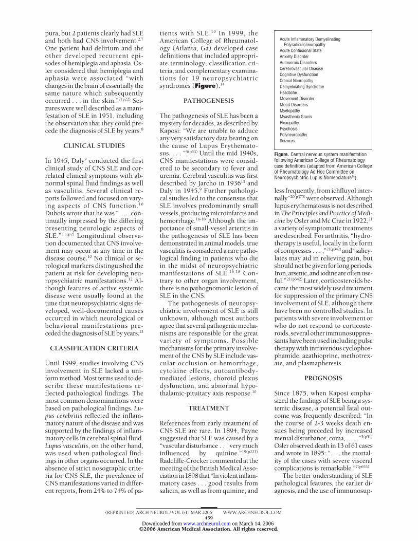
pura, but 2 patients clearly had SLEand both had CNS involvement.2,7
One patient had delirium and theother developed recurrent epi-sodes of hemiplegia and aphasia. Os-ler considered that hemiplegia andaphasia were associated “withchanges in the brain of essentially thesame nature which subsequentlyoccurred . . . in the skin.”7(p22) Sei-zures were well described as a mani-festation of SLE in 1951, includingthe observation that they could pre-cede the diagnosis of SLE by years.8
CLINICAL STUDIES
In 1945, Daly9 conducted the firstclinical study of CNS SLE and cor-related clinical symptoms with ab-normal spinal fluid findings as wellas vasculitis. Several clinical re-ports followed and focused on vary-ing aspects of CNS function.10
Dubois wrote that he was “ . . . con-tinually impressed by the differingpresenting neurologic aspects ofSLE.”11(p2) Longitudinal observa-tion documented that CNS involve-ment may occur at any time in thedisease course.10 No clinical or se-rological markers distinguished thepatient at risk for developing neu-ropsychiatric manifestations.12 Al-though features of active systemicdisease were usually found at thetime that neuropsychiatric signs de-veloped, well-documented causesoccurred in which neurological orbehavioral manifestations pre-ceded the diagnosis of SLE by years.13
CLASSIFICATION CRITERIA
Until 1999, studies involving CNSinvolvement in SLE lacked a uni-form method. Most terms used to de-scribe these manifestations re-flected pathological findings. Themost common denominations werebased on pathological findings. Lu-pus cerebritis reflected the inflam-matory nature of the disease and wassupported by the findings of inflam-matory cells in cerebral spinal fluid.Lupus vasculitis, on the other hand,was used when pathological find-ings in other organs occurred. In theabsence of strict nosographic crite-ria for CNS SLE, the prevalence ofCNS manifestations varied in differ-ent reports, from 24% to 74% of pa-
tients with SLE.10 In 1999, theAmerican College of Rheumatol-ogy (Atlanta, Ga) developed casedefinitions that included appropri-ate terminology, classification cri-teria, and complementary examina-tions for 19 neuropsychiatricsyndromes (Figure).14
PATHOGENESIS
The pathogenesis of SLE has been amystery for decades, as described byKaposi: “We are unable to adduceany very satisfactory data bearing onthe cause of Lupus Erythemato-sus. . . . ”5(p53) Until the mid 1940s,CNS manifestations were consid-ered to be secondary to fever anduremia. Cerebral vasculitis was firstdescribed by Jarcho in 193615 andDaly in 1945.9 Further pathologi-cal studies led to the consensus thatSLE involves predominantly smallvessels, producing microinfarcts andhemorrhage.16-18 Although the im-portance of small-vessel arteritis inthe pathogenesis of SLE has beendemonstrated in animal models, truevasculitis is considered a rare patho-logical finding in patients who diein the midst of neuropsychiatricmanifestations of SLE.16-18 Con-trary to other organ involvement,there is no pathognomonic lesion ofSLE in the CNS.
The pathogenesis of neuropsy-chiatric involvement of SLE is stillunknown, although most authorsagree that several pathogenic mecha-nisms are responsible for the greatvariety of symptoms. Possiblemechanisms for the primary involve-ment of the CNS by SLE include vas-cular occlusion or hemorrhage,cytokine effects, autoantibody-mediated lesions, choroid plexusdysfunction, and abnormal hypo-thalamic-pituitary axis response.10
TREATMENT
References from early treatment ofCNS SLE are rare. In 1894, Paynesuggested that SLE was caused by a“vasculardisturbance . . . verymuchinfluenced by quinine.”19(p223)
Radcliffe-Crocker commented at themeeting of the British Medical Asso-ciationin1898that“Inviolentinflam-matory cases . . . good results fromsalicin, as well as from quinine, and
less frequently, from ichfluyol inter-nally”20(p375) wereobserved.Althoughlupuserythematosus isnotdescribedinThe Principles and Practice of Medi-cine by Osler and Mc Crae in 1922,21
a variety of symptomatic treatmentsare described. For arthritis, “hydro-therapy is useful, locally in the formofcompresses . . . ,”21(p342) and“salicy-lates may aid in relieving pain, butshould not be given for long periods.Iron,arsenic,andiodineareoftenuse-ful.”21(p342) Later, corticosteroids be-camethemostwidelyused treatmentfor suppression of the primary CNSinvolvement of SLE, although therehave been no controlled studies. Inpatients with severe involvement orwho do not respond to corticoste-roids, severalother immunosuppres-sants have been used including pulsetherapywith intravenouscyclophos-phamide, azathioprine, methotrex-ate, and plasmapheresis.
PROGNOSIS
Since 1875, when Kaposi empha-sized the findings of SLE being a sys-temic disease, a potential fatal out-come was frequently described: “Inthe course of 2-3 weeks death en-sues being preceded by increasedmental disturbance, coma, . . . .”5(p51)
Osler observed death in 13 of 61 casesand wrote in 1895: “ . . . the mortal-ity of the cases with severe visceralcomplications is remarkable.”7(p633)
The better understanding of SLEpathological features, the earlier di-agnosis, and the use of immunosup-
Acute Inflammatory Demyelinating Polyradiculoneuropathy
Acute Confusional StateAnxiety DisorderAutonomic DisordersCerebrovascular DiseaseCognitive DysfunctionCranial NeuropathyDemyelinating SyndromeHeadacheMovement DisorderMood DisordersMyelopathyMyasthenia GravisPlexopathyPsychosisPolyneuropathySeizures
Figure. Central nervous system manifestationfollowing American College of Rheumatologycase definitions (adapted from American Collegeof Rheumatology Ad Hoc Committee onNeuropsychiatric Lupus Nomenclature14).
(REPRINTED) ARCH NEUROL / VOL 63, MAR 2006 WWW.ARCHNEUROL.COM459
©2006 American Medical Association. All rights reserved. on March 14, 2006 www.archneurol.comDownloaded from
pressants for SLE activity and anti-biotics for secondary infections hasimproved the survival of patients withSLE in the last decades. Despite theaggressive treatment, CNS SLE stillhas a guarded prognosis and mortal-ity is elevated, occurring in 7% to 19%of patients. In particular, seizures,stroke, and acute confusional state arepoor-prognosis markers.
Accepted for Publication: May 30,2005.Correspondence : S imoneAppenzeller, MD, Department ofInternal Medicine, State Universityof Campinas (UNICAMP), CEP13081-970, Campinas, Brazil([email protected]).Author Contributions: Study con-cept and design: Appenzeller andCostallat. Acquisition of data:Appenzeller and Cendes. Drafting ofthe manuscript: Appenzeller. Criti-cal revision of the manuscript forimportant intellectual content :Appenzeller, Costallat, and Cendes.Statistical analysis: Appenzeller. Ob-tained funding: Appenzeller. Study su-pervision: Costallat and Cendes.
Funding/Support: This study wassupported by a grant from Funda-cao de Amparo à Pesquisa do Es-tado de Sao Paulo, Sao Paulo, Brazil.
REFERENCES
1. Skinner A. The Origin of Medical Terms. Balti-more, Md: Williams & Wilkins Co; 1949:219.
2. Smith CD, Cyr M. The history of systemic lupuserythematosus from Hippocrates to Osler. RheumDis Clin North Am. 1988;14:1-19.
3. Virchow R. Historische Notizen uber Lupus. Ar-chiv Fur Pathologische Anat. 1865;32:139-143.
4. Cazenave PLA, Chausit M. Du lupus. Ann Mal dela Peau et de la syphilis. 1851;3:297-300.
5. Kaposi M. Lupus erythematosus. In: Hebra F, KaposiM, eds, London TW, trans-ed. On Diseases of theSkin Including the Exanthemata. London, En-gland: The New Sydenham Society; 1875:49-64.
6. Bowen JT. Lupus erythematosus. In: Stedman TL,ed. Twentieth Century Practice. Vol 5. New York,NY: W Wood; 1896:691-708.
7. Osler W. On the visceral complications of ery-thema exudativum multiforme. Am J Med Sci.1895;110:629-646.
8. Russel PW, Haserick JR, Zucker EM. Epilepsy in sys-temic lupus erythematosus: effect of cortisone andACTH. Arch Intern Med. 1951;88:78-92.
9. Daly D. Central nervous system in acute dissemi-nated lupus erythematosus. J Nerv Ment Dis. 1945;102:461-465.
10. West SG. Neuropsychiatric lupus. Rheum Dis ClinNorth Am. 1994;20:129-158.
11. Dubois EL. The clinical picture of systemic lupus
erythematosus. In: Dubois EL, ed. Lupus Ery-thematosus. 2nd ed. Los Angeles: University ofSouthern California Press; 1974.
12. Abel T, Gladman DD, Urowitz MB. Neuropsychi-atric lupus. J Rheumatol. 1980;7:325-333.
13. Feinglass EJ, Arnett FC, Dorsch CA, et al. Neuro-psychiatric manifestations of systemic lupus ery-thematosus: diagnosis, clinical spectrum and re-lationship to other features of the disease.Medicine. 1976;55:323-327.
14. The American College of Rheumatology nomen-clature and case definitions for neuropsychiatriclupus syndromes. Arthritis Rheum. 1999;42:599-608.
15. Jarcho S. Lupus erythematosus associated withvisceral vascular lesions. Bull Johns Hopkins Hosp.1936;59:262-270.
16. Johnson RT, Richardson EP. The neurological mani-festations of systemic lupus erythematosus: a clini-cal-pathological study of 24 cases and review of theliterature. Medicine. 1968;47:337-369.
17. Dubois EL, Tuffanelli DL. Clinical manifestationsof systemic lupus erythematosus. JAMA. 1964;190:104-111.
18. Ellis SG, Verity MA. Central nervous system involve-ment in systemic lupus erythematosus: a review ofneuropathologic findings in 57 cases, 1955-1977.Semin Arthritis Rheum. 1979;8:212-221.
19. Payne JF. A post-graduate lecture on lupuserythematosus. Clin J. 1894;4:223.
20. Radcliffe-Crocker H. Meeting of the BritishMedical Association: discussion on lupuserythematosus. Br J Dermatol. 1898;10:375.
21. Osler W, Mc Crae T. The Principles and Practiceof Medicine. 9th ed. New York, NY: D Appleton &Co; 1922.
Announcement
Topic Collections. Archives offers collections of articlesin specific topic areas to make it easier for physicians tofind the most recent publications in a field. These areavailable by subspecialty, study type, disease, or prob-lem. In addition, you can sign up to receive a Collec-tion E-Mail Alert when new articles on specific topicsare published. Go to http://archneur.ama-assn.org/collections to see these collections of articles.
(REPRINTED) ARCH NEUROL / VOL 63, MAR 2006 WWW.ARCHNEUROL.COM460
©2006 American Medical Association. All rights reserved. on March 14, 2006 www.archneurol.comDownloaded from
Resultados 77
ARTIGO 2
Central Nervous System Manifestations in Systemic Lupus Erythematosus
Appenzeller S, Costallat LT, Cendes F
Central Nervous System Manifestations in Systemic Lupus Erythematosus
submetido
Resultados 78
Central Nervous System Manifestations in Systemic Lupus Erythematosus
Simone Appenzeller MD1, 2, Lilian Tereza Lavras Costallat MD, PhD 1, Fernando Cendes
MD, PhD 2, 3
1Rheumatology Unit - State University of Campinas 2 Neuroimaging Lab-State University
of Campinas; 3 Department of Neurology, State University of Campinas
The authors have nothing to disclose.
Running Title: CNS in SLE
Address all correspondence to:
Dr Fernando Cendes, Department of Neurology,
State University of Campinas (UNICAMP),
Cidade Universitaria Zeferino Vaz, CEP 13083970 Campinas-SP-Brazil
Tel: +55 1937887734
Fax: +55 19 32891818
E-mail: [email protected]
Key words: CNS involvement, SLE
Grants: Fundação de Amparo a Pesquisa do Estado de São Paulo (FAPESP), Centro
Nacional de Pesquisa e Desenvolvimento (CNPq)
Resultados 79
Abstract
Neurologists are often called to evaluate central nervous system (CNS)
manifestations in patients with suspected or definite systemic lupus erythematosus (SLE).
The manifestations are highly diverse and often have major prognostic consequences. The
major difficulties are to determine if the given manifestation is primarily due to SLE
activity in the brain, or a consequence of metabolic disturbances, infection, or
corticosteroid use.
The true incidence of CNS manifestations attributable to SLE is not entirely
clear, but several studies show prevalence rate between 15-75%, depending on different
methods and classification criteria applied.
This paper has the objective to review the main clinical manifestations
according to the American College of Rheumatology (ACR) Criteria, possible etiological
mechanism and neuroimaging features associated with these manifestations. We further
discuss the most important tools that can help bedside diagnosis.
Introduction
Systemic lupus erythematosus (SLE) is an autoimmune disease with central
nervous system (CNS) involvement occurring in up to 75% of patients. However the
frequency of these manifestations in SLE studies varies widely, depending on the type of
manifestations included and the method use for evaluation (1-3). CNS involvement may be
considered primary if directly related to SLE activity in the CNS or secondary when related
to treatment, infections, metabolic abnormalities or other systemic manifestations as uremia
and hypertension (4).
The involvement may vary from subtle signs such as headache and mood
disorders to severe, life threatening conditions, as stroke, myelopathy and acute confusional
state. Any part of the peripheral or CNS may be affected by the disease (5). The diagnosis
of primary CNS involvement by SLE is often difficult, as both focal and diffuse
manifestations may occur and there is no gold standard for diagnosis (4).
Resultados 80
The aim of this study was to review the main clinical manifestations according
to the American College of Rheumatology Criteria (ACR) (6), possible etiological
mechanism and neuroimaging features associated with these manifestations. We further
analyze the most important tools that can help bedside diagnosis.
Search strategy and selection criteria
References for this review were identified by searches of PubMed from 1966
until February 2006 with the terms "central nervous system", "neuropsychiatric", "systemic
lupus erythematosus". Only papers published in English were reviewed. Reviews and
original publications (articles and letters), including those about single cases, were
accepted. In order to uniform description of clinical manifestations we included only
studies that included the 1999 ACR case definitions for NP SLE (6).
We asked first for articles covering the combinations ‘SLE and CNS’ and ‘SLE
and CNS diseases’ . Subsequently, we asked for combinations of SLE and specific CNS
syndromes. In addition to the selected articles, the references in these articles were screened
for other studies of interest.
Classification criteria
The large number of papers published about CNS manifestations in SLE
evidence more manifestations attributable to SLE than seizures and psychosis described in
the original classification criteria by Tan (7). In addition, the several distinct pathologic
mechanisms of CNS disease predispose to different presentations, including focal and
diffuse disease, in addition to central and peripheral involvement. The lack of definitions of
individual manifestations and the absence of standardization for investigation were
reflected in the prevalence and frequency of CNS in different reports, varying from 24-74%
of SLE patients (1, 8).
In 1999, the American College of Rheumatology developed case definitions
that included appropriate terminology, classification criteria and complementary
Resultados 81
examinations for 19 neuropsychiatric syndromes (Table 1) (6). These criteria were a result
of a consensus meeting of experts of several subspecialties (rheumatologist, neurologist,
immunology, and psychiatrist). Furthermore, in 2001, these criteria have been validated in a
cross-sectional study with a specificity of 46% (9). Several studies have used these
classification criteria in order to determine frequency and prevalence of CNS involvement
in SLE population (Table 2) (10-19). Although this studies show a substantial variability
between the frequency of CNS manifestations, suggesting differences between cohorts or
bias in data acquisition, this classification allows us to compare the results (4).
Clinical relevance
The clinical relevance of NP manifestations in SLE has been determined by
analyzing the impact of these manifestations in mortality, quality of life and overall damage
scores (16, 17, 20-29).
Using mortality as indicative for poor outcome in NP manifestations, there are
studies suggesting that patients with NP have increased mortality when compared to
patients without these manifestations (20-24). Although some studies did not find an
increased mortality among patients with CNS manifestations when compared to SLE
patients without CNS manifestations and controls (24-28), the presence of CNS
manifestations, independently of its etiology, seems to have a negative impact in quality of
life (4), increased disability scores (29), and higher fatigue scores (16).
The ACR damage score (ACR/SLICC-DI) has been developed to determine
irreversible damage in SLE patients, irrespectively if attributed to disease itself or
secondary to comorbidities or medications. In the ACR/SLICC-DI scores seizures,
psychosis, mood disorders, cerebrovascular disease, neuropathy, mononeuritis multiplx,
acute confusional state and myelopathy are scored in addition to several other clinical
manifestations in order to determine the global damage score. Using the items of NP in
order to create a NP damage score, the strongest risk factors for the development of
significant NP damage was the presence of greater disease activity at the time of CNS
involvement onset and the presence of antiphospholipid antibodies (17).
Resultados 82
Furthermore it is necessary to determine the course of NP manifestations. There
are only a limited amount of follow-up studies evaluating NP manifestations in SLE
patients (30-35). CNS involvement seems to have a general good prognosis, with
improvement or stabilization of symptoms in most of the cases (31). The presence of
antiphospholipid antibodies, higher number of NP events and hippocampal atrophy were
negative prognostic factors (31, 32). Furthermore the progression of hippocampal atrophy
was a predictor for progressive cognitive impairment (32). Perhaps the absence of
abnormalities on MRI may suggest reversibility or stabilization of NP manifestations,
whereas the presence of progressive atrophy may be related to worse prognosis over time.
In another study cognitive impairment was a stable symptom over time and more frequently
observed in patients with other NP manifestations in SLE, although intellectual
deterioration may occur in patients without other symptoms of NP-SLE (30). In this study,
four SLE patients without other NP involvement showed stable cognitive impairment over
time that did not differ from that in NP-SLE subjects. These findings were consistent with
the hypothesis of subclinical CNS functional involvement, as suggested by magnetic
resonance spectroscopy study (36, 37).
Pathology
The rationale for identifying the etiology and pathogenic mechanisms
underlying NP disease in SLE is to facilitate the logical development of appropriate and
effective therapies (4, 38).
Histopathological studies of brains of SLE patients with and without CNS
manifestations revealed a predominant small vessel infarction, with little signs of true
vasculitis (4, 39-43). Multiple microinfarcts, noninflammatory thickening of small vessels
with intimae proliferation, small-vessel occlusion, and intracranial embolism or
hemorrhage have all been shown in SLE patients (39-43). Although small vessel
vasculopathy is frequently found in autopsy findings, a parallel between these and CNS
symptoms was not always evident. Therefore, in addition to vasculopathy, autoantibodies
and inflammatory mediators may be involved in different disease expression in CNS SLE.
Resultados 83
Autoantibodies directed against neurons, ribosomes and phospholipids-
associated proteins have been associated with CNS manifestations and may be locally
produced or cross the blood-brain barrier (38, 44). Antineuronal antibodies have been
shown to induce memory deficits, seizures and neuropathological changes in animal models
(45, 46). In SLE patients, the presence of antineuronal antibodies has been increased in
patients with NP manifestations, although no clinical manifestations and no diagnostic
specificity could be identified (38). The NMDA (N-methyl-D-aspartate) receptors NR2a
and NR2b have been shown to occur in patients with NP manifestations and appear to have
a functional consequence leading to neuronal injury. Anti NR2 antibodies have been shown
to be involved in learning, memory (47) and psychosis (48). Anti-ribosomal P (anti-P)
antibodies are quite specific for SLE with a prevalence of 13–20%, depending on the ethnic
group (49, 50). Clinically they are associated with psychosis and depression (51-53).
Antiphospholipid antibodies are associated with predominately focal manifestations of NP-
SLE. The most common neurological disorders are those of vascular origin, such as
transient cerebral ischemia or stroke, but other associations include seizures, chorea,
transverse myelitis and cognitive dysfunction (12, 54-57). More recently, serum S100B
protein level have been shown to be increased in NPSLE, reflecting continuing
neurological damage. (58).
Several studies have analyzed the role of inflammatory process in the
manifestations of CNS manifestations. Interleukins (58-60), tumor necrosis factors (61) and
metaloproteinases (62, 63) have been shown to be increased in CSF in patients with CNS
manifestations and even associated with some specific clinical manifestations and MRI
findings (60, 62).
Therefore the three primary immunopathogenic mechanisms involved in CNS
manifestations of SLE patients seem to have a final common pathway: the involvement of
the cerebral microvasculature (4, 38).
The strict exclusion of patients with other etiologies of CNS than SLE disease,
in addition to the analysis of individual manifestations may provide a more homogenous
clinical population and may favor elucidation of pathological mechanism involved.
Resultados 84
Diagnosis
The correct diagnosis of CNS manifestations, attributing individual
manifestations to SLE disease activity or to a secondary cause remains a challenge in
clinical practice (4). Because of the absence of diagnostic gold standard for most of the
individual manifestations, clinical, laboratory and neuroimaging features are necessary for
exclusion of alternative etiologies (4). The ACR nomenclature provides tools for accessing
these manifestations in a systematic manner (4). Using these guidelines, Hanly et al (13)
were able to determine that 41% of the CNS manifestation in their cohort was secondary to
non-SLE causes. Furthermore, several studies have shown the occurrence of subclinical NP
involvement, which clinical significance has still to be determined (31, 37).
Clinical and laboratory investigation
CNS infection should always be excluded by cerebrospinal fluid (CSF)
examinations. Non-specific abnormalities may be found in the CSF of 33% of patients with
NP disease and include pleocytosis and elevated protein levels (64). The clinical usefulness
of measuring CSF autoantibodies, cytokines and biomarkers of neurological damage (65) is
still a subject of research (4, 38). In considering circulating autoantibodies, those that are
most likely to provide the greatest diagnostic yield are antiphospholipid antibodies. The
value of measuring anti-P antibodies remains uncertain, given the conflicting results to
date, and the role of anti-NR2 antibodies in NP-SLE is currently unknown (4).
Neuroimaging
In SLE, both structural and functional neuroimaging methods may be useful for
determine CNS abnormalities. Cranial tomography (CT) may be the preferred technique in
several centers for the diagnosis of gross structural abnormalities, especially because it may
be used in severe ill patients and its availability in most centers around the word. But
magnetic resonance imaging (MRI) has largely replaced CT, because of the excellent soft-
tissue contrast observed with MRI and the ability to acquire multiplanar images (66).
Resultados 85
Although MRI abnormalities may be found in 19-70% of SLE patients, its
clinical significance has still to be determined, because these abnormalities may occur in
both, patients with and without CNS manifestations (4, 67-69). Atrophy was described in 6-
12% of SLE patients, depending upon linear or segmentation methods have been applied.
Age, disease activity, the presence of past history of CNS manifestations and the use of
corticosteroid have all been associated with the occurrence of atrophy (64, 70-75).
Although most studies have analyzed cerebral atrophy as a hole, some studies (66, 76, 77)
have determined that that there are different patterns in cortical and subcortical involvement
in SLE patients. The impact of cerebral atrophy has been studied, indicating that cognitive
impairment may be present more frequently in both corpus callosum and hippocampal
atrophy (33, 66).
White matter lesions have been detected SLE patients, but may occur in both
symptomatic in asymptomatic patients. In a large prospective population-based study
involving healthy individuals, the presence of these lesions were associated with cognitive
impairment (NEJM, 2000-78). Fluid-attenuating inversion recovery (FLAIR) images are
more sensible for detecting theses lesions than T2 images (69). Although these white matter
lesions are often considered nonspecific, they may be attributed to age, hypertension,
disease duration, small vessel disease and the presence of NP manifestations (69, 72). In the
presence of large lesions, the different diagnosis with multiple sclerosis is mandatory (72).
Magnetization transfer imaging (MTI) is particularly suited to the detection and
quantification of diffuse brain damage (79, 80). Diffusion weighted imaging (DWI) is
highly effective in the detection of hyperacute brain injury, in particular acute ischemia
following stroke (81).
Magnetic resonance angiography (MRA) permits visualization of cerebral
blood flow, although it is probably not optimum for visualization of flow in small caliber
vessels that are the ones primarily involved in NP-SLE (82).
Functional studies may be performed using SPECT, PET, MRS. Positron
emission tomography (PET) scanning, but practical considerations limit its applicability
(4). Single photon emission computed tomography (SPECT) scanning provides semi-
quantitative analysis of regional cerebral blood flow and metabolism. It is exquisitely
Resultados 86
sensitive and in studies of SLE patients has identified both diffuse and focal deficits which
may be fixed or reversible (84-87). Magnetic resonance spectroscopy (MRS) allows the
identification and quantification of brain metabolites, which reflect the quantity and
integrity of neuronal cells, is reduced in SLE patients (36, 37, 88).
Treatment of primary CNS manifestations
Due to the large number of CNS manifestations and the great spectra of
different diagnosis, individual approach is necessary for each patient. But some
recommendations may be applied to all patients. First to identify and treat potential
aggravating factors such as hypertension, infection and metabolic abnormalities and
second, symptomatic therapy should be considered, such as anticonvulsants,
antidepressants and antipsychotic medications, if necessary (4).
Immunosuppressive therapies with high-dose corticosteroids, azathioprine and
cyclophosphamide, mycophenolate mofetil and rituximab have all been used in association
with corticosteroids in patients with CNS disease.(89-100) But there are only a few
controlled studies for treatment of CNS manifestations in SLE (89-100.). Anticoagulation is
indicated for focal disease when antiphospholipid antibodies are implicated (101, 102)
Resultados 87
References
1. West SG. Neuropsychiatric lupus. Rheumatic Diseases Clinics of North America 1994;
20(1): 129–158.
2. Hanly JG, Walsh NM, Sangalang V. Brain pathology in systemic lupus erythematosus.
Journal of Rheumatology 1992; 19: 732–741.
3. Johnson RT, Richardson EP. The neurological manifestations of systemic lupus
erythematosus. Medicine (Baltimore) 1968; 47: 337–369.
4. Hanly JG. Neuropsychiatric lupus. Rheum Dis Clin North Am. 2005;31:273-98
5. Kozora E, Arciniegas DB, Filley CM, Ellison MC, West SG, Brown MS, Simon JH.
Cognition, MRS neurometabolites, and MRI volumetrics in non-neuropsychiatric
systemic lupus erythematosus: preliminary data. Cogn Behav Neurol. 2005;18:159-62.
6. American College of Rheumatology (ACR), The American College of Rheumatology
nomenclature and case definitions for neuropsychiatric lupus syndromes. Arthritis and
Rheumatism 1999; 42: 599–608.
7. Tan EM, Cohen AS, Fries JF, Masi AT, McShane DJ, Rothfield NF, et al. The 1982
revised criteria for the classification of systemic lupus erythematosus. Arthritis Rheum
1982;25:1271–7.
8. Ainiala H, Hietaharju A, Loukkola J, Peltola J, Korpela M, Metsanoja R, Auvinen A..
Validity of the new American College of Rheumatology criteria for neuropsychiatric
lupus syndromes: a population-based evaluation. Arthritis and Rheumatism 2001; 45:
419–423.
9. Ainiala H, Loukkola J, Peltola J, Korpela M, Hietaharju A. The prevalence of
neuropsychiatric syndromes in systemic lupus erythematosus. Neurology. 2001;5:496-
500.
10. Brey RL, Holliday SL, Saklad AR, et al. Neuropsychiatric syndromes in lupus:
prevalence using standardized definitions. Neurology 2002; 58: 1214–1220.
11. Afeltra A, Garzia P, Mitterhofer AP, Vadacca M, Galluzzo S, Del Porto F, Finamore L,
Pascucci S, Gasparini M, Lagana B, Caccavo D, Ferri GM, Amoroso A, Francia A.
Resultados 88
Neuropsychiatric lupus syndromes: relationship with antiphospholipid antibodies.
Neurology. 2003;61:108-10.
12. Hanly JG, Fisk JD, McCurdy G, Fougere L, Douglas JA. Neuropsychiatric syndromes
in patients with systemic lupus erythematosus and rheumatoid arthritis. J Rheumatol.
2005;32:1459-6.
13. Sanna G, Bertolaccini ML, Cuadrado MJ, Laing H, Khamashta MA, Mathieu A,
Hughes GR.., Neuropsychiatric manifestations in systemic lupus erythematosus:
prevalence and association with antiphospholipid antibodies. J Rheumatol.
2003;30:985-92.
14. Mok CC, Lau CS, Wong RW. Neuropsychiatric manifestations and their clinical
associations in southern Chinese patients with systemic lupus erythematosus.J
Rheumatol 2001,28:766–71)
15. Hanly JG, McCurdy G, Fougere L, Douglas JA et al. Neuropsychiatric events in
systemic lupus erythematosus: attribution and clinical significance, Journal of
Rheumatology 2004; 31: 2156–2162.
16. Mikdashi J, Handwerger B. Predictors of neuropsychiatric damage in systemic lupus
erythematosus: data from the Maryland lupus cohort. Rheumatology (Oxford).
2004;43:1555-60
17. Robert M, Sunitha R, Thulaseedharan NK. Neuropsychiatric manifestations systemic
lupus erythematosus: a study from South India. Neurol India. 2006 Mar;54(1):75-7.
18. Shimojima Y, Matsuda M, Gono T, Ishii W, Ikeda S. Relationship between clinical
factors and neuropsychiatric manifestations in systemic lupus erythematosus. Clin
Rheumatol. 2005;24:469-75
19. Estes D, Christian CL. The natural history of systemic lupus erythematosus by
prospective analysis. Medicine (Baltimore). 1971;50:85-95.
20. Feng PH, Cheah PS, Lee YK. Mortality in systemic lupus erythematosus: a 10-year
review.
Br Med J. 1973; 4(5895):772-4.
Resultados 89
21. Cheatum DE, Hurd ER, Strunk SW, Ziff M. Renal histology and clinical course of
systemic lupus erythematosus. A prospective study. Arthritis Rheum. 1973;16:670-6.
22. Lee P, Urowitz MB, Bookman AA, Koehler BE, Smythe HA, Gordon DA, Ogryzlo
MA. Systemic lupus erythematosus. A review of 110 cases with reference to nephritis,
the nervous system, infections, aseptic necrosis and prognosis. Q J Med. 1977;46:1-32.
23. Ginzler EM, Diamond HS, Weiner M, Schlesinger M, Fries JF, Wasner C, Medsger TA
Jr, Ziegler G, Klippel JH, Hadler NM, Albert DA, Hess EV, Spencer-Green G, Grayzel
A, Worth D, Hahn BH, Barnett EV. Arthrits and Rheum 1982; 25: 601-611
24. Sibley JT, Olszynski WP, Decoteau WE, Sundaram MB.. The incidence and prognosis
of central nervous system disease in systemic lupus erythematosus. J Rheumatol 1992;
19: 47-52.
25. Sergent JS, Lockshin MD, Klempner MS, Lipsky BA. Central nervous system disease
in systemic lupus erythematosus. Therapy and prognosis. Am J Med. 1975;58:644-54.
26. Feinglass EJ, Arnett FC, Dorsch CA, Zizic TM, Stevens MB.. Neuropsychiatric
manifestations of systemic lupus erythematosus: diagnosis, clinical spectrum, and
relationship to other features of the disease. Medicine (Baltimore). 1976;55:323-39..
27. Kovacs JA, Urowitz MB, Gladman DD.. Dilemmas in neuropsychiatric lupus.
Rheum Dis Clin North Am. 1993;19:795-814.
28. Jonsen A, Bengston AA, Nived O, Rayberg B, Sturfelt G. Outcome of neuropsychiatric
systemic lupus erythematosus within a defined Swedish population: increased
morbidity but low mortality. Rheumatology 2002; 41:1308-12.
29. Carlomagno S, Migliaresi S, Ambrosone L, Sannino M, Sanges G, Di Iorio G.
Cognitive impairment in systemic lupus erythematosus: a follow-up study. J Neurol.
2000;247:273-9.
30. Karassa FB, Ioannidis JP, Boki KA, Touloumi G, Argyropoulou MI, Strigaris KA,
Moutsopoulos HM. Predictors of clinical outcome and radiologic progression in
patients with neuropsychiatric manifestations of systemic lupus erythematosus. Am J
Med. 2000; 109:628-34..
Resultados 90
31. Appenzeller S, Carnevalle AD, Li LM, Costallat LT, Cendes F. Hippocampal atrophy
in Systemic lupus erythematosus. Ann Rheum Dis. 2006 Jan 26; [Epub ahead of print]
32. Hanly JG, Fisk JD, Sherwood G, Eastwood B.Clinical course of cognitive dysfunction
in systemic lupus erythematosus. J Rheumatol 1994; 2:1825–1831
33. Hay EM, Huddy A, Black D, et al.A prospective study of psychiatric disorder and
cognitive function in systemic lupus erythematosus. Ann Rheum Dis 1994;53:298–303
34. Waterloo K, Omdal R, Husby G, Mellgren SI. Neuropsychological function in systemic
lupus erythematosus: a five-year longitudinal study. Rheumatology (Oxford) 2002; 41:
411–415.
35. Brooks WM, Jung RE, Ford CC, et al.Relationship between neurometabolite
derangement and neurocognitive dysfunction in systemic lupus erythematosus. J
Rheumatol 1999; 26:81–85
36. Appenzeller S, Li LM, Costallat LT, Cendes F. Evidence of reversible axonal
dysfunction in systemic lupus erythematosus: a proton MRS study. Brain.
2005;128:2933-40.
37. Hanly JG, Harrison MJ. Management of neuropsychiatric lupus. Best Pract Res Clin
Rheumatol. 2005;19:799-821.
38. Devinsky O, Petito CK, Alonso DR. Clinical and neuropathological findings in
systemic lupus erythematosus: the role of vasculitis, heart emboli, and thrombotic
thrombocytopenic purpura. Annals of Neurology 1988; 23: 380–384.
39. Ellis SG, Verity MA. Central nervous system involvement in systemic lupus
erythematosus: a review of neuropathologic findings in 57 cases, 1955–1977. Seminars
in Arthritis and Rheumatism 1979; 8: 212–221.
40. Zvaifler NJ, Bluestein HG. The pathogenesis of central nervous system manifestations
of systemic lupus erythematosus. Arthritis and Rheumatism 1982; 25: 862–866.
41. Johnson RT, Richardson EP. The neurological manifestations of systemic lupus
erythematosus. Medicine (Baltimore) 1968; 47: 337–69
Resultados 91
42. Hanly JG, Walsh NMG, Sangalang V. Brain pathology in systemic lupus
erythematosus. J Rheumatol 1992; 19: 732–41
43. Abbott NJ, Mendonca LL, Dolman DE. The blood-brain barrier in systemic lupus
erythematosus. Lupus. 2003;12:908-15.
44. Karpiak SE, Graf L, Rapport MM. Antiserum to brain gangliosides produces recurrent
epileptiform activity. Science. 1976;194:735-7.
45. Kobiler D, Allweis C. The effect of antisynaptosomal plasma membrane antibodies on
memory. Brain Res. 1976;115:129-38.
46. Morris RG, Anderson E, Lynch GS, Baudry M.. Selective impairment of learning and
blockade of long-term potentiation by an Nmethyl- D-aspartate receptor antagonist,
AP5. Nature 1986; 319: 774–776.
47. Akbarian S, Sucher NJ, Bradley D, Tafazzoli A, Trinh D, Hetrick WP, Potkin SG,
Sandman CA, Bunney WE Jr, Jones EG.. Selective alterations in gene expression for
NMDA receptor subunits in prefrontal cortex of schizophrenics. Journal of
Neuroscience 1996; 16: 19–30.
48. Teh LS,Isenberg DA. Antiribosomal P protein antibodies in systemic lupus
erythematosus. A reappraisal. Arthritis and Rheumatism 1994; 37: 307–315.
49. Arnett FC, Reveille JD, Moutsopoulos HM, Georgescu L, Elkon KB.. Ribosomal P
autoantibodies in systemic lupus erythematosus. Frequencies in different ethnic groups
and clinical and immunogenetic associations. Arthritis and Rheumatism 1996; 39:
1833–1839.
50. Bonfa E, Golombek SJ, Kaufman LD, Skelly S, Weissabach H, Brot N, Elkon KB..
Association between lupus psychosis and anti-ribosomal P protein antibodies. New
England Journal of Medicine 1987; 317: 265–271.
51. Tzioufas AG, Tzortzakis NG, Panou-Pomonis E, Boki KA, Sakarellos-Daitsiotis M,
Sakarellos C, Moutsopoulos HM. The clinical relevance of antibodies to ribosomal-P
common epitope in two targeted systemic lupus erythematosus populations: a large
cohort of consecutive patients and patients with active central nervous system disease.
Annals of The Rheumatic Diseases 2000; 59: 99–104.
Resultados 92
52. Gerli R, Caponi L, Tincani A, Scorza R, Sabbadini MG, Danieli MG, De Angelis V,
Cesarotti M, Piccirilli M, Quartesan R, Moretti P, Cantoni C, Franceschini F,
Cavazzana I, Origgi L, Vanoli M, Bozzolo E, Ferrario L, Padovani A, Gambini O,
Vanzulli L, Croce D, Bombardieri S. Clinical and serological associations of ribosomal
P autoantibodies in systemic lupus erythematosus: prospective evaluation in a large
cohort of Italian patients. Rheumatology (Oxford) 2002; 41: 1357–1366.
53. Hanly JG. Antiphospholipid syndrome: an overview. Canadian Medical Association
Journal 2003; 168: 1675–1682.
54. Love PE, Santoro SA. Antiphospholipid antibodies: anticardiolipin and the lupus
anticoagulant in systemic lupus erythematosus (SLE) and in non-SLE disorders.
Prevalence and clinical significance. Annals of Internal Medicine 1990; 112: 682–698.
55. Menon S, Jameson-Shortall E, Newman SP, Hall-Craggs MR, Chinn R, Isenberg DA.
A longitudinal study of anticardiolipin antibody levels and cognitive functioning in
systemic lupus erythematosus. Arthritis and Rheumatism 1999; 42: 735–741.
56. Chapman J, Cohen-Armon M, Shoenfeld Y, Korczyn AD.. Antiphospholipid antibodies
permeabilize and depolarize brain synaptoneurosomes. Lupus 1999; 8: 127–133.
57. Schenatto CB, Xavier RM, Bredemeier M, Portela LV, Tort AB, Dedavid E Silva TL,
Souza DO, Brenol JC. Raised serum S100B protein levels in neuropsychiatric
lupus.Ann Rheum Dis. 2006;65:829-31
58. Hirohata S, Miyamoto T. Elevated levels of interleukin-6 in cerebrospinal fluid from
patients with systemic lupus erythematosus and central nervous system involvement.
Arthritis and Rheumatism 1990; 33: 644–649.
59. Jara LJ, Irigoyen L, Ortiz MJ, Zazueta B, Bravo G, Espinoza LR.. Prolactin and
interleukin-6 in neuropsychiatric lupus erythematosus. Clinical Rheumatology 1998;
17: 110–114.
60. Trysberg E, Carlsten H, Tarkowski A. Intrathecal cytokines in systemic lupus
erythematosus with central nervous system involvement. Lupus 2000; 9: 498–503.
61. Shiozawa S, Kuroki Y, Kim M, Hirohata S, Ogino T.Interferon-alpha in lupus
psychosis. Arthritis and Rheumatism 1992; 35: 417–422.
Resultados 93
62. Faber-Elmann A, Sthoeger Z, Tcherniack A, Dayan M, Mozes E. Activity of matrix
metalloproteinase-9 is elevated in sera of patients with systemic lupus erythematosus.
Clinical and Experimental Immunology 2002; 127: 393–398.
63. Ainiala H, Hietaharju A, Dastidar P, Loukkola J, Lehtimaki T, Peltola J, Korpela M,
Heinonen T, Nikkari ST.Increased serum matrix metalloproteinase 9 levels in systemic
lupus erythematosus patients with neuropsychiatric manifestations and brain magnetic
resonance imaging abnormalities. Arthritis and Rheumatism 2004; 50(3): 858-65.
64. Small P, Mass MF, Kohler PF, Harbeck RJ. Central nervous system involvement in
SLE. Diagnostic profile and clinical features. Arthritis and Rheumatism 1977; 20: 869–
878.
65. Trysberg E et al. Neuronal and astrocytic damage in systemic lupus erythematosus
patients with central nervous system involvement. Arthritis and Rheumatism 2003; 48:
2881–2887.
66. Appenzeller S, Rondina JM, Li LM, Costallat LT, Cendes F. Cerebral and corpus
callosum atrophy in systemic lupus erythematosus. Arthritis Rheum. 2005;52:2783-9.
67. McCune WJ,MacGuirre A,Aisen A,Gebarski S. Identification of brain lesions in
neuropsychiatric systemic lupus erythematosus by magnetic resonance scanning.
Arthritis Rheum 1988, 31, 159-166
68. Rovaris M, Viti B, Ciboddo G, Gerevini S, Capra R, Iannucci G, Comi G, Filippi
M.Brain involvement in systemic immune mediated diseases: magnetic resonance and
magnetisation transfer imaging study. J Neurol Neurosurg Psychiatry. 2000;68:170-7.
69. Sibbitt WL Jr, Schmidt PJ, Hart BL, Brooks WM. Fluid attenuated inversion recovery
(FLAIR) imaging in neuropsychiatric systemic lupus erythematosus. J Rheumatol.
2003;30:1983-9.
70. Cotton F, Bouffard-Vercelli J, Hermier M, Tebib J, Vital Durand D, Tran Minh VA, et
al. MRI of central nervous system in a series of 58 systemic lupus erythematosus (SLE)
patients with or without overt neuropsychiatric manifestations. Rev Med Interne
2004;25: 8–15.
Resultados 94
71. Peterson PL, Howe FA, Clark CA, Axford JS. Quantitative magnetic resonance
imaging in neuropsychiatric systemic lupus erythematosus. Lupus 2003;12:897–902.
72. Graham JW, Jan W. MRI and the brain in systemic lupus erythematosus. Lupus
2003;12:891–6.
73. Csepany T, Bereczki D, Kollar J, Sikula J, Kiss E, Csiba L. MRI findings in central
nervous system systemic lupus erythematosus are associated with immunoserological
parameters and hypertension. J Neurol 2003;250:1348–54.
74. Jennings JE, Sundgren PC, Attwood J, McCune J, Maly P. Value of MRI of the brain
in patients with systemic lupus erythematosus and neurologic disturbance.
Neuroradiology 2004;46:15–21.
75. Oku K, Atsumi T, Furukawa S, Horita T, Sakai Y, Yodo S, Amasaki Y, Ishikawa K,
Amenguai O, Koike T. Cerebral imaging by magnetic resonance imaging and single
photon emission computed tomography in systemic lupus erythematosus with central
nervous system involvement. Rheumatology (Oxford) 2003; 12: 773-7.
76. Holliday SL, Navarrete MG, Hermosillo-Romo D, Valdez CR, Saklad AR, Escalante A,
Brey RL. Validating a computerized neuropsychological test battery for mixed ethnic
lupus patients. Lupus. 2003;12:697-703.
77. Steens SC, Admiraal-Behloul F, Bosma GP, Steup-Beekman GM, Olofsen H, Le Cessie
S, Huizinga TW, Van Buchem MA. Selective gray matter damage in neuropsychiatric
lupus. Arthritis Rheum. 2004;50:2877-81.
78. Katzman GL, Dagher AP, Patronas NJ. Incidental findings on brain magnetic resonance
from 1000 asymptomatic volunteers. JAMA. 1999; 282: 36-39.
79. Bosma GP, Rood MJ, Huizinga TW, de Jong BA, Bollen EL, van Buchem MA.,
Detection of cerebral involvement in patients with active neuropsychiatric systemic
lupus erythematosus by the use of volumetric magnetization transfer imaging.
Arthritis Rheum. 2000;43:2428-36.
80. Bosma GP, Rood MJ, Zwinderman AH, Huizinga TW, van Buchem MA. Evidence of
central nervous system damage in patients with neuropsychiatric systemic lupus
Resultados 95
erythematosus, demonstrated by magnetization transfer imaging. Arthritis Rheum.
2000;43:48-54.
81. Moritani T, Shrier DA, Numaguchi Y, Takahashi C, Yano T, Nakai K, Zhong J, Wang
HZ, Shibata DK, Naselli SM. Diffusion-weighted echo-planar MR imaging of CNS
involvement in systemic lupus erythematosus. Acad Radiol. 2001;8:741-53.
82. Demaerel P, De Ruyter N, Maes F, Velghe B, Wilms G. Magnetic resonance
angiography in suspected cerebral vasculitis. Eur Radiol. 2004; 14:1005-12.
83. Hanly JG. Single photon emission computed tomography scanning in neuropsychiatric
systemic lupus erythematosus. J Rheumatol. 1998;25:401-3.
84. Nossent JC, Hovestadt A, Schonfeld DH, Swaak AJ. Single-photon-emission computed
tomography of the brain in the evaluation of cerebral lupus. Arthritis Rheum
1991;34:1397–403.
85. Rubbert A, Marienhagen J, Pirner K et al. Single-photon emission computed
tomography analysis of cerebral blood flow in the evaluation of central nervous system
involvement in patients with systemic lupus erythematosus. Arthritis Rheum
1993;36:1253–62.
86. Rogers MP, Waterhouse E, Nagel JS, et al. I-23 iofetamine SPECT scan in systemic
lupus erythematousus with cognitive and other minor neuropsychiatric symptoms: a
pilot study. Lupus, 1992; 215-219.
87. Kovacs JA, Urowitz MB, Gladman DD, Zeman R. The use of single photon emission
computerized tomography in neuropsychiatric SLE: a pilot study. J Rheumatol 1995;
22:1247–53.
88. Sabet A, Sibbitt WL Jr, Stidley CA, Danska J, Brooks WM. Neurometabolite markers
of cerebral injury in the antiphospholipid antibody syndrome of systemic lupus
erythematosus. Stroke. 1998;29:2254-60.
89. Denburg SD, Carbotte RM & Denburg JA. Corticosteroids and neuropsychological
functioning in patients with systemic lupus erythematosus. Arthritis and Rheumatism
1994; 37: 1311–1320.
Resultados 96
90. Barile L,Lavalle C. Transverse myelitis in systemic lupus erythematosus—the effect of
IV pulse methylprednisolone and cyclophosphamide. Journal of Rheumatology 1992;
19: 370–372.
91. Eyanson S, Passo MH, Aldo-Benson MA, Benson MD. Methylprednisolone pulse
therapy for nonrenal lupus erythematosus. Annals of The Rheumatic Diseases 1980; 39:
377–380.
92. Mok CC, Lau CS, Wong RW.. Treatment of lupus psychosis with oral
cyclophosphamide followed by azathioprine maintenance: an open-label study.
American Journal of Medicine 2003; 115: 59–62.
93. Barile-Fabris L, Ariza-Andraca R, Olguin-Ortega L, Jara LJ, Fraga-Mouret A, Miranda-
Limon JM, Fuentes de la Mata J, Clark P, Vargas F, Alocer-Varela J. Controlled
clinical trial of IV cyclophosphamide versus IV methylprednisolone in severe
neurological manifestations in systemic lupus erythematosus. Ann Rheum Dis.
2005;64:620-5.
94. Leung FK, Fortin PR. Intravenous cyclophosphamide and high dose corticosteroids
improve MRI lesions in demyelinating syndrome in systemic lupus erythematosus.
Journal of Rheumatology 2003; 30:1871–1873.
95. Boumpas DT, Yamada H, Patronas NJ, Scott D, Klippel JH, Balow JE. Pulse
cyclophosphamide for severe neuropsychiatric lupus. Quantum Journal of Medicine
1991; 81(296): 975–984.
96. Neuwelt CM, Lacks S, Kaye BR, Ellman JB, Borenstein DG. Role of intravenous
cyclophosphamide in the treatment of severe neuropsychiatric systemic lupus
erythematosus. American Journal of Medicine 1995; 98: 32–41.
97. Ramos PC, Mendez MJ, Ames PR, Khamashta MA, Hughes GR. Pulse
cyclophosphamide in the treatment of neuropsychiatric systemic lupus erythematosus.
Clinical and Experimental Rheumatology 1996; 14: 295–299.
98. Mok CC, Lau CS, Chan EY, Wong RW. Acute transverse myelopathy in systemic
lupus erythematosus: clinical presentation, treatment, and outcome. Journal of
Rheumatology 1998; 25: 467–473.
Resultados 97
99. Galindo-Rodriguez G, Avina-Zubieta JA, Pizarro S, Diaz de Leon V, Saucedo N,
Fuentes M, Lavalle C. Cyclophosphamide pulse therapy in optic neuritis due to
systemic lupus erythematosus: an open trial. American Journal of Medicine 1999; 106:
65–69.
100. McCune WJ, Golbus J, Zeldes W, Bohlke P, Dunne R, Fox DA. Clinical and
immunologic effects of monthly administration of intravenous cyclophosphamide in
severe systemic lupus erythematosus. New England Journal of Medicine 1988; 318:
1423–1431.
101. Khamashta MA, Cuadrado MJ, Mujic F, Taub NA, Hunt BJ, Hughes GR. The
management of thrombosis in the antiphospholipid-antibody syndrome. N Engl J
Med. 1995; 332: 993–997.
102. Crowther MA, Ginsberg JS, Julian J, Denburg J, Hirsh J, Douketis J, Laskin C, Fortin
P, Anderson D, Kearon C, Clarke A, Geerts W, Forgie M, Green D, Costantini L,
Yacura W, Wilson S, Gent M, Kovacs MJ. A comparison of two intensities of
warfarin for the prevention of recurrent thrombosis in patients with the
antiphospholipid antibody syndrome. N Engl J Med. 2003; 349: 1133–1138.
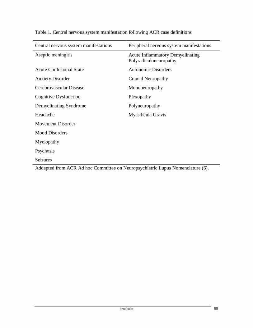
Resultados 98
Table 1. Central nervous system manifestation following ACR case definitions
Central nervous system manifestations Peripheral nervous system manifestations
Aseptic meningitis Acute Inflammatory Demyelinating Polyradiculoneuropathy
Acute Confusional State Autonomic Disorders
Anxiety Disorder Cranial Neuropathy
Cerebrovascular Disease Mononeuropathy
Cognitive Dysfunction Plexopathy
Demyelinating Syndrome Polyneuropathy
Headache Myasthenia Gravis
Movement Disorder
Mood Disorders
Myelopathy
Psychosis
Seizures
Addapted from ACR Ad hoc Committee on Neuropsychiatric Lupus Nomenclature (6).

Res
ulta
dos
99
Tab
le 2
. NP
man
ifes
tatio
ns in
stu
dies
usi
ng A
CR
cla
ssif
icat
ion
crite
ria
Aut
hors
N
umbe
r of
pa
tient
s
Ove
rall
prev
alen
ce
(%)
Cog
niti
ve
dysf
unct
ion
Moo
d di
sord
ers
Cer
ebro
vasc
ular
di
seas
e Se
izur
es
Hea
dach
e Ps
ycho
sis
Poli
neur
opat
hy
Ain
iala
et a
l, 20
01
(10)
46
91
81
43
15
9
54
0 28
Bre
y et
al,
2002
(11)
12
8 80
79
23
.3
2 16
57
6.
5 22
Alf
reta
et a
l, 20
03
(12)
61
72
52
27
24
11
21
0
13
Han
ly e
t al,
2005
(13)
53
31
1.
9 0
0 0
9.4
0 0
Sann
a et
al,
2003
(14)
32
3 57
.3
10.8
16
.7
14.5
8.
3 24
7.
7 2.
8
Moc
c et
al,
2001
(15)
51
8 19
N
A
6 19
28
4
11
1
Han
ly e
t al,
2004
(16)
11
1 37
7.
3 9.
8 9.
8 2.
4 24
.4
7.3
4.9
Mik
dash
i et a
l, 20
04
(17)
13
0 56
.9
27.3
N
A
25.7
7.
6 N
A
15.1
18
.2
Rob
ert e
t al,
2006
(18)
50
78
%
18
0
20.5
55
.6
16.2
0
Shim
ojim
a Y
et a
l, 20
05
(19)
25
N
A
12
0 24
36
12
32
12
NA
: not
ava
ilabl
e
Resultados 100
ARTIGO 3
Magnetic resonance spectroscopy in the evaluation of central nervous system
manifestations of systemic lupus erythematosus
Appenzeller S, Costallat LT, Li ML, Cendes F
Magnetic resonance spectroscopy in the evaluation of central nervous system
manifestations of systemic lupus erythematosus
Arthritis and Rheumatology Care and Research (in press)
Magnetic Resonance Spectroscopy in theEvaluation of Central Nervous SystemManifestations of Systemic Lupus ErythematosusSIMONE APPENZELLER, LILIAN T. L. COSTALLAT, LI MIN LI, AND FERNANDO CENDES
IntroductionNeuropsychiatric (NP) systemic lupus erythematosus(SLE) is characterized by a large spectrum of physical andbehavioral manifestations. One major difficulty is the ab-sence of diagnostic tools for assessing disease activity andseverity of NP manifestation. The neurologic symptomscan be of new onset, chronic, or of a former or resolvednature (1). Although several studies have used differentneuroimaging tools, including computed tomography,magnetic resonance imaging (MRI), and single-photon–emission computed tomography, no single technique hasproven to be definitive for diagnosis of NP manifestationsin persons with SLE (1).
Magnetic resonance spectroscopy (MRS) permits chem-ically specific, noninvasive measurements of some com-pounds of biologic importance in living tissues. MRS wasdiscovered in 1946, but was only first used in living ani-mal brain in 1980 (2), followed by use in human brains inseveral pathologies. In the human brain, phosphate energystores, intracellular pH, lactate concentrations, and theneuronal marker N-acetylaspartate are examples of MRS-measurable variables (3).
The purpose of this article is to review studies usingMRS in SLE and to discuss the clinical utility of thistechnique in determining central nervous system (CNS)involvement in individuals with SLE. We will also discussfuture applications of MRS in the evaluation and treatmentof NP manifestations in patients with SLE.
HistoryThe nuclear magnetic resonance (NMR) phenomenon wasdiscovered independently in 2 laboratories in 1946 by
Bloch and Purcell, which led them to receive the NobelPrize for physics in 1952. When imaging methods usingthe NMR signal were first developed, the term NMR imag-ing had been applied. But because of increasing danger ofnuclear energy in the 1980s and because MR techniques donot use ionizing radiation, the term nuclear was droppedin clinical use, being maintained only to describe thephysical phenomenon itself (3).
MRS physicsSpectroscopy deals with the interaction of electromagneticradiation with matter; therefore, because the structure ofatomic nuclei have magnetic properties, they respond tostrong magnetic fields. During relaxation from the excita-tion of a magnetic field, atomic nuclei emit oscillatingsignals at a frequency that perturbs the nuclei. These sig-nals may be detected by coils and then converted intospectra or images. The position of peaks in the spectrum isdetermined by its molecular characteristics. Informationabout their metabolites can be extracted based upon theamplitude or area under a given peak (3).
Advantages of MRSThere are several advantages to performing MRS in vivo.Metabolic studies of organs in their normal environmentcan increase understanding of the function of complexorganisms and enable researchers to evaluate changes dur-ing diseases. The noninvasive nature of MRS allows re-peated measurements in order to evaluate kinetic and lon-gitudinal studies and to study human tissues that areinaccessible by invasive techniques. At the strength of themagnetic field needed for human studies in vivo, no del-eterious effect on living tissue has been noted (3). Precau-tions such as excluding magnetic objects from the magnetare the main recommendation.
Disadvantages of MRSThe major disadvantage of MRS is its lack of sensitivity,which depends on a wide range of factors, including thenucleus investigated, the volume of the region of interest,and the magnetic field strength, among others. In general,
Supported by grants from the FAPESP and CNPq.Simone Appenzeller, MD, Lilian T. L. Costallat, MD, PhD,
Li Min Li, MD, PhD, Fernando Cendes, MD, PhD: StateUniversity of Campinas, Campinas, Brazil.
Address correspondence to Fernando Cendes, MD, PhD,Department of Neurology, University of Campinas-UNICAMP, Cidade Universitaria, Campinas SP, Brazil, CEP13083-970. E-mail: [email protected].
Submitted for publication November 27, 2005; accepted inrevised form March 21, 2006.
Arthritis & Rheumatism (Arthritis Care & Research)Vol. 55, No. 5, October 15, 2006, pp 000–000DOI 10.1002/art.●© 2006, American College of Rheumatology
REVIEW ARTICLE
1
AQ: 1-2
AQ: 5
AQ: 3
AQ: 4
AQ: 6
AQ: 7
AQ: 8
rich3/z1x-acr/z1x-acr/z1x00506/z1x5549d06a halej S�4 6/13/06 10:14 Art: ACR05-0449
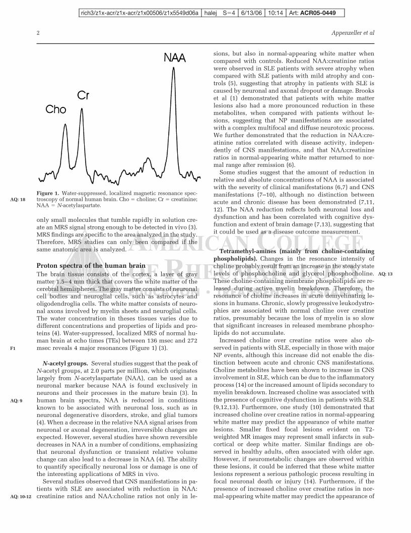
only small molecules that tumble rapidly in solution cre-ate an MRS signal strong enough to be detected in vivo (3).MRS findings are specific to the area analyzed in the study.Therefore, MRS studies can only been compared if thesame anatomic area is analyzed.
Proton spectra of the human brainThe brain tissue consists of the cortex, a layer of graymatter 1.5–4 mm thick that covers the white matter of thecerebral hemispheres. The gray matter consists of neuronalcell bodies and neuroglial cells, such as astrocytes andoligodendroglia cells. The white matter consists of neuro-nal axons involved by myelin sheets and neuroglial cells.The water concentration in theses tissues varies due todifferent concentrations and properties of lipids and pro-teins (4). Water-suppressed, localized MRS of normal hu-man brain at echo times (TEs) between 136 msec and 272msec reveals 4 major resonances (Figure 1) (3).
N-acetyl groups. Several studies suggest that the peak ofN-acetyl groups, at 2.0 parts per million, which originateslargely from N-acetylaspartate (NAA), can be used as aneuronal marker because NAA is found exclusively inneurons and their processes in the mature brain (3). Inhuman brain spectra, NAA is reduced in conditionsknown to be associated with neuronal loss, such as inneuronal degenerative disorders, stroke, and glial tumors(4). When a decrease in the relative NAA signal arises fromneuronal or axonal degeneration, irreversible changes areexpected. However, several studies have shown reversibledecreases in NAA in a number of conditions, emphasizingthat neuronal dysfunction or transient relative volumechange can also lead to a decrease in NAA (4). The abilityto quantify specifically neuronal loss or damage is one ofthe interesting applications of MRS in vivo.
Several studies observed that CNS manifestations in pa-tients with SLE are associated with reduction in NAA:creatinine ratios and NAA:choline ratios not only in le-
sions, but also in normal-appearing white matter whencompared with controls. Reduced NAA:creatinine ratioswere observed in SLE patients with severe atrophy whencompared with SLE patients with mild atrophy and con-trols (5), suggesting that atrophy in patients with SLE iscaused by neuronal and axonal dropout or damage. Brookset al (1) demonstrated that patients with white matterlesions also had a more pronounced reduction in thesemetabolites, when compared with patients without le-sions, suggesting that NP manifestations are associatedwith a complex multifocal and diffuse neurotoxic process.We further demonstrated that the reduction in NAA:cre-atinine ratios correlated with disease activity, indepen-dently of CNS manifestations, and that NAA:creatinineratios in normal-appearing white matter returned to nor-mal range after remission (6).
Some studies suggest that the amount of reduction inrelative and absolute concentrations of NAA is associatedwith the severity of clinical manifestations (6,7) and CNSmanifestations (7–10), although no distinction betweenacute and chronic disease has been demonstrated (7,11,12). The NAA reduction reflects both neuronal loss anddysfunction and has been correlated with cognitive dys-function and extent of brain damage (7,13), suggesting thatit could be used as a disease outcome measurement.
Tetramethyl-amines (mainly from choline-containingphospholipids). Changes in the resonance intensity ofcholine probably result from an increase in the steady statelevels of phosphocholine and glycerol phosphocholine.These choline-containing membrane phospholipids are re-leased during active myelin breakdown. Therefore, theresonance of choline increases in acute demyelinating le-sions in humans. Chronic, slowly progressive leukodystro-phies are associated with normal choline over creatineratios, presumably because the loss of myelin is so slowthat significant increases in released membrane phospho-lipids do not accumulate.
Increased choline over creatine ratios were also ob-served in patients with SLE, especially in those with majorNP events, although this increase did not enable the dis-tinction between acute and chronic CNS manifestations.Choline metabolites have been shown to increase in CNSinvolvement in SLE, which can be due to the inflammatoryprocess (14) or the increased amount of lipids secondary tomyelin breakdown. Increased choline was associated withthe presence of cognitive dysfunction in patients with SLE(9,12,13). Furthermore, one study (10) demonstrated thatincreased choline over creatine ratios in normal-appearingwhite matter may predict the appearance of white matterlesions. Smaller fixed focal lesions evident on T2-weighted MR images may represent small infarcts in sub-cortical or deep white matter. Similar findings are ob-served in healthy adults, often associated with older age.However, if neurometabolic changes are observed withinthese lesions, it could be inferred that these white matterlesions represent a serious pathologic process resulting infocal neuronal death or injury (14). Furthermore, if thepresence of increased choline over creatine ratios in nor-mal-appearing white matter may predict the appearance of
Figure 1. Water-suppressed, localized magnetic resonance spec-troscopy of normal human brain. Cho � choline; Cr � creatinine;NAA � N-acetylaspartate.
2 Appenzeller et al
F1
AQ: 9
AQ: 10-12
AQ: 18
AQ: 13
rich3/z1x-acr/z1x-acr/z1x00506/z1x5549d06a halej S�4 6/13/06 10:14 Art: ACR05-0449
fixed white matter lesions, new treatment strategies couldbe introduced.
Creatine and phosphocreatine. Total creatine concen-tration is relatively constant throughout the brain andtends to be relatively resistant to changes; however, vari-ations in creatine levels do occur, as in the gradual loss ofcreatine together with other major metabolites in tissuedeath or necrosis (4). Creatine may increase as a hyperos-molar response to craniocerebral trauma, or may be absentas in the case of creatine deficiency, a rare congenitaldisease (3,6). It is reasonable to use creatine as an internalstandard to normalize NAA and choline resonances tocorrect for artifactual variations in signal intensities due tomagnetic fields and radiofrequency inhomogeneity (7).However, some studies suggest the loss of information byusing this approach. An alternative is the use of externalconcentration reference, but factors such as radiofre-quency field inhomogeneity and coil tuning and couplinghave to be controlled (3).
In a large study, we observed constant creatine values in50 patients with SLE followed for 19 months (6). Similarfindings were observed by Axford et al (8).
Lactate. Lactic acid is the end product of glycolysis andaccumulates when oxidative metabolism cannot meet en-ergy requirements. Elevation of lactic acid in cerebral neo-plasms correlates approximately with relative rates of glu-cose uptake. However, because lactic acid is present in theintracellular and extracellular compartments, a largeamount can be accumulated outside actively anaerobictissue (4). In inflammation, lactate accumulation may alsoreflect metabolism of inflammatory cells rather than brainparenchyma itself. The lactate peak is above the baselinewhen the TE is low (20–35 msec) or high (270–288 msec).At an intermediate TE (135–144 msec), the lactate peakinverts to project below the baseline, a feature that enablesits distinction from lipids and some macromolecules seenat a similar location on the spectrum (15).
Only a few studies have analyzed the presence of lactatein patients with SLE. Brooks et al (14) did not find anincreased lactate over creatine in patients with normal-appearing white matter or white matter lesions when com-pared with controls.
Other metabolites. Myo-inositol (mI) is an osmolyte andastrocyte marker. Its resonance at 3.56 ppm is visualizedwhen performing MRS using a short TE (15,16). Only inone study was mI measured, where all patients with majorCNS involvement had higher values of mI than patientswith minor manifestations (8).
Normal variations
There are age-related and regional variations in the con-centrations of various metabolites in the brain, especially aconstant reduction of NAA:creatinine ratios in the elderly.Regional variations of metabolite concentrations in thebrain are seen between gray and white matter (NAA ishigher in white matter and creatine and choline are higher
in gray matter) and within different parts of the brain(1,15,16).
MRS techniquesCommonly used spectroscopic techniques include the sin-gle-voxel spectroscopy, with a spatial resolution in theorder of 1–8 cm3 (16), and the multivoxel technique, al-lowing the derivation of metabolite maps (16). Althoughsingle-voxel spectroscopy allows evaluation of only smallvolumes of tissue, it is time efficient and allows the acqui-sition of quantitative data. Multivoxel MRS allows exam-ination of different areas of the brain at the same time(15,16). Most SLE studies have used single-voxel MRS,especially because patients with SLE included in the stud-ies were severely ill and needed shorter time necessary forexaminations (Table 1).
The selection of appropriate MRS techniques, includingmeasurement parameters such as repetition time (TR) andTE, depends on the clinical question. Short TE (20–35msec) evaluations are required when there is a need fordetection of metabolites with short relaxation times, suchas glutamine, glutamate, mI, and certain amino acids(15,16), whereas long TE studies (135–270 msec) are suf-ficient for the detection of the major metabolites such asNAA, choline, creatine, and lactate/lipids (16). DifferentTR and TE used in patient with SLE are shown in Table 1.
Proton MRS studies in specific NP manifestationsMost studies using MRS in patients with SLE have in-cluded patients with or without CNS involvement. Nostudy has used MRS for investigating specific manifesta-tions. Brooks et al (1) and Kozora et al (17) observedincreased choline:creatinine ratios in patients with cogni-tive impairment.
Laboratory and treatment featuresThe use of corticosteroids and the presence of antiphos-pholipid antibodies may also influence neurometabolicmarkers. One study (18) demonstrated that patients receiv-ing corticosteroids had lower NAA:creatinine ratios thanpatients not receiving corticosteroids.
The presence of antiphospholipid antibodies is associ-ated with epilepsy and stroke in patients with SLE. We didnot observe a difference in NAA:creatinine ratios betweenpatients with and those without antiphospholipid anti-bodies (6), although one previous study showed a correla-tion between the presence of IgG antiphospholipid anti-bodies and NAA:creatinine ratios (19).
ConclusionMRS allows the quantification of changes in neuronalmarkers and the monitoring of disease progression. MRSseems to be more sensitive than MRI in detecting neuronaldamage or dysfunction in patients with SLE. Althoughdifferent MRS techniques and different localization of theMRS volume of interest were used in SLE studies, weobserved that most authors report a significant decrease inNAA:creatinine ratios in patients with SLE when com-pared with controls. Although some studies correlate the
SLE and Magnetic Resonance Spectroscopy 3
AQ: 14
T1
AQ: 15
rich3/z1x-acr/z1x-acr/z1x00506/z1x5549d06a halej S�4 6/13/06 10:14 Art: ACR05-0449

Tab
le1.
MR
Sfi
nd
ings
insy
stem
iclu
pu
ser
yth
emat
osu
s:li
tera
ture
revi
ew*
Au
thor
(ref
eren
ce)
Loc
aliz
atio
nM
RS
No.
ofp
atie
nts
Vox
elsi
zeM
RS
par
amet
ers
MR
Sfi
nd
ings
Cli
nic
alas
soci
atio
ns
Oth
ers
Sib
bitt
etal
(5)
Wh
ite
mat
ter,
sup
erio
rto
the
ven
tric
les,
exte
nd
ing
thro
ugh
both
hem
isp
her
es
212
�2
�2
cm3
TR
�1,
500;
TE
�19
2N
AA
/Cr
Mor
ep
ron
oun
ced
inse
vere
atro
ph
y
Dav
ieet
al(1
1)W
hit
em
atte
rle
sion
s(5
pat
ien
ts)
orN
AW
Min
fron
tal
regi
on(7
pat
ien
ts)
133.
5–6
ml
TR
�2,
000
mse
c;T
E�
10an
d13
5m
sec
2N
AA
/Cr
No
corr
elat
ion
wit
hn
euro
logi
can
dp
sych
iatr
icin
volv
emen
t
2N
AA
/Cr
inle
sion
s
Bro
oks
etal
(14)
Les
ion
san
dN
AW
Min
per
iven
tric
ula
rw
hit
e,oc
cip
ital
wh
ite,
and
occi
pit
algr
aym
atte
r
141
ml
TE
�27
0m
sec;
TR
�2,
300
mse
c2
NA
A/C
rIn
all
regi
ons
ofth
ebr
ain
Mor
ep
ron
oun
ced
inle
sion
s
Ch
inn
etal
(19)
Fro
nta
lw
hit
em
atte
ran
dth
ep
arie
to-o
ccip
ital
wh
ite
mat
ter
478
ml
TR
�1,
600
ms;
TE
�13
5m
sec
2N
AA
/Cr;2
Ch
o/C
rC
orti
cost
eroi
ds
Sib
bitt
etal
(7)
NA
WM
par
ieta
llo
be36
8cm
3T
E�
26an
d13
6m
sec;
TR
�2,
000
mse
c2
NA
A/C
rM
ajor
NP
;d
isea
seac
tivi
tyS
abet
etal
(18)
Dee
poc
cip
itop
arie
tal
wh
ite
mat
ter;
mu
ltis
lice
4310
cm3
TE
�19
mse
c;T
R�
2000
mse
cT
E�
270
mse
c;T
R�
2,30
0m
sec
2N
AA
/Cr
1C
ho/
Cr
inS
AA
F
Fri
edm
anet
al(1
2)N
AW
Min
occi
pit
opar
ieta
lre
gion
424
cm3
TE
�19
mse
c;T
R�
2,00
0m
sec
2N
AA
/Cr;1
Ch
o/C
r2
NA
A/C
ras
soci
ated
wit
hsm
all
foca
lle
sion
s1
Ch
o/C
ras
soci
ated
wit
hce
rebr
alin
farc
tB
rook
set
al(1
)L
esio
ns
and
NA
WM
inp
eriv
entr
icu
lar
wh
ite,
occi
pit
alw
hit
e,an
doc
cip
ital
gray
mat
ter
121
ml
TE
�27
0m
sec;
TR
�2,
300
mse
c1
Ch
o/C
rC
ogn
itiv
eim
pai
rmen
t;S
LIC
C
Lim
etal
(13)
Bas
alga
ngl
iaan
dle
ftp
erit
rigo
nal
per
iven
tric
ula
rw
hit
em
atte
r
268
cm3
TE
�30
mse
c;T
R�
3,00
0m
sec
2N
AA
/Cr
inba
sal
gan
glia
1C
ho/
Cr
inp
eriv
entr
icu
lar
wh
ite
mat
ter
Maj
orN
Pm
anif
esta
tion
sN
oco
rrel
atio
nw
ith
MR
Ifi
nd
ings
Axf
ord
etal
(8)
Par
ieta
lN
AW
M9
8cm
3T
E�
30m
sec;
TR
�2,
020
mse
c2
NA
A,1
mI;1
Ch
oM
ajor
NP
man
ifes
tati
ons;
Min
orN
Pm
anif
esta
tion
s
1m
Iin
NA
WM
Han
da
etal
(9)
Fro
nta
lan
dp
arie
to-o
ccip
ital
NA
WM
201–
8m
lT
E�
135
mse
c,27
0m
sec;
TR
�3,
000
mse
c
2N
AA
/Cr
NP
man
ifes
tati
ons
Cas
tell
ino
etal
(10)
Hyp
oper
fuse
dor
nor
mop
erfu
sed
fron
tal
area
sby
SP
EC
T
88
cm3
TE
�13
5m
sec;
TR
�2,
000
mse
c2
NA
A/
Ch
oin
hyp
oper
fuse
d1
Ch
o/C
rin
nor
mop
erfu
sed
area
Wh
ite
mat
ter
lesi
ons
New
wh
ite
mat
ter
lesi
ons
onp
revi
ous
area
sof
hyp
oper
fusi
onan
d2
NA
A/C
ho
Ap
pen
zell
eret
al(6
)N
AW
Msu
per
ior
top
oste
rior
regi
onof
corp
us
call
osu
m90
10cm
3T
E�
135
mse
c;T
R�
1,50
0m
sec
2N
AA
/Cr
Act
ive
dis
ease
,re
vers
ible
wit
hin
acti
vity
Koz
ora
etal
(17)
NA
WM
fron
tal
per
iven
tric
ula
r7
2�
2�
2cm
TE
�13
5m
sec;
TR
�1,
500
mse
c1
Ch
o/C
r1
inco
gnit
ive
imp
airm
ent
*M
RS
�m
agn
etic
reso
nan
cesp
ectr
osco
py;
TR
�re
pet
itio
nti
me;
TE
�ec
ho
tim
e;2
�d
ecre
ased
;1�
incr
ease
d;N
AA
�N
-ace
tyla
spar
tate
;Cr
�cr
eati
nin
e;N
AW
M�
nor
mal
-ap
pea
rin
gw
hit
em
atte
r;C
ho
�ch
olin
e;N
P�
neu
rop
sych
iatr
icm
anif
esta
tion
s;S
AA
F�
XX
XX
X;
SL
ICC
�S
yste
mic
Lu
pu
sIn
tern
atio
nal
Col
labo
rati
ng
Cli
nic
s;M
RI
�m
agn
etic
reso
nan
ceim
agin
g;S
PE
CT
�si
ngl
e-p
hot
on–
emis
sion
com
pu
ted
tom
ogra
ph
y.
4 Appenzeller et al
rich3/z1x-acr/z1x-acr/z1x00506/z1x5549d06a halej S�4 6/13/06 10:14 Art: ACR05-0449
Resultados 106
ARTIGO 4
Epileptic seizures in systemic lupus erythematosus
Appenzeller S, Cendes F, Costallat LT
Epileptic seizures in systemic lupus erythematosus
Neurology. 2004 Nov 23; 63(10):1808-12
Epileptic seizures in systemic lupuserythematosus
Simone Appenzeller, MD; Fernando Cendes, MD, PhD; and Lilian T.L. Costallat, MD, PhD
Abstract—Objective: To evaluate the frequency and risk factors of epileptic seizures in a large cohort of patients withsystemic lupus erythematosus (SLE). Methods: Five hundred nineteen consecutive patients with SLE were studied, withfollow-up ranging from 4 to 7.8 years. The type and frequency of risk factors associated with acute and recurrent epilepticseizures in SLE were determined. Results: Sixty (11.6%) patients with epileptic seizures were identified. Epileptic seizuresoccurred at the onset of SLE symptoms in 19 (31.6%) and after the onset of SLE in 41 of 60 (68.3%) patients. Fifty-three of60 (88.3%) patients had acute symptomatic epileptic seizures, and 7 of 60 (11.7%) had recurrent epileptic seizures.Variables associated with acute epileptic seizures at SLE onset were stroke (p � 0.0004) and antiphospholipid antibodies(p � 0.0013). Epileptic seizures during follow-up were related to nephritis (p � 0.001), antiphospholipid antibodies (p �0.005), and epileptic seizures at disease onset (p � 0.00001). All seven patients who presented recurrent epileptic seizureshad antiphospholipid syndrome and interictal epileptic abnormalities on EEG. Conclusions: Epileptic seizures wereobserved in 11.2% of systemic lupus erythematosus (SLE) patients. Antiphospholipid antibodies and stroke were related toepileptic seizures at SLE disease onset. Patients with renal flares, epileptic seizures at SLE disease onset, and antiphos-pholipid antibodies were at greater risk for acute symptomatic seizures during follow-up. Recurrence of epileptic seizuresoccurred in 1.3% of patients and was associated with antiphospholipid syndrome.
NEUROLOGY 2004;63:1808–1812
Neuropsychiatric involvement in systemic lupus ery-thematosus (SLE) is considered as one of the majormanifestations of the disease.1-3 Single epileptic sei-zure episodes have been documented in about 10 to20% of patients with SLE.1-6 Most studies analyzedrisk factors associated with the occurrence of epilep-tic seizures,1-12 whereas specific risk factors associ-ated with recurrence of epileptic seizures have beenrarely reported.
Epileptic seizures can be a primary event result-ing from the direct effect of active SLE manifestationin the CNS or occur independently of lupus activityitself, being associated with CNS infections, uremia,hypertension, or electrolytic disturbance.
Generalized tonic-clonic seizures are by far themost common, but simple partial and complex par-tial seizures have also been described.3,6 The patho-genesis of epileptic seizures in SLE remainsunknown, but ischemic vascular disease or antibod-ies that bind to cerebral tissues have been consid-ered as probable causes.2,13-15
We sought to determine the frequency and riskfactors for epileptic seizures in a large cohort of pa-tients with SLE. We also analyzed clinical and labo-ratory features associated with the occurrence ofsingle episodes and recurrence of epileptic seizures.
Patients and methods. The records of 519 consecutive patientswith four or more criteria for SLE diagnosis16 from January 1974to December 2002 had their medical histories and clinical and
serologic characteristics documented in computer database pro-grams. All patients were followed and examined by one of theauthors at the Rheumatology and Neurology Unit of the StateUniversity of Campinas. Therefore, medical records and protocolsfor investigations were homogeneous among patients. All patientshad their clinical and laboratory evaluation performed at diagno-sis and quarterly during the follow-up period. Patients with in-complete clinical and laboratory evaluations or who were lost tofollow-up were not included in this series. The mean duration offollow-up of these patients was 5.7 years (SD 1.2 years), rangingfrom 4.0 to 7.8 years.
Only patients with epileptic seizures indicating CNS involve-ment by SLE17 were included in this study. Patients who hadepilepsy and associated structural MRI abnormalities diagnosedprior to SLE and patients with seizures secondary to acute meta-bolic causes such as uremia, hypertension, diabetes mellitus, andelectrolytic abnormalities were not included in the frequency de-termination and in analysis of epileptic seizures, although theywere followed to observe if they would have recurrent seizuresattributable to SLE.
Neurologic evaluation. Epileptic seizures were classified ac-cording to the criteria suggested by the International LeagueAgainst Epilepy.18 EEGs were recorded in the interictal period in a16-channel analog or 32-channel digital EEG recorder with theInternational 10–20 System of electrode placement for 20 to 30minutes in 38 (63.3%) patients. We tabulated the presence andlocalization of interictal epileptiform abnormality and slow waveabnormality.
Clinical features. The age at onset of epileptic seizures wasdetermined in relation to the age at which the first well-describedsign or symptom indicating SLE occurred. Patients with a diagno-sis of seizures before the onset of SLE had their clinical historycarefully evaluated to determine if SLE was drug induced or oc-curred concomitantly to seizures. In seizures occurring at diseaseonset or during follow-up, SLE disease flares and other triggeringevents were searched for. To determine the risk factors for occur-rence of epileptic seizures, we analyzed clinical manifestations
From the Rheumatology Unit (Drs. Appenzeller and Costallat), Department of Internal Medicine, and Department of Neurology (Dr. Cendes), StateUniversity of Campinas, Brazil.Supported by Fundação de Amparo à Pesquisa do Estado de São Paulo (FAPESP) grant no. 03/015270.Received February 16, 2004. Accepted in final form July 21, 2004.Address correspondence and reprint requests to Dr. F. Cendes, Department of Neurology, State University of Campinas (UNICAMP), Cidade UniversitariaZeferino Vaz CEP, 13083970 Campinas-SP, Brazil; e-mail: [email protected]
1808 Copyright © 2004 by AAN Enterprises, Inc.
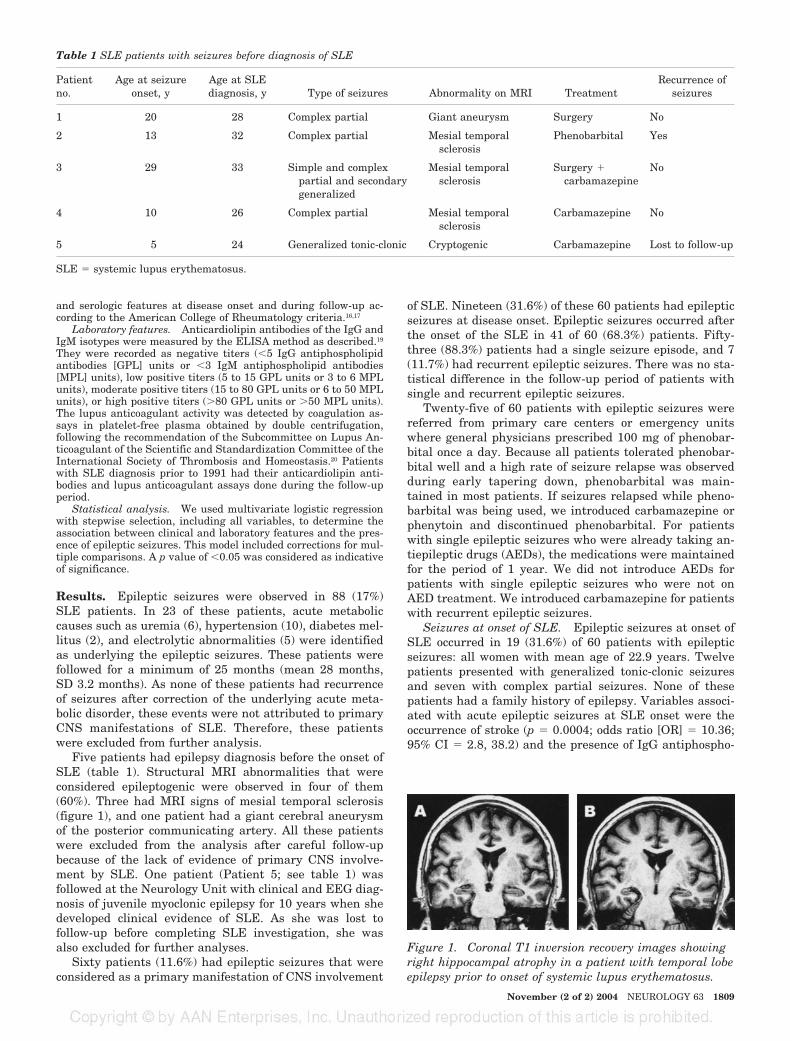
and serologic features at disease onset and during follow-up ac-cording to the American College of Rheumatology criteria.16,17
Laboratory features. Anticardiolipin antibodies of the IgG andIgM isotypes were measured by the ELISA method as described.19
They were recorded as negative titers (�5 IgG antiphospholipidantibodies [GPL] units or �3 IgM antiphospholipid antibodies[MPL] units), low positive titers (5 to 15 GPL units or 3 to 6 MPLunits), moderate positive titers (15 to 80 GPL units or 6 to 50 MPLunits), or high positive titers (�80 GPL units or �50 MPL units).The lupus anticoagulant activity was detected by coagulation as-says in platelet-free plasma obtained by double centrifugation,following the recommendation of the Subcommittee on Lupus An-ticoagulant of the Scientific and Standardization Committee of theInternational Society of Thrombosis and Homeostasis.20 Patientswith SLE diagnosis prior to 1991 had their anticardiolipin anti-bodies and lupus anticoagulant assays done during the follow-upperiod.
Statistical analysis. We used multivariate logistic regressionwith stepwise selection, including all variables, to determine theassociation between clinical and laboratory features and the pres-ence of epileptic seizures. This model included corrections for mul-tiple comparisons. A p value of �0.05 was considered as indicativeof significance.
Results. Epileptic seizures were observed in 88 (17%)SLE patients. In 23 of these patients, acute metaboliccauses such as uremia (6), hypertension (10), diabetes mel-litus (2), and electrolytic abnormalities (5) were identifiedas underlying the epileptic seizures. These patients werefollowed for a minimum of 25 months (mean 28 months,SD 3.2 months). As none of these patients had recurrenceof seizures after correction of the underlying acute meta-bolic disorder, these events were not attributed to primaryCNS manifestations of SLE. Therefore, these patientswere excluded from further analysis.
Five patients had epilepsy diagnosis before the onset ofSLE (table 1). Structural MRI abnormalities that wereconsidered epileptogenic were observed in four of them(60%). Three had MRI signs of mesial temporal sclerosis(figure 1), and one patient had a giant cerebral aneurysmof the posterior communicating artery. All these patientswere excluded from the analysis after careful follow-upbecause of the lack of evidence of primary CNS involve-ment by SLE. One patient (Patient 5; see table 1) wasfollowed at the Neurology Unit with clinical and EEG diag-nosis of juvenile myoclonic epilepsy for 10 years when shedeveloped clinical evidence of SLE. As she was lost tofollow-up before completing SLE investigation, she wasalso excluded for further analyses.
Sixty patients (11.6%) had epileptic seizures that wereconsidered as a primary manifestation of CNS involvement
of SLE. Nineteen (31.6%) of these 60 patients had epilepticseizures at disease onset. Epileptic seizures occurred afterthe onset of the SLE in 41 of 60 (68.3%) patients. Fifty-three (88.3%) patients had a single seizure episode, and 7(11.7%) had recurrent epileptic seizures. There was no sta-tistical difference in the follow-up period of patients withsingle and recurrent epileptic seizures.
Twenty-five of 60 patients with epileptic seizures werereferred from primary care centers or emergency unitswhere general physicians prescribed 100 mg of phenobar-bital once a day. Because all patients tolerated phenobar-bital well and a high rate of seizure relapse was observedduring early tapering down, phenobarbital was main-tained in most patients. If seizures relapsed while pheno-barbital was being used, we introduced carbamazepine orphenytoin and discontinued phenobarbital. For patientswith single epileptic seizures who were already taking an-tiepileptic drugs (AEDs), the medications were maintainedfor the period of 1 year. We did not introduce AEDs forpatients with single epileptic seizures who were not onAED treatment. We introduced carbamazepine for patientswith recurrent epileptic seizures.
Seizures at onset of SLE. Epileptic seizures at onset ofSLE occurred in 19 (31.6%) of 60 patients with epilepticseizures: all women with mean age of 22.9 years. Twelvepatients presented with generalized tonic-clonic seizuresand seven with complex partial seizures. None of thesepatients had a family history of epilepsy. Variables associ-ated with acute epileptic seizures at SLE onset were theoccurrence of stroke (p � 0.0004; odds ratio [OR] � 10.36;95% CI � 2.8, 38.2) and the presence of IgG antiphospho-
Table 1 SLE patients with seizures before diagnosis of SLE
Patientno.
Age at seizureonset, y
Age at SLEdiagnosis, y Type of seizures Abnormality on MRI Treatment
Recurrence ofseizures
1 20 28 Complex partial Giant aneurysm Surgery No
2 13 32 Complex partial Mesial temporalsclerosis
Phenobarbital Yes
3 29 33 Simple and complexpartial and secondarygeneralized
Mesial temporalsclerosis
Surgery �carbamazepine
No
4 10 26 Complex partial Mesial temporalsclerosis
Carbamazepine No
5 5 24 Generalized tonic-clonic Cryptogenic Carbamazepine Lost to follow-up
SLE � systemic lupus erythematosus.
Figure 1. Coronal T1 inversion recovery images showingright hippocampal atrophy in a patient with temporal lobeepilepsy prior to onset of systemic lupus erythematosus.
November (2 of 2) 2004 NEUROLOGY 63 1809
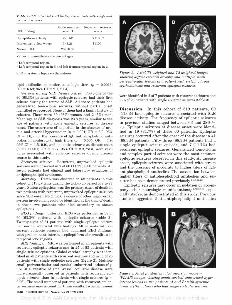
lipid antibodies in moderate to high titers (p � 0.0013;OR � 6.69; 95% CI � 2.1, 21.4).
Seizures during SLE disease course. Forty-one of the60 (68.3%) patients with epileptic seizures had their firstseizure during the course of SLE. All these patients hadgeneralized tonic-clonic seizures, without partial onsetidentified or recorded. None of them had a family history ofseizures. There were 39 (95%) women and 2 (5%) men.Mean age at SLE diagnosis was 23.8 years, similar to theage of patients with acute epileptic seizures at diseaseonset. The occurrence of nephritis, in the absence of ure-mia and arterial hypertension (p � 0.001; OR � 3.2; 95%CI � 1.6, 6.5), the presence of IgG antiphospholipid anti-bodies in moderate to high titers (p � 0.005; OR � 3.9;95% CI � 1.5, 9.9), and epileptic seizures at disease onset(p � 0.00001; OR � 8.27; 95% CI � 2.9, 23.3) were vari-ables associated with epileptic seizures during diseasecourse in this study.
Recurrent seizures. Recurrent, unprovoked epilepticseizures were observed in 7 of 60 (11.7%) SLE patients. Allseven patients had clinical and laboratory evidences ofantiphospholipid syndrome.
Mortality. Death was observed in 58 patients in thiscohort of 519 patients during the follow-up period of 2 to 27years. Status epilepticus was the primary cause of death intwo patients with recurrent, unprovoked epileptic seizuressince SLE onset. No clinical evidence of other major organsystem involvement could be identified at the time of deathin these two patients who died secondary to statusepilepticus.
EEG findings. Interictal EEG was performed in 38 of60 (63.3%) patients with epileptic seizures (table 2).Twenty-eight of 31 patients with single epileptic seizurehad normal interictal EEG findings. All patients with re-current epileptic seizures had abnormal EEG findings,with predominant interictal epileptiform abnormalities intemporal lobe regions.
MRI findings. MRI was performed in all patients withrecurrent epileptic seizures and in 25 of 53 patients withsingle seizure episodes. Global cerebral atrophy was iden-tified in all patients with recurrent seizures and in 11 of 25patients with single epileptic seizures (figure 2). Multiplesmall periventricular and cortical–subcortical lesions (fig-ure 3) suggestive of small-vessel occlusive disease weremore frequently observed in patients with recurrent epi-leptic seizures than in patients with single seizures (p �0.06). The small number of patients with recurrent epilep-tic seizures may account for these results. Ischemic lesions
were identified in 2 of 7 patients with recurrent seizures andin 6 of 25 patients with single epileptic seizures (table 3).
Discussion. In this cohort of 519 patients, 60(11.6%) had epileptic seizures associated with SLEdisease activity. The frequency of epileptic seizuresin previous studies ranged between 8.3 and 28%.1,5-
10,21 Epileptic seizures at disease onset were identi-fied in 19 (31.7%) of these 60 patients. Epilepticseizures occurred after the onset of the disease in 41(68.3%) patients. Fifty-three (88.3%) patients had asingle epileptic seizure episode, and 7 (11.7%) hadrecurrent epileptic seizures. Generalized tonic-clonicand complex partial seizures were the most commonepileptic seizures observed in this study. At diseaseonset, epileptic seizures were associated with strokeand the presence of moderate to higher titers of IgGantiphospholipid antibodies. The association betweenhigher titers of antiphospholipid antibodies and sei-zures has been demonstrated previously.2,4,9,22-26
Epileptic seizures may occur in isolation or accom-pany other neurologic manifestations,4,5,10,27-29 espe-cially stroke, as demonstrated in this study. Previousstudies suggested that antiphospholipid antibodies
Table 2 SLE: interictal EEG findings in patients with single andrecurrent seizures
EEG findingSingle seizures,
n � 31Recurrent seizures,
n � 7
Epileptiform activity 2 (6.5)* 7 (100)†
Intermittent slow waves 1 (3.2) 7 (100)
Normal EEG 28 (90.3) 0
Values in parentheses are percentages.
* Left temporal region.† Left temporal region in 5 and left frontotemporal region in 2.
SLE � systemic lupus erythematosus. Figure 2. Axial T1-weighted and T2-weighted imagesshowing diffuse cerebral atrophy and multiple smallperiventricular lesions in a patient with systemic lupuserythematosus and recurrent epileptic seizures.
Figure 3. Axial fluid-attenuated inversion recovery(FLAIR) images showing small cortical–subcortical hyper-intense lesions in two patients (A and B) with systemiclupus erythematosus who had single epileptic seizures.
1810 NEUROLOGY 63 November (2 of 2) 2004
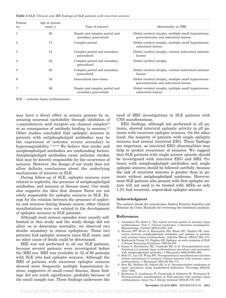
may have a direct effect in seizure genesis by in-creasing neuronal excitability through inhibition of�-aminobutyric acid receptor–ion channel complex30
or as consequence of antibody binding to neurons.31
Other studies concluded that epileptic seizures inpatients with antiphospholipid antibodies may bethe expression of ischemic events secondary tohypercoagulability.11,32-34 We believe that stroke andantiphospholipid antibodies are confounding factors:Antiphospholipid antibodies cause ischemic strokesthat may be directly responsible for the occurrence ofseizures. However, the design of our study does notallow definite conclusions about the underlyingmechanisms of seizures in SLE.
During follow-up of SLE, epileptic seizures wererelated to nephritis, the presence of antiphospholipidantibodies, and seizures at disease onset. Our studyalso supports the idea that disease flares are notsolely responsible for epileptic seizures in SLE. Ex-cept for the relation between the presence of nephri-tis and seizures during disease course, other clinicalmanifestations were not related to the developmentof epileptic seizures in SLE patients.
Although most seizure episodes were usually self-limited in this study and the study design did notallow us to determine mortality, we observed twodeaths secondary to status epilepticus. These twopatients had epileptic seizures since SLE onset, andno other cause of death could be determined.
MRI was not performed in all our SLE patients,because several patients were investigated beforethe MRI era. MRI was available in 32 of 60 patientswith SLE who had epileptic seizures. Although theMRI of patients with recurrent epileptic seizuresshowed more frequently multiple hyperintense le-sions, suggestive of small-vessel disease, these find-ings did not reach significance, probably because ofthe small sample size. These findings underscore the
need of MRI investigations in SLE patients withCNS manifestations.
EEG findings, although not performed in all pa-tients, showed interictal epileptic activity in all pa-tients with recurrent epileptic seizures. On the otherhand, the majority of patients with single epilepticseizures had normal interictal EEG. These findingsare important, as interictal EEG abnormalities mayhelp to predict recurrence of seizures. We suggestthat SLE patients with single seizure episode shouldbe investigated with interictal EEG and MRI. Pa-tients with antiphospholipid antibodies and singleepileptic seizures should be followed carefully, becausethe risk of recurrent seizures is greater than in pa-tients without antiphospholipid syndrome. However,most SLE patients who present with first epileptic sei-zure will not need to be treated with AEDs as only1.3% had recurrent, unprovoked epileptic seizures.
AcknowledgmentThe authors thank the statisticians Andrea Ferreira Semolini andHelymar da Costa Machado for reviewing the statistical analysis.
References1. Jennekens FG, Kater L. The central nervous system in systemic lupus
erythematosus. Part 1. Clinical syndromes: a literature investigation.Rheumatology (Oxford) 2002;41:605–618.
2. Herranz MT, Rivier G, Khamashta MA, Blaser KU, Hughes GR. Asso-ciation between antiphospholipid antibodies and epilepsy in patientswith systemic lupus erythematosus. Arthritis Rheum 1994;37:568–571.
3. Mackworth-Young CG, Hughes GR. Epilepsy: an early symptom of SLE.J Neurol Neurosurg Psychiatry 1985;48:185.
4. Sanna G, Bertolaccini ML, Cuadrado MJ, et al. Neuropsychiatric man-ifestations in systemic lupus erythematosus: prevalence and associationwith antiphospholipid antibodies. J Rheumatol 2003;30:985–992.
5. Mok CC, Lau CS, Wong RW. Neuropsychiatric manifestations and theirclinical associations in southern Chinese patients with systemic lupuserythematosus. J Rheumatol 2001;28:766–771.
6. Brey RL, Holliday SL, Saklad AR, et al. Neuropsychiatric syndromes inlupus: prevalence using standardized definitions. Neurology 2002;58:1214–1220.
7. Kasitanon N, Louthrenoo W, Piyasirisilp S, Sukitawu W, Wichainun R.Neuropsychiatric manifestations in Thai patients with systemic lupuserythematosus. Asian Pac J Allergy Immunol 2002;20:179–185.
Table 3 SLE: Clinical and MR findings of SLE patients with recurrent seizures
Patientno.
Age at seizureonset, y Type of seizures Abnormality on MRI
1 20 Simple and complex partial andsecondary generalized
Global cerebral atrophy, multiple small hyperintenseperiventricular and subcortical lesions
2 17 Complex partial Global cerebral atrophy, multiple small hyperintensesubcortical lesions
3 14 Complex partial and secondarygeneralized
Global cerebral atrophy, cortical–subcortical ischemiclesions
4 22 Complex partial and secondarygeneralized
Global cerebral atrophy
5 25 Complex partial and secondarygeneralized
Global cerebral atrophy, cortical–subcortical ischemiclesions
6 32 Generalized tonic-clonic Global cerebral atrophy, multiple small hyperintenseperiventricular and subcortical lesions
7 28 Simple and complex partial andsecondary generalized
Global cerebral atrophy, multiple small hyperintensesubcortical lesions
SLE � systemic lupus erythematosus.
November (2 of 2) 2004 NEUROLOGY 63 1811
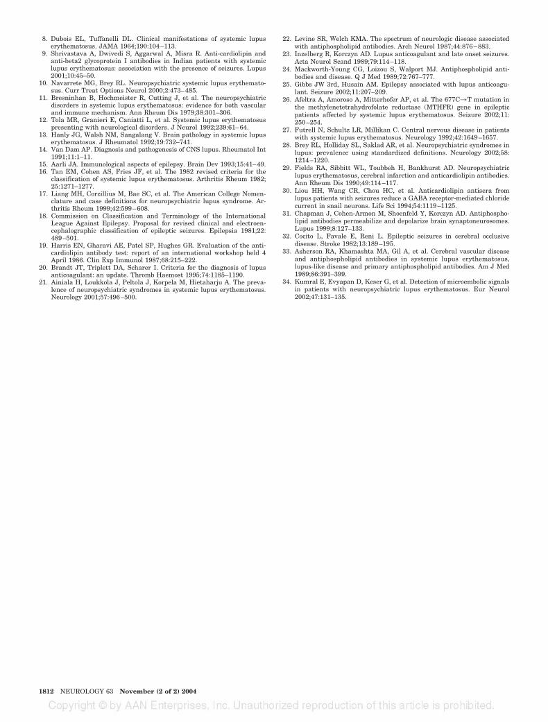
8. Dubois EL, Tuffanelli DL. Clinical manifestations of systemic lupuserythematosus. JAMA 1964;190:104–113.
9. Shrivastava A, Dwivedi S, Aggarwal A, Misra R. Anti-cardiolipin andanti-beta2 glycoprotein I antibodies in Indian patients with systemiclupus erythematosus: association with the presence of seizures. Lupus2001;10:45–50.
10. Navarrete MG, Brey RL. Neuropsychiatric systemic lupus erythemato-sus. Curr Treat Options Neurol 2000;2:473–485.
11. Bresninhan B, Hochmeister R, Cutting J, et al. The neuropsychiatricdisorders in systemic lupus erythematosus: evidence for both vascularand immune mechanism. Ann Rheum Dis 1979;38:301–306.
12. Tola MR, Granieri E, Caniatti L, et al. Systemic lupus erythematosuspresenting with neurological disorders. J Neurol 1992;239:61–64.
13. Hanly JG, Walsh NM, Sangalang V. Brain pathology in systemic lupuserythematosus. J Rheumatol 1992;19:732–741.
14. Van Dam AP. Diagnosis and pathogenesis of CNS lupus. Rheumatol Int1991;11:1–11.
15. Aarli JA. Immunological aspects of epilepsy. Brain Dev 1993;15:41–49.16. Tan EM, Cohen AS, Fries JF, et al. The 1982 revised criteria for the
classification of systemic lupus erythematosus. Arthritis Rheum 1982;25:1271–1277.
17. Liang MH, Corzillius M, Bae SC, et al. The American College Nomen-clature and case definitions for neuropsychiatric lupus syndrome. Ar-thritis Rheum 1999;42:599–608.
18. Commission on Classification and Terminology of the InternationalLeague Against Epilepsy. Proposal for revised clinical and electroen-cephalographic classification of epileptic seizures. Epilepsia 1981;22:489–501.
19. Harris EN, Gharavi AE, Patel SP, Hughes GR. Evaluation of the anti-cardiolipin antibody test: report of an international workshop held 4April 1986. Clin Exp Immunol 1987;68:215–222.
20. Brandt JT, Triplett DA, Scharer I. Criteria for the diagnosis of lupusanticoagulant: an update. Thromb Haemost 1995;74:1185–1190.
21. Ainiala H, Loukkola J, Peltola J, Korpela M, Hietaharju A. The preva-lence of neuropsychiatric syndromes in systemic lupus erythematosus.Neurology 2001;57:496–500.
22. Levine SR, Welch KMA. The spectrum of neurologic disease associatedwith antiphospholipid antibodies. Arch Neurol 1987;44:876–883.
23. Inzelberg R, Korczyn AD. Lupus anticoagulant and late onset seizures.Acta Neurol Scand 1989;79:114–118.
24. Mackworth-Young CG, Loizou S, Walport MJ. Antiphospholipid anti-bodies and disease. Q J Med 1989;72:767–777.
25. Gibbs JW 3rd, Husain AM. Epilepsy associated with lupus anticoagu-lant. Seizure 2002;11:207–209.
26. Afeltra A, Amoroso A, Mitterhofer AP, et al. The 677C3T mutation inthe methylenetetrahydrofolate reductase (MTHFR) gene in epilepticpatients affected by systemic lupus erythematosus. Seizure 2002;11:250–254.
27. Futrell N, Schultz LR, Millikan C. Central nervous disease in patientswith systemic lupus erythematosus. Neurology 1992;42:1649–1657.
28. Brey RL, Holliday SL, Saklad AR, et al. Neuropsychiatric syndromes inlupus: prevalence using standardized definitions. Neurology 2002;58:1214–1220.
29. Fields RA, Sibbitt WL, Toubbeh H, Bankhurst AD. Neuropsychiatriclupus erythematosus, cerebral infarction and anticardiolipin antibodies.Ann Rheum Dis 1990;49:114–117.
30. Liou HH, Wang CR, Chou HC, et al. Anticardiolipin antisera fromlupus patients with seizures reduce a GABA receptor-mediated chloridecurrent in snail neurons. Life Sci 1994;54:1119–1125.
31. Chapman J, Cohen-Armon M, Shoenfeld Y, Korczyn AD. Antiphospho-lipid antibodies permeabilize and depolarize brain synaptoneurosomes.Lupus 1999;8:127–133.
32. Cocito L, Favale E, Reni L. Epileptic seizures in cerebral occlusivedisease. Stroke 1982;13:189–195.
33. Asherson RA, Khamashta MA, Gil A, et al. Cerebral vascular diseaseand antiphospholipid antibodies in systemic lupus erythematosus,lupus-like disease and primary antiphospholipid antibodies. Am J Med1989;86:391–399.
34. Kumral E, Evyapan D, Keser G, et al. Detection of microembolic signalsin patients with neuropsychiatric lupus erythematosus. Eur Neurol2002;47:131–135.
1812 NEUROLOGY 63 November (2 of 2) 2004
Resultados 112
ARTIGO 5
Clinical implications of migraine in systemic lupus erythematosus: relation to
cumulative organ damage
Appenzeller S, Costallat LT
Clinical implications of migraine in systemic lupus erythematosus: relation to cumulative
organ damage
Cephalalgia. 2004 Dec; 24(12):1024-30
1024 © Blackwell Publishing Ltd
Cephalalgia,
2004,
24
, 1024–1030
Blackwell Science, Ltd
Oxford, UKCHA
Cephalalgia
1468-2982Blackwell Science, 2004
241210241030
Original Article
Migraine in SLES Appenzeller & LTL Costallat
Clinical implications of migraine in systemic lupus erythematosus: relation to cumulative organ damage
S Appenzeller & LTL Costallat
Unit of Rheumatology, Department of Internal Medicine State University of Campinas Faculty of Medical Sciences, Campinas, Brazil
Appenzeller S & Costallat LTL. Clinical implications of migraine in systemic lupuserythematosus. relation to cumulative organ damage. Cephalalgia 2004; 24:1024–1030. London. ISSN 0333-1024
The aim of this study was to determine the clinical implications of migraine insystemic lupus erythematosus (SLE) using the cumulative organ damage scores(SLICC-DI). Eighty SLE, 40 rheumatoid arthritis (RA) patients and 40 controls(non SLE, nor RA out-patients), all women, were included. Migraine was definedaccording to the International Headache Society (IHS) criteria for neuropsychiatricSLE. Disease activity was measured by MEX-SLEDAI and cumulative organ dam-age by SLICC-DI. Statistics were obtained by Chi-square and Fischer’s exact tests.
ANOVA
was used for comparing means. Migraine was identified in 42.5% of SLEpatients, compared to 12.5% of RA patients (
P
<
0.05) and 10.0% (
P
<
0.05) in thecontrol group. In the SLE group, a significant association between migraine andRaynaud’s phenomenon (
P =
0.003, OR = 10.1; 95%CI 2.9–35) and antiphospho-lipid antibodies (
P =
0.0012; OR = 7.5; 95%CI 2.5–22.9) was noted. SLE patientswith active migraine had higher MEX-SLEDAI scores than SLE patients withoutmigraine. SLE patients with past history of migraine had significantly higherSLICC scores than SLE patients without migraine. History of migraine was asso-ciated with greater organ damage. Active migraine was associated with higherdisease activity, antiphospholipid antibodies and worsening of Raynaud’s phe-nomenon. The increased cumulative organ damage in SLE patients with pasthistory of migraine justifies the routine evaluation of migraine in clinical practice.
�
Migraine, systemic lupus erythematosus, cumulative organ damage, antiphospholipidantibodies
Dr Simone Appenzeller, Departamento de Clínica Médica, Faculdade de CiênciasMédicas/UNICAMP, CEP 13081–970 Campinas, SP-Brazil. Tel. +55 193788369,fax +55 0193788369, e-mail [email protected] Received 20 September 2003, accepted 27 February 2004
Introduction
Systemic lupus erythematosus (SLE) is a chronic,inflammatory, immune-mediated disease withdiverse clinical manifestations. Central nervoussystem (CNS) involvement in SLE has been morefrequently recognized and reported in recent years,occurring in up to 50% of the patients during thedisease course (1). The incidence varies because ofthe heterogeneity of methods applied and thesmall number of patients in different studies (2).Several neurological manifestations have been con-sidered to be important features of SLE and indica-
tive of CNS involvement (2–4), but only a fewstudies (3, 5, 6) included headache as a CNSmanifestation.
The exact prevalence of migraine in SLE patientsis unknown (7), but several studies published prev-alence that varied between 31 and 45% (1, 6–9).The limitations of research in this area are thesmall sample size, the large variability betweenstudy designs and the different classification crite-ria applied (6).
The clinical importance of migraine has beenaddressed by several studies (1–7, 10), but theassociation between migraine and disease activity,
Migraine in SLE
1025
© Blackwell Publishing Ltd
Cephalalgia,
2004,
24
, 1024–1030
antiphospholipid antibodies and Raynaud’s phe-nomenon remains unclear.
Headaches are considered to be associated with asignificant source of patient disability (7); thereforethe aim of this study was to determine the relationbetween migraine and cumulative organ damage ina SLE cohort followed prospectively during a one-year period.
Patients and methods
Patients and controls
The frequency of migraine in 80 patients with SLE(11), classified according to the revised criteria of theACR (12) was compared to that found in 40 controls.
In order to study the relationship betweenmigraine and the chronic conditions of rheumaticdiseases, the frequency of migraine in SLE patientswas also compared to that found in 40 RA patients(13). All patients and controls were women and werefollowed up in the outpatient Rheumatology Unit ofthe State University of Campinas, Brazil, a tertiaryreference centre for rheumatic diseases. The controlsubjects were selected among out-patients womenattending other clinics in our hospital and were notrelated to the RA patients or to other patients withautoimmune diseases.
The patients and controls were examined by thesame investigator (SA) on a quarterly basis, duringa one-year period. All of the patients and controlssigned an informed consent document prior tobeginning study procedures.
Exclusion criteria
Migraine has a high rate of familial occurrence,suggesting an underlying genetic factor. In orderto determine if SLE influences the occurrence ofmigraine, patients and controls with a family his-tory of migraine were excluded from this study.Patients with history of migraine prior to diagnosisof rheumatic disease, suggesting that migraine andSLE or RA were coexisting conditions, were alsoexcluded. Patients with headache secondary toinfection, hypertension, uraemia, metabolic disor-ders, lesions or traction of intra- and extra-cranialstructures were excluded through history, clinicaland laboratorial examinations, performed at everyvisit. Acute episodes of nonrecurrent headache ofvery low intensity or frequency were not includedin the analysis. As hormonal changes influence theoccurrence of migraine, we also excluded post-menopausal women.
Demographic, clinical, serological andtreatment features
In SLE patients, constitutional, cutaneous, muscu-loskeletal, respiratory, cardiac, haematological andrenal manifestations of the disease were scrutinized.The diagnosis of Raynaud’s phenomenon was basedon at least two-phase colour reactions of bilateraldistribution described by the patient or observed bya physician. Worsening of Raynaud’s phenomenonwas defined as worsening of the pain referred by thepatient or appearance or worsening of pre-existingdigital ulcers. Data on sex, race, age at disease onsetand disease duration were collected for each patient.All clinical manifestations and laboratory test find-ings were recorded. Nephritis was diagnosed on thebasis of proteinuria exceeding 1.0 g/l with abnormalurinary sediment and/or histological findings.Nephrotic syndrome was defined as proteinuria inexcess of 3.5 g/day. Haematological alterations wereascribed to lupus only in the absence of bone mar-row suppression (leucopenia
<
4
¥
10
6
cells/l; throm-bocytopenia
<
100
¥
10
6
cells/l; haemolytic anaemiawith positive Coombs test). Antinuclear antibodies(ANA) were determined by indirect immunofluores-cence using mouse liver as the substrate andregarded as positive if higher than 1 : 40. Anti-double-stranded DNA (AdsDNA) antibodies weredetermined by indirect immunofluorescence usingChrithidia as substrate and considered positive ifhigher than 1 : 10. Precipitating antibodies to extract-able nuclear antigens (ENA), including Ro (SSA), La(SSB) and Sm were detected by immunodiffusionand/or microhemagglutination. Anticardiolipinantibodies (aCL) of the IgG and IgM isotypes weremeasured by the ELISA method as described (14).Lupus anticoagulant (LA) activity was detected bycoagulation assays in platelet free plasma obtainedby double centrifugation, following the recommen-dation of the subcommittee on LA of the Scientificand Standardization Committee of the InternationalSociety of Thrombosis and Homeostasis (15).
Current treatment options were analysed andchanges were made when considered necessary.
RA patients and controls had also a complete clin-ical examination performed during the visits. Anti-nuclear antibodies, antiphospholipid antibodies andlupus coagulant were searched in these groups.
Disease activity and cumulative organ damage
At every visit, SLE patients had their systemic dis-ease activity measured by MEX-SLEDAI, a simpli-fied modification of the SLEDAI with a good
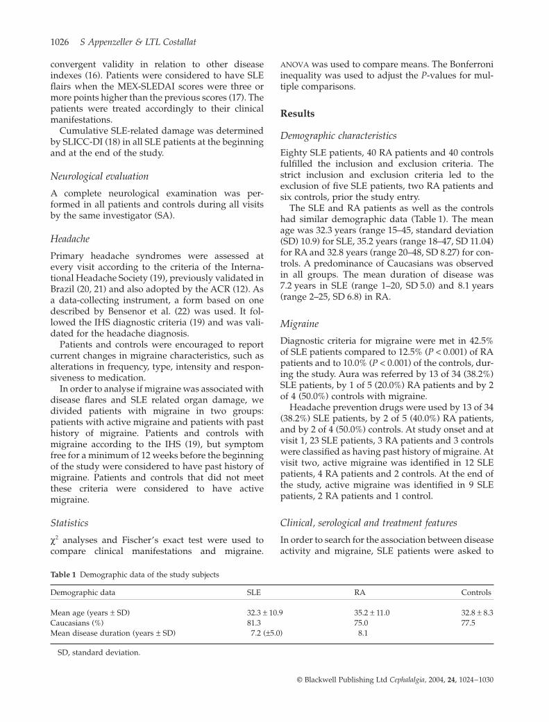
1026
S Appenzeller & LTL Costallat
© Blackwell Publishing Ltd
Cephalalgia,
2004,
24
, 1024–1030
convergent validity in relation to other diseaseindexes (16). Patients were considered to have SLEflairs when the MEX-SLEDAI scores were three ormore points higher than the previous scores (17). Thepatients were treated accordingly to their clinicalmanifestations.
Cumulative SLE-related damage was determinedby SLICC-DI (18) in all SLE patients at the beginningand at the end of the study.
Neurological evaluation
A complete neurological examination was per-formed in all patients and controls during all visitsby the same investigator (SA).
Headache
Primary headache syndromes were assessed atevery visit according to the criteria of the Interna-tional Headache Society (19), previously validated inBrazil (20, 21) and also adopted by the ACR (12). Asa data-collecting instrument, a form based on onedescribed by Bensenor et al. (22) was used. It fol-lowed the IHS diagnostic criteria (19) and was vali-dated for the headache diagnosis.
Patients and controls were encouraged to reportcurrent changes in migraine characteristics, such asalterations in frequency, type, intensity and respon-siveness to medication.
In order to analyse if migraine was associated withdisease flares and SLE related organ damage, wedivided patients with migraine in two groups:patients with active migraine and patients with pasthistory of migraine. Patients and controls withmigraine according to the IHS (19), but symptomfree for a minimum of 12 weeks before the beginningof the study were considered to have past history ofmigraine. Patients and controls that did not meetthese criteria were considered to have activemigraine.
Statistics
c
2
analyses and Fischer’s exact test were used tocompare clinical manifestations and migraine.
ANOVA
was used to compare means. The Bonferroniinequality was used to adjust the
P
-values for mul-tiple comparisons.
Results
Demographic characteristics
Eighty SLE patients, 40 RA patients and 40 controlsfulfilled the inclusion and exclusion criteria. Thestrict inclusion and exclusion criteria led to theexclusion of five SLE patients, two RA patients andsix controls, prior the study entry.
The SLE and RA patients as well as the controlshad similar demographic data (Table 1). The meanage was 32.3 years (range 15–45, standard deviation(SD) 10.9) for SLE, 35.2 years (range 18–47, SD 11.04)for RA and 32.8 years (range 20–48, SD 8.27) for con-trols. A predominance of Caucasians was observedin all groups. The mean duration of disease was7.2 years in SLE (range 1–20, SD 5.0) and 8.1 years(range 2–25, SD 6.8) in RA.
Migraine
Diagnostic criteria for migraine were met in 42.5%of SLE patients compared to 12.5% (
P
<
0.001) of RApatients and to 10.0% (
P
<
0.001) of the controls, dur-ing the study. Aura was referred by 13 of 34 (38.2%)SLE patients, by 1 of 5 (20.0%) RA patients and by 2of 4 (50.0%) controls with migraine.
Headache prevention drugs were used by 13 of 34(38.2%) SLE patients, by 2 of 5 (40.0%) RA patients,and by 2 of 4 (50.0%) controls. At study onset and atvisit 1, 23 SLE patients, 3 RA patients and 3 controlswere classified as having past history of migraine. Atvisit two, active migraine was identified in 12 SLEpatients, 4 RA patients and 2 controls. At the end ofthe study, active migraine was identified in 9 SLEpatients, 2 RA patients and 1 control.
Clinical, serological and treatment features
In order to search for the association between diseaseactivity and migraine, SLE patients were asked to
Table 1
Demographic data of the study subjects
Demographic data SLE RA Controls
Mean age (years
±
SD) 32.3
±
10.9 35.2
±
11.0 32.8
±
8.3Caucasians (%) 81.3 75.0 77.5Mean disease duration (years
±
SD) 7.2 (
±
5.0) 8.1
SD, standard deviation.

Migraine in SLE
1027
© Blackwell Publishing Ltd
Cephalalgia,
2004,
24
, 1024–1030
record recent changes in their symptoms. The mostfrequent changes observed were greater intensity ofpain (37.5%), new episodes of migraine after beingon effective therapy for 12 weeks (39.1%) and changein aura characteristics (46.1%).
No difference was noted when we compared themean MEX-SLEDAI scores of SLE patients duringall 3 visits. However, when we compared themean MEX-SLEDAI scores of patients with activemigraine (13.7
±
2.19) to patients without them(4.6
±
1.5), at the end of the study, a statistically sig-nificant difference was observed (
P
<
0.001) (Fig. 1).Raynaud’s phenomenon was more frequent in
SLE patients with active migraine (
P
<
0.0001,OR = 14.0; 95% CI = 4.2–41.0) (Table 2). Worseningof Raynaud’s phenomenon shortly before severemigraine episodes was referred by 44.1% of SLE
patients. Eleven of 13 (84.6%) patients with migrainereferred worsening of Raynaud’s phenomenonduring aura.
Antiphospholipid antibodies were more frequentin SLE with migraine when compared to SLE with-out migraine (
P
<
0.0001; OR = 11.7; 95% CI = 3.7–37.1). No difference between the frequencies of otherauto antibodies in these two groups was observed(Table 3).
No difference was noted when mean SLICC scoresfrom SLE patients at the beginning and at the end ofthe study were compared. However, the mean valueof SLICC scores was 4.0 (
±
2.19) for SLE patients withpast history of migraine compared to 0.8 (
±
0.83)(
P
<
0.001) for SLE patients without past history ofmigraine (Fig. 2) at the end of the study. Renal, mus-culoskeletal and peripheral vascular systems weresignificantly more frequently affected (
P
<
0.05) inSLE patients with past history of migraine (Fig. 3).
Figure 1
SLE: MEX-SLEDAI scores in patients with (
�
) and without (
�
) active migraine at the end of the study.
0.02.04.06.08.0
10.012.014.016.018.020.0
1 3 5 7 9 11 13 15 17 19 21 23 25
MEX-SLEDAI scores
No.
of p
atie
nts
(%)
Table 2
SLE: Clinical manifestations in patients with and without migraine
ManifestationsMigraine
n
(%)No migraine
n
(%)Total
n
(%)
Total no. patients 34 46 80
Arthritis 27 (79.4) 42 (91.3) 69 (86.3)Avascular necrosis 6 (17.6) 1 (2.3) 10 (12.5)Discoid rash 13 (38.2) 10 (21.7) 23 (28.7)Fever 31 (91.2) 40 (87.0) 71 (88.8)Haemolytic anaemia 10 (29.4) 8 (17.4) 18 (22.5)Leucopenia 15 (44.1) 27 (58.7) 42 (52.5)Malar rash 17 (50.0) 20 (43.5) 37 (46.2)Nephropathy 15 (44.1) 14 (30.4) 29 (36.3)Oral ulcers 6 (17.6) 8 (17.4) 14 (17.5)Photosensitivity 24 (72.0) 29 (63.0) 53 (66.3)Raynaud’s phenomenon 30 (88.2) 16 (34.8)* 36 (45.0)Serositis 19 (56.0) 22 (47.8) 41 (51.3)Thrombocytopenia 5 (16.0) 6 (13.3) 11 (13.8)Thrombosis 7 (20.6) 2 (4.3) 9 (11.3)
*
P
<
0.003.
Table 3
SLE: Immunological features in patients with and without migraine
Immunologicalfeature
Migraine
n
(%)No migraine
n
(%)Total
n
(%)
ANA 32 (94.1) 43 (93.5) 75 (93.8)Antiphospholipid
antibodies20 (58.8) 5 (10.9)* 25 (31.3)
ds-DNA 23 (67.6) 24 (52.2) 47 (58.8)SSA/Ro 8 (23.5) 14 (30.4) 22 (27.5)SSB/La 3 (8.8) 7 (15.2) 10 (12.5)Sm 7 (20.6) 12 (26.0) 19 (23.7)
*
P
<
0.008.
Figure 2
SLE: SLICC scores in patients with (
�
) and without (
�
) past history of migraine at the end of the study.
0
10
20
30
40
50
60
1 2 43 5 6 7 8 9 10 11SLICC scores
No.
of p
atie
nts
(%)
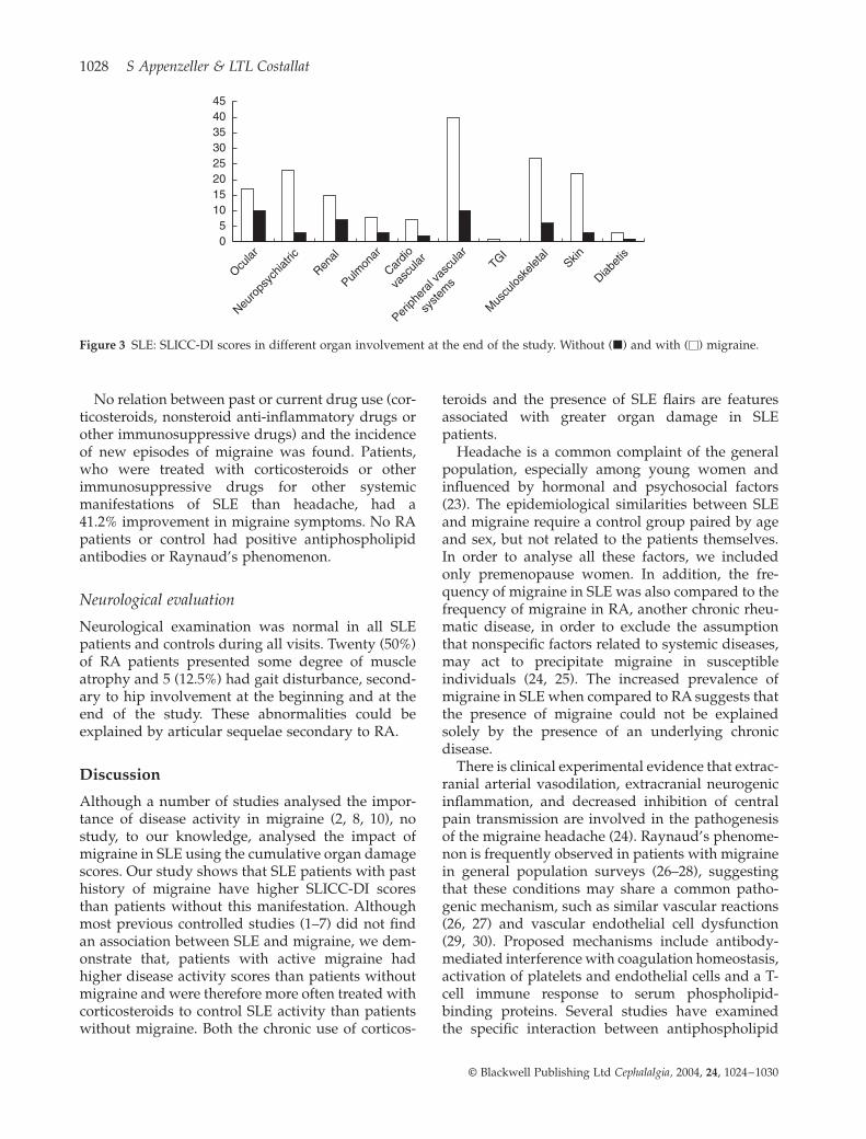
1028
S Appenzeller & LTL Costallat
© Blackwell Publishing Ltd
Cephalalgia,
2004,
24
, 1024–1030
No relation between past or current drug use (cor-ticosteroids, nonsteroid anti-inflammatory drugs orother immunosuppressive drugs) and the incidenceof new episodes of migraine was found. Patients,who were treated with corticosteroids or otherimmunosuppressive drugs for other systemicmanifestations of SLE than headache, had a41.2% improvement in migraine symptoms. No RApatients or control had positive antiphospholipidantibodies or Raynaud’s phenomenon.
Neurological evaluation
Neurological examination was normal in all SLEpatients and controls during all visits. Twenty (50%)of RA patients presented some degree of muscleatrophy and 5 (12.5%) had gait disturbance, second-ary to hip involvement at the beginning and at theend of the study. These abnormalities could beexplained by articular sequelae secondary to RA.
Discussion
Although a number of studies analysed the impor-tance of disease activity in migraine (2, 8, 10), nostudy, to our knowledge, analysed the impact ofmigraine in SLE using the cumulative organ damagescores. Our study shows that SLE patients with pasthistory of migraine have higher SLICC-DI scoresthan patients without this manifestation. Althoughmost previous controlled studies (1–7) did not findan association between SLE and migraine, we dem-onstrate that, patients with active migraine hadhigher disease activity scores than patients withoutmigraine and were therefore more often treated withcorticosteroids to control SLE activity than patientswithout migraine. Both the chronic use of corticos-
teroids and the presence of SLE flairs are featuresassociated with greater organ damage in SLEpatients.
Headache is a common complaint of the generalpopulation, especially among young women andinfluenced by hormonal and psychosocial factors(23). The epidemiological similarities between SLEand migraine require a control group paired by ageand sex, but not related to the patients themselves.In order to analyse all these factors, we includedonly premenopause women. In addition, the fre-quency of migraine in SLE was also compared to thefrequency of migraine in RA, another chronic rheu-matic disease, in order to exclude the assumptionthat nonspecific factors related to systemic diseases,may act to precipitate migraine in susceptibleindividuals (24, 25). The increased prevalence ofmigraine in SLE when compared to RA suggests thatthe presence of migraine could not be explainedsolely by the presence of an underlying chronicdisease.
There is clinical experimental evidence that extrac-ranial arterial vasodilation, extracranial neurogenicinflammation, and decreased inhibition of centralpain transmission are involved in the pathogenesisof the migraine headache (24). Raynaud’s phenome-non is frequently observed in patients with migrainein general population surveys (26–28), suggestingthat these conditions may share a common patho-genic mechanism, such as similar vascular reactions(26, 27) and vascular endothelial cell dysfunction(29, 30). Proposed mechanisms include antibody-mediated interference with coagulation homeostasis,activation of platelets and endothelial cells and a T-cell immune response to serum phospholipid-binding proteins. Several studies have examinedthe specific interaction between antiphospholipid
Figure 3
SLE: SLICC-DI scores in different organ involvement at the end of the study. Without (
�
) and with (
�
) migraine.
05
1015202530354045
Ocular
Neuro
psyc
hiatri
c
Renal
Pulmon
ar
Cardio
vasc
ular
Periph
eral
vasc
ular
syste
ms
TGI
Mus
culos
kelet
alSkin
Diabet
is
Migraine in SLE
1029
© Blackwell Publishing Ltd
Cephalalgia,
2004,
24
, 1024–1030
antibodies, in especially, antibodies to
b
2
-glycopro-tein I and in-vitro endothelial cell function (31–36).The direct binding of
b
2
-glycoprotein I to the endot-helial cell surface is facilitated by the constitutivenegative charge on the surface of endothelial cells,enhanced surface expression of negatively chargedphosphatidylserine during apoptosis (31) and the factthat annexin II acts as a receptor for the binding of
b
2
-glycoprotein I to cultured endothelial cells (32).Thus, antiphospholipid antibody binding to theendothelial cell surface in a
b
2
-glycoprotein-I–depen-dent manner leads to endothelial cell activation,which is manifested by up regulation of cell surfaceadhesion molecules and increased secretion of inter-leukin-6 and prostaglandins (33–36). So they mayinduce endothelial damage by complement and orantibody dependent cytotoxicity. Endothelial cellsare involved in the regulation of many substancesthat are involved in the pathogenesis of migraine:inactivation of vasoactive substances such as sero-tonin and bradicynin and production, for example,of endothelin 1 and prosatcycline. Endothelial celldysfunction is a relevant pathogenic mechanismexplaining the interaction between migraine,antiphospholipid antibodies and Raynaud’sphenomenon.
Some studies have analysed the associationsbetween migraine and Raynaud’s phenomenon inSLE (24, 25, 37, 38). We found not only a higherprevalence of Raynaud’s phenomenon in SLEpatients with migraine, but also a worsening ofRaynaud’s phenomenon prior to migraine episodesin 44.1% of our SLE patients. The majority of ourpatients with aura referred worsening of Raynaud’sphenomenon during the aura occurrence, support-ing the idea that both conditions may have a com-mon pathogenic mechanism. Several neurologicaldisorders have been associated with the presence ofantiphospholipid antibodies (39–42), but there arestill controversies in relation to migraine (7, 24, 25,37, 38). However, the majority of study groups aresmall, making statistic analysis more difficult. In ourstudy, the presence of antiphospholipid antibodieswas more frequent in SLE patients with migraine.
The Rheumatology Unit of the Sate University ofCampinas is a reference centre for rheumatic diseasesand this fact explains the high frequency of someclinical SLE manifestations, such as nephropathy.
Our study has some limitations. First, this is notan unselected sample of SLE patients. Only womenwere included in this study because both migraineand SLE have hormonal influence. Second, the num-ber of patients with active migraine is small and noheadache diary was used, making difficult to extract
conclusion in a prospective manner. Also, the strictinclusion and exclusion criteria used could intro-duce bias, especially in relation to the number ofpersons affected by migraine in all three groups. Butas the primary objective of our study was to deter-mine the relation of disease activity and cumulativeorgan damage and migraine in SLE, and not to deter-mine the overall prevalence of migraine in this pop-ulation, we consider that these limitations were notrelevant to determine this issue. Third, no neurolog-ical consultation was performed in this study.
In summary, history of migraine was associatedwith greater organ damage using SLICC-DI. Activemigraine was associated with higher disease activity,measured by MEX-SLEDAI, antiphospholipid anti-bodies and worsening of Raynaud’s phenomenon.The increased cumulative organ damage in SLEpatients with past history of migraine justifies theroutine evaluation of migraine in clinical practice.
Acknowledgements
This work was supported by Fundos Remanescentes daSociedade Brazileira de Reumatologia and FAPESP (03/01527-0).
References
1 Omdal R, Mellgren SI, Husby G. Clinical neuropsychiatricand neuromuscular manifestations in systemic lupuserythematosus. Scand J Rheumatol 1988; 17:113–7.
2 Feinglass EJ, Arnett FC, Dorsch CA, Zizic TM, Stevens MB.Neuropsychiatric manifestations of systemic lupus erythe-matosus: diagnosis, clinical spectrum and relationship toother features of the disease. Medicine 1976; 55:323–39.
3 Adelman DC, Saltiel E, Klinenberg JR. The neuropsychiat-ric manifestations of systemic lupus erythematosus: Anoverview. Semin Arthritis Rheum 1986; 15:185–99.
4 Johnson RT, Richardson EP. The neurological manifesta-tions of systemic lupus erythematosus. Medicine 1968;47:337–69.
5 Fernandez-Nebro A, Palacios-Munoz R, Gordillo J,Abarca-Costalgo M, De Haro-Liger M, Rodriguez-AndreuJ et al. Chronic or recurrent headache in patients withsystemic lupus erythematosus: a case control study. Lupus1999; 8:151–6.
6 Vazquez-Cruz J, Traboulssi H, Rodriguez-De la Serna A,Geli C, Roig C, Diaz C. A prospective study of chronic orrecurrent headache in systemic lupus erythematosus.Headache 1990; 30:232–5.
7 Glanz BI, Venkatesan A, Schur PH, Lew RA, Khoshbin S.Prevalence of migraine in patients with systemic lupuserythematosus. Headache 2001; 41:285–9.
8 Sfikakis PP, Mitsikostas DD, Manoussakis MN, FoukaneliD, Moutsopoulos HM. Headache in systemic lupus erythe-matosus: a controlled Study. Br J Rheumatol 1998; 37:300–3.
9 Omdal R, Waterloo K, Koldingsnes W, Husby G, MellgrenSI. Somatic and psychological features of headache in sys-temic lupus erythematosus. J Rheumatol 2001; 28:772–9.
1030
S Appenzeller & LTL Costallat
© Blackwell Publishing Ltd
Cephalalgia,
2004,
24
, 1024–1030
10 Ainiala H, Loukkola J, Peltola J, Korpela M, Hietaharju A.The prevalence of neuropsychiatric syndromes in systemiclupus erythematosus. Neurology 2001; 57:496–500.
11 Tan EM, Cohen AS, Fries JF, Masi AT, McShane DJ,Rothfield NF, et al. The 1982 revised criteria for the classi-fication of systemic lupus erythematosus. Arthritis Rheum1982; 25:1271–7.
12 The American College of Rheumatology Nomenclatureand case definitions for neuropsychiatric lupus syn-dromes. Arthritis Rheum 1999; 42:599–608.
13 Arnett FC. Revised criteria for the classification of rheu-matoid arthritis. Bull Rheum Dis 1989; 38:1–6.
14 Harris EN, Gharavi AE, Patel SP, Hughes GR. Evaluationof the anticardiolipine antibody test: report of an interna-tional workshop held 4 April 1986. Clin Exp Immunol1987; 68:215–22.
15 Brandt JT, Triplett DA, Alving B, Scharer I. Criteria for thediagnosis of lupus anticoagulant: an update. On behalf ofthe Subcommittee on Lupus Anticoagulant/Antiphospho-lipid Antibody of the Scientific and Standardisation Com-mittee of the ISTH. Thromb Haemost 1995; 74:1185–90.
16 Guzman J, Cardiel MH, Arce-Salinas A, Sanchez-GuerreroJ, Alarcon-Segovia D. Measurement of disease activity inSLE. Prospective validation of 3 clinical indices. J Rheuma-tol 1992; 19:1551–8.
17 Gladman DD, Urowitz MB, Kagal A, Hallett D. Accuratelydescribing changes in disease activity in systemic lupuserythematosus. J Rheumatol 2000; 27:377–9.
18 Gladman DD, Urowitz MB, Goldsmith CH, Fortin P, Gin-zler E, Gordon C et al. The reliability of the Systemic LupusInternational Collaborating Clinics/American College ofRheumatology Damage Index in patients with systemiclupus erythematosus. Arthritis Rheum 1997; 40:809–13.
19 Headache Classification Committee of the InternationalHeadache Society. Classification and diagnostic criteria forheadache disorders, cranial neuralgias and facial pain.Cephalalgia 1988; 8 Suppl 7:1–96.
20 Solomon S. Diagnosis of primary headache disorders.Validity of the International Headache Society criteria inclinical practice. Neurol Clin 1997; 15:15–26.
21 Pajaron E, Lainez JM, Monzon MJ, Parra J, Peiro C, SanchoJ. The validity of the classification criteria of the Interna-tional Headache Society for migraine, tension headacheand chronic tension headache. Neurologia 1999; 14:283–8.
22 Bensenor IJ, Lotufo PA, Pereira AC, Tannuri AC, Issa FK,Akashi D et al. Validation of a questionnaire for the diag-nosis of headache in an outpatient clinic at a Universityhospital. Arq Neuropsiquiatr 1997; 55:364–9.
23 Weitzel KW, Strickland JM, Smith KM, Goode JV. Gender-specific issues in the treatment of migraine. J Gend SpecifMed 2001; 4:64–74.
24 Isenberg DA, Meyrick-Thomas D, Snaith ML, McKeranRO, Royston JP. A study of migraine in systemic lupuserythematosus. Ann Rheum Dis 1982; 41:30–2.
25 Markus HS, Hopkinson N. Migraine and headache in sys-temic lupus erythematosus and their relationship with anti-bodies against phospholipids. J Neurol 1992; 239:39–42.
26 Silman A, Holligan S, Brennan P, Maddison P. Prevalenceof symptoms of Raynaud’s phenomenon in general prac-tice. Br Med J 1990; 301:590–2.
27 Heslop J, Coggon D, Acheson ED. The prevalence of inter-mittent digital ischaemia in a general practice. J Roy CollGeneral Pract 1983; 33:85–9.
28 O’Keeffe ST, Tsapatsaris NP, Beetham WP Jr. Associationbetween Raynaud’s phenomenon and migraine in a ran-dom population of hospital employees. J Rheumatol 1993;20:1187–8.
29 Spierings EL. Pathogenesis of the migraine attack. Clin JPain 2003; 19:255–62.
30 Hanly JG. Antiphospholipid syndrome: an overview.CMAJ 2003; 168:1675–82.
31 Bombeli T, Karsan A, Tait JF, Harlan JM. Apoptotic vascu-lar endothelial cells become procoagulant. Blood 1997;89:2429–42.
32 Ma K, Simantov R, Zhang JC, Silverstein R, Hajjar KA,McCrae KR. High affinity binding of beta 2-glycoprotein Ito human endothelial cells is mediated by annexin II. J BiolChem 2000; 275:15541–8.
33 Del Papa N, Moroni PL, Tincani A, Harris EN, PierangeliSS, Barcellini W et al. Relationship between anti-phospholipid and anti-endothelial cell antibodies: furthercharacterization of the reactivity on resting and cytokine-activated endothelial cells. Clin Exp Rheumatol 1992;10:37–42.
34 Simantov R, LaSala JM, Lo SK, Gharavi AE, SammaritanoLR, Salmon JE et al.Activation of cultured vascular endot-helial cells by antiphospholipid antibodies. J Clin Invest1995; 96:2211–9.
35 Del Papa N, Guidali L, Sala A, Buccellati C, KhamashtaMA, Ishikawa K et al. Endothelial cells as target forantiphospholipid antibodies. Human polyclonal andmonoclonal anti-beta 2-glycoprotein I antibodies react invitro with endothelial cells through adherent beta 2-glycoprotein I and induce endothelial activation. ArthritisRheum 1997; 40:551–61.
36 Hanly JG, Hong C, Issekutz A. Beta 2-glycoprotein I andanticardiolipin antibody binding to resting and activatedcultured human endothelial cells. J Rheumatol 1996;23:1543–9.
37 Montalban J, Cervera R, Font J, Ordi J, Vianna J, Haga HTet al. Lack of association between anticardiolipin antibod-ies and migraine in systemic lupus erythematosus. Neu-rology 1992; 3:681–2.
38 Uziel Y. Headache in SLE. Clin Exp Rheumatol 2000; 18(3):421.
39 Aarli JA. Immunological aspects of epilepsy. Brain Dev1993; 15:41–9.
40 Herranz MT, Rivier G, Khamashta MA, Blaser KU,Hughes GR. Association between antiphospholipid anti-bodies and epilepsy in patients with systemic lupuserythematosus. Arthritis Rheum 1994; 37:568–71.
41 Whitlaw DA, Spangenberg JJ, Rickman R, Hugo FH,Roberts M. An association between the AntiphospholipidAntibody and neurological Impairment in SLE. Lupus1999; 8:444–8.
42 Shrivastava A, Dwivedi S, Aggarwal A, Misra R. Anti-cardiolipin and anti-beta2 glycoprotein I antibodies inIndian patients with systemic lupus erythematosus. asso-ciation with the presence of seizures. Lupus 2001; 10:45–50.
Resultados 120
ARTIGO 6
Acute psychosis in SLE
Appenzeller S, Cendes F, Costallat LT
Acute psychosis in SLE
submetido
Resultados 121
Acute psychosis in systemic lupus erythematosus
Simone Appenzeller MD1, 2, Fernando Cendes MD, PhD 2, 3, Lilan Tereza Lavras Costallat
MD, PhD 1
1Rheumatology Unit - State University of Campinas 2 Neuroimaging Lab-State University
of Campinas; 3 Department of Neurology, State University of Campinas
Running Title: acute psychosis in SLE
Address all correspondence to: Prof Dr Lilian TL Costallat, Department of Internal
medicine, State University of Campinas (UNICAMP), Cidade Universitaria Zeferino Vaz,
CEP 13083970 Campinas-SP-Brazil
Tel: +55 1937887734
Fax: +55 19 32135106
E-mail: [email protected]
Key words: psychosis, neuropsychiatric, systemic lupus erythematosus
Grants: Fundação de Amparo a Pesquisa do Estado de São Paulo (FAPESP), Conselho
Nacional de Pesquisa e Desenvolvimento (CNPq).
Resultados 122
Abstract
Objective: To evaluate the frequency and risk factors of acute psychosis in a
large cohort of patients with systemic lupus erythematosous (SLE). To identify clinical and
laboratory variables useful in differentiating acute psychosis as a primary manifestation of
CNS from corticosteroid induced psychosis.
Methods: Five hundred thirty seven consecutive patients with SLE were
studied, with follow-up ranging from 4 to 8.8 years. A standardized medical history,
neurological, rheumatologic, and psychiatric examinations and serologic testing were
performed in all patients. The type and frequency of risk factors associated with acute
psychosis as a primary manifestation of CNS system and corticosteroid induced psychosis
was determined using multivariate regression with automatic backward stepwise selection.
Results: We identified acute psychosis in 89 of 520 (17.1%) SLE patients.
Psychosis primary to CNS involvement was diagnosed in 59 of these patients,
corticosteroid induced psychosis in 28 and primary psychotic disorder not related to SLE or
medication in 2 patients. Psychosis secondary to SLE at disease onset occurred in 19
patients and was associated with disease activity (p=0.001; OR=2.4; CI=1.5-6.2). Psychosis
during follow-up of SLE was observed in 40 patients and associated with positive
antiphospholipid antibodies (p=0.004; OR=3.2; CI=1.9-4.5) and less frequently with renal
(p=0.002; OR=1.9; CI=0.0-0.6) and cutaneous (p=0.04; OR=1.1; CI=0.0-0.8) involvement.
We identified 28 patients with 38 episodes of psychosis associated with corticosteroid
therapy. All patients had severe active disease and 10 of these patients had
hypoalbuminemia when psychosis developed. At time of psychotic event, all patients were
taking prednisone in doses varying from 0.75 to 1 mg/kg/day. Psychosis resolved after
tapering prednisone down in all patients.
Conclusion: Acute psychosis related to SLE was observed in 11.3% of our
cohort. Recurrence of primary psychosis was associated with other CNS manifestations
related to SLE.
Resultados 123
Introduction
Central nervous system (CNS) manifestations in systemic lupus erythematosus
(SLE) are very heterogeneous in clinical presentations, involving multiple
pathophysiological mechanisms (1-3). Neuropsychiatric involvement in SLE could be
defined as neurological syndromes of the central, peripheral and autonomic nervous system
and psychiatric syndromes observed in SLE patients, after excluding other possible causes
not related with SLE (3).
A challenging problem in the diagnosis and management of SLE is psychiatric
disorder. Abnormal behavior was first described in SLE by Hebra and Kaposi (4), followed
by Osler (5). Many psychiatric symptoms can be observed in SLE patients, such as
depression, anxiety, mood disorders, but psychosis was considered one of the most
important and included in the classification criteria for SLE in 1982, despite the widely
diverse clinical manifestations (6) and varying degree of severity (7, 8). Psychosis may be a
primary event of CNS manifestation of SLE or associated with a variety of drugs and
infections, such as corticosteroids and antimalarics (9). Adverse psychiatric effects,
including mild euphoria, emotional lability, alteration of behavior, panic attacks, psychosis
and delirium, are seen in 3 to 10% of all patients receiving corticosteroids and are
unpredictable from the regimens of corticosteroids used (10-14).
In this study we reviewed the frequency of acute psychosis in our cohort and
determined the frequency of corticosteroid induced psychosis. We further tried to identify
risk factors for occurrence of acute and corticosteroid induced psychosis, as well as for the
recurrence of psychosis.
Patients and Methods
537 consecutive patients with four or more criteria for SLE diagnosis (6), had
their medical histories, clinical and serological characteristics documented regularly in
computer database programs. All patients had their clinical and laboratory evaluation
performed at diagnosis and quarterly during follow-up period and were followed and
examined by one of the authors (LTLC, SA) throughout the years. Therefore, medical
records and protocols for investigations were very homogenous among patients followed at
Resultados 124
the Rheumatology Unit of the State University of Campinas. In this study, we included all
patients that were regularly evaluated in our unit from January 1999 to December 2003.
The mean duration of follow-up of these patients was 5.3 years (SD 1.1 years), ranging
from 4.0 to 8.8 years.
Seven patients with incomplete clinical and laboratory evaluations or who lost
follow up were not included in this series. This study has been approved by our local Ethics
Committee.
Psychiatr ic evaluation
Acute psychosis as a primary manifestation of CNS involvement by SLE was
defined by the presence of disturbance of reality characterized by delusions and/or
hallucinations, severe enough to cause significant distress or social impairment. All
symptomatic patients were evaluated by a psychiatrist and events were classified using the
Diagnostic and Statistical Manual of Mental Disorders (4th edition) (15). Patients with
psychosis were carefully evaluated, in order to differentiate primary psychotic disorders
unrelated to SLE, substance or drug-induced psychotic disorders and psychologically
mediated reactions to SLE as major stressor. In all patients with psychosis, other causes
such as infection, metabolic disturbance and structural lesions were carefully excluded by
clinical, laboratory and MRI when indicated.
Corticosteroid-induced psychiatric disturbances were defined as new symptoms
that appeared temporally within 8 weeks of institution or augmentation of steroids and
resolved completely after reduction of steroid dosage without additional
immunosuppressive agents. Ten patients who had psychosis previously to SLE diagnosis or
previously to the evaluations in our service were not included in the analysis, although they
were followed in order to observe if they would have recurrent psychotic episodes. Patients
with diagnosis of psychosis before 1999, had their medical charts carefully reviewed in
order to determine if they complete ACR criteria (16). Therefore 17 patients were excluded
and the remaining 520 patients were included in the final analysis.
Resultados 125
Clinical features
The age at onset of psychosis was determined in relation to the age at which the
first well-described sign or symptom indicating SLE occurred. To determine the risk factors
for occurrence of psychosis we analyzed clinical manifestations, serologic features at
disease onset and during follow-up, according to the American College of Rheumatology
(ACR) criteria (6, 16). For the purpose of statistical analyses, some of the clinical features
were grouped under renal, neuropsychiatric, and hematologic disease. Renal disease was
defined as any 1 of the following: 1) persistent proteinuria of >=0.5 g/day; 2) the presence
of cellular casts; and/or 3) biopsy evidence of lupus glomerulonephritis. Neuropsychiatric
disease referred to any of the manifestations defined by ACR criteria (16), after careful
review of medical records. Hematologic disease was defined as any 1 of the following: 1)
hemolytic anemia; 2) leukopenia (<4.0 × 10⁹/L); and 3) thrombocytopenia (<100 × 109/L),
on at least 2 occasions that were not due to the effect of medications.
Global disease activity was quantified by the SLE disease activity Index
(SLEDAI) (17), cumulative organ damage by the ACR/Systemic Lupus (SLICC) (18).
Patients with neuropsychiatric manifestations or complains were evaluated. A
complete neurological examination, as well as cognitive and psychiatric charts, were
applied. Cognitive impairment was analyzed using a standardized neuropsychological tests
in order to screen for possible impairment in one or more of the subsequent cognitive
domains: simple attention, complex attention, memory, visuo-spatial processing, language,
reasoning/problem solving, psychomotor speed, and executive functions (19-22). The
individual test results were converted into standard scores, which were compared with the
available normative data (19-22). Regarding any of the eight cognitive domains, subjects
with a total score of two or more standard deviations (SD) below the normative value were
considered to be impaired. Cognitive dysfunction was classified as mild if there were
deficits in less than three dimensions, as moderate if there were deficits in three or four
dimensions, and as severe if there were deficits in at least five dimensions (23, 24).
Assessment of depression was based on clinical interview and the Beck
Depression Inventory (BDI) (25, 26). On BDI, scores from 10 to 17 were considered to
indicate mild depression, from 18 to 24 moderate depression, and greater than 24 severe
Resultados 126
depression. Anxiety was evaluated by anxiety through the Hospital Anxiety and Depression
scale (27). The presence of psychosis was determined through the Brief Psychiatry Rating
Scale (aBPRS) (28).
Laboratory features
Anticardiolipin antibodies (aCL) of the IgG and IgM isotypes were measured
by the ELISA method as described previously (29). They were recorded as negative
(<5GPL units or <3MPL units), low positive titers (5-15 GPL units or 3-6 MPL units),
moderate positive titers (15-80 GPL units or 6-50 MPL units), or high positive titers (>80
GPL units or >50 MPL units). The lupus anticoagulant (LA) activity was detected by
coagulation assays in platelet free plasma obtained by double centrifugation (30). Patients
with SLE diagnosis prior to 1991 had their aCL and LA essays done during the follow-up
period.
Statistical analysis
Values in this study were expressed as mean ± standard deviation (SD).
Comparison of categorical data among groups was made by the chi-square test.
Comparison of continuous data was performed by 1-way ANOVA. Post-hoc multiple
comparisons were performed using the Tukey test for unequal samples.
First, the baseline features of those patients with psychosis were compared to
patients without psychosis. Comparisons were done by 2 for categorical variables and by
analyses of variance for continuous variables. Furthermore, patients were divided into
groups as follows: psychosis at disease onset, psychosis during follow-up period and
corticosteroid induced psychosis. Because of the small sample size, we excluded patients
with psychosis not related to SLE from the analysis and used the data only in a descriptive
manner. Their baseline features were also compared. The independent variables chosen to
enter the multivariate analysis model were those that presented a level of statistical
significance � 5% at the univariate analyses.
Resultados 127
We initially performed one multiple regression including significant variables
to differentiate acute psychosis due to SLE, independently to the time of disease onset, to
patients with acute psychosis related to corticosteroids therapy. Then we performed an
automatic backward stepwise multiple regression, including the variables SLEDAI and
SLICC scores, malar rash, photosensitivity, arthritis, renal, neuropsychiatric, and
hematologic disease, moderate to high antiphospholipid antibodies, ANA antibodies, anti
DNA antibodies, hypoalbuminemia in the model, to determine clinical and laboratory
features associated with psychosis at disease onset and at follow-up. These models included
corrections for multiple comparisons. A p value <0.05 was considered as indicative of
statistical significance. MRI findings and CSF were not included in the analysis.
Results
Acute psychosis was identified in 89 of 520 (17.1%) patients (78 women) of
our cohort with mean age of 26.3 years. We did not observe any statistical difference in
mean age of disease onset, time to disease diagnosis and any other demographic variable
between patients with acute psychosis and without psychosis (Table 1). Nineteen (21.3%)
patients had acute psychosis at disease onset and 40 (45%) during course of SLE. Twenty
eight (31.5%) patients had corticosteroid induced psychosis and 2 (2.2%) patients were
identified with psychosis not related to SLE or drugs.
Acute psychosis at disease onset
Acute psychosis at disease onset was identified in 19 of 89 patients (17 women)
with mean age of 25.6 years (SD=5.6; range 16-38 years). All patients had acute psychosis
as one of the first manifestations of SLE and were off steroid when psychotic episode
occurred. They were subsequently referred to our service because of clinical manifestations
suggestive of SLE during further investigations. Further investigations excluded infections
and revealed positive ANA titers in all patients. Cerebral spinal fluid (CSF) analysis was
normal in 14 and revealed mild pleocitosis in 5 patients. MRI was normal in 8 of 9 patients
(Table 2). All patients completed classification criteria for SLE during follow-up period.
The psychotic episodes resolved after introduction of psychiatric medication and
Resultados 128
corticosteroid doses that varied from 0.5 to 1 mg/kg/day of prednisone, depending on the
severity of clinical manifestations. After a mean time of follow-up of 6.2 years (SD=2.3,
range 2.3-10.2 years), 11 patients were off psychiatric medications.
In automatic backward stepwise regression analyses we observed that the
disease activity was independently associated with psychosis (p=0.001; OR=2.4; CI=1.5-
6.2). Furthermore, the absence of malar rash (p=0.002; OR=1.6; CI=0.1-0.9) and
photosensitivity (p=0.01; OR=0.5; CI=0.01-0.085) were also variables independently
associated with psychosis when compared to patients without psychosis at disease onset.
Recurrence of acute psychosis not related to corticotheraphy was observed in 8
patients. They all had other CNS manifestations related to SLE in addition to psychosis,
including severe cognitive impairment in 5, headache in 5, mood disorder in 4, seizures in
3, anxiety in 2 and stroke in 2 patients. Recurrence of psychosis in this group of patients
was associated with disease activity (p=0.001; OR=3.4; CI=1.9-6.0) and the presence of
other CNS manifestations (p=0.02; OR=1.9; CI=1.3-3.5) in automatic backward stepwise
regression analyses.
Acute psychosis during follow-up
During the course of SLE, acute psychosis was observed in 40 patients (35
women). Mean time of SLE diagnosis before psychosis occurred was 14 months (range 7-
40 months) (Table 1). CSF analyses during acute psychosis were performed in 32 of 40
patients and were negative for infections in all. Fifteen patients had mild pleocitosis and 10
mild protein elevations in CSF. MRI performed in 15 patients was abnormal in 10 and
revealed hyperintense lesions in 10, cerebral atrophy in 8 and diffuse white matter
involvement in 2 patients (Table 2).
In automatic backward stepwise regression analyses, the presence of positive
antiphospholipid antibodies in moderate to high titers (p=0.004; OR=3.2; CI=1.9-4.5) and
SLEDAI scores � 8 (p=0.001; OR=2.0; CI=1.5-2.3) were independent risk factors for
occurrence of psychosis. The absence of renal (p=0.002; OR=1.9; CI=0.0-0.6) and
cutaneous manifestations (p=0.04; OR=1.1; CI=0.0-0.8) were also variables associated with
psychosis during follow-up period.
Resultados 129
During mean follow-up period of 4.6 years after the occurrence of psychosis,
these patients developed more frequently other primary CNS manifestations related to SLE,
including cognitive impairment in 20, mood disorder in 7, stroke in 5, seizures in 4 and
anxiety in 2 patients.
Recurrence of acute psychosis was identified in 10 of 40 patients. After
carefully investigations, recurrences were associated with SLE disease activity in all
patients (p=0.001; OR=3.2; CI=1.6-4.6).
Acute psychosis related to corticosteroids therapy
We identified 28 patients (25 women) with 38 episodes of acute psychosis
associated with corticosteroid treatment. All patients had severe active disease and 10 of
these patients had hypoalbuminemia (albumin < 2mg/dl) when psychosis developed.
Nephritis was diagnosed in 11, serositis in 6, systemic vasculitis in 5, autoimmune hepatitis
in 3, and pericarditis in 3 patients. At time of psychosis, all patients were using prednisone
in dose varying 0.75 to 1 mg/kg/day. Psychosis resolved after tapering prednisone down in
all cases after a median time of 13.3 (SD±5.2) days. In 15 episodes of psychosis,
antipsychotic medications were required. Haloperidol was used in 13 of 15 patients. CSF
analyses during psychosis were performed in 14 of 28 patients and were normal in 14 and
negative for infections in all.
In the automatic backward stepwise regression analyses, the only variable
associated with psychosis associated with corticosteroid treatment was hypoalbuminemia
(p=0.03; OR=2.2; CI=1.9-2.5). Recurrences of psychosis were observed in 10 of these
patients: in eight after increment of prednisone dose for systemic manifestations and in 2
with lower doses.
Acute psychosis not related to SLE
Two patients (2 women) were identified with acute psychosis not related to
SLE. One patient had mental retardation secondary to perinatal anoxia and developed
severe psychiatric disturbance during development. The other patient had initially several
Resultados 130
hallucinations and psychotic manifestations after epileptic seizures, and was diagnosed
during the follow-up period as having temporal lobe epilepsy due to her clinical and
electroencephalogram findings. In addition, her MRI showed signs of hippocampal
sclerosis ipsilateral to the epileptic focus. Psychotic episodes resolved after introduction of
antiepileptic medication.
Acute psychosis due to SLE x acute psychosis related to corticosteroids therapy
When we compared SLE patients with acute psychosis due to SLE,
independently to the time of disease onset, to patients with acute psychosis related to
corticosteroids therapy using automatic backward stepwise regression analyses, we
observed that patients with psychosis due to SLE had more frequently other CNS
manifestations related to SLE (p=0.03; OR=2.1; CI=1.2-3.9) and positive antiphospholipid
antibodies (p=0.01; OR=2.2; CI=1.4-3.5) than patients with acute psychosis related to
corticosteroids therapy. The presence of hypoalbuminemia was a risk factor (p=0.03;
OR=2.2; CI=1.9-2.5) for development of corticosteroid induced psychosis in automatic
backward stepwise regression analyses.
Discussion
We identified acute psychosis in 89 of 520 (17.1%) symptomatic SLE patients.
Acute psychosis primary to CNS involvement was diagnosed in 59, corticosteroid induced
psychosis in 28 and primary psychotic disorder not related to SLE neither to medication in
2 patients. We observed that the presence of other CNS manifestations associated to SLE
and positive antiphospholipid antibodies were more frequently observed in patients with
psychosis due to SLE when compared to corticosteroid induced psychosis. We further
observed that corticosteroid induced psychosis was more frequently observed in patients
with hypoalbuminemia. Because of the small sample size, we excluded patients with
psychosis not related to SLE from the analysis and used the data only in a descriptive
manner.
Resultados 131
We observed that acute psychosis secondary to SLE at disease onset was more
frequently associated with disease activity, whereas cutaneous manifestations were less
frequently seen. This may be one of the reasons why these patients are usually first seen by
psychiatrist. However a carefully investigation is necessary, especially because all patients
are usually young women, and develop severe systemic manifestations if not treated
promptly. In patients who had acute psychosis during the course of disease, it was
associated with the presence of antiphospholipid antibodies and they had more frequently
other CNS manifestations related to SLE activity. Depression, stroke, seizures, cognitive
dysfunction, and psychosis have all been associated with antiphospholipid antibodies (2, 3).
The presumed pathophysiological mechanism underlying these manifestations is thought to
be a result of cerebral ischemia in some, but not all patients. An interaction between
antiphospholipid antibodies and central nervous system cellular elements rather than
antiphospholipid antibodies associated thrombosis seems to be a more plausible mechanism
for most of these clinical manifestations (2, 3, 31). Renal and cutaneous manifestations of
SLE were less frequently seen. Recurrence of acute psychosis was observed in 18 of 59
patients. These patients had also more frequently other CNS events due to SLE activity.
Therefore patients with acute psychosis should be followed carefully in order to determine
if they develop other primary CNS manifestations secondary to SLE.
In our study we observed 38 episodes of corticosteroid induced psychosis in 28
patients during the follow-up period. All patients had active SLE disease and were
receiving prednisone in moderate to high doses. As previously described (11-13, 19-33),
hypoalbuminemia may be a risk factor for corticosteroid induced psychosis. In our study
we observed hypoalbuminemia in 10 patients who developed corticosteroid induced
psychosis. The explanation for these findings may be that corticosteroid binding globulin
does not bind to synthetic steroids, whose transport depends on serum albumin which, by
contrast, presents low affinity but a great capacity to transport the steroids because of its
high plasma concentration. Steroids are biologically inactive when bound to albumin.
Therefore, the free (and active) fraction of steroids is higher in patients with low plasma
albumin levels, and this will expose the patient to more adverse effects (35). Due to the
retrospective nature of our study, we were not able to determine the albumin level of all our
patients; therefore we could not determine the cut off values for an increased risk of
Resultados 132
psychosis. The incidence of psychiatric reactions to corticosteroid treatment in SLE is not
significantly higher than the expected one in ulcerative colitis, rheumatoid arthritis or
lymphoma. However, patients with SLE do have a higher incidence of corticosteroid-
induced psychiatric symptoms (35).
In patients with SLE, corticosteroid-induced psychiatric events may be difficult
to distinguish from primary neuropsychiatric manifestations of SLE. The antiribosomal P
antibody is specific for SLE and has been shown to be associated with psychosis related to
disease activity and other CNS manifestations, including depression (33), but the low
sensitivity limits its clinical usefulness for the diagnosis of lupus psychosis (13). Therefore,
a temporal relationship between the beginning of corticosteroid therapy and psychiatric
events, and the resolution of symptoms after reduction of corticosteroids is an important
feature in diagnosis of corticosteroid induced psychosis. We did not search for
antiribosomal P antibodies in our studies and steroid induced psychosis was based on
clinical judgment (34).
MRIs were done in 30 patients during acute psychosis. Patients with psychosis
at disease onset had more frequently normal MRI when compared to patients with
psychosis at follow-up and corticosteroid induced psychosis, although none of the findings
observed in MRI could be directly related to psychotic event.
A limitation of this study is its retrospective nature. However, all patients’
clinical and laboratory findings were constantly updated on our computer database by one
of the authors, reducing the bias. Recurrence of psychosis and factors associated with its
occurrence has not been frequently reported before.
In conclusion we observed acute psychosis related to SLE in 11.3% of our
cohort. Recurrence occurred in 30% of these patients and was related to the presence of
other CNS manifestations. Hypoalbuminemia was a risk factor for development of
corticosteroid induced psychosis, whereas disease activity, CNS manifestations and the
presence of antiphospholipid antibodies were risk factors for psychosis due to SLE.

Resultados 133
References
1. Jennekens FG, Kater L. The central nervous system in systemic lupus erythematosus.
Part 1. Clinical syndromes: a literature investigation. Rheumatology (Oxford)
2002;41:605-618.
2. Sanna G, Bertolaccini ML, Cuadrado MJ, et al. Neuropsychiatric manifestations in
systemic lupus erythematosus: prevalence and association with antiphospholipid
antibodies. J Rheumatol 2003; 30:985-992.
3. Mok CC, Lau CS, Wong RW. Neuropsychiatric manifestations and their clinical
associations in southern Chinese patients with systemic lupus erythematosus. J
Rheumatol 2001;28:766-771.
4. Hebra F, Kaposi M. On diseases of the skin, including the exanthemata, vol 4. Tay W,
trans, Fragge CH, ed. London: New Sydenham Society 1866-1880, 1874.
5. Osler W. The visceral lesions of the erythema group. Br J Dermatol 1900; 12: 227-245.
6. Tan EM, Cohen AS, Fries JF, et al. The 1982 revised criteria for the classification of
systemic lupus erythematosus. Arthritis Rheum 1982; 25: 1271-1277.
7. Kassan SS, Kagan LJ. Central nervous system lupus erythematosus: measurment of
CSF cyclic GMP and other clinical markers of disease activity. Arthritis Rheum 1979;
22: 449-457.
8. Singer J, Denburg JA, and the Ad Hoc Neuropsychiatric Lupus Workshop Group:
diagnostic criteria for neuropsychiatric systemic lupus erythematosus: the result of a
consensus meeting. J Rheumatol 1990; 17: 1397-1402.
9. Brey RL, Holliday SL, Saklad AR, et al.Neuropsychiatric syndromes in lupus:
prevalence using standardized definitions. Neurology 2002; 58:1214-1220.
10. Kasitanon N, Louthrenoo W, Piyasirisilp S, Sukitawu W, Wichainun R.
Neuropsychiatric manifestations in Thai patients with systemic lupus erythematosus.
Asian Pac J Allergy Immunol 2002;20:179-185.
11. Patten SB, Neutel CI. Corticosteroid-induced adverse psychiatric effects: incidence,
diagnosis and management. Drug Saf. 2000; 22: 111–122.
Resultados 134
12. Boston Collaborative Drug Surveillance Program. Acute adverse reactions to
prednisone in relation to dosage. Clin Pharmacol Ther. 1972; 13: 694–698.
13. Lopez-Medrano F, Cervera R, Trejo O, Font J, Ingelmo M. Steroid induced psychosis
in systemic lupus erythematosus: a possible role of serum albumin level. Ann Rheum
Dis. 2002; 61: 562–563.
14. Chau SY, Mok CC. Factors predictive of corticosteroid psychosis in patients with
systemic lupus erythematosus. Neurology 2003; 61: 104-107.
15. American Psychiatric Association. Diagnostic and Statistical Manual of Mental
Disorders (4th edition). Washington, DC: APA, 1994.
16. Liang MH, Corzillius M, Bae SC et al. The American College Nomenclature and case
definitions for neuropsychiatric lupus syndrome. Arthritis Rheum 1999; 42: 599-608.
17. The Committee on prognosis studies in SLE, C. Bombardier, D.D. Gladman, M.
Urowitz, D. Caron and C. Chang, Derivation of the SLEDAI: A disease activity index
for lupus patients. Arthritis Rheum 1992; 35: 630–640
18. Gladman DD, Urowitz MB, Goldsmith CH, Fortin P, Ginzler E, Gordon C, Hanly JG,
et al. The reliability of the Systemic Lupus International Collaborating
Clinics/American College of Rheumatology Damage Index in patients with systemic
lupus erythematosus. Arthritis Rheum. 1997; 40:809-813.
19. Folstein MF, Folstein SE, Folstein PR: “Mini mental state” A practical method for
grading the cognitive state of patient for the clinician. J Psychiatr Res 12: 189-198,
1975.
20. Spranoel HZ. The psychoeducational use and interpretation of the Wechsler Adult
Intelligence Scale–Revised. Springfield: CC Thomas, 1992.
21. Wechsler in Muistiasteikko, Suom. Anja Manner. Helsinki: Psykologien Kustannus,
1986.
22. Delis DC, Kramer JH, Kaplan E, et al. California Verbal Learning Test Research
Edition Manual. San Antonio: The Psychological Corporation, 1987.
23. Lezak MD. Neuropsychological assessment. New York: Oxford University Press, 1995.
Resultados 135
24. Heaton RK, Chelune GJ, Talley JL, et al. Wisconsin Card Sorting Test Manual, revised
and expanded. Odessa: Psychological Assessment Resources, Inc., 1993.
25. Beck AT, Beamesderfer A. Assessment of depression: the depression inventory. Mod
Probl Pharmacopsychiat. 1974; 7: 151–169.
26. Beck AT, Rial WY, Rickels K. Short form of depression inventory: cross-validation.
Psychol Rep. 1974; 34: 1184–1186.
27. Herrmann C. International experiences with the Hospital Anxiety and Depression
Scale--a review of validation data and clinical results. J Psychosom Res. 1997;42:17-41.
28. Overall JE, Beller SA. The Brief Psychiatric Rating Scale (BPRS) in geropsychiatric
research: I. Factor structure on an inpatient unit. J Gerontol. 1984;39:187-93.
29. Harris EN, Gharavi AE, Patel SP, Hughes GR. Evaluation of the anticardiolipine
antibody test: report of an international workshop held 4 April 1986. Clin Exp Immunol
1987; 68: 215-222.
30. Brandt JT, Triplett DA, Scharer I. Criteria for the diagnosis of lupus anticoagulant: an
update. Thromb Haemost 1995; 74: 1185-90.
31. Wysenbeek AJ, Leibovici L, Zoldan J. Acute central nervous system complications
after pulse steroid therapy in patients with SLE. J Rheumatol 1990;17:1695–6.
32. Lewis GP, Jusko WJ, Graves L, Burke CW. Prednisone side-effects and serum-protein
levels. A collaborative study. Lancet. 1971; 2: 778–780.
33. Kohen M, Asherson RA, Gharavi AE, Lahita RG. Lupus psychosis: differentiation from
the steroid-induced state. Clin Exp Rheumatol 1993;11:323–6.
34. Reichlin M. Ribosomal P antibodies and CNS lupus. Lupus 2003; 12: 916-918.
35. Sirois F. Steroid psychosis: a review. Gen Hosp Psychiatry. 2003; 25:27-33.
36. Isshi K, Hirohata S. Differential roles of the anti-ribosomal P antibody and antineuronal
antibody in the pathogenesis of central nervous system involvement in systemic lupus
erythematosus. Arthritis Rheum 1998; 41: 1819–1827
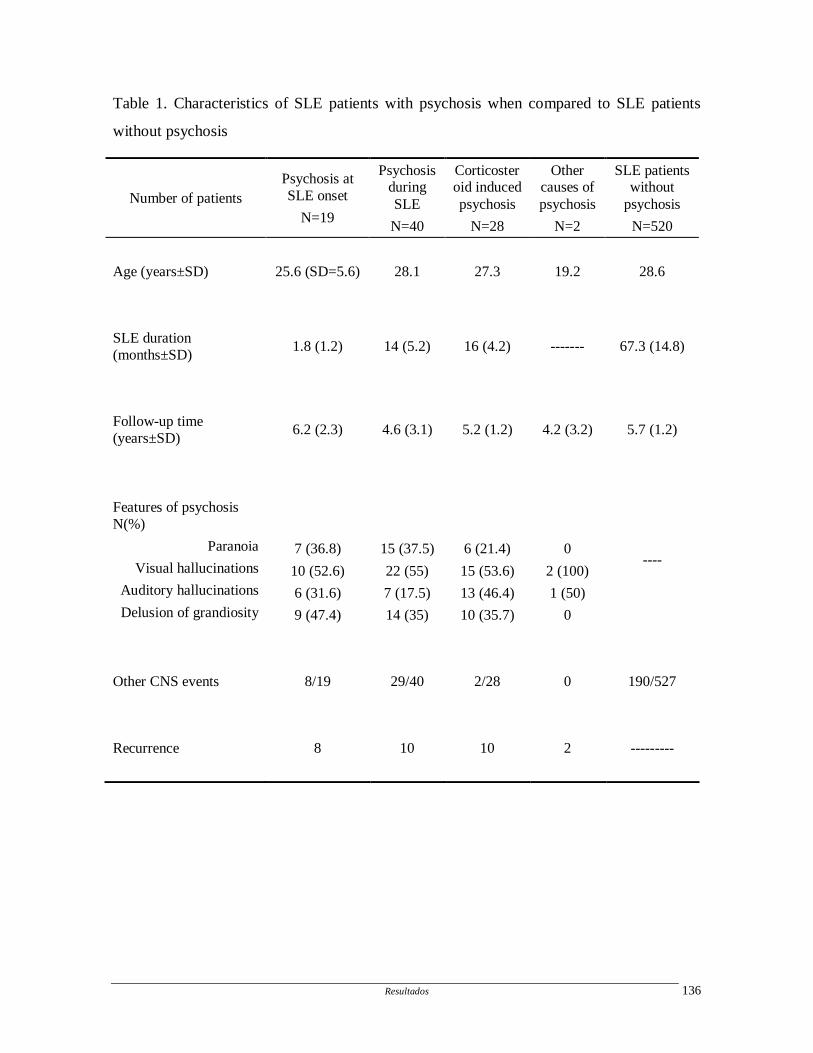
Resultados 136
Table 1. Characteristics of SLE patients with psychosis when compared to SLE patients
without psychosis
Number of patients Psychosis at SLE onset
N=19
Psychosis during SLE
N=40
Corticosteroid induced psychosis
N=28
Other causes of psychosis
N=2
SLE patients without
psychosis
N=520
Age (years±SD) 25.6 (SD=5.6) 28.1 27.3 19.2 28.6
SLE duration (months±SD)
1.8 (1.2) 14 (5.2) 16 (4.2) ------- 67.3 (14.8)
Follow-up time (years±SD)
6.2 (2.3) 4.6 (3.1) 5.2 (1.2) 4.2 (3.2) 5.7 (1.2)
Features of psychosis N(%)
Paranoia
Visual hallucinations
Auditory hallucinations
Delusion of grandiosity
7 (36.8)
10 (52.6)
6 (31.6)
9 (47.4)
15 (37.5)
22 (55)
7 (17.5)
14 (35)
6 (21.4)
15 (53.6)
13 (46.4)
10 (35.7)
0
2 (100)
1 (50)
0
----
Other CNS events 8/19 29/40 2/28 0 190/527
Recurrence 8 10 10 2 ---------
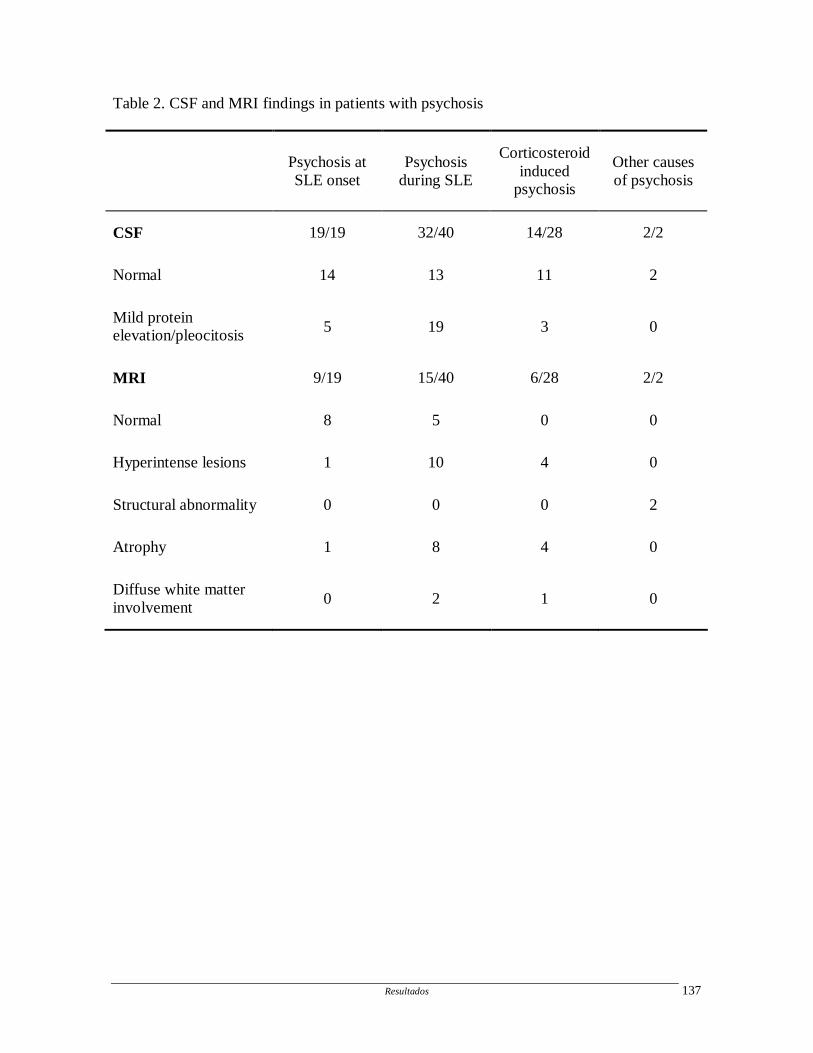
Resultados 137
Table 2. CSF and MRI findings in patients with psychosis
Psychosis at SLE onset
Psychosis during SLE
Corticosteroid induced
psychosis
Other causes of psychosis
CSF 19/19 32/40 14/28 2/2
Normal 14 13 11 2
Mild protein elevation/pleocitosis 5 19 3 0
MRI 9/19 15/40 6/28 2/2
Normal 8 5 0 0
Hyperintense lesions 1 10 4 0
Structural abnormality 0 0 0 2
Atrophy 1 8 4 0
Diffuse white matter involvement
0 2 1 0
Resultados 138
ARTIGO 7
Cerebral venous thrombosis: influence of r isk factors and imaging findings on
prognosis
Appenzeller S, Zeller CB, Annichino-Bizzachi JM, Costallat LT, Deus-Silva L, Voetsch B,
Faria AV, Zanardi VA, Damasceno BP, Cendes F
Cerebral venous thrombosis: influence of risk factors and imaging findings on prognosis
Clin Neurol Neurosurg. 2005 Aug; 107(5):371-8
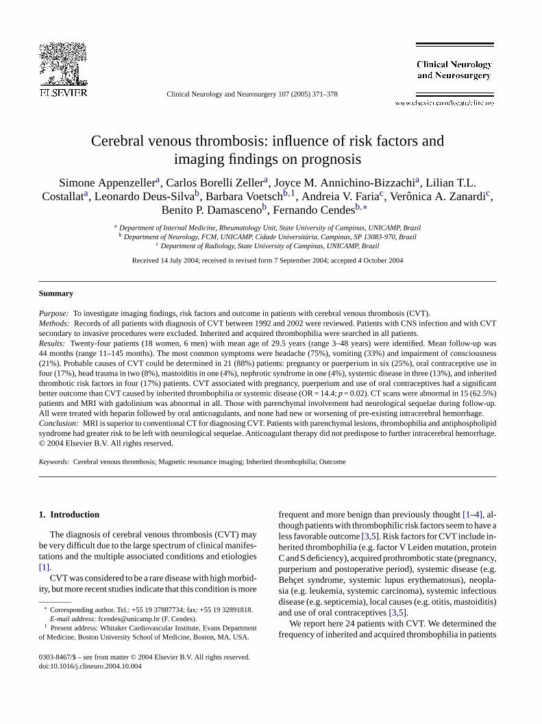
Clinical Neurology and Neurosurgery 107 (2005) 371–378
Cerebral venous thrombosis: influence of risk factors andimaging findings on prognosis
Simone Appenzellera, Carlos Borelli Zellera, Joyce M. Annichino-Bizzachia, Lilian T.L.Costallata, Leonardo Deus-Silvab, Barbara Voetschb,1, Andreia V. Fariac, Veronica A. Zanardic,
Benito P. Damascenob, Fernando Cendesb,∗a Department of Internal Medicine, Rheumatology Unit, State University of Campinas, UNICAMP, Brazil
b Department of Neurology, FCM, UNICAMP, Cidade Universit´aria, Campinas, SP 13083-970, Brazilc Department of Radiology, State University of Campinas, UNICAMP, Brazil
Received 14 July 2004; received in revised form 7 September 2004; accepted 4 October 2004
Summary
Purpose: To investigate imaging findings, risk factors and outcome in patients with cerebral venous thrombosis (CVT).M ith CVTsR w-up was4 sciousness( tive use inf d inheritedt significantb %)p ollow-up.A rhage.C pholipids emorrhage.©
K
1
bt[
i
o
ve a-teinancy,(e.g.
opla-tiousiditis)
theents
0d
ethods:Records of all patients with diagnosis of CVT between 1992 and 2002 were reviewed. Patients with CNS infection and wecondary to invasive procedures were excluded. Inherited and acquired thrombophilia were searched in all patients.esults: Twenty-four patients (18 women, 6 men) with mean age of 29.5 years (range 3–48 years) were identified. Mean follo4 months (range 11–145 months). The most common symptoms were headache (75%), vomiting (33%) and impairment of con21%). Probable causes of CVT could be determined in 21 (88%) patients: pregnancy or puerperium in six (25%), oral contracepour (17%), head trauma in two (8%), mastoiditis in one (4%), nephrotic syndrome in one (4%), systemic disease in three (13%), anhrombotic risk factors in four (17%) patients. CVT associated with pregnancy, puerperium and use of oral contraceptives had aetter outcome than CVT caused by inherited thrombophilia or systemic disease (OR = 14.4;p= 0.02). CT scans were abnormal in 15 (62.5atients and MRI with gadolinium was abnormal in all. Those with parenchymal involvement had neurological sequelae during fll were treated with heparin followed by oral anticoagulants, and none had new or worsening of pre-existing intracerebral hemoronclusion:MRI is superior to conventional CT for diagnosing CVT. Patients with parenchymal lesions, thrombophilia and antiphosyndrome had greater risk to be left with neurological sequelae. Anticoagulant therapy did not predispose to further intracerebral h2004 Elsevier B.V. All rights reserved.
eywords:Cerebral venous thrombosis; Magnetic resonance imaging; Inherited thrombophilia; Outcome
. Introduction
The diagnosis of cerebral venous thrombosis (CVT) maye very difficult due to the large spectrum of clinical manifes-
ations and the multiple associated conditions and etiologies1].
CVT was considered to be a rare disease with high morbid-ty, but more recent studies indicate that this condition is more
∗ Corresponding author. Tel.: +55 19 37887734; fax: +55 19 32891818.E-mail address:[email protected] (F. Cendes).
1 Present address: Whitaker Cardiovascular Institute, Evans Departmentf Medicine, Boston University School of Medicine, Boston, MA, USA.
frequent and more benign than previously thought[1–4], al-though patients with thrombophilic risk factors seem to haless favorable outcome[3,5]. Risk factors for CVT include inherited thrombophilia (e.g. factor V Leiden mutation, proC and S deficiency), acquired prothrombotic state (pregnpurperium and postoperative period), systemic diseaseBehcet syndrome, systemic lupus erythematosus), nesia (e.g. leukemia, systemic carcinoma), systemic infecdisease (e.g. septicemia), local causes (e.g. otitis, mastoand use of oral contraceptives[3,5].
We report here 24 patients with CVT. We determinedfrequency of inherited and acquired thrombophilia in pati
303-8467/$ – see front matter © 2004 Elsevier B.V. All rights reserved.oi:10.1016/j.clineuro.2004.10.004
372 S. Appenzeller et al. / Clinical Neurology and Neurosurgery 107 (2005) 371–378
with CVT and their influence on clinical outcome. In addi-tion, we searched whether other clinical, laboratory and neu-roimaging features influenced outcome in these patients.
2. Methods
2.1. Subjects
The records of 24 patients with a diagnosis of CVT fol-lowed in the Neurology and in the Hematology unit between1992 and 2002 were revised. We included patients with aclinical hypothesis of CVT (headache, focal neurologicaldeficits, and cranial hypertension) supported by appropri-ate neuroimaging studies including “delta sign” on cranialcomputed tomography (CT) or magnetic resonance imag-ing (MRI), a partial or complete absence of filling of onedural sinus on two projections using gadolinium enhancedT1-wheighted MRI. Magnetic resonance venography (MRV)was used as additional evidence whenever possible.
Patients with CNS infections, patients who did not com-plete laboratory investigations and patients with CVT sec-ondary to invasive procedures (central venous catheter, neu-rosurgery proceedings) were excluded from this study.
All patients had a complete neurological examination ata
2
in-c andu medi tem( s toe temicd
d onK om( t forLo me-l angew ntsm tran-s
thes ttinga ora enics truc-t er-i thept gene(
2.3. Imaging investigation
The site and extent of the thrombus and the appearance ofassociated brain lesions were determined for all patients.
Enhanced CTs were obtained with a conventional scan-ner (Elscint HeliCat, Elscint, Haifa, Israel; or Somatom AR,Siemmens, Erlangen, Germany). The section thickness was2.5 mm for the posterior fossa and 5 mm for the supratentorialregion. Radiological techniques ranged from 200 to 275 mAsand 100–120 kVp, using a 512× 512 matrix.
MRIs were performed in a 2T scanner (Elscint Prestige®,Haifa, Israel), with T1 and T2 acquisitions in three or-thogonal planes, including T1-weighted spin echo (SE)gadolinium enhanced images. MRI acquisition parame-ters were: Sagital T1 SE, 6 mm thick, flip angle = 180◦;repetition time (TR) = 430, echo time (TE) = 12, ma-trix 200× 350, field of view (FOV) = 25 cm× 25 cm; T2-weighted and proton density “fast spin echo” (FSE),3 mm thick, flip angle = 160◦; TR = 4800, TE = 108/18, ma-trix 256× 256, FOV = 22 cm× 22 cm; coronal T1-weightedinversion recovery (IR), 3 mm thick, flip angle = 200◦;TR = 2800, TE = 14, inversion time (TI) = 840, matrix130× 256, FOV = 16 cm× 18 cm or T1-weighted SE; ax-ial T1-weighted SE and T2-weighted fluid-attenuated in-version recovery (FLAIR) images TR = 8500 and 2000 or100 and 2200, TE = 72 or 90, matrix of 256× 296 andF iume gonalp
mmtF
2
aret inter-v
3
3
di-a entst re-m picalm oneh gen-e
from3 ientsw Thef ean4 logy
dmission and during the follow-up period.
.2. Laboratory investigation
The laboratory investigation performed in all patientsluded: complete blood count, basic blood biochemistryrinalysis. Cerebrospinal fluid (CSF) study was perfor
n all patients in order to exclude central nervous sysCNS) infections. Antinuclear antibodies and antibodiextractable nuclear antigens were performed when sysisease was suspected.
The diagnosis of lupus anticoagulant (LA) was baseaolim clotting time (KCT) and diluted Russel viper ven
dVV) as screening methodologies. The confirmatory tesA was carried out by dVV confirmatory test[6]. Anticardi-lipin antibodies were determined in serum by an enzy
inked immunoasorbent assay (ELISA), and the normal ras considered <5 GPL U/ml or MPL U/ml. For all patieore than one sample was obtained in order to avoid a
ient LA diagnosis.The general screening for thrombophilia includes
earch for deficiency of protein C and protein S by a clossay (Stago®Protein C, Stago®Protein S Stago, France),ntithrombin by an amydolitic assay, using the chromogubstrate S-2338, according to the manufacturer’s insion (Kabivitrum, Stockholm, Sweden). Detection of inhted thrombophilia risk factors such as factor V Leiden,rothrombin gene variant (allele 20210 A) and the 677C→ T
ransition in the methylenetetrahydrofolate reductaseMTHFR) were carried out as previously described[7–9].
OV of 22 versus 22 cm. T1-weighted SE gadolinnhanced images were obtained in the three ortholanes.
MRVs were performed with 2D phase contrast, 1.2hick, flip angle = 28◦, TR = 38, TE = 6, matrix 170× 256,OV = 17 mm× 23 mm.
.4. Statistical analysis
Differences in proportions were tested with chi-squest, or Fisher’s exact test when required. Confidenceals and odd ratios were also obtained when indicated.
. Results
.1. Demographics
During the study period, 33 patients had an initialgnosis of CVT based on clinical features. In 24 pati
he diagnosis was confirmed with CT or MRI. Of theaining patients, seven had a final diagnosis of atyigraine, one had a central nervous system tumor andad nonconvulsive status epilepticus due to cortical dyssis.
There were 18 women and 6 men, with age rangingto 48 years (mean 29.5 years). Twenty (83%) pat
ere Caucasians and four (17%) were Afro-Brazilians.ollow-up period ranged between 11 and 145 months (m6 months). The patients were followed-up in the Neuro
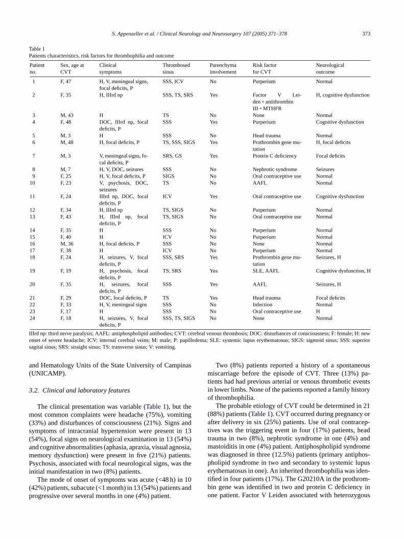
S. Appenzeller et al. / Clinical Neurology and Neurosurgery 107 (2005) 371–378 373
Table 1Patients characteristics, risk factors for thrombophilia and outcome
Patientno.
Sex, age atCVT
Clinicalsymptoms
Thrombosedsinus
Parenchymainvolvement
Risk factorfor CVT
Neurologicaloutcome
1 F, 47 H, V, meningeal signs,focal deficits, P
SSS, ICV No Purperium Normal
2 F, 35 H, IIIrd np SSS, TS, SRS Yes Factor V Lei-den + antithrombinIII + MTHFR
H, cognitive dysfunction
3 M, 43 H TS No None Normal4 F, 48 DOC, IIIrd np, focal
deficits, PSSS Yes Purperium Cognitive dysfunction
5 M, 3 H SSS No Head trauma Normal6 M, 48 H, focal deficits, P TS, SSS, SIGS Yes Prothrombin gene mu-
tationH, focal deficits
7 M, 3 V, meningeal signs, fo-cal deficits, P
SRS, GS Yes Protein C deficiency Focal deficits
8 M, 7 H, V, DOC, seizures SSS No Nephrotic syndrome Seizures9 F, 25 H, V, focal deficits, P SIGS No Oral contraceptive use Normal
10 F, 23 V, psychosis, DOC,seizures
TS No AAFL Normal
11 F, 24 IIIrd np, DOC, focaldeficits, P
ICV Yes Oral contraceptive use Cognitive dysfunction
12 F, 34 H, IIIrd np TS, SIGS No Purperium Normal13 F, 43 H, IIIrd np, focal
deficits, PTS, SIGS No Oral contraceptive use Normal
14 F, 35 H SSS No Purperium Normal15 F, 40 H ICV No Purperium Normal16 M, 36 H, focal deficits, P SSS No None Normal17 F, 38 H ICV No Purperium Normal18 F, 24 H, seizures, V, focal
deficits, PSSS, SRS Yes Prothrombin gene mu-
tationSeizures, H
19 F, 19 H, psychosis, focaldeficits, P
TS, SRS Yes SLE, AAFL Cognitive dysfunction, H
20 F, 35 H, seizures, focaldeficits, P
SSS Yes AAFL Seizures, H
21 F, 29 DOC, focal deficits, P TS Yes Head trauma Focal deficits22 F, 33 H, V, meningeal signs SSS No Infection Normal23 F, 17 H SSS No Oral contraceptive use H24 F, 18 H, seizures, V, focal
deficits, PSSS, TS, SIGS No None Normal
IIIrd np: third nerve paralysis; AAFL: antiphospholipid antibodies; CVT: cerebral venous thrombosis; DOC: disturbances of consciousness; F: female; H: newonset of severe headache; ICV: internal cerebral veins; M: male; P: papilledema; SLE: systemic lupus erythematosus; SIGS: sigmoid sinus; SSS: superiorsagital sinus; SRS: straight sinus; TS: transverse sinus; V: vomiting.
and Hematology Units of the State University of Campinas(UNICAMP).
3.2. Clinical and laboratory features
The clinical presentation was variable (Table 1), but themost common complaints were headache (75%), vomiting(33%) and disturbances of consciousness (21%). Signs andsymptoms of intracranial hypertension were present in 13(54%), focal signs on neurological examination in 13 (54%)and cognitive abnormalities (aphasia, apraxia, visual agnosia,memory dysfunction) were present in five (21%) patients.Psychosis, associated with focal neurological signs, was theinitial manifestation in two (8%) patients.
The mode of onset of symptoms was acute (<48 h) in 10(42%) patients, subacute (<1 month) in 13 (54%) patients andprogressive over several months in one (4%) patient.
Two (8%) patients reported a history of a spontaneousmiscarriage before the episode of CVT. Three (13%) pa-tients had had previous arterial or venous thrombotic eventsin lower limbs. None of the patients reported a family historyof thrombophilia.
The probable etiology of CVT could be determined in 21(88%) patients (Table 1). CVT occurred during pregnancy orafter delivery in six (25%) patients. Use of oral contracep-tives was the triggering event in four (17%) patients, headtrauma in two (8%), nephrotic syndrome in one (4%) andmastoiditis in one (4%) patient. Antiphospholipid syndromewas diagnosed in three (12.5%) patients (primary antiphos-pholipid syndrome in two and secondary to systemic lupuserythematosus in one). An inherited thrombophilia was iden-tified in four patients (17%). The G20210A in the prothrom-bin gene was identified in two and protein C deficiency inone patient. Factor V Leiden associated with heterozygous

374 S. Appenzeller et al. / Clinical Neurology and Neurosurgery 107 (2005) 371–378
Fig. 1. MRI of patient (#6) with CVT secondary to the G20210A mutation in the prothrombin gene showing axial T1-weighted image (A) and proton densityimage (B) showing intraparenchymal hemorrhage and perilesional edema in the convexity of the left frontal region secondary to CVT. MRV (C, D) showingasignal dropout of blood flow in the left transverse sinus (small arrow) (D) with hyperintensity (large arrow) corresponding to the area of hemorrhagicinfarct.There is also signal dropout in the superior sagital sinus.
antithrombin III deficiency and C677T homozygosity in theMTHFR gene was identified in one patient.
3.3. Neuroimaging features
Neuroimaging studies were available for review in all pa-tients. Cranial CT was performed in all patients, MRI in 17patients and MRV in seven patients.
CT images were abnormal in 15 (62.5%) patients.Parenchymal involvement was seen in 9 of 24 (37.5%)patients: Eight had hemorrhagic and one had ischemicparenchymal lesions. Signs of diffuse cerebral edema wereobserved in four (17%) patients. The sites of CVT are shownin Table 1.
MRI scans were performed in 17 patients, including allnine patients who had normal CT scans. All patients withnormal CT had abnormal MRI. MRI showed signs of si-nus thrombosis in all 17 patients. MRI showed parenchymallesions in nine patients (Table 1): in all four patients withthrombophilia (Fig. 1), in two of six patients with CVT dur-
ing purperium (Fig. 2), in one of two patients with a historyof head trauma, in one of four patients with CVT associatedwith the use of oral contraceptives and in one of three patientswith antiphospholipid syndrome (Fig. 3). Four patients withdiffuse cerebral edema on CT had this diagnosis confirmedby T2-weigheted MRI.
MRV showed a stop in the involved sinus in all seven pa-tients. Hyperintensity, corresponding to parenchymal hemor-rhagic infarct, was seen in one patient.
3.4. Treatment
All patients received heparin for 3–5 days followed by oralanticoagulants for 6 months, unless there was an underlyingdisease carrying a thrombotic risk, in which case anticoagula-tion was not interrupted. Hemorrhagic infarcts were identifiedprior to anticoagulation in nine (37.5%) patients. There wasno clinical worsening after introduction of heparin and nosigns of further intracranial hemorrhage on follow-up scan.Anticoagulation did not cause intracerebral bleeding in those
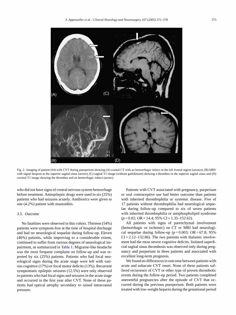
S. Appenzeller et al. / Clinical Neurology and Neurosurgery 107 (2005) 371–378 375
Fig. 2. Imaging of patient (#4) with CVT during puerperium showing (A) cranial CT with an hemorrhagic infarct in the left frontal region (arrows); (B) MRVwith signal dropout at the superior sagital sinus (arrow); (C) sagital T1 image (without gadolinium) showing a thrombus in the superior sagital sinusand (D)coronal T1 image showing the thrombus and an hemorrhagic infarct (arrow).
who did not have signs of central nervous system hemorrhagebefore treatment. Antiepileptic drugs were used in six (25%)patients who had seizures acutely. Antibiotics were given toone (4.2%) patient with mastoiditis.
3.5. Outcome
No fatalities were observed in this cohort. Thirteen (54%)patients were symptom-free at the time of hospital dischargeand had no neurological sequelae during follow-up. Eleven(46%) patients, while improving to a considerable extent,continued to suffer from various degrees of neurological im-pairment, as summarized inTable 1. Migraine-like headachewas the most frequent complaint on follow-up and was re-ported by six (25%) patients. Patients who had focal neu-rological signs during the acute stage were left with vari-ous cognitive (17%) or focal motor deficits (13%). Recurrentsymptomatic epileptic seizures (12.5%) were only observedin patients who had focal signs and seizures in the acute stageand occurred in the first year after CVT. None of these pa-tients had optical atrophy secondary to raised intracranialpressure.
Patients with CVT associated with pregnancy, purperiumor oral contraceptive use had better outcome than patientswith inherited thrombophilia or systemic disease. Five of17 patients without thrombophilia had neurological seque-lae during follow-up compared to six of seven patientswith inherited thrombophilia or antiphospholipid syndrome(p= 0.02; OR = 14.4; 95% CI = 1.35–152.62).
All patients with signs of parenchymal involvement(hemorrhagic or ischemic) on CT or MRI had neurologi-cal sequelae during follow-up (p= 0.003; OR = 67.8; 95%CI = 2.12–132.86). The two patients with thalamic involve-ment had the most severe cognitive deficits. Isolated superfi-cial sagital sinus thrombosis was observed only during preg-nancy and purperium in three patients and associated withexcellent long-term prognosis.
We found no differences in outcome between patients withacute and subacute CVT onset. None of these patients suf-fered recurrence of CVT or other type of proven thromboticevents during the follow-up period. Two patients completeduneventful pregnancies after the episode of CVT that oc-curred during the previous puerperium. Both patients weretreated with low-weight heparin during the gestational period
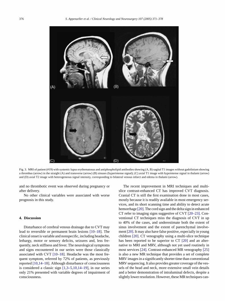
376 S. Appenzeller et al. / Clinical Neurology and Neurosurgery 107 (2005) 371–378
Fig. 3. MRI of patient (#19) with systemic lupus erythematosus and antiphospholipid antibodies showing (A, B) sagital T1 images without gadolinium showinga thrombus (arrow) in the straight (A) and transverse (arrow) (B) sinuses (hyperintense signal); (C) axial T1 image with hypointense signal in thalami (arrow)and (D) axial T2 image with heterogeneous signal intensity, corresponding to bilateral venous infarct and edema in thalami (arrow).
and no thrombotic event was observed during pregnancy orafter delivery.
No other clinical variables were associated with worseprognosis in this study.
4. Discussion
Disturbance of cerebral venous drainage due to CVT maylead to reversible or permanent brain lesions[10–18]. Theclinical onset is variable and nonspecific, including headache,lethargy, motor or sensory deficits, seizures and, less fre-quently, neck stiffness and fever. The neurological symptomsand signs encountered in our series were those classicallyassociated with CVT[10–18]. Headache was the most fre-quent symptom, referred by 72% of patients, as previouslyreported[10,14–18]. Although disturbance of consciousnessis considered a classic sign[1,3–5,10,14–19], in our seriesonly 21% presented with variable degrees of impairment ofconsciousness.
The recent improvement in MRI techniques and multi-slice contrast-enhanced CT has improved CVT diagnosis.Cranial CT is still the first examination done in most cases,mostly because it is readily available in most emergency ser-vices, and its short scanning time and ability to detect acutehemorrhage[20]. The cord sign and the delta sign in enhancedCT refer to imaging signs suggestive of CVT[20–23]. Con-ventional CT techniques miss the diagnosis of CVT in upto 40% of the cases, and underestimate both the extent ofsinus involvement and the extent of parenchymal involve-ment[20]. It may also have false positive, especially in youngchildren[20]. CT venography using a multi-slice techniquehas been reported to be superior to CT[20] and an alter-native to MRI and MRV, although not yet used routinely inmost services[24]. Contrast-enhanced MR venography[25]is also a new MR technique that provides a set of completeMRV images in a significantly shorter time than conventionalMRV sequencing. It also provides greater coverage of the ves-sels of the head and neck, more extensive small vein detailsand a better demonstration of intraluminal defects, despite aslightly lower resolution. However, these MR techniques can-
S. Appenzeller et al. / Clinical Neurology and Neurosurgery 107 (2005) 371–378 377
not difference from reversible and irreversible tissue changes.Diffusion-weighted MRI (DWI), on the other hand, may dis-criminate between areas at risk of undergoing infarction andthose, which are going to recover[3]. In our study, DWIwas used in only two patients, because most of the patientswere diagnosed before DWI technique was available in ourservice. In addition to new imaging techniques, newer labora-tory measurements also increased CVT diagnosis. Althoughnot performed in this study, d-dimer measurement is usefulfor acute CVT diagnosis. As previously reported[26], valuesbelow 500 mg/ml make acute CVT unlikely.
CVT is associated with coagulation defects or risk factorssuch as hormonal therapy, pregnancy or increased blood vis-cosity in about 75% of cases[10]. In our series, etiologieswere identified in 88% of the patients. Four (17%) of thesepatients were found to have inherited thrombophilia. Factor VLeiden is the most frequent encountered mutation in patientswith CVT. The frequency of this mutation varies accordingto ethnicity and has been described as being 2% in the overallBrazilian population[7]. In our study, only one (4%) patientwas homozygous for the factor V Leiden mutation. The pro-thrombin G20210A mutation is found at a rate of 0.7% inour population[8]. The finding of two patients with this mu-tation in this study suggests that it may be associated withan increased risk for CVT. Contrary to previous study[8] wefound no elevation of factor VIII plasma level associated withC ti-fi hane kelyr ourc iatedw VT.Tb ,C 0%o aret gc rredb thani ipids lipids .2%)o bei
ermp ibed[ eeno nceo 6%)w log-i nich reg-n ettero sys-t dis-e gical
sequelae compared with patients with CVT during pregnancyor purperium. On the other hand, patients with parenchymalinvolvement documented by CT or MRI had a 67.8-fold in-crease in risk of suffering neurological sequelae, which is inaccordance with previously reported findings[29–36]. Pa-tients with thalamic involvement had the worse prognosis,with important cognitive deficits. Isolated superficial sagi-tal sinus thrombosis was observed only during pregnancyand purperium in three patients and associated with excellentlong-term prognosis. We found no difference in outcome be-tween patients with acute and subacute CVT onset.
All patients received heparin for 3–5 days followed by oralanticoagulants[37–42]. No clinical worsening after introduc-tion of heparin or signs of further intracranial hemorrhage onfollow-up scans was observed in this series. In addition toheparin treatment, local thrombolisis has been reported insmall series[4]. The ability to lise the thrombus more rapidlythan heparin has made thrombolisis and alternative option inCVT treatment. However, local thrombolisis is not consid-ered first line treatment because of higher risk of cerebralhemorrhage and the absence of correlation between the reso-lution of thrombosis and clinical improvement. Until furtherevidence, local thrombolisis should be used when heparintreatment fails[4].
CVT is probably underdiagnosed and should be consid-ered in patients with new onset of severe headache, pro-g uresa thor-o st-e ful asa ncedC andn orm ven-t ctedc icalv pa-t iliaa ring( n ando rtheri
R
osedogy
nousGy-
a L,cyto-throm-
Neu-
VT in this study. Other risk factors for CVT could be idened in 20 patients. The frequency of infection was lower txpected, occurring in only 4% of the cases. This most lieflects a selection bias, since patients with infections inenter are referred to other clinics and symptoms associth infections may overwhelm the symptomatology of Chis prevalence is similar to that observed in one study[10],ut less than observed by other authors[17,18]. In contrastVT during gestation and purperium was identified in 2f our series. It is believed that infection and purperium
he main causes of CVT[19,27–29]especially in developinountries[28]. The use of oral contraceptives was refey four of 18 (22%) female patients, a frequency higher
n the overall Brazilian population. Primary antiphospholyndrome was diagnosed in two (8.3%) and antiphosphoyndrome associated with SLE was identified in one (4f our patients. No other thrombophilic risk factor could
dentified in these four patients.Although this was a retrospective study, the long-t
rognosis of CVT was favorable, as previously descr29–36]. No fatalities were observed in our series. Thirtf 24 (54%) patients with CVT recovered without evidef permanent neurological damage. Eleven patients (4ere left with sequelae: seizures in three, focal neuro
cal signs in three, cognitive deficits in four, and chroeadache in six. Patients with CVT associated with pancy, purperium and use of oral contraceptives had a butcome than patients with inherited thrombophilia or
emic disease. Patients with thrombophilia and systemicase had a 14.4-fold increase in risk of having neurolo
ressive unexplained cognitive deficit, unexplained seiznd somnolence. The etiology should be investigatedughly and MRI with gadolinium or multi-slice contranhanced CT are the exams of choice. MRV may be helpcomplementary exam when multi-slice contrast-enhaT is not available. Due to the high yield of diagnosison-invasiveness of MRI and MRV (“MR venography”)ulti-slice contrast-enhanced CT, we believe that con
ional cerebral angiography should be reserved for seleases, particularly in cases of CVT involving only the corteins[43,44]. The course of the disease is variable, butients with parenchymal involvement and with thrombophnd antiphospholipid syndrome have greater risk of suffeor sustaining) neurological sequelae. The use of hepariral anticoagulants in this series did not predispose to fu
ntracerebral hemorrhage.
eferences
[1] Raizer JJ, DeAngelis LM. Cerebral sinus thrombosis diagnby MRI and MR venography in cancer patients. Neurol2000;54:1222–6.
[2] Lanska DJ, Kryscio RJ. Peripartum stroke and intracranial vethrombosis in the National Hospital Discharge Survey. Obstetnecol 1997;89:413–8.
[3] Lovblad KO, Bassetti C, Schneider J, Ozdoba C, RemondSchroth G. Diffusion-weighted MRI suggests the coexistence oftoxic and vasogenic edema in a case of deep cerebral venousbosis. Neuroradiology 2000;42:728–31.
[4] Niclot P, Bousser MG. Cerebral venous thrombosis. Curr Treatrol 2000;2:343–52.
378 S. Appenzeller et al. / Clinical Neurology and Neurosurgery 107 (2005) 371–378
[5] Stolz E, Kemkes-Matthes B, Potzsch B, Hahn M, Kraus J, WirbartzA, et al. Screening for thrombophilic risk factors among 25 Ger-man patients with cerebral venous thrombosis. Acta Neurol Scand2000;102:31–6.
[6] Brandt JT, Triplett DA, Alving B, Scharrer I. Criteria for the di-agnosis of lupus anticoagulants: an update. On behalf of the Sub-committee on Lupus Anticoagulant/Antiphospholipid Antibody ofthe Scientific and Standardisation Committee of the ISTH. ThrombHaemost 1995;74:1185–90.
[7] Arruda VR, Annichino-Bizzacchi JM, Costa FF, Reitsma PH. FactorV (FVQ 506) is common in a Brazilian population. Am J Haematol1995;49:242–3.
[8] Arruda VR, Annichino-Bizzacchi JM, Gonc¸alves MS, Costa FC.Prevalence of the prothombin gene variant (nt20210A) in venousthrombosis and arterial disease. Thromb Haemost 1997;78:1430–3.
[9] Arruda VR, von Zuben PM, Chiaparini LC, Annichino-Bizzachi JM,Costa FC. The mutation Ala 677→ Val in the methylenetetrahydro-folate reductase gene: a risk factor for arterial disease and venousthrombosis. Thromb Haemost 1997;77:818–21.
[10] Daif M, Awada A, Al Rajeh S, Abduljabbar M, al Tahan AR,Obeid T, et al. Cerebral venous thrombosis in Saudi Arabia. Stroke1995;26:1193–5.
[11] Biousse V, Ameri A, Bousser MG. Isolated intracranial hyperten-sion as the only sign of cerebral venous thrombosis. Neurology1999;53:1537–42.
[12] Murray BJ, Llinas R, Caplan LR, Scammell T, Pascual-Leone A.Cerebral deep venous thrombosis presenting as acute micrographiaand hypophonia. Neurology 2000;54:751–3.
[13] Ameri A, Bousser MG. Cerebral venous thrombosis. Neurol Clin1992;10:87–111.
[14] Einhaupl KM, Masuhr F. Cerebral venous and sinus thrombosis: an
[ ost-
[ rom-
[ hrom-
[ iller.
[ bosis.
[ img
[ bral
[ ebral
[ iag-ance:
[ lixfor
[ R,aphy.
[ mil-iag-
nosis of cerebral venous thrombosis? Neurology 2003;61:1057–60.
[27] Cantu C, Barinagarrementeria F. Cerebral venous thrombosis asso-ciated with pregnancy and puerperium. Review of 67 cases. Stroke1993;24:1880–4.
[28] Srinavasan K. Ischemic cerebrovascular disease in young: two com-mon causes in India. Stroke 1984;15:733–5.
[29] de Bruijn SF, Budde M, Teunisse S, de Haan RJ, Stam J. Long-termoutcome of cognition and functional health after cerebral venoussinus thrombosis. Neurology 2000;54:1687–9.
[30] Lanska DJ, Kryscio RJ. Risk factors for peripartum and postpar-tum stroke and intracranial venous thrombosis. Stroke 2000;31:1274–82.
[31] Breteau G, Mounier-Vehier F, Godefroy O, Gauvrit JY, Mackowiak-Cordoliani MA, Girot M, et al. Cerebral venous thrombosis3-year clinical outcome in 55 consecutive patients. J Neurol2003;250:29–35.
[32] Strupp M, Covi M, Seelos K, Dichgans M, Brandt T. Cerebralvenous thrombosis: correlation between recanalization and clin-ical outcome—a long-term follow-up of 40 patients. J Neurol2002;249:1123–4.
[33] Preter M, Tzourio C, Ameri A, Bousser MG. Long-term progno-sis in cerebral venous thrombosis. Follow-up of 77 patients. Stroke1996;27:243–6.
[34] de Bruijn SF, de Haan RJ, Stam J. Clinical features and prognosticfactors of cerebral venous sinus thrombosis in a prospective seriesof 59 patients. J Neurol Neurosurg Psychiatry 2001;70:105–8, forThe Cerebral Venous Sinus Thrombosis Study Group.
[35] Cakmak S, Derex L, Berruyer M, Nighoghossian N, PhilippeauF, Adeleine P, et al. Cerebral venous thrombosis: clinical out-come and systematic screening of prothrombotic factors. Neurology
[ F. IS-rom-ural
[ anti-bral
[ lberenousases.
[ F.tients
[ Curr
[ Curr
[ ve-rology
[ ault;21:
[ O,ous
update. Eur J Neurol 1994;1:109–26.15] Allroggen H, Abbott RJ. Cerebral venous sinus thrombosis. P
graduate Med J 2000;76:12–5.16] Lafitte F, Boukozobzda M, Guichard JP. Deep cerebral venous th
bosis: imaging in eight cases. Neuroradiology 1999;41:410–8.17] Bousser MG, Chiras J, Sauron B, Castaign P. Cerebral venous t
bosis: a review of 38 cases. Stroke 1985;16:199–213.18] Shell CL, Rathe RJ. Superior sagital sinus thrombosis. Still a k
West J Med 1988;149:304–7.19] Lanska DJ, Kryscio RJ. Stroke and intracranial venous throm
during pregnancy and puerperium. Neurology 1998;51:1622–820] Shroff M, de Veber G. Sinovenous thrombosis in children. Neuro
Clin N Am 2003;13:115–38.21] Buonanno FS, Mood DM, Ball MR. CT scan findings in cere
sinovenous occlusion. Neurology 1979;29:1433–4.22] Chiras J, Bousser MG, Meder JF, Koussa A, Bories J. CT in cer
thromboflebitis. Neuroradiology 1985;27:145–54.23] Bianchi D, Maeder P, Bogousslavsky J, Schnyder P, Meuli RA. D
nosis of cerebral venous thrombosis with routine magnetic resonan update. Eur Neurol 1998;40:179–90.
24] Vogl TJ, Bergman C, Villringer A, Einhaupl K, Lissner J, FeR. Dural sinus thrombosis: value of venous MR angiographydiagnosis and follow-up. Am J Roentgenol 1994;162:1191–8.
25] Lovblad KO, Schneider J, Bassetti C, El-Koussy M, GuzmanHeid O, et al. Fast contrast-enhanced MR whole-brain venogrNeuroradiology 2002;44:681–8.
26] Lalive PH, de Moerloose P, Loveblad K, Sarasin FP, Merlod B, Sztajzel R. Is measurement of d-dimer useful in the
2003;60:1175–8.36] Ferro JM, Canhao P, Stam J, Bousser MG, Barinagarrementeria
CVT Investigators. Prognosis of cerebral vein and dural sinus thbosis: results of the International Study on Cerebral Vein and DSinus Thrombosis (ISCVT). Stroke 2004;35:664–70.
37] de Bruijn SF, Stam J. Randomized, placebo-controlled trial ofcoagulant treatment with low-molecular-weight heparin for ceresinus thrombosis. Stroke 1999;30:484–8.
38] Brucker AB, Vollert-Rogenhofer H, Wagner M, Stieglbauer K, FeS, Trenkler J, et al. Heparin treatment in acute cerebral sinus vthrombosis: a retrospective clinical and MR analysis of 42 cCerebrovasc Dis 1998;8:331–7.
39] Mehraein S, Schmidtke K, Villringer A, Valdueza JM, MasuhrHeparin treatment in cerebral sinus and venous thrombosis: paat risk of fatal outcome. Cerebrovasc Dis 2003;15:17–21.
40] Biousse V, Tong F, Newman NJ. Cerebral venous thrombosis.Treat Options Neurol 2003;5:409–20.
41] Biousse V, Tong F, Newman NJ. Cerebral venous thrombosis.Treat Options Cardiovasc Med 2003;5:181–92.
42] Fink JN, McAuley DL. Safety of anticoagulation for cerebralnous thrombosis associated with intracerebral hematoma. Neu2001;57:1138–9.
43] Dormont D, Anxionnat R, Evrard S, Louaille C, Chiras J, MarsC. MRI in cerebral venous thrombosis. J Neuroradiol 199481–99.
44] Lafitte F, Boukobza M, Guichard JP, Hoeffel C, Reizine D, Illeet al. MRI and MRA for diagnosis and follow-up of cerebral venthrombosis (CVT). Clin Radiol 1997;52:672–9.
Resultados 147
ARTIGO 8
Cerebral and corpus callosum atrophy in systemic lupus erythematosus
Appenzeller S, Rondina JM, Li LM, Costallat LT, Cendes F
Cerebral and corpus callosum atrophy in systemic lupus erythematosus
Arthritis Rheum. 2005 Sep; 52(9):2783-9.
ARTHRITIS & RHEUMATISMVol. 52, No. 9, September 2005, pp 2783–2789DOI 10.1002/art.21271© 2005, American College of Rheumatology
Cerebral and Corpus Callosum Atrophy inSystemic Lupus Erythematosus
Simone Appenzeller, Jane Maryam Rondina, Li Min Li, Lilian T. L. Costallat,and Fernando Cendes
Objective. To determine cerebral and corpus cal-losum volumes in patients with systemic lupus erythem-atosus (SLE), using semiautomatic magnetic resonanceimaging (MRI) volumetric measurements, and to deter-mine possible relationships between a reduction incerebral volume and disease duration, total cortico-steroid dose, neuropsychiatric manifestations, and thepresence of antiphospholipid antibodies.
Methods. We studied 115 consecutive patientswith SLE and 44 healthy volunteers. A complete clinical,laboratory, and neurologic evaluation was performed.MRI scans were obtained through a standardized pro-tocol. Sagittal T1-weighted images were used for semi-automatic volumetric measurements. We compared SLEpatients with controls using the 2-sample t-test. Analysisof variance was used to test for differences betweengroups, followed by Tukey’s post hoc test for pairwisecomparisons, when necessary. Linear regression wasused to analyze the association between cerebral atro-phy and disease duration and total corticosteroid dose.
Results. Cerebral and corpus callosum volumeswere significantly smaller in patients with SLE com-pared with healthy volunteers (P < 0.001). Reducedcerebral and corpus callosum volumes were related todisease duration (P < 0.001). Patients with a history ofcentral nervous system (CNS) involvement more fre-quently had a reduction in cerebral and corpus callo-sum volumes (P < 0.001). Patients with cognitive im-pairment had significantly reduced corpus callosum
and cerebral volumes when compared with SLE patientswithout cognitive impairment (P � 0.001). Cerebral andcorpus callosum volumes were not associated with thetotal corticosteroid dose or the presence of antiphospho-lipid antibodies.
Conclusion. In patients with SLE, a reduction incerebral and corpus callosum volumes is associated withdisease duration, a history of CNS involvement, andcognitive impairment. The total corticosteroid dose andthe presence of antiphospholipid antibodies were notassociated with more pronounced atrophy.
The central nervous system (CNS) is frequentlyaffected in patients with systemic lupus erythematosus(SLE) (1–3). Neuropsychiatric symptoms vary fromovert neurologic and psychiatric disorders to more sub-tle signs and symptoms such as headache, mood disor-ders, and impairment of cognitive function (1–5). Al-though clinical assessment is still the cornerstone of thediagnosis of neuropsychiatric SLE, the diagnosis is oftendifficult and remains presumptive in some patients (1–5). Magnetic resonance imaging (MRI) is known to bemore sensitive than computed tomography (CT) for thedetection of structural brain abnormalities in patientswith neuropsychiatric SLE, because of the excellentsoft-tissue contrast observed with MRI and the ability toacquire multiplanar images (6). In patients with SLE,the frequency of cerebral atrophy has been reported tobe variable (7–12). Aging, systemic diseases, cortico-steroid treatment, and CNS involvement may lead tocerebral atrophy. Various methods for evaluating cere-bral atrophy in the setting of SLE have been described.CT and MRI are the most frequently used methods, butmost studies (6,8,13–20) have used linear measurements.Although these procedures have been shown to beuseful, new methods, such as semiautomatic quantifica-tion, have demonstrated superiority in detecting brainabnormalities in several diseases (21–23).
Supported by Fundacao de Amparo a Pesquisa do Estado deSao Paulo.
Simone Appenzeller, MD, Jane Maryam Rondina, Li Min Li,MD, PhD, Lilian T. L. Costallat, MD, PhD, Fernando Cendes, MD,PhD: University of Campinas, Sao Paulo, Brazil.
Address correspondence and reprint requests to FernandoCendes, MD, PhD, Department of Neurology, University ofCampinas-UNICAMP, Cidade Universitaria, Campinas SP, Sao PauloCEP 13083-970, Brazil. E-mail: [email protected].
Submitted for publication December 9, 2004; accepted inrevised form June 13, 2005.
2783
The aim of this study was to analyze cerebralvolume in patients with SLE, using validated semiauto-mated MRI segmentation. We also analyzed corpuscallosum volume in order to determine neuronal loss. Inaddition, we investigated the relationships between ce-rebral and corpus callosum volumes and disease dura-tion, corticosteroid treatment, CNS involvement, andthe presence of antiphospholipid antibodies.
PATIENTS AND METHODS
Patients. A total of 150 consecutive patients fulfilling�4 of the American College of Rheumatology (ACR) criteriafor a diagnosis of SLE (24), who were seen regularly at ourrheumatology unit, were screened prospectively for participa-tion in this study. All SLE patients were followed up by thesame investigators (LTLC and SA), using a standardizedprotocol. We excluded patients who were unable to undergoMRI, such as those with claustrophobia (8 patients) or apacemaker (2 patients), as well as patients with previousclinical conditions that could influence cerebral atrophy, suchas a history of stroke (10 patients), arterial hypertension (5patients), diabetes mellitus (5 patients), alcohol and drugabuse (1 patient), and malignancy (1 patient). Patients whofulfilled the ACR criteria for rheumatoid arthritis, systemicsclerosis, Sjogren’s syndrome (primary or secondary) (3 pa-tients), or other connective tissue disease and those withdrug-induced SLE were also excluded. No patient had renalinsufficiency or other pathologic conditions that could influ-ence cerebral atrophy. The remaining 115 patients (109 ofwhom were women) were included in this study.
To analyze neuropsychiatric involvement, we used theclassification system for neuropsychiatric lupus proposed bythe ACR (25). The patients’ medical records were reviewed inorder to determine past CNS events. Patients with CNSmanifestations secondary to clinical conditions such as infec-tion, arterial hypertension, uremia, diabetes, and drugs wereexcluded. The control group consisted of 44 healthy volun-teers. This study was approved by the ethics committee at ourinstitution, and informed written consent was obtained fromeach participant.
Clinical, serologic, and treatment features of patientswith SLE. Data on sex, age at disease onset, and diseaseduration were collected for each patient. Disease duration wasdefined as the time from the appearance of the initial mani-festation clearly attributable to SLE until the day of MRIacquisition. All clinical manifestations and laboratory testresults were recorded. The following clinical manifestationswere analyzed: malar rash, discoid lesions, subacute cutaneouslesions, photosensitivity, oral ulcers, arthritis, serositis, nephri-tis, neurologic and psychiatric involvement, thrombocytopenia,hemolytic anemia, Raynaud’s phenomenon, thrombosis, myo-sitis, lung involvement, and lymphadenopathy.
Nephritis was diagnosed on the basis of proteinuria(�0.5 gm/liter) with abnormal urinary sediment and/or histo-logic findings. Nephrotic syndrome was defined as proteinuriain excess of 3.5 gm/day. Hematologic alterations were ascribedto lupus only in the absence of bone marrow suppression(leukopenia �4,000 cells/mm3; thrombocytopenia �100,000/
mm3; hemolytic anemia with positive Coombs’ test). Antinu-clear antibodies (ANAs) were determined by indirect immu-nofluorescence using HEp-2 as the substrate, and a titer �1:40was considered positive. Anti–double-stranded DNA antibod-ies were determined by indirect immunofluorescence usingCrithidia as substrate, and a titer �1:10 was considered posi-tive. Precipitating antibodies to extractable nuclear antigen,including Ro/SSA, La/SSB, and Sm, were detected by immu-nodiffusion and/or microhemagglutination. Anticardiolipinantibodies of the IgG and IgM isotypes were measured byenzyme-linked immunosorbent assay, as previously described(26). Lupus anticoagulant activity was detected by coagulationassays in platelet-free plasma obtained by double centrifuga-tion, following the recommendation of the Subcommittee onLupus Anticoagulant of the Scientific and StandardizationCommittee of the International Society of Thrombosis andHomeostasis (27).
CNS manifestations were classified according to ACRcase definitions for neuropsychiatric lupus (25) and wererecorded as being present (the patient had active CNS involve-ment or a history of CNS involvement) or absent (the patientnever presented with CNS involvement). A complete neuro-logic examination (including cognitive and psychiatric charts)was prospectively applied to all patients in order to identifyCNS involvement. The mini-mental state examination (28) wasapplied to all participants.
All patients and controls underwent a battery of stan-dardized neuropsychological tests in order to screen for pos-sible impairments in 1 or more of the following cognitivedomains: simple attention, complex attention, memory, visual–spatial processing, language, reasoning/problem-solving, psy-chomotor speed, and executive functions (29–32). The individ-ual test results were converted into standard scores, whichwere compared with the available normative data (29–32).Regarding any of the 8 cognitive domains, subjects with a totalscore �2 SD lower than the normative value were consideredto be impaired. Cognitive dysfunction was classified as mild ifthe patient had deficits in fewer than 3 dimensions, as moder-ate if there were deficits in 3 or 4 dimensions, and as severe ifthe patient had deficits in at least 5 dimensions (32,33).
The assessment of depression was based on a clinicalinterview and the Beck Depression Inventory (BDI) (34,35).On the BDI, scores of 10–17 are indicative of mild depression,scores of 18–24 indicate moderate depression, and scores �24indicate the presence of severe depression. Anxiety was eval-uated using the Hospital Anxiety and Depression scale (36).The presence of psychosis was determined using the BriefPsychiatric Rating Scale (37).
A history of CNS involvement was determined byreviewing the medical charts of patients. Disease activity wasmeasured by the Systemic Lupus Erythematosus Disease Ac-tivity Index (SLEDAI), and a score of �8 represented activedisease (38). Cumulative SLE-related damage in all patientswas determined using the Systemic Lupus International Col-laborating Clinics (SLICC)/ACR Damage Index (SDI) (39) atthe time of MRI.
The total doses of corticosteroids and other immuno-suppressant medications used since the onset of disease werecalculated by careful review of the medical charts. Doses oforal and parenteral corticosteroids were analyzed and con-verted to the equivalent doses of prednisone. The cumulative
2784 APPENZELLER ET AL
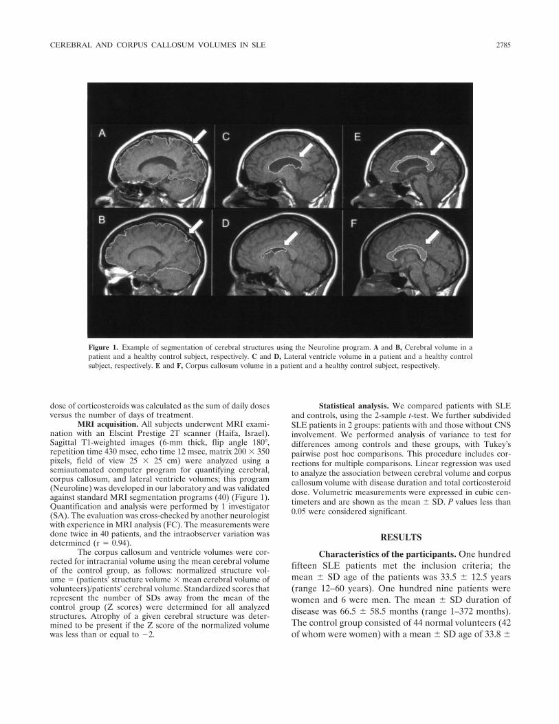
dose of corticosteroids was calculated as the sum of daily dosesversus the number of days of treatment.
MRI acquisition. All subjects underwent MRI exami-nation with an Elscint Prestige 2T scanner (Haifa, Israel).Sagittal T1-weighted images (6-mm thick, flip angle 180°,repetition time 430 msec, echo time 12 msec, matrix 200 � 350pixels, field of view 25 � 25 cm) were analyzed using asemiautomated computer program for quantifying cerebral,corpus callosum, and lateral ventricle volumes; this program(Neuroline) was developed in our laboratory and was validatedagainst standard MRI segmentation programs (40) (Figure 1).Quantification and analysis were performed by 1 investigator(SA). The evaluation was cross-checked by another neurologistwith experience in MRI analysis (FC). The measurements weredone twice in 40 patients, and the intraobserver variation wasdetermined (r � 0.94).
The corpus callosum and ventricle volumes were cor-rected for intracranial volume using the mean cerebral volumeof the control group, as follows: normalized structure vol-ume � (patients’ structure volume � mean cerebral volume ofvolunteers)/patients’ cerebral volume. Standardized scores thatrepresent the number of SDs away from the mean of thecontrol group (Z scores) were determined for all analyzedstructures. Atrophy of a given cerebral structure was deter-mined to be present if the Z score of the normalized volumewas less than or equal to �2.
Statistical analysis. We compared patients with SLEand controls, using the 2-sample t-test. We further subdividedSLE patients in 2 groups: patients with and those without CNSinvolvement. We performed analysis of variance to test fordifferences among controls and these groups, with Tukey’spairwise post hoc comparisons. This procedure includes cor-rections for multiple comparisons. Linear regression was usedto analyze the association between cerebral volume and corpuscallosum volume with disease duration and total corticosteroiddose. Volumetric measurements were expressed in cubic cen-timeters and are shown as the mean � SD. P values less than0.05 were considered significant.
RESULTS
Characteristics of the participants. One hundredfifteen SLE patients met the inclusion criteria; themean � SD age of the patients was 33.5 � 12.5 years(range 12–60 years). One hundred nine patients werewomen and 6 were men. The mean � SD duration ofdisease was 66.5 � 58.5 months (range 1–372 months).The control group consisted of 44 normal volunteers (42of whom were women) with a mean � SD age of 33.8 �
Figure 1. Example of segmentation of cerebral structures using the Neuroline program. A and B, Cerebral volume in apatient and a healthy control subject, respectively. C and D, Lateral ventricle volume in a patient and a healthy controlsubject, respectively. E and F, Corpus callosum volume in a patient and a healthy control subject, respectively.
CEREBRAL AND CORPUS CALLOSUM VOLUMES IN SLE 2785
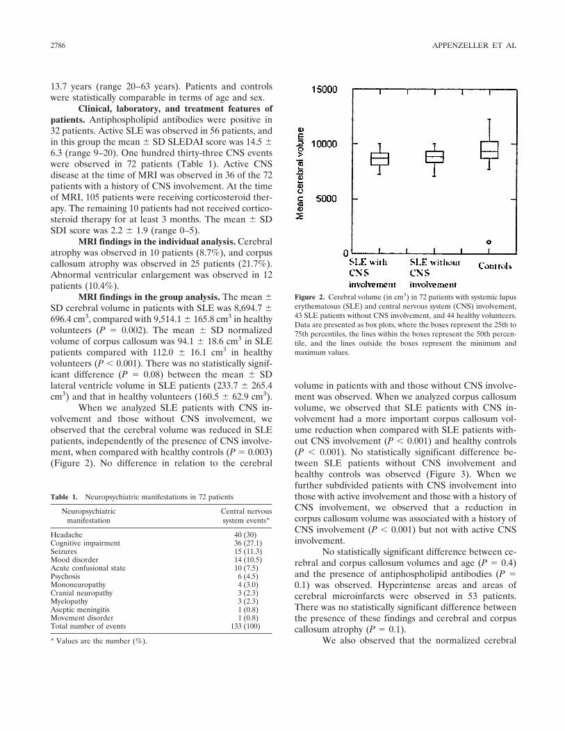
13.7 years (range 20–63 years). Patients and controlswere statistically comparable in terms of age and sex.
Clinical, laboratory, and treatment features ofpatients. Antiphospholipid antibodies were positive in32 patients. Active SLE was observed in 56 patients, andin this group the mean � SD SLEDAI score was 14.5 �6.3 (range 9–20). One hundred thirty-three CNS eventswere observed in 72 patients (Table 1). Active CNSdisease at the time of MRI was observed in 36 of the 72patients with a history of CNS involvement. At the timeof MRI, 105 patients were receiving corticosteroid ther-apy. The remaining 10 patients had not received cortico-steroid therapy for at least 3 months. The mean � SDSDI score was 2.2 � 1.9 (range 0–5).
MRI findings in the individual analysis. Cerebralatrophy was observed in 10 patients (8.7%), and corpuscallosum atrophy was observed in 25 patients (21.7%).Abnormal ventricular enlargement was observed in 12patients (10.4%).
MRI findings in the group analysis. The mean �SD cerebral volume in patients with SLE was 8,694.7 �696.4 cm3, compared with 9,514.1 � 165.8 cm3 in healthyvolunteers (P � 0.002). The mean � SD normalizedvolume of corpus callosum was 94.1 � 18.6 cm3 in SLEpatients compared with 112.0 � 16.1 cm3 in healthyvolunteers (P � 0.001). There was no statistically signif-icant difference (P � 0.08) between the mean � SDlateral ventricle volume in SLE patients (233.7 � 265.4cm3) and that in healthy volunteers (160.5 � 62.9 cm3).
When we analyzed SLE patients with CNS in-volvement and those without CNS involvement, weobserved that the cerebral volume was reduced in SLEpatients, independently of the presence of CNS involve-ment, when compared with healthy controls (P � 0.003)(Figure 2). No difference in relation to the cerebral
volume in patients with and those without CNS involve-ment was observed. When we analyzed corpus callosumvolume, we observed that SLE patients with CNS in-volvement had a more important corpus callosum vol-ume reduction when compared with SLE patients with-out CNS involvement (P � 0.001) and healthy controls(P � 0.001). No statistically significant difference be-tween SLE patients without CNS involvement andhealthy controls was observed (Figure 3). When wefurther subdivided patients with CNS involvement intothose with active involvement and those with a history ofCNS involvement, we observed that a reduction incorpus callosum volume was associated with a history ofCNS involvement (P � 0.001) but not with active CNSinvolvement.
No statistically significant difference between ce-rebral and corpus callosum volumes and age (P � 0.4)and the presence of antiphospholipid antibodies (P �0.1) was observed. Hyperintense areas and areas ofcerebral microinfarcts were observed in 53 patients.There was no statistically significant difference betweenthe presence of these findings and cerebral and corpuscallosum atrophy (P � 0.1).
We also observed that the normalized cerebral
Table 1. Neuropsychiatric manifestations in 72 patients
Neuropsychiatricmanifestation
Central nervoussystem events*
Headache 40 (30)Cognitive impairment 36 (27.1)Seizures 15 (11.3)Mood disorder 14 (10.5)Acute confusional state 10 (7.5)Psychosis 6 (4.5)Mononeuropathy 4 (3.0)Cranial neuropathy 3 (2.3)Myelopathy 3 (2.3)Aseptic meningitis 1 (0.8)Movement disorder 1 (0.8)Total number of events 133 (100)
* Values are the number (%).
Figure 2. Cerebral volume (in cm3) in 72 patients with systemic lupuserythematosus (SLE) and central nervous system (CNS) involvement,43 SLE patients without CNS involvement, and 44 healthy volunteers.Data are presented as box plots, where the boxes represent the 25th to75th percentiles, the lines within the boxes represent the 50th percen-tile, and the lines outside the boxes represent the minimum andmaximum values.
2786 APPENZELLER ET AL
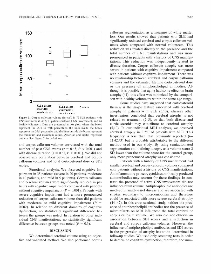
and corpus callosum volumes correlated with the totalnumber of past CNS events (r � 0.45, P � 0.001) andwith disease duration (r � 0.81, P � 0.001). We did notobserve any correlation between cerebral and corpuscallosum volumes and total corticosteroid dose or SDIscores.
Functional analysis. We observed cognitive im-pairment in 35 patients (severe in 20 patients, moderatein 10 patients, and mild in 5 patients). Corpus callosumand cerebral volumes were significantly reduced in pa-tients with cognitive impairment compared with patientswithout cognitive impairment (P � 0.001). Patients withsevere cognitive impairment had a more pronouncedreduction of corpus callosum volume than did patientswith moderate or mild cognitive impairment (P �0.002). In relation to different domains of cognitivedysfunction, no statistically significant difference be-tween the groups was noted. In relation to other indi-vidual CNS manifestations, no statistically significantdifference between groups was noted (P � 0.3).
DISCUSSION
We determined cerebral volume using an objec-tive and validated method. We also performed corpus
callosum segmentation as a measure of white matterloss. Our results showed that patients with SLE hadsignificantly reduced cerebral and corpus callosum vol-umes when compared with normal volunteers. Thisreduction was related directly to the presence and thetotal number of CNS manifestations and was morepronounced in patients with a history of CNS manifes-tations. This reduction was independently related todisease duration. Corpus callosum atrophy was moresevere in patients with cognitive impairment comparedwith patients without cognitive impairment. There wasno relationship between cerebral and corpus callosumvolumes and the estimated lifetime corticosteroid doseor the presence of antiphospholipid antibodies. Al-though it is possible that aging had some effect on brainatrophy (41), this effect was minimized by the compari-son with healthy volunteers within the same age range.
Some studies have suggested that corticosteroidtherapy is the major feature associated with cerebralatrophy in patients with SLE (6,10), whereas otherinvestigators concluded that cerebral atrophy is notrelated to treatment (2–5), or that both disease andcorticosteroids may contribute to cerebral atrophy(5,10). In our individual MRI analyses, we observedcerebral atrophy in 8.7% of patients with SLE. Thisfrequency is less than that previously reported (6–11,42,43) but is probably attributable to the differentmethod used in our study. By using semiautomatedsegmentation and defining atrophy as a volume score 2SD lower than the volume score for the control group,only more pronounced atrophy was considered.
Patients with a history of CNS involvement hadsmaller cerebral and corpus callosum volumes comparedwith patients without a history of CNS manifestations.An inflammatory process, cytokines, or locally producedautoantibodies may account for these findings. In con-trast, the presence of active CNS involvement did notinfluence brain volume. Antiphospholipid antibodies areinvolved in small-vessel disease and are associated withstrokes secondary to microembolism and thereforecould be associated with more severe cerebral atrophy(44–47). In this cross-sectional study, neither the pres-ence of antiphospholipid antibodies nor the presence ofmicroinfarcts on MRI influenced the total cerebral orcorpus callosum volume. We also did not observe anassociation between SDI scores and a reduction incerebral and corpus callosum volumes. However, theinfluence of antiphospholipid antibodies and SDI scoresin the progression of atrophy has to be determined infollowup studies. We used only screening tests in orderto determine cognitive dysfunction; therefore, the num-
Figure 3. Corpus callosum volume (in cm3) in 72 SLE patients withCNS involvement, 43 SLE patients without CNS involvement, and 44healthy volunteers. Data are presented as box plots, where the boxesrepresent the 25th to 75th percentiles, the lines inside the boxesrepresent the 50th percentile, and the lines outside the boxes representthe minimum and maximum values. Asterisks and circles representoutliers. See Figure 2 for definitions.
CEREBRAL AND CORPUS CALLOSUM VOLUMES IN SLE 2787
ber of patients with mild cognitive dysfunction is ratherlow.
In conclusion, a reduction in cerebral and corpuscallosum volumes was associated mainly with the dura-tion of SLE and a history of CNS involvement. Thepresence of active CNS involvement did not influencecerebral and corpus callosum volumes. The rate ofprogression of cerebral atrophy and the predictor vari-ables for clinical CNS manifestations of SLE remain tobe determined.
REFERENCES
1. Omdal R, Mellgren SI, Husby G. Clinical neuropsychiatric andneuromuscular manifestations in systemic lupus erythematosus.Scand J Rheumatol 1988;17:113–7.
2. Feinglass EJ, Arnett FC, Dorsch CA, Zizic TM, Stevens MB.Neuropsychiatric manifestations of systemic lupus erythematosus:diagnosis, clinical spectrum and relationship to other features ofthe disease. Medicine (Baltimore) 1976;55:323–39.
3. Sanna G, Piga M, Terryberry JW, Peltz MT, Giagheddu S, Satta L,et al. Central nervous system involvement in systemic lupuserythematosus: cerebral imaging and serological profile in patientswith and without overt neuropsychiatric manifestations. Lupus2000;9:573–83.
4. Adelman DC, Saltiel E, Klinenberg JR. The neuropsychiatricmanifestations of systemic lupus erythematosus: an overview.Semin Arthritis Rheum 1986;15:185–99.
5. Johnson RT, Richardson EP. The neurological manifestations ofsystemic lupus erythematosus. Medicine (Baltimore) 1968;47:337–69.
6. Zanardi VA, Magna LA, Costallat LT. Cerebral atrophy related tocorticotherapy in systemic lupus erythematosus (SLE). Clin Rheu-matol 2001;20:245–50.
7. Carette S, Urowitz MB, Grosman H, St Louis EL. Cranialcomputerized tomography in systemic lupus erythematosus.J Rheumatol 1982;9:855–9.
8. Kaell AT, Shetty M, Lee BC, Lockshin MD. The diversity ofneurologic events in systemic lupus erythematosus: prospectiveclinical and computed tomographic classification of 82 events in 71patients. Arch Neurol 1986;43:273–6.
9. McCune WJ, MacGuire A, Aisen A, Gebarski S. Identification ofbrain lesions in neuropsychiatric systemic lupus erythematosus bymagnetic resonance scanning. Arthritis Rheum 1988;3:159–66.
10. Omdal R, Selseth B, Klow NE, Husby G, Mellgren SI. Clinicalneurological, electrophysiological, and cerebral CT scan findingsin systemic lupus erythematosus. Scand J Rheumatol 1989;18:283–9.
11. Ostrov SG, Quencer RM, Gaylis NB, Altman RD. Cerebralatrophy in systemic lupus erythematosus: steroid- or disease-induced phenomenon? AJNR Am J Neuroradiol 1982;3:21–3.
12. Waterloo K, Omdal R, Jacobsen EA, Klow NE, Husby G, Tor-bergsen T, et al. Cerebral computed tomography and electroen-cephalography compared with neuropsychological findings in sys-temic lupus erythematosus. J Neurol 1999;246:706–11.
13. Cotton F, Bouffard-Vercelli J, Hermier M, Tebib J, Vital DurandD, Tran Minh VA, et al. MRI of central nervous system in a seriesof 58 systemic lupus erythematosus (SLE) patients with or withoutovert neuropsychiatric manifestations. Rev Med Interne 2004;25:8–15.
14. Peterson PL, Howe FA, Clark CA, Axford JS. Quantitativemagnetic resonance imaging in neuropsychiatric systemic lupuserythematosus. Lupus 2003;12:897–902.
15. Graham JW, Jan W. MRI and the brain in systemic lupuserythematosus. Lupus 2003;12:891–6.
16. Csepany T, Bereczki D, Kollar J, Sikula J, Kiss E, Csiba L. MRIfindings in central nervous system systemic lupus erythematosusare associated with immunoserological parameters and hyperten-sion. J Neurol 2003;250:1348–54.
17. Jennings JE, Sundgren PC, Attwood J, McCune J, Maly P. Valueof MRI of the brain in patients with systemic lupus erythematosusand neurologic disturbance. Neuroradiology 2004;46:15–21.
18. Oku K, Atsumi T, Furukawa S, Horita T, Sakai Y, Jodo S, et al.Cerebral imaging by magnetic resonance imaging and singlephoton emission computed tomography in systemic lupus ery-thematosus with central nervous system involvement. Rheumatol-ogy (Oxford) 2003;42:773–7.
19. Walecki J, Sierakowski S, Lewszuk A, Sulik A, Tarasow E,Lebkowska U. MR in neurological syndromes of connective tissuediseases. Med Sci Monit 2002;8:105–11.
20. Huizinga TW, Steens SC, van Buchem MA. Imaging modalities incentral nervous system systemic lupus erythematosus. Curr OpinRheumatol 2001;13:383–8.
21. Hogan RE, Mark KE, Wang L, Joshi S, Miller MI, Bucholz RD.Mesial temporal sclerosis and temporal lobe epilepsy: MR imagingdeformation-based segmentation of the hippocampus in five pa-tients. Radiology 2000;216:291–7.
22. Tilak Ratnanather J, Wang L, Nebel MB, Hosakere M, Han X,Csernansky JG, et al. Validation of semiautomated methods forquantifying cingulate cortical metrics in schizophrenia. PsychiatryRes 2004;132:53–68.
23. Lukas C, Hahn HK, Bellenberg B, Rexilius J, Schmid G, SchimrigkSK, et al. Sensitivity and reproducibility of a new fast 3D segmen-tation technique for clinical MR-based brain volumetry in multiplesclerosis. Neuroradiology 2004;46:906–15.
24. Tan EM, Cohen AS, Fries JF, Masi AT, McShane DJ, RothfieldNF, et al. The 1982 revised criteria for the classification of systemiclupus erythematosus. Arthritis Rheum 1982;25:1271–7.
25. ACR Ad Hoc Committee on Neuropsychiatric Lupus Nomencla-ture. The American College of Rheumatology nomenclature andcase definitions for neuropsychiatric lupus syndromes. ArthritisRheum 1999;42:599–608.
26. Harris EN, Gharavi AE, Patel SP, Hughes GR. Evaluation of theanti-cardiolipin antibody test: report of an international workshopheld 4 April 1986. Clin Exp Immunol 1987;68:215–22.
27. Brandt JT, Triplett DA, Alving B, Scharrer I, on behalf of theSubcommittee on Lupus Anticoagulant/Antiphospholipid Anti-body of the Scientific and Standardisation Committee of the ISTH.Criteria for the diagnosis of lupus anticoagulants: an update.Thromb Haemost 1995;74:1185–90.
28. Folstein MF, Folstein SE, McHugh PR. “Mini-mental state”: apractical method for grading the cognitive state of patients for theclinician. J Psychiatr Res 1975;12:189–98.
29. Spranoel HZ. The psychoeducational use and interpretation of theWechsler Adult Intelligence Scale: revised. Springfield (IL): CCThomas; 1992.
30. Manner A. Wechsler in Muistiasteikko, Suom. Helsinki: Psykolo-gien Kustannus; 1986.
31. Delis DC, Kramer JH, Kaplan E, Ober BA. California VerbalLearning Test manual. 2nd ed. San Antonio: The PsychologicalCorporation; 1987.
32. Lezak MD. Neuropsychological assessment. New York: OxfordUniversity Press; 1995.
33. Heaton RK, Chelune GJ, Talley JL, Kay GG, Curtiss G. WisconsinCard Sorting Test manual: revised and expanded. Odessa: Psycho-logical Assessment Resources, Inc; 1993.
34. Beck AT, Beamesderfer A. Assessment of depression: the depres-sion inventory. Mod Probl Pharmacopsychiatry 1974;7:151–69.
35. Beck AT, Rial WY, Rickels K. Short form of depression inventory:cross-validation. Psychol Rep 1974;34:1184–6.
2788 APPENZELLER ET AL
36. Herrmann C. International experiences with the Hospital Anxietyand Depression Scale: a review of validation data and clinicalresults. J Psychosom Res 1997;42:17–41.
37. Overall JE, Beller SA. The Brief Psychiatric Rating Scale (BPRS)in geropsychiatric research. I. Factor structure on an inpatientunit. J Gerontol 1984;39:187–93.
38. Gladman DD, Urowitz MB, Kagal A, Hallett D. Accuratelydescribing changes in disease activity in systemic lupus erythema-tosus. J Rheumatol 2000;27:377–9.
39. Gladman DD, Urowitz MB, Goldsmith CH, Fortin P, Ginzler E,Gordon C, et al. The Reliability of the Systemic Lupus Interna-tional Collaborating Clinics/American College of RheumatologyDamage Index in patients with systemic lupus erythematosus.Arthritis Rheum 1997;40:809–13.
40. Carnevalle AD, Rondina JM, Kobayashi E, Cendes F. Validationof a semi-automated system for MRI-based hippocampal volum-etry in patients with temporal lobe epilepsy. J Epilepsy ClinNeurophysiol 2003;9:97–104.
41. Miller AK, Alston RL, Corselli JA. Variation with age in thevolumes of gray and white matter in the cerebral hemisphere ofman: measurements with an imaging analyzer. Neuropathol ApplNeurobiol 1980;6:119–32.
42. Stimmler MM, Coletti PM, Quismorio FP. Magnetic resonanceimaging of the brain in neuropsychiatric systemic lupus erythem-atosus. Semin Arthritis Rheum 1993;22:335–49.
43. Sibbitt WL Jr, Sibbitt RR, Griffey RH, Eckel C, Bankhurst AD.Magnetic resonance and computed tomographic imaging in theevaluation of acute neuropsychiatric disease in systemic lupuserythematosus. Ann Rheum Dis 1989;48:1014–22.
44. Fields RA, Sibbitt WL, Toubbeh H, Bankhurst AD. Neuropsychi-atric lupus erythematosus, cerebral infarctions, and anticardiolipinantibodies. Ann Rheum Dis 1990;49:114–7.
45. Aisen AM, Gabrielsen TO, McCune WJ. MR imaging of systemiclupus erythematosus involving the brain. AJR Am J Roentgenol1985;144:1027–31.
46. Asherson RA, Mercey D, Phillips G, Sheehan N, Gharavi AE,Harris EN, et al. Recurrent stroke and multi-infarct dementia insystemic lupus erythematosus: association with antiphospholipidantibodies. Ann Rheum Dis 1987;46:605–11.
47. Bell CL, Partington C, Robbins M, Graziano F, Turski P, Korn-guth S. Magnetic resonance imaging of central nervous systemlesions in patients with lupus erythematosus: correlation withclinical remission and antineurofilament and anticardiolipin anti-body titers. Arthritis Rheum 1991;34:432–41.
CEREBRAL AND CORPUS CALLOSUM VOLUMES IN SLE 2789
Resultados 155
ARTIGO 9
Longitudinal analysis of gray and white matter loss in patients with systemic lupus
erythematosus
Simone Appenzeller, Leonardo Bonilha, Pablo A Rio, Li Min Li, Lilan Tereza Lavras
Costallat, Fernando Cendes
Longitudinal analysis of gray and white matter loss in patients with systemic lupus
erythematosus
submetido

Resultados 156
Longitudinal analysis of gray and white matter loss in patients with systemic lupus
erythematosus
Simone Appenzeller MD1, 2, Leonardo Bonilha, MD, PhD3, Pablo A Rio 2, Li Min Li, MD,
PhD2, 4, Lilan Tereza Lavras Costallat MD, PhD 1, Fernando Cendes MD, PhD 2, 4
1 Rheumatology Unit
2 Neuroimaging Laboratory
4 Department of Neurology
State University of Campinas, Brazil
3 Departments of Neuropsychiatry and Communication Sciences and Disorders
University of South Carolina, USA
Running Title: VBM of SLE
Address all correspondence to: Dr Fernando Cendes, Department of Neurology, State
University of Campinas (UNICAMP), Cidade Universitária Zeferino Vaz, CEP 13083970
Campinas-SP-Brazil
Tel: +55 1937887734
Fax: +55 1937887483
E-mail: [email protected]
Word count (text):
Key words: White matter, gray matter, VBM, SLE, MRI
Resultados 157
Abstract
Cerebral atrophy has been described to occur in systemic lupus erythematosus
(SLE) with variable frequency. The aim of this study was to determine white and gray
matter abnormalities in brain magnetic resonance imaging (MRI) of patients with SLE and
to determine if these abnormalities progress over a one year period. Seventy-five patients
with SLE and 44 healthy age and sex-matched controls were enrolled in this study. T1-
weighted volumetric images were used for voxel based morphometry (VBM) analyses. SLE
patients exhibited a significant reduction in white matter and gray matter volume compared
to controls (p=0.001). Follow-up images, after an average interval of 19 months, revealed a
progressive white matter and gray matter atrophy (p=0.001). Reduced white and gray
matter volume was associated with disease duration and the presence of antiphospholipid
antibodies. Patients with severe cognitive impairment had a more pronounced white and
gray matter reduction than patients with moderate cognitive impairment. Total
corticosteroid dose was associated with gray matter reduction and not with white matter
loss in SLE patients. We concluded that brain tissue loss associated with SLE is significant
and progresses over a relatively short period of time. Disease duration, the presence of
antiphospholipid antibodies and cognitive impairment were associated with white and gray
matter loss. Corticosteroid was associated only with gray matter atrophy.
Introduction
Systemic lupus erythematosus (SLE) is a multisystemic autoimmune disease
(Feinglass et al., 1976; Denburg & Denburg, 2003; Adelmann et al., 1986) with frequent
central nervous system involvement (Waterloo et al., 1999; Omdal et al., 1989; Weisberg et
al., 1986; Carette et al 1982; Sibbitt et al 1989; Cotton et al 2004; Csepany et al., 2003;
Zanardi et al., 2001). Several methods, including computer tomography (CT) (Waterloo et
al., 1999; Omdal et al., 1989; Weisberg et al., 1986; Carette et al., 1982) and magnetic
resonance imaging (MRI) (Sibbitt et al., 1989; Cotton et al., 2004; Csepany et al., 2003;
Resultados 158
Zanardi et al., 2001), have been applied to analyze structural abnormalities and cerebral
atrophy in SLE. MRI is known to be more sensitive and accurate than CT for the detection
of anatomic brain abnormalities in patients with neuropsychiatric SLE (Sibbitt et al., 1989;
Huizinga et al., 2001).
Cerebral atrophy has been described to occur in SLE with variable frequency
(Waterloo et al., 1999; Omdal et al., 1989; Weisberg 1986; Carette et al., 1982; Sibbitt et
al., 1989; Cotton et al., 2004; Csepany et al., 2003; Zanardi et al., 2001, Hachulla et al.,
1998; Chinn et al., 1997; Baum et al., 1993), but its exact cause remains unclear. For
instance, brain pathology in SLE may be due to axonal damage secondary to axonal injury.
Axonal injury is associated with neuronal dystrophy through both anterograde and
retrograde changes. Altogether, direct cortical pathology or cortical neuronal changes
related to white matter pathology can contribute to the brain atrophy in SLE, as previously
described in MS (Losseff et al., 1996; Evangelou et al., 2000; Bjartmar et al., 2001).
Several studies have analyzed the frequency of cerebral atrophy in SLE
(Waterloo et al., 1999; Omdal et al., 1989; Weisberg 1986; Carette et al., 1982; Sibbitt et
al., 1989; Cotton et al., 2004; Csepany et al., 2003; Zanardi et al., 2001; Hachulla et al.,
1998; Chinn et al., 1997; Baum et al., 1993). In a previous study (Appenzeller et al., 2005)
we have shown that there is a different pattern of white matter atrophy when compared to
whole brain atrophy, including different clinical implications. Measurements of brain
volume are sensitive to both neuronal and axonal loss. Total and regional brain atrophy can
be accurately assessed from conventional T1-wheighted images by means of computational
methods allowing automatic or semiautomatic measurements of cerebral volumes (Losseff
et al., 1996; Rudick et al., 1999; Fox et al., 2000; Smith et al., 2002). Voxel-based
morphometry (VBM) is an automated method used for characterizing regional cerebral
volume and tissue volumes differences in structural MRI (Ashburner & Friston, 1997;
Ashburner & Friston, 2000; Ashburner et al., 2001; Friston et al., 1995; Genovese et al.,
2002).
The purpose of this study is to investigate abnormalities in volume of white and
gray matter in SLE patients using VBM. We also aim to determine clinical factors
associated with SLE that may contribute to regional and white matter atrophy. Furthermore,
Resultados 159
we aim to investigate, using a longitudinal design, if there is a significant progression of
regional brain abnormalities in SLE.
Methods
Patients
Eighty nine consecutive patients with SLE, with four or more criteria for SLE
(Tan et al., 1982), seen regularly at our Rheumatology Unit, were screened prospectively to
participate in the study. All SLE patients were followed using a standardized protocol and
followed by the same investigators in the Rheumatology Unit (LTLC, SA). We excluded
patients that were not able to undergo MRI, such as patients with claustrophobia (2
patients) and pacemaker (1 patients), as well as patients with previous clinical conditions
that could influence cerebral atrophy, such as history of stroke (2 patients), arterial
hypertension (2 patients), diabetes mellitus (1 patients), alcohol and drug abuse (0 patient),
and malignancy (0 patient). Patients who fulfilled the American College of Rheumatology
(ACR) criteria for rheumatoid arthritis, systemic sclerosis, Sjögren syndrome (primary or
secondary) (8 patients, 6 associated with other exclusion criteria) or other connective tissue
disease and with drug-induced SLE were also excluded. There were no patients with renal
insufficiency, or other pathologies that could influence cerebral atrophy. The remaining 79
patients (71 women) were included in this study.
We used the classification proposed by the ACR to analyze neuropsychiatric
involvement (ACR 1999). We considered solemnly primary central nervous system (CNS)
involvement.
Controls
The control group consisted of 44 healthy controls with age and gender
distribution similar to the patients’ group.
Resultados 160
All patients and controls agreed in participate in the study and signed a written
informed consent form, approved by our local ethics committee.
Clinical, serologic and treatment features of SLE patients
Data on gender, age at disease onset and disease duration were collected for
each patient. Disease duration was defined as the initial manifestation clearly attributable to
SLE until the day of MRI acquisition. All clinical manifestations and laboratory test
findings were recorded. Nephritis was diagnosed on the basis of proteinuria exceeding 0.5
g/L with abnormal urinary sediment and/or histological findings. Nephrotic syndrome was
defined as proteinuria in excess of 3.5 g/day. Hematologic alterations were ascribed to
lupus only in the absence of bone marrow suppression (leukopenia <4000 cells/mm3;
thrombocytopenia <100.000/mm3; hemolytic anemia with positive Coombs test).
Antinuclear antibodies (ANA) were determined by indirect immunofluorescence using Hep
2 as the substrate and regarded as positive if higher than 1:40. Anti-double-stranded DNA
(AdsDNA) antibodies were determined by indirect immunofluorescence using Chrithidia as
substrate and considered positive if higher than 1:10. Precipitating antibodies to extractable
nuclear antigens (ENA), including Ro (SSA), La (SSB) and Sm were detected by
immunodiffusion and/or microhemagglutination. Anticardiolipin antibodies (aCL) of the
IgG and IgM isotypes were measured by the ELISA method as described (Brandt et al.,
1995). Lupus anticoagulant (LA) activity was detected by coagulation assays in platelet
free plasma obtained by double centrifugation, following the recommendation of the
subcommittee on LA of the Scientific and Standardization Committee of the International
Society of Thrombosis and Homeostasis (Harris et al., 1987). CNS manifestations were
recorded following ACR case definitions (ACR 1999) and divided into present (active or
past history of CNS involvement) or absent (never presented CNS involvement). A
complete neurological examination, as well as cognitive and psychiatric charts, was
prospectively applied to all patients in order to identify CNS involvement. Mini Mental
State Examination (Folstein et al., 1975) was applied to all participants.
All patients and controls were submitted to a battery of standardized
neuropsychological tests in order to screen for possible impairment in one or more of the
Resultados 161
subsequent cognitive domains: simple attention, complex attention, memory, visuo-spatial
processing, language, reasoning/problem solving, psychomotor speed, and executive
functions (Spranoel 1992; Wechsler 1986; Dellis et al., 1987; Lezak 1995). The individual
test results were converted into standard scores, which were compared with the available
normative data (Spranoel 1992; Wechsler 1986; Dellis et al., 1987; Lezak 1995). Regarding
any of the eight cognitive domains, subjects with a total score of two or more standard
deviations (SD) below the normative value were considered to be impaired. Cognitive
dysfunction was classified as mild if there were deficits in less than three dimensions, as
moderate if there were deficits in three or four dimensions, and as severe if there were
deficits in at least five dimensions (Heaton et al., 1993).
Assessment of depression was based on clinical interview and the Beck
Depression Inventory (BDI) (Beck &Beamesderfer 1993; Beck et al., 1974). On BDI,
scores from 10 to 17 were considered to indicate mild depression, from 18 to 24 moderate
depression, and greater than 24 severe depression. Anxiety was evaluated by anxiety
through the Hospital Anxiety and Depression scale (Herrmann 1997). The presence of
psychosis was determined through the Brief Psychiatry Rating Scale (aBPRS) (Overall et
al., 1984).
We reviewed the medical charts of patients to determine past history of CNS
involvement.
Disease activity was measured by systemic lupus disease activity index
(SLEDAI) and considered active with scores higher than eight (Bombardier et al.,1992).
Cumulative SLE-related damage was determined by Systemic Lupus International
Collaborating Clinics/American College of Rheumatology Damage Index (SLICC/ACR
DI) (Gladman et al., 1997) in all SLE patients at time of MRI.
Total dose of corticosteroids and other immunosuppressant medications used
since the onset of disease were calculated by data obtained by careful review of the medical
charts. Four patients with incomplete charts were excluded from the analysis. Doses of oral
and parenteral corticosteroids were analyzed and the doses converted to the equivalent
doses of prednisone to homogenize the data. The cumulative dose of corticosteroids used
was calculated by the sum of daily dosages versus time (days) of treatment.
Resultados 162
Therefore, 75 patients (70 women) were eligible for this study. MRIs were
repeated in these 75 patients after a mean follow-up time of a minimum of 12 months.
Structural MRI scanning protocol
MRI was performed on a 2 Tesla scanner (Elscint Prestige). A volumetric
structural MRI was acquired on each subject using a T1-weighted gradient-echo sequence,
with slice thickness of 1 mm (TR=22ms, TE=9ms, flip angle=35o, matrix=256x22).
Data pre-processing and analysis
Data were analyzed using SPM 2 (http://www.fil.ion.ucl.ac.uk/spm/). Before
processing, all the structural images were checked for artifacts, and when present, these
images were excluded. Images were then transformed from Dicom to analyze format using
MRIcro (www.mricro.com). VBM of MRI data involves several fully automated pre-
processing steps, including spatial normalization of all images to the same stereotactic
space, segmentation into white and gray matter and cerebrospinal fluid compartments,
correction for volume changes induced by spatial normalization (modulation) and
smoothing. Spatial normalization was performed by matching the individual’s image to a
standard template by estimating the optimum 12-parameter affine transformation (Smith et
al., 2002; Ashburner & Friston 2000) to correct for global brain shape differences
(Ashburner & Friston 1997). The spatially normalized images were then partitioned into
gray matter (GM), white matter (WM) and cerebrospinal fluid (CSF) by using segmentation
in-built SPM2 routines. Images were modulated to correct for spatial deformation induced
by spaced normalization (Good et al., 2001). Segmented white and gray matter modulated
images were smoothed with a 10-mm full width at half maximum (FWHM) Isotropic
Gaussian Kernel, rendering the data more normally distributed.
Resultados 163
VBM statistical analysis
Normalized, segmented, modulated and smoothed data were analyzed using
statistical parametric mapping (SPM 2) (Ashburner et al., 2001). Regional specific
differences in white matter between groups were assessed using a two-tailed contrast,
namely testing for an increased or decreased probability of voxel being white or gray
matter. This analysis included grand mean scaling and proportional threshold masking and
implicit masking. A p-value of 0.001, corrected for multiple comparisons using FDR (False
Discovery Rate) (Friston et al., 1995) was used, with an extended threshold of clusters of at
least 32 contiguous voxels. Stereotaxic coordinates were visually confirmed and further re-
assessed using the Talairach Daemon client
(http://ric.uthscsa.edu/projects/talairachdaemon.html).
Results
Demographic data
Seventy five patients with SLE with mean age of 32.3 years (range 18-60 years,
SD=12.5) met inclusion and exclusion criteria and were included in the analysis. Seventy
patients were women and 5 were men. The control group consisted of 44 healthy volunteers
(40 women) with mean age 33.8 (range 18-63, SD=13.7 years).
Clinical, laboratory and treatment features
The disease duration ranged between 1 and 340 months [mean 64.5 (SD=53.5)].
Active SLE disease at the time of MRI scan was observed in 36 of 75 (48%) patients with
mean SLEDAI score of 15.3 (range 9-24; SD=8.3). At the dates of MRI, all patients were
on steroid use. Antiphospholipid antibodies were positive in 28 patients. 78 episodes of
CNS manifestations were observed in 36 patients. Active CNS disease at the time of MRI
scan was observed in 15 of 36 patients with CNS involvement. Mean SLICC/ACR DI
scores were 2.0 (range 0-7; SD=2.1).
Resultados 164
White and gray matter abnormalities
We observed a significant reduction in white and gray matter volume,
especially in the corpus callosum, frontal, occipital, temporal lobes, limbic areas and
cerebellum of SLE patients when compared to controls (p=0.001) (Figure 1; Table 1).
Reduced white and gray matter volume was associated with disease duration (Table 2) and
the presence of antiphospholipid antibodies (Table 3). When we compared SLE patients
with CNS involvement to SLE patients without CNS involvement and healthy volunteers
we observed that the first had a more important white and gray matter reduction than the
other two groups (p=0.001) and healthy controls (p=0.001). No difference between SLE
patients without CNS involvement and healthy controls was observed. When we further
subdivided patients with CNS involvement in active and past history of CNS involvement
we observed a more pronounced voxel reduction in the corpus callosum region in patients
with past history (Table 4) (p=0.001) and not with active CNS involvement. SLICC/ACR
DI scores correlated strongly with the degree of white matter reduction, but when cognitive
dysfunction was excluded from the SLICC scores, no difference in white and gray matter
reduction of SLE patients was observed. Patients with severe cognitive impairment had a
more pronounced white and gray matter reduction than patients with moderate cognitive
impairment (Table 5). Total corticosteroid dose was associated with gray matter reduction
and not with white matter loss in SLE patients (Table 6).
No relation to disease activity, clinical or laboratory manifestations was
observed. We also did not observe an association between age and white matter volume in
patients and controls.
Follow-up study
MRI were repeated after a mean follow-up time of 19 months (SD=1.2; range
12-24 months) in all patients included in this study. Analyzing these images we observed a
significant reduction in white matter, especially in the frontal and posterior part of the
corpus callosum (p=0.001). When we analyzed gray matter, we observed a more
Resultados 165
widespread reduction in gray matter in the frontal, dorsolateral and medial temporal lobe
(p=0.001) (Table 7).
Discussion
In agreement with several studies (Waterloo et al., 1999; Omdal et al., 1989;
Weisberg et al., 1986; Carette et al., 1982; Sibbitt et al., 1989; Cotton et al., 2004; Csepany
et al., 2003; Zanardi et al., 2001, Hachulla et al., 1998; Chinn et al., 1997; Baum et al.,
1993, Appenzeller et al., 2005) we found cerebral atrophy in patients with SLE when
compared to healthy volunteers with similar and gender distribution. We observed that this
atrophy was related to both white and gray matter atrophy.
MRI of healthy elderly adults frequently reveals corpus callosum atrophy
(Black et al., 2000; Hampel et al., 1998; Janowsky et al., 1996). The largest numbers of
neurons projected into corpus callosum are those found in the large pyramidal cells of
cortical layer III-IV of the contralateral hemisphere (Leys et al., 1991). Cerebral ischemia
may result in damage to neurons in layer III (Leys et al., 1991), thus leading to Wallerian
degeneration of the corpus callosum. Therefore corpus callosum atrophy may be considered
a marker of neuronal loss (Yamanoushi et al., 1993). In our study, a more pronounced
white matter reduction was observed in the corpus callosum and frontal cortex in patients
with CNS involvement of SLE. This finding can be explained by locally produced
inflammatory mediators may cause axonal injury. Furthermore, we demonstrated that
atrophy is progressive in follow up MRIs.
We also observed a substantial reduction of gray matter in SLE patients when
compared to controls. Gray matter atrophy was widespread and more diffuse that white
matter loss. Gray matter atrophy was significant in the frontal, dorsolateral and medial
temporal lobe regions. In addition, gray matter loss was associated with disease duration,
number of CNS manifestations and total corticosteroid dose. Longitudinally, gray matter
atrophy in these areas was also progressive and associated with cumulative corticosteroid
dose.
Resultados 166
We previously demonstrated corpus callosum and cerebral atrophy in SLE
patients using a semi-automated method (Appenzeller et al., 2005). In this present study
using VBM, we confirm our previous findings, but we also demonstrated that the pattern of
white and gray matter in patients with SLE is associated with different clinical profiles, and
varies in intensity across patients.
The observation that cerebral atrophy is not uniform in all SLE patients and that
some cerebral structure may be more affected than others may help determining the
etiology of CNS involvement in SLE. We determined that brain tissue loss associated with
SLE is not only significant, but progresses over a relatively short period of time. The fact
that some clinical variables associated with SLE further increase brain atrophy can help
elucidate the mechanisms underlying brain damage in SLE. In particular, the understanding
of which mechanisms are primarily involved with brain atrophy may dictate emphasis on
their clinical control.
Resultados 167
References
ACR 1999 ACR Ad Hoc Committee on Neuropsychiatric Lupus, 1999. The American
College of Rheumatology nomenclature and case definitions for neuropsychiatric lupus
syndromes. Arthritis Rheum 42, 599-608.
Adelmann et al., 1986 DC Adelmann, E Saltiel, JR Klinenberg 1986. The neuropsichiatric
manifestations of SLE: an overview. Semin Arthr Rheum 15, 185-199
Appenzeller et al., 2005 S Appenzeller, JM Rondina, LM Li, LT Costallat, F Cendes, 2005.
Cerebral and corpus callosum atrophy in systemic lupus erythematosus. Arthritis Rheum. 52,
2783-9.
Ashburner & Friston 1997 J Ashburner and KJ Friston, 1997. Voxel-based morphometry-the
method. NeuroImage 11, 805-821.
Ashburner & Friston 2000 J Ashburner and KJ Friston, 2000. Multimodal image
coregistration and partitioning--a unified framework. Neuroimage. 6, 209-17
Ashburner et al., 2001 J Ashburner, P,Neelin DL Collins, AC Evans, K Friston, 2001.
Incorporating prior knowledge into imaging registration. NeuroImage, 344-352.
Baum et al., 1993 KA Baum, U Hopf, C Nehrig, M Stover, W Schorner 1993. Systemic
lupus erythematosus: neuropsychiatric signs and symptoms related to cerebral MRI findings.
Clin Neurol Neurosurg. 95, 29-34.
Beck et al., 1974 AT Beck, WY Rial, K Rickels, 1974. Short form of depression inventory:
cross-validation. Psychol Rep. 34, 1184–1186.
Beck &Beamesderfer 1993 AT Beck, A Beamesderfer, 1993. Assessment of depression: the
depression inventory. Mod Probl Pharmacopsychiat. 7, 151–169.
Bjartmar et al., 2001 C Bjartmar, BD Trapp 2001. Axonal and neuronal degeneration in
multiple sclerosis: mechanism and functional consequences. Curr Opin Neurol 14, 271-278.
Black et al., 2000 SE Black, SD Moffat, DC Yu, et al, 2000. Callosal atrophy correlates with
temporal lobe volume and mental status in Alzheimer's disease.Can J Neurol Sci. 27, 204-9.
Resultados 168
Bombardier et al.,1992 C. Bombardier, D.D. Gladman, M. Urowitz, D, 1992. Caron and C.
Chang, Derivation of the SLEDAI: A disease activity index for lupus patients. Arthritis
Rheum 35, 630–640.
Brandt et al., 1995 JT Brandt, DA Triplett, B Alving, I Scharrer, 1995. Criteria for the
diagnosis of lupus anticoagulants: an update. On behalf of the Subcommittee on Lupus
Anticoagulant/Antiphospholipid Antibody of the Scientific and Standardisation Committee
of the ISTH. Thromb Haemost 74, 1185-1190.
Carette et al., 1982 S Carette, MB Urowitz, H Grosman, EL St Louis, 1982. Cranial
computerized tomography in systemic lupus erythematosus. J Rheumatol. 9, 855-9.
Chinn et al., 1997 RJ Chinn, ID Wilkinson, MA Hall-Craggs, et al, 1997. Magnetic
resonance imaging of the brain and cerebral proton spectroscopy in patients with systemic
lupus erythematosus. Arthritis Rheum. 40, 36-46.
Cotton et al., 2004 F Cotton, J Bouffard-Vercelli, M Hermier, et al, 2004. MRI of central
nervous system in a series of 58 systemic lupus erythematosus (SLE) patients with or
without overt neuropsychiatric manifestations. Rev Med Interne. 25, 8-15.
Csepany et al., 2003 T Csepany, D Bereczki, J Kollar, et al, 2003. MRI findings in central
nervous system systemic lupus erythematosus are associated with immunoserological
parameters and hypertension. J Neurol. 250, 1348-54.
Dellis et al., 1987 DC Delis, JH Kramer, E Kaplan, et al, 1987. California Verbal Learning
Test Research Edition Manual. San Antonio: The Psychological Corporation.
Denburg & Denburg, 2003 SD Denburg & JA Denburg, 2003. Cognitive dysfunction and
antiphospholipd antibodies in systemic lupus erythematosus. Lupus 12, 883-890
Evangelou et al., 2000 N Evangelou, MM Esiri, S Smith, J Palace, PM Matthews 2000.
Quantitative pathological evidence for axonal loss in normal appearing white matter in
multiple sclerosis. Ann Neurol 47, 391-395.
Feinglass et al., 1976 EJ Feinglass, FC Arnett, CA Dorsh, TM Zizic, MB Stevens, 1976.
Neuropsychiatric manifestations of systemic lupus erythematosus: diagnosis, clinical
spectrum and relationship to the other features of the disease. Medicine 55, 323-339.
Resultados 169
Folstein et al., 1975 MF Folstein, SE Folstein, PR Folstein, 1975. “Mini mental state” A
practical method for grading the cognitive state of patient for the clinician. J Psychiatr Res
12, 189-198.
Fox et al., 2000 NC Fox, R Jenkins, SM Leary, VL Stevenson, et al, 2000. Progressive
cerebral atrophy in MS: a serial study using registered, volumetric MRI. Neurology 54, 807-
812.
Friston et al., 1995 KJ Friston, AP Holmes, KP Worsley, et al, 1995. Statistical parametric
maps in functional imaging: a general linear approach. Human Brain Mapping 2, 189-210.
Genovese et al., 2002 CR Genovese, NA Lazar, T Nichols, 2002. Thresholding of statistical
maps in functional neuroimaging using the false discovery rate. Neuroimage. 15, 870-8.
Gladman et al., 1997 DD Gladman, MB Urowitz, CH Goldsmith, et al, 1997. The reliability
of the Systemic Lupus International Collaborating Clinics/American College of
Rheumatology Damage Index in patients with systemic lupus erythematosus. Arthritis
Rheum. 40, 809-813.
Good et al., 2001 CD Good, I S Johnsrude, J Ashburner,.et al, 2001. A voxel-based
morphometric study of ageing in 465 normal adult human brains. Neuroimage. 14, 21-36
Hachulla et al., 1998 E Hachulla, U Michon-Pasturel, D Leys, JP Pruvo, et al, 1998. Cerebral
magnetic resonance imaging in patients with or without antiphospholipid antibodies. Lupus.
7, 24-31.
Hampel et al., 1998 H Hampel, SJ Teipel, GE Alexander, et al, 1998. Corpus callosum
atrophy is a possible indicator of region- and cell type-specific neuronal degeneration in
Alzheimer disease: a magnetic resonance imaging analysis. Arch Neurol. 55, 193-8.
Harris et al., 1987 EN Harris, AE Gharavi, SP Patel, et al, 1987. Evaluation of the
anticardiolipine antibody test: report of an international workshop held 4 April 1986. Clin
Exp Immunol 68, 215-222.
Heaton et al., 1993 RK Heaton, GJ Chelune, JL Talley, et al, 1993. Wisconsin Card Sorting
Test Manual, revised and expanded. Odessa: Psychological Assessment Resources, Inc.
Resultados 170
Herrmann 1997 C Herrmann, 1997. International experiences with the Hospital Anxiety and
Depression Scale--a review of validation data and clinical results. J Psychosom Res. 42,17-
41.
Huizinga et al., 2001 TW Huizinga, SC Steens, MA van Buchem 2001. Imaging modalities
in central nervous system systemic lupus erythematosus. Curr Opin Rheumatol. 13, 383-8.
Janowsky et al., 1996 JS Janowsky, JA Kaye, RA Carper, 1996. Atrophy of the corpus
callosum in Alzheimer's disease versus healthy aging.J Am Geriatr Soc. 44, 798-803.
Leys et al., 1991 D Leys, JP Pruvo, M Parent, et al, 1991. Could Wallerian degeneration
contribute to "leuko-araiosis" in subjects free of any vascular disorder? J Neurol Neurosurg
Psychiatry. 54, 46-50.
Lezak 1995 MD Lezak, 1995. Neuropsychological assessment. New York: Oxford
University Press.
Losseff et al., 1996 NA Losseff, L Wang, HM Lai, et al, 1996. Progressive cerebral atrophy
in multiple sclerosis. A serial MRI study. Brain 119, 2009-2019.
Omdal et al., 1989 R Omdal, B Selseth, NE Klow, G Husby, SI Mellgren, 1989. Clinical
neurological, electrophysiological, and cerebral CT scan findings in systemic lupus
erythematosus. Scand J Rheumatol. 18, 283-9.
Overall et al., 1984 JE Overall, SA Beller, 1984. The Brief Psychiatric Rating Scale (BPRS)
in geropsychiatric research: I. Factor structure on an inpatient unit. J Gerontol. 39,187-93.
Rudick et al., 1999 RA Rudick, E Fischer, JC Lee, J Simon, L Jacobs 1999. Use of the brain
parenchymal fraction to measure whole brain atrophy in relapsing-remitting MS. Neurology
53, 1698-1704.
Sibbitt et al., 1989 WL Jr Sibbitt, RR Sibbitt, RH Griffey, C Eckel, AD Bankhurst, 1989.
Magnetic resonance and computed tomographic imaging in the evaluation of acute
neuropsychiatric disease in systemic lupus erythematosus. Ann Rheum Dis. 48, 1014-22.
Smith et al., 2002 SM Smith, Y Zhang, M Jenkinson, et al, 2002. Accurate, robust and
automated longitudinal and cross-sectional brain changes analysis. Neuroimage 17, 479-489.
Resultados 171
Spranoel 1992 HZ Spranoel, 1992. The psychoeducational use and interpretation of the
Wechsler Adult Intelligence Scale–Revised. Springfield: CC Thomas.
Tan et al., 1982 EM Tan, AS Cohen, JF Fries, et al, 1982. The 1982 revised criteria for the
classification of systemic lupus erythematosus. Arthritis Rheum 25,1271-1277.
Waterloo et al., 1999 K Waterloo, R Omdal, EA Jacobsen, NE Klow, G Husby, T
Torbergsen, et al, 1999. Cerebral computed tomography and electroencephalography
compared with neuropsychological findings in systemic lupus erythematosus. J Neurol. 246,
706-11.
Wechsler 1986 Wechsler 1986. In Muistiasteikko, Suom. Anja Manner. Helsinki:
Psykologien Kustannus.
Weisberg et al., 1986 LA Weisberg, 1986. The cranial computed tomographic findings in
patients with neurologic manifestations of systemic lupus erythematosus. Comput Radiol.
10, 63-8.
Yamanoushi et al., 1993 H Yamanoushi, H Fukuyama, K Harada, et al, 1993. Callosal
atrophy parallels decreased cortical oxygen metabolism and neuropsychological impairment
in Alzheimer’s disease. Archives of Neurology 50, 1070-1074.
Zanardi et al., 2001 VA Zanardi, LA Magna, LT Costallat, 2001. Cerebral atrophy related to
corticotherapy in systemic lupus erythematosus (SLE). Clin Rheumatol. 20, 245-50.

Resultados 172
Table 1. Brain sites where gray and white matter volumes were observed in patients with
SLE compared to controls
Brain sites where patients with SLE have less volume of white matter (WM) and gray matter (GM) compared to controls.
Height threshold: T = 1.81, cluster � 20 voxels, FDR corrected (p<0.05)
Voxel wise spatial coordinates anatomical location Cluster
size T Z X Y Z Side Location
1449 5.36 5.04 -33 -32 13 Left Temporal lobe, transverse temporal gyrus, WM
143 5.11 4.83 -10 -35 32 Left Limbic lobe, cingulated gyrus, WM
23 2.38 2.35 19 -73 -41 Right Cerebellum, posterior lobe
90 2.38 2.34 36 27 24 Right Frontal lobe, sub-gyral, WM
60 2.72 2.69 33 16 41 Right Frontal lobe, middle frontal gyrus, WM
203 3.14 3.00 14 -38 23 Right Sub-lobar, extra-nuclear, corpus callosum, WM
199 5.12 5.09 18 -38 -25 Right Sub-lobar, extra-nuclear, corpus callosum, WM
245 4.67 4.13 -14 -37 23 Left Sub-lobar, extra-nuclear, corpus callosum, WM
100 3.18 3.15 -52 47 -7 Left Frontal lobe, middle frontal gyrus, GM
143 3.15 3.15 -4 22 38 Left Limbic lobe, cingulated gyrus, GM
123 3.00 2.99 -33 -92 -11 Left Occipital lbe, inferior occipital gyrus, GM
456 2.15 2.13 -51 23 22 Left Frontal lobe, middle frontal gyrus, GM
342 2.15 2.13 46 -26 16 Right Temporal lobe, insula, GM
233 2.14 2.13 -58 -69 -1 Left Temporal lobe, inferior temporal gyrus, GM
312 2.12 2.09 -51 19 22 Left Frontal lobe, inferior temporal gyrus, GM
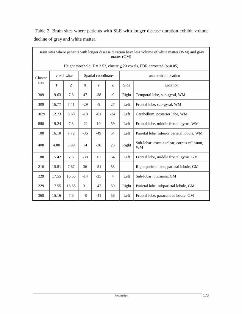
Resultados 173
Table 2. Brain sites where patients with SLE with longer disease duration exhibit volume
decline of gray and white matter.
Brain sites where patients with longer disease duration have less volume of white matter (WM) and gray matter (GM)
Height threshold: T = 3.53, cluster � 20 voxels, FDR corrected (p<0.05)
voxel wise Spatial coordinates anatomical location Cluster
size T Z X Y Z Side Location
309 19.63 7.8 47 -38 -9 Right Temporal lobe, sub-gyral, WM
309 16.77 7.41 -29 -9 27 Left Frontal lobe, sub-gyral, WM
1029 12.73 6.68 -18 -61 -34 Left Cerebellum, posterior lobe, WM
888 19.24 7.8 -21 10 59 Left Frontal lobe, middle frontal gyrus, WM
100 16.10 7.72 -36 -49 54 Left Parietal lobe, inferior parietal lobule, WM
400 4.00 3.99 14 -38 23 Right Sub-lobar, extra-nuclear, corpus callosum, WM
180 15.42 7.6 -38 10 54 Left Frontal lobe, middle frontal gyrus, GM
210 15.81 7.67 36 -51 53 Right parietal lobe, parietal lobule, GM
229 17.55 16.65 -14 -25 4 Left Sub-lobar, thalamus, GM
229 17.55 16.65 31 -47 59 Right Parietal lobe, subparietal lobule, GM
368 15.16 7.6 -8 -41 56 Left Frontal lobe, paracentral lobule, GM
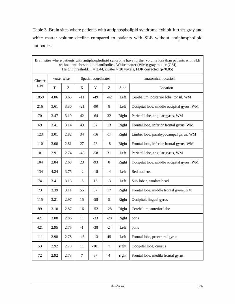
Resultados 174
Table 3. Brain sites where patients with antiphospholipid syndrome exhibit further gray and
white matter volume decline compared to patients with SLE without antiphospholipid
antibodies
Brain sites where patients with antiphospholipid syndrome have further volume loss than patients with SLE without antiphospholipid antibodies. White matter (WM); gray matter (GM)
Height threshold: T = 2.44, cluster � 20 voxels, FDR corrected (p<0.05)
voxel wise Spatial coordinates anatomical location Cluster
size T Z X Y Z Side Location
1859 4.06 3.65 -11 -49 -42 Left Cerebelum, posterior lobe, tonsil, WM
216 3.61 3.30 -21 -90 8 Left Occipital lobe, middle occipital gyrus, WM
70 3.47 3.19 42 -64 32 Right Parietal lobe, angular gyrus, WM
69 3.41 3.14 43 37 13 Right Frontal lobe, inferior frontal gyrus, WM
123 3.01 2.82 34 -16 -14 Right Limbic lobe, parahypocampal gyrus, WM
110 3.00 2.81 27 28 -8 Right Frontal lobe, inferior frontal gyrus, WM
101 2.91 2.74 -45 -58 31 Left Parietal lobe, angular gyrus, WM
104 2.84 2.68 23 -93 8 Right Occipital lobe, middle occipital gyrus, WM
134 4.24 3.75 -2 -18 -4 Left Red nucleus
74 3.41 3.13 -5 13 -3 Left Sub-lobar, caudate head
73 3.39 3.11 55 37 17 Right Frontal lobe, middle frontal gyrus, GM
115 3.21 2.97 15 -58 5 Right Occipital, lingual gyrus
99 3.10 2.87 16 -52 -28 Right Cerebelum, anterior lobe
421 3.08 2.86 11 -33 -28 Right pons
421 2.95 2.75 -1 -38 -24 Left pons
111 2.98 2.78 -45 -13 45 Left Frontal lobe, precentral gyrus
53 2.92 2.73 11 -101 7 right Occipital lobe, cuneus
72 2.92 2.73 7 67 4 right Frontal lobe, medila frontal gyrus
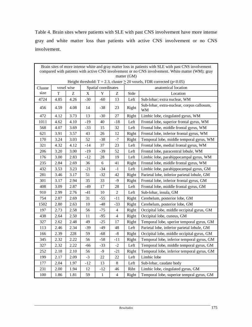
Resultados 175
Table 4. Brain sites where patients with SLE with past CNS involvement have more intense
gray and white matter loss than patients with active CNS involvement or no CNS
involvement.
Brain sites of more intense white and gray matter loss in patients with SLE with past CNS involvement compared with patients with active CNS involvement or no CNS involvement. White matter (WM); gray
matter (GM) Height threshold: T = 2.3, cluster � 20 voxels, FDR corrected (p<0.05)
voxel wise Spatial coordinates anatomical location Cluster size T Z X Y Z Side Location
4724 4.85 4.26 -30 -60 13 Left Sub-lobar; extra nuclear, WM
456 4.59 4.08 14 -38 23 Right Sub-lobar, extra-nuclear, corpus callosum, WM
472 4.12 3.73 13 -30 27 Right Limbic lobe, cingulated gyrus, WM
1011 4.62 4.10 -19 40 -18 Left Frontal lobe, superior frontal gyrus, WM
568 4.07 3.69 -33 15 32 Left Frontal lobe, middle frontal gyrus, WM
621 3.91 3.57 43 26 12 Right Frontal lobe, inferior frontal gyrus, WM
170 3.24 3.03 52 -38 -7 Right Temporal lobe, middle temporal gyrus, WM
321 4.32 4.12 -14 37 23 Left Frontal lobe, medial frontal gyrus, WM
206 3.20 3.00 -19 -39 52 Left Frontal lobe, paracentral lobule, WM
176 3.00 2.83 -12 28 19 Left Limbic lobe, parahippocampal gyrus, WM
235 2.84 2.69 36 6 41 Right Frontal lobe, middle frontal gyrus, WM
432 3.53 3.23 -21 -34 -1 Left Limbic lobe, parahippocampal gyrus, GM
281 3.46 3.17 51 -32 42 Right Parietal lobe, inferior parietal lobule, GM
301 3.17 2.94 35 33 -9 Right Frontal lobe, inferior frontal gyrus, GM
408 3.09 2.87 -49 17 28 Left Frontal lobe, middle frontal gyrus, GM
910 2.99 2.76 -41 10 2 Left Sub-lobar, insula, GM
754 2.87 2.69 31 -55 -11 Right Cerebelum, posterior lobe, GM
1502 2.80 2.63 10 -48 -33 Right Cerebelum, posterior lobe, GM
197 2.73 2.58 56 -75 4 Right Occipital lobe, middle occipital gyrus, GM
438 2.64 2.50 11 -95 4 Right Occipital lobe, cuneus, GM
327 2.62 2.48 49 -25 17 Right Temporal lobe, uperior temporal gyrus, GM
113 2.46 2.34 -39 -49 48 Left Parietal lobe, inferior parietal lobule, GM
166 2.39 228 59 -68 -8 Right Occipital lobe, middle occipital gyrus, GM
345 2.32 2.22 56 -58 -11 Right Temporal lobe, inferior temporal gyrus, GM
327 2.32 2.22 -66 -33 -2 Left Temporal lobe, middle temporal gyrus, GM
252 2.18 2.10 56 -9 -21 Right Temporal lobe, inferior temporal gyrus, GM
199 2.17 2.09 -3 22 22 Left Limbic lobe
177 2.04 1.97 -12 13 8 Left Sub-lobar, caudate body
231 2.00 1.94 12 -12 46 Riht Limbic lobe, cingulated gyrus, GM
100 1.86 1.81 59 1 4 Right Temporal lobe, superior tempral gyrus, GM
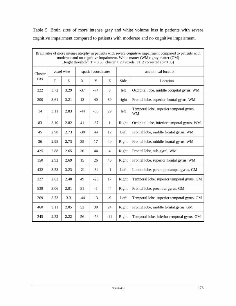
Resultados 176
Table 5. Brain sites of more intense gray and white volume loss in patients with severe
cognitive impairment compared to patients with moderate and no cognitive impairment.
Brain sites of more intense atrophy in patients with severe cognitive impairment compared to patients with moderate and no cognitive impairment. White matter (WM); gray matter (GM)
Height threshold: T = 3.30, cluster � 20 voxels, FDR corrected (p<0.05)
voxel wise spatial coordinates anatomical location Cluster
size T Z X Y Z Side Location
222 3.72 3.29 -37 -74 8 left Occipital lobe, middle occipital gyrus, WM
200 3.61 3.21 13 40 39 right Frontal lobe, superior frontal gyrus, WM
14 3.11 2.83 -44 -56 29 left Temporal lobe, superior temporal gyrus, WM
83 3.10 2.82 41 -67 1 Right Occipital lobe, inferior temporal gyrus, WM
45 2.98 2.73 -38 44 12 Left Frontal lobe, middle frontal gyrus, WM
36 2.98 2.73 35 17 40 Right Frontal lobe, middle frontal gyrus, WM
425 2.88 2.65 30 44 4 Right Frontal lobe, sub-gyral, WM
150 2.92 2.69 15 26 46 Right Frontal lobe, superior frontal gyrus, WM
432 3.53 3.23 -21 -34 -1 Left Limbic lobe, parahippocampal gyrus, GM
327 2.62 2.48 49 -25 17 Right Temporal lobe, superior temporal gyrus, GM
539 3.06 2.81 51 -5 44 Right Frontal lobe, precntral gyrus, GM
269 3.73 3.3 -44 13 -9 Left Temporal lobe, superior temporal gyrus, GM
460 3.11 2.85 53 38 24 Right Frontal lobe, middle frontal gyrus, GM
345 2.32 2.22 56 -58 -11 Right Temporal lobe, inferior temporal gyrus, GM
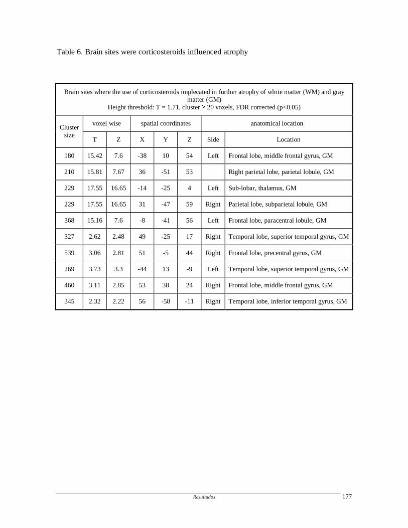
Resultados 177
Table 6. Brain sites were corticosteroids influenced atrophy
Brain sites where the use of corticosteroids implecated in further atrophy of white matter (WM) and gray matter (GM)
Height threshold: T = 1.71, cluster � 20 voxels, FDR corrected (p<0.05)
voxel wise spatial coordinates anatomical location Cluster
size T Z X Y Z Side Location
180 15.42 7.6 -38 10 54 Left Frontal lobe, middle frontal gyrus, GM
210 15.81 7.67 36 -51 53 Right parietal lobe, parietal lobule, GM
229 17.55 16.65 -14 -25 4 Left Sub-lobar, thalamus, GM
229 17.55 16.65 31 -47 59 Right Parietal lobe, subparietal lobule, GM
368 15.16 7.6 -8 -41 56 Left Frontal lobe, paracentral lobule, GM
327 2.62 2.48 49 -25 17 Right Temporal lobe, superior temporal gyrus, GM
539 3.06 2.81 51 -5 44 Right Frontal lobe, precentral gyrus, GM
269 3.73 3.3 -44 13 -9 Left Temporal lobe, superior temporal gyrus, GM
460 3.11 2.85 53 38 24 Right Frontal lobe, middle frontal gyrus, GM
345 2.32 2.22 56 -58 -11 Right Temporal lobe, inferior temporal gyrus, GM
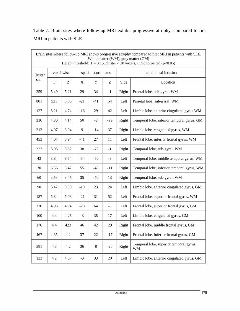
Resultados 178
Table 7. Brain sites where follow-up MRI exhibit progressive atrophy, compared to first
MRI in patients with SLE
Brain sites where follow-up MRI shows progressive atrophy compared to first MRI in patients with SLE. White matter (WM); gray matter (GM)
Height threshold: T = 3.15, cluster � 20 voxels, FDR corrected (p<0.05)
voxel wise spatial coordinates anatomical location Cluster
size T Z X Y Z Side Location
259 5.49 5.21 29 34 -1 Right Frontal lobe, sub-gyral, WM
801 531 5.06 -21 -41 54 Left Parietal lobe, sub-gyral, WM
127 5.21 4.74 -16 29 42 Left Limbic lobe, anterior cingulated gyrus WM
216 4.30 4.14 50 -3 -29 Right Temporal lobe, inferior temporal gyrus, GM
212 4.07 3.94 9 -14 37 Right Limbic lobe, cingulated gyrus, WM
453 4.07 3.94 -41 27 11 Left Frontal lobe, inferior frontal gyrus, WM
227 3.93 3.82 38 -72 -1 Right Temporal lobe, sub-gyral, WM
43 3.84 3.74 -54 -50 -8 Left Temporal lobe, middle temporal gyrus, WM
30 3.56 3.47 55 -45 -11 Right Temporal lobe, inferior temporal gyrus, WM
60 3.53 3.45 35 -70 13 Right Temporal lobe, sub-gyral, WM
90 3.47 3.39 -10 23 24 Left Limbic lobe, anterior cingulated gyrus, GM
187 5.34 5.08 -21 31 52 Left Frontal lobe, superior frontal gyrus, WM
330 4.98 4.94 -28 64 -8 Left Frontal lobe, superior frontal gyrus, GM
100 4.4 4.25 -3 35 17 Left Limbic lobe, cingulated gyrus, GM
176 4.4 423 46 42 29 Right Frontal lobe, middle frontal gyrus, GM
467 4.35 4.2 37 22 -17 Right Frontal lobe, inferior frontal gyrus, GM
581 4.3 4.2 36 8 -26 Right Temporal lobe, superior temporal gyrus, WM
122 4.2 4.07 -3 33 20 Left Limbic lobe, anterior cingulated gyrus, GM
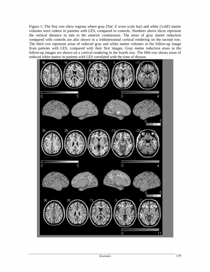
Resultados 179
Figure 1: The first row show regions where gray ('hot' Z score scale bar) and white ('cold') matter volumes were reduce in patients with LES, compared to controls. Numbers above slices represent the vertical distance in mm to the anterior commissure. The areas of gray matter reduction compared with controls are also shown in a tridimensional cortical rendering on the second row. The third row represent areas of reduced gray and white matter volumes in the follow-up image from patients with LES, compared with their first images. Gray matter reduction areas in the follow-up images are shown on a cortical rendering in the fourth row. The fifth row shows areas of reduced white matter in patients with LES correlated with the time of disease.
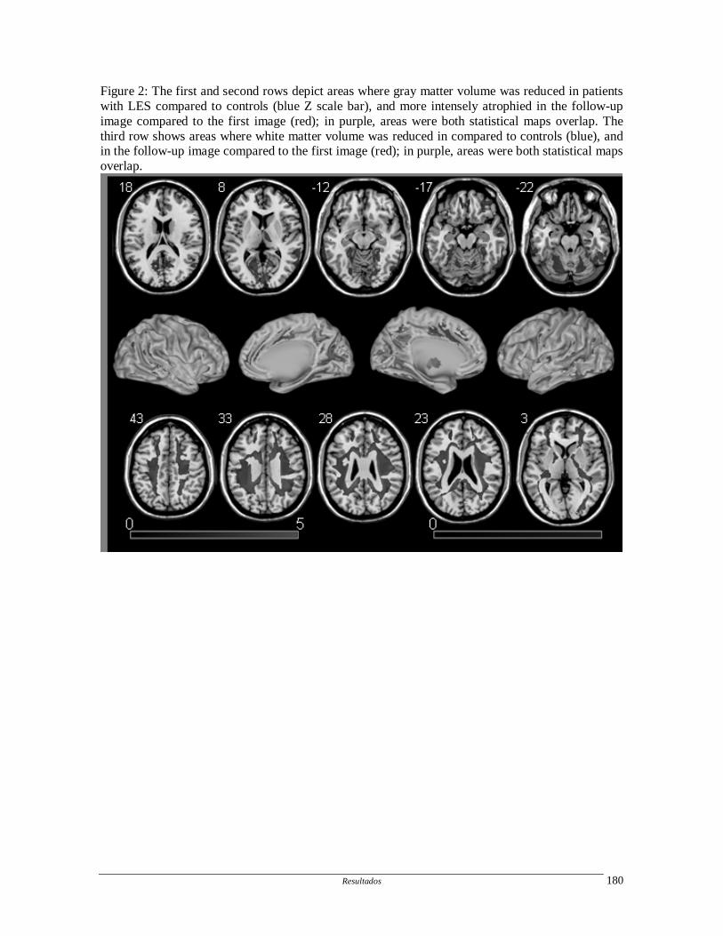
Resultados 180
Figure 2: The first and second rows depict areas where gray matter volume was reduced in patients with LES compared to controls (blue Z scale bar), and more intensely atrophied in the follow-up image compared to the first image (red); in purple, areas were both statistical maps overlap. The third row shows areas where white matter volume was reduced in compared to controls (blue), and in the follow-up image compared to the first image (red); in purple, areas were both statistical maps overlap.
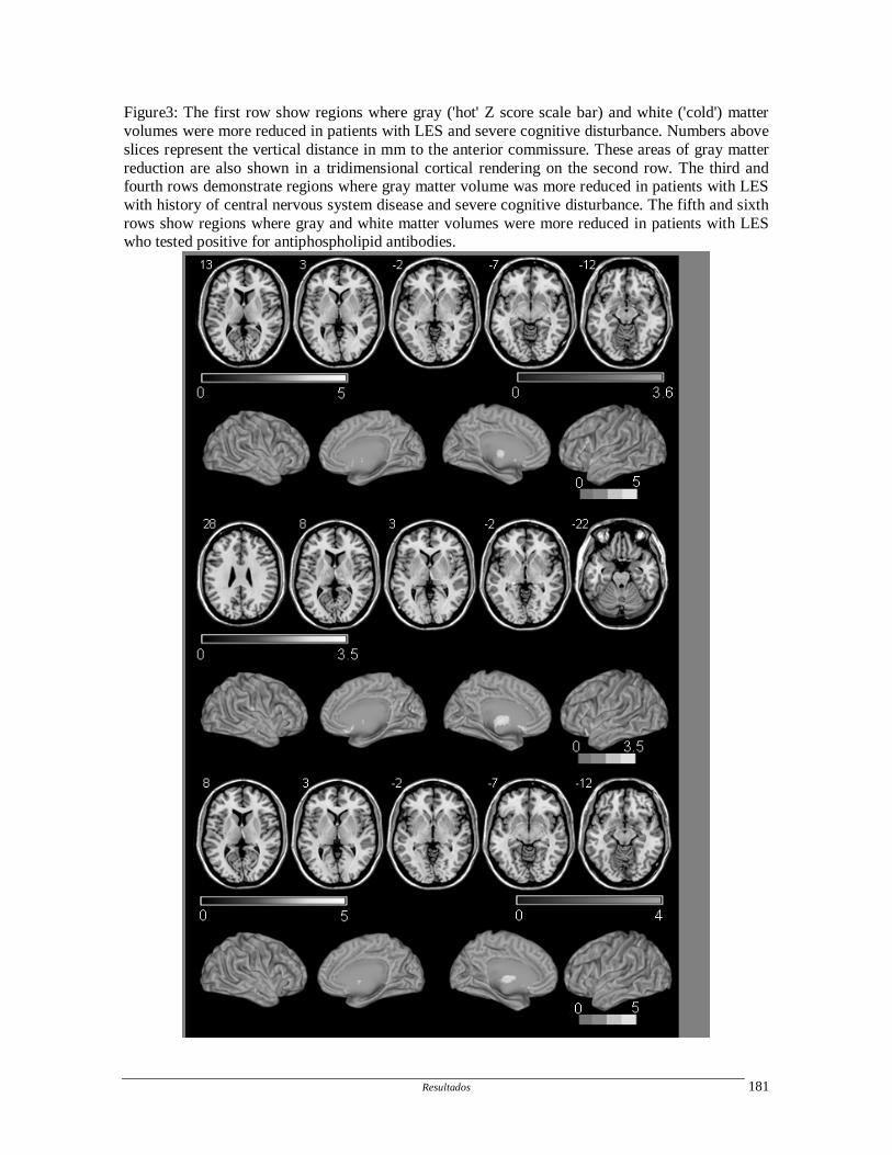
Resultados 181
Figure3: The first row show regions where gray ('hot' Z score scale bar) and white ('cold') matter volumes were more reduced in patients with LES and severe cognitive disturbance. Numbers above slices represent the vertical distance in mm to the anterior commissure. These areas of gray matter reduction are also shown in a tridimensional cortical rendering on the second row. The third and fourth rows demonstrate regions where gray matter volume was more reduced in patients with LES with history of central nervous system disease and severe cognitive disturbance. The fifth and sixth rows show regions where gray and white matter volumes were more reduced in patients with LES who tested positive for antiphospholipid antibodies.
Resultados 182
ARTIGO 10
Hippocampal atrophy in Systemic lupus erythematosus
Appenzeller S, Carnevalle AD, Li LM, Costallat LT, Cendes F.
Hippocampal atrophy in Systemic lupus erythematosus
Ann Rheum Dis. 2006 Jan 26; [Epub ahead of print]
Resultados 183
Hippocampal atrophy in systemic lupus erythematosus
Simone Appenzeller1,3, MD, Aline Daiane Carnevalle3, Li Min Li2,3, MD, PhD, Lilian TL
Costallat1, MD, PhD, Fernando Cendes2,3, MD, PhD
Departments of Rheumatology1 and Neurology2, Neuroimaging Laboratory3
University of Campinas, São Paulo – Brazil
Key words: systemic lupus erythematosus- atrophy – progression-MRI- semiautomatic
MRI segmentation-hippocampus
Grants: FAPESP and CNPq (479133/2004-2)
Correspondence to: Fernando Cendes, MD, PhD
Department of Neurology, University of Campinas-UNICAMP
Cidade Universitária, Campinas SP, Brazil, CEP 13083-970
FAX: +55 19 3289-1818
Email: [email protected]
Resultados 184
Abstract
Objective: To determine the frequency and progression of hippocampal atrophy in systemic
lupus erythematosus (SLE) and to determine clinical, laboratory and treatment features
associated with its occurrence.
Methods: A total of 150 SLE patients and 40 healthy volunteers were enrolled in this study.
A complete clinical, laboratory and neurological evaluation was performed. MRI scans
were performed in a 2T scanner (Elscint Prestige®) and coronal T1 weighted images were
used for manual volumetric measurements. Atrophy was defined as values below 2
standard deviations (SD) from the means of controls.
Results: At study entry, the mean right and left hippocampal volumes of patients were
significantly smaller than the hippocampal volumes of healthy controls (p<0.001). After the
follow-up MRI we observed a significant progression of reduction of right and left
hippocampal volume in patients (p<0.0001). At study entry we identified atrophy in 43.9%
and at follow up in 66.7% of SLE patients. Hippocampal atrophy was related to disease
duration, total corticosteroid dose and past history of CNS manifestations. Progression of
atrophy was associated with cumulative corticosteroid dose and number of CNS events.
Patients with cognitive impairment had more severe hippocampal atrophy than patients
without this manifestation.
Conclusion: Disease duration, total corticosteroid dose and greater number of CNS
manifestations were associated with hippocampal atrophy in SLE patients. We observed a
significant progression of hippocampal atrophy related to total corticosteroid dose and
number of CNS events. Further studies are necessary to confirm these findings.
Introduction
Systemic lupus erythematosus (SLE) is a multiorgan autoimmune disease with
abnormalities in immune regulation (1-3). Disease activity is characterized through intense
inflammatory activity and treatment includes corticosteroids and other immunosuppressive
agents (4).
Resultados 185
The hippocampus, located in the temporal lobe, is a structure intimately
involved with certain aspects of learning and memory consolidation (5). Patients exposed to
high level exogenous (6-8) or endogenous (9-11) corticosteroids are more prone to short-
term memory deficits. Previous studies have analyzed reversible and irreversible changes in
hippocampus after corticosteroid exposure (12, 13). The hippocampus provides negative
feedback to the hypothalamic-pituitary-adrenal axis and plays a critical role in declarative
memory (14-16). In two studies the reduction of hippocampal volume correlated with mean
cortisol level and atrophy was reversible after normalization of cortisol levels (10, 11).
The aim of this study was to determine the prevalence of hippocampal atrophy
in SLE through validated manual MRI segmentation and to determine clinical, laboratory
and treatment features associated with its occurrence. We also wanted to determine if this
atrophy is progressive after follow-up period and which factors were associated with the
continuous reduction of hippocampal volume.
Subjects and Methods
Subjects
A total of 150 consecutive SLE patients with four or more criteria for SLE (17)
seen regularly at our Rheumatology Unit were screened prospectively to participate in the
study. All SLE patients were followed using a standardized protocol and followed by the
same investigators in the Rheumatology Unit (LTLC, SA). We excluded patients that were
not able to undergo MRI, such as patients with claustrophobia (8 patients) and pacemaker
(2 patients), as well as patients with previous clinical conditions that could influence
cerebral and hipocamal atrophy, such as history of stroke (10 patients), arterial
hypertension (5 patients), diabetes mellitus (5 patients), alcohol and drug abuse (1 patient),
epilepsy (8) and malignancy (1 patient). There were no patients with renal insufficiency, or
other pathologies that could influence cerebral atrophy. Patients who fulfilled the ACR
criteria for rheumatoid arthritis, systemic sclerosis, Sjogren syndrome (primary or
secondary) (3 patients) or other connective tissue disease and with drug-induced SLE were
also excluded. The remaining 107 patients (100 women) were included. We used the
Resultados 186
classification proposed by the ACR to analyze neuropsychiatric involvement (18). Records
were reviewed in order to determine past CNS events. Patients with CNS manifestations
secondary to clinical conditions such as infections, arterial hypertension, uremia, diabetes
and drugs were excluded from this study.
This study was approved by Ethical Committee of our institution and informed
written consent was obtained from each subject.
Clinical, serologic and treatment features of SLE patients
Data on gender, age at disease onset and disease duration were collected for
each patient. Disease duration was defined as the initial manifestation clearly attributable to
SLE until the day of MRI acquisition. All clinical manifestations and laboratory test
findings were recorded. The following clinical manifestations were analyzed: malar rash,
discoid lesions, subacute cutaneous lesions, photosensitivity, oral ulcers, arthritis, serositis,
nephritis, neurological and psychiatric involvement, thrombocytopenia, hemolytic anemia,
Raynaud’s phenomenon, thrombosis, myositis, lung involvement and lymphadenopathy.
Nephritis was diagnosed on the basis of proteinuria exceeding 0.5 g/L with
abnormal urinary sediment and/or histological findings. Nephrotic syndrome was defined
as proteinuria in excess of 3.5 g/day. Hematologic alterations were ascribed to lupus only in
the absence of bone marrow suppression (leukopenia <4000 cells/mm3; thrombocytopenia
<100.000/mm3; hemolytic anemia with positive Coombs test). Antinuclear antibodies
(ANA) were determined by indirect immunofluorescence using HEp 2 cells as the substrate
and regarded as positive if higher than 1:40. Anti-double-stranded DNA (AdsDNA)
antibodies were determined by indirect immunofluorescence using Chrithidia as substrate
and considered positive if higher than 1:10. Precipitating antibodies to extractable nuclear
antigens (ENA), including Ro (SSA), La (SSB) and Sm were detected by immunodiffusion
and/or microhemagglutination. Anticardiolipin antibodies (aCL) of the IgG and IgM
isotypes were measured by the ELISA method as described (19). Lupus anticoagulant (LA)
activity was detected by coagulation assays in platelet free plasma obtained by double
centrifugation, following the recommendation of the subcommittee on LA of the Scientific
Resultados 187
and Standardization Committee of the International Society of Thrombosis and
Homeostasis (20).
CNS manifestations were recorded following ACR case definitions (18) and
divided into present (active or past history of CNS involvement) or absent (never presented
CNS involvement). A complete neurological examination, as well as cognitive and
psychiatric charts, was prospectively applied to all patients in order to identify CNS
involvement. Mini Mental State Examination (21) was applied to all participants.
All patients and controls were submitted to a battery of standardized
neuropsychologic tests in order to screen for possible impairment in one or more of the
subsequent cognitive domains: simple attention, complex attention, memory, visuo-spatial
processing, language, reasoning/problem solving, psychomotor speed, and executive
functions (21-24). The individual test results were converted into standard scores, which
were compared with the available normative data (21-24). Regarding any of the eight
cognitive domains, subjects with a total score of two or more standard deviations (SD)
below the normative value were considered to be impaired. Cognitive dysfunction was
classified as mild if there were deficits in less than three dimensions, as moderate if there
were deficits in three or four dimensions, and as severe if there were deficits in at least five
dimensions (25, 26).
Assessment of depression was based on clinical interview and the Beck
Depression Inventory (BDI) (27, 28). On BDI, scores from 10 to 17 were considered to
indicate mild depression, from 18 to 24 moderate depression, and greater than 24 severe
depression. Anxiety was evaluated by anxiety through the Hospital Anxiety and Depression
scale (29). The presence of psychosis was determined through the Brief Psychiatry Rating
Scale (aBPRS) (30).
For past history of CNS involvement we reviewed the medical charts of
patients. Disease activity was measured by SLEDAI and considered active if scores were
higher than eight points (31). Cumulative SLE-related damage was determined by Systemic
Lupus International Collaborating Clinics/American College of Rheumatology Damage
Index (SLICC/ACR DI) (32) in all SLE patients at time of MRI.
Resultados 188
Total doses of corticosteroids and other immunosuppressant medications used
since the onset of disease were calculated by careful review of the medical charts. Doses of
oral and parenteral corticosteroids were analyzed and converted to the equivalent doses of
prednisone. The cumulative dose of corticosteroids used was calculated by the sum of daily
dosages versus time (days) of treatment.
MRI acquisition
All subjects had MRI examination using Escint Prestige 2T scanner (Haifa,
Israel). Coronal T1-IR (4mm thick, flip angle=120º, repetition time=6800 ms, echo
time=129 ms, matrix 252x328, field of view=21x23cm) images were analyzed using
anatomic guidelines obtained from a standardized protocol (33) for manual segmentations
for hippocampal and total brain volume (Figure 1). Quantification and analysis were
performed by one investigator (ADC), blind to patients clinical data. The evaluation was
cross-checked by another investigators with experience in MRI analysis (SA, FC). This
method has been previously compared to other segmentation programs (34) and intra-
observer (r=0.90)and inter-observer (r=0.85) variation was determined.
The control group for MRI protocol consisted of 40 healthy volunteers (36
women) with mean age of 31.8 (range 20-55, SD=10.2 years). Patients and controls were
statistically comparable in age and gender.
Image processing
Manual delineation of hippocampi boundaries was performed using Scion®
Image program (NIH). Anatomic guidelines for outlining the hippocampus followed a
specific protocol previously described (35). Once the outline had been defined, the slice
area was calculated automatically by the computer program. We then calculated the total
volumes expressed in cubic millimeters by the sum of each area multiplied by the slice
thickness (mm3). In order to determine hippocampal atrophy even in the presence of diffuse
atrophy, and also to correct for individual variation of the size of the head, we performed a
correction of all hippocampal absolute volumes for each respective individual cerebral

Resultados 189
volume. This correction consisted of dividing the mean total brain volume of the control
group by the patient’s brain volume. In each patient, the calculated hippocampal volume
was then multiplied by this ratio (36). This correction for ‘brain volume’ assumes a linear
relationship between hippocampi and brain volumes (36).
In addition, asymmetry index (AI) was determined by the ratio of the smaller by
the larger structure for each subject.
Atrophy was determined when corrected volumes and/or AI were bellow two
standard deviations (SD) from the mean of control group.
Statistical analysis
We compared hippocampal volumes of SLE patients to controls using two
sample t-test. We further subdivided SLE patients in two groups: patients with and without
CNS involvement. We performed analysis of variance (ANOVA) to test for differences
among hippocampal volumes in these groups, with Tukey’s pairwised post hoc
comparisons when necessary. This procedure includes corrections for multiple
comparisons. Demographic data between groups were compared with chi-square test.
Follow-up volumes were compared by paired t-test with Bonferronis correction for
multiple comparisons. Linear regression was used to analyze the association between
cerebral and hippocampi volume with disease duration and total corticosteroid use.
Volumetric measurements were expressed in cm3 and are shown in mean ±
standard deviation (SD). Statistical significance was considered at � of 5%.
Results
Demographic data
One hundred and seven SLE patients (100 women) met the inclusion and
exclusion criteria with mean age of 32.2 years (SD=11.2; range=18 to 54 years). The mean
disease duration at study entry was 64.5 months (range 1-372 months; SD=48.50). All
Resultados 190
patients were on corticosteroid use with doses varying from 5 to 100 mg of prednisone.
Antiphospholipid antibodies were positive in 32 patients. Active SLE disease was observed
in 54 patients with mean SLEDAI score of 14.0 (range 9-20; SD=5.9). One hundred twenty
two episodes of CNS manifestations were observed in 64 patients (Table 1). Active CNS
disease at the time of MRI scan was observed in 30 of 64 patients with history of CNS
involvement. Mean SLICC/ACR DI scores at study entry were 2.3 (range 0-5; SD=1.8).
The cumulative clinical and CNS events at study entry are shown in Table 1.
MRI scans were repeated in 60 patients after a mean follow-up period of 19
months (SD=2.2, range=12-25 months). Forty of these patients presented CNS events
during follow-up period with mean SLEDAI scores of 12.4 (range 3-20; SD=3.8). Mean
SLICC/ACR DI scores at follow-up were 3.1 (SD=1.7; range 0-6).
Hippocampal volumes
At study entry, the mean right hippocampal volumes of patients was 3260 mm3
(SD=330) and the mean left hippocampus was 3080 mm m3 (SD=330). In controls we
observed a mean right hippocampal volume of 3570 mm m3 (DP=310) and a mean left
hippocampal volume of 3480 mm m3 (SD=330). There was a statistically difference
between the right (p=0.002) and the left (p=0.001) hippocampal volume of patients and
controls (Figure 2).
Individual analysis
Hippocampal atrophy was identified in 47 of 107 (43.9%) patients and
distributed as follows: 20 of 47 (42.6%) with left hippocampal atrophy, 10 (8.3%) with
right hippocampal atrophy and 17 (10%) with bilateral hippocampal atrophy.
Group analysis
The degree of hippocampal volume loss correlated independently with disease
duration (r=0.89; p<0.001), the presence of CNS manifestations (r=0.65; p=0.01) and
Resultados 191
cumulative corticosteroid dose (r=0.74; p=0.01). When we further subdivided patients with
CNS involvement in active and past history of CNS involvement we observed that
hippocampal reduction was associated with past history (r=0.9; p<0.001) and not with
active CNS involvement.
SLICC/ACR DI scores correlated strongly with the degree of hippocampal
atrophy (r=0.87; p=0.001), but when cognitive dysfunction was excluded from the SLICC
scores, the correlation was not statistically significant (r=0.3; p=0.4). There was a trend
between the association of hippocampal atrophy and the presence of antiphospholipid
antibodies (r=0.5; p=0.06). No association between hippocampal atrophy and SLEDAI
(r=0.43; p=0.08) was observed. On visual analysis, hyperintense areas and areas of cerebral
micro-infarcts were observed in 50 patients. There was a statistically significance between
the presence of these findings and hippocampal atrophy (p=0.01).
Cognitive evaluation
We observed cognitive impairment in 35 patients, 20 with severe, 10 with
moderate and 5 with mild cognitive impairment. We observed that that patients with
cognitive impairment had a more significant reduced hippocampal volume when compared
to SLE patients without cognitive impairment (r=0.9; p=0.001). Patients with severe
cognitive impairment had a more pronounced reduction of hippocampal volumes than
patients with moderate and mild cognitive impairment (p=0.002). In relation to different
domains of cognitive dysfunction, hippocampal atrophy was associated to lower scores on
general memory (p=0.015), verbal memory (p=0.01), and delayed recall (p=0.01).
Follow-up analysis
After the follow-up MRI, group analysis showed a significant progression of
reduction of right (mean=28.2 cm3, SD=3.1) and left (mean=27.9 cm3, SD=1.2)
hippocampal volumes in patients (p<0.0001) (Figure 3).
Individual analysis showed that the follow-up MRI demonstrated a significant
increase in number of patients with hippocampal atrophy. We observed atrophy in 40 of 60
Resultados 192
(66.7%) patients: 32 (53.3%) bilaterally, 4 (6.7%) with right hippocampal atrophy and 4
(6.7%) with left hippocampal atrophy. Progression of hippocampal atrophy correlated with
cumulative corticosteroid dose (r=0.69; p=0.01) and number of CNS events during the
follow-up period (r= 0.72, p=0.01).
We observed cognitive impairment in 40 of 60 patients after the follow-up
period, 22 with severe, 12 with moderate and 6 with mild cognitive impairment. We
observed that patients with hippocampal atrophy and normal cognitive function at study
entry, presented with cognitive impairment during the follow-up period (Figure 3). The
severity of cognitive impairment was directly associated with the degree of hippocampal
volume loss (r=0.89; p=0.001). In relation to different domains of cognitive dysfunction, no
difference in relation to study entry was observed, being hippocampal atrophy associated to
lower scores on general memory (p=0.01), verbal memory (p=0.02), and delayed recall
(p=0.01).
Discussion
We observed a frequency of 44% of hippocampal atrophy in our study and a
significant progression of hippocampal volume loss and atrophy during follow-up period.
Both the presence and the progression of hippocampal atrophy correlated with disease
duration, total corticosteroid dose and the presence of past history of CNS manifestations.
The number of CNS events was associated with progression of hippocampal atrophy during
the follow-up period. The short follow-up period did not allow us to determine if
hippocampal volumes may return to normal ranges or if the progression of hippocampal
atrophy stops after corticosteroid withdrawal.
At study entry we observed a small proportion of bilateral atrophy. We did not
identify any cause for the predominance of unilateral atrophy in these patients, although
after a follow-up period the majority of the patients presented bilateral hippocampal
atrophy.
Hipocampal atrophy has been shown in several diseases before, such as
epilepsy (36), and it is related to memory impairment (5-11). In addition to the exclusion of
clinical conditions that are associated with cerebral atrophy, such as stroke, arterial
Resultados 193
hypertension (37) and diabetes mellitus, we excluded patients with epilepsy. The
relationship between epilepsy and hippocampal atrophy is well determined in the literature
(38).
Although it is possible that aging had some effect in brain and hippocampal
atrophy (39), this effect was minimized by the comparison with volunteers with same age
range.
Hippocampus is the prominent target structure for the activity of corticosteroids
in the brain (40). In animals, corticosteroid hormones have been shown to reduce the
number of branch points and length of dendrites in hippocampus of rats, to cause dendritic
atrophy of pyramidal neurons (41) and to lead to reduction of CA3 and CA4 hippocampal
neurons (42). In patients with Cushing syndrome, previous studies have shown that the
reduction of hippocampal volume correlated with mean cortisol level and that atrophy was
reversible after normalization of cortisol levels (10, 11).
If corticosteroids cause cell death and neuronal loss or if they cause atrophy or
degeneration is not clearly understood. The majority of animal models report only atrophy
of dendritic branches (40-42). Although this can be the first step and progress to neuronal
loss, neuronal cell death was found only in a few studies (40). Our study suggest that
hippocampal atrophy in SLE is one of the consequences of CNS damage induced by the
inflammatory process, which is most likely potentiated by prolonged use of corticosteroids.
We observed that hippocampal atrophy was associated with the presence of
cognitive dysfunction in SLE, especially memory function. Patients with severe cognitive
impairment had more pronounced hippocampal volume loss when compared to patients
with mild cognitive impairment and patients without this manifestation. The elevated
presence of hippocampal atrophy observed in this study may explain the high frequency of
cognitive impairment observed in SLE patients (1-4). We also observed that patients with
white matter lesions had more frequently hippocampal atrophy than patients without these
MRI findings. Similar findings were described in patients with Alzheimer disease (43). We
also observed that the presence of antiphospholipid antibodies had a tendency to be
associated with hippocampal atrophy, supporting the theory that small vessel disease may
contribute to hippocampal atrophy and cognitive impairment (43). We also observed that
Resultados 194
hippocampal atrophy was a predictor for cognitive impairment. The patients that had
normal cognitive function test and hippocampal atrophy at study entry presented cognitive
impairment during the follow-up study.
In this study we demonstrated new evidence that structural MRI abnormalities
may be associated with cognitive dysfunction in SLE patients. We also demonstrated that
hippocampal atrophy is progressive over time. Furthermore we showed that the presence of
hippocampal atrophy may be predictive of cognitive impairment in SLE patients. Longer
follow-up studies are necessary to confirm these findings and to determine if the
hippocampal loss is reversible and which factors may contribute to it.
Resultados 195
References
1. Omdal R, Mellgren SI, Husby G. Clinical neuropsychiatric and neuromuscular
manifestations in systemic lupus erythematosus. Scand J Rheumatol 1988; 17: 113-117.
2. Feinglass EJ, Arnett FC, Dorsch CA, Zizic TM, Stevens MB. Neuropsychiatric
manifestations of systemic lupus erythematosus: diagnosis, clinical spectrum and
relationship to other features of the disease. Medicine 1976; 55: 323-339.
3. Adelman DC, Saltiel E, Klinenberg JR. The neuropsychiatric manifestations of
systemic lupus erythematosus: An overview. Semin Arthritis Rheum 1986; 15: 185-
199.
4. Johnson RT, Richardson EP. The neurological manifestations of systemic lupus
erythematosus. Medicine 1968, 47: 337-369.
5. Brunner R, Schaefer D, Hess K, Parzer P, Resch F, Schwab S. Effect of corticosteroids
on short-term and long-term memory. Neurology 2005; 64: 335-337.
6. Wolkowitz OM, Reus VI, Weingartner H, Thompson K, Breier A, Doran A, Rubinow
D, Pickar D. Cognitive effects of corticosteroids. Am J Psychiatry. 1990;147:1297-303.
7. Wolkowitz OM, Lupien SJ, Bigler E, Levin RB, Canick J. The "steroid dementia
syndrome": an unrecognized complication of glucocorticoid treatment. Ann N Y Acad
Sci. 2004;1032:191-4.
8. Newcomer JW, Craft S, Hershey T, Askins K, Bardgett ME. Glucocorticoid-induced
impairment in declarative memory performance in adult humans. J Neurosci.
1994;14:2047-53.
9. Mauri M, Sinforiani E, Bono G, Vignati F, Berselli ME, Attanasio R, Nappi G.
Memory impairment in Cushing's disease. Acta Neurol Scand. 1993;87:52-5.
10. Starkman MN, Gebarski SS, Berent S, Schteingart DE. Hippocampal formation
volume, memory dysfunction, and cortisol levels in patients with Cushing's
syndrome.Biol Psychiatry. 1992;32:756-65.
11. Starkman MN, Giordani B, Berent S, Schork MA, Schteingart DE. Elevated cortisol
levels in Cushing's disease are associated with cognitive decrements. Psychosom Med.
2001;63:985-93.

Resultados 196
12. Starkman MN, Giordani B, Gebarski SS, Berent S, Schork MA, Schteingart DE.
Decrease in cortisol reverses human hippocampal atrophy following treatment of
Cushing's disease. Biol Psychiatry. 1999;46:1595-602.
13. Lupien SJ, Mc Ewen BS. The acute effects of corticosteroids on cognition: integration
of animal and human model studies. Brain Research Reviews 1997; 24: 1-27
14. Brown ES, J Woolston D, Frol A, Bobadilla L, Khan DA, Hanczyc M, Rush AJ,
Fleckenstein J, Babcock E, Cullum CM. Hippocampal volume, spectroscopy, cognition,
and mood in patients receiving corticosteroid therapy. Biol Psychiatry. 2004;55:538-45.
15. Brown ES, Rush AJ, Mc Ewwn BS. Hippocampal remodeling and damage by
corticosteroids: Implications for mood disorders. Neuropsychopharmacology 1999; 21:
474-484.
16. Squire LR. Memory and the hippocampus: A synthesis from findings with rats,
monkeys, and humans. Psychol Rew 1992; 99: 195-231.
17. Tan EM, Cohen AS, Fries JF, Masi AT, McShane DJ, Rothfield NF, et al. The 1982
revised criteria for the classification of systemic lupus erythematosus. Arthritis Rheum
1982; 25: 1271-1277.
18. ACR Ad Hoc Committee on Neuropsychiatric Lupus. The American College of
Rheumatology nomenclature and case definitions for neuropsychiatric lupus
syndromes. Arthritis Rheum 1999; 42: 599-608.
19. Harris EN, Gharavi AE, Patel SP, et al. Evaluation of the anticardiolipine antibody test:
report of an international workshop held 4 April 1986. Clin Exp Immunol 1987; 68:
215-222.
20. Brandt JT, Triplett DA, Alving B, Scharrer I. Criteria for the diagnosis of lupus
anticoagulants: an update. On behalf of the Subcommittee on Lupus
Anticoagulant/Antiphospholipid Antibody of the Scientific and Standardization
Committee of the ISTH. Thromb Haemost 1995; 74: 1185-1190.
21. Folstein MF, Folstein SE, Folstein PR: “Mini mental state” A practical method for
grading the cognitive state of patient for the clinician. J Psychiatr Res 12: 189-198,
1975.
Resultados 197
22. Spranoel HZ. The psychoeducational use and interpretation of the Wechsler Adult
Intelligence Scale–Revised. Springfield: CC Thomas, 1992.
23. Wechsler in Muistiasteikko, Suom. Anja Manner. Helsinki: Psykologien Kustannus,
1986.
24. Delis DC, Kramer JH, Kaplan E, et al. California Verbal Learning Test Research
Edition Manual. San Antonio: The Psychological Corporation, 1987.
25. Lezak MD. Neuropsychological assessment. New York: Oxford University Press, 1995.
26. Heaton RK, Chelune GJ, Talley JL, et al. Wisconsin Card Sorting Test Manual, revised
and expanded. Odessa: Psychological Assessment Resources, Inc., 1993.
27. Beck AT, Beamesderfer A. Assessment of depression: the depression inventory. Mod
Probl Pharmacopsychiat. 1974; 7: 151–169.
28. Beck AT, Rial WY, Rickels K. Short form of depression inventory: cross-validation.
Psychol Rep. 1974; 34: 1184–1186.
29. Herrmann C. International experiences with the Hospital Anxiety and Depression
Scale--a review of validation data and clinical results. J Psychosom Res. 1997;42:17-41.
30. Overall JE, Beller SA. The Brief Psychiatric Rating Scale (BPRS) in geropsychiatric
research: I. Factor structure on an inpatient unit. J Gerontol. 1984;39:187-93.
31. Gladman DD, Urowitz MB, Kagal A, Hallett D. Accurately describing changes in
disease activity in systemic lupus erythematosus. J Rheumatol 2000; 27: 377-379.
32. Gladman DD, Urowitz MB, Goldsmith CH, Fortin P, Ginzler E, Gordon C, Hanly JG,
et al. The reliability of the Systemic Lupus International Collaborating
Clinics/American College of Rheumatology Damage Index in patients with systemic
lupus erythematosus. Arthritis Rheum. 1997; 40:809-813.
33. Bonilha L, Kobayashi E, Cendes F, Li LM. The importance of accurate anatomic
assessment for the volumetric analysis of the amygdala. Braz J Med Biol Res.
2005;38:409-18.
Resultados 198
34. Carnevalle, A.D., Rondina J.M., Kobayashi E., Lotufo R.A., Cendes F. Validation of a
semi-automated system for MRI-based hippocampal volumetry in patients with TLE. J
Epilepsy Clin Neurophysiol 2003; 9: 97-104.
35. Watson C, Andermann F, Gloor P, et al. Anatomic basis of amygdaloid and
hippocampal volume measurments by magnetic ressonance imaging. Neurology 1992;
689-703.
36. Cendes F, Andermann F, Gloor P, Lopes-Cendes I, Andermann E, Melanson D et al.
Relationship between atrophy of the amygdala and ictal fear in temporal lobe epilepsy.
Brain 1994; 117: 739-746.
37. den Heijer T, Launer LJ, Prins ND, van Dijk EJ, Vermeer SE, Hofman A, Koudstaal PJ,
Breteler MM. Association between blood pressure, white matter lesions, and atrophy of
the medial temporal lobe. Neurology. 2005;64:263-7.
38. Cendes F, Andermann F, Gloor P, Evans A, Jones-Gotman M, Watson C, Melanson D,
Olivier A, Peters T, Lopes-Cendes I, et al. MRI volumetric measurement of amygdala
and hippocampus in temporal lobe epilepsy. Neurology. 1993;43:719-25.
39. Lemaitre H, Crivello F, Grassiot B, Alperovitch A, Tzourio C, Mazoyer B. Age- and
sex-related effects on the neuroanatomy of healthy elderly. Neuroimage. 2005;26:900-
11.
40. Hoeschl C, Hajek T. Hippocampal damagemediated by corticosteroids-a
neuropsychiatric research challenge. Eur Arch Psychiatry Clin Neurosci 2001; 251: 81-
88.
41. Watanabe Y, Gould E, Cameron HA, Daniels DC, McEwen BS. Phenytoin prevents
stress- and corticosterone-induced atrophy of CA3 pyramidal neurons. Hippocampus.
1992;2:431-5.
42. Mizoguchi K, Kunishita T, Chui DH, Tabira T. Stress induces neuronal death in the
hippocampus of castrated rats. Neurosci Lett. 1992;138:157-60
43. Mungas D, Jagust WJ, Reed BR. MRI predictors of cognition in subcortical ischemic
vascular disease and Alzheimer’s disease. Neurology 2001; 57: 2229-2235.

Resultados 199
Table 1: Neuropsychiatric manifestations in 64 patients at study entry and 40 patients at
follow-up
Neuropsychiatric
manifestations
Number of CNS events (%)
at study entry
Number of CNS events (%)
during follow-up
Headache 39 (32) 7 (10.3)
Cognitive impairment 35 (27) 40 (58.8)
Mood disorder 13 (11) 5 (7.3)
Acute confusional state 10 (8.1) 3 (4.4)
Anxiety 7 (5.7) 3 (4.4)
Psychosis 6 (4.9) 3 (4.4)
Mononeuropathy 4 (3.3) 0
Cranial neuropathy 3 (2.5) 1 (1.4)
Myelopathy 3 (2.5) 4 (5.9)
Aseptic meningitis 1 (0.8) 2 (2.9)
Movement disorder 1 (0.8) 0
Total number of events 122 68
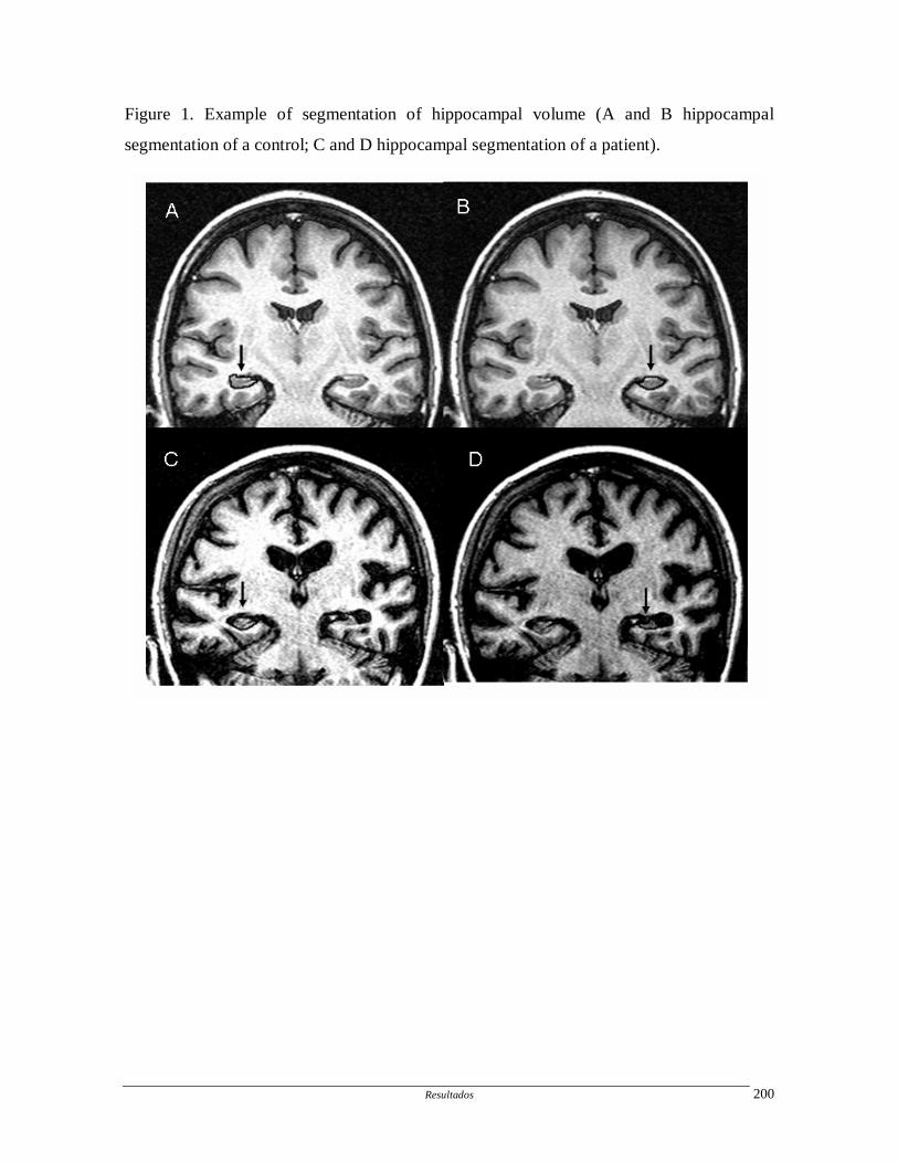
Resultados 200
Figure 1. Example of segmentation of hippocampal volume (A and B hippocampal
segmentation of a control; C and D hippocampal segmentation of a patient).
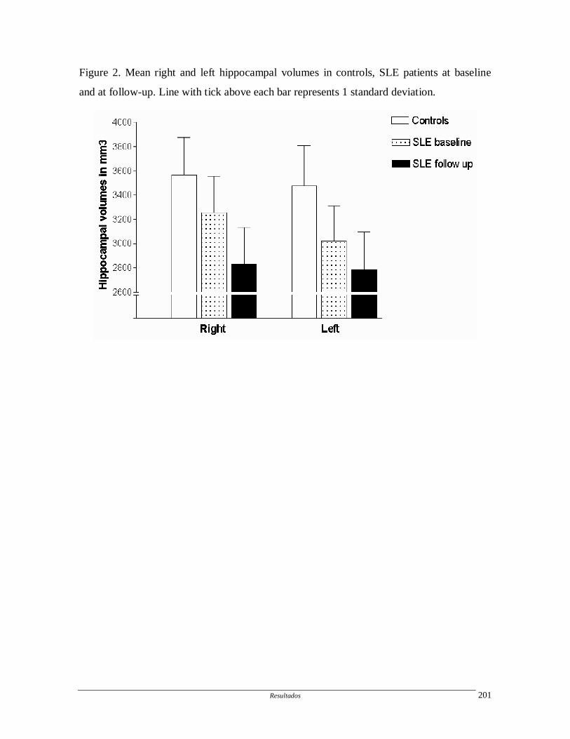
Resultados 201
Figure 2. Mean right and left hippocampal volumes in controls, SLE patients at baseline
and at follow-up. Line with tick above each bar represents 1 standard deviation.
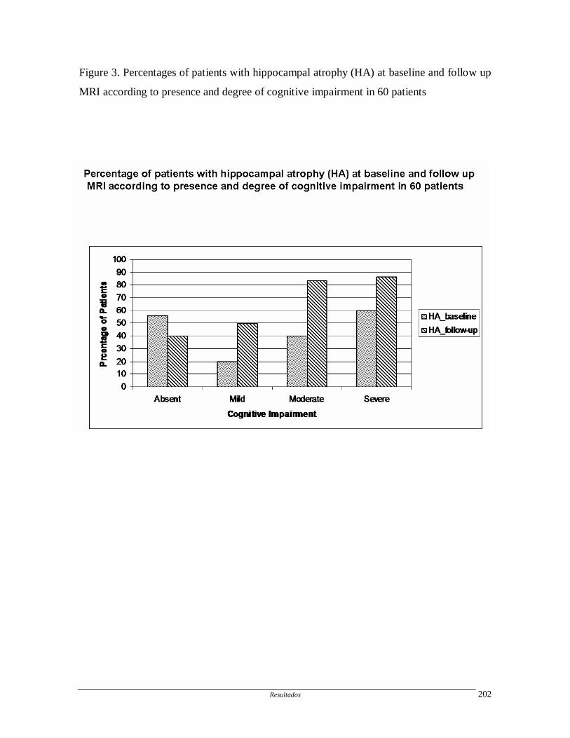
Resultados 202
Figure 3. Percentages of patients with hippocampal atrophy (HA) at baseline and follow up
MRI according to presence and degree of cognitive impairment in 60 patients
Resultados 203
ARTIGO 11
Voxel-based morphometry of brain SPECT can detect the presence of active central
nervous system involvement in systemic lupus erythematosus
Simone Appenzeller; Bárbara Juarez Amorim; Celso Dario Ramos; Pablo A Rio; Elba
Cristina Sá de Camargo Etchebehere; Edwaldo Eduardo Camargo; Fernando Cendes, Lilian
TL Costallat
Voxel-based morphometry of brain SPECT can detect the presence of active central
nervous system involvement in systemic lupus erythematosus
Rheumatology in press
[Ver: A3B2-WIN8.07r/W-Standard] [7.7.2006–5:40pm] [1–6] [Page No. 1] FIRST PROOFS
{OUP_FPP}rheumatology/kel255.3d (RHEUMATOLOGY)
Paper: kel255 OUP
Voxel-based morphometry of brain SPECT can detectthe presence of active central nervous systeminvolvement in systemic lupus erythematosus
S. Appenzeller1,2, B. J. Amorim3, C. D. Ramos3, P. A. Rio2, E. C. S. de C. Etchebehere3,
5 E. E. Camargo3, F. Cendes2,4 and L. T. L. Costallat1
Objective. To determine the value of voxel-based morphometry (VBM) of brain SPECT (single-photon emission computed
tomography) images (BSI) in discriminating active central nervous system (CNS) manifestations in systemic lupus
erythematosus (SLE) patients.
Patients and Methods. Forty SLE patients (mean age 33 yrs) and 33 normal volunteers were submitted to BSI. SLE patients
10 were screened for the presence of CNS involvement following the American College of Rheumatology (ACR) case definition.
Patients with CNS infections, uraemia, diabetes and previous ischaemic or haemorrhagic stroke were excluded. Magnetic
resonance imaging (MRI) scans were obtained in a 2T scanner (Elscint Prestige) with T1- and T2-weighted images. BSI were
performed after injection of 1110 MBq (30mCi) of 99mTc-ECD (ethyl-cysteinate-dimer). BSI were analysed using the
statistical parametric mapping. After normalization, segmentation and smoothing the groups of SLE patients with active
15 and inactive CNS manifestations and healthy volunteers were compared using VBM. Post-processed images were compared
voxel-by-voxel using t-test in order to determine differences of intensity between groups. This analysis included grand mean
scaling, proportional threshold masking (set to 0.4) and implicit masking. A P-value of 0.001 and cluster size of 32 were taken
into consideration.
Results. VBM analyses of BSI did not show any differences between SLE patients with inactive CNS involvement and normal
20 controls. However, the group of SLE patients with active CNS involvement had a global hypoperfusion, more intense in
the frontal, dorsolateral and medial temporal lobe when compared with SLE patients without CNS involvement (P¼ 0.001)
and healthy volunteers (P¼ 0.001).
Conclusion. VBM of BSI is a useful and objective method for detecting perfusion abnormalities in SLE patients, which is
indicative of active CNS involvement. However, it is not helpful in differentiating the clinical sub-types of CNS involvement
25 according to the ACR classification.
KEY WORDS: SPECT, VBM, SLE.
Introduction
Central nervous system (CNS) involvement is seen in as manyas 11–60% of systemic lupus erythematosus (SLE) patients
30 and may cause transient neurological manifestations or chronicbrain injury [1, 2]. The diagnosis of CNS involvement of SLEis difficult because of the need to differentiate primary fromsecondary causes of neurological involvement such as CNSinfections and metabolic encephalopathy. In addition, the absence
35 of reliable serum markers and an ideal imaging modality increasesthis difficulty [3].
Magnetic resonance imaging (MRI) is the preferred anatomicimaging modality [3]. MRI is more likely to show abnormalitiesif there are focal neurological deficits, seizures, chronic
40 cognitive dysfunction or the antiphospholipid syndrome.However, in many patients with obvious CNS involvement,MRI may not show abnormalities, especially in patientswith affective disorders, confusional states or headaches [3, 4].
Another drawback of MRI is the difficulty in differentiating45lesions of active CNS manifestations from old lesions [5, 6].
MRI frequently (25–50%) reveals chronic lesions in patients withand without active disease, and the incidence of these lesionsincreases with age, disease severity and past history of CNSinvolvement [3, 6, 7]. Therefore, techniques for detecting
50functional brain abnormalities may be useful. Several functionaltechniques have been used in SLE, including positron emissiontomography (PET), magnetic resonance spectroscopy (MRS),and single-photon emission computed tomography (SPECT).PET with 18-fluoro-2-deoxyglucose (FDG) shows cerebral
55involvement in patients with NP-SLE who have no morphologicalchanges detectable by CT and MRI. PET is considered to bea sensitive and reliable method for evaluating SLE patients withCNS involvement. However, it is not yet established in routineclinical use because it is expensive and not available in
60most hospitals [8]. MRS is another functional method that maybe used for detecting CNS manifestations in SLE. Although it
1Rheumatology Unit, State University of Campinas, 2Neuroimaging Lab, State University of Campinas, 3Division of Nuclear Medicine, Department of
Radiology and4Department of Neurology, FCM, State University of Campinas (UNICAMP), Campinas, Brazil.
Submitted 3 January 2006; revised version accepted 21 June 2006.
Correspondence to: Dr. F. Cendes, Department of Neurology, State University of Campinas (UNICAMP), Cidade Universitaria Zeferino Vaz CEP
13083-970 Campinas-SP, Brazil. E-mail: [email protected]
Rheumatology 2006; 1 of 6 doi:10.1093/rheumatology/kel255
54
1 of 6� The Author 2006. Published by Oxford University Press on behalf of the British Society for Rheumatology. All rights reserved. For Permissions, please email: [email protected]
[Ver: A3B2-WIN8.07r/W-Standard] [7.7.2006–5:40pm] [1–6] [Page No. 1] FIRST PROOFS
{OUP_FPP}rheumatology/kel255.3d (RHEUMATOLOGY)
Paper: kel255 OUP
is more frequently used than PET, most studies use single-voxelMRS, and so only a predetermined volume of the brain may beanalysed [9]. Multi-voxel MRS is still rare in clinical practice [9].Therefore, brain SPECT may be an alternative functional method
5 for detecting functional abnormalities in CNS manifestationsin clinical practice, providing important clinical information byimaging regional blood flow changes. Several studies used brainSPECT images (BSI) to investigate patients with SLE [3, 8, 10–20].Hypoperfusion, suggesting decreased regional blood flow, were
10 identified in SLE patients with neuropsychiatric manifestations[10, 12–17], although others have also found perfusionabnormalities in SLE patients without neuropsychiatric manifes-tations [18–20]. Most studies use visual analysis with regionalquantification of perfusion abnormalities. Semi-quantitative
15 analysis of BSI have increased diagnostic yield of BSI in otherdiseases [21–23]. Statistical parametric mapping (SPM) is anincreasingly established form of neuroimaging analysis to detectstatistically significant differences in spatially normalized imageson a voxel-by-voxel basis [24–26]. The use of SPM eliminates
20 observer subjectivity inherent in visual analysis [27–29].The purpose of this study was to determine the value of voxel-based morphometry (VBM) of brain SPECT images (BSI) indiscriminating active CNS manifestations in SLE patients.
Subjects and method
25 Subjects
Sixty consecutive SLE patients fulfilling four or more criteria forclassification of SLE [30] with CNS involvement were invitedto participate in the study. All patients had active or past historyof CNS involvement as defined by the American College of
30 Rheumatology (ACR) case definition [31]. Active CNS disease, asdefined by the new onset or persistence of a CNS manifestationat the time of examination, was identified in 27 patients.The remaining 33 patients had inactive (past history) CNSinvolvement. Patients were followed-up prospectively in the
35 Rheumatology Unit of the State University of Campinas(UNICAMP).
We excluded patients with associated clinical conditions thatcould cause cerebral atrophy, such as stroke (10 patients), arterialhypertension (one patient), diabetes mellitus, alcohol and drug
40 abuse and malignancy. Patients satisfying the ACR criteriafor rheumatoid arthritis, systemic sclerosis, Sjogren syndrome(two patients) or other connective tissue disease and with drug-induced SLE were also excluded.
Total dose of corticosteroids and other immunosuppressant45 medications used since the onset of disease were estimated
using the data obtained by careful review of the medical charts.Seven patients with incomplete charts were excluded fromthe analysis.
Therefore, 40 patients (20 with active and 20 with inactive50 CNS manifestations) were submitted to BSI. The control group
consisted of 50 healthy age- and sex-matched volunteers.This study was approved by the Ethical Committee of our
institution and informed, written consent was obtained fromeach subject.
55 Clinical, serological and treatment features of SLE patients
Clinical manifestations. Data on gender and age at diseaseonset and disease duration were collected for each patient.Disease duration was defined as the initial manifestation clearlyattributable to SLE until the day of BSI acquisition. All clinical
60 manifestations and laboratory test findings were recordedaccording to ACR criteria [30, 31]. Disease activity was measuredin all visits using the systemic lupus erythematosus disease activityindex (SLEDAI), which is a standardized score for SLE patientsand includes 24 items [32]. The SLEDAI score is calculated by
65summing the predetermined weights for the items that arepresent. Items that are life-threatening have higher weights, withpossible scores ranging from 0 to 105. Cumulative organ damagewas analysed by validated damage index (SLICC/ACR-DI) [33].SLICC/ACR-DI is an unweighted index composed of 41 items
70grouped in 12 domains, with a maximum possible score of 47.As previously established, damage was considered when theirreversible lesions were present for at least 6 months unrelatedto active inflammation and had occurred after SLE diagnosis [33].
CNS involvement. A complete neurological examination,75as well as cognitive and psychiatric charts, was prospectively
applied to all patients during their clinical visit in order to identifyactive CNS involvement [31]. Mini Mental State Examination [34]was applied to all participants. All patients were submitted toa battery of standardized neuropsychological tests in order to
80screen for possible impairment in one or more of the subsequentcognitive domains: simple attention, complex attention, memory,visuospatial processing, language, reasoning/problem solving,psychomotor speed and executive functions [35–38]. These testshave not been validated for SLE patients, but are widely used for
85patients with CNS disorders in clinical practice and research.The individual test results were converted into standard scores,which were compared with the available normative data [35–38].Regarding any of the eight cognitive domains, subjects with atotal score of �2 S.D. below the normative value were considered
90to be impaired. Cognitive dysfunction was classified as mildif there were deficits in less than three dimensions, as moderate ifthere were deficits in three or four dimensions and as severeif there were deficits in at least five dimensions [38, 39].
Assessment of depression was based on clinical interview and95the Beck Depression Inventory (BDI) [40, 41]. On BDI, scores
from 10 to 17 were considered to indicate mild depression,from 18 to 24 moderate depression and greater than 24 severedepression. Anxiety was evaluated by anxiety through theHospital Anxiety and Depression scale [42]. The presence of
100psychosis was determined through the Brief Psychiatry RatingScale (aBPRS) [43].
For past history of CNS involvement we reviewed the medicalcharts of patients.
Laboratory features. Antinuclear antibodies (ANA) were105determined by indirect immunofluorescence using mouse liver
as the substrate, and were regarded as positive if higherthan 1:40. Anti-double-stranded DNA (AdsDNA) antibodieswere determined by indirect immunofluorescence usingChrithidia as substrate, and were considered positive if higher
110than 1:10. Precipitating antibodies to extractable nuclearantigens (ENA), including Ro (SSA), La (SSB) and Sm weredetected by immunodiffusion and/or microhaemagglutination.Anticardiolipin antibodies (aCL) of the IgG and IgM isotypeswere measured by the ELISA method [44]. Lupus anticoagulant
115(LA) activity was detected by coagulation assays in platelet-free plasma obtained by double centrifugation, following therecommendation of the subcommittee on LA of the Scientificand Standardization Committee of the International Society ofThrombosis and Homeostasis [45].
120BSI acquisition. After completing inclusion and exclusioncriteria, 40 patients (20 with active and 20 with inactive CNSmanifestations) were required to remain resting in a dark, quietroom for 15min, with a permanent intravenous access througha butterfly connected to a catheter with saline solution. While at
125rest, 1110MBq (30mCi) of 99mTc-ECD (ethyl-cysteinate-dimer)was injected. The patients remained resting for another 10min.BSI was performed in a computed scintillation camera with afan-beam collimator. Sixty images were acquired in a 64� 64matrix, every 68, in a total of 3608. Raw data were reconstructed
130by filtered backprojection, and attenuation correction was
2 of 6 S. Appenzeller et al.
[Ver: A3B2-WIN8.07r/W-Standard] [7.7.2006–5:40pm] [1–6] [Page No. 1] FIRST PROOFS
{OUP_FPP}rheumatology/kel255.3d (RHEUMATOLOGY)
Paper: kel255 OUP
performed using Chang’s method with a 0.115 attenuationcoefficient.
BSI analysis. The reconstructed BSI were converted intoAnalyze format using MRIcro software (www.mricro.com).
5 Voxel-based analysis was performed using SPM2 (WellcomeDepartment of Cognitive Neurology, London)
To allow group comparisons, the size and shape ofeach individual’s scan were normalized to sterotaxic space byestimating the optimum 12-parameter affine transformation
10 [46–48]. The 99mTc-ECD uptake was standardized to the meanglobal uptake using a proportional scaling. At this point, standardVBM protocols include segmentation of brain tissues, but becauseBSI signals are acquired from radio tracers dispersed in bloodflow and most of blood vessels are in the GM portion of brain,
15 this step was not necessary and is not recommended for nuclearmedicine images to avoid spatial resolution worsening. The imageswere subsequently smoothed by convolving its voxels with anisotropic Gaussian kernel (IGK) of 10mm in order to minimizeborder effects caused by gyral inter-individual variability and
20 create images that can be spatially compared with a goodcorrespondence of analogous tissues.
MRI acquisition protocol and analysis. MRI scanswere obtained in a 2T Elscint Prestige scanner. T1-weightedgradient-echo sequence with 1mm thickness (TR¼ 22ms,
25 TE¼ 9ms, flip angle¼ 358 and matrix¼ 256� 220) was usedfor voxel-based morphometry (VBM) analysis. Images werenormalized to the standard space using 12 linear parametersand 7� 8� 7 nonlinear basis functions, using a brain mask.Spatially normalized images were re-sliced to isotropic voxels of
30 1.5mm and underwent segmentation of white and gray matter.The images were smoothed by convolution with an IGK of 10mmin order to minimize inter-individual gyral variability. Theresulting images were then compared voxel-by-voxel by usingt-test to determine differences in gray matter between patients
35 with active and inactive CNS involvement and controlsusing statistical parametric mapping (SPM 2). This analysisincluded grand mean scaling and proportional thresholdmasking (set to 0.4) and implicit masking. A P-value of 0.001was taken into analysis, and the minimum cluster size taken
40 into account was 32.
Statistical analysis. Group differences for age were assessedusing one-way ANOVA, and the gender distribution wasevaluated with the chi-square test.
The statistical analysis of the normalized and smoothed BSI45 data was performed using the SPM2. Statistical analysis was
performed by comparing both groups of patients (with andwithout CNS manifestations) with the control group. It wasalso performed a comparison between the group of patientswith CNS manifestation and the group of patients without
50 manifestation. These comparisons between these groups wereperformed using a non-paired two-sample t-test. Only voxels withsignal intensity above a threshold of 0.4 were entered in eachanalysis. It was used with a P-value of 0.001 with false discoveryrate (FDR) and a cluster size of 32. The tool FDRs minimize
55 errors from multiple comparisons and eliminates, from the finalstatistical t-map, all values with a probability of being a falsepositive discovery. This step is important since the naturalvariability can produce false discoveries and lead to incorrectresults [49]. The areas of hypoperfusion on BSI were compared
60 with areas of reduced voxel number (atrophy) on MRI in orderto determine if the hypoperfusion is due to cerebral atrophyin SLE patients.
In order to determine the relationship between specific clinicalmanifestation and pattern of reduced blood flow assessment,
65 multiple regression was used.
Results
Demographic data
We included 40 SLE patients with mean age of 33.3 yrs(range 18–45 yrs, S.D.¼ 12.46). Thirty-eight were women. The
70mean duration of disease was 31.5 months (range 1–150,S.D.¼ 58.50) (Table 1).
The control group consisted of 33 healthy volunteers(29 women and 4 men) with mean age 30.6 yrs (range 20–53 yrs;S.D.¼ 8.65 yrs).
75Clinical, laboratory and treatment features
Clinical features are summarized in Table 2. Mean SLEDAIscores was 4.7 points (S.D.¼ 0.74). Mean SLICC score was3.9 (S.D.¼ 2.3) (Table 1). Of 48 CNS manifestations observedin SLE patients, 27 manifestations were active in 20 patients
80(Table 3). Although headache is the most frequently observedCNS manifestation, it occurred isolated only in two patients withinactive CNS manifestations. The other patients had other CNSmanifestations, especially cognitive dysfunction, associated.
All patients were on corticosteroid dose with doses ranging85from 5 to 60mg/day. Fifteen patients were on chloroquine and
eight patients (three with active and five with inactive CNSmanifestations) were receiving azathioprine.
MRI findings
Areas of abnormal T2 signal, identified as small white matter90lesions, were observed in 10 (25%) patients. Five of these patients
had cognitive impairment, four had seizures and one patienthad aseptic meningitis. All patients with MRI abnormalities hadactive (four patients) or past history (six patients) of CNSmanifestations. On visual analysis, no further abnormalities
TABLE 1. Demographic characteristics in groupsa
Data
SLE withactive
CNS disease
SLE withpast history
of CNS diseaseHealthycontrols
Age (mean� S.D. yrs) 32.4� 11.8 32.6� 13.1 30.6� 8.6Female: male ratio 18/2 20/0 29/4Disease duration
(mean months� S.D.)30.2� 10.2 33� 8.4 –
SLEDAI (mean� S.D.) 5.1� 0.6 4.8� 0.9 –SLICC (mean� S.D.) 4.1� 1.5 3.9� 1.9 –
aP> 0.05 in all comparisons.
TABLE 2. Clinical manifestations in SLE patients with active and inactiveCNS involvement
Manifestations
SLE patientswith active CNS
involvement, n (%)
SLE patients withinactive CNS
involvement, n (%)
Arthritis 16 (80) 15 (75)Avascular necrosis 1 (5) 2 (10)Discoid rash 6 (30) 7 (35)Fever 18 (90) 18 (20)Hemolytic anaemia 4 (20) 3 (15)Leucopenia 10 (50) 12 (60)Malar rash 12 (60.0) 10 (50)Nephropathy 6 (30) 7 (35)Oral Ulcers 4 (20) 4 (20)Photosensitivity 14 (70) 15 (75)Raynaud’s phenomenon 6 (30) 7 (35)Serositis 7 (35) 5 (25)Thrombocytopenia 3 (15) 4 (20)
SPECT VBM in SLE 3 of 6
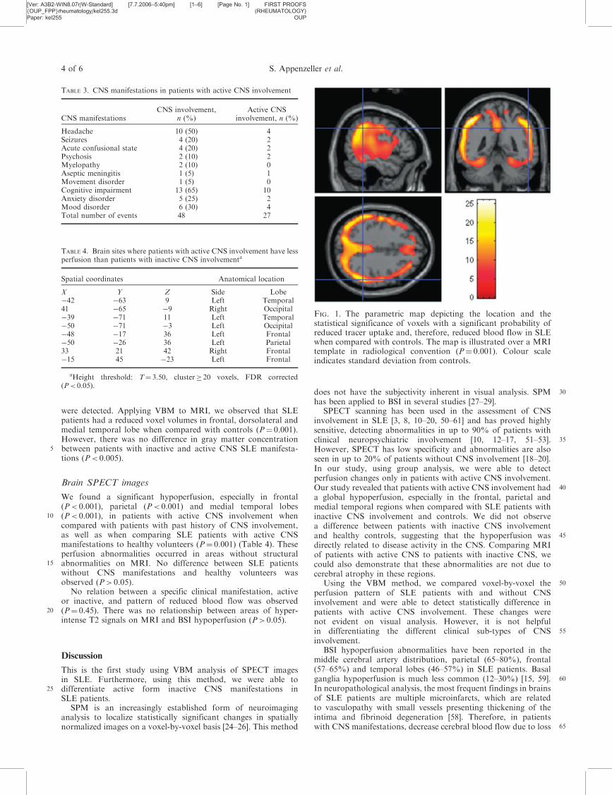
[Ver: A3B2-WIN8.07r/W-Standard] [7.7.2006–5:40pm] [1–6] [Page No. 1] FIRST PROOFS
{OUP_FPP}rheumatology/kel255.3d (RHEUMATOLOGY)
Paper: kel255 OUP
were detected. Applying VBM to MRI, we observed that SLEpatients had a reduced voxel volumes in frontal, dorsolateral andmedial temporal lobe when compared with controls (P¼ 0.001).However, there was no difference in gray matter concentration
5 between patients with inactive and active CNS SLE manifesta-tions (P< 0.005).
Brain SPECT images
We found a significant hypoperfusion, especially in frontal(P< 0.001), parietal (P< 0.001) and medial temporal lobes
10 (P< 0.001), in patients with active CNS involvement whencompared with patients with past history of CNS involvement,as well as when comparing SLE patients with active CNSmanifestations to healthy volunteers (P¼ 0.001) (Table 4). Theseperfusion abnormalities occurred in areas without structural
15 abnormalities on MRI. No difference between SLE patientswithout CNS manifestations and healthy volunteers wasobserved (P> 0.05).
No relation between a specific clinical manifestation, activeor inactive, and pattern of reduced blood flow was observed
20 (P¼ 0.45). There was no relationship between areas of hyper-intense T2 signals on MRI and BSI hypoperfusion (P> 0.05).
Discussion
This is the first study using VBM analysis of SPECT imagesin SLE. Furthermore, using this method, we were able to
25 differentiate active form inactive CNS manifestations inSLE patients.
SPM is an increasingly established form of neuroimaginganalysis to localize statistically significant changes in spatiallynormalized images on a voxel-by-voxel basis [24–26]. This method
30does not have the subjectivity inherent in visual analysis. SPMhas been applied to BSI in several studies [27–29].
SPECT scanning has been used in the assessment of CNSinvolvement in SLE [3, 8, 10–20, 50–61] and has proved highlysensitive, detecting abnormalities in up to 90% of patients with
35clinical neuropsychiatric involvement [10, 12–17, 51–53].However, SPECT has low specificity and abnormalities are alsoseen in up to 20% of patients without CNS involvement [18–20].In our study, using group analysis, we were able to detectperfusion changes only in patients with active CNS involvement.
40Our study revealed that patients with active CNS involvement hada global hypoperfusion, especially in the frontal, parietal andmedial temporal regions when compared with SLE patients withinactive CNS involvement and controls. We did not observea difference between patients with inactive CNS involvement
45and healthy controls, suggesting that the hypoperfusion wasdirectly related to disease activity in the CNS. Comparing MRIof patients with active CNS to patients with inactive CNS, wecould also demonstrate that these abnormalities are not due tocerebral atrophy in these regions.
50Using the VBM method, we compared voxel-by-voxel theperfusion pattern of SLE patients with and without CNSinvolvement and were able to detect statistically difference inpatients with active CNS involvement. These changes werenot evident on visual analysis. However, it is not helpful
55in differentiating the different clinical sub-types of CNSinvolvement.
BSI hypoperfusion abnormalities have been reported in themiddle cerebral artery distribution, parietal (65–80%), frontal(57–65%) and temporal lobes (46–57%) in SLE patients. Basal
60ganglia hypoperfusion is much less common (12–30%) [15, 59].In neuropathological analysis, the most frequent findings in brainsof SLE patients are multiple microinfarcts, which are relatedto vasculopathy with small vessels presenting thickening of theintima and fibrinoid degeneration [58]. Therefore, in patients
65with CNS manifestations, decrease cerebral blood flow due to loss
TABLE 4. Brain sites where patients with active CNS involvement have lessperfusion than patients with inactive CNS involvementa
Spatial coordinates Anatomical location
X Y Z Side Lobe�42 �63 9 Left Temporal41 �65 �9 Right Occipital�39 �71 11 Left Temporal�50 �71 �3 Left Occipital�48 �17 36 Left Frontal�50 �26 36 Left Parietal33 21 42 Right Frontal�15 45 �23 Left Frontal
aHeight threshold: T¼ 3.50, cluster� 20 voxels, FDR corrected(P< 0.05).
TABLE 3. CNS manifestations in patients with active CNS involvement
CNS manifestationsCNS involvement,
n (%)Active CNS
involvement, n (%)
Headache 10 (50) 4Seizures 4 (20) 2Acute confusional state 4 (20) 2Psychosis 2 (10) 2Myelopathy 2 (10) 0Aseptic meningitis 1 (5) 1Movement disorder 1 (5) 0Cognitive impairment 13 (65) 10Anxiety disorder 5 (25) 2Mood disorder 6 (30) 4Total number of events 48 27
FIG. 1. The parametric map depicting the location and thestatistical significance of voxels with a significant probability ofreduced tracer uptake and, therefore, reduced blood flow in SLEwhen compared with controls. The map is illustrated over a MRItemplate in radiological convention (P¼ 0.001). Colour scaleindicates standard deviation from controls.
4 of 6 S. Appenzeller et al.
[Ver: A3B2-WIN8.07r/W-Standard] [7.7.2006–5:40pm] [1–6] [Page No. 1] FIRST PROOFS
{OUP_FPP}rheumatology/kel255.3d (RHEUMATOLOGY)
Paper: kel255 OUP
of cerebral perfusion reserve occurs earlier than detected onstructural MRI. This explains why our patients with active CNSmanifestations presented hypoperfusion on BSI images.
In agreement with previous data [10, 13, 15], we did not find5 a relationship between the type of CNS manifestations and the
pattern of perfusion abnormalities. Thus, BSI scanning may beused mainly to support a clinical diagnosis of active neuro-psychiatric involvement as a syndrome and not to differentiatebetween different types of manifestations. Perhaps the inclusion of
10 a greater number of individual manifestations could differentiatedifferent patterns of hypoperfusion on BSI. In order to determineif hypoperfusion could be secondary to cerebral atrophy, weanalysed the MRI data of patients and controls. Although weobserved a statistical difference in relation to gray matter volume
15 reduction in patients when compared with controls, there was nodifference in gray matter volume between patients with active andinactive CNS involvement. These findings support the idea thathypoperfusion could be secondary to disease activity in the CNSand not only due to the cortical atrophy.
20 The absence of SLE patients with quiescent SLE and SLEpatients with active disease without CNS involvement is alimitation of this study. Further studies are necessary to determinethe relationships between structural and functional abnormalitiesin these groups of patients.
25 Because functional abnormalities may precede anatomicabnormalities, perfusion and metabolic studies employing BSIare complementary to MRI in diagnosing CNS involvement.Qualitative analysis of BSI has an interobserver variabilityand the importance of combining neuroimaging studies has been
30 emphasized in order to assess both brain structure and function.
The authors have declared no conflicts of interest.
References
1. West SG. Neuropsychiatric lupus. Rheum Dis Clin North Am
1994;20:129–58.35 2. Ward MM, Pyun E, Studenski S. Mortality risks associated with
specific clinical manifestations of systemic lupus erythematosus.
Arch Int Med 1996;156:1337–44.
3. Sibbitt WL, Sibbitt RR, Brooks WM. Neuroimaging in neuro-
psychiatric systemic lupus erythematosus. Arthritis Rheum40 1999;42:2026–38.
4. West SG, Emlen W, Wener MH, Kotzin BL. Neuropsychiatric lupus
erythematosus: a 10-year prospective study on the value of diagnostic
tests. Am J Med 1995;99:153–63.
5. McCune WJ, MacGuire A, Aisen A, Gebarski S. Identification of45 brain lesions in neuropsychiatric systemic lupus erythematosus by
magnetic resonance scanning. Arthritis Rheum 1988;31:159–66.
6. Jarek MJ, West SG, Baker MR, Rak KM. Magnetic resonance
imaging in systemic lupus erythematosus patients without a
history of neuropsychiatric lupus erythematosus. Arthritis Rheum50 1994;37:1609–13.
7. Sanna G, Piga M, Terryherry JW et al. Central nervous system
involvement in systemic lupus erythematosus: cerebral imaging and
serological profile in patients with and without overt neuropsychiatric
manifestations. Lupus 2000;9:573–83.558. Huang WS, Chiu PY, Tsai CH, Hao A, Lee CC. Objective evidence
of abnormal regional cerebral blood flow in patients with systemic
lupus erythematosus on Tc-99mECD brain SPECT. Rheumatol Int
2002;22:178–81.
9. Sundgren PC, Jennings J, Attwood JT et al. MRI and 2D-CSI MR60spectroscopy of the brain in the evaluation of patients with acute
onset of neuropsychiatric systemic lupus erythematosus.
Neuroradiology 2005;47:576–85.
10. Oku K, Atsumi T, Furukawa S et al. Cerebral imaging by magnetic
resonance imaging and single photon emission computed tomography65in systemic lupus erythematosus with central nervous system
involvement. Rheumatology 2003;12:773–7.
11. Liu FY, Huang WS, Kao CH, Yean RF, Wang JI, Hu ST. Usefulness
of Tc-99 ECD brain SPECT to evaluate the effects of
methylpredinisolone pulse therapy in lupus erythematosus with70brain involvement: a preliminary report. Rheumatol Int
2003;23:182–5.
12. Handa R, Sahota P, Kumar M et al. In vivo proton magnetic
resonance spectroscopy (MRS) and single photon emission compu-
terized tomography (SPECT) in systemic lupus erythematosus (SLE).75Magn Reson Imaging 2003;21:1033–7.
13. Colamussi P, Trotta F, Ricci R et al. Brain perfusion SPET and
proton magnetic resonance spectroscopy in the evaluation of two
systemic lupus erythematosus patients with mild neuropsychiatric
manifestations. Nuclear Medicine Communications 1997;18:269–73.8014. Zhang X, Zhu Z, Zhang F, Shu H, Li F, Dong Y. Diagnostic value of
single-photon-emission computed tomography in severe central
nervous system involvement of systemic lupus erythematosus: a
case-control study. Arthritis Rheum 2005;53:845–9.
15. Lin WY, Wang SJ, Yen TC, Lan JL. Technetium-99m-HMPAO brain85SPECT in systemic lupus erythematosus with CNS involvement. J
Nucl Med 1997;38:1112–5.
16. Kao CH, Lan JL, ChangLai SP, Liao KK, Yen RF, Chieng PU.
The role of FDG-PET, HMPAO-SPET and MRI in the detection of
brain involvement in patients with systemic lupus erythematosus.90Eur J Nucl Med 1999;26:129–34.
17. Huang WS, Chiu PY, Tsai CH, Kao A, Lee CC. Objective evidence of
abnormal regional cerebral blood flow in patients with systemic lupus
erythematosus on Tc-99m ECD brain SPECT. Rheumatol Int
2002;22:178–81.9518. Chen JJ, Yen RF, Kao A, Lin CC, Lee CC. Abnormal regional
cerebral blood flow found by technetium-99m ethyl cysteinate dimer
brain single photon emission computed tomography in systemic lupus
erythematosus patients with normal brain MRI findings. Clin
Rheumatol 2002;21:516–9.10019. Waterloo K, Omdal R, Sjoholm H et al. Neuropsychological
dysfunction in systemic lupus erythematosus is not associated with
changes in cerebral blood flow. J Neurol 2001;248:595–602.
20. Lopez-Longo FJ, Carol N, Almoguera MI et al. Cerebral hypo-
perfusion detected by SPECT in patients with systemic lupus105erythematosus is related to clinical activity and cumulative tissue
damage. Lupus 2003;21:813–9.
21. Zugbal IG, Spencer SS, Iman K et al. Differences images calculated
from ictal and interictal technesium-00m-HMPAO SPECT scans of
epilepsy. J Nucl Med 1995;36:684–9.11022. Spanaki MV, Spencer SS, Wiesnieski G et al. Evolution and
localization of postictal blood flow changes in partial seizures
demonstrated by SPECT: use of quantitative differences images.
J Epilepsy 1998;11:25–33.
23. O’Brien TJ, So EL, Mullan BP et al. Subtraction ictal SPECT115co-registered to MRI improves clinical usefulness of SPECT in
localizing surgical seizure focus. Neurology 1998;50:445–54.
24. Stefan H, Bauer J, Feistel H et al. Regional cerebral blood flow during
focal seizures of temporal and frontocentral onset. Ann Neurol
1990;27:162–6.
Rheumatology
Key message
� VBM of BSI is a useful and objectivemethod for detecting perfusionabnormalities.
� VBM was able to differentiate activefrom inactive CNS manifestations inSLE patients.
� It is not helpful in differentiating theclinical sub-types of CNS involvement.
SPECT VBM in SLE 5 of 6
[Ver: A3B2-WIN8.07r/W-Standard] [7.7.2006–5:40pm] [1–6] [Page No. 1] FIRST PROOFS
{OUP_FPP}rheumatology/kel255.3d (RHEUMATOLOGY)
Paper: kel255 OUP
25. Rowe CC, Berkovic SF, Austin MC et al. Patterns of postictal
cerebral blood flow in temporal lobe epilepsy. Neurology
1991;41:1096–103.
26. Duncan R, Patterson J, Roberts R et al. Ictal/postictal SPECT in the5 pre-surgical localization of complex partial seizures. J Neurol
Neurosurg Psychiatry 1993;56:141–8.
27. Friston KJ, Holmes A, Poline JB et al. Detecting activations in
PET and fMRI: levels of inference and power. Neuroimage
1996;40:223–35.10 28. Friston KJ, Holmes AP, Worsley KJ et al. Statistical parametric maps
in functional imaging: a general linear approach. Hum Brain Mapp
1995;2:89–210.
29. West J, Fitzpatrick JM, Wang MY et al. Comparison and evaluation
of retrospective intermodality image registration techniques.15 J Comput Assist Tomogr 1997;21:554–66.
30. Tan EM, Cohen AS, Fries JF et al. The 1982 revised criteria for the
classification of systemic lupus erythematosus. Arthritis Rheum
1982;25:1271–7.
31. Liang MH, Corzillius M, Bae SC et al. The American College20 Nomenclature and case definitions for neuropsychiatric lupus
syndrome. Arthritis Rheum 1999;42:599–608.
32. The Committee on prognosis studies in SLE, Bombardier C,
Gladman DD, Urowitz M, Caron D, Chang C. Derivation of the
SLEDAI: a disease activity index for lupus patients. Arthritis Rheum25 1992;35:630–40.
33. Gladman D, Glinzler E, Goldsmith C et al. The development and
initial validation of the Systemic Lupus International Collaborating
Clinics/American College of Rheumatology damage index for
systemic lupus erythematosus. Arthritis Rheum 1996;39:363–78.30 34. Folstein MF, Folstein SE, Folstein PR. ‘‘Mini mental state’’ A
practical method for grading the cognitive state of patient for the
clinician. J Psychiatr Res 1975;12:189–98.
35. Spranoel HZ. The psychoeducational use and interpretation of the
Wechsler Adult Intelligence Scale–Revised. Springfield: CC Thomas,35 1992.
36. Wechsler in Muistiasteikko, Suom. Anja Manner. Helsinki:
Psykologien Kustannus, 1986.
37. Delis DC, Kramer JH, Kaplan E et al. California verbal learning test
research edition manual. San Antonio: The Psychological40 Corporation, 1987.
38. Lezak MD. Neuropsychological assessment. New York: Oxford
University Press, 1995.
39. Heaton RK, Chelune GJ, Talley JL et al. Wisconsin Card Sorting
Test Manual, revised and expanded. Odessa: Psychological45 Assessment Resources Inc., 1993.
40. Beck AT, Beamesderfer A. Assessment of depression: the depression
inventory. Mod Probl Pharmacopsychiat 1974;7:151–69.
41. Beck AT, Rial WY, Rickels K. Short form of depression inventory:
cross-validation. Psychol Rep 1974;34:1184–6.50 42. Herrmann C. International experiences with the Hospital Anxiety and
Depression Scale – a review of validation data and clinical results.
J Psychosom Res 1997;42:17–41.
43. Overall JE, Beller SA. The Brief Psychiatric Rating Scale (BPRS) in
geropsychiatric research: I. Factor structure on an inpatient unit.55 J Gerontol 1984;39:187–93.
44. Harris EN, Gharavi AE, Patel SP, Hughes GR. Evaluation of the
anticardiolipine antibody test: report of an international workshop
held 4 April 1986. Clin Exp Immunol 1987;68:215–22.
45. Brandt JT, Triplett DA, Scharer I. Criteria for the diagnosis of lupus60anticoagulant: an update. Thromb Haemost 1995;74:1185–90.
46. Ashburner J, Friston KJ. Multimodal image coregistration and
partitioning – a unified framework. Neuroimage 1997;6:209–17.
47. Ashburner J, Neelin P, Collins DL, Evans AC, Friston K.
Incorporating prior knowledge into imaging registration.65NeuroImage 2001;6:344–52.
48. Friston KJ, Holmes AP, Worsley KP, Proline JB, Frith CD,
Frackowiak RSJ. Statistical parametric maps in functional imaging:
a general linear approach. Human Brain Mapp 1995;2:189–210.
49. Genovese CR, Lazar NA, Nichols T. Thresholding of statistical maps70in functional neuroimaging using the false discovery rate. Neuroimage
2002;15:870–8.
50. Rubbert A, Marienhagen J, Pirner K et al. Single-photon emission
computed tomography analysis of cerebral blood flow in the
evaluation of central nervous system involvement in patients with75systemic lupus erythematosus. Arthritis Rheum 1993;36:1253–62.
51. Emmi L, Bramati M, De Cristofaro MT et al. MRI and SPECT
investigations of the CNS in SLE patients. Clin Exp Rheumatol
1993;11:13–20.
52. Nossent JC, Hovestadt A, Schonfeld DH, Swaak AJ. Single-photon-80emission computed tomography of the brain in the evaluation of
cerebral lupus. Arthritis Rheum 1991;34:1397–403.
53. Szer IS, Miller JH, Rawlings D, Shaham B, Bernstein B. Cerebral
perfusion abnormalities in children with central nervous system
manifestations of lupus detected by single photon emission computed85tomography. J Rheumatol 1993;20:2143–8.
54. Kovacs JA, Urowitz MB, Gladman DD, Zeman R. The use of single
photon emission computerized tomography in neuropsychiatric SLE:
a pilot study. J Rheumatol 1995;22:1247–53.
55. Reiff A, Miller J, Shaham B, Bernstein B, Szer IS. Childhood central90nervous system lupus: longitudinal assessment using single photon
emission computed tomography. J Rheumatol 1997;24:2461–5.
56. Aisen AM, Gabrielsen TO, McCune WJ. MR imaging of systemic
lupus erythematosus involving the brain. Am J Roentgenol
1985;144:1027–31.9557. Stoppe G, Wildhagen K, Seidel JW et al. Position emission
tomography in neuropsychiatric lupus erythematosus. Neurology
1990;40:304–8.
58. Chia-Hung K, Yung-Jen HL, Jung-Liang C, Sheng-K Ping L,
Ko-Kaung L, Poon-Ung C. Discrepancy between regional cerebral100blood flow and glucose metabolism of the brain in systemic lupus
erythematosus patients with normal brain magnetic resonance
imaging findings. Arthritis Rheum 1999;42:61–8.
59. Colamussi P, Giganti M, Cittanti C et al. Brain single photon
emission tomography with 99m Tc-HMPAO in neuropsychiatric105systemic lupus erythematosus: relations with EEG and MRI findings
and clinical manifestations. Eur J Nucl Med 1995;22:17–24.
60. Oda K, Matsushima E, Okubo Y et al. Abnormal regional cerebral
blood flow in systemic lupus erythematosus patients with psychiatric
symptoms. J Clin Psychiatry 2005;66:907–13.11061. Abreu MR, Jakosky A, Folgerini M, Brenol JC, Xavier RM,
Kapczinsky F. Neuropsychiatric systemic lupus erythematosus:
correlation of brain MR imaging, CT, and SPECT. Clin Imaging
2005;29:215–21.
62. Castellino G, Govoni M, Padovan M, Colamussi P, Borrelli M,115Trotta F. Proton magnetic resonance spectroscopy may predict future
brain lesions in SLE patients: a functional multi-imaging approach
and follow up. Ann Rheum Dis 2005;64:1022–7.
6 of 6 S. Appenzeller et al.
Resultados 210
ARTIGO 12
Evidence of reversible axonal dysfunction in systemic lupus erythematosus: a proton
MRS study
Appenzeller S, Li LM, Costallat LT, Cendes F.
Evidence of reversible axonal dysfunction in systemic lupus erythematosus: a proton MRS
study
Brain. 2005 Dec; 128(Pt 12):2933-40.
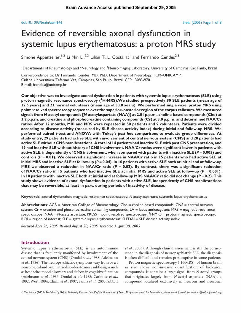
doi:10.1093/brain/awh646 Brain (2005) Page 1 of 8
Evidence of reversible axonal dysfunction insystemic lupus erythematosus: a proton MRS study
Simone Appenzeller,1,3 Li Min Li,2,3 Lilian T. L. Costallat1 and Fernando Cendes2,3
1Departments of Rheumatology and 2Neurology and 3Neuroimaging Laboratory, University of Campinas, Sao Paulo, Brazil
Correspondence to: Dr Fernando Cendes, MD, PhD, Department of Neurology, FCM–UNICAMP,Cidade Universitaria Zeferino Vaz, Campinas, Sao Paulo, Brazil, CEP 13083-970E-mail: [email protected]
Our objective was to investigate axonal dysfunction in patients with systemic lupus erythematosus (SLE) usingproton magnetic resonance spectroscopy (1H-MRS).We studied prospectively 90 SLE patients (mean age of32.5 years) and 23 normal volunteers (mean age of 33.8 years). We performed single voxel proton MRS usingpoint resolved spectroscopy sequence over the superior–posterior region of the corpus callosum. We measuredsignals from N-acetyl compounds [N-acetylaspartate (NAA)] at 2.01 p.p.m., choline-based compounds (Cho) at3.2 p.p.m. and creatine and phosphocreatine containing compounds (Cr) at 3.0 p.p.m. and determined NAA/Crratios. After 12 months, MRI and MRS were repeated in 50 patients and 9 volunteers. Patients were dividedaccording to disease activity (measured by SLE disease activity index) during initial and follow-up MRS. Weperformed paired t-test and ANOVA with Tukey’s post hoc comparisons to evaluate group differences. Atstudy entry, 29 patients had active SLE with involvement of central nervous system (CNS) and 28 patients hadactive SLE without CNS manifestations. A total of 14 patients had inactive SLE with past CNS presentation, and19 had inactive SLE without history of CNS involvement. NAA/Cr ratios were significant lower in patients withactive SLE, independently of CNS involvement, when compared with patients with inactive SLE (P = 0.005) andcontrols (P = 0.01). We observed a significant increase in NAA/Cr ratio in 15 patients who had active SLE atinitial MRS and inactive SLE at follow-up (P = 0.04). In 10 patients with active SLE both at initial and at follow-upMRS we observed a reduction in NAA/Cr ratio (P = 0.02). By contrast, there was a significant reductionof NAA/Cr ratio in 15 patients who had inactive SLE at initial MRS and active SLE at follow-up (P = 0.001).In 10 patients with inactive SLE both at initial and at follow-up MRS NAA/Cr ratio did not change (P = 0.2). Thisstudy shows evidence of axonal dysfunction in patients with active SLE, independently of CNS manifestationsthat may be reversible, at least in part, during periods of inactivity of disease.
Keywords: axonal dysfunction; magnetic resonance spectroscopy; N-acetylaspartate; systemic lupus erythematosus
Abbreviations: ACR = American College of Rheumatology; Cho = choline-based compounds; CNS = central nervoussystem; Cr = creatine and phosphocreatine containing compounds; LA = lupus anticoagulant; MRS = magnetic resonancespectroscopy; NAA = N-acetylaspartate; PRESS = point resolved spectroscopy; 1H-MRS = proton magnetic spectroscopy;ROI = region of interest; SLE = systemic lupus erythematosus; SLEDAI = SLE disease activity index
Received April 26, 2005. Revised August 20, 2005. Accepted August 30, 2005
IntroductionSystemic lupus erythematosus (SLE) is an autoimmune
disease that is frequently manifested by involvement of the
central nervous system (CNS) (Omdal et al., 1988; Adelmann
et al., 1986). The neuropsychiatric symptoms vary from overt
neurologicalandpsychiatricdisorders tomoresubtle signssuch
as headache, mood disorders and defects in cognitive function
(Adelmann et al., 1986; Omdal et al., 1988; Carbotte et al.,
1992; West, 1994; Chinn et al., 1997; Sanna et al., 2003; Sibbitt
et al., 2003). Although clinical assessment is still the corner-
stone in the diagnosis of neuropsychiatric SLE, the diagnosis
is often difficult and remains presumptive in some patients.
Proton magnetic spectroscopy (1H-MRS) of human brain
in vivo allows non-invasive quantification of biological
compounds. It contains a large signal from N-acetyl groups
that originates largely from N-acetyl aspartate (NAA), a
compound localized exclusively in neurons and neuronal
# The Author (2005). Published by Oxford University Press on behalf of the Guarantors of Brain. All rights reserved. For Permissions, please email: [email protected]
Brain Advance Access published September 29, 2005
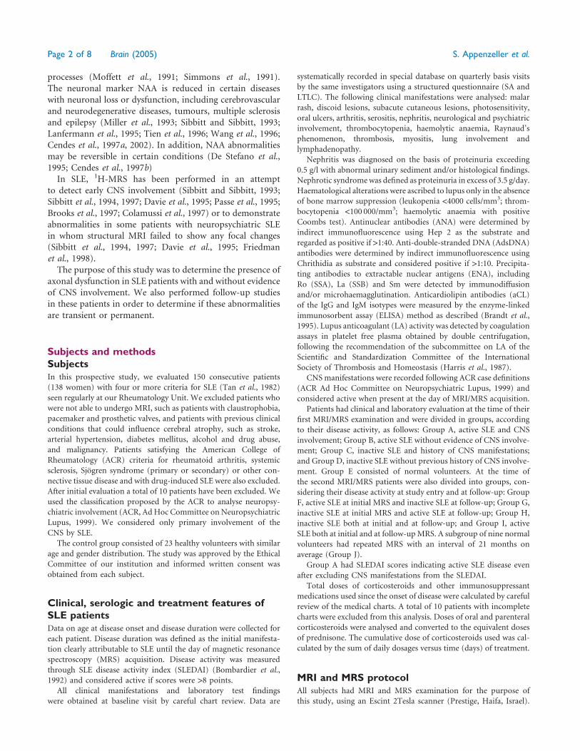
processes (Moffett et al., 1991; Simmons et al., 1991).
The neuronal marker NAA is reduced in certain diseases
with neuronal loss or dysfunction, including cerebrovascular
and neurodegenerative diseases, tumours, multiple sclerosis
and epilepsy (Miller et al., 1993; Sibbitt and Sibbitt, 1993;
Lanfermann et al., 1995; Tien et al., 1996; Wang et al., 1996;
Cendes et al., 1997a, 2002). In addition, NAA abnormalities
may be reversible in certain conditions (De Stefano et al.,
1995; Cendes et al., 1997b)
In SLE, 1H-MRS has been performed in an attempt
to detect early CNS involvement (Sibbitt and Sibbitt, 1993;
Sibbitt et al., 1994, 1997; Davie et al., 1995; Passe et al., 1995;
Brooks et al., 1997; Colamussi et al., 1997) or to demonstrate
abnormalities in some patients with neuropsychiatric SLE
in whom structural MRI failed to show any focal changes
(Sibbitt et al., 1994, 1997; Davie et al., 1995; Friedman
et al., 1998).
The purpose of this study was to determine the presence of
axonal dysfunction in SLE patients with and without evidence
of CNS involvement. We also performed follow-up studies
in these patients in order to determine if these abnormalities
are transient or permanent.
Subjects and methodsSubjectsIn this prospective study, we evaluated 150 consecutive patients
(138 women) with four or more criteria for SLE (Tan et al., 1982)
seen regularly at our Rheumatology Unit. We excluded patients who
were not able to undergo MRI, such as patients with claustrophobia,
pacemaker and prosthetic valves, and patients with previous clinical
conditions that could influence cerebral atrophy, such as stroke,
arterial hypertension, diabetes mellitus, alcohol and drug abuse,
and malignancy. Patients satisfying the American College of
Rheumatology (ACR) criteria for rheumatoid arthritis, systemic
sclerosis, Sjogren syndrome (primary or secondary) or other con-
nective tissue disease and with drug-induced SLE were also excluded.
After initial evaluation a total of 10 patients have been excluded. We
used the classification proposed by the ACR to analyse neuropsy-
chiatric involvement (ACR, Ad Hoc Committee on Neuropsychiatric
Lupus, 1999). We considered only primary involvement of the
CNS by SLE.
The control group consisted of 23 healthy volunteers with similar
age and gender distribution. The study was approved by the Ethical
Committee of our institution and informed written consent was
obtained from each subject.
Clinical, serologic and treatment features ofSLE patientsData on age at disease onset and disease duration were collected for
each patient. Disease duration was defined as the initial manifesta-
tion clearly attributable to SLE until the day of magnetic resonance
spectroscopy (MRS) acquisition. Disease activity was measured
through SLE disease activity index (SLEDAI) (Bombardier et al.,
1992) and considered active if scores were >8 points.
All clinical manifestations and laboratory test findings
were obtained at baseline visit by careful chart review. Data are
systematically recorded in special database on quarterly basis visits
by the same investigators using a structured questionnaire (SA and
LTLC). The following clinical manifestations were analysed: malar
rash, discoid lesions, subacute cutaneous lesions, photosensitivity,
oral ulcers, arthritis, serositis, nephritis, neurological and psychiatric
involvement, thrombocytopenia, haemolytic anaemia, Raynaud’s
phenomenon, thrombosis, myositis, lung involvement and
lymphadenopathy.
Nephritis was diagnosed on the basis of proteinuria exceeding
0.5 g/l with abnormal urinary sediment and/or histological findings.
Nephrotic syndrome was defined as proteinuria in excess of 3.5 g/day.
Haematological alterations were ascribed to lupus only in the absence
of bone marrow suppression (leukopenia <4000 cells/mm3; throm-
bocytopenia <100 000/mm3; haemolytic anaemia with positive
Coombs test). Antinuclear antibodies (ANA) were determined by
indirect immunofluorescence using Hep 2 as the substrate and
regarded as positive if >1:40. Anti-double-stranded DNA (AdsDNA)
antibodies were determined by indirect immunofluorescence using
Chrithidia as substrate and considered positive if >1:10. Precipita-
ting antibodies to extractable nuclear antigens (ENA), including
Ro (SSA), La (SSB) and Sm were detected by immunodiffusion
and/or microhaemagglutination. Anticardiolipin antibodies (aCL)
of the IgG and IgM isotypes were measured by the enzyme-linked
immunosorbent assay (ELISA) method as described (Brandt et al.,
1995). Lupus anticoagulant (LA) activity was detected by coagulation
assays in platelet free plasma obtained by double centrifugation,
following the recommendation of the subcommittee on LA of the
Scientific and Standardization Committee of the International
Society of Thrombosis and Homeostasis (Harris et al., 1987).
CNS manifestations were recorded following ACR case definitions
(ACR Ad Hoc Committee on Neuropsychiatric Lupus, 1999) and
considered active when present at the day of MRI/MRS acquisition.
Patients had clinical and laboratory evaluation at the time of their
first MRI/MRS examination and were divided in groups, according
to their disease activity, as follows: Group A, active SLE and CNS
involvement; Group B, active SLE without evidence of CNS involve-
ment; Group C, inactive SLE and history of CNS manifestations;
and Group D, inactive SLE without previous history of CNS involve-
ment. Group E consisted of normal volunteers. At the time of
the second MRI/MRS patients were also divided into groups, con-
sidering their disease activity at study entry and at follow-up: Group
F, active SLE at initial MRS and inactive SLE at follow-up; Group G,
inactive SLE at initial MRS and active SLE at follow-up; Group H,
inactive SLE both at initial and at follow-up; and Group I, active
SLE both at initial and at follow-up MRS. A subgroup of nine normal
volunteers had repeated MRS with an interval of 21 months on
average (Group J).
Group A had SLEDAI scores indicating active SLE disease even
after excluding CNS manifestations from the SLEDAI.
Total doses of corticosteroids and other immunosuppressant
medications used since the onset of disease were calculated by careful
review of the medical charts. A total of 10 patients with incomplete
charts were excluded from this analysis. Doses of oral and parenteral
corticosteroids were analysed and converted to the equivalent doses
of prednisone. The cumulative dose of corticosteroids used was cal-
culated by the sum of daily dosages versus time (days) of treatment.
MRI and MRS protocolAll subjects had MRI and MRS examination for the purpose of
this study, using an Escint 2Tesla scanner (Prestige, Haifa, Israel).
Page 2 of 8 Brain (2005) S. Appenzeller et al.
Our MRI protocol consisted of:
(i) Sagittal T1 spin echo = 6 mm thick, flip angle = 180�, repetition
time (TR) = 430, echo time (TE) = 12, matrix 200 · 350, field of
view (FOV) = 25 · 25 cm;(ii) Coronal images, perpendicular to long axis of hippocampus,
defined by the sagittal images:
(a) T2-weighted ‘fast-spin echo’ (FSE) = 3 mm thick, flip
angle = 120�, TR = 4800, TE = 129, matrix 252 · 320,
FOV = 18 · 18 cm;
(b) T1-weighted inversion recovery (IR) = 3 mm thick, flip
angle = 200�, TR = 2800–3000, TE = 14, inversion time
(TI) = 840, matrix 130 · 256, FOV = 16 · 18 cm;(iii) Axial images parallel to the long axis of the hippocampi:
(a) T1-weighted gradient echo = 3 mm thick, flip angle = 70�,
TR = 200, TE = 5, matrix 180 · 232, FOV = 22 · 22 cm;
(b) Fluid attenuated inversion recovery (FLAIR) = 4 mm thick,
flip angle = 120�, TR = 6800, TE = 129, matrix 252 · 328,
FOV = 21 · 23 cm;(iv) T1-weighted 3D gradient echo, acquired in the sagittal plane for
multiplanar and reconstruction: 1 mm thick, flip angle = 35�,
TR = 22, TE = 9, matrix 256 · 220, FOV = 23 · 25 cm.
Single voxel 1H-MRS was acquired using point resolved spectro-
scopy (PRESS) sequence (Bottomley, 1987 ) (TR = 1500 ms, TE =
135 ms, NEX = 200) over the superior–posterior region of the left
hemisphere at the level of corpus callosum. This area was previously
analysed using T1-weighted, T2-weighted and FLAIR sequences.
Patients with white matter lesions in this region were not included
(n = 30). Therefore, all patients evaluated in this study had normal
appearing white matter within the MRS region of interest (ROI).
After the acquisition of scout anatomical images in sagittal planes for
localization of corpus callosum, one single voxel (2 · 5 · 1 cm) was
placed over the ROI (Fig. 1). Prior to the acquisition, a localized
shimming at the ROI was performed to ensure adequate field homo-
geneity followed by water suppression adjustment.
The spectra were post-processed using software supplied by the
machine manufacturer (Elscint 2T Prestige, Haifa, Israel). After zero-
filling and baseline correction we determined peak areas by integ-
ration of the corresponding signals from N-acetyl compounds
(NAA) at 2.01 p.p.m., choline-base compounds (Cho) at 3.2 p.p.m.
and creatine and phosphocreatine containing compounds (Cr) at
3.0 p.p.m.. The spectra were scaled in relation to creatine values.
Ratios of NAA/Cr were used for analyses.
Spectral acquisition, quantification and analysis were performed
by one investigator (S.A.). The evaluation was cross-checked by two
spectroscopists (L.M.L. and F.C.), blinded to the name and clinical
data of patients and volunteers. The quality of the spectral analysis
was judged independently by these two investigators from the para-
meters linewidth and signal-to-noise ratio (Fig. 2) and spectra with
broad peaks and poor separation of individual peaks were excluded
from analysis. Values <2 SD from the mean of controls were con-
sidered abnormal. A total of 10 patients were excluded because of bad
quality spectra.
Therefore, 90 patients (88 women) with mean age of 32.5 years
(range 18–59 years, SD = 13.1) were available for evaluation for this
study. We repeated MRI and MRS exams in 50 of these patients
(48 women) and in 9 volunteers after a minimum interval of 1 year.
StatisticsWe performed analysis of variance (ANOVA) to test differences
among the groups, followed by post hoc Tukey’s HSD for pairwise
comparison if necessary. Follow-up MRS results were analysed using
paired t-test with Bonferroni’s correction for multiple comparisons.
Statistical significance was considered to be present for P < 0.05.
ResultsDemographic dataWe analysed MRS data of 90 SLE patients. In relation to
the different patients groups at study entry, we observedFig. 1 Placement of region of interest (ROI).
Fig. 2 Illustrative proton magnetic spectra from posterior supraventricular region from a volunteer (left) and one SLE patient (right).
Reversibility of axonal dysfunction in SLE Brain (2005) Page 3 of 8
the following age and gender distribution: Group A: 29
patients (28 women) with mean age of 32.2 (range 18–56;
SD = 12.9); Group B: 28 patients (27 women) with mean age
of 33.0 (range 18–59; SD = 13.1); Group C: 14 patients (14
women) with mean age of 31.9 (range 18–50; SD = 13.7);
Group D: 19 patients (19 women) with mean age of 33.5
(range 18–56; SD = 12.9).
The control group (Group E) consisted of 23 healthy
volunteers (19 women) with mean age of 33.8 years (range
20–60, SD = 13.7 years)
MRI and 1H-MRS studies were repeated in 50 SLE patients
(48 women) with mean age of 33.3 years (ranging from 18 to
57 years; SD = 12.2) at study entry. In relation to the different
patients groups at follow-up, we observed the following gen-
der and age distribution: Group F, 15 patients (14 women)
with mean age of 32.5 (range 18–55; SD = 12); Group G,
15 patients (14 women) with mean of age 33.0 (range
18–57; SD = 13.1); Group H, 10 patients (10 women) with
mean age of 32.3 (range 18–50; SD = 13); Group I, 10 patients
(10 women) with mean age of 33.6 (range 18–50; SD = 13).
The mean interval between the two 1H-MRS of SLE patients
was 19 months (range 12–24 months; SD = 2.3)
MRS studies were repeated in 9 volunteers (7 women) of
Group J with mean age of 32.1 (range 20–60; SD = 14.1) at
study entry. The mean interval between MRS examinations
was 21 months (range 18–24 months; SD = 1.8).
There was no statistical difference between the age and
gender distribution among the different groups of patients
(Groups A–D and F–I) and volunteers (Groups E and J).
Clinical, laboratory and treatmentfeaturesThe mean disease duration was 64.5 months (range 1–362
months, SD = 48.50) at study entry and 93 months (range
12–421 months, SD = 45.45) at follow-up MRS. At baseline,
65 episodes of CNS manifestations had occurred in 43 patients
(29 patients with active and 14 with inactive CNS manifesta-
tions) (Table 1). At the time of MRS scans, 57 patients had
active SLE, with SLEDAI scores ranging between 10 and
20 (mean 14.56, SD = 6.52). Active CNS manifestations at
the first MRS scan were observed in 29 of 57 patients with
active SLE. Number of patients who had inactive SLE at the
time of first MRS was 33 and 14 of them had past history of
CNS involvement and 19 did not. All patients were on steroid
use on the day of MRS study, with doses ranging from 5 to
80 mg/day (mean 43 mg/day). IgG antiphospholipid anti-
bodies were positive in 32 patients.
Disease activity and MRSMedian NAA/Cr values for each group of patients were:
(A), active SLE and CNS involvement, 1.65 (SD = 0.25);
(B), active SLE without evidence of CNS involvement, 1.67
(SD = 0.27); (C), inactive SLE and history of CNS manifesta-
tions, 1.82 (SD = 0.23); (D), inactive SLE without previous
history of CNS involvement, 1.98 (SD = 0.21); (E), volunteers,
1.86 (SD = 0.15) (Table 2).
Median NAA/Cr ratios were significantly lower in patients
with active SLE (Groups A and B), when compared with
patients with inactive SLE (Groups C and D) (P = 0.005)
and volunteers (Group E) (P = 0.01) (Fig. 3).
We did not find a correlation between daily corticosteroid
dose or cumulative corticosteroid dose and median NAA/Cr
value (r = 0.4).
Follow-up studyPatients were divided, according to their disease activity at
study entry and at follow-up MRS, into four groups. Group F
(n = 15) with active SLE at initial MRS (median NAA/Cr = 1.6;
SD = 0.36) and inactive SLE at follow-up (median NAA/Cr =
2.1; SD = 0.31); P = 0.04. Group G (n = 15) with inactive SLE
at initial MRS (median NAA/Cr = 1.9; SD = 0.12) and active
SLE at follow-up (median NAA/Cr = 1.3; SD = 0.21);
P = 0.001. Group H (n = 10) with inactive SLE both at initial
(median NAA/Cr = 2.0; SD = 0.48) and at follow-up (median
NAA/Cr = 2.1; SD = 0.42) MRS; P = 0.2. Group I (n = 10) with
active SLE both at initial (median NAA/Cr = 1.7; SD = 0.21)
and at follow-up (median NAA/Cr = 1.38; SD = 0.48) MRS;
P = 0.02 (Fig. 4). The NAA/Cr ratios remained constant in
Table 2 Median NAA/Cr and Cho/Cr values
Groups Median NAA/Cr atinitial MRS (6SD)
Median Cho/Cr atinitial MRS (6SD)
A 1.65 (0.25) # 1.03 (0.3)B 1.67 (0.27) # 0.98 (0.4)C 1.82 (0.23) 1.1 (0.2)D 1.98 (0.21) 1.0 (0.3)E 1.86 (0.15) 0.96 (0.2)
Values in 90 patients and 23 normal volunteers at study entry.Groups: (A), Active SLE and CNS involvement; (B), active SLEwithout evidence of CNS involvement; (C), inactive SLE andhistory of CNS manifestations; (D), inactive SLE without previoushistory of CNS involvement; and (E), normal volunteers.#: Significantly lower than normal volunteers.
Table 1 Summary of cumulative neuropsychiatricmanifestations that occurred in 43 patients at study entry
Neuropsychiatric manifestations Number ofCNS events (%)
Headache 18 (27.7)Cognitive impairment 20 (30.8)Mood disorder 10 (15.4)Seizures 7 (10.8)Acute confusional state 5 (7.7)Psychosis 2 (3.1)Mononeuropathy 1 (1.5)Cranial neuropathy 1 (1.5)Aseptic meningitis 1 (1.5)Total number of events 65
Page 4 of 8 Brain (2005) S. Appenzeller et al.
normal volunteers (initial median NAA/Cr = 1.86; SD = 0.17;
follow-up MRS: median NAA/Cr = 1.89; SD = 0.18. Cho and
Cr values remained stable during follow-up periods in
patients and controls (Table 3).
MRI findings and MRSSubtle abnormal MRI findings, in areas outside and far from
the MRS ROI, (hyperintense areas suggestive of cerebral
microinfarcts) in cortical and subcortical regions were
observed in 53 patients at baseline study. These MRI abnor-
malities were more frequently observed in patients with anti-
phospholipid antibodies (P = 0.04). The number of lesions
was counted in all MRI scans. Visual analysis of MRI did not
demonstrate significant increase in the number of these
lesions during the follow-up study as compared with baseline
study.
NAA/Cr ratios were lower in SLE patients with MRI
abnormalities when compared with patients with normal
MRI (P = 0.028). No difference in Cho/Cr values between
patients with and without MRI abnormalities was observed.
Antiphospholipid antibodies and MRSSLE patients with positive antiphospholipid antibodies had
lower NAA/Cr (P = 0.021) values when compared with
patients without antiphospholipid antibodies. The frequency
of antiphospholipid antibodies was distributed equally among
these groups.
DiscussionAlthough several studies (Sibbitt et al., 1994, 1997; Davie et al.,
1995; Friedman et al., 1998, Castellino et al., 2005) showed the
usefulness of 1H-MRS in CNS manifestations in SLE, only one
previous study (Castellino et al., 2005) analysed SLE patients
without CNS manifestations. In our study we observed that
SLE patients with active disease had low relative NAA signal
intensity, indicating axonal dysfunction, when compared
with SLE patients with inactive disease and volunteers. This
occurred independently of clinical CNS involvement as
defined by the ACR criteria (ACR Ad Hoc Committee on
Neuropsychiatric Lupus, 1999).
Cerebrovascular abnormalities may be the basis of diffuse
cerebral injury in SLE. Small-vessel injury is primarily asso-
ciated with decreased NAA/Cr ratio, while medium-vessel
injury is primarily associated with increased Cho/Cr ratio
(Davie et al., 1995). In our study we observed that patients
with white matter abnormalities in regions outside the MRS
ROI and patients with antiphospholipid antibodies had more
pronounced decreased NAA/Cr ratios, supporting the theory
of small vessel involvement in SLE. We did not observe dif-
ferences in Cho/Cr ratio among the subgroups of SLE patients
and normal volunteers.
The ROI for MRS examination in patients with SLE should
best reflect the area where the metabolic changes might
precede the morphological changes. MRS studies have already
been performed in the supraventricular and subcortical white
matter (Sibbitt and Sibbitt, 1993; Sibbitt et al., 1994; Friedman
et al., 1998; Lim et al., 2000) and in the basal ganglia (Lim et al.,
2000). In this study the normal appearing white matter was
chosen, despite the presence of small hyperintense lesions in
other areas of the brain, because most of the time these MRI
abnormalities are referred to as non-specific findings. MRS
abnormalities in these regions would support the idea that
MRS could precede the appearance of hyperintense lesions
in T2 or FLAIR sequences due to CNS involvement of SLE
(Castellino et al., 2005). Other studies (Sanna et al., 2003;
Sibbitt et al., 2003) have shown the association of non-
specific white matter abnormalities and signs and symptoms
of CNS manifestations in SLE. The specific susceptibility of
the white matter to small vascular lesions is thought to be due
to unique vascularization of this tissue. Blood is supplied to
the white matter by means of single sources, rendering the
deep white matter more vulnerable to vascular insults. In SLE
the precise biochemical mechanism that explains the basis
of CNS involvement is still unknown (Lim et al., 2000).
We therefore choose the supraventricular region, in order
to test the hypothesis that early NAA changes might occur
in this area in patients with overt CNS manifestations. Our
results confirm previous findings (Sibbitt and Sibbitt, 1993;
Sibbitt et al.,1994, Castellino et al., 2005) and support this
hypothesis. However, in the present study, after 19 months of
follow-up, we did not observe that low NAA/Cr ratio pre-
disposed to the appearance of structural lesions detectable by
MRI. Perhaps longer periods of observation are necessary to
Fig. 3 Box-and-whiskers plot showing median NAA/Cr ratio inSLE patients with active SLE and CNS manifestations. No. 1represents Group A, patients with active SLE without CNSmanifestations; No. 2 represents Group B, inactive SLE patientswith past history of CNS involvement; No. 3 represents Group C,inactive patients without history of CNS involvement; No. 4represents Group D and No. 5 represents volunteers, Group E.The box extends from the 25th percentile to the 75th percentile,with a horizontal line at the median (50th percentile). Whiskersextend down to the smallest value and up to the largest.Outliers are represented by an asterisk (*).
Reversibility of axonal dysfunction in SLE Brain (2005) Page 5 of 8
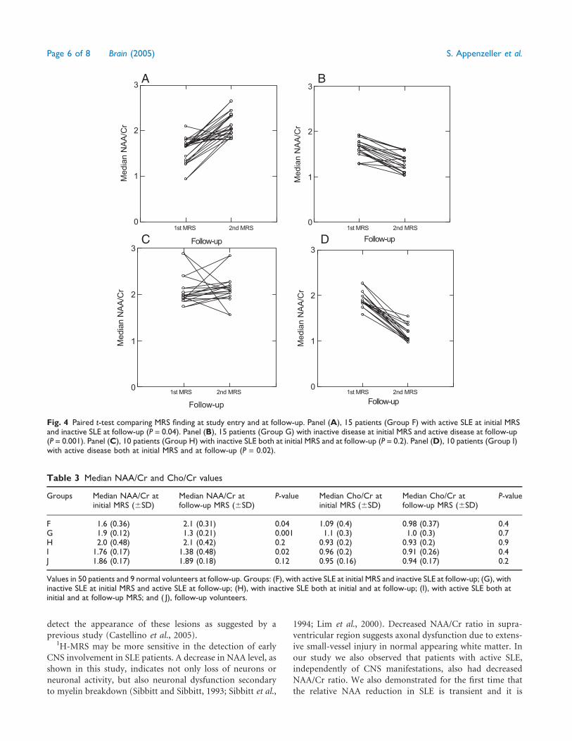
detect the appearance of these lesions as suggested by a
previous study (Castellino et al., 2005).1H-MRS may be more sensitive in the detection of early
CNS involvement in SLE patients. A decrease in NAA level, as
shown in this study, indicates not only loss of neurons or
neuronal activity, but also neuronal dysfunction secondary
to myelin breakdown (Sibbitt and Sibbitt, 1993; Sibbitt et al.,
1994; Lim et al., 2000). Decreased NAA/Cr ratio in supra-
ventricular region suggests axonal dysfunction due to extens-
ive small-vessel injury in normal appearing white matter. In
our study we also observed that patients with active SLE,
independently of CNS manifestations, also had decreased
NAA/Cr ratio. We also demonstrated for the first time that
the relative NAA reduction in SLE is transient and it is
Follow-up
0
1
2
3
Me
dia
n N
AA
/Cr
Follow-up
0
1
2
3
Me
dia
n N
AA
/Cr
Follow-up
0
1
2
3
Media
n N
AA
/Cr
Follow-up
0
1
2
3
Me
dia
n N
AA
/Cr
A B
1st MRS 1st MRS2nd MRS 2nd MRS
C D1st MRS 1st MRS2nd MRS 2nd MRS
Fig. 4 Paired t-test comparing MRS finding at study entry and at follow-up. Panel (A), 15 patients (Group F) with active SLE at initial MRSand inactive SLE at follow-up (P = 0.04). Panel (B), 15 patients (Group G) with inactive disease at initial MRS and active disease at follow-up(P = 0.001). Panel (C), 10 patients (Group H) with inactive SLE both at initial MRS and at follow-up (P = 0.2). Panel (D), 10 patients (Group I)with active disease both at initial MRS and at follow-up (P = 0.02).
Table 3 Median NAA/Cr and Cho/Cr values
Groups Median NAA/Cr atinitial MRS (6SD)
Median NAA/Cr atfollow-up MRS (6SD)
P-value Median Cho/Cr atinitial MRS (6SD)
Median Cho/Cr atfollow-up MRS (6SD)
P-value
F 1.6 (0.36) 2.1 (0.31) 0.04 1.09 (0.4) 0.98 (0.37) 0.4G 1.9 (0.12) 1.3 (0.21) 0.001 1.1 (0.3) 1.0 (0.3) 0.7H 2.0 (0.48) 2.1 (0.42) 0.2 0.93 (0.2) 0.93 (0.2) 0.9I 1.76 (0.17) 1.38 (0.48) 0.02 0.96 (0.2) 0.91 (0.26) 0.4J 1.86 (0.17) 1.89 (0.18) 0.12 0.95 (0.16) 0.94 (0.17) 0.2
Values in 50 patients and 9 normal volunteers at follow-up. Groups: (F), with active SLE at initial MRS and inactive SLE at follow-up; (G), withinactive SLE at initial MRS and active SLE at follow-up; (H), with inactive SLE both at initial and at follow-up; (I), with active SLE both atinitial and at follow-up MRS; and ( J), follow-up volunteers.
Page 6 of 8 Brain (2005) S. Appenzeller et al.
probably dependent on disease activity. We observed that SLE
patients with active disease had low relative NAA signal
intensity that returned to normal range after disease remis-
sion. In the group of patients with inactive disease both at
baseline and follow-up MRS, there was no difference in NAA
values as compared with the control group. On the other
hand, in the group of patients with active disease both at
baseline and follow-up MRS, there was a progressive decrease
in NAA values. These findings suggest that axonal dysfunction
in SLE patients may be transient and related to disease
activity, independently of the presence of CNS involvement
or corticosteroid use.
In the follow-up graphs (Fig. 4) we observed that not
all patients had the same pattern of NAA/Cr increase or
reduction. Further studies are necessary to determine the
factors associated with the intensity and rate of NAA/Cr
loss or recovery. These factors may help to explain the outliers
in Fig. 4. It is possible that the NAA reduction precedes the
clinical signs and symptoms of SLE disease activity, among
other factors. This could explain the drop of relative NAA/Cr
values in the three patients who had inactive SLE disease at
both initial and follow-up MRS. However, it is difficult to
explain the two outlier patients in Group H who had a more
pronounced increase in NAA/Cr values.
The fall of NAA reflects neuronal loss or dysfunction (Passe
et al., 1995; Tsai and Coyle, 1995). Previous studies have
correlated NAA loss with cerebral atrophy (Sibbitt et al.,
1994), cognitive dysfunction and damage index (Brooks
et al., 1999). NAA recovery, after initial reduction, has not
been reported in SLE before, but was observed in multiple
sclerosis (Wolinsky and Narayana, 2002), stroke (Saunders
et al., 1995), schizophrenia (Haussinger et al., 1994) and
epilepsy (Cendes et al., 1997b)
Cho concentrations, on the other hand, are reported to
rise in SLE patients with CNS involvement (Brooks et al.,
1997, 1999; Sibbitt et al., 1997; Castellino et al., 2005), but
the cause has not been determined, although it may be due
to myelin breakdown secondary to neuronal loss or due
to inflammatory process (Brooks et al., 1997; Brooks
et al., 1999).
In this study we used Cr values as an internal reference,
although it has not been demonstrated that Cr is stable in
SLE. The facts that we performed MRS in normal appearing
white matter and that Cho/Cr ratios were not different among
groups give support to the assumption that eventual changes
in Cr were minimal and did not produce a great influence in
our results.
In conclusion, our findings suggest that SLE activity,
independently of CNS involvement, is associated with white
matter insult, often not evident by structural MRI or by
clinical manifestations. The relative NAA decrease may be a
surrogate marker for disease activity in SLE patients and
may be useful for follow-up of disease activity. These findings
are limited to the area in the brain studied here, and further
studies are necessary to confirm these findings.
AcknowledgementsStudy supported by Fundacao de Amparo a Pesquisa do
Estado de Sao Paulo (FAPESP) grant # 03/015270.
References
ACR Ad Hoc Committee on Neuropsychiatric Lupus. The American College
of Rheumatology nomenclature and case definitions for neuropsychiatric
lupus syndromes. Arthritis Rheum 1999; 42: 599–608.
Adelman DC, Saltiel E, Klinenberg JR. The Neuropsychiatric Manifestations
of Systemic Lupus Erythematosus: An Overview. Semin Arthritis Rheum
1986; 15: 185–99.
Bombardier C, Gladman DD, Urowitz MB, Caron D, Chang CH. Derivation
of the SLEDAI. A disease activity index for lupus patients. The Committee
on Prognosis Studies in SLE. Arthritis Rheum 1992; 35: 630–40.
Bottomley PA. Spatial localization in NMR spectroscopy in vivo. Ann NY
Acad Sci 1987; 508: 333–48.
Brandt JT, Triplett DA, Alving B, Scharrer I. Criteria for the diagnosis of
lupus anticoagulants: an update. On behalf of the Subcommittee on Lupus
Anticoagulant/Antiphospholipid Antibody of the Scientific and Standard-
isation Committee of the ISTH. Thromb Haemost 1995; 74: 1185–90.
Brooks WM, Sabet A, Sibbitt WL Jr, Barker PB, van Zijl PC, Duyn JH, et al.
Neurochemistry of brain lesions determined by spectroscopic imaging in
systemic lupus erythematosus. J Rheumatol 1997; 24: 2323–2329.
Brooks WM, Jung RE, Ford CC, Greinel EJ, Sibbitt WL Jr. Relationship
between neurometabolite derangement and neurocognitive dysfunction
in systemic lupus erythematosus. J Rheumatol 1999; 26: 81–85.
Castellino G, Govoni M, Padovan M, Colamussi P, Borrelli M, Trotta F.
Proton magnetic resonance spectroscopy may predict future brain lesions
in SLE patients: a functional multi-imaging approach and follow up.
Ann Rheum Dis 2005; 64: 1022–7.
Carbotte RM, Denburg SD, Denburg JA. Cognitive dysfunction and systemic
lupus erythematosus. New York, NY: Churchill Livingstone, 1992; 865–81.
Cendes F, Stanley JA, Dubeau F, Andermann F, Arnold DL. Proton magnetic
resonance spectroscopic imaging for discrimination of absence and
complex partial seizures. Ann Neurol 1997a; 41: 74–80
Cendes F, Andermann F, Dubeau F, Matthews PM, Arnold DL. Normal-
ization of neuronal metabolic dysfunction after surgery for temporal
lobe epilepsy. Evidence from proton MRS imaging. Neurology 1997b;
49: 1525–33.
Cendes F, Knowlton RC, Novotny E, Li ML, Antel S, Sawrie S, et al. Magnetic
resonance spectroscopy in epilepsy: clinical issue. Epilepsia 2002; 43: 32–9.
Chinn RJ, Wilkinson ID, Hall-Craggs MA, Paley MN, Shortall E, Carter S,
et al. Magnetic resonance imaging of the brain and cerebral proton
spectroscopy in patients with systemic lupus erythematosus. Arthritis
Rheum 1997; 40: 36–46.
Colamussi P, Trotta F, Ricci R, Cittanti C, Govoni M, Barbarella G, et al.
Brain perfusion SPECT and proton magnetic resonance spectroscopy in
the evaluation of two systemic lupus erythematosus patients with mild
neuropsychiatric manifestations. Nucl Med Commun 1997; 18: 269–73.
Davie CA, Feinstein A, Kartsounis LD, Barker GJ, McHugh NJ, Walport MJ,
et al. Proton magnetic resonance spectroscopy of systemic lupus erythem-
atosus involving the central nervous system. J Neurol 1995; 242: 522–8.
De Stefano N, Matthews PM, Arnold DL. Reversible decreases in N-
acetylaspartate after acute brain injury. Magn Reson Med 1995; 34: 721–7.
Friedman SD, Stidley CA, Brooks WM, Hart BL, Sibbitt WL Jr. Brain injury
and neurometabolic abnormalities in systemic lupus erythematosus.
Radiology 1998; 209: 79–84.
Harris EN, Gharavi AE, Patel SP, Hughes GR. Evaluation of the anti-
cardiolipin antibody test: report of an international workshop held
4 April 1986. Clin Exp Immunol 1987; 68: 215–22.
Haussinger D, Laubenberger J, vom Dahl S, Ernst T, Bayer S, Langer M,
et al. Proton magnetic resonance spectroscopy studies on human brain
myo-inositol in hypo-osmolarity and hepatic encephalopathy. Gastro-
enterology 1994; 107: 1475–80.
Reversibility of axonal dysfunction in SLE Brain (2005) Page 7 of 8
Lanfermann H, Kugel H, Heindel W, Herholz K, Heiss WD, Lackner K.
Metabolic changes in acute and subacute cerebral infarctions: findings
at proton MR spectroscopic imaging. Radiology 1995; 196: 203–10.
Lim MK, Suh CH, Kim HJ, Cho YK, Choi SH, Kang JH, et al. Systemic
lupus erythematosus: Brain MR imaging and single voxel hydrogen
1MR spectroscopy. Radiology 2000; 217: 43–9.
Miller BL, Moats RA, Shonk T, Ernst T, Woolley S, Ross BD. Alzheimer
disease: depiction of increased cerebral myo-inositol with proton MR
spectroscopy. Radiology 1993; 187: 433–7.
Moffett JR, Namboodiri MA, Cangro CB, Neale JH. Immunohistochemical
localization of N-acetylaspartate in rat brain. Neuroreport 1991; 2: 131–4.
Omdal R, Mellgren SI, Husby G. Clinical neuropsychiatric and neuromus-
cular manifestations in systemic lupus erythematosus. Scand J Rheumatol
1988; 17: 113–7.
Passe TJ, Charles HC, Rajagopalan P, Krishnan KR. Nuclear magnetic
resonance spectroscopy: a review of neuropsychiatric applications. Prog
Neuropsychopharmacol Biol Psychiatry 1995; 19: 541–63.
Sanna G, Bertolaccini ML, Cuadrado MJ, Laing H, Khamashta MA, Mathieu A,
et al. Neuropsychiatric manifestations in systemic lupus erythematosus:
prevalence and association with antiphospholipid antibodies. J Rheumatol
2003; 30: 985–92.
Saunders DE, Howe FA, van den Boogaart A, McLean MA, Griffiths JR,
Brown MM. Continuing ischemic damage after acute middle cerebral
artery infarction in humans demonstrated by short-echo proton spectro-
scopy. Stroke 1995; 26: 1007–13.
Sibbitt WL, Sibbitt RR. Magnetic resonance spectroscopy and positron
emission tomography scanning in neuropsychiatric systemic lupus
erythematosus. Rheum Dis Clin North Am 1993; 19: 851–68.
Sibbitt WL Jr, Haseler LJ, Griffey RH, Hart BL, Sibbitt RR, Matwiyoff NA.
Analysis of cerebral structural changes in systemic lupus erythematosus
by proton MR spectroscopy. AJNR Am J Neuroradiol 1994; 15: 923–8.
Sibbitt WL Jr, Haseler LJ, Griffey RR, Friedman SD, Brooks WM.
Neurometabolism of active neuropsychiatric lupus determined with
proton MR spectroscopy. AJNR Am J Neuroradiol 1997; 18: 1271–7.
Sibbitt WL Jr, Schmidt PJ, Hart BL, Brooks WM. Fluid Attenuated Inversion
Recovery (FLAIR) imaging in neuropsychiatric systemic lupus erythem-
atosus. J Rheumatol 2003; 30: 1983–9.
Simmons ML, Frondoza CG, Coyle JT. Immunocytochemical localization
of N-acetyl-aspartate with monoclonal antibodies. Neuroscience 1991;
45: 37–45.
Tan EM, Cohen AS, Fries JF, Masi AT, McShane DJ, Rothfield NF, et al. The
1982 revised criteria for the classification of systemic lupus erythematosus.
Arthritis Rheum 1982; 25: 1271–7.
Tien RD, Lau PH, Smith JS, Lazeyas F. Single-voxel proton brain spectroscopy
exam (PROBE/SV) in patients with primary brain tumors. AJR Am J
Roentgenol 1996; 167: 201–9.
Tsai G, Coyle JT. N-acetylaspartate in neuropsychiatric disorders. Prog
Neurobiol 1995; 46: 531–40.
Wang Z, Zimmermann RA, Sauter R. Proton MR spectroscopy of the brain:
clinically useful information obtained in assessing CNS disease in children.
AJR Am J Roentgenol 1996; 167: 191–9.
West SG. Neuropsychiatric lupus. Rheum Dis Clin North Am 1994; 20:
129–58.
Wolinsky JS. Narayana PA. Magnetic resonance spectroscopy in multiple
sclerosis: window into the diseased brain. Curr Opin Neurol 2002;
15: 247–51.
Page 8 of 8 Brain (2005) S. Appenzeller et al.
Resultados 219
ARTIGO 13
Increased choline/creatinine ratio on MRS may predict appearance of white matter
lesions in systemic lupus erythematosus
Appenzeller S, Li LM, Costallat LT, Cendes F.
Increased choline/creatinine ratio on MRS may predict appearance of white matter lesions
in systemic lupus erythematosus
Submetido
Resultados 220
Increased choline/creatinine ratio on MRS may predict appearance of white matter
lesions in systemic lupus erythematosus
Simone Appenzeller1,3, MD, Li Min Li2,3, MD, PhD, Lilian TL Costallat1, MD, PhD,
Fernando Cendes2,3, MD, PhD
Departments of Rheumatology1 and Neurology2, and Neuroimaging Laboratory3
University of Campinas, São Paulo – Brazil
Key words: systemic lupus erythematosus-spectroscopy-white matter lesions- Choline
Grants: FAPESP and CNPq (479133/2004-2)
Counts for references: 32
Running title: MRS and white matter lesions
Correspondence to: Fernando Cendes, MD, PhD
Department of Neurology,
FCM – UNICAMP
Cidade Universitária Zeferino Vaz
Campinas SP, Brazil, CEP 13083-970
FAX: +55 19 3289-1818
Email: [email protected]
Resultados 221
Abstract
Objective: To investigate proton magnetic resonance spectroscopy (MRS) in
patients with systemic lupus erythematosus (SLE) with small hyperintense lesions on T2-
weithed MRI.
Methods: We studied 30 SLE patients who had lesions in MRS ROI and 23
controls. We performed single voxel proton MRS over the superior-posterior region of the
corpus callosum. We measured signals from N-acetyl-compounds (NAA), choline (Cho)
and creatine+phosphocreatine (Cr) and determined NAA/Cr and Cho/Cr ratios. After a
minimum of 12 months, MRI and MRS were repeated in all patients and 9 volunteers. We
performed paired T-test and Anova with Tukey´s post hoc comparisons to evaluate group
differences.
Results: Ten patients had MRI hyperintense lesions in the MRS-ROI at
baseline, and 20 had lesions in follow-up MRI but no lesions at baseline MRI. They had
increased Cho/Cr values at both MRS when compared to normal controls (p=0.001). In
addition, there was an increase in Cho/Cr values when patients’ baseline and follow up
MRS were compared (p=0.001). Controls had no change in Cho/Cr between scans
(p=0.56). NAA/Cr was lower in patients with active disease and returned to normal range in
patients with inactivity.
Conclusion: Increased Cho/Cr in normal appearing white matter may be
indicative of future appearance of hyperintense T2-weighet MRI lesions.
Introduction
Magnetic resonance imaging (MRI) is currently considered the standard
technique for determination of morphological brain abnormalities in systemic lupus
erythematosus (SLE) patients (Castellino et al., 2005; Stimmler et al., 1993). Reported
prevalence of detectable lesions varies from 62% to 100% (Stimmler et al., 1993; McCune
et al., 1998; Bell et al., 1991; Sibbitt et al., 1989; Aisen et al., 1985; West et al., 1995;
Karassa et al., 2000). Imaging findings in SLE patients vary from ischemic lesion to
frequently found small hyperintense lesions in deep white matter. These lesions often
referred to as nonspecific lesion, may be related to small foci of ischemia and may be
Resultados 222
associated with CNS manifestations (West et al., 1995; Karassa et al., 2000) and with the
presence of antiphospholipid antibodies (Karassa et al., 2000; Sanna et al., 2000).
Proton magnetic resonance spectroscopy (1H-MRS) has proved to be a non-
invasive tool for detecting neuronal metabolic dysfunction in several neurological diseases,
including SLE (Colamussi et al., 1995; Nossent et al., 1991; Rubbert et al., 1993; Sibbitt et
al., 1993; Castillo et al., 1996). This technique shows four major spectra, depending of the
echo time used during acquisition, corresponding to different metabolites: N-acetylaspartate
(NAA), choline (Cho), and creatine (Cr). Altered metabolite ratios have been observed even
in the absence of MRI lesions; however, few studies have been carried out in SLE patients
with and without overt neurological involvement (Sibbitt et al., 1994; Handa et al., 2003;
Lim et al., 2000; Axford et al., 2001; Peterson et al., 2003; Appenzeller et al., 2005).
In a previous study (Appenzeller et al., 2005), we demonstrated that patients
with active SLE without white matter lesions in the MRS region of interest (ROI) had a
decrease in NAA/Cr values, independently of the presence of CNS manifestations.
Furthermore, there was a recover in NAA/Cr values after disease control. In the present
study we analyzed only patients who had lesions in MRS ROI in order to determine if MRS
may be helpful to predict the appearance of new white matter lesions in patients with SLE.
Subjects and Methods
Subjects
We selected 30 patients who had follow-up MRIs and white matter lesions
inside the MRS region of interest (ROI) in at least one MRI. The presence of these lesions
was determined by visual analysis of MRI using FLAIR and T2-wheighted sequences.
None of them were included in a previous study (Appenzeller et al., 2005) in which we did
evaluate patients with normal appearing white matter in both baseline and follow up MRS
ROI. These patients were selected among a group of 150 consecutive patients (138 women)
with four or more criteria for SLE (Tan et al., 1982) seen regularly at our Rheumatology
Unit. From this initial group, we excluded patients that were not able to undergo MRI, such
as patients with claustrophobia, pacemaker and prosthetic valves, and patients with
previous clinical conditions that could influence cerebral atrophy, such as stroke, arterial
hypertension, diabetes mellitus, alcohol and drug abuse, and malignancy. Patients satisfying
Resultados 223
the American College of Rheumatology (ACR) criteria for rheumatoid arthritis, systemic
sclerosis, Sjögren syndrome (primary or secondary) or other connective tissue disease and
with drug-induced SLE were also excluded (Appenzeller et al., 2005).
The control group consisted of 23 healthy volunteers with similar age and
gender distribution. The study was approved by Ethical Committee of our institution and
informed written consent was obtained from each subject.
Clinical, serologic and treatment features of SLE patients
Data on age at disease onset and disease duration were collected for each
patient. Disease duration was defined as the initial manifestation clearly attributable to SLE
until the day of MRS acquisition. Disease activity was measured through SLE disease
activity index (SLEDAI) (Bombardier et al., 1992) and considered active if scores were
higher than eight points.
All clinical manifestations and laboratory test findings were obtained at
baseline visit by careful chart review. Data are systematically recorded in special database
on quarterly basis visits by the same investigators using a structured questionnaire (SA and
LTLC). The following clinical manifestations were analyzed: malar rash, discoid lesions,
subacute cutaneous lesions, photosensitivity, oral ulcers, arthritis, serositis, nephritis,
neurological and psychiatric involvement, thrombocytopenia, hemolytic anemia, Raynaud’s
phenomenon, thrombosis, myositis, lung involvement and lymphadenopathy.
Nephritis was diagnosed on the basis of proteinuria exceeding 0.5 g/L with
abnormal urinary sediment and/or histological findings. Nephrotic syndrome was defined
as proteinuria in excess of 3.5 g/day. Hematologic alterations were ascribed to lupus only in
the absence of bone marrow suppression (leukopenia <4000 cells/mm3; thrombocytopenia
<100.000/mm3; hemolytic anemia with positive Coombs test). Antinuclear antibodies
(ANA) were determined by indirect immunofluorescence using Hep 2 as the substrate and
regarded as positive if higher than 1:40. Anti-double-stranded DNA (AdsDNA) antibodies
were determined by indirect immunofluorescence using Chrithidia as substrate and
considered positive if higher than 1:10. Precipitating antibodies to extractable nuclear
antigens (ENA), including Ro (SSA), La (SSB) and Sm were detected by immunodiffusion
and/or microhemagglutination. Anticardiolipin antibodies (aCL) of the IgG and IgM
isotypes were measured by the ELISA method as described (Harris et al., 1987). Lupus
Resultados 224
anticoagulant (LA) activity was detected by coagulation assays in platelet free plasma
obtained by double centrifugation, following the recommendation of the subcommittee on
LA of the Scientific and Standardization Committee of the International Society of
Thrombosis and Homeostasis (Brandt et al., 1995).
CNS manifestations were recorded following ACR case definitions (ACR
1999) and considered active, when present at the day of MRI/MRS acquisition.
Patients had clinical and laboratory evaluation at time of their first MRI/MRS
examination and were divided in groups, according to the presence of white matter lesions
in the ROI, as follows: (A) 10 patients with white matter lesions at study entry; (B) 20
patients without white matter lesions at study entry, but with white matter lesions at follow-
up study.
Total doses of corticosteroids and other immunosuppressant medications used
since the onset of disease were calculated by careful review of the medical charts.
MRI and MRS Protocol
All subjects had MRI and MRS examination for the purpose of this study, using
an Escint 2Tesla scanner (Prestige, Haifa, Israel). Our MRI protocol consisted of: (1)
Sagittal T1 spin echo, 6 millimeters (mm) thick, flip angle= 180o; repetition time
(TR)=430, echo time (TE)=12, matrix 200X350, field of view (FOV)=25X25 centimeters
(cm); (2) Coronal images, perpendicular to long axis of hippocampus, defined by the
sagittal images; (2.a) T2-weighted “ fast spin echo” (FSE), 3mm thick, flip angle= 120o;
TR=4800, TE=129, matrix 252X320, FOV=18X18cm; (2.b) T1-weighted inversion
recovery (IR), 3mm thick, flip angle=200o; TR=2800-3000, TE=14, inversion time
(TI)=840, matrix 130X256, FOV=16X18cm; (3) Axial images (3.a) T1-weighted gradient
echo, 3mm thick, flip angle=70o, TR=200, TE=5, matrix 180X232, FOV=22X22 cm; (3.b)
Fluid attenuated inversion recovery (FLAIR), 4mm thick, flip angle=120o, TR=6800,
TE=129, matrix 252X328, FOV=21X23cm (4) T1-weighted 3D gradient echo, acquired in
the sagittal plane for multiplanar and reconstruction (1mm thick, flip angle=35o; TR=22,
TE=9, matrix 256X220, FOV=23X25cm).
Single voxel 1H-MRS was acquired using point resolved spectroscopy
(PRESS) sequence (26) (TR= 1500 ms, TE=135ms, NEX=200) over the superior-posterior
region of the left hemisphere at the level of corpus callosum. This area was previously
Resultados 225
analyzed using T1-weighted, T2-weighted and FLAIR sequences. After the acquisition of
scout anatomical images in sagittal planes for localization of corpus callosum, one single
voxel (2x5x1cm) was placed over the ROI (Appenzeller et al., 2005). Prior to the
acquisition, a localized shimming at the ROI was performed to ensure adequate field
homogeneity followed by water suppression adjustment.
The spectra were post-processed using software supplied by the machine
manufacturer (Elscint 2T Prestige, Haifa, Israel). After zero-filling and baseline correction
we determined peak areas by integration of the corresponding signals from N-acetyl
compounds (NAA) at 2.01 parts per million (ppm), choline-base compounds (Cho) at 3.2
ppm and creatine and phosphocreatine contained compounds (Cr) at 3.0 ppm. The spectra
were scaled in relation to creatine values. Ratios of NAA/Cr and Cho/Cr were used for
analyses.
Spectral acquisition, quantification and analysis were performed by one
investigator (SA). The evaluation was cross-checked by two spectroscopists (LML and
FC), blinded to the name and clinical data of patients and volunteers. The quality of the
spectral analysis was judged independently by these two investigators from the parameters
linewidth and signal-to-noise ratio and spectra with broad peaks and poor separation of
individual peaks were excluded from analysis. None of these patients or controls was
excluded because of poor quality spectra.
Statistics
We performed analysis of variance (ANOVA) to test differences among the
groups, followed by post hoc Tukey’s test for pairwise comparisons if necessary. Follow-up
MRS results were analyzed using paired t-test with Bonferroni’s correction for multiple
comparisons.
Results
Demographic data
We analyzed MRS data of 30 SLE patients. The age and gender distribution
was: group (A): 10 patients (10 women) with mean age of 32.2 (range 18-56; SD=12.9);
group (B): 20 patients (18 women) with mean of age 33.0 (range 18-59; SD=13.1).
The mean interval between the two proton MRS of SLE patients was 19 months
(range 12-24 months; SD=2.3).
Resultados 226
The control group consisted of 23 healthy volunteers (19 women) with mean
age of 33.8 years (range 20-60, SD=13.7 years) (Group C). MRS studies were repeated in 9
volunteers (7 women) with mean age of 32.1 (range 20-60; SD=14.1) at study entry (Group
D). The mean interval between MRS examinations was 21 months (range 18-24 months;
SD= 1.8).
There was no statistical difference between the age and gender distribution
among the different groups of patients (groups A and B) and volunteers (groups C and D).
Clinical, laboratory and treatment features
The mean disease duration was 59 months (range 1-312 months, SD=20.2) at
study entry and 79 months (range 12-331 months, SD=20.45) at follow-up MRS. At
baseline, 39 episodes of CNS manifestations had occurred in 20 patients (12 patients with
active and 8 with inactive CNS manifestations) (Table 1). At the time of MRS scans, 15
patients had active SLE, with SLEDAI scores ranging between 10 and 20 (mean 12.6,
SD=4.6). Active CNS manifestations at the first MRS scan were observed in 12 of 15
patients with active SLE. Fifteen patients had inactive SLE at the time of first MRS. Eight
of them had past history of CNS involvement and 7 did not. All patients were on steroid use
at the day of MRS study, with doses ranging from 5 to 60 mg/day (mean 38 mg/day). IgG
antiphospholipid antibodies were positive in 10 patients.
MRI findings
Subtle abnormal MRI findings, inside the MRS ROI, (areas of hyperintense
signal on T2-weighted images) in subcortical regions were observed in 10 patients at
baseline study. Visual analysis of MRI demonstrated a significant increase in the number of
these lesions during the follow-up study as compared to baseline study (p=0.03).
Follow-up MRI demonstrated white matter lesions inside the ROI in all 30
patients. A greater number of white matter lesions were observed in patients with
antiphospholipid antibodies (p=0.04). We observed a significant correlation between the
presence of CNS involvement and white matter lesions (r=0.7; p=0.01).
MRS findings
Group A had increased Cho/Cr ratios (median Cho/Cr=1.13; SD=0.11) when
compared normal controls (median Cho/Cr=0.95; SD=0.15; p=0.008). Group B had similar
Resultados 227
Cho/Cr ratios when compared to group A (median Cho/Cr=1.07; SD=0.12; p=0.19) and
increased in relation to controls (p=0.001) (Table 2 and 3).
After 19 months follow-up, we observed a significant increase in median
Cho/Cr ratio in patients of group A (median Cho/Cr=1.6; SD=0.2; p=0.001) and B (median
Cho/Cr=1.2; SD=0.2; p=0.006) (Table 2). We observed a correlation between Cho/Cr ratios
and number of white matter lesions (p=0.001). Patients with antiphospholipid antibodies
had more pronounced Cho/Cr increase than patients without this antibody (p=0.01).
Patients with CNS manifestations had an increase in Cho/Cr ratios. Controls who had
follow up MRS had no change in Cho/Cr values (median Cho/Cr=0.94; SD=0.15; p=0.88)
(Figure 1).
NAA/Cr ratios were lower in patients with active disease at study entry when
compared to controls. No difference in NAA/Cr values of patients with and without CNS
involvement could be observed. In patients with active disease at study entry and inactive at
follow-up we observed that NAA/Cr ratios returned to normal range, similar to controls. In
patients with active disease at study entry and at follow-up MRS we observed a reduction
in NAA/Cr ratios, although not statistically significant. In patients with inactive disease at
both initial and follow-up MRS, constant NAA/Cr ratios were observed. NAA/Cr ratios
were lower in SLE patients with MRI abnormalities when compared to patients with
normal MRI at study entry (p=0.03).No difference in relation to individual clinical or
laboratory variables could be observed.
Discussion
MRI is currently considered the neuroimaging method of choice for
morphological brain evaluation SLE, being capable of detecting a large proportion of the
brain lesions, which are frequently represented by small focal T2-weighted hyperintense
lesions in the white matter (Castellino et al., 2005; Stimmler et al., 1993). Although MRI
appears sensitive for detecting abnormalities in patients with major clinical manifestations
such as focal neurological defects, seizures, and cerebrovascular disease, its sensitivity is
very low in SLE patients with neuropsychiatric disturbances such as headache, cognitive
dysfunction, affective disorders, or confusional states (Castellino et al., 2005; Stimmler et
al., 1993; West et al., 1995; Sibbitt et al., 1995; Rozell et al., 1998; Sabet et al., 1998). In
these patients functional or metabolic brain imaging may reveal abnormalities before the

Resultados 228
appearance in conventional MRI. Furthermore the interpretation of small hyperintense MRI
lesions which are often observed even in normal subjects is still debated, as is the normal
MRI found in SLE patients with CNS involvement.
Although neuro-metabolic abnormalities in SLE have been described in several
studies (1, 5, 10, 13-18, 30-32), there are few follow-up studies (Castellino et al., 2005).
Castellino et al (Castellino et al., 2005) observed the appearance of white matter lesions 3
of 5 patients in areas of normal appearing white matter with hypoperfusion in SPECT
images and increase in Cho/Cr ratios.
In this study we observed that Cho/Cr ratios were increased in patients with
white matter lesions inside the MRS ROI when compared to controls and this increase
correlated independently with the presence of antiphospholipid antibodies. At follow-up
MRS Cho/Cr ratios increased further and were associated with an increase in the number of
white matter lesions inside the ROI. We further observed that patients with normal
appearing white matter at baseline MRS who presented with white matter lesions in the
follow-up MRI also had an increased Cho/Cr ratio at both MRS studies. In our previous
study, SLE patients with normal appearing white matter inside the ROI both at baseline and
follow up MRS had Cho/Cr ratios similar to normal controls (Appenzeller et al., 2005).
Cho concentrations is reported to rise in SLE patients with CNS involvement
(Castellino et al., 2005; Sibbitt et al., 1997; Brooks et al., 1974; Brooks et al., 1999), but its
cause has not been determined, although it may be due to myelin breakdown secondary to
neuronal loss or due to inflammatory process (Brooks et al., 1974; Brooks et al., 1999). We
observed that patients with CNS involvement had higher Cho/Cr ratios than patients
without CNS involvement. The reduction of NAA/Cr ratios observed in the present series,
is similar as in our previous study (Appenzeller et al., 2005). We used Cr values as an
internal reference, although it has not been demonstrated that Cr is stable in SLE
(Appenzeller et al., 2005).
In conclusion, we demonstrated a progressive increase of Cho/Cr ratios
associated with the number of white matter lesions and the presence of antiphospholipid
antibodies. In addition, increased Cho/Cr ratios in normal appearing white matter may
predict the appearance of white matter lesions in patients with SLE. Cho/Cr and NAA/Cr
ratios may be used as a surrogate marker in follow-up studies of SLE.
Resultados 229
Reference ACR Ad Hoc Committee on Neuropsychiatric Lupus. The American College of Rheumatology nomenclature and case definitions for neuropsychiatric lupus syndromes. Arthritis Rheum 1999; 42: 599-608. Aisen AM, Gabrielsen TO and McCune WJ. MR imaging of systemic lupus erythematosus involving the brain. Am J Roentgenol 1985; 144: 1027–1031. Appenzeller S, Li LM, Costallat LT, Cendes F. Evidence of reversible axonal dysfunction in systemic lupus erythematosus: a proton MRS study. Brain 2005; 128: 2933-2940. Axford JS, Howe FA, Heron C, Griffiths JR. Sensitivity of quantitative (1)H magnetic resonance spectroscopy of the brain in detecting early neuronal damage in systemic lupus erythematosus. Ann Rheum Dis 2001; 60:106–11. Bell CL, Partington C, Robbins M et al. Magnetic resonance imaging of central nervous system lesions in patients with lupus erythematosus. Correlation with clinical remission and antineurofilament and anticardiolipin antibody titers. Arthritis Rheum 1991; 34: 432–441. Bombardier C, Gladman DD, Urowitz MB, et al. Derivation of the SLEDAI. A disease activity index for lupus patients. The Committee on Prognosis Studies in SLE. Arthritis Rheum. 1992; 35:630-40. Bottomley PA. Spatial localization in NMR spectroscopy in vivo. Ann NY Acad Sci 1987; 508: 333–48. Brandt JT, Triplett DA, Alving B, Scharrer I. Criteria for the diagnosis of lupus anticoagulants: an update. On behalf of the Subcommittee on Lupus Anticoagulant/Antiphospholipid Antibody of the Scientific and Standardization Committee of the ISTH. Thromb Haemost 1995; 74: 1185-1190. Brooks WM, Sabet A, Sibbitt WL Jr, et al. Neurochemistry of brain lesions determined by spectroscopic imaging in systemic lupus erythematosus. J Rheumatol 1997; 24: 2323-2329. Brooks WM, Jung RE, Ford CC, et al. Relationship between neurometabolite derangement and neurocognitive dysfunction in systemic lupus erythematosus. J Rheumatol 1999; 26: 81-85. Castellino G, Govoni M, Padovan M, et al. Proton magnetic resonance spectroscopy may predict future brain lesions in SLE patients: a functional multi-imaging approach and follow up. Ann Rheum Dis. 2005;64:1022-7. Castillo M, Kwock L, Mukhery I. Clinical applications of proton MR spectroscopy. Am J Neuroradiol 1996; 17:1–15. Colamussi P, Giganti M, Cittanti C, et al. Brain single-photon emission tomography with 99mTc HMPAO in neuropsychiatric systemic lupus erythematosus: Relations with EEG and MRI findings and clinical manifestations. Eur J Nucl Med 1995; 22:17–24. Handa R, Sahota P, Kumar M, et al. In vivo proton magnetic resonance spectroscopy (MRS) and single photon emission computerized tomography (SPECT) in systemic lupus erythematosus (SLE). Magn Reson Imaging 2003; 21:1033–7. Harris EN, Gharavi AE, Patel SP, et al. Evaluation of the anticardiolipine antibody test: report of an international workshop held 4 April 1986. Clin Exp Immunol 1987; 68: 215-222.
Resultados 230
Karassa FB, Ioannidis JP, Boki KA, et al. Predictors of clinical outcome and radiologic progression in patients with neuropsychiatric manifestations of systemic lupus erythematosus. Am J Med. 2000;109:628-34. Lim MK, Suh CH, Kim HJ, et al. Systemic lupus erythematosus: brain MR imaging and single-voxel hydrogen MR spectroscopy. Radiology 2000; 217:43–9. McCune WJ, MacGuire A, Aisen A, Gebarski S. Identification of brain lesions in neuropsychiatric systemic lupus erythematosus by magnetic resonance scanning. Arthritis Rheum 1998; 31: 159–166. Nossent JC, Hovestadt DH, Schonfeld DHW, Swaak AJG. Single-photon emission computed tomography of the brain in the evaluation of cerebral lupus. Arthritis Rheum 1991; 34:1397–403. Peterson PL, Howe FA, Clark CA, Axford JS. Quantitative magnetic resonance imaging in neuropsychiatric systemic lupus erythematosus. Lupus 2003; 12:897–902. Rozell CL, Sibbitt WL, Brooks WM. Structural and neurochemical markers of brain injury in the migraine diathesis of systemic lupus erythematosus. Cephalalgia 1998; 18:209–15. Rubbert A, Marienhagen J, Pirner K, et al. Single photon emission computed tomography analysis of cerebral blood flow in the evaluation of central nervous system involvement in patients with systemic lupus erythematosus. Arthritis Rheum 1993; 36:1253–62. Sabet A, Sibbitt WL, Stidley CA, et al. Neurometabolite markers of cerebral injury in the antiphospholipid antibody syndrome of systemic lupus erythematosus. Stroke 1998; 29:2254–60. Sanna G, Piga M, Terryberry JW, et al. Central nervous system involvement in systemic lupus erythematosus: cerebral imaging and serological profile in patients with and without overt neuropsychiatric manifestations. Lupus. 2000;9:573-83. Sibbitt WL, Sibbitt RR, Griffey RH et al., Magnetic resonance and computed tomographic imaging in the evaluation of acute neuropsychiatric disease in systemic lupus erythematosus. Ann Rheum Dis 1989; 48: 1014–1022. Sibbitt WL, Sibbitt RR. Magnetic resonance spectroscopy and positron emission tomography scanning in neuropsychiatric systemic lupus erythematosus. Rheum Dis Clin N Am 1993; 19:851–68. Sibbitt WL, Luke JH, Griffey RH, et al. Analysis of cerebral structural changes in systemic lupus erythematosus by proton MR spectroscopy. Am J Neuroradiol 1994; 15:923–8. Sibbitt WL Jr, Haseler LJ, Griffey RR, et al. Neurometabolism of active neuropsychiatric lupus determined with proton MR spectroscopy. AJNR Am J Neuroradiol. 1997; 18:1271-7. Sibbitt WL, Brooks WM, Haseler LJ, et al. Spin-spin relaxation of brain tissues in systemic lupus erythematosus: method for increasing the sensitivity of magnetic resonance imaging for neuropsychiatric lupus. Arthritis Rheum 1995; 38:810–18. Stimmler MM, Coletti PM, Quismorio FP. Magnetic resonance imaging of the brain in neuropsychiatric systemic lupus erythematosus. Semin Arthritis Rheum 1993; 22: 335–349. Tan EM, Cohen AS, Fries JF, et al. The 1982 revised criteria for the classification of systemic lupus erythematosus. Arthritis Rheum 1982; 25:1271–7 West SG, Woodruff E, Wener MH, Kotzin BL. Neuropsychiatric lupus erythematosus: a 10-year prospective study on the value of diagnostic tests. Am J Med 1995; 99: 153–163.
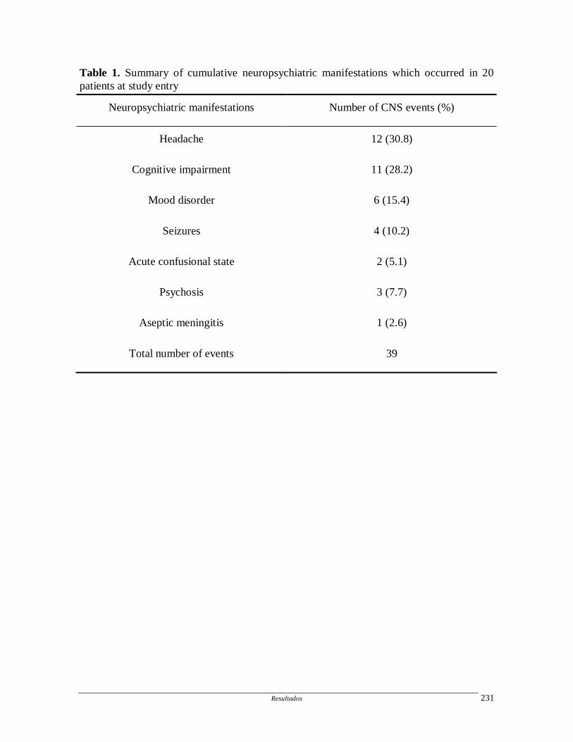
Resultados 231
Table 1. Summary of cumulative neuropsychiatric manifestations which occurred in 20 patients at study entry
Neuropsychiatric manifestations Number of CNS events (%)
Headache 12 (30.8)
Cognitive impairment 11 (28.2)
Mood disorder 6 (15.4)
Seizures 4 (10.2)
Acute confusional state 2 (5.1)
Psychosis 3 (7.7)
Aseptic meningitis 1 (2.6)
Total number of events 39

Resultados 232
Table 2. Median Cho/Cr values in 30 patients and 9 controls.
Groups Median Cho/Cr at initial
MRS (±SD) Median Cho/Cr at follow-up
MRS (±SD) p-value
A 1.13 (0.11) � 1.6 (0.2) � <0.001
B 1.07 (0.12) � 1.2 (0.2) � 0.006
D 0.95 (0.15) 0.94 (0.15) 0.88
(A) SLE patients with WM lesions in MRS ROI initial MRS
(B) SLE patients without WM lesions at initial MRI but with WM lesions at follow-up MRI
within MRS ROI
(C) Controls at follow-up
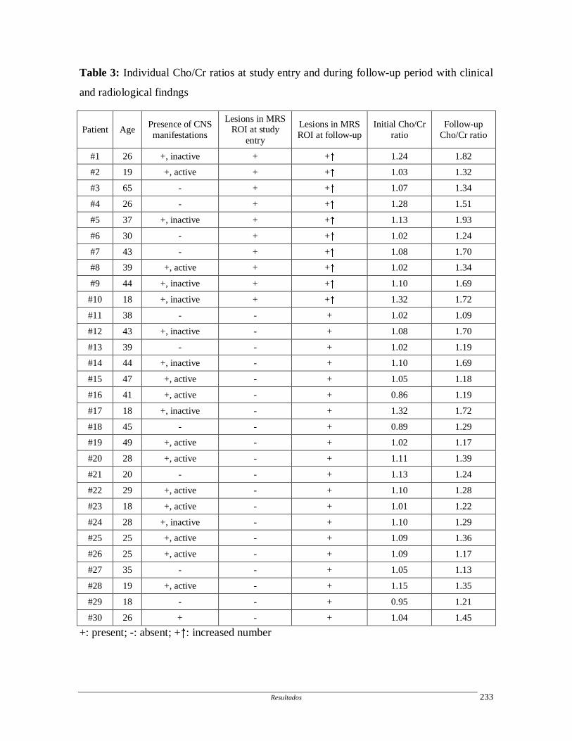
Resultados 233
Table 3: Individual Cho/Cr ratios at study entry and during follow-up period with clinical
and radiological findngs
Patient Age Presence of CNS manifestations
Lesions in MRS ROI at study
entry
Lesions in MRS ROI at follow-up
Initial Cho/Cr ratio
Follow-up Cho/Cr ratio
#1 26 +, inactive + + � 1.24 1.82
#2 19 +, active + + � 1.03 1.32
#3 65 - + + � 1.07 1.34
#4 26 - + + � 1.28 1.51
#5 37 +, inactive + + � 1.13 1.93
#6 30 - + + � 1.02 1.24
#7 43 - + + � 1.08 1.70
#8 39 +, active + + � 1.02 1.34
#9 44 +, inactive + + � 1.10 1.69
#10 18 +, inactive + + � 1.32 1.72
#11 38 - - + 1.02 1.09
#12 43 +, inactive - + 1.08 1.70
#13 39 - - + 1.02 1.19
#14 44 +, inactive - + 1.10 1.69
#15 47 +, active - + 1.05 1.18
#16 41 +, active - + 0.86 1.19
#17 18 +, inactive - + 1.32 1.72
#18 45 - - + 0.89 1.29
#19 49 +, active - + 1.02 1.17
#20 28 +, active - + 1.11 1.39
#21 20 - - + 1.13 1.24
#22 29 +, active - + 1.10 1.28
#23 18 +, active - + 1.01 1.22
#24 28 +, inactive - + 1.10 1.29
#25 25 +, active - + 1.09 1.36
#26 25 +, active - + 1.09 1.17
#27 35 - - + 1.05 1.13
#28 19 +, active - + 1.15 1.35
#29 18 - - + 0.95 1.21
#30 26 + - + 1.04 1.45
+: present; -: absent; + � : increased number
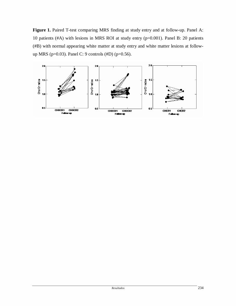
Resultados 234
Figure 1. Paired T-test comparing MRS finding at study entry and at follow-up. Panel A:
10 patients (#A) with lesions in MRS ROI at study entry (p=0.001). Panel B: 20 patients
(#B) with normal appearing white matter at study entry and white matter lesions at follow-
up MRS (p=0.03). Panel C: 9 controls (#D) (p=0.56).

235
5. DISCUSSÃO
Discussão 237
Os três primeiros artigos da tese são trabalhos de revisão. O artigo Neurolupus
(artigo #1) é uma revisão histórica sobre as primeiras menções do LES na literatura e as
primeiras descrições clínicas, mostrando que há muito tempo tinha sido reconhecida uma
variedade destas manifestações e um pior prognóstico de cada uma delas. Neste artigo
também enfatiza-se a importância da uniformização dos critérios diagnósticos e do
tratamento, ressaltando o fato interessante que os antimaláricos (quinina) serem utilizados
para o tratamento do LES desde o século 19. Ainda com relação ao tratamento, o uso de
corticosteróides e imunosupressores melhorou muito a sobrevida dos pacientes (Urowitz et
al., 1997), apesar de ainda observarmos uma elevada morbi-mortalidade naqueles com
manifestações do SNC (Blanco et al., 1998; Kasitanon et al., 2002; Jonsen et al., 2002).
No artigo sobre as manifestações do SNC no LES (artigo #2) apresentamos uma
revisão mais ampla, enfatizando além da dificuldade diagnóstica, a patogênese, a
investigação clínica e neuroimagem das manifestações do SNC. Comparamos os diferentes
estudos que utilizam os critérios diagnósticos do Colégio Americano de Reumatologia
(1999) e observamos que, apesar de uma tentativa de uniformização, ainda ocorrem
diferentes freqüências das manifestações. Isto pode ser explicado por diferenças loco-
regionais ou por viés na inclusão dos pacientes nos trabalhos (Hanly, 2005). Enfatizamos
também a importância do diagnóstico diferencial, visto que até 40% das manifestações do
SNC no LES podem ser atribuídas a outras causas, e não a doença propriamente dita (Hanly
et al., 2004).
Juntamente com a RM, a ERM (artigo #3) é uma ferramenta que pode ser
utilizada na investigação do comprometimento do SNC no LES. Ela permite uma avaliação
das alterações estruturais e pode ser realizada juntamente com a RM. Uma revisão sobre os
mecanismos físicos, dificuldades de aquisição e os resultados obtidos, muitas vezes por
diferentes técnicas de aquisição, é discutido neste trabalho. A maioria dos autores concorda
que há dano neuronal no LES (Axford et al., 2001; Handa et al., 2003), porém, a associação
destes achados com as manifestações do SNC (Sibbitt et al., 1997; Brooks et al., 1999; Lim
et al., 2000), o uso de corticsteróides (Chinn et al., 1997) ou alterações estruturais (Davie et
al., 1995; Lim et al., 2000) ainda é controversa.
Discussão 238
A maioria dos estudos envolvendo as manifestações NP no LES, mesmo os
mais atuais, (Sabbadini et al., 1999; Sanna et al., 2000; Ainiala et al., 2001; Mok et al.,
2001; Kasitanon et al., 2002; Sanna et al., 2003; Hanly et al., 2004; Mikdashi et al., 2004;
Hanly et al., 2005; Shimojima et al., 2005; Robert et al., 2006) apresenta uma descrição
generalizada destas manifestações, dificultando muitas vezes a sua análise individual.
Assim, ao estudar algumas destas manifestações isoladamente, pudemos avaliar melhor a
relação entre estas e outras variáveis clínicas e laboratoriais e de neuroimagem.
Ao estudar a freqüência de epilepsia em 519 pacientes (artigo #4), observamos
que a prevalência de crises epilépticas em nossa casuística (11,6%) é similar (8,3-28%) ao
que foi previamente descrito (Mackworth-Young et al., 1985; Herranz et al., 1994;
Jennekens e Kater, 2002; Brey et al., 2002; Kassitanon et al., 2002; Sanna et al., 2003;
Cimaz et al., 2006; Robert et al., 2006). A maioria destes pacientes (88,3%) apresentou
crises únicas e somente 11,7% apresentaram crises recorrentes, caracterizando, assim,
epilepsia. Crises tônico-clônicas generalizadas e crises parciais complexas foram mais
freqüentemente observadas em nossa casuística. A ocorrência de crises epilépticas no início
do LES ocorreu em 19 de 60 pacientes (31,7%) e esteve associada à presença de acidente
vascular cerebral (AVC) e aos anticorpos antifosfolípides, em títulos moderados a elevados.
Ambos os fatores devem estar associados à gênese das crises epilépticas, sejam secundárias
a eventos isquêmicos (Bresninhan et al., 1979; Cocito et al., 1982; Asherson et al., 1989;
Kumeral et al., 2002) ou ao aumento da excitabilidade neuronal (Liou et al., 1994). No
entanto, por se tratar de um estudo retrospectivo, não foi possível definir as causas de crises
em nossa casuística. Assim, como observado em nossos pacientes com crises epilépticas,
que apresentaram mais freqüentemente AVC, outros estudos demonstraram a associação de
crises epilépticas com outras manifestações NP no LES (Futrell et al., 1992; Mok et al.,
2001; Brey et al., 2002; Sanna et al., 2003).Estes achados, portanto, sugerem que, na
presença de manifestação NP, outras manifestações NP devam ser pesquisadas. Embora
tinha sido observada uma associação entre a ocorrência de crises epilépticas e nefrite, a
atividade da doença não parece ser responsável pela crise, visto que outras manifestações
clínicas não estavam associadas à presença de crises nesta casuística. A associação de crises
epilépticas com a presença de AVC e de sinais de doença de pequenos vasos à RM, reforça
a importância deste exame na investigação destes pacientes. O eletroencefalograma (EEG)
Discussão 239
foi útil na identificação de pacientes com maior risco de recorrência de crises, pois estes
apresentaram atividade epileptiforme interictal ao exame. Contudo, a recorrência das crises
foi rara, e ocorreu somente em pacientes com anticorpos antifosfolípides. O tratamento com
drogas antiepiléticas, não necessitaria, portanto, ser iniciado para todos os pacientes com
LES crises epilépticas isoladas. No entanto, pacientes com EEG demonstrando atividade
epileptiforme interictal e pacientes com anticorpos antifosfolípides poderiam ser medicados
com drogas antiepilépticas precocemente, mesmo após crises isoladas, visto que
apresentam maior risco de recorrência de crises.
Ainda há controvérsias se existe associação entre migrânea e o LES (Glanz et
al., 2001;Whitelaw et al., 2004), ou trata-se de ocorrência fortuita (Sfikakis et al., 1998;
Fernandez-Nebro et al., 1999; Mitsikostas et al., 2004). Realizamos um estudo prospectivo
de pacientes com LES com e sem migrânea comparando-os a dois grupos controles
(indivíduos normais e pacientes com artrite reumatóide) com objetivo de analisar a
importância clínica desta manifestação no LES (artigo #5). Optou-se por incluir um grupo
de pacientes com artrite reumatóide, com o intuito de excluir o fator doença crônica na
presença de cefaléia.Neste trabalho, pacientes com LES apresentaram migrânea com
freqüência significativamente maior do que aqueles com artrite reumatóide e controles.
Observamos ainda que na vigência da migrânea, os pacientes com LES apresentavam mais
frequentemente atividade da doença e piora do fenômeno de Raynaud. Desta forma, a
atividade de doença deve ser suspeitada naqueles que apresentam piora do quadro de
migrânea. A associação do fenômeno de Raynaud com migrânea no LES também foi
observada por outros autores (Cervera et al., 2002; Lessa et al., 2006), assim como na
população geral (Heslop et al., 1983; Silman et al., 1990; O’Keeffe et al., 1993), sugerindo
mecanismos patogênicos comuns, como reação vascular (Heslop et al., 1983; Silman et al.,
1990) e disfunção endotelial (Spierings, 2003; Hanly, 2003). A presença de migrânea
relacionou-se também a presença dos anticorpos antifosfolípides. Vários estudos analisaram
a interação entre a presença deste anticorpo e a disfunção endotelial. O anticorpo
antifosfolípide ao se ligar à superfície endotelial, levaria a uma ativação celular, que, por
sua vez, promoveria um aumento das moléculas de adesão e um aumento da secreção de
interleucina 6 e prostaglandinas. Isto, por sua vez, levaria a uma lesão endotelial induzida
por complemento ou por citotoxicidade induzida por anticorpos. Portanto, a disfunção
Discussão 240
endotelial é um dos mecanismos patogênicos que explicaria a associação entre migrânea,
anticorpos antifosfolípides e fenômeno de Raynaud (Del Papa et al., 1992; Del Papa et al.,
1997; Simantov et al., 1995; Hanly et al., 1996). Analisando o índice de dano nestes
pacientes, observamos uma associação positiva com a história pregressa de migrânea. Uma
possibilidade plausível é que, como se observou associação entre atividade de doença e
migrânea, esta atividade, juntamente com uma possível dose maior de corticosteróides e
outras drogas, poderiam levar a um maior dano permanente nestes pacientes. A importância
da RM neste grupo de pacientes não foi avaliada neste trabalho, fazendo parte de um estudo
já em andamento.
A freqüência de psicose, avaliada em 537 pacientes com LES (artigo #6)
também foi semelhante à publicações prévias, ou seja 17,5% (Mok et al., 2001, Brey et al.,
2002, Kasitanon et al., 2002, Sanna et al., 2003). A ocorrência da psicose no início do LES
apresentou associação positiva com atividade de doença, mas uma associação negativa com
lesões cutâneas, ou seja, pacientes com psicose apresentaram menor freqüência de
comprometimento de pele. Pacientes com psicose, no início do LES, provavelmente são
encaminhados inicialmente ao psiquiatra, o que pode levar a uma subestimação deste
sintoma no início do LES.
Uma das questões mais frequentes na prática clínica e durante a evolução da
doença, é se a psicose apresentada pelos pacientes com LES é primária ou secundária ao
uso de corticosteróides. Observou-se que pacientes com psicose primária apresentavam
mais frequentemente anticorpos antifosfolípides e outras manifestações do SNC, como
depressão, AVC, crise epilépticas e distúrbios cognitivos. Isto pode ser devido à interação
dos anticorpos antifosfolípides com a membrana neuronal, não necessariamente
secundários a fenômenos trombóticos (Wysenbeek et al., 1999, Mok et al., 2001, Sanna et
al., 2003). Apesar do anticorpo anti-P ser de grande utilidade para o diagnóstico da psicose
no LES (Bonfa et al., 1987), sua realização de rotina ainda não ocorre na maioria dos
centros de atendimento. Por isso é importante determinar outros fatores que possam
diferenciar psicose primária daquela induzida por corticosteróides, além das evidências
clínicas e sua relação temporal com o uso da droga. Um dado interessante neste trabalho
(artigo #6) foi a associação da psicose induzida por corticosteróides com a
Discussão 241
hipoalbuminemia. Por se tratar de um estudo retrospectivo, não foi possível determinar um
nível crítico de albumina a partir do qual os pacientes teriam uma maior chance de
desenvolver psicose. Como os corticosteróides sintéticos ligam-se a albumina para o
transporte, tornando-se assim inativos, a hipoalbuminemia estaria relacionada a níveis mais
elevados de corticosteróides livres circulantes, o que pode justificar o maior efeito colateral
nestes pacientes, incluíndo-se a psicose (Kohen et al., 1993; Patten &Neutel 2000, Lopez-
Medrano et al., 2002; Sirois, 2003).
Quando se estudou a recorrência da psicose nestes pacientes, observou-se que
esta ocorreu mais frequentemente em pacientes com atividade de doença e outras
manifestações do SNC. No entanto, não foi possível identificar substratos anatômicos na
RM associados à recorrência de psicose nestes pacientes.
A trombose venosa central (TVC) é considerada rara no LES (Vidailhet et al.,
1990; Flusser et al., 1994; Laversuch et al., 1995; Lee et al., 2001; Uthman et al., 2004).
Em nosso estudo analisando 24 pacientes com TVC de diferentes etiologias (artigo #7),
somente três pacientes apresentaram síndrome do anticorpo antifosfolípide e destes,
somente um tinha também o diagnóstico de LES. Os principais sintomas dos pacientes com
TVC foram, em freqüência decrescente, cefaléia, vômitos e alterações do nível de
consciência. Portanto, é recomendável que pacientes com LES, principalmente aqueles com
anticorpos antifosfolípides associados, que apresentem cefaléia intensa de início recente e
aqueles com distúrbios cognitivos progressivos, sejam avaliados por método de imagem
para afastar a presença de TVC.
Estes trabalhos acima descritos, ao analisarem algumas manifestações do SNC
no LES puderam destacar a utilidade da RM estrutural como método de investigação
complementar. A partir destes trabalhos procurou-se estudar de forma mais minuciosa o
papel de diferentes métodos de neuroimagem aplicados à pacientes com LES para avaliar,
do ponto de vista estrutural e funcional, o envolvimento do SNC.
Analisando a RM estrutural de 115 pacientes com LES, 72 com manifestações
do SNC (artigo #8), observamos que pacientes com LES apresentavam mais
frequentemente atrofia de corpo caloso e do volume cerebral do que controles normais.
Observamos atrofia cerebral grave em 8,7% dos casos, inferior ao previamente relatado
Discussão 242
(Bilaniuk et al., 1977; Killian et al., 1979; Gonzalez-Scarano et al., 1979; Gayliset al.,
1982; Carette et al., 1982; Kaell et al., 1986; McCune e Golbus, 1988; McCune et al., 1988;
Omdal et al., 1989; Ostrov et al., 1982; Waterloo et al., 1999). Isto decorre, possivelmente,
do método utilizado para análise, pois a maioria dos estudos descritos acima utilizou uma
análise visual ou medição manual em imagens bidimensionais (Tabelas 4 e 5), enquanto
que, em nosso trabalho, com a utilização de um método de segmentação semi-automática e
a elaboração de protocolos de segmentação bem definidos, pode-se obter resultados mais
fidedignos e reproduzíveis. A atrofia cerebral no LES não estava associada aos diferentes
tipos de manifestações do SNC, nem a dose cumulativa de corticosteróides, como sugerido
por alguns autores (Carette et al., 1982; Ostrov et al., 1982; Zanardi et al., 2001), porém
correlacionou-se ao número de manifestações pregressas do SNC e ao maior tempo de
doença.
Apesar da atrofia cerebral ter sido estudada por vários autores (Bilaniuk et al.,
1977; Killian et al., 1979; Gonzalez-Scarano et al., 1979; Gayliset al., 1982; Carette et al.,
1982; Ostrov et al., 1982; Kaell et al., 1986; McCune e Golbus, 1988; McCune et al., 1988;
Omdal et al., 1989; Waterloo et al., 1999; Zanardi et al., 2001;), apenas um trabalho (Steens
et al., 2004) procurou determinar se há uma região cerebral predominantemente acometida.
Nossos trabalhos permitiram observar, através de dois métodos distintos [VBM (artigo #9)
e segmentação semi-automática (artigo #8)], que o corpo caloso é a região de substância
branca mais freqüentemente afetada. Na análise semi-automática a atrofia do corpo caloso
esteve associada à história pregressa de manifestações do SNC, ao número total destas
manifestações e ao maior tempo de doença. A atrofia do corpo caloso é frequentemente
observada em RM estrutural de idosos (Janowsky et al., 1996; Hampel et al., 1988; Black et
al., 2000; Bjartmar et al., 2001). A explicação é que a maioria dos neurônios projetados
para o corpo caloso são das células piramidais gigantes das camadas corticais III e IV do
hemisfério contralateral (Hampel et al., 1998) e a isquemia cerebral resulta em lesão da
camada III (Innocenti et al., 1986), levando a degeneração Walleriana do corpo caloso
(Grahm et al., 1992). Portanto, a atrofia do corpo caloso pode ser considerada um marcador
de perda neuronal em idosos (Grahm et al., 1992). Esta hipótese foi testada pelo método de
VBM, e uma maior perda de substância branca foi também observada na região frontal e
occipital, além do corpo caloso, em nossos pacientes. Este achado poderia representar um
Discussão 243
substrato anatômico para a presença de distúrbios cognitivos no LES, que envolve
principalmente funções subcorticais frontais (Leritz et al., 2000). Entre os fatores
associados observados, além da história pregressa de manifestações NP e maior tempo de
doença, já descritas, observamos também uma correlação entre atrofia de substância branca
e presença dos anticorpos antifosfolípides pela técnica de VBM. As lesões de substância
branca que ocorrem frequentemente em idosos (Awald et al., 1986; Katzman t al., 1999),
assim como em pacientes lúpicos (Chinn et al., 1997; Walcki et al., 2002; Cotton et al.,
2004), ainda que muitas vezes consideradas inespecíficas, podem ser decorrentes de
vasculopatia (Wen et al., 2004). Na etiologia da vasculopatia, parece haver a participação
dos anticorpos antifosfolípides, observada mais frequentemente naqueles pacientes com
lesões de substância branca, embora estudos anátomopatológicos devam confirmar esta
hipótese. A vasculopatia, por sua vez, leva a atrofia de substância branca que por sua vez
leva a atrofia de corpo caloso.
Em relação à substância cinzenta, a análise pela técnica de VBM (artigo #9)
demonstrou, principalmente a redução do volume dos lobos temporais e outras áreas
límbicas, associada à dose total de corticosteróides utilizada, entre outros fatores.
Analisando os volumes dos hipocampos, isoladamente, (artigo #10) observamos, no início
do estudo, 43,9% de atrofia hipocampal, associada ao maior tempo de doença, dose
cumulativa de corticosteroides e ao número de manifestações do SNC, indicando que o
hipocampo parece ser uma estrutura muito sensível aos insultos sistêmicos. A presença da
atrofia hipocampal esteve associada à presença e ao grau dos distúrbios cognitivos no LES.
A partir da padronização destas técnicas, pode-se avaliar, no artigo #9 e #10, se
a atrofia cerebral no LES é progressiva e qual os fatores associados à sua progressão.
Observamos que, após um tempo médio de 19 meses de seguimento ocorreu progressão
significativa da perda tanto de substância branca como de cinzenta. Pela técnica de VBM
(artigo #9), observamos também uma redução do volume cortical em pacientes com LES,
ocorrendo predominantemente em lobos frontal, dorsolateral e temporal medial.
Já a progressão da atrofia hipocampal (artigo #10) esteve associada não
somente ao número de manifestações NP, mas também à dose cumulativa de
corticosteróides. Clinicamente, a progressão da atrofia hipocampal esteve associada à piora

Discussão 244
do distúrbio cognitivo. Estudos prévios demonstraram que no LES, grande parte dos
pacientes com distúrbios cognitivos não piora ao longo da doença (Karassa et al., 2000;
Carlomagno et al., 2000). Isto sugere que pacientes com flutuações na cognição
provavelmente não apresentam alteração estrutural, enquanto que aqueles com persistência
e progressão de distúrbios cognitivos provavelmente apresentam atrofia hipocampal.
Portanto, a análise volumétrica dos hipocampos pode ser uma importante ferramenta para
predizer o curso clínico dos distúrbios cognitivos no LES.
Observamos também que a atrofia hipocampal progressiva esteve associada à
presença pregressa de manifestações do SNC e não à atividade do LES, indicando que o
dano estrutural causado pelo envolvimento do SNC ocorre de forma gradual e lenta. Além
disto, nossos resultados mostram que a dose cumulativa de corticosteróides esteve
associada com a perda de substância cinzenta, como o hipocampo, enquanto o tempo de
doença influenciou a progressão de atrofia tanto de substância branca como cinzenta. A
piora do distúrbio cognitivo nos pacientes com LES esteve claramente associada à perda de
substância cinzenta (atrofia hipocampal) neste estudo (artigo #10).
A aplicação de técnicas de neuroimagem funcional revelou que as alterações
funcionais observadas ocorrem mais precocemente que as alterações estruturais e parecem
estar relacionados mais à presença de atividade sistêmica de doença do que com
manifestações do SNC isoladamente, indicando que o cérebro é mais amplamente afetado
pela doença ativa do que se supunha.
O estudo com SPECT cerebral foi realizado em 40 pacientes, sendo que 20
apresentavam manifestações NP ativas e 20 pacientes história pregressa de
comprometimento do SNC (artigo #11). Analisando-se as imagens do SPECT cerebral
através da técnica do VBM, foi possível identificar alterações em pacientes com
manifestações NP ativas, não identificadas visualmente. Estes apresentavam uma
hipoperfusão cerebral difusa, independentemente do tipo da manifestação apresentada.
Pacientes sem manifestações neuropsiquiátricas ativas apresentavam um padrão de
perfusão cerebral semelhante aos controles. Para determinar se os achados de hipoperfusão
cerebral não são decorrentes da presença da atrofia, as RM destes pacientes foram
analisadas pela técnica do VBM. Como não foi observada diferença quanto à atrofia nos
Discussão 245
dois grupos de pacientes, podemos considerar o hipofluxo secundário a presença de
manifestações NP ativas. Utilizando-se, portanto, a técnica do VBM para análise de SPECT
cerebral, foi possível identificar alterações de hipoperfusão em pacientes com
manifestações NP ativas, porém pelo pequeno número de pacientes, não foi possível avaliar
se esta alteração foi decorrente da atividade sistêmica da doença ou da manifestação do
SNC propriamente dita. Para responder a esta pergunta, utilizamos outro método funcional,
a ERM. Neste estudo (artigo #12) foram incluídos 90 pacientes, sendo 29 com doença ativa
e comprometimento do SNC ativo, 28 pacientes com doença ativa sem evidência de
envolvimento do SNC, 14 pacientes com doença inativa e história pregressa de
manifestações NP e 19 pacientes com doença inativa sem evidências de comprometimento
do SNC. Observamos uma redução significativa do marcador neuronal NAA (relativo ao
sinal de cratina -NAA/Cr) em pacientes com doença ativa, independentemente da presença
do comprometimento do SNC. O fato da redução relativa do NAA ser semelhante em
pacientes com e sem história pregressa de comprometimento do SNC, sugeriu que a perda
neuronal poderia ser, até certo ponto, transitória e relacionada à presença de atividade
sistêmica do LES, fato demonstrado no estudo de seguimento destes pacientes. A
normalização da relação NAA/Cr em pacientes que apresentaram doença ativa no início do
estudo e doença inativa no seguimento e a piora da relação NAA/Cr em pacientes com
doença inativa que se tornou ativa, indica a transitoriedade destes achados. Em pacientes
com doença ativa tanto no início como na evolução observou-se uma progressiva piora da
relação NAA/Cr. Não observamos elevação da relação cholina/creatina nos pacientes com
substância branca normal. Em outro estudo (artigo #13) em que a ERM foi realizada em
regiões com grande número de lesões de substância branca observamos um aumento na
relação Cho/Cr em relação aos controles. Neste mesmo estudo também observamos que
houve um aumento do número de lesões de substância branca após uma média de 19 meses
de seguimento, assim como um aumento da relação Cho/Cr nesta mesma região. Em
pacientes que, no início do estudo, não apresentavam lesões de substância branca, mas
apresentavam aumento da relação Cho/Cr, a RM realizada após 19 meses evidenciou a
presença de lesões de substância branca, demonstrando que o aumento da relação Cho/Cr
em substância branca normal pode predizer o aparecimento de lesões de substância branca.
Este achado, além de ressaltar a importância da ERM no acompanhamento destes pacientes,
Discussão 246
contribui para que se valorize as lesões de substância branca no LES, muitas vezes
consideradas inespecíficas, porém, como demonstrado, associadas a alterações
neurometabólicas.
Pelos resultados dos nossos trabalhos, podemos concluir que a padronização de
técnicas de neuroimagem estrutural e funcional pode, portanto, acrescentar informações
valiosas quanto ao comprometimento do SNC no LES e auxiliar a correlação entre a RM e
a clínica destes pacientes, influenciando na conduta terapêutica e no prognóstico destes
pacientes.

247
6. CONCLUSÕES
Conclusões 249
1. Crises epilépticas em pacientes com LES são geralmente únicas e, quando há
recorrência,o EEG e a RM são ferramentas úteis para identificar estes casos.
2. Os anticorpos antifosfolípides estão associados a manifestações do SNC no LES,
em especial a ocorrência e recorrência de crises epilépticas, a migrânea, e a psicose
na evolução.
3. A migrânea está associada à atividade de doença, à presença de anticorpos
antifosfolípides e à presença do Fenômeno de Raynaud. Pacientes com história
pregressa de migrânea apresentaram mais dano, avaliado pelo SLICC, que
pacientes sem migrânea.
4. A trombose venosa central é rara no LES, mas, quando ocorre, pode estar associada
à presença de distúrbios cognitivos.
5. A atrofia cerebral, tanto de substância branca como de substância cinzenta, é mais
freqüente no LES que em controles e ocorre de forma progressiva. A atrofia está
associada ao número e a história pregressa de manifestações do SNC e não ocorre
na vigência da manifestação ativa. O uso de corticosteróides está associado à
atrofia de substância cinzenta e não à atrofia de substância branca.
6. A presença e o grau de atrofia do corpo caloso e do hipocampo estão associados à
presença e a gravidade dos distúrbios cognitivos.
7. A análise do SPECT cerebral pela técnica de VBM é capaz de diferenciar pacientes
com manifestações do SNC ativas de inativas.
8. A ERM demonstra disfunção axonal na substância branca normal, associada à
atividade de doença, independentemente do comprometimento do SNC. Estes
achados são transitórios, melhorando com controle da atividade da doença.
9. Pacientes com lesões de substância branca apresentam elevação da relação Cho/Cr.
A elevação da Cho/Cr na substância branca normal pode predizer o aparecimento
de lesões de substância branca.
Conclusões 250
10. A padronização de técnicas de neuroimagem é importante para avaliação da
presença e da progressão da atrofia cerebral. Métodos de neuroimagem estruturais e
funcionais são úteis na identificação do comprometimento do SNC no LES e
podem demonstrar um maior envolvimento, independentemente da presença de
manifestações do SNC.
251
7. REFERÊNCIAS BIBLIOGRÁFICAS
Referências Bibliográficas 253
Abbott NJ, Mendonca LL, Dolman DE. The blood-brain barrier in systemic lupus
erythematosus. Lupus. 2003; 12:908-15
Abreu MR, Jakosky A, Folgerini M, Brenol JC, Xavier RM, Kapczinsky F.
Neuropsychiatric systemic lupus erythematosus: correlation of brain MR imaging, CT, and
SPECT. Clin Imaging. 2005; 29:215-21.
ACR Ad hoc Committee on Neuropsychiatric Lupus Nomenclature. The American College
of rheumatology Nomenclature and Case definitions for Neuropsychiatric Lupus
syndromes. Arthritis Rheum 1999; 42:599-608.
Adelmann DC, Saltiel E, Klinenberg JR. The neuropsichiatric manifestations of SLE: an
overview. Semin Arthr Rheum 1986: 15:185-199.
Afeltra A, Garzia P, Mitterhofer AP, Vadacca M, Galluzzo S, Del Porto F, et al.
Neuropsychiatric lupus syndromes: relationship with antiphospholipid antibodies.
Neurology. 2003; 61:108-10.
Ainiala H, Hietaharju A, Loukkola J, Peltola J, Korpela M, Metsanoja R, et al. Validity of
the new American College of Rheumatology criteria for neuropsychiatric lupus syndromes:
a population-based evaluation. Arthritis Rheum. 2001; 45:419-23.
Ainiala H, Loukkola J, Peltola J, Korpela M, Hietaharju A. The prevalence of
neuropsychiatric syndromes in systemic lupus erythematosus. Neurology. 2001; 57:496-
500.
Ainiala H, Hietaharju A, Dastidar P, Loukkola J, Lehtimaki T, Peltola J, et al. Increased
serum matrix metalloproteinase 9 levels in systemic lupus erythematosus patients with
neuropsychiatric manifestations and brain magnetic resonance imaging abnormalities.
Arthritis and Rheumatism 2004; 50:858-65.
Ainiala H, Dastidar P, Loukkola J, Lehtimaki T, Korpela M, Peltola J, et al. Cerebral MRI
abnormalities and their association with neuropsychiatric manifestations in SLE: a
population-based study. Scand J Rheumatol. 2005; 34:376-82.
Referências Bibliográficas 254
Akbarian S, Sucher NJ, Bradley D, Tafazzoli A, Trinh D, Hetrick WP, et al. Selective
alterations in gene expression for NMDA receptor subunits in prefrontal cortex of
schizophrenics. Journal of Neuroscience 1996; 16:19-30.
Alarcon GS. Of ethnicity, race and lupus. Lupus 2001; 10:594–596.
Arnett FC, Reveille JD, Moutsopoulos HM, Georgescu L, Elkon KB. Ribosomal P
autoantibodies in systemic lupus erythematosus. Frequencies in different ethnic groups and
clinical and immunogenetic associations. Arthritis and Rheumatism 1996; 39:1833–1839.
Arnold DL, Shoubridge EA, Villemure JG, Feindel W. Proton and phosphorus magnetic
ressonance spectroscopy of human astrocytomas in vivo. Preliminar observations in tumor
gradient. Radiology, 183:711-718, 1992.
Arnold DL, Matthews PL, Francis GS, O´Connor J, Antel JP. Proton magnetic ressonace
spectroscopic imaging for metabolic characterization of demyelinating plaques. Ann
Neurol, 31:235-241, 1992.
Ashburner J, Friston KJ. Voxel-based morphometry-the method. NeuroImage 1997;
11:805-821.
Ashburner J, Friston KJ. Multimodal image coregistration and partitioning--a unified
framework. Neuroimage 2000; 6:209-17.
Asherson RA, Khamashta MA, Gil A, Vasquez J-J, Chan O, Baguley E, et al.
Cerebrovascular disease and antiphospholipid antibodies in systemic lupus erythematosus,
lupus-like disease, and the primary antiphospholipid syndrome. Am J Med 1989, 86:391-
399.
Awad IA, Spetzler RF, Hodak JÁ, Hodak JA. Incidental subcortical lesions identified on
magnetic resonance imaging in the elderly. I Correlation with age and cerebrovascular risk
factors. Stoke. 1986; 17:1084-1089.
Axford JS, Howe FA, Heron C, Griffiths JR. Sensitivity of quantitative (1)H magnetic
resonance spectroscopy of the brain in detecting early neuronal damage in systemic lupus
erythematosus. Ann Rheum Dis. 2001; 60:106-11.
Referências Bibliográficas 255
Barkhof F, Filippi M, van Waesberghe JH, Molyneux P, Rovaris M, Lycklama a Nijeholt
G, et al. Comparison of MRI criteria at first presentation to predict conversion to clinically
definite multiple esclerosis. Brain 1997; 120:2059-2069.
Baum KA, Hopf U, Nehrig C, Stover M, Schorner W. Systemic lupus erythematosus:
neuropsychiatric signs and symptoms related to cerebral MRI findings. Clin Neurol
Neurosurg. 1993; 95:29-34.
Beck AT, Beamesderfer A. Assessment of depression: the depression inventory. Mod Probl
Pharmacopsychiat. 1974; 7:151-169.
Beck AT, Rial WY, Rickels K. Short form of depression inventory: cross-validation.
Psychol Rep. 1974; 34:1184–1186.
Bilaniuk LT, Patel S, Zimmerman RA. Computed tomography of systemic lupus
erythematosus. Radiology. 1977; 124:119-21.
Birken DL, Oldendorf WH. N-acetyl-L-aspartic acid: a literature review of a compound
prominent in 1H-NMR spectroscopic studies of brain. Neurosci Biobehav Rev. 1989;
13:23-31.
Black SE, Moffat SD, Yu DC, Parker J, Stanchev P, Bronskill M. Callosal atrophy
correlates with temporal lobe volume and mental status in Alzheimer's disease. Can J
Neurol Sci 2000; 27:204-9.
Blanco FJ, Gomez-Reino JJ, de la Mata J, Corrales A, Rodriguez-Valverde V, Rosas JC, et
al. Survival analysis of 306 European Spanish patients with systemic lupus erythematosus.
Lupus 1998; 7:159-63.
Bluestein HG, Zvaifler NJ. Antibodies reactive with central nervous system antigens. Hum
Pathol. 1983; 14:424-8.
Bluestein HG, Woods VL Jr. Antineuronal antibodies in systemic lupus erythematosus.
Arthritis Rheum. 1982; 25:773-8.
Referências Bibliográficas 256
Bluestein HG, Williams GW, Steinberg AD. Cerebrospinal fluid antibodies to neuronal
cells: association with neuropsychiatric manifestations of systemic lupus erythematosus.
Am J Med. 1981; 70: 240-6.
Bonfa E, Golombek SJ, Kaufman LD, Skelly S, Weissabach H, Brot N, Elkon KB.
Association between lupus psychosis and anti-ribosomal P protein antibodies. N Engl. J.
Med 1987; 317:265-271.
Borrelli M, Tamarozzi R, Colamussi P, Govoni M, Trotta F, Lappi S. Evaluation with MR,
perfusion MR and cerebral flowSPECT in NPSLE patients. Radiol Med 2003; 105:482-9.
Bosma GP, Middelkoop HA, Rood MJ, Bollen EL, Huizinga TW, Van Buchem MA.
Association of global brain damage and clinical functioning in neuropsychiatric lupus
erythematosus. Arthritis Rheum 2002; 46:2665-72.
Brandt JT, Triplett DA, Alving B, Scharrer I. Criteria for the diagnosis of lupus
anticoagulants: an update. On behalf of the Subcommittee on Lupus
Anticoagulant/Antiphospholipid Antibody of the Scientific and Standardization Committee
of the ISTH. Thromb Haemost 1995; 74:1185-1190.
Bresnihan B, Hohmeister R, Cutting J, Travers RL, Waldburger M, Black C, et al. The
neuropsychiatric disorder in systemic lupus erythematosus: evidence for both vascular and
immune mechanisms. Ann Rheum Dis. 1979; 38:301-6.
Brey RL, Holliday SL, Saklad AR, Navarrete MG, Hermosillo-Romo D, Stallworth CL, et
al. Neuropsychiatric syndromes in lupus: prevalence using standardized definitions.
Neurology 2002; 58:1214–1220.
Brooks WM, Jung RE, Ford CC, Greinel EJ, Sibbitt WL Jr. Relationship between
neurometabolite derangement and neurocognitive dysfunction in systemic lupus
erythematosus. J Rheumatol 1999; 26:81-85.
Brooks WM, Sabet A, Sibbitt WL Jr, Barker PB, van Zijl PC, Duyn JH, et al.
Neurochemistry of brain lesions determined by spectroscopic imaging in systemic lupus
erythematosus. J Rheumatol. 1997; 24:2323-9.
Referências Bibliográficas 257
Brooks WM, Jung RE, Ford CC, Greinel EJ, Sibbitt WL Jr. Relationship between
neurometabolite derangement and neurocognitive dysfunction in systemic lupus
erythematosus. J Rheumatol. 1999; 26:81-5.
Carette S, Urowitz MB, Grosman H, St Louis EL. Cranial computerized tomography in
systemic lupus erythematosus.J Rheumatol. 1982; 9:855-9.
Carlomagno S, Migliaresi S, Ambrosone L, Sannino M, Sanges G, Di Iorio G. Cognitive
impairment in systemic lupus erythematosus: a follow-up study. J Neurol. 2000; 247:273-9.
Castellino G, Govoni M, Padovan M, Colamussi P, Borrelli M, Trotta F. Proton magnetic
resonance spectroscopy may predict future brain lesions in SLE patients: a functional multi-
imaging approach and follow up. Ann Rheum Dis. 2005; 64:1022-7.
Cauli A, Montaldo C, Peltz MT, Nurchis P, Sanna G, Garau P, et al. Abnormalities of
magnetic resonance imaging of the central nervous system in patients with systemic lupus
erythematosus correlate with disease severity. Clin Rheumatol. 1994; 13:615-8.
Cendes F, Andermann F, Gloor P, Evans A, Jones-Gotman M, Watson C, et al. MRI
volumetric measurement of amygdala and hippocampus in temporal lobe epilepsy.
Neurology. 1993; 43:719-25.
Cervera R, Piette JC, Font J, Khamashta MA, Shoenfeld Y, Camps MT, et al.
Antiphospholipid syndrome: clinical and immunologic manifestations and patterns of
disease expression in a cohort of 1,000 patients. Arthritis Rheum. 2002; 46:1019-27.
Chahade WH, Sato EI, Moura JE Jr, Costallat LT, Andrade LE. Systemic lupus
erythematosus in Sao Paulo/Brazil: a clinical and laboratory overview. Lupus. 1995; 4:100-
3.
Chapman J, Cohen-Armon M, Shoenfeld Y, Korczyn AD. Antiphospholipid antibodies
permeabilize and depolarize brain synaptoneurosomes. Lupus 1999; 8:127-133
Cheatum DE, Hurd ER, Strunk SW, Ziff M. Renal histology and clinical course of systemic
lupus erythematosus. A prospective study. Arthritis Rheum. 1973; 16:670-6
Referências Bibliográficas 258
Chinn RJ, Wilkinson ID, Hall-Craggs MA, Paley MN, Shortall E, Carter S, et al. Magnetic
resonance imaging of the brain and cerebral proton spectroscopy in patients with systemic
lupus erythematosus. Arthritis Rheum. 1997; 40:36-46.
Chen JJ, Yen RF, Kao A, Lin CC, Lee CC. Abnormal regional cerebral blood flow found
by technetium-99m ethyl cysteinate dimer brain single photon emission computed
tomography in systemic lupus erythematosus patients with normal brain MRI findings.
Clin Rheumatol. 2002; 21:516-9.
Cimaz R, Meroni PL, Shoenfeld Y. Epilepsy as part of systemic lupus erythematosus and
systemic antiphospholipid syndrome (Hughes syndrome). Lupus. 2006; 15:191-7.
Clark EC, Bailey AA. Nurological and psychiatric signs associated with systemic lupus
erythematosus. JAMA 1956; 160:455-457.
Colamussi P, Giganti M, Cittanti C, Dovigo L, Trotta F, Tola MR, et al. Brain single
photon emission tomography with 99m Tc-HMPAO in neuropsychiatric systemic lupus
erythematosus: relations with EEG and MRI findings and clinical manifestations. Eur J
Nucl Med 1995; 22:17-24.
Colamussi P, Trotta F, Ricci R, Cittanti C, Govoni M, Barbarella G, et al. Brain perfusion
SPET and proton magnetic resonance spectroscopy in the evaluation of two systemic lupus
erythematosus patients with mild neuropsychiatric manifestations. Nucl Med Commun.
1997; 18:269-73.
Cocito L, Favale E, Reni L. Epileptic seizures in cerebral occlusive disease. Stroke. 1982;
13:189-195
Costallat LT, Appenzeller S, Bértolo MB. Lúpus neuropsiquiátrico de acordo com a nova
nomenclatura e definição de casos do Colégio Americano de Reumatologia (ACR): análise
de 527 pacientes. Rev Bras Reumatol 2001; 41:133-141.
Costallat LTL, Appenzeller S, Marini R. Evolução e fatores prognósticos do lúpus
eritematoso sistêmico em relação com a idade de início. Rev Bras Reumatol 2002; 42:91-
98.
Referências Bibliográficas 259
Costallat LT, Quagliato EMAB, Zanardi VA. Evoked potentials in the assessment of
neuropsychiatric manifestations in SLE. Clin Rheum 1997; 16:217-221.
Costallat LTL, Oliveira RM, Santiago MB, Cossermelli W, Samara AM. Neuropsychiatric
manifestations of SLE: the value of anticardiolipin, antigangliosides and
antigalactocerebrosides antibodies. Clin Rheum 1990; 4:489-497.
Cotton F, Bouffard-Vercelli J, Hermier M, Tebib J, Vital Durand D, Tran Minh VA, et al.
MRI of central nervous system in a series of 58 systemic lupus erythematosus (SLE)
patients with or without overt neuropsychiatric manifestations. Rev Med Interne. 2004;
25:8-15.
Daly D. Central nervous system in acute disseminated lupus erythematosus. J Nerv ment
Dis 1945; 012:461-464
Davie CA, Hawkins CP, Barker GJ, Brennan A, Tofts PS, Miller DH, et al. Serial proton
magnetic ressonance spectroscopy in acute multiple sclerosis lesions. Brain 1994; 117:49-
58.
Davie CA, Feinstein A, Kartsounis LD, Barker GJ, McHugh NJ, Walport MJ, et al. Proton
magnetic resonance spectroscopy of systemic lupus erythematosus involving the central
nervous system. J Neurol. 1995; 242:522-8.
Del Papa N, Meroni PL, Tincani A, Harris EN, Pierangeli SS, Barcellini W, et al.
Relationship between anti-phospholipid and anti-endothelial cell antibodies: further
characterization of the reactivity on resting and cytokine-activated endothelial cells. Clin
Exp Rheumatol. 1992; 10:37-42.
Del Papa N, Guidali L, Sala A, Buccellati C, Khamashta MA, Ichikawa K,et al. Endothelial
cells as target for antiphospholipid antibodies. Human polyclonal and monoclonal anti-beta
2-glycoprotein I antibodies react in vitro with endothelial cells through adherent beta 2-
glycoprotein I and induce endothelial activation. Arthritis Rheum. 1997; 40:551-61.
Duijn JH, Matson GB, Maudsley AA, Hugg JW, Weiner MW. Human brain infarction:
proton MR spectroscopy. Radiology. 1992; 183:711-8.
Referências Bibliográficas 260
Delis DC, Kramer JH, Kaplan E, et al. California Verbal Learning Test Research Edition
Manual. San Antonio: The Psychological Corporation, 1987.
Denburg SD, Behmann SA, Carbotte RM, Denburg JA. Lymphocyte antigens in
neuropsychiatric systemic lupus erythematosus. Relationship of lymphocyte antibody
specificities to clinical disease. Arthritis Rheum. 1994; 37:369-75.
Destefano N, Matthews PM, Antel JP, Preul M, Francis G, Arnold DL. Chemical pathology
of acute demyelinating lesions and its correlation with disability. Ann Neurology 1995;
38:901-909.
Destefano N, Matthews PM, Arnold DL. Reversible decrease in NAA after acute brain
injury. Mag res Méd 1995; 117:721-727.
Devinsky O, Petito CK, Alonso DR. Clinical and neuropatological findings in systemic
lupus erythematosus: the role of vasculitis, heart emboli, and thrombotic thrombocytopenic
purpura. Ann Neurol 1988; 23:380-384.
Dubois EL, Tuffanelli DL. Clinical manifestations of systemic lupus erythematosus:
computer analysis of 520 cases. JAMA 1964; 190:104-111.
Dubois EL, Tuffanelli DL. Clinical manifestations of systemic lupus erythematosus.
JAMA, 1964; 190:104-111.
Ellis SG, Verity AM. Central nervous system involvement in SLE. Sem Arthr Rheum 1979:
8:3:212-221.
Emmi L, Bramati M, De Cristofaro MT, Mascalchi M, Dal Pozzo G, Marconi GP, et al.
MRI and SPECT investigations of the CNS in SLE patients. Clin Exp Rheumatol 1993;
11:13–20.
Estes D, Christian CL. The natural history of systemic lupus erythematosus studied by
prospective analysis. Medicine, 1971: 50:85-95.
Faber-Elmann A, Sthoeger Z, Tcherniack A, Dayan M, Mozes E. Activity of matrix
metalloproteinase-9 is elevated in sera of patients with systemic lupus erythematosus.
Clinical and Experimental Immunology 2002; 127:393–398.
Referências Bibliográficas 261
Falcini F, De Cristofaro MT, Ermini M, Guarnieri M, Massai G, Olmastroni M, et al.
Regional cerebral blood flow in juvenile systemic lupus erythematosus: a prospective
SPECT study. Single photon emission computed tomography. J Rheumatol 1998; 25:583-8.
Fazekas F, Barkhof F, Filippi M, Grossman RI, Li DK, McDonald WI, et al. The
contribution of magnetic resonance imaging to the diagnosis of multiple sclerosis.
Neurology 1999; 53:448-456.
Feinglass EJ, Arnett FC, Dorsh CA, Zizic TM, Stevens MB. Neuropsychiatric
manifestations of systemic lupus erythematosus: diagnosis, clinical spectrum and
relationship to the other features of the disease. Medicine 1976; 55:323-339.
Feng PH, Cheah PS, Lee YK. Mortality in systemic lupus erythematosus: a 10-year review.
Br Med J. 1973; 4:772-4.
Fessel WJ. Systemic lupus erythematosus in the community: incidence, prevalence,
outcome, and first symptoms; the high prevalence in black women. Archives of Internal
Medicine 1974; 134:1027-1035.
Flusser D, Abu-Shakra M, Baumgarten-Kleiner A, Flusser G, Sukenik S. Superior sagittal
sinus thrombosis in a patient with systemic lupus erythematosus. Lupus. 1996; 5:334-6.
Folstein MF, Folstein SE, McHugh PR. “Mini-mental state” . A practical method for
grading the cognitive state of patients for the clinician. J Psychiatric Res. 1975; 12: 189-98.
Friedman SD, Stidley CA, Brooks WM, Hart BL, Sibbitt WL Jr. Brain injury and
neurometabolic abnormalities in systemic lupus erythematosus. Radiology 1998; 209:79-
84.
Futrell N, Schultz LR, Millikan C. Central nervous system disease in patients with systemic
lupus erythematosus. Neurology 1992; 42:1649-57.
Gaylis NB, Altman RD, Ostrov S, Quencer R The selective value of computed tomography
of the brain in cerebritis due to systemic lupus erythematosus. J Rheumatol. 1982; 9:850-4.
Gerli R, Caponi L, Tincani A, Scorza R, Sabbadini MG, Danieli MG,et al. Clinical and
serological associations of ribosomal P autoantibodies in systemic lupus erythematosus:
Referências Bibliográficas 262
prospective evaluation in a large cohort of Italian patients. Rheumatology (Oxford) 2002;
41:1357-1366.
Gharavi AE, Harris EN, Asherson R A, Hughes GRV. Anticardiolipin antibodies: isotype
distribution and phospholipid specificity. Ann Rheum Dis, 1987; 4:61-66.
Gladman DD, Urowitz MB, Goldsmith CH, Fortin P, Ginzler E, Gordon C, et al. The
reliability of the Systemic Lupus International Collaborating Clinics/American College of
Rheumatology Damage Index in patients with systemic lupus erythematosus. Arthritis
Rheum. 1997; 40:809-813.
Gladman DD, Urowitz MB, Kagal A, Hallett D. Accurately describing changes in disease
activity in systemic lupus erythematosus. J Rheumatol 2000; 27:377-379.
Glanz BI, Venkatesan A, Schur PH, Lew RA, Khoshbin S. Prevalence of migraine in
patients with systemic lupus erythematosus. Headache 2001; 41:285-9.
Ginzler EM, Diamond HS, Weiner M, Schlesinger M, Fries JF, Wasner C, et al. A
multicenter study of outcome in systemic lupus erythematosus. I. Entry variables as
predictors of prognosis. Arthritis Rheum. 1982; 25:601-11.
Grahm DI. Hypoxia and vascular disorders. In JHAdams &LWDuchen (Eds.), Greenfield’s
neuropathology (1992; 153-268). London: Edward Arnold.
Graham GD, Blamire AM, Howseman AM, Rothman DL, Fayad PB, Brass LM,et al.
Proton magnetic resonance spectroscopy of cerebral lactate and other metabolites in stroke
patients. Stroke. 1992; 23:333-40.
Gonzalez-Scarano F, Lisak RP, Bilaniuk LT, Zimmerman RA, Atkins PC, Zweiman B.
Cranial computed tomography in the diagnosis of systemic lupus erythematosus. Ann
Neurol. 1979; 5:158-65.
Guzman J, Cardiel MH, Arce-Salinas A, Sanchez-Guerrero J, Alarcon-Segovia D.
Measurement of disease activity in systemic lupus erythematosus. Prospective validation of
3 clinical indices. J Rheumatol. 1992; 19:1551-8.
Referências Bibliográficas 263
Hampel H, Teipel SJ, Alexander GE, Horwitz B, Teichberg D, Schapiro MB, et al. Corpus
callosum atrophy is a possible indicator of region- and cell type-specific neuronal
degeneration in Alzheimer disease: a magnetic resonance imaging analysis.Arch Neurol
1998; 55:193-8.
Handa R, Sahota P, Kumar M, Jagannathan NR, Bal CS, Gulati M, et al. In vivo proton
magnetic resonance spectroscopy (MRS) and single photon emission computerized
tomography (SPECT) in systemic lupus erythematosus (SLE). Magn Reson Imaging. 2003;
21:1033-7.
Hanly JG, Walsh NMG, Sangalang V. Brain pathology in systemic lupus erythematosus. J
Rheumatol 1992; 19:732–41.
Hanly JG, Fisk JD, Sherwood G, Eastwood B. Clinical course of cognitive dysfunction in
systemic lupus erythematosus. J Rheumatol 1994; 2:1825-1831.
Hanly JG, Hong C, Issekutz A. Beta 2-glycoprotein I and anticardiolipin antibody binding
to resting and activated cultured human endothelial cells. J Rheumatol. 1996; 23:1543-9.
Hanly JG. The evaluation of patients with CNS involvement in SLE. Bailliers Clin
Rheumatol 1998; 12:415-431.
Hanly JG. Antiphospholipid syndrome: an overview. Canadian Medical Association
Journal 2003; 168:1675-1682.
Hanly JG. ACR classification criteria for systemic lupus erythematosus: limitations and
revisions to neuropsychiatric variables. Lupus 2004; 13:861-864.
Hanly JG, McCurdy G, Fougere L, Douglas JA, Thompson K.. Neuropsychiatric events in
systemic lupus erythematosus: attribution and clinical significance. J Rheumatol 2004;
32:2156-62.
Hanly JG. Neuropsychiatric lupus. Rheum Dis Clin North Am. 2005; 31:273-98.
Hanly JG, Harrison MJ. Management of neuropsychiatric lupus. Best Pract Res Clin
Rheumatol. 2005; 19:799-821.

Referências Bibliográficas 264
Hanly JG, Fisk JD, McCurdy G, Fougere L, Douglas JA. Neuropsychiatric syndromes in
patients with systemic lupus erythematosus and rheumatoid arthritis. J Rheumatol. 2005;
32:1459-6.
Hanson VG, Horowitz M, Rosenbluth D, Spiera H, Puszkin S. Systemic lupus
erythematosus patients with central nervous system involvement show autoantibodies to a
50-kD neuronal membrane protein. J Exp Med. 1992; 176:565-73.
Harris EN, Gharavi AE, Patel SP, Hughes GR. Evaluation of the anticardiolipine antibody
test: report of an international workshop held 4 April 1986. Clin Exp Immunol 1987; 68:
215-222.
Hay EM, Huddy A, Black D, Mbaya P, Tomenson B, Bernstein RM, et al. A prospective
study of psychiatric disorder and cognitive function in systemic lupus erythematosus. Ann
Rheum Dis 1994; 53:298–303.
Heaton RK, Chelune GJ, Talley JL, et al. Wisconsin Card Sorting Test Manual, revised and
expanded. Odessa: Psychological Assessment Resources, Inc., 1993.
Hebra F, Kaposi M. On diseases of the skin including the exanthemata. Vol IV. Tay W,
editor/translator. London: The New Syndeham Society; 1875:14-47.
Herrmann C. International experiences with the Hospital Anxiety and Depression Scale--a
review of validation data and clinical results. J Psychosom Res. 1997; 42:17-41
Herranz MT, Rivier G, Khamashta MA, Blaser KU, Hughes GR. Association between
antiphospholipid antibodies and epilepsy in patients with systemic lupus erythematosus.
Arthritis Rheum. 1994; 37:568–571.
Heslop J, Coggon D, Acheson ED. The prevalence of intermittent digital ischaemia
(Raynaud's phenomenon) in a general practice. J R Coll Gen Pract. 1983; 33:85-9.
Hirohata S, Miyamoto T. Elevated levels of interleukin-6 in cerebrospinal fluid from
patients with systemic lupus erythematosus and central nervous system involvement.
Arthritis and Rheumatism 1990; 33:644–649.
Referências Bibliográficas 265
Hochberg MC. Updating the American College of Rheumatology Revised Criteria for the
Classification of Systemic Lupus Erythematosus. Arthritis Rheum. 40:1725, 1997.
Hogan MJ, Brunet DG, Ford PM, Lillicrap D. Lupus anticoagulant, antiphospholipid
antibodies and migraine. Can J Neurol Sci 1988; 15:420-425.
Hopkinson ND, Doherty M, Powell RJ. Clinical features and race-specific
incidence/prevalence rates of systemic lupus erythematosus in a geographically complete
cohort of patients. Annals of the Rheumatic Diseases 1994; 53: 675–680.
How A, Dent PB, Liao SK, Denburg JA. Antineuronal antibodies in systemic lupus
erythematosus. Arthritis Rheum 1985; 28:789-795.
Huang WS, Chiu PY, Tsai CH, Hao A, Lee CC. Objective evidence of abnormal regional
cerebral blood flow in patients with systemic lupus erythematosus on Tc-99mECD brain
SPECT. Rheumatol Int 2002; 22:178-81.
Innocenti GM. General organization of callosal connections in cerebral cortex. In EG Jones
& A Peters (Eds.), Cerebral cortex (5th ed., 1986; 291-353). New York: Plenum
Isshi K, Hirohata S. Differential roles of the anti-ribosomal P antibody and antineuronal
antibody in the pathogenesis of central nervous system involvement in systemic lupus
erythematosus. Arthritis Rheum 1998; 41:1819–1827
Jara LJ, Irigoyen L, Ortiz MJ, Zazueta B, Bravo G, Espinoza LR. Prolactin and interleukin-
6 in neuropsychiatric lupus erythematosus. Clinical Rheumatology 1998; 17:110–114.
Janowsky JS, Kaye JA, Carper RA. Atrophy of the corpus callosum in Alzheimer's disease
versus healthy aging.J Am Geriatr Soc; 44:798-803, 1996.
Jennekens FG, Kater L. The central nervous system in systemic lupus erythematosus. Part
1. Clinical syndromes: a literature investigation. Rheumatology (Oxford). 2002; 41:605-
618.
Jarek MJ, West SG, Baker MR, Rak KM. Magnetic resonance imaging in systemic lupus
erythematosus patients without a history of neuropsychiatric lupus erythematosus. Arthritis
Rheum. 1994; 37:1609-13.
Referências Bibliográficas 266
Johnson RT, Richardson EP. The neurological manifestations of systemic lupus
erythematosus. Medicine, 1968; 47:337-369.
Jonsen A, Bengtsson AA, Nived O, Ryberg B, Sturfelt G. Outcome of neuropsychiatric
systemic lupus erythematosus within a defined Swedish population: increased morbidity
but low mortality. Rheumatology (Oxford). 2002; 41:1308-12.
Jonsson H, Nived O, Sturfelt G. Outcome in systemic lupus erythematosus: A prospective
study of patients from a defined population. Medicine (Baltimore) 1989; 68:141–50.
Kaell AT, Shetty M, Lee BC, Lockshin MD. The diversity of neurologic events in systemic
lupus erythematosus: prospective clinical and computer tomographic classification of 82
events in 71 patients. Arch Neurol 1986; 43:273-276
Kasitanon N, Louthrenoo W, Sukitawut W, Vichainun R. Causes of death and prognostic
factors in Thai patients with systemic lupus erythematosus. Asian Pac J Allergy Immunol.
2002; 20:85-91.
Kasitanon N, Louthrenoo W, Piyasirisilp S, Sukitawu W, Wichainun R. Neuropsychiatric
manifestations in Thai patients with systemic lupus erythematosus. Asian Pac J Allergy
Immunol. 2002; 20:179–185.
Kassan SS, Lockshin MD. Central Nervous system lupus erythematosus. The need for
classification. Arthritis Rheum 1979; 22:1382-1385.
Kaposi M. Lupus erythematosus. In Hebra F, Kaposi M (eds). On diseases of the skin
including the exanthemata. Translated and edited by Tay W London: The New Syndeham
Society, 1875; 49-64.
Karassa FB, Ioannidis JP, Boki KA, Touloumi G, Argyropoulou MI, Strigaris KA, et al.
Predictors of clinical outcome and radiologic progression in patients with neuropsychiatric
manifestations of systemic lupus erythematosus. Am J Med. 2000; 109:628-34.
Katzman GL, Dagher AP, Patronas NJ. Incidental findings on brain magnetic resonance
from 1000 asymptomatic volunteers. JAMA. 1999; 282:36-39.
Referências Bibliográficas 267
Kao CH, Lan JL, ChangLai SP, Liao KK, Yen RF, Chieng PU. The role of FDG-PET,
HMPAO-SPET and MRI in the detection of brain involvement in patients with systemic
lupus erythematosus. Eur J Nucl Med. 1999; 26:129-34.
Kao CH, Ho YJ, Lan JL, Changlai SP, Liao KK, Chieng PU. Discrepancy between regional
cerebral blood flow and glucose metabolism of the brain in systemic lupus erythematosus
patients with normal brain magnetic resonance imaging findings. Arthritis Rheum. 1999;
42:61-8.
Kelly M, Denburg J. Cerebrospinal fluid Immunoglobulins and neuronal antibodies in
neurosychiatric systemic lupus erythematosus and related conditions. J Rheum 1987;
14:740-744.
Klippel JH, Zvaifler NJ. Neuropsychiatric abnormalities in systemic lupus erythematosus.
Clin Rheumatol Dis 1975; 1:621-638
Kobiler D, Allweis C. The effect of antisynaptosomal plasma membrane antibodies on
memory. Brain Res. 1976; 115:129-38.
Kodama K, Okada S, Hino T, Takabayashi K, Nawata Y, Uchida Y, et al. Single photon
emission computed tomography in systemic lupus erythematosus with psychiatric
symptoms. J. Neurosurg Psychiatry.1995; 58:307-311.
Kohen M, Asherson RA, Gharavi AE, Lahita RG. Lupus psychosis: differentiation from the
steroid-induced state. Clin Exp Rheumatol 1993; 11:323–6.
Kovacs JA, Urowitz MB, Gladman DD. Dilemmas in neuropsychiatric lupus. Rheum Dis
Clin North Am. 1993; 19:795-814.
Kovacs JA, Urowitz MB, Gladman DD, Zeman R. The use of single photon emission
computerized tomography in neuropsychiatric SLE: a pilot study. J Rheumatol 1995;
22:1247–53.
Kozora E, Sterling GW, Kotzin BL, Julian L, Porter S, Biglere E. Magnetic resonance
abnormalities and cognitive deficits in systemic lupus erythematosus patients without overt
central nervous system disease. Arth Rheum 1998, 41:41-47.
Referências Bibliográficas 268
Kozora E, Arciniegas DB, Filley CM, Ellison MC, West SG, Brown MS, et al. Cognition,
MRS neurometabolites, and MRI volumetrics in non-neuropsychiatric systemic lupus
erythematosus: preliminary data. Cogn Behav Neurol. 2005; 18:159-62.
Kumral E, Evyapan D, Keser G, Kabasakal Y, Oksel F, Aksu K, et al. Detection of
microembolic signals in patients with neuropsychiatric lupus erythematosus. Eur Neurol.
2002; 47:131–135
Laversuch CJ, Brown MM, Clifton A, Bourke BE. Cerebral venous thrombosis and
acquired protein S deficiency: an uncommon cause of headache in systemic lupus
erythematosus. Br J Rheumatol. 1995; 34:572-5.
Lee P, Urowitz MB, Bookman AA, Koehler BE, Smythe HA, Gordon DA, et al. Systemic
lupus erythematosus. A review of 110 cases with reference to nephritis, the nervous system,
infections, aseptic necrosis and prognosis. Q J Med. 1977; 46:1-32.
Lee MK, Kim JH, Kang HR, Rho HJ, Nam EJ, Kim SW, et al. Systemic lupus
erythematosus complicated with cerebral venous sinus thrombosis: a report of two cases.
J Korean Med Sci. 2001; 16:351-4.
Leritz E, Brandt J, Minor M, Reis-Jensen F, Petri M. "Subcortical" cognitive impairment in
patients with systemic lupus erythematosus. J Int Neuropsychol Soc. 2000; 6:821-5.
Lessa B, Santana A, Lima I, Almeida JM, Santiago M. Prevalence and classification of
headache in patients with systemic lupus erythematosus. Clin Rheumatol. 2006 in press.
Lewis GP, Jusko WJ, Graves L, Burke CW. Prednisone side-effects and serum-protein
levels. A collaborative study. Lancet 1971; 2:778–780
Lezak MD. Neuropsychological assessment. New York: Oxford University Press, 1995.
Lim MK, Suh CH, Kim HJ, Cho YK, Choi SH, Kang JH, et al. Systemic lupus
erythematosus: brain MR imaging and single-voxel hydrogen 1 MR spectroscopy.
Radiology 2000; 217:43-9.
Lin WY, Wang SJ, Yen TC, Lan JL. Technetium-99m-HMPAO brain SPECT in systemic
lupus erythematosus with CNS involvement. J Nucl Med 1997; 38:1112-5.
Referências Bibliográficas 269
Lipton SA, Rosenberg PA. Excitatory amino acids as a final common pathway for
neurologic disorders. N Engl J Med. 1994; 330:613-22.
Liou HH, Wang CR, Chou HC, Arvanov VL, Chen RC, Chang YC, et al. Anticardiolipin
antisera from lupus patients with seizures reduce a GABA receptor-mediated chloride
current in snail neurons. Life Sci. 1994; 54:1119–1125.
Liu FY, Huang WS, Kao CH, Yean RF, Wang JI, Hu ST. Usefulness of Tc-99 ECD brain
SPECT to evaluate the effects of methylpredinisolone pulse therapy in lupus erythematosus
with brain involvement: a preliminary report. Rheumatol Int 2003; 23: 182-185.
Lopez-Longo FJ, Carol N, Almoguera MI, Olazaran J, Alonso-Farto JC, Ortega A, et al.
Cerebral hypoperfusion detected by SPECT in patients with systemic lupus erythematosus
is related to clinical activity and cumulative tissue damage. Lupus. 2003; 12:813-9.
Lopez-Medrano F, Cervera R, Trejo O, Font J, Ingelmo M. Steroid induced psychosis in
systemic lupus erythematosus: a possible role of serum albumin level. Ann Rheum Dis.
2002; 61:562–563.
Love PE, Santoro SA. Antiphospholipid antibodies: anticardiolipin and the lupus
anticoagulant in systemic lupus erythematosus (SLE) and in non-SLE disorders. Prevalence
and clinical significance. Annals of Internal Medicine 1990; 112:682–698.
Mackworth-Young CG, Hughes GR. Epilepsy: an early symptom of SLE. J Neurol
Neurosurg Psychiatry. 1985; 48:185.
Marini R, Costallat LT. Young age at onset, renal involvement, and arterial hypertension
are of adverse prognostic significance in juvenile systemic lupus erythematosus. Rev Rhum
Engl Ed. 1999; 66:303-9.
McCune WJ, Golbus I. Neuropsychiatric lupus. Rheum Dis Clin North Amer 1988; 14:149-
166.
McCune WJ, MacGuirre A, Aisen A, Gebarski S. Identification of brain lesions in
neuropsychiatric systemic lupus erythematosus by magnetic resonance scanning. Arthritis
Rheum 1988, 31:159-166.
Referências Bibliográficas 270
Menon S, Jameson-Shortall E, Newman SP, Hall-Craggs MR, Chinn R, Isenberg DA. A
longitudinal study of anticardiolipin antibody levels and cognitive functioning in systemic
lupus erythematosus. Arthritis and Rheumatism 1999; 42:735-741.
Miller DH, Buchanan N, Barker G, Morrissey SP, Kendall BE, Rudge P, et al. Gadolinium-
enhanced magnetic resonance imaging of the central nervous system in systemic lupus
erythematosus. J Neurol. 1992; 239:460-4.
Mikdashi J, Handwerger B. Predictors of neuropsychiatric damage in systemic lupus
erythematosus: data from the Maryland lupus cohort. Rheumatology (Oxford). 2004;
43:1555-60
Mitsikostas DD, Sfikakis PP, Goadsby PJ. A meta-analysis for headache in systemic lupus
erythematosus: the evidence and the myth. Brain. 2004; 127:1200-9
Moffett JR, Namboodiri MA, Cangro CB, Neale JH. Immunohistochemical localization of
N-acetylaspartate in rat brain. Neuroreport. 1991; 2:131-4.
Mok CC, Lau CS, Wong RW. Neuropsychiatric manifestations and their clinical
associations in southern Chinese patients with systemic lupus erythematosus. J Rheumatol.
2001; 28:766-71.
Molina J F, Molina J and Garcia C. Ethnic differences in the clinical expression of systemic
lupus erythematosus: a comparative study between African-Americans and Latin
Americans. Lupus 1997; 6:63–67
Molokhia M, McKeigue PM, Cuadrado MJ and Hughes G. Systemic lupus erythematosus
in migrants from west Africa compared with Afro-Caribbean people in the UK. Lancet
2001; 357:1414–1415
Morris RG, Anderson E, Lynch GS, Baudry M. Selective impairment of learning and
blockade of long-term potentiation by an Nmethyl- D-aspartate receptor antagonist, AP5.
Nature 1986; 319: 774–776.
Referências Bibliográficas 271
Nossent JC, Hovestadt A, Schonfeld DH, Swaak AJ.Single-photon-emission computed
tomography of the brain in the evaluation of cerebral lupus. Arthritis Rheum 1991;
34:1397–403.
O'Keeffe ST, Tsapatsaris NP, Beetham WP Jr. Association between Raynaud's
phenomenon and migraine in a random population of hospital employees. J Rheumatol.
1993; 20:1187-8.
Oda K, Matsushima E, Okubo Y, Ohta K, Murata Y, Koike R, et al. Abnormal regional
cerebral blood flow in systemic lupus erythematosus patients with psychiatric symptoms. J
Clin Psychiatry. 2005; 66:907-13.
Oku K, Atsumi T, Furukawa S, Horita T, Sakai Y, Yodo S, et al. Cerebral imaging by
magnetic resonance imaging and single photon emission computed tomography in systemic
lupus erythematosus with central nervous system involvement. Rheumatology (Oxford)
2003; 12: 773-7.
Omdal R, Selseth B, Klow NE, Husby G, Mellgren SI. Clinical neurological,
electrophysiological, and cerebral CT scan findings in systemic lupus erythematosus. Scand
J Rheumatol 1989; 18:283–9.
Omdal R, Henriksen OA, Mellgren SI, Husby G. Peripheral neuropathy in systemic lupus
erythematosus. Neurology. 1991; 41:808-11.
Omdal R, Mellgren SI, Husby G, Salvesen R, Henriksen OA, Torbergsen T. A controlled
study of peripheral neuropathy in systemic lupus erythematosus. Acta Neurol Scand. 1993;
88:41-6.
Omdal R, Bekkelund SI, Mellgren SI, Husby G. C-fibre function in systemic lupus
erythematosus. Lupus. 1996; 5:613-7.
Omdal R, Sjoholm H, Koldingsnes W, Sundsfjord JA, Jacobsen EA, Husby G, et al.
Fatigue in patients with lupus is not associated with disturbances in cerebral blood flow as
detected by SPECT. J Neurol. 2005; 252:78-83.
Referências Bibliográficas 272
Ostrov SG, Quencer RM, Gaylis NB, Altman RD. Cerebral atrophy in systemic lupus
erythematosus: steroid- or diseaseinduced phenomenon? AJNR Am J Neuroradiol 1982;
3:21–3.
Osler W. On the visceral complications of erythema exudativum multiforme. Am J Med Sci
1904; 127:1-23.
Overall JE, Beller SA. The Brief Psychiatric Rating Scale (BPRS) in geropsychiatric
research: I. Factor structure on an inpatient unit. J Gerontol. 1984; 39:187-93.
Patten SB, Neutel CI. Corticosteroid-induced adverse psychiatric effects: incidence,
diagnosis and management. Drug Saf. 2000; 22:111–122.
Petri M. Epidemiology of systemic lupus erythematosus. Best Pract Res Clin Rheumatol.
2002; 16:847-58
Petroff OA. GABA and glutamate in the human brain. Neuroscientist. 2002; 8:562-73.
Pistiner M, Wallace DJ, Nessim S, Metzger AL, Klinenberg JR. Lupus erythematosus in
the 1980s: A survey of 570 patients. Sem Arthr Rheum 1991; 21:55-64.
Preul MC, Caramanos Z, Collins DL, Villemure JG, Leblanc R, Olivier A, et al. Accurate,
noninvasive diagnosis of human brain tumors by NMRS. Nature Medicine 1996; 2:323-
325.
Reichlin M. Ribosomal P antibodies and CNS lupus. Lupus 2003; 12:916-918.
Robbins ML, Kornguth SE, Bell CL, Kalinke T, England D, Turski P, et al.
Antineurofilament antibody evaluation in neuropsychiatric systemic lupus erythematosus.
Combination with anticardiolipin antibody assay and magnetic resonance imaging. Arthritis
Rheum. 1988 May; 31(5):623-31.
Robert M, Sunitha R, Thulaseedharan NK. Neuropsychiatric manifestations systemic lupus
erythematosus: a study from South India. Neurol India. 2006 Mar; 54(1):75-7.
Referências Bibliográficas 273
Rogers MP, Waterhouse E, Nagel JS, Roberts NW, Stern SH, Fraser P, et al. I-23
iofetamine SPECT scan in systemic lupus erythematousus with cognitive and other minor
neuropsychiatric symptoms: a pilot study. Lupus, 1992; 215-219.
Rubbert A, Marienhagen J, Pirner K, Manger B, Grebmeier J, Engelhardt A, et al. Single-
photon emission computed tomography analysis of cerebral blood flow in the evaluation of
central nervous system involvement in patients with systemic lupus erythematosus.
Arthritis Rheum 1993; 36:1253–62.
Russo R, Gilday D, Laxer RM, Eddy A, Silverman ED. Single photon emission computed
tomography scanning in chilhood systemic lupus erythematosus. J. Rheumatol. 1998;
25:576-582.
Sabbadini MG, Manfredi AA, Bozzolo E, Ferrario L, Rugarli C, Scorza R, et al. Central
nervous system involvement in systemic lupus erythematosus patients without overt
neuropsychiatric manifestations. Lupus. 1999; 8(1):11-9.
Sabet A, Sibbitt WL Jr, Stidley CA, Danska J, Brooks WM. Neurometabolite markers of
cerebral injury in the antiphospholipid antibody syndrome of systemic lupus erythematosus.
Stroke. 1998; 29:2254-60.
Sanna G, Piga M, Terryberry JW, Peltz MT, Giagheddu S, Satta L, et al. Central nervous
system involvement in systemic lupus erythematosus: cerebral imaging and serological
profile in patients with and without overt neuropsychiatric manifestations. Lupus. 2000;
9(8):573-83.
Sanna G, Bertolaccini ML, Cuadrado MJ, Laing H, Khamashta MA, Mathieu A, et al.
Neuropsychiatric manifestations in systemic lupus erythematosus: prevalence and
association with antiphospholipid antibodies. J Rheumatol. 2003 May; 30(5):985-92.
Schenatto CB, Xavier RM, Bredemeier M, Portela LV, Tort AB, Dedavid E, et al. Raised
serum S100B protein levels in neuropsychiatric lupus.Ann Rheum Dis. 2006 Jun;
65(6):829-31.
Sergent JS, Lockshin MD, Klemperer MS, Lipsky BA. Central Nervous system disease in
systemic lupus erythematosus: theraphy and prognosis. Am J Med 1975; 58: 644-54.
Referências Bibliográficas 274
Shen YY, Kao CH, Ho YJ, Lee JK. Regional cerebral blood flow in patients with systemic
lupus erythematosus. J Neuroimaging. 1999 Jul; 9(3):160-4.
Shimojima Y, Matsuda M, Gono T, Ishii W, Ikeda S. Relationship between clinical factors
and neuropsychiatric manifestations in systemic lupus erythematosus. Clin Rheumatol.
2005 Sep; 24(5):469-75
Shiozawa S, Kuroki Y, Kim M, Hirohata S, Ogino T. Interferon-alpha in lupus psychosis.
Arthritis Rheum. 1992 Apr; 35(4):417-22.
Sibbitt WL Jr, Sibbitt RR, Griffey RH, Eckel C, Bankhurst AD. Magnetic resonance and
computed tomographic imaging in the evaluation of acute neuropsychiatric disease in
systemic lupus erythematosus. Ann Rheum Dis. 1989 Dec; 48(12):1014-22.
Sibbitt WL Jr, Haseler LJ, Griffey RH, Hart BL, Sibbitt RR, Matwiyoff NA. Analysis of
cerebral structural changes in systemic lupus erythematosus by proton MR spectroscopy.
AJNR Am J Neuroradiol. 1994; 15:923-8.
Sibbitt WL Jr, Haseler LJ, Griffey RR, Friedman SD, Brooks WM. Neurometabolism of
active neuropsychiatric lupus determined with proton MR spectroscopy. AJNR Am J
Neuroradiol. 1997; 18:1271-7.
Sibley JT, Olszynski WP, Decoteau WE, Sundaram MB. The incidence and prognosis of
central nervous system disease in systemic lupus erythematosus. J Rheumatol. 1992 Jan;
19(1):47-52.
Sfikakis PP, Mitsikostas DD, Manoussakis MN, Foukaneli D, Moutsopoulos HM.
Headache in systemic lupus erythematosus: a controlled Study. Br J Rheumatol 1998; 37:
300–3.
Siegel M and Lee SL. The epidemiology of systemic lupus erythematosus. Seminars in
Arthritis and Rheumatism 1973; 3:1–54.
Silman A, Holligan S, Brennan P, Maddison P. Prevalence of symptoms of Raynaud's
phenomenon in general practice. BMJ. 1990 Sep 22; 301(6752):590-2.
Referências Bibliográficas 275
Simantov R, LaSala JM, Lo SK, Gharavi AE, Sammaritano LR, Salmon JE, et al.
Activation of cultured vascular endothelial cells by antiphospholipid antibodies. J Clin
Invest. 1995 Nov; 96(5):2211-9.
Simmons ML, Frondoza CG, Coyle JT. Immunocytochemical localization of NAA with
monoclonal antibodies. Neuriscience 1991; 45:37-45.
Singer J, Denburg AJA. Diagnostic criteria for neuropsychiatric systemic lupus
erythematosus: the result of a consensus meeting. The Ad Hoc Neuropsychiatric Lupus
workshop Group. J Rheumatol 1990; 17:1397-1402
Sirois F. Steroid psychosis: a review. Gen Hosp Psychiatry. 2003; 25:27-33.
Skinner A. The Origin of medical terms. Baltimore, Williams and Wilkins Co, 1949; 219.
Smith CD, Cyr M. The history of systemic lupus erythematosus from Hipoccrates to Osler.
Rheum Dis Clin North Am 1988; 14: 1-19.
Spierings EL. Pathogenesis of the migraine attack. Clin J Pain. 2003 Jul-Aug; 19(4):255-
62.
Spranoel HZ. The psychoeducational use and interpretation of the Wechsler Adult
Intelligence Scale–Revised. Springfield: CC Thomas, 1992.
Steens SC, Admiraal-Behloul F, Bosma GP, Steup-Beekman GM, Olofsen H, Le Cessie S,
et al. Selective gray matter damage in neuropsychiatric lupus. Arthritis Rheum. 2004 Sep;
50(9):2877-81.
Stimmler MM, Coletti PM, Quismorio FP Jr. Magnetic resonance imaging of the brain in
neuropsychiatric systemic lupus erythematosus. Semin Arthritis Rheum. 1993 Apr;
22(5):335-49.
Sun SS, Huang WS, Chen JJ, Chang CP, Kao CH, Wang JJ. Evaluation of the effects of
methylprednisolone pulse therapy in patients with systemic lupus erythematosus with brain
involvement by Tc-99m HMPAO brain SPECT. Eur Radiol. 2004 Jul; 14(7):1311-5. Epub
2003 Dec 9.
Referências Bibliográficas 276
Sundgren PC, Jennings J, Attwood JT, Nan B, Gebarski S, McCune WJ, et al. MRI and 2D-
CSI MR spectroscopy of the brain in the evaluation of patients with acute onset of
neuropsychiatric systemic lupus erythematosus. Neuroradiology. 2005; 47:576-85.
Swaak AJ, Nossent JC, Bronsveld W, van Rooyen A, Nieuwenhuys EJ, Theuns L, et al.
Systemic lupus erythematosus. I. Outcome and survival: Dutch experience with 110
patients studied prospectively. Ann Rheum Dis 1989; 48:447–54.
Szer IS, Miller JH, Rawlings D, Shaham B, Bernstein B. Cerebral perfusion abnormalities
in children with central nervous system manifestations of lupus detected by single photon
emission computed tomography. J Rheumatol 1993; 20:2143–8.
Tan EM, Cohen AS, Fries JF, Masi AT, McShane DJ, Rothfield NF, et al.The 1982
Revised Criteria for the Classification of SLE. Arthritis and Rheumatism. 1982; 25:1271-
1277, 1982.
Tatsukawa H, Ishii K, Haranaka M, Kumagi M, Hino I, Yoshimatsu H. Evaluation of
average amount of cerebral blood flow measured by brain perfusion index in patients with
neuropsychiatric systemic lupus erythematosus. Lupus. 2005; 14(6):445-9.
Teh LS, Isenberg DA. Antiribosomal P protein antibodies in systemic lupus erythematosus.
A reappraisal. Arthritis and Rheumatism 1994; 37(3):307–315.
Temesvari P, Denburg J, Denburg S, Carbotte R, Bensen W, Singal D. Serum
lymphocytotoxic antibodies in neuropsychiatric lupus: a serial study. Clin Immunol
Immunopathol. 1983 Aug; 28(2):243-51.
Trysberg E, Carlsten H, Tarkowski A. Intrathecal cytokines in systemic lupus
erythematosus with central nervous system involvement. Lupus 2000; 9(7):498–503.
Tzioufas AG, Tzortzakis NG, Panou-Pomonis E, Boki KA, Sakarellos-Daitsiotis M,
Sakarellos C, et al. The clinical relevance of antibodies to ribosomal-P common epitope in
two targeted systemic lupus erythematosus populations: a large cohort of consecutive
patients and patients with active central nervous system disease. Annals of The Rheumatic
Diseases 2000; 59(2):99–104.
Referências Bibliográficas 277
Uribe AG, Vila LM, McGwin G Jr, Sanchez ML, Reveille JD, Alarcon GS. The Systemic
Lupus Activity Measure-revised, the Mexican Systemic Lupus Erythematosus Disease
Activity Index (SLEDAI), and a modified SLEDAI-2K are adequate instruments to
measure disease activity in systemic lupus erythematosus. J Rheumatol. 2004 Oct;
31(10):1934-40.
Uthman I, Khalil I, Sawaya R, Taher A. Lupus anticoagulant, factor V Leiden, and
methylenetetrahydrofolate reductase gene mutation in a lupus patient with cerebral venous
thrombosis. Clin Rheumatol. 2004 Aug; 23(4):362-3.
van der Knaap MS, van der Grond J, Luyten PR, den Hollander JA, Nauta JJ, Valk J. 1H
and 31P magnetic resonance spectroscopy of the brain in degenerative cerebral disorders.
Ann Neurol. 1992; 31:202-11.
Vermess M, Bernstein RM, Bydder GM, Steiner RE, Young IR, Hughes GR. Nuclear
magnetic resonance (NMR) imaging of the brain in systemic lupus erythematosus. J
Comput Assist Tomogr. 1983 Jun; 7(3):461-7.
Vidailhet M, Piette J, Wechsler B, Bousser MG, Brunet P. Cerebral venous thrombosis in
systemic lupus erythematosus. Stroke. 1990 Aug; 21(8):1226-31.
Walecki J, Sierakowski S, Lewszuk A, Sulik A, Tarasow E, Lebkowska U. MR in
neurological syndromes of connective tissue diseases. Med Sci Monit. 2002; 8: 105-111.
Waterloo K, Omdal R, Sjoholm H, Koldingsnes W, Jacobsen EA, Sundsfjord JA, et al.
Central nervous system involvement in systemic lupus erythematosus: cerebral imaging and
serological profile in patients with and without overt neuropsychiatric manifestations.
Lupus. 2000; 9(8):573-83. Erratum in: Lupus 2001; 10(2):134.
Waterloo K, Omdal R, Husby G, Mellgren SI. Neuropsychological function in systemic
lupus erythematosus: a five-year longitudinal study. Rheumatology (Oxford) 2002; 41(4):
411–415.
Watson C, Andermann F, Gloor P, Jones-Gotman M, Peters T, Evans A, et al. Anatomic
basis of amygdaloid and hippocampal volume measurments by magnetic ressonance
imaging. Neurology 1992; 689-703.
Referências Bibliográficas 278
Wechsler in Muistiasteikko, Suom. Anja Manner. Helsinki: Psykologien Kustannus, 1986.
Weisberg LA. The cranial computed tomographic findings in patients with neurologic
manifestations of systemic lupus erythematosus. Comput Radiol. 1986 Jan-Feb; 10(1):63-8.
Wen W, Sachdev P, Shnier R, Brodaty H. Effect of white matter hyperintensities on
cortical cerebral blood volume using perfusion MRI. Neuroimage. 2004; 21:1350-6.
West SG. Neuropsychiatric lupus. Rheum Dis Clin North AM 19994; 20:129-158
Whitelaw DA, Hugo F, Spangenberg JJ, Rickman R. Headaches in patients with systemic
lupus erythematosus: a comparative study. Lupus. 2004; 13:501-5.
Wysenbeek AJ, Leibovici L, Zoldan J. Acute central nervous system complications after
pulse steroid therapy in patients with SLE. J Rheumatol 1990; 17:1695–6.
Yang WT, Daly BD, Li EK, Hutchinson R. Cranial computed tomography in the
assessment of neurological complications in critically ill patients with systemic lupus
erythematosus. Anaesth Intensive Care. 1993 Aug; 21(4):400-4.
Zanardi VA, Magna LA, Costallat LTL. Cerebral atrophy related to corticotherapy in
systemic lupus erythematosus (SLE). Clin Rheumatol. 2001; 20(4):245-50.
Zhang X, Zhu Z, Zhang F, Shu H, Li F, Dong Y. Diagnostic value of single-photon-
emission computed tomography in severe central nervous system involvement of systemic
lupus erythematosus: a case-control study. Arthritis Rheum. 2005 Dec 15; 53(6):845-9.
Zvaifler NJ and Bluestein HG. The pathogenesis of central nervous system manifestations
of systemic lupus erythematosus, Arthritis and Rheumatism 1982; 25: 862–866.