Embed Size (px)
Citation preview
UNIVERSIDADE FEDERAL DE PERNAMBUCOCENTRO DE CIÊNCIAS BIOLÓGICAS
MESTRADO EM BIOQUÍMICA E FISIOLOGIA
INFLUÊNCIA DA OVARIECTOMIA SOBRE O
DESENVOLVIMENTO NEURAL: ANÁLISE ELETROFISIOLÓGICA
NORANEGE EPIFÂNIO ACCIOLY
Orientador:
Prof . Dr Rubem Carlos Araújo Guedes
Co-orientadora:
Prof. Dra Belmira Lara Silveira Andrade da Costa

RECIFE, 2011
2

4
DEDICATÓRIA
ÍNDICE ANALÍTICO
5
Ao prof. Dr. Rubem Carlos Araújo Guedes que com exemplo, dedicação e profissionalismo faz do seu trabalho uma missão tornando o mundo um lugar melhor pra viver!
• Agradecimentos 06
• Lista de ilustrações 07
• Lista de figuras e tabela - Artigo 07
• Resumo 08
• Abstract 09
• Introdução 10
• Objetivos 16
• Artigo científico 17
- Abstract 18
- Keywords 18
- Introduction 19
- Materials and methods 21
Animals 21
Bilateral ovariectomy 21
CSD elicitation and recording 22
Statistics 23
- Results 24
Body weights 24
Uterus and adrenals weights 25
CSD propagation 26
6
- Discussion
29
Ovariectomy in developing rats. 29
CSD propagation 30
- Acknowledgements 32
- References
33
• Conclusão 41
• Referências Bibliográficas 42
• Anexo 01: Guia para autores 55
• Anexo 02: Parecer do Comitê de ética em pesquisa 56
• Anexo 03: Apresentação de trabalho em congresso 57
• Anexo 04: Trabalho a ser apresentado em congresso 58
• Anexo 05: Comprovante de concessão de bolsa de iniciação científica
para dar continuidade à linha de pesquisa desta dissertação. 59
• Anexo 06 Comprovante de submissão do artigo 60
7
AGRADECIMENTOS
“Bendize, ó minha alma, ao Senhor e não te esqueças de nenhum só de seus
benefícios.” Salmo 103:2.
Sou imensamente grata, sobretudo a Jesus que morreu e ressuscitou para que
pudéssemos ter livre acesso ao Pai e vida em abundância. Conhecê-lo mudou toda minha
vida!!
Agradeço profundamente a TODA minha família. Aos meus pais, tão amados,
muito obrigada por tudo, dedico este trabalho a vocês! Agradeço à minha irmã, por todo
apoio, exemplo e confiança que recebo até hoje. Agradeço à minha tia Maria por todo o
cuidado comigo, à Viturina, tão linda e especial, ao Alexandre sempre disponível e paciente
aos meus tios e primos: Graça, Jorge, Junior, Juliana... Também agradeço ao casal Liz e
Valter que se tornaram meus 2° pais e ao Rodrigo, meu super namorado que realmente
merece tal título.
Agradeço ao prof. Euclides, que me inspirou, apoiou e orientou nos meus primeiros
passos na pesquisa. Ao meu orientador, prof. Rubem, exemplo de dedicação,
8
profissionalismo, humildade e resiliência. À prof. Belmira, uma pessoa encantadora e
sempre disposta a ajudar e agradeço também a todos de seu laboratório.
Agradeço de forma especial ao companheirismo da Cássia e Manuella, à
empolgação da Andréa, aos ensinamentos do Ricardo, à ajuda da Cinthia, Rosangela, Thais,
Mariana e Heloisa bem como de Evelise, Rafael e principalmente Regina que perseverou
até o fim.
Agradeço ao Sr França e principalmente aos animais sem os quais nada disso teria
sido possível.
LISTA DE ILUSTRAÇÕES
PágFigura 1: Etapas de desenvolvimento do sistema nervoso 10
Figura 2: Esquema da depressão alastrante cortical (DAC) 12
LISTA DE TABELAS
Pág
Tabela 1: Algumas condições que dificultam a propagação da DAC 13
Tabela 2: Algumas condições que facilitam a propagação da DAC 14
LISTA DE FIGURAS E TABELA – ARTIGO
Pág
Figura 1:Evolução ponderal 24
9
Figura 2:Gráfico com os pesos dos úteros e glândulas adrenais e respectivas fotos 25
Figura 3:Registro da DAC dos três grupos e esquema da DAC 26
Figura 4:Gráficos com as velocidades de propagação da DAC e pesos encefálicos
dos três grupos 27
Tabela 1: Amplitudes e durações da DAC . 28
RESUMO
Nos mamíferos, evidências experimentais e clínicas demonstram que os hormônios
ovarianos influenciam o cérebro, desde o seu desenvolvimento até a idade adulta. Durante o
desenvolvimento, essa influência hormonal inclui vários mecanismos com importantes
repercussões nas propriedades eletrofisiológicas do cérebro adulto, muitas delas
influenciadas pela sua atividade sináptica. Neste trabalho, nós caracterizamos, em ratas
adultas ovariectomizadas durante o desenvolvimento, a habilidade cerebral em propagar o
fenômeno da depressão alastrante cortical (DAC) como indicador dos efeitos da ausência
dos hormônios ovarianos no cérebro eletrofisiologicamente desenvolvido. Ratas wistar
recém-nascidas (7 dias de idade) foram submetidas à ovariectomia (grupo Ovx), ou à
cirurgia fictícia (grupo Sham), ou deixadas sem cirurgia (grupo “Intacto”, ou ingênuo).
Quando atingiram a idade de 90-130 dias, foram submetidas ao registro da DAC
(eletrocorticograma e variação lenta de voltagem – DC) em dois pontos da superfície
cortical durante 4h. Ovariectomia bilateral aos 7 dias de vida resultou em pesos corporais
maiores (de 50-65 dias em diante). Houve também redução dos pesos uterinos e da
10
velocidade de propagação da DAC, em comparação com ambos os grupos controle (Intacto
e Sham). Conclui-se que a ovariectomia durante o período do desenvolvimento cerebral
está associada, de forma causal, com a redução da propagação da DAC no cérebro adulto,
indicando um efeito de longo prazo. Sugere-se que esse efeito está relacionado com a
supressão duradoura da ação dos hormônios ovarianos sobre a transmissão sináptica
cerebral.
Palavras-chave: Desenvolvimento cerebral; hormônios ovarianos; depressão alastrante
cortical; ratas.
ABSTRACT
The brain of mammals is one important target organ for the action of gonadal
steroids and, when occurring during development, this hormonal influence may result in
important repercussion on the brain electrophysiological properties at adulthood, some of
which depending on the synaptic activity. Here we have characterized in early
ovariectomized adult rats the brain ability to propagate cortical spreading depression
(CSD), as an index of the cerebral electrophysiological effects of the early-induced absence
of the ovarian hormones. Wistar female rat pups (7 days old) underwent bilateral
ovariectomy (Ovx group; n=21) or sham surgery (Sham group; n=22), or no surgery (Naive
group; n=22). When the pups became adult (90-130 days), they were anesthetized and
submitted to the recording of CSD (electrocorticogram and slow DC voltage variation) in
two points of the cortical surface during 4h. Compared with both Naïve and Sham controls,
bilateral ovariectomy early in life resulted in significantly higher body weights (from day
50-65 onwards) and severely reduced uterus weights at adulthood. Furthermore, in the Ovx
11
animals the amplitudes and durations of the DC potential changes of CSD were higher, and
the CSD propagation velocities were reduced. It is concluded that ovariectomy during the
period of brain development is causally associated with the impairment of CSD propagation
in the adult brain, indicating a long-lasting effect, which we suggest as being related to the
long-term suppression of the action of the ovarian hormones on synaptic transmission.
Keywords: Brain development, Ovarian hormones, Cortical spreading depression, Rats
INTRODUÇÃO
O sistema nervoso central é um órgão-alvo importante para as ações dos hormônios
esteróides gonadais, desde o desenvolvimento à vida adulta (Genazzani et al., 2005,
Kawata, 1995). O cérebro é susceptível às variações sistêmicas dos hormônios ovarianos,
pois eles podem atravessar a barreira hematoencefálica e exercer efeitos profundos sobre a
função cerebral (Eikermann-Haerter et al., 2007; Kawata, 1995).
No sistema nervoso embrionário, os processos de neurogênese, gliogênese e
migração neuronal ocorrem mais intensamente na fase chamada ―período de crescimento
rápido do cérebro ou simplesmente período crítico. Essa fase é considerada crítica para o
perfeito desenvolvimento e funcionamento neurológico, sendo uma etapa de grande
vulnerabilidade do cérebro a agressões internas e/ou externas (Dobbing, 1968). Tal fase
acha-se compreendida entre o terceiro trimestre gestacional e o segundo ano de vida, no
homem, e corresponde ao período de aleitamento, no rato (Fig. 1; Morgane et al., 1993).
12
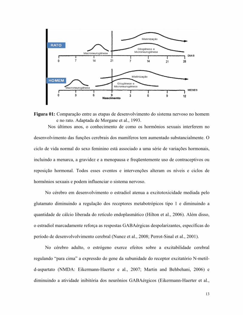
Figura 01: Comparação entre as etapas de desenvolvimento do sistema nervoso no homem e no rato. Adaptada de Morgane et al., 1993.
Nos últimos anos, o conhecimento de como os hormônios sexuais interferem no
desenvolvimento das funções cerebrais dos mamíferos tem aumentado substancialmente. O
ciclo de vida normal do sexo feminino está associado a uma série de variações hormonais,
incluindo a menarca, a gravidez e a menopausa e freqüentemente uso de contraceptivos ou
reposição hormonal. Todos esses eventos e intervenções alteram os níveis e ciclos de
hormônios sexuais e podem influenciar o sistema nervoso.
No cérebro em desenvolvimento o estradiol atenua a excitotoxicidade mediada pelo
glutamato diminuindo a regulação dos receptores metabotrópicos tipo 1 e diminuindo a
quantidade de cálcio liberada do retículo endoplasmático (Hilton et al., 2006). Além disso,
o estradiol marcadamente reforça as respostas GABAérgicas despolarizantes, específicas do
período de desenvolvolvimento cerebral (Nunez et al., 2008; Perrot-Sinal et al., 2001).
No cérebro adulto, o estrógeno exerce efeitos sobre a excitabilidade cerebral
regulando “para cima” a expressão do gene da subunidade do receptor excitatório N-metil-
d-aspartato (NMDA: Eikermann-Haerter e al., 2007; Martin and Behbehani, 2006) e
diminuindo a atividade inibitória dos neurônios GABAérgicos (Eikermann-Haerter et al.,
13
2007). Tanto o β-estradiol quanto a progesterona podem aumentar a potenciação de longo
termo (LTP) nos tecidos neocorticais (Sachs et al., 2007).
Estados hipoestrogênicos no cérebro adulto podem causar mudanças neuroendócrinas
em diferentes áreas cerebrais. A ausência de produção hormonal ovariana gera sintomas
específicos devido ao desarranjo do sistema nervoso central, no hipotálamo, por exemplo,
pode originar sintomas vasomotores, bem como distúrbios do comportamento alimentar e
controle alterado da pressão arterial (Genazzani et al., 1999).
Estudos a partir de modelos animais indicam que o cérebro é realmente sensível à
progesterona durante períodos críticos de desenvolvimento e maturação (López and
Wagner, 2009). Receptores de progesterona (PR) são transitoriamente expressos durante
desenvolvimento fetal e neonatal (Wagner, 2008). A progesterona é capaz de promover
crescimento dendrítico, spinogênese e sinaptogênense nas células de Purkinje em
desenvolvimento (Tsutsui, 2008).
Um modelo interessante para o estudo das relações entre hormônios gonadais
femininos e excitabilidade cerebral constitui-se no fenômeno da depressão alastrante
cortical (DAC), que foi empregado no presente trabalho. A DAC é um fenômeno
eletrofisiológico caracterizado por uma onda de excitação neuronal seguida de inibição. O
fenômeno se auto-propaga como uma onda de despolarização com características iônicas,
metabólicas e hemodinâmicas peculiares, plenamente reversíveis ao cabo de alguns
minutos, acompanhada por supressão transitória da atividade neuronal (Leão, 1944a,b).
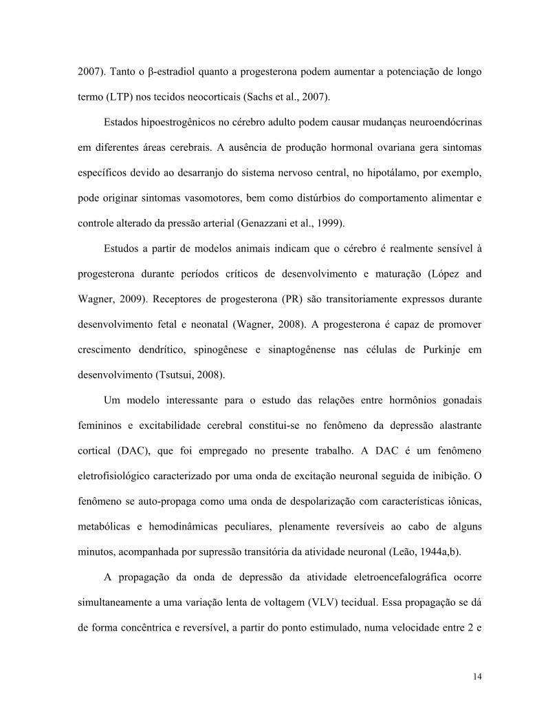
A propagação da onda de depressão da atividade eletroencefalográfica ocorre
simultaneamente a uma variação lenta de voltagem (VLV) tecidual. Essa propagação se dá
de forma concêntrica e reversível, a partir do ponto estimulado, numa velocidade entre 2 e
14
5 mm/min, sendo sua latência de reversão de 10 a 15min (Martins-Ferreira, 1983). A Fig. 2
ilustra o fenômeno.
Figura 02: À esquerda observa-se a seqüência temporal cíclica dos eventos que ocorrem durante a propagação da DAC. R1 e R2 indicam pontos de registro. Um estímulo externo (x) deu início ao fenômeno (etapa 1) que se propaga de forma concêntrica (etapas 2-4). As áreas escuras (etapas 2, 3 e 4) representam áreas corticais na vigência do fenômeno, enquanto que as áreas quadriculadas (etapas 3 a 6) indicam o princípio da recuperação tissular. As áreas claras indicam o tecido recuperado (etapas 5-6), o que também ocorre de forma concêntrica, retornando à condição inicial (etapa 1). À direita observa-se o eletrocorticograma (ECoG) e a variação lenta de voltagem (VLV), esta última presente durante a DAC, quando o ECoG diminui sua amplitude. Tais registros, obtidos em nosso laboratório foram feitos simultaneamente nos pontos R1 e R2. Observe a recuperação do ECoG após a passagem do fenômeno (Guedes et al., 2004).
Estudos experimentais prévios indicam que o tecido nervoso apresenta naturalmente
uma resistência à passagem da DAC (Guedes e Do-Carmo, 1980), e que esta resistência
pode diminuir ou aumentar na vigência de alguns tratamentos, modificando assim a sua
velocidade de propagação (Abadie-Guedes et al., 2008). Diversas modificações de
condições sistêmicas podem alterar a propagação da DAC (Guedes, 1984; Guedes et al.,
15
1987; Andrade et al., 1990; Guedes et al., 1992; Rocha-de-Melo e Guedes, 1997).
Tratamentos locais do tecido cortical podem também modificar a sua propagação (Richter
et al., 2005; Guedes et al., 1987; Amâncio-dos-Santos et al., 2006).
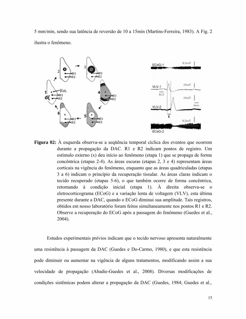
As Tabelas 01 e 02 apresentam diversas condições, já estudadas, que podem
dificultar ou facilitar a propagação da DAC.
Tabela 01: Algumas condições que dificultam a propagação da DAC
Condição experimental Autor/Ano Tratamento dietético com lítio Guedes et al., 1989 Hiperglicemia Ximenes-da-Silva e Guedes,1991; Costa-Cruz
et al., 2001 Anestésicos Guedes e Barreto, 1992 Hipotireoidismo Guedes e Pereira-da-Silva, 1993 Envelhecimento Guedes et al., 1996 Dieta hiperlipídica Paixão et al., 2007 Epilepsia crônica provocada pela pilocarpina
Guedes e Cavalheiro, 1997; Costa-Cruz et al., 2006
Estimulação ambiental Santos-Monteiro et al., 2000 Ativação do Sistema Serotoninérgico Guedes et al., 2002; Amâncio-dos-Santos et
al., 2006 Estimulação Elétrica Cerebral direta e trans-craniana
Fregni et al., 2005; 2007
Condições favoráveis de aleitamento Rocha-de-Melo et al., 2006
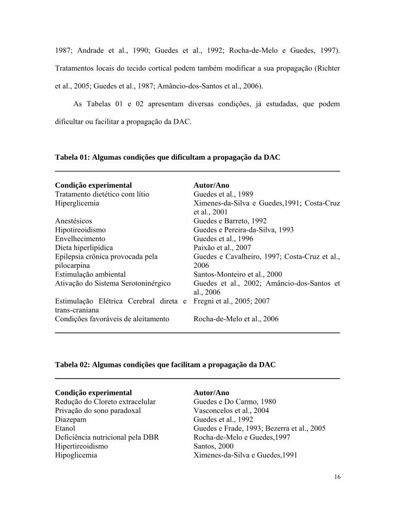
Tabela 02: Algumas condições que facilitam a propagação da DAC
Condição experimental Autor/Ano Redução do Cloreto extracelular Guedes e Do Carmo, 1980 Privação do sono paradoxal Vasconcelos et al., 2004 Diazepam Guedes et al., 1992 Etanol Guedes e Frade, 1993; Bezerra et al., 2005 Deficiência nutricional pela DBR Rocha-de-Melo e Guedes,1997 Hipertireoidismo Santos, 2000 Hipoglicemia Ximenes-da-Silva e Guedes,1991
16
Privação sensorial Tenório et al., 2009 Arginina durante o desenvolvimento Maia et al., 2009 Hipertermia ambiental Farias-Santos et al., 2009 Glutamina durante o desenvolvimento Lima et al., 2009 Uso de dipirona no início da vida Amaral et al., 2009
Estudos prévios têm mostrado que alterações hormonais durante o desenvolvimento
influenciam a propagação da DAC (Guedes e Pereira-da-Silva, 1993; Santos, 2000). Um
estudo in vitro em ratos adultos sugere uma possível influência, dose-dependente, dos
hormônios ovarianos sobre a DAC (Sachs et al., 2007). Entretanto, pouca atenção tem sido
dada aos efeitos sobre a DAC dos hormônios ovarianos, ou da sua ausência, in vivo,
quando atuando no cérebro em desenvolvimento.
Frente ao exposto, o presente trabalho se propôs a estudar o efeito da deficiência
ovariana durante o desenvolvimento cerebral sobre a DAC em ratas adultas. Este trabalho é
a continuação de uma linha de pesquisa do “Laboratório de Fisiologia da Nutrição Naíde
Teodósio” (LAFINNT) que utiliza o fenômeno da DAC para estudar o efeito de fatores
nutricionais, ambientais, farmacológicos e hormonais sobre o cérebro em desenvolvimento
(Guedes, 2011).
17
OBJETIVOS
Geral
Avaliar, em ratas, o impacto da ovariectomia aos sete dias de vida sobre o
desenvolvimento eletrofisiológico do sistema nervoso através da susceptibilidade cortical à
DAC, na idade adulta.
Específicos
• Avaliar a eficácia da ovariectomia, analisando-se, nos animais castrados, alterações
da evolução ponderal ao longo do desenvolvimento e, na idade adulta, a presença de
18
padrão atrófico do epitélio vaginal e do peso uterino, em comparação com os dois
grupos controle.
• Quando os filhotes se tornarem adultos, avaliar, nos grupos estudados 1) Grupo Ovx
(ratas submetidas à ovariectomia; 2) Grupo Sham (ratas submetidas à cirurgia
fictícia à ovariectomia) e 3) Grupo Intacto (ratas mantidas sem tratamento), a
incidência e propagação da DAC, por meio do seu registro eletrofisiológico
(eletrocorticograma – EcoG - e variação lenta de voltagem), quantificando-se a
velocidade de propagação, bem como a amplitude e a duração da variação lenta de
voltagem que caracterizam o fenômeno.
ARTIGO CIENTÍFICO
Title: Ovariectomy in the developing rat decelerates cortical spreading
depression in adult brain
19
Authors: Noranege Epifânio Accioly², Regina de Deus Lira Benevides², Belmira Lara da
Silveira Andrade da Costa¹, Rubem Carlos Araujo Guedes²CA
/
Affiliation: Departamento de Fisiologia e Farmacologia; ²Departamento de Nutrição,
Universidade Federal de Pernambuco, 50670901, Recife, PE, BrazilCA=
Corresponding author; address, please see address #2 above
E-mail: [email protected] or [email protected]
Telephone: +55-81-21268936 Fax: +55-81-21268473
Abstract
The brain of mammals is one important target organ for the action of gonadal
steroids and, when occurring during development, this hormonal influence may result in
important repercussion on the brain electrophysiological properties at adulthood, some of
which depending on the synaptic activity. Here we have characterized in early
ovariectomized adult rats the brain ability to propagate cortical spreading depression
(CSD), as an index of the cerebral electrophysiological effects of the early-induced absence
of the ovarian hormones. Wistar female rat pups (7 days old) underwent bilateral
ovariectomy (Ovx group; n=21) or sham surgery (Sham group; n=22), or no surgery (Naive
group; n=22). When the pups became adult (90-130 days), they were anesthetized and
submitted to the recording of CSD (electrocorticogram and slow DC voltage variation) in
two points of the cortical surface during 4h. Compared with both Naïve and Sham controls,
20
bilateral ovariectomy early in life resulted in significantly higher body weights (from day
50-65 onwards) and severely reduced uterus weights at adulthood. Furthermore, in the Ovx
animals the amplitudes and durations of the DC potential changes of CSD were higher, and
the CSD propagation velocities were reduced. It is concluded that ovariectomy during the
period of brain development is causally associated with the impairment of CSD propagation
in the adult brain, indicating a long-lasting effect, which we suggest as being related to the
long-term suppression of the action of the ovarian hormones on synaptic transmission.
Keywords: Brain development, Ovarian hormones, Cortical spreading depression, Rats
Introduction
In mammals, the fetal environment is rich in estradiol and progesterone derived
from the maternal organism (McCarthy, 2009; Sanyal, 1978). In the female fetal brain of
the rat, α-fetoprotein (AFP), a steroid binding globulin, sequesters circulating estrogens to
avoid their brain masculinizing effects early in life (Bakker et al., 2006, Gillies and
MCarthur, 2010) and selectively deliver estradiol to specific neuronal populations (Bakker
and Baum, 2008; Bakker et al., 2006). As a result, the female fetal brain is exposed to lower
levels of estradiol, as compared with the male fetal brain. Nevertheless, there is evidence
that estradiol can be de novo synthesized (locally from cholesterol) directly in fetal and
neonatal neurons during the female developing brain (Amateau et al., 2004; Bakker et al.,
2002; McCarthy, 2008; Mellon and Vaudry, 2001). This hormonal scenario extends
21
postnatally and in the offspring it influences the sexual differentiation of the developing
brain (McCarthy and Konkle, 2005; Amateau et al., 2004).
The brain enzyme aromatase, that synthesizes estradiol, presents its highest activity
in the immature brain as compared to the mature brain (McCarthy, 2009) and the AFP
activity no longer plays a significant role postnatally when the ovaries start to produce
estrogens (Bakker and Baum, 2008). During this initial period of life, ovarian hormones can
influence developmental processes in the brain (Bakker et al., 2002). Estradiol is capable of
modulating brain development by enhancing depolarizing GABA responses, which are
specific of the neonatal period, causing a trophic effect (Perrot-Sinal et al., 2003) and
preventing glutamate-induced cell death (Hilton et al., 2006). In addition, progesterone is
capable of promoting dendritic growth, spinogenesis, and synaptogenesis in the developing
Purkinje cell (Tsutsui, 2008).
In the fully developed brain, estrogen can exert effects on excitability by
upregulating the gene expression of excitatory N-metil-D-aspartate (NMDA) receptor
subunit (Eikermann-Haerter et al., 2007; Martin and Behbehani, 2006) and by decreasing
the inhibitory activity of γ-aminobutyric acid (GABA)-ergic neurons (Eikermann-Haerter et
al., 2007). Both β-estradiol and progesterone may enhance long-term potentiation (LTP)
induction in neocortical tissues (Sachs et al., 2007). These data indicate a relationship
between ovarian hormones and neuronal excitability, which can be experimentally explored
by using the electrophysiological phenomenon denominated as cortical spreading
depression (CSD). CSD is characterized by a wave of self-propagating depolarization with
characteristic ionic, metabolic, and hemodynamic changes followed by transient
suppression of neuronal activity (Leao, 1944a,b). In one in vitro study, ovarian hormones
applied to neocortical slices obtained from adult rats facilitated CSD (Sachs et al., 2007).
22
However, little attention has been paid to the effects of ovarian deficiency during brain
development on CSD features in the cerebral cortex of adult rats.
The present study aimed to address these issues in female rats that had been
previously ovariectomized early in life. Our hypothesis is that ovariectomy during the
period of brain development is causally associated in adulthood with impairment of CSD
propagation.
Material and methods
Animals
Wistar female newborn rats (n=65) from the colony of Departamento de Nutrição of
Universidade Federal de Pernambuco (Brazil) were randomly distributed to three groups,
submitted respectively on the postnatal day 7 to the following treatments: a) bilateral
ovariectomy (OVX group; n=21); b) sham surgery (n=22); c) no surgery (naïve group;
n=22).
The handling procedures involving the animals were in accordance with the
Institution’s guidelines, which comply with the ―Principles of Laboratory Animal Care
(National Institutes of Health, Bethesda, USA). The experimental design was approved by
the University Committee on Ethics in animal research, which complies with the
“Principles of Laboratory Animal Care” (National Institutes of Health, Bethesda, USA).
23
Animals were reared in polypropylene cages (51 cm X 35.5 cm X 18.5 cm) in a room
maintained at 22± 1°C with a 12h light/ 12h dark cycle (lights on at 7:00 a.m.) with free
access to water and food.
Bilateral ovariectomy
Under deep surgical cryoanesthesia (Phifer and Terry, 1986; see also Tenório et al.,
2009), the ovaries of the 7-days old rat pups were removed through a dorsal midline
incision on the lumbar region, as described elsewhere (Brouwer et al, 1980). In the Sham
group, all rat pups received the same incisions as the OVX animals; the ovaries were
identified and palpated, but not removed. Suture procedures were the same in both groups.
After recovering from anesthesia, the pups were returned to the maternal cage. After
weaning, they were housed in cages similar to the maternal ones (3-4 rats per cage). Total
surgery time was 10-15 min and the post-surgery mortality was very low (3 out of 46
operated pups).
The effectiveness of the early ovariectomy was histologically confirmed on two
occasions: at 60-90 days of life (by the atrophic pattern of the genital epithelium as well as
by the delayed vaginal opening, as compared to the Sham and Naïve controls) and on the
day of CSD recording (90-130 days of life), when the animal was killed and the uterus was
removed, showing severe atrophy.
CSD elicitation and recording
When the pups were 90 to 130 days old, they were submitted to the CSD recording
for a 4-hour period. In the sham- and naïve groups, the CSD recordings were performed
24
only when the animals were in the proestrus phase of the estrous cycle, which was
histologically confirmed on the day of the CSD recording.
Under anesthesia (1 g/kg urethane plus 40 mg/kg chloralose, ip), three trephine
holes (2–3 mm in diameter) were drilled on the right side of the skull. The first hole (on the
frontal bone) was used to apply the stimulus (KCl solution) to elicit CSD. The propagating
CSD wave was then recorded on two points of the parietal cortex surface through the other
two holes, drilled on the parietal bone. Rectal temperature was continuously monitored and
maintained at 37 ± 1ºC by a heating blanket. CSD was elicited at 20 min intervals by
applying a cotton ball (1–2 mm diameter), soaked in 2% KCl solution (approximately 0.27
M) to the anterior hole drilled at the frontal region for 1 min. The electrocorticogram
(ECoG) and the slow DC potential change accompanying CSD were recorded
simultaneously at the two parietal points on the cortical surface by using a pair of Ag-AgCl
agar-Ringer electrodes. These electrodes consisted of plastic conic pipettes (5 cm length,
0.5 mm tip inner diameter), filled with Ringer solution and solidified with the addition of
0.5% agar, into which a chlorided silver wire was inserted. The pipettes were fixed together
pair-wise with cyanoacrylate glue, so that the interelectrode distance was kept constant for
each pair (range: 4–5.5 mm). Each pair of electrodes was connected to a lever that could be
vertically moved by turning around a screw, so that the recording electrodes could be
gently placed on the intact dura-mater, under low-power microscope control, without any
excessive pressure on the cortical surface. A third electrode, of the same type, placed on the
nasal bones, served as common reference electrode. The velocity of CSD propagation was
calculated based on the time required for a CSD wave to cross the distance between the two
recording electrodes. In the measurement of CSD velocities, the initial point of each DC
negative rising phase was used as the reference point.
25
Statistics
Body-, uterus-, adrenals- and brain weights and CSD propagation rates were
compared between groups by ANOVA, followed by a post-hoc (Tukey–Kramer) test when
indicated. Differences were considered significant when p≤0.05. All values are presented in
the text as means ± standard deviations.
Results
Body weights
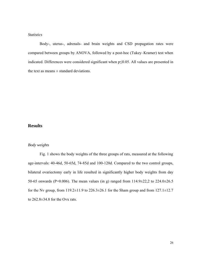
Fig. 1 shows the body weights of the three groups of rats, measured at the following
age-intervals: 40-46d, 50-65d, 74-85d and 100-120d. Compared to the two control groups,
bilateral ovariectomy early in life resulted in significantly higher body weights from day
50-65 onwards (P=0.006). The mean values (in g) ranged from 114.9±22,2 to 224.0±26.5
for the Nv group, from 119.2±11.9 to 226.3±26.1 for the Sham group and from 127.1±12.7
to 262.8±34.8 for the Ovx rats.
26
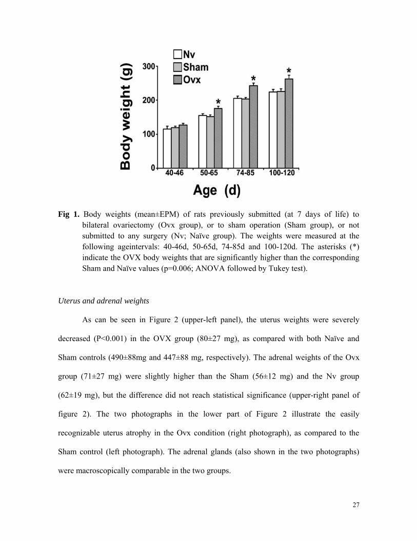
Fig 1. Body weights (mean±EPM) of rats previously submitted (at 7 days of life) to bilateral ovariectomy (Ovx group), or to sham operation (Sham group), or not submitted to any surgery (Nv; Naïve group). The weights were measured at the following ageintervals: 40-46d, 50-65d, 74-85d and 100-120d. The asterisks (*) indicate the OVX body weights that are significantly higher than the corresponding Sham and Naïve values (p=0.006; ANOVA followed by Tukey test).
Uterus and adrenal weights
As can be seen in Figure 2 (upper-left panel), the uterus weights were severely
decreased (P<0.001) in the OVX group (80±27 mg), as compared with both Naïve and
Sham controls (490±88mg and 447±88 mg, respectively). The adrenal weights of the Ovx
group (71±27 mg) were slightly higher than the Sham (56±12 mg) and the Nv group
(62±19 mg), but the difference did not reach statistical significance (upper-right panel of
figure 2). The two photographs in the lower part of Figure 2 illustrate the easily
recognizable uterus atrophy in the Ovx condition (right photograph), as compared to the
Sham control (left photograph). The adrenal glands (also shown in the two photographs)
were macroscopically comparable in the two groups.
27

Fig 2. Uterus and adrenal weights (mean± EPM) of the Nv, Sham and Ovx groups, as defined in figure 1. The asterisk (*) in the upper-left panel indicates that the Ovx group presents uterus weights significantly lower (p<0.001) than those of the Sham and Naïve controls. The mean adrenal weights of the three groups were comparable (upper-right panel). The two photographs of the lower part of the figure illustrate the uterus atrophy in the Ovx condition (right photograph), as compared to the Sham control (left photograph). The adrenal glands are also shown in the two photographs, and no macroscopically appreciable intergroup difference could be detected. (ANOVA followed by Tukey test).
CSD propagation
Figure 3 shows a typical electrophysiological recording (slow DC-potential change
and ECoG) in one Nv, one Sham and one Ovx rat. In all groups, the 1-min stimulation with
2% KCl at one point of the frontal cortex elicited a single CSD wave that propagated
without interruption and was recorded by the two electrodes located more posterior in the
parietal cortex (see stimulation- and recording points in the inset of the figure). One can
notice that the ECoG depression and the slow potential change confirmed the presence of
CSD, after KCl application.
28
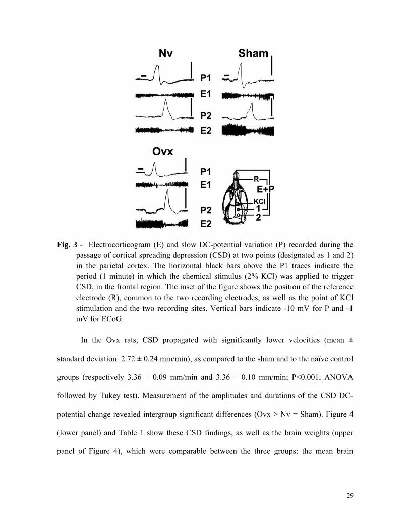
Fig. 3 - Electrocorticogram (E) and slow DC-potential variation (P) recorded during the passage of cortical spreading depression (CSD) at two points (designated as 1 and 2) in the parietal cortex. The horizontal black bars above the P1 traces indicate the period (1 minute) in which the chemical stimulus (2% KCl) was applied to trigger CSD, in the frontal region. The inset of the figure shows the position of the reference electrode (R), common to the two recording electrodes, as well as the point of KCl stimulation and the two recording sites. Vertical bars indicate -10 mV for P and -1 mV for ECoG.
In the Ovx rats, CSD propagated with significantly lower velocities (mean ±
standard deviation: 2.72 ± 0.24 mm/min), as compared to the sham and to the naïve control
groups (respectively 3.36 ± 0.09 mm/min and 3.36 ± 0.10 mm/min; P<0.001, ANOVA
followed by Tukey test). Measurement of the amplitudes and durations of the CSD DC-
potential change revealed intergroup significant differences (Ovx > Nv = Sham). Figure 4
(lower panel) and Table 1 show these CSD findings, as well as the brain weights (upper
panel of Figure 4), which were comparable between the three groups: the mean brain
29
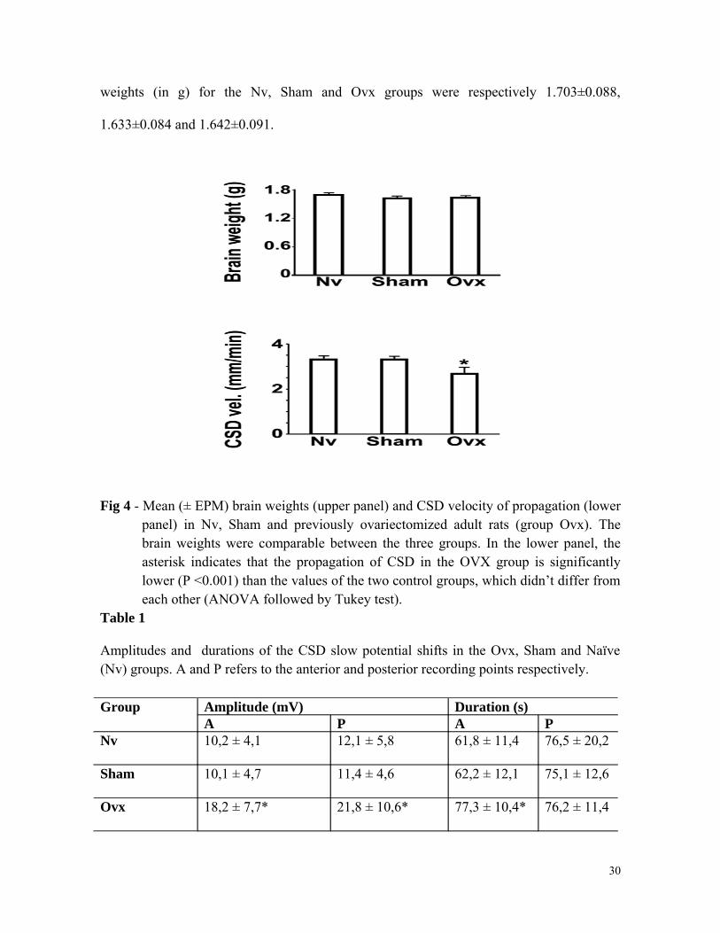
weights (in g) for the Nv, Sham and Ovx groups were respectively 1.703±0.088,
1.633±0.084 and 1.642±0.091.
Fig 4 - Mean (± EPM) brain weights (upper panel) and CSD velocity of propagation (lower panel) in Nv, Sham and previously ovariectomized adult rats (group Ovx). The brain weights were comparable between the three groups. In the lower panel, the asterisk indicates that the propagation of CSD in the OVX group is significantly lower (P <0.001) than the values of the two control groups, which didn’t differ from each other (ANOVA followed by Tukey test).
Table 1
Amplitudes and durations of the CSD slow potential shifts in the Ovx, Sham and Naïve (Nv) groups. A and P refers to the anterior and posterior recording points respectively.
Group Amplitude (mV) Duration (s)A P A P
Nv 10,2 ± 4,1 12,1 ± 5,8 61,8 ± 11,4 76,5 ± 20,2
Sham 10,1 ± 4,7 11,4 ± 4,6 62,2 ± 12,1 75,1 ± 12,6
Ovx 18,2 ± 7,7* 21,8 ± 10,6* 77,3 ± 10,4* 76,2 ± 11,4
30

Data are expressed as mean ± standard deviation. The asterisks indicate values significantly different from the corresponding Sham and Naïve group.
31
Discussion
Ovariectomy in developing rats
In this study we observed important developmental systemic and localized
alterations resulting from bilateral ovariectomy, which was performed in the developing
rats as early as the postnatal day 7. We have chosen this age because it corresponds to the
time-point in which the ovaries become capable of secreting significant amounts of their
hormones (Lamprecht et al, 1976). In accordance with a previous report (Gitlin, 1974), the
effectiveness of the ovariectomy was presently evidenced by the severe atrophic patterns of
uterus- and genital epithelium, delayed vaginal opening (as compared to the controls), as
well as body weight increment. Regarding the ovariectomy-induced increase in body
weights, it is known that the hypoestrogenic status participates in the increased food intake
and the resulting higher body weight gain, as compared to the controls (Wade, 1975;
Roesch, 2006). The post-ovariectomy deficiency in the uterus development would be
expected, since it is well described in the literature; ovaries participate in the uterine growth
observed between the second and the fourth postnatal weeks (Branham and Sheehan, 1995).
Concerning adrenal weights the lack of intergroup statistically significant difference is in
agreement with data of (Ramaley (1973). Taken together, our data on the systemic and
localized developmental effects seen in early ovariectomized rats, besides assuring the
effectiveness of the ovariectomy procedure also supports the causal link between ovarian
hormones deficiency and the here described brain CSD effects.
32
CSD propagation
The main electrophysiological finding of the present study was that chronic ovarian
hormones deficiency that was provoked during development reduced brain capability to
propagate CSD in adulthood, as indexed by its lower velocities in comparison to the
velocities of the sham and naïve controls. The alterations in CSD amplitude and duration
also reinforce this conclusion. It is well established that the gonadal steroids exert some of
its action on the central nervous system, which is one important target organ for their
actions, both during development and in adult life (Genazzani et al., 2005; Kawata, 1995).
In contrast to its role on the adult brain, in the developing brain GABA is the predominant
source of excitation via membrane depolarization, and estradiol markedly enhance
depolarizing GABA responses in neonatal neurons (Nunez et al., 2008; Perrot-Sinal et al.,
2001). Furthermore, in the developing brain estradiol dampens glutamate-mediated
excitotoxicity by downregulating their receptors (mGluR1 and mGluR5) and by decreasing
the amount of calcium released from the endoplasmic reticulum (Hilton et al., 2006).
Therefore, it appears to us unequivocal accepting that ovariectomy early in life changes
excitability of the developing brain, and our CSD findings support that.
Considering that CSD is influenced by changes in the brain excitability (Leão, 1944;
1972; Guedes and Cavalheiro, 1997; Guedes et al, 2009; Guedes, 2011), it is reasonable to
raise the question of if, and how early ovarian deficiency (or ovarian absence, as in the
ovariectomy paradigm) would modulate brain excitability during development, and
therefore influencing phenomena like seizures and CSD at adulthood. One possibility to
explain the effects of ovariectomy on the excitability of the adult brain would be based on
the influence of ovarian hormones on the glutamatergic neurotransmitter system. Estrogens
can increase neuronal excitability via upregulating the gene expression of NMDA receptor
33
subunit and by decreasing the inhibitory action of GABAergic neurons (Eikermann-Haerter
et al, 2007; Martin and Behbehani, 2006). Estrogen also can inhibit L-glutamate uptake by
astrocytes (Sato et al, 2003), and increases the number of dendritic spines, which are
densely populated with NMDA receptors (Woolley et al., 1997). As a rule, higher levels of
estrogen are associated with an increased seizure frequency in females (Klein and Herzog,
1998) and seizure thresholds are decreased during peak estrogen levels (Woolley and
Timiras, 1962), while progesterone is associated with seizure control in catamenial epilepsy
(Herzog, 2009). Application of exogenous estrogen to the cerebellum of female rats
significantly potentiates the excitatory neuronal response of Purkinje cells to glutamate
applied by iontophoresis (Smith et al, 1988). Besides the glutamatergic, the GABAergic
neurons also are strongly modulated by ovarian steroids. Estrogen can increase GABA
release and upregulate the number of GABA receptors (Shughrue and Merchenthaler,
2000). Progesterone has been shown to have depressant effects on CNS excitability via
modulation of the gamma-aminobutiric acid type A (GABAA; Lambert et al., 2003). These,
and several other pieces of evidence showing that gonadal hormones can affect brain
excitability (Eikermann-Haerter et al., 2007; 2009; Sachs et al., 2007; Woolley et al., 1997;
Martin and Behbehani, 2006; Scharfman and MacLusky, 2006; Scharfman et al., 2005),
collectively suggest that ovarian hormones could have a critical role in epilepsy, and
perhaps could also play a role in epilepsy treatment (Herzog, 2009).
Concerning the ovarian hormones/CSD relationship, several findings from others
deserve comment. The CSD susceptibility in familial hemiplegic migraine type 1 (FHM1)
knockin mice is higher in females than in males; ovariectomy reverses this gender
difference, which is partially restored by estradiol replacement, suggesting that actually
estrogens can modulate CSD susceptibility (Eikermann-Haerter et al. 2009). The thresholds
34
for CSD elicitation with KCl- and electrical stimulation are lower in female mice compared
to males (Brennan et al. 2007). In rat neocortical slices, application of both β-estradiol and
progesterone enhances CSD features (Sachs et al. 2007). This compelling evidence from
the literature favors our initial hypothesis that ovariectomy during the period of brain
development is causally associated in adulthood with CSD impairment.
In conclusion we demonstrated for the first time that the brains of adult rats that had
been ovariectomized during their development are more resistant (or less sensible) to CSD
propagation and we suggest that this effect is related to the long-term suppression of the
physiological action of the ovarian hormones on synaptic transmission. Our findings
document the importance of further searching for the molecular mechanisms underlying the
role of the ovarian hormones on the brain development and their electrophysiological
properties.
Acknowledgments
The authors thank the Brazilian agencies CAPES (Procad/2007), CNPq (INCT de
Neurociencia Translacional–No. 573604/2008-8), MS/SCTIE/DECIT (No. 17/2006),
Facepe (APQ0975-4.05/08), and IBN-Net/Finep (No. 4191) for financial support. R.C.A.
Guedes is Research Fellow from CNPq (No. 301190/2010-0).
35
References
Amateau, S.K., Alt, J.J., Stamps, C.L., McCarthy, M.M., 2004. Brain estradiol content in
newborn rats: sex differences, regional heterogeneity, and possible de novo synthesis by the
female telencephalon. Endocrinol. 145, 2906–2917.
Bakker, J., Honda, S., Harada, N., Balthazart, J., 2002. The aromatase knock-out mouse
provides new evidence that estradiol is required during development in the female for the
expression of sociosexual behaviors in adulthood. J. Neurosci. 22, 9104-9112.
Bakker, J., Baum, M.J., 2008. Role for estradiol in female-typical brain and behavioral
sexual differentiation. Front. Neuroendocrinol. 29, 1–16.
Bakker, J., De Mees, C., Douhard, Q., Balthazart, J., Gabant, P., Szpirer, J., Szpirer, C.,
2006. Alpha-fetoprotein protects the developing female mouse brain from masculinization
and defeminization by estrogens. Nat. Neurosci. 9, 220–226.
Branham, W.S., Sheehan, D.M., 1995. Ovarian and Adrenal Contributions to Postnatal
Growth and Differentiation of the Rat Uterus. Biol. Reprod. 53, 863-872.
Brennan, K.C., Reyes, M. R., Valdés, H.E.L., Arnold, A.P., Charles, A.C., 2007. Reduced
threshold for cortical spreading depression in female mice. Ann. Neurol. 61, 603–606.
36
Brouwer, E.A., Dailey, R., Brouwer, J.B., 1980. Ovariectomy of newborn rats: a descriptive
surgical procedure. Lab. An. Sci. 30, 546-548.
Eikermann-Haerter, K., Kudo, C., Moskowitz, M.A., 2007. Cortical spreading depression
and estrogen. Headache. 47[suppl 2], S79-S85.
Eikermann-Haerter, K., Dileköz, E., Kudo, C., Savitz, S.I., Waeber, C., Baum, M.J.,
Ferrari, M.D., van den Maagdenberg, A.M.J.M., Moskowitz, M.A., Ayata, C., 2009.
Genetic and hormonal factors modulate spreading depression and transient hemiparesis in
mouse models of familial hemiplegic migraine type 1. J. Clin. Invest. 119, 99-109.
Genazzani, A.R., Bernardi, F., Pluchino, N., Begliuomini, S., Lenzi, E., Casarosa, E., Luisi,
M., 2005. Endocrinology of menopausal transition and its brain implications. CNS Spectr.
6, 449-57.
Gillies, G.E., MCarthur, S., 2010. Estrogen Actions in the Brain and the Basis for
Differential Action in Men and Women: A Case for Sex-Specific Med. Pharmacol. Rev. 62,
155–198.
Gitlin, G., 1974. Vaginal opening and vaginal epithelium following ovariectomy in
newborn rats. Acta Anat. 90, 117-132.
37
Guedes, R.C.A., 2011. Cortical Spreading Depression: A Model for Studying Brain
Consequences of Malnutrition. in: Preedy, V., Watson, R., Martin, C. (Eds.), Handbook of
Behavior, Food and Nutrition. Springer, Berlin, pp. 2343-2355.
Guedes, R.C.A., Cabral-Filho, J.E., Teodósio, N.R., 1992. Gabaergic mechanisms involved
in cortical spreading depression in normal and malnourished rats. in: Do Carmo, R.J. (Ed.).
Spreading Depression. (Experimental Brain Research Series No. 23). Springer, Berlin, pp
17-26.
Guedes, R.C.A., Cavalheiro, E.A., 1997. Blockade of spreading depression in chronic
epileptic rats: reversion by diazepam. Epil. Res. 27, 33-40.
Guedes R.C.A., Do Carmo R.J., 1980. Influence of ionic disturbances produced by gastric
washing on cortical spreading depression, Exp. Brain Res. 39, 341–349.
Guedes, R.C.A., Oliveira, J.A.C., Amâncio-dos-Santos, A., García-Cairasco, N., 2009.
Sexual Differentiation of Cortical Spreading Depression propagation after Acute and
Kindled Audiogenic Seizures in the Wistar Audiogenic Rat (WAR). Epil. Res. 83, 207-214.
Guedes, R.C.A., Santos, A.A., Manhães-de-Castro R., Cruz, R.R.G.C., 2002. Citalopram
has an antagonistic action on cortical spreading depression in well-nourished and early-
malnourished adult rats. Nutr. Neurosci. 5;115-123
Herzog, A.G., 2009. Hormonal Therapies: Progesterone. Neurotherapeutics. 6, 383–391.
38
Hilton, G.D., Nunez, J.L., Bambrick, L., Thompson, S.M., McCarthy, M.M., 2006.
Glutamate-mediated excitotoxicity in neonatal hippocampal neurons is mediated by
mGluR-induced release of Ca++ from intracellular stores and is prevented by estradiol.
Eur. J. Neurosci. 24, 3008–3016.
Klein, P., Herzog, A.G., 1998. Hormonal effects on epilepsy in women. Epilepsia. 39, S9–
S16.
Kawata, M., 1995. Roles of steroid hormones and their receptors in organization in the
nervous system structural. Neurosci. Res. 24, 1-46.
Lambert, J.J., Belelli. D., Peden. D.R., Vardy, A.W., Peters, J.A., 2003. Neurosteroid
Modulation of GABAA receptors. Progr. Neurobiol. 71, 67–80.
Lamprecht, S.A., Kohen, F., Ausher, J., Zor, U., Lindner, H.R., 1976. Hormonal
stimulation of estradiol-17 beta release from the rat ovary during early postnatal
development. J. Endocrinol. 68, 343–344.
Leao, A.A.P., 1944a. Spreading depression of activity in the cerebral cortex. J.
Neurophysiol. 7, 359–390.
Leão, A.A.P., 1944b. Pial circulation and spreading depression of activity in the cerebral
cortex. J. Neurophysiol. 7, 391-396.
39
Leão, A.A.P., 1972. Spreading depression. in: Purpura, D.P., Penry, K., Tower, D.B.,
Woodbury, D.M.,Walter, R.D. (Eds.), Experimental Models of epilepsy. Raven Press, N.
York, pp. 173-195.
Martin, V.T., Behbehani, M., 2006. Ovarian hormones and migraine headache:
understanding mechanisms and pathogenesis-part I. Headache. 46, 3-23.
McCarthy, M.M., 2009. The two faces of estradiol: effects on the developing brain.
Neuroscientist. 15, 599–610.
McCarthy, M.M., 2008. Estradiol and the developing brain. Physiol. Rev. 88, 91–124.
McCarthy, M.M., Konkle, A.T.M., 2005. When is a sex difference not a sex difference?
Front. Neuroendocrinol. 26, 85–102.
Mellon, S.H., Vaudry, H., 2001. Biosynthesis of neurosteroids and regulation of their
synthesis. Internat. Rev. Neurobiol. 46, 33-78.
Nunez, J.L., Aberdeen, G.W., Albrecht, E.D., McCarthy, M.M., 2008. Impact of estradiol
on GABA- and glutamate-mediated calcium responses of fetal baboon (papio anubis)
hippocampal and cortical neurons. Endocrinology. 149, 6433–6443.
40
Perrot-Sinal, T.S., Auger, A.P., McCarthy, M.M., 2003. Excitatory GABA-induced pCREB
in developing brain is mediated by L-type Ca+2 channels and dependent on age, sex and
brain region. Neuroscience. 116, 995–1003.
Phifer, C.B., Terry, L.M., 1986. Use of hypothermia for general anesthesia in preweanling
rodents. Physiol. Behav. 38, 887-890.
Ramaley, J.A., 1973. Adrenal function in female rats before puberty: The effects of
gonadectomy. Steroids. 22, 597-608.
Roesch, D.M., 2006. Effects of selective estrogen receptor agonists on food intake and
body weight gain in rats. Physiol. Behav. 87, 39–44.
Sachs, M., Pape, H.C., Seckmann, E.J., Gorji, A., 2007. The effect of estrogen and
progesterone on spreading depression in rat neocortical tissues. Neurobiol. Dis. 25, 27-34.
Sanyal, M.K., 1978. Secretion of progesterone during gestation in the rat. J. Endocrinol. 79,
179 –190.
Sato, K., Matsuki, N., Ohno, Y., Nakazawa, K., 2003. Estrogens inhibit l-glutamate uptake
activity of astrocytes via membrane estrogen receptor alpha. J. Neurochem. 86, 1498–1505.
Scharfman, H.E., Goodman, J.H., Rigoulot, M.A., Berger, R.E., Walling, S.G., Mercurio,
T.C., Stormes, K., Maclusky, N.J., 2005.Seizure susceptibility in intact and ovariectomized
female rats treated with the convulsant pilocarpine. Exp. Neurol. 196, 73-86.
41
Scharfman, H.E., MacLusky, N.J., 2006. The influence of gonadal hormones on neuronal
excitability, seizures, and epilepsy in the female. Epilepsia. 47, 1423-1440.
Shughrue, P.J., Merchenthaler, I., 2000. Estrogen is more than just a “sex hormone”: novel
sites for estrogen action in the hippocampus and cerebral cortex. Front. Neuroendocrinol.
21, 95-101.
Smith, S.S., Waterhouse, B.D., Woodward, D.J., 1988. Locally applied estrogens potentiate
glutamate-evoked excitation of cerebellar Purkinje cells. Brain Res. 475, 272-282.
Tenório, A.S., Oliveira, I.D.V.A., Guedes, R.C.A., 2009. Early vibrissae removal facilitates
cortical spreading depression propagation in the brain of well-nourished and malnourished
developing rats. Int. J. Devel. Neurosci. 27, 431–437.
Tsutsui, K., 2008. Minireview: Progesterone Biosynthesis and Action in the Developing
Neuron. Endocrinology. 149, 2757–2761.
Wade, G.N., 1975. Some effects of ovarian hormones on food intake and body weight in
female rats. J. Comp. Physiol. Psychol. 88, 183–193.
Woolley, C.S., Weiland, N.G., McEwen, B.S., Schwartzkroin, P.A., 1997. Estradiol
increases the sensitivity of hippocampal CA1 pyramidal cells to NMDA receptor-mediated
synaptic input: correlation with dendritic spine density. J. Neurosci. 17, 1848–1859.
42

Woolley, D.E., Timiras, P.S., 1962. Estrous and circadian periodicity and electroshock
convulsions in rats. Am. J. Physiol. 202, 379–382
43
CONCLUSÕES
A análise dos resultados desta dissertação permite as duas seguintes conclusões e uma
sugestão:
• A técnica presentemente utilizada, de remoção cirúrgica dos ovários, em ratas com 7
dias de vida, foi eficaz em alterar o desenvolvimento das suas estruturas genitais e
reprodutoras.
• Tal efeito é baseado na análise macroscópica do padrão atrófico vaginal e uterino.
• Esta conclusão é também reforçada pelo ganho de peso corporal, significante a partir de
50-65 dias de vida.
• Os cérebros das ratas adultas que foram ovariectomizadas durante seu desenvolvimento
tornaram-se mais resistentes (ou menos sensíveis) à propagação da DAC.
• Sugere-se que este efeito da ovariectomia precoce sobre a DAC seja devido à supressão
da ação dos hormônios ovarianos sobre mecanismos sinápticos cerebrais, o que deverá
ser investigado em futuros experimentos.
44

45
REFERÊNCIAS BIBLIOGRÁFICAS
Abadie-Guedes, R., Santos, S.D., Cahú, T.B., Guedes, R.C.A., Bezerra, R.S., 2008. Dose-
dependent effects of astaxanthin on cortical spreading depression in chronically ethanol-
treated adult rats. Alcohol. Clin. Exp. Res. 32, 1417-1421.
Amaral, A.P.B., Barbosa, M.S.S., Souza, V.C., Ramos, I.L.T., Guedes, R.C.A., 2009.
Drug/nutrition interaction in the developing brain: dipyrone enhances spreading depression
in rats. Exp. Neur. 219, 492-498.
Amateau, S.K., Alt, J.J., Stamps, C.L., McCarthy, M.M., 2004. Brain estradiol content in
newborn rats: sex differences, regional heterogeneity, and possible de novo synthesis by the
female telencephalon. Endocrinol. 145, 2906–2917.
Amâncio-dos-Santos A., Pinheiro, P. C. F., Lima, D. S. C., Ozias, M. G., Oliveira, M. B.,
Guimaraes, N. X., Guedes, R. C. A., 2006. Fluoxetine inhibits cortical spreading
depression in weaned and adult rats suckled under favorable and unfavorable lactation
conditions. Exp. Neurol. 200,275-282.
Andrade, A. F. D., Guedes, R. C. A., Teodósio, N. R., 1990. Enhanced rate of cortical
spreading depression due to malnutrition: Prevention by dietary protein suplementation.
Braz. J. Med. Biol. Res. 23, 889-893.
46
Bakker, J., Honda, S., Harada, N., Balthazart, J., 2002. The aromatase knock-out mouse
provides new evidence that estradiol is required during development in the female for the
expression of sociosexual behaviors in adulthood. J. Neurosci. 22, 9104-9112.
Bakker, J., Baum, M.J., 2008. Role for estradiol in female-typical brain and behavioral
sexual differentiation. Front. Neuroendocrinol. 29, 1–16.
Bakker, J., De Mees, C., Douhard, Q., Balthazart, J., Gabant, P., Szpirer, J., Szpirer, C.,
2006. Alpha-fetoprotein protects the developing female mouse brain from masculinization
and defeminization by estrogens. Nat. Neurosci. 9, 220–226.
Bezerra, R.S., Abadie-Guedes, R., Melo, F.R.M., Paiva, A.M.A., Santos, A.A., Guedes,
R.C.A., 2005. Shrimp carotenoids protect the developing rat cerebral cortex agains. the
effects of ethanol on cortical spreading depression. Neurosc. Let. 391, 51-55.
Branham, W.S., Sheehan, D.M., 1995. Ovarian and adrenal contributions to postnatal
growth and differentiation of the rat uterus. Biol. Reprod. 53, 863-872.
Brennan, K.C., Reyes, M. R., Valdés, H.E.L., Arnold, A.P., Charles, A.C., 2007. Reduced
threshold for cortical spreading depression in female mice. Ann. Neurol. 61, 603–606.
Brouwer, E.A., Dailey, R., Brouwer, J.B., 1980. Ovariectomy of newborn rats: a descriptive
surgical procedure. Lab. An. Sci. 30, 546-548.
47
Costa-Cruz, R.R.G.; Guedes, R.C.A., 2001. Cortical spreading depression during
streptozotocin-induced hyperglycemia in nutritionally normal and early-malnourished rats.
Neurosc. Let. 303, 177-180.
Costa-Cruz, R.R.G., Santos, A.A., Guedes, R.C.A., 2006. Characterization of cortical
spreading depression in adult well-nourished and malnourished rats submitted to the
association of pilocarpine-induced epilepsy plus streptozotocin-induced hyperglycemia.
Neurosc. Let. 401, 271-275.
Dobbing ,J., 1969. Vulnerable periods in developing brain. in: Davison, A.N., Dobbing, J.
(Eds), Applied Neurochemistry, Blackwell. Oxford, pp 287–316.
Eikermann-Haerter, K., Kudo, C., Moskowitz, M.A., 2007. Cortical spreading depression
and estrogen. Headache. 47[suppl 2], S79-S85.
Eikermann-Haerter, K., Dileköz, E., Kudo, C., Savitz, S.I., Waeber, C., Baum, M.J.,
Ferrari, M.D., van den Maagdenberg, A.M.J.M., Moskowitz, M.A., Ayata, C., 2009.
Genetic and hormonal factors modulate spreading depression and transient hemiparesis in
mouse models of familial hemiplegic migraine type 1. J. Clin. Invest. 119, 99-109.
Farias-Santos, R.C., Lira, M.C.A., Pereira, D.E.S., Sá, I.R., Pimentel, M.R.F., Araújo, L.L.,
Guedes, R.C.A., 2009. Exposure of developing well-nourished and malnourished rats to
environmental heating facilitates cortical spreading depression propagation at adulthood.
Neurosc. Let. 454, 218-222.
48
Fregni, F., Liebetanz, D., Monte-Silva, K.K., Oliveira, M.B., Amancio-dos-Santos, A.,
Nitsche, M.A., Pascual-Leone, A., Guedes, R.C.A., 2007. Effects of transcranial direct
current stimulation coupled with repetitive electrical stimulation on cortical spreading
depression. Exp. Neurol. 204, 462-466.
Fregni, F., Monte-Silva, K.K., Oliveira, M.B., Freedman, S., Pascual-Leone, A., Guedes,
R.C.A., 2005. Lasting accelerative effects of 1 Hz and 20 Hz electrical stimulation on
cortical spreading depression: relevance for clinical applications of brain stimulation..
Europ. J. Neurosc. 21, 2278-2284.
Genazzani, A.R., Bernardi, F., Pluchino, N., Begliuomini, S., Lenzi, E., Casarosa, E., Luisi,
M., 2005. Endocrinology of menopausal transition and its brain implications. CNS Spectr.
6, 449-57.
Genazzani, A.R., Spinetti, A., Bernardi, F., 1999. Menopause and the central nervous
system: intervention options. Maturitas, 31, 103-110.
Gillies, G.E., MCarthur, S., 2010. Estrogen Actions in the Brain and the Basis for
Differential Action in Men and Women: A Case for Sex-Specific Med. Pharmacol. Rev. 62,
155–198.
Gitlin, G., 1974. Vaginal opening and vaginal epithelium following ovariectomy in
newborn rats. Acta Anat. 90, 117-132.
49
Guedes, R.C.A., 1984. On some conditions that influence cortical spreading depression.
Anais da Academia Brasileira de Ciências. 56, 445-455.
Guedes, R.C.A., 2011. Cortical spreading depression: a model for studying brain
consequences of malnutrition. in: Preedy, V., Watson, R., Martin, C. (Eds.), Handbook of
Behavior, Food and Nutrition. Springer, Berlin, pp. 2343-2355.
Guedes, R.C.A., Amorim, L.F., Teodósio, N.R., 1996. Effect of aging on cortical spreading
depression. Braz. J. Med. Biol. Res. 29, 1407-1412.
Guedes, R.C.A., Andrade, A.F.D., Cabral-Filho, J.E., 1987. Propagation of cortical
spreading depression in malnourished rats: facilitatory effect of dietary protein deficiency.
Braz. J. Med. Biol. Res. 20, 639-642.
Guedes, R.C.A., Barreto, J., 1992. Effect of anesthesia on the propagation of cortical
spreading depression. Braz. J. Med. Biol. Res. 25, 393-397.
Guedes, R.C.A., Cabral-Filho, J.E., Teodósio, N.R., 1992. Gabaergic mechanisms
involved in cortical spreading depression in normal and malnourished rats. in: Do Carmo,
R.J. (Ed.), Spreading depression. experimental brain research series. Springer, Berlin, pp
17-26.
Guedes, R.C.A., Cavalheiro, E.A., 1997. Blockade of spreading depression in chronic
epileptic rats: reversion by diazepam. Epil. Res. 27, 33-40.
50
Guedes R.C.A., Do Carmo, R.J., 1980. Influence of ionic disturbances produced by gastric
washing on cortical spreading depression. Exp. Brain Res.39, 341–349.
Guedes, R.C.A., Frade, S.F., 1993. Effect of ethanol on cortical spreading depression. Braz.
J. Med. Biol. Res. 26, 1241-1244.
Guedes, R.C.A., Oliveira, J.A.C., Amâncio-dos-Santos, A., García-Cairasco, N., 2009.
sexual differentiation of cortical spreading depression propagation after acute and kindled
audiogenic seizures in the wistar audiogenic rat (WAR). Epil. Res. 83, 207-214.
Guedes, R.C.A., Pereira-da-Silva, M., 1993. Effect of pre- and postnatal propylthiouracil
administration on the propagation of cortical spreading depression of adult rats. Braz. J.
Med. Biol. Res. 26, 1123-1128.
Guedes, R.C.A., Rocha-de-Melo, A.P., Teodósio, N.R., 2004. Nutrição adequada: a base do
funcionamento cerebral. Ciência e Cultura. 56, 32-35.
Guedes, R.C.A., Silva, A. T. Teodósio, N.R., Amorim, L.F., 1989. Effect of dietary lithium
on cortical spreading depression. Braz. J. Med. Biol. Res.. 22, 923-925.
Herzog, A.G., 2009. Hormonal Therapies: Progesterone. Neurotherapeutics. 6, 383–391.
Hilton, G.D., Nunez, J.L., Bambrick, L., Thompson, S.M., McCarthy, M.M., 2006.
Glutamate-mediated excitotoxicity in neonatal hippocampal neurons is mediated by
51
mGluR-induced release of Ca++ from intracellular stores and is prevented by estradiol.
Eur. J. Neurosci. 24, 3008–3016.
Klein, P., Herzog, A.G., 1998. Hormonal effects on epilepsy in women. Epilepsia. 39, S9–
S16.
Kawata, M., 1995. Roles of steroid hormones and their receptors in organization in the
nervous system structural. Neurosci. Res. 24, 1-46.
Lambert, J.J., Belelli. D., Peden. D.R., Vardy, A.W., Peters, J.A., 2003. Neurosteroid
Modulation of GABAA receptors. Progr. Neurobiol. 71, 67–80.
Lamprecht, S.A., Kohen, F., Ausher, J., Zor, U., Lindner, H.R., 1976. Hormonal
stimulation of estradiol-17 beta release from the rat ovary during early postnatal
development. J. Endocrinol. 68, 343–344.
Leão, A.A.P., 1944a. Spreading depression of activity in the cerebral cortex. J.
Neurophysiol. 7, 359–390.
Leão, A.A.P., 1944b. Pial circulation and spreading depression of activity in the cerebral
cortex. J. Neurophysiol. 7, 391-396.
52
Leão, A.A.P., 1972. Spreading depression. in: Purpura, D.P., Penry, K., Tower, D.B.,
Woodbury, D.M.,Walter, R.D. (Eds.), Experimental Models of epilepsy. Raven Press, N.
York, pp. 173-195.
Lima, D. S. C., Maia, L. M. S. S. , Barboza, E. A., Duarte, R.A. , Souza, L.S., Guedes, R.
C. A., 2009. L-glutamine supplementation during the lactation period facilitates cortical
spreading depression in well-nourished and early-malnourished rats. Life Sciences. 85,
241-247.
López, V., Wagner, C.K., Progestin receptor is transiently expressed perinatally in neurons
of rat isocortex. J. Comp. Neurol. 512, 124–139.
Maia, L.M.S.S., Amancio-dos-Santos, A., Duda-de-Oliveira, D., Angelim, M.K.C.,
Germano, P.C.P., Santos, S.F., Guedes, R.C.A., 2009. L-Arginine administration during rat
brain development facilitates spreading depression propagation: evidence for a dose- and
nutrition-dependent effect. Nut. Neurosc. 12, 73-80.
Martin, V.T., Behbehani, M., 2006. Ovarian hormones and migraine headache:
understanding mechanisms and pathogenesis-part I. Headache. 46, 3-23.
Martins-Ferreira, H., 1983. Spreading depression in chick retina. In: Ookawa, T. (Ed.) The
Brain and Behavior of the Fowl. Japan Scientific Societies Press, Tokyo, pp 317-333
53
McCarthy, M.M., 2009. The two faces of estradiol: effects on the developing brain.
Neuroscientist. 15, 599–610.
McCarthy, M.M., 2008. Estradiol and the developing brain. Physiol. Rev. 88, 91–124.
McCarthy, M.M., Konkle, A.T.M., 2005. When is a sex difference not a sex difference?
Front. Neuroendocrinol. 26, 85–102.
Mellon, S.H., Vaudry, H., 2001. Biosynthesis of neurosteroids and regulation of their
synthesis. Internat. Rev. Neurobiol. 46, 33-78.
Morgane, P.J., Austin-La France, R., Bronzino, J., Tonkiss, J., Díaz-Cintra, S., Cintra, L.,
Kemper, T., Galler, J.R., 1993. Prenatal malnutrition and development of the brain.
Neurosci. Biobehav. Rev. 17, 91–128.
Nunez, J.L., Aberdeen, G.W., Albrecht, E.D., McCarthy, M.M., 2008. Impact of estradiol
on GABA- and glutamate-mediated calcium responses of fetal baboon (Papio anubis)
hippocampal and cortical neurons. Endocrinol. 149, 6433–6443.
Paixão, A.D.O., Trindade, A.S., Dantas, A.C., Barreto, I.S.S., Vieira-Filho, L.D., Medeiros
M.C., Teodósio N.R., Guedes, R.C.A., 2007. Impact of two early malnutrition models on
renal and neural functions in rats. in: Vesler LW (Ed) Malnutrition in the 21st Century.
Nova Science Publishers, Inc., N.York, Chapter 13, pp 239-263.
54
Perrot-Sinal, T.S., Auger, A.P., McCarthy, M.M., 2003. Excitatory GABA-induced pCREB
in developing brain is mediated by L-type Ca+2 channels and dependent on age, sex and
brain region. Neuroscience. 116, 995–1003.
Perrot-Sinal,T.S., Davis, A.M., Gregerson, K.A., Kao, J.P., McCarthy, M.M., 2001.
Estradiol enhances excitatory gamma-aminobutyric [corrected] acid-mediated calcium
signaling in neonatal hypothalamic neurons. Endocrinol. 142, 2238-2243.
Phifer, C.B., Terry, L.M., 1986. Use of hypothermia for general anesthesia in preweanling
rodents. Physiol. Behav. 38, 887-890.
Ramaley, J.A., 1973. Adrenal function in female rats before puberty: the effects of
gonadectomy. Steroids. 22, 597-608.
Richter, F.; Lehmenkühler, A.; Schaible, H.G., 2005. Voltage-gated calcium channels are
not involved in generation and propagation of spreading depression (SD) in the brainstem
of immature rats. Neurosci Lett. 390,15-20.
Rocha-de-Melo, A.P., Cavalcanti, J.B., Barros, A.S., Guedes, R.C.A., 2006. Manipulation
of rat litter size during suckling influences cortical spreading depression after weaning and
at adulthood. Nut. Neurosc. 9,155-160.
55
Rocha-de-Melo, A.P., Guedes, R.C.A., 1997. Spreading depression is facilitated in adult
rats previously submitted to short episodes of malnutrition within the lactation period. Braz.
J. Med. Biol. Res. 30, 663-670.
Roesch, D.M., 2006. Effects of selective estrogen receptor agonists on food intake and
body weight gain in rats. Physiol. Behav. 87, 39–44.
Sachs, M., Pape, H.C., Seckmann, E.J., Gorji, A., 2007. The effect of estrogen and
progesterone on spreading depression in rat neocortical tissues. Neurobiol. Dis. 25, 27-34.
Santos-Monteiro, J.S., Teodósio, N.R.T., Guedes, R.C.A., 2000. Long-lasting effects of
early environmental stimulation on cortical spreading depression in normal and early
malnourished adult rats. Nut. Neurosc. 3, 29-40.
Santos, R.S., 2000. Nutrição, hipertireoidismo precoce e desenvolvimento cerebral: estudo
em ratos recém-desmamados. Dissertação de Mestrado. Depto. De Nutrição, UFPE.
Sanyal, M.K., 1978. Secretion of progesterone during gestation in the rat. J. Endocrinol. 79,
179 –190.
Sato, K., Matsuki, N., Ohno, Y., Nakazawa, K., 2003. Estrogens inhibit l-glutamate uptake
activity of astrocytes via membrane estrogen receptor alpha. J. Neurochem. 86, 1498–1505.
56
Scharfman, H.E., Goodman, J.H., Rigoulot, M.A., Berger, R.E., Walling, S.G., Mercurio,
T.C., Stormes, K., Maclusky, N.J., 2005.Seizure susceptibility in intact and ovariectomized
female rats treated with the convulsant pilocarpine. Exp. Neurol. 196, 73-86.
Scharfman, H.E., MacLusky, N.J., 2006. The influence of gonadal hormones on neuronal
excitability, seizures, and epilepsy in the female. Epilepsia. 47, 1423-1440.
Shughrue, P.J., Merchenthaler, I., 2000. Estrogen is more than just a “sex hormone”: novel
sites for estrogen action in the hippocampus and cerebral cortex. Front. Neuroendocrinol.
21, 95-101.
Smith, S.S., Waterhouse, B.D., Woodward, D.J., 1988. Locally applied estrogens potentiate
glutamate-evoked excitation of cerebellar Purkinje cells. Brain Res. 475, 272-282.
Tenório, A.S., Oliveira, I.D.V.A., Guedes, R.C.A., 2009. Early vibrissae removal facilitates
cortical spreading depression propagation in the brain of well-nourished and malnourished
developing rats. Int. J. Devel. Neurosci. 27, 431–437.
Tsutsui, K., 2008. Minireview: Progesterone Biosynthesis and Action in the Developing
Neuron. Endocrinology. 149, 2757–2761.
Vasconcelos, C.A.C., Oliveira, J.A.F., Costa, L.A.O., Guedes, R.C.A., 2004. Malnutrition
and REM-sleep deprivation modulate in rats the impairment of spreading depression by a
single sub-convulsing dose of pilocarpine. Nutr. Neurosci. 7, 163-170.
57
Wade, G.N., 1975. Some effects of ovarian hormones on food intake and body weight in
female rats. J. Comp. Physiol. Psychol. 88, 183–193.
Wagner, C.K., 2008. Progesterone Receptors and Neural Development: A Gap between
Bench and Bedside? Endocrinol. 149, 2743-2749.
Woolley, C.S., Weiland, N.G., McEwen, B.S., Schwartzkroin, P.A., 1997. Estradiol
increases the sensitivity of hippocampal CA1 pyramidal cells to NMDA receptor-mediated
synaptic input: correlation with dendritic spine density. J. Neurosci. 17, 1848–1859.
Woolley, D.E., Timiras, P.S., 1962. Estrous and circadian periodicity and electroshock
convulsions in rats. Am. J. Physiol. 202, 379–382
Ximenes-da-Silva, A., Guedes, R.C.A., 1991. Differential effect of changes in blood
glucose levels on the velocity of propagation of cortical spreading depression in normal and
malnourished rats. Braz. J. Med. Biol. Res. 24, 1277-1281.
58
ANEXO 1: Guia para autores
INTERNATIONAL JOURNAL OF DEVELOPMENTAL NEUROSCIENCE
Article structure
Introduction Material and methods Results Discussion Conclusions
Essential title page information
Title. Author names and affiliations. Corresponding author. Present/permanent address.
Abstract
Keywords Acknowledgements
References
Examples:• Reference to a journal publication:
Van der Geer, J., Hanraads, J.A.J., Lupton, R.A., 2000. The art of writing a scientific article. J. Sci. Commun. 163, 51–59.
• Reference to a chapter in an edited book:Mettam, G.R., Adams, L.B., 1999. How to prepare an electronic version of your article, in: Jones, B.S., Smith , R.Z. (Eds.), Introduction to the Electronic Age. E-Publishing Inc., New York, pp. 281–304.
59
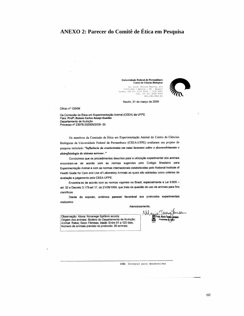
ANEXO 2: Parecer do Comitê de Ética em Pesquisa
60
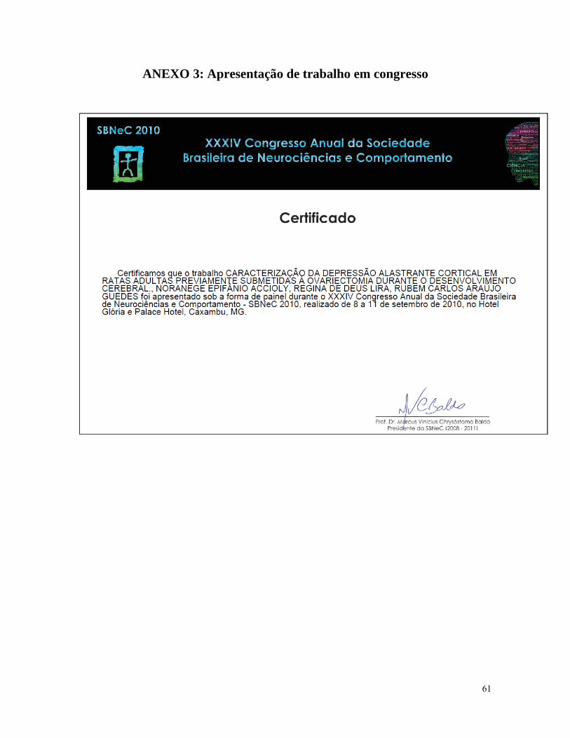
ANEXO 3: Apresentação de trabalho em congresso
61
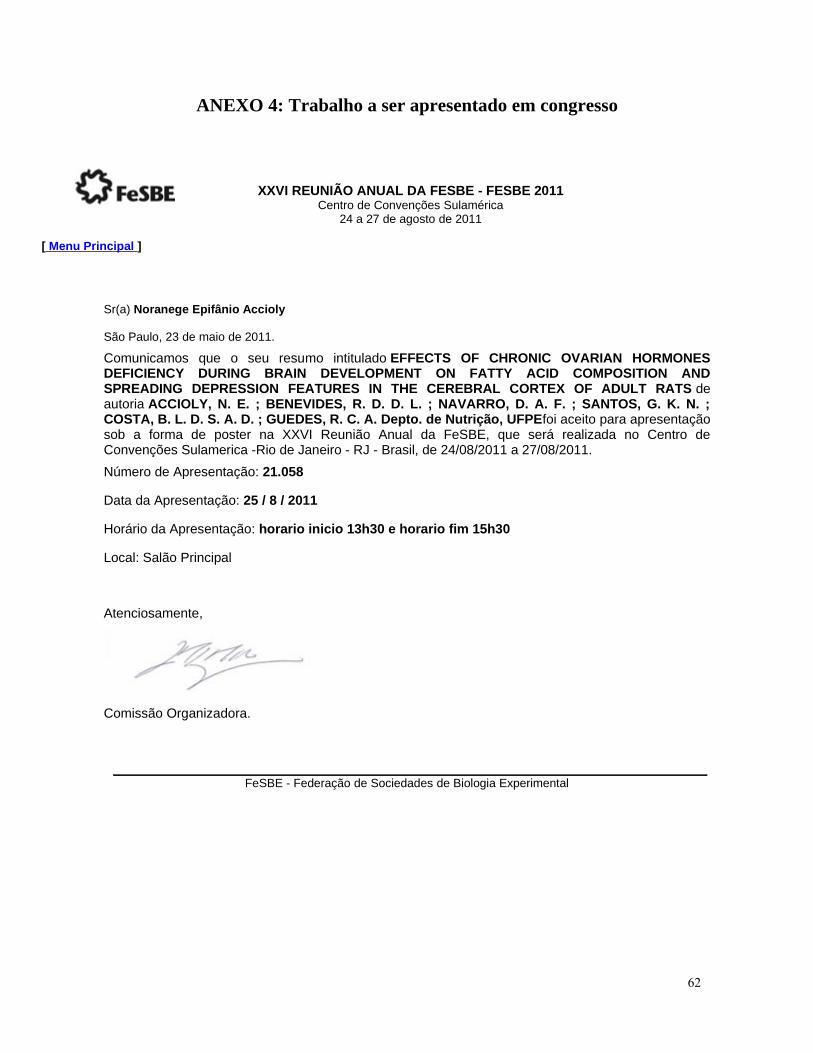
ANEXO 4: Trabalho a ser apresentado em congresso
XXVI REUNIÃO ANUAL DA FESBE - FESBE 2011Centro de Convenções Sulamérica
24 a 27 de agosto de 2011
[ Menu Principal ]
Sr(a) Noranege Epifânio Accioly
São Paulo, 23 de maio de 2011.
Comunicamos que o seu resumo intitulado EFFECTS OF CHRONIC OVARIAN HORMONES DEFICIENCY DURING BRAIN DEVELOPMENT ON FATTY ACID COMPOSITION AND SPREADING DEPRESSION FEATURES IN THE CEREBRAL CORTEX OF ADULT RATS de autoria ACCIOLY, N. E. ; BENEVIDES, R. D. D. L. ; NAVARRO, D. A. F. ; SANTOS, G. K. N. ; COSTA, B. L. D. S. A. D. ; GUEDES, R. C. A. Depto. de Nutrição, UFPEfoi aceito para apresentação sob a forma de poster na XXVI Reunião Anual da FeSBE, que será realizada no Centro de Convenções Sulamerica -Rio de Janeiro - RJ - Brasil, de 24/08/2011 a 27/08/2011.Número de Apresentação: 21.058
Data da Apresentação: 25 / 8 / 2011
Horário da Apresentação: horario inicio 13h30 e horario fim 15h30
Local: Salão Principal
Atenciosamente,
Comissão Organizadora.
FeSBE - Federação de Sociedades de Biologia Experimental
62
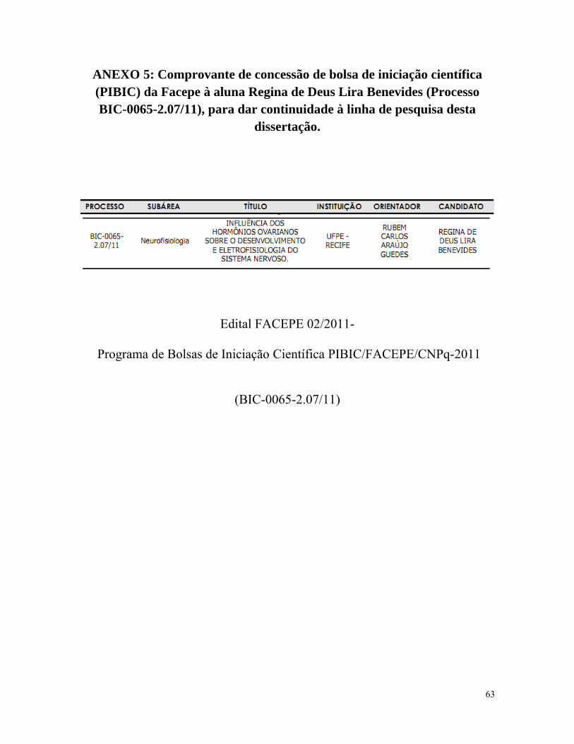
ANEXO 5: Comprovante de concessão de bolsa de iniciação científica (PIBIC) da Facepe à aluna Regina de Deus Lira Benevides (Processo BIC-0065-2.07/11), para dar continuidade à linha de pesquisa desta
dissertação.
Edital FACEPE 02/2011-
Programa de Bolsas de Iniciação Científica PIBIC/FACEPE/CNPq-2011
(BIC-0065-2.07/11)
63

ANEXO 6: Comprovante de submissão do artigo
From: "Int. J. Developmental Neuroscience" [email protected]
Sent: Sex 29/07/11 23:17
Subject: Fwd: Submission Confirmation
Dear Rubem,
Your submission entitled "Ovariectomy in the developing rat decelerates cortical spreading depression in
adult brain" has been received by International Journal of Developmental Neuroscience.
You may check on the progress of your paper by logging on to the Elsevier Editorial System as an author. The
URL is http://ees.elsevier.com/dn/ Your username is Your username is: Guedes.
If you need to retrieve password details, please go to: http://ees.elsevier.com/dn/automail_query.asp
Your manuscript will be given a reference number once an Editor has been assigned.
Thank you for submitting your work to this journal.
Kind regards,
Elsevier Editorial System
International Journal of Developmental Neuroscience
64

65


![JUCELY APARECIDA DA ROSA - teses.usp.br · fêmures e nos componentes moleculares do osso em ratas ovariectomizadas [tese]. São Paulo: ... pela ovariectomia e diminuiu as ligações](https://img.document.onl/doc/110x75/5c03e71709d3f21e408ceb21/jucely-aparecida-da-rosa-tesesuspbr-femures-e-nos-componentes-moleculares.jpg)