Embed Size (px)
Citation preview
UNIVERSIDADE FEDERAL DE UBERLÂNDIA INSTITUTO DE GENÉTICA E BIOQUÍMICA
PÓS-GRADUAÇÃO EM GENÉTICA E BIOQUÍMICA
Aspectos da lesão tecidual local e da regeneração induzidas pela
BnSP-7, uma miotoxina isolada da peçonha da serpente Bothrops
pauloensis: Um estudo da liberação de citocinas pró-
inflamatórias e da expressão de metaloproteases de matriz
(MMP-9 e MMP-2)
Carolina de Freitas Oliveira
UBERLÂNDIA – MG
2008

UNIVERSIDADE FEDERAL DE UBERLÂNDIA INSTITUTO DE GENÉTICA E BIOQUÍMICA
PÓS-GRADUAÇÃO EM GENÉTICA E BIOQUÍMICA
Aspectos da lesão tecidual local e da regeneração induzidas pela
BnSP-7, uma miotoxina isolada da peçonha da serpente Bothrops
pauloensis: Um estudo da liberação de citocinas pró-inflamatórias e
da expressão de metaloproteases de matriz (MMP-9 e MMP-2)
Carolina de Freitas Oliveira
Profª. Drª. Veridiana de Melo Rodrigues Ávila
UBERLÂNDIA – MG
2008

ii
UNIVERSIDADE FEDERAL DE UBERLÂNDIA INSTITUTO DE GENÉTICA E BIOQUÍMICA
PÓS-GRADUAÇÃO EM GENÉTICA E BIOQUÍMICA
Aspectos da lesão tecidual local e da regeneração induzidas pela
BnSP-7, uma miotoxina isolada da peçonha da serpente Bothrops
pauloensis: Um estudo da liberação de citocinas pró-inflamatórias e
da expressão de metaloproteases de matriz (MMP-9 e MMP-2)
Carolina de Freitas Oliveira
Profª. Drª. Veridiana de Melo Rodrigues Ávila
Dissertação apresentada à Universidade
Federal de Uberlândia como parte dos
requisitos para a obtenção do Título de
Mestre em Genética e Bioquímica (Área
Bioquímica)
UBERLÂNDIA – MG
2008
Dados Internacionais de Catalogação na Publicação (CIP)
O48a
Oliveira, Carolina de Freitas, 1984- Aspectos da lesão tecidual local e da regeneração induzidas pela BnSP-7, uma miotoxina isolada da peçonha da serpente Bothrops pauloensis: um estudo da liberação de citocinas pró-inflamatórias e da expressão de metaloproteases de matriz (MMP-9 e MMP-2) / Carolina de Freitas Oliveira. - 2008. 86 f. : il. Orientador: Veridiana de Melo Rodrigues Ávila. Dissertação (mestrado) – Universidade Federal de Uberlândia, Pro-grama de Pós-Graduação em Genética e Bioquímica. Inclui bibliografia.
1. Cobra venenosa - Veneno - Teses. I. Ávila, Veridiana de Melo Rodrigues. II. Universidade Federal de Uberlândia. Programa de Pós-Graduação em Genética e Bioquímica. III. Título.
CDU: 615.919:598.126 Elaborado pelo Sistema de Bibliotecas da UFU / Setor de Catalogação e Classificação

iii
UNIVERSIDADE FEDERAL DE UBERLÂNDIA INSTITUTO DE GENÉTICA E BIOQUÍMICA
PÓS-GRADUAÇÃO EM GENÉTICA E BIOQUÍMICA
Aspectos da lesão tecidual local e da regeneração induzidas pela BnSP-7, uma
miotoxina isolada da peçonha da serpente Bothrops pauloensis: Um estudo da
liberação de citocinas pró-inflamatórias e da expressão de metaloproteases de
matriz (MMP-9 e MMP-2)
Carolina de Freitas Oliveira
COMISSÃO EXAMINADORA
Presidente: _________________________________________ Drª. Veridiana de Melo Rodrigues Ávila
Examinadores: _________________________________________ Drª. Patrícia Bianca Clissa
_________________________________________ Drª. Eloisa Amália Vieira Ferro
Data da Defesa: _____/_____/_____
As sugestões da Comissão Examinadora e as Normas PGGB para o formato da
Dissertação foram contempladas
_________________________________________
Drª. Veridiana de Melo Rodrigues Ávila

iv
Dedico este trabalho a meus pais, que são o
porto seguro da minha vida! Obrigada meu
Deus por escolhê-los para mim!!!
v
Agradecimentos
À Deus, por se fazer presente em todos os momentos da minha vida.
À professora Drª Veridiana de Melo Rodrigues Ávila, pela orientação e confiança
durante todos estes anos. Você foi essencial para meu crescimento! Nunca vou me
esquecer de você e do seu carinho sempre!!!
Aos meus pais, Luiza e Júnior, aos meus irmãos, Frá e Marco e ao Claudinho por
sempre estarem me incentivando e dando forças quando eu desanimava e por todo
o amor e compreensão. Vocês são essenciais para minha felicidade! Amo vocês!!!
À Daiana, amiga e companheira sempre presente, mesmo estando longe agora...
Você também fez possível este trabalho! Obrigada por tudo! Seremos a “dupla
dinâmica” por toda a vida, espero!!!
À Drª Patrícia Bianca Clissa, pela parceria, pela grande contribuição a este trabalho
e por todos os ensinamentos. Adorei te conhecer!!!
Aos colegas do Laboratório de Química de Proteínas e Produtos Naturais: Renata,
Johara, Débora, Letícia, Dayane, Sâmela, Malson, Francis, Fábio, Luciana,
Robson, Tomás e Luiz Fernando, pela convivência agradável e pelo auxílio no dia
a dia do laboratório.
Ao pessoal do Laboratório de Imunopatologia do Instituto Butantan, Drª Maísa,
Cristiani e Stela pelo auxílio nos experimentos e pela ótima convivência. Saudades!
À Mirian, por todos os ensinamentos e auxílio nos experimentos e análises.
Obrigada pela amizade sempre!!!
vi
À Drª Tânia Machado de Alcântara pela parceria e sugestões na primeira parte do
trabalho.
À Drª Ana Moura pela parceria e colaborações.
Ao Drª Roberto Octaviano, pela experiência e garra que só acrescentaram ao
nosso laboratório.
Às amigas Carol e Ana Paula pelo grande auxílio nas técnicas de Biologia
Molecular. Adorei trabalhar com vocês!
À Drª Maria Inês Homsi Brandeburgo e à Drª Amélia Hamaguchi, pelo apoio
financeiro.
Ao Dr. Luiz Ricardo Goulart Filho, pelo apoio técnico.
Aos funcionários Tianinha, Marlene, D. Nenzinha e Gerson pela disposição em
ajudar sempre.
Ao Conselho Nacional de Desenvolvimento Científico e Tecnológico (CNPq) e à
Fundação de Amparo à Pesquisa do Estado de São Paulo (FAPESP) pelo apoio
financeiro.
A todos que diretamente ou indiretamente contribuíram para a realização deste
trabalho.
vii
Sumário
Apresentação .......................................................................................................... 1
Capítulo I: Peçonhas botrópicas e seus efeitos .................................................. 4
1. Fundamentação Teórica...................................................................................... 5
1.1. Características epidemiológicas e biológicas do envenenamento ofídico ... 5
1.2. Alterações teciduais locais induzidas pelo envenenamento botrópico ........ 6
1.2.1. Resposta inflamatória ....................................................................... 10
1.3. Características estruturais e funcionais das PLA2s .....................................16
2. Referências Bibliográficas ................................................................................ 21
Capítulo II: Aspectos da lesão tecidual local e da regeneração induzidas pela
BnSP-7, uma miotoxina isolada da peçonha da serpente Bothrops
pauloensis: Um estudo de liberação de citocinas pró-inflamatórias e da
expressão de metaloproteases de matrix (MMP-9 e MMP-2). ........................ 35
1. Introdução ......................................................................................................... 40
2. Materiais e Métodos.......................................................................................... 42
2.1. Peçonha e toxinas ...................................................................................... 42
2.2. Animais ....................................................................................................... 42
2.3. Atividade edematogênica ........................................................................... 43
2.4. Atividade miotóxica .................................................................................... 43
2.5. Análises histológicas .................................................................................. 44
viii
2.6. Extração de RNA ........................................................................................ 44
2.7. RT-PCR - Análise semi-quantitativa ........................................................... 45
2.8. Células peritoneais e cultura ...................................................................... 46
2.9. Dosagem de citocinas ................................................................................ 47
2.10. Análise estatística ............................................................................. 48
3. Resultados ........................................................................................................ 48
3.1. Atividade edematogênica ........................................................................... 48
3.2. Atividade miotóxica .................................................................................... 49
3.3. Análises histológicas .................................................................................. 49
3.4. Expressão de MMPs .................................................................................. 50
3.5. Efeito nas citocinas .................................................................................... 51
4. Discussão ......................................................................................................... 51
5. Agradecimentos ................................................................................................ 60
6. Referências ....................................................................................................... 60
Anexos .................................................................................................................. 81
Anexo I ........................................................................................................ 82

ix
Lista de Legendas
1. Fotomicrografia de músculo gastrocnêmio de camundongo inoculado com
CV ................................................................................................................ 73
2. Fotomicrografia de músculo gastrocnêmio de camundongo inoculado com
BnSP-7 ........................................................................................................ 73
3. Expressão semi-quantitativa dos genes MMP-9 e MMP-2 em músculo
gastrocnêmio de camundongo induzida pela BnSP-7 ................................. 73
4. Liberação de citocinas por MPAC ............................................................... 74
5. Liberação de citocinas pelo músculo gastrocnêmio .................................... 74

x
Lista de Figuras
1. Fotomicrografia de músculo gastrocnêmio de camundongo inoculado com
CV ................................................................................................................ 75
2. Fotomicrografia de músculo gastrocnêmio de camundongo inoculado com
BnSP-7 ........................................................................................................ 76
3. Expressão semi-quantitativa dos genes MMP-9 e MMP-2 em músculo
gastrocnêmio de camundongo induzida pela BnSP-7 ................................. 77
4. Liberação de citocinas por MPAC ............................................................... 78
5. Liberação de citocinas pelo músculo gastrocnêmio .................................... 79

xi
Lista de Tabelas
1. Indução de edema tempo-dependente e liberação de creatina quinase .... 80
xii
Lista de Abreviaturas
PBS - tampão fosfato salina
FCS - soro fetal bovino (fetal calf serum)
MTT - brometo 3-(4,5-dimetiltiazol-2-il) 2,5 difenil-tetrazolio
MPAC - células aderentes peritoneais de murinos (murine peritoneal adherent
cells)
i.m. - intramuscular
RNA - ácido ribonucléico
cDNA - ácido desoxirribonucléico complementar
BPB - brometo de 4-bromophenacil
CV - peçonha bruta de Bothrops pauloensis (crude venom)
PLA2 - fosfolipase A2 (E.C.3.1.1.4)
MMPs - metaloprotases de matriz (matrix metalloproteinases)
MEC - matriz extracelular
SVMPs - metaloproteases de peçonha de serpentes (snake venom
metalloproteinases)
PAF - fator de agregação plaquetária (platelet agragation factor)
CK - creatina quinase (creatine kinase)
HE - hematoxilina-eosina (hematoxin-eosin)

Apresentação

2
O envenenamento ofídico constitui um problema de saúde pública no Brasil
devido à sua grande incidência e seqüelas deixadas nos acidentados. Na maioria
dos casos o ataque é de serpentes do gênero Bothrops, conhecidas popularmente
como jararacas. A letalidade desta peçonha não é tão alta, porém seu efeito no
local da picada é bastante grave. Observa-se o surgimento de bolhas de sangue,
necrose e hemorragia que muitas das vezes levam à amputação do membro
afetado.
Atualmente, o único tratamento que se faz é a aplicação do soro antiofídico,
que é eficiente para neutralizar os efeitos sistêmicos das toxinas da peçonha. No
entanto, o soro não consegue neutralizar os efeitos locais do envenenamento,
mesmo quando administrado imediatamente após o acidente. As toxinas da
peçonha agem muito rapidamente no tecido afetado e induzem uma resposta
inflamatória associada com a liberação de mediadores endógenos antes que o soro
possa neutralizar a atividade dos vários componentes da peçonha.
Desta forma, o estudo dos componentes das peçonhas e suas atividades
biológicas nos permitem compreender os mecanismos pelos quais elas causam a
lesão, favorecendo assim, o desenvolvimento de terapias alternativas ao soro, que
possam neutralizar os efeitos locais da peçonha.
Além disso, as toxinas da peçonha de serpentes podem funcionar como
ferramentas importantes para o entendimento de uma variedade de processos
biológicos, assim como funcionarem como base estrutural para o desenvolvimento
de medicamentos sintéticos com seus princípios de ação para outras patologias.
Este trabalho teve como objetivo avaliar a lesão tecidual local e o processo
inflamatório induzido pela BnSP-7, uma miotoxina isolada da peçonha de Bothrops
pauloensis. Além disso, a evolução dos processos de degeneração e regeneração
do músculo gastrocnemius também foi avaliada.
A apresentação deste trabalho foi realizada segundo as normas da Pós-
Graduação em Genética e Bioquímica da Universidade Federal de Uberlândia
(www.cogeb.ufu.br). A dissertação foi dividida em: Capítulo 1 – Revisão de
literatura sobre os aspectos epidemiológicos e biológicos de envenenamento
ofídico, alterações teciduais locais e resposta inflamatória induzidas pelo
envenenamento botrópico, e características estruturais e funcionais das
fosfolipases A2 (PLA2s) e Capítulo 2 – Aspectos da lesão tecidual local e da

3
regeneração induzidas pela BnSP-7, uma miotoxina isolada da peçonha da
serpente Bothrops pauloensis: Um estudo de liberação de citocinas pró-
inflamatórias e da expressão de metaloproteases de matrix (MMP-9 e MMP-2). A
formatação deste capítulo foi baseada nas normas da revista Toxicon (Anexo I).

4
Capítulo I
Peçonhas botrópicas e seus efeitos

5
1. Fundamentação Teórica
1.1. Características epidemiológicas e biológicas do
envenenamento ofídico
Os acidentes ocasionados por animais peçonhentos constituem problema de
saúde pública (Soerensen, 1990) nos países tropicais em desenvolvimento, dadas
à incidência, a gravidade e as seqüelas deixadas nos acidentados (Barraviera,
1991; Pinho & Pereira, 2001).
Das 20 famílias de serpentes conhecidas no mundo, nove são encontradas
no Brasil, sendo as principais Boidae (10 espécies), Colubridae (189 espécies),
Elapidae (18 espécies) e Viperidae (22 espécies). Destas, somente a Elapidae e a
Viperidae são consideradas peçonhentas. A família Viperidae, maior responsável
pelos acidentes ofídicos, é representada por três gêneros, Lachesis, Crotalus e
Bothrops, serpentes conhecidas popularmente como surucucus, cascavéis e
jararacas, respectivamente (Barraviera, 1990; Cardoso, 2003).
Estima-se que acidentes ofídicos afetem mais de 2,5 milhões de pessoas
anualmente no mundo, onde 100 mil dos casos resultam em morte (White, 2005).
No Brasil a epidemiologia aponta para um perfil que se mantém inalterado ao longo
dos últimos 100 anos. Ocorrem 19,000 a 22,000 acidentes ofídicos por ano, com
letalidade ao redor de 0,45% (Ministério da Saúde, 2001). As serpentes do gênero
Bothrops são as principais responsáveis por acidentes ofídicos no Brasil (90%),
mas com reduzidos índices de letalidade (0,3%) (Ministério da Saúde, 2001; Silva
et al., 2003).
A subespécie Bothrops neuwiedi pauloensis, conhecida como jararaca
pintada ou boca de sapo, descrita por Amaral em 1925, habita preferencialmente
áreas secas, campos e cerrados (Peters e Orejas, 1970). Uma revisão sistemática
do complexo Bothrops neuwiedi foi realizada por Silva (2000) e resultou na
modificação das doze subespécies existentes para sete espécies distintas. Nessa
reclassificação Bothrops neuwiedi pauloensis passou a ser denominada Bothrops
pauloensis. Esta nova classificação foi aceita pela Sociedade Brasileira de
6
Herpetologia – SBH (2005). A serpente Bothrops pauloensis é encontrada nos
estados de São Paulo, Sul de Goiás e Triângulo Mineiro (Campbell e Lamar, 1989).
A peçonha das serpentes do gênero Bothrops induz um quadro
fisiopatológico caracterizado por efeitos locais e/ou sistêmicos (Gutiérrez e
Lomonte, 1989; Kamiguti et al.,1996). Os efeitos locais freqüentemente incluem
dor, edema, hemorragia local, inflamação e equimose (Mandelbaum et al., 1988;
Mebs e Ownby, 1990; Gutiérrez e Lomonte, 1995; Voronov et al., 1999). Este
quadro clínico na maioria das vezes evolui para necrose tecidual (Gutiérrez e
Lomonte, 1989). Efeitos sistêmicos são representados por alterações na
coagulação sangüínea, alterações cardiovasculares, choque hipovolêmico,
alterações renais e hemorragias distantes dos locais da picada tais como
hemorragia gengival, macro-hematúria, hemorragia uterina e gastrintestinal
(Kamiguti et al., 1996).
Estes efeitos do envenenamento botrópico são atribuídos a uma variedade
de toxinas presentes na peçonha, as quais incluem fosfolipases A2,
metaloproteases, serinoproteases, desintegrinas, L-aminoácido oxidases,
fosfomonoesterases, fosfodiesterases, acetilcolinesterase, arginina esterase,
hialuronidase e 5’-nucleotidase (Russel, 1980; Tu, 1988; Meier, 1990; Stocker,
1990). Estas proteínas constituem cerca de 90 a 95% do peso seco da peçonha,
que também é constituída por citrato, íons metálicos, carboidratos, nucleotídeos e
aminoácidos livres e lipídeos em menor proporção (Souza, et al 2001).
1.2. Alterações teciduais locais induzidas pelo envenenamento
botrópico
A lesão tecidual local induzida pelo envenenamento botrópico caracteriza-se
por hemorragia, edema, inflamação e mionecrose. A magnitude destes efeitos
depende do tipo de peçonha, da dose injetada e do hospedeiro. Em muitos casos,
o dano tecidual é tão intenso, levando a conseqüências severas como lesão
vascular e isquemia, as quais podem culminar com a amputação do membro
afetado (Nishioka et al., 1992). Estes efeitos são causados pela ação combinada
de vários componentes das peçonhas tais como metaloproteases (SVMPs),
fosfolipases A2 (PLA2s) e também pela ação de mediadores endógenos liberados

7
pela ação da resposta inflamatória (Gutiérrez, et al., 1989; Gutiérrez & Rucavado,
2000).
A necrose muscular ou mionecrose, efeito bastante comum após
envenenamento botrópico, pode ocorrer devido à ação direta de fosfolipases A2
miotóxicas sobre as membranas plasmáticas de células musculares, ou indireta,
como conseqüência de lesões vasculares e isquemia, causadas por
metaloproteases hemorrágicas presentes nas peçonhas de serpentes.
O mecanismo pelo qual as metaloproteases hemorrágicas causam danos
teciduais locais e ou sistêmicos se deve à sua ação proteolítica sobre proteínas da
membrana basal (Bjarnason e Fox, 1994; Esclante et al., 2006). Essa ação
enfraquece sua estabilidade mecânica e as forças hemodinâmicas biofísicas que
normalmente operam na circulação, provocando uma distensão na parede capilar
até que sua integridade seja rompida e ocorra o extravasamento de um modo
explosivo, levando à hipóxia tecidual e, consequentemente, à morte celular
(Gutiérrez e Rucavado, 2000; Gutiérrez et al., 2005; Gutiérrez et al., 2006). Por
esta hipótese, é sugerido que esta distensão e ruptura abruptas resultem na perda
da integridade não só das células endoteliais, mas também dos componentes da
membrana basal (Moreira et al., 1994; Gutiérrez et al., 2006). As lesões
hemorrágicas também podem ser provocadas pelo efeito inibitório das SVMPs na
agregação plaquetária (Bjarnason e Fox, 1994, Kamiguti, et al., 2003). Além disso,
as SVMPs podem inibir a adesão das células endoteliais às proteínas da matriz
extracelular culminando em um processo apoptótico (Tanjoni et al., 2005; Diaz et
al., 2005; You et al., 2003).
Estudos para a compreensão dos mecanismos de ação das PLA2s de
peçonhas botrópicas sobre o tecido muscular vêm sendo realizados desde as
últimas décadas. Estudos realizados com miotoxinas isoladas de B. asper e B.
numminifer demonstraram que estas enzimas podem induzir mionecrose por
agirem na membrana sarcoplasmática, induzindo desorganização dos
componentes fosfolipídicos, que permitiriam a saída de moléculas intracelulares,
como a creatina e a creatina quinase (Gutiérrez et al., 1986; Gutiérrez et al., 1989).
Os efeitos celulares causados por fosfolipases A2 miotóxicas, segundo Harris
(1991), são: (a) 0-1 hora: edema confinado ao espaço extravascular; (b) 1-3 horas:
degeneração e hipercontração das miofibrilas e acúmulo de fagócitos na luz dos
8
vasos sanguíneos e no espaço perivascular; (c) 3-6 horas: invasão das fibras
musculares necrosadas pelas células fagocíticas, colapso do potencial das fibras
musculares e rompimento da membrana sarcoplasmática; (d) 6-24 horas:
degeneração total das fibras musculares individuais. Estas enzimas podem destruir
os terminais nervo-motores dos músculos, mas não interferem com as arteríolas,
capilares, vênulas ou nervos intramusculares, o que permite a regeneração das
fibras musculares lesionadas por estas toxinas, a partir de células satélites.
As células satélites fazem parte de uma população de células com grande
atividade mitogênica que contribuem para o crescimento muscular pós-natal, o
reparo de fibras musculares danificadas e a manutenção do músculo esquelético
adulto. São células indiferenciadas e mononucleadas, cuja membrana basal está
em continuidade com a membrana basal da fibra muscular (Harris, 2003). Estas
células se tornam ativadas durante o processo degenerativo. As células filhas
migram para formar cadeias de células satélites longitudinalmente organizadas na
lâmina basal. A fusão das células leva à formação de uma nova fibra muscular
multinucleada (Snow, 1977). Este processo é regulado por vários fatores
transcricionais, incluindo Pax3 e Pax7, e fatores regulatórios miogênicos como
Myf5, MyoD e miogenina (Dhawan e Rando, 2005; Charge e Rudnicki, 2004).
O mecanismo molecular pelo qual as PLA2s induzem mionecrose foi
inicialmente proposto por Gutiérrez e Lomonte em 1995. Estes autores propuseram
um modelo hipotético para o entendimento do mecanismo de ação das miotoxinas
Lys49, que pode ser descrito da seguinte forma: 1- ligação da miotoxina a um sítio
não identificado localizado na membrana sarcoplasmática; 2- interação
eletrostática entre o sítio catiônico da toxina e grupos carregados negativamente na
membrana; 3- penetração da miotoxina na bicamada lipídica por interação
hidrofóbica mediada pela região citotóxica da molécula; 4- penetração da região
citotóxica no centro da bicamada lipídica (o efeito seria a desorganização e ruptura
da membrana, com conseqüente prejuízo na regulação da permeabilidade
seletiva); 5- grande influxo de íons cálcio e início de uma variedade de
mecanismos degenerativos.
Atualmente, muitos autores buscam esclarecer uma via intracelular e um
possível sítio de ligação na membrana plasmática que poderia ser alvo dessas
PLA2s. Dessa forma, vários estudos visam identificar na estrutura das PLA2s
9
resíduos de aminoácidos específicos que estariam envolvidos no reconhecimento
celular, permitindo ação catalítica local da enzima a fim de iniciar o processo de
transdução de sinais, o que levaria a formação de mensageiros celulares que
modulariam a ação dessas enzimas ao nível celular.
O uso de peptídeos sintéticos evidenciou a região C-terminal (115-129)
como efetora das atividades tóxicas em algumas PLA2 Lys49 miotóxicas (Núnez et
al., 2001; Lomonte et al., 2003a). Esta região combina aminoácidos catiônicos e
hidrofóbicos responsáveis pelo mecanismo de dano às membranas celulares.
Segundo Lomonte et al. (2003b), os resíduos catiônicos interagiriam
eletrostaticamente com grupos aniônicos de um sítio aceptor (possivelmente
fosfolipídeos da membrana negativamente carregados) enquanto os resíduos
hidrofóbicos, especialmente os aromáticos, interagiriam e possivelmente
penetrariam a bicamada fosfolipídica, resultando na sua desestabilização (Núnez et
al., 2001). No entanto, este fato de um único peptídeo reproduzir todas as
principais atividades tóxicas da molécula de origem, não impede a existência de
outros domínios estruturais que possam participar ou complementar a ação do sítio
tóxico efetor (Lomonte et al., 2003b). Em contraste, peptídeos da região C-terminal
de algumas PLA2 Asp49 miotóxicas não apresentaram atividade de dano direto na
membrana, o que sugere que o mecanismo tóxico exercido por estas proteínas,
que provavelmente envolve sua atividade catalítica como um passo relevante, seja
diferente daquele utilizado pelas PLA2 Lys49 miotóxicas (Núnez et al., 2001).
Kini (2003) propôs ainda que as diferentes ações farmacológicas induzidas
pelas PLA2s se devem à capacidade que estas enzimas possuem de se ligar com
alta afinidade a aceptores celulares, por meio de interações iônicas, hidrofóbicas e
de van der Waals. Uma vez ligada ao seu alvo, essas enzimas podem induzir seus
efeitos farmacológicos por mecanismos dependentes ou independentes de sua
atividade catalítica.
Devido ao largo espectro de alvos específicos em vários tecidos e órgãos, a
identificação destes sítios farmacológicos presentes na estrutura dessas enzimas
se torna um campo vasto de investigações para as áreas da saúde e biotecnologia.
Envenenamentos botrópicos também são caracterizados pelo rápido
desenvolvimento de edema e inflamação no local da picada. O edema induzido
pelas peçonhas de serpentes é bioquimicamente heterogêneo e pode agravar
10
ainda mais o dano tecidual local. Em maior magnitude, o edema pode acentuar o
efeito hipovolêmico e hipotensivo da peçonha e culminar em choque cardiovascular
(Lomonte et al., 1993). O edema é provavelmente causado pelo efeito direto de
toxinas hemorrágicas nos vasos sangüíneos, induzindo a liberação de mediadores
endógenos, como a histamina, cininas, prostaglandinas, devido à ação de
componentes da peçonha em mastócitos, cininogênio e fosfolipídios,
respectivamente. Além disso, fosfolipases A2, esterases e aminas biogênicas
podem estar envolvidas na indução do edema (Gutiérrez, 1990).
Entre os vários mecanismos descritos na indução do edema podemos citar:
a degranulação de mastócitos, com liberação de histamina e serotonina,
recrutamento de células polimorfonucleares, com formação de radical superóxido,
produção de prostaglandinas e leucotrienos (LTB4), liberação de bradicinina e
oxido nítrico, potencialização da atividade da bradicinina por peptídeos inibidores
da enzima conversora de angiostensina, ativação do sistema complemento e
liberação das anafilotoxinas C3a e C5a (Lomonte et al., 1993).
1.2.1 Resposta inflamatória
A resposta inflamatória que se estabelece logo após o envenenamento
também é de relevância para o progresso da lesão tecidual, uma vez que além das
toxinas da peçonha presentes no local da picada existe também a participação de
mediadores endógenos que podem contribuir com estas alterações.
A inflamação é a reação do organismo à invasão por agente infeccioso, por
desafio com antígeno ou mesmo apenas uma lesão física. A resposta inflamatória
compreende três eventos principais: (1) aumento do suprimento sangüíneo para a
área; (2) aumento da permeabilidade capilar, ocasionado pela retração das células
endoteliais, com conseqüente escape de moléculas maiores, permitindo, então,
que os mediadores solúveis da imunidade atinjam o local da infecção; (3) migração
de leucócitos dos capilares para os tecidos circundantes, sendo que na fase inicial
da inflamação, os neutrófilos são particularmente prevalentes e, mais tardiamente
no processo, os monócitos e linfócitos também migram para o local inflamado.
(Ryan e Majno, 1997; Rosenfeld, 1971).

11
Dependendo da sua duração, as inflamações podem ser divididas em
agudas e crônicas. Assim, as inflamações que duram desde poucos minutos até
poucos dias são chamadas de agudas, enquanto as que persistem por semanas e
meses são chamadas de crônicas. Do ponto de vista funcional e morfológico, as
inflamações agudas caracterizam-se pelo predomínio de fenômenos exsudativos,
ou seja, conseqüentes alterações na permeabilidade vascular, permitindo o
acúmulo de líquido na região inflamada (edema), fibrina, leucócitos, especialmente
neutrófilos, e hemácias. Nas inflamações crônicas, além destes elementos, ocorre
proliferação de vasos, migração e proliferação de monócitos e linfócitos
(Montenegro et al., 1999).
A resposta inflamatória envolve também a sinalização entre os vários
leucócitos, além das células teciduais, que ocorre tanto por interações diretas
célula-célula, envolvendo moléculas da superfície celular, quanto por citocinas.
(Roitt et al., 2003).
As citocinas são pequenas “proteínas mensageiras” (8-80 kDa), mediadores
solúveis da comunicação intercelular que, em associação aos hormônios e
neurotransmissores, constituem uma linguagem química de sinalização que
controla o desenvolvimento, o reparo tecidual e a resposta imune em organismos
multicelulares (Arend e Gabay, 2004). Paralelamente a outros sinais oriundos do
contato célula-célula ou célula-antígeno, as citocinas propiciam uma rede de
controle das respostas imunes inatas e específicas, incluindo a inflamação, a
defesa contra infecções virais, a proliferação de clones específicos e células T e B
e o controle de suas funções. Elas atuam através da ligação a receptores
específicos na membrana celular, estabelecendo uma cascata que leva à indução,
ao favorecimento ou à inibição de inúmeros genes citocina-regulados no núcleo
celular (Roitt et al., 2003).
A produção de citocinas pro e antiinflamatórias é controlada por um
complexo mecanismo de feedback. As citocinas proinflamatórias são
primeiramente responsáveis por iniciar um efeito contra um patógeno exógeno.
Entretanto, uma produção excessiva desses mediadores pode contribuir
significativamente para o choque, falência múltipla de órgãos, e morte (Van der
Meide e Schellekend, 1996). Em contraste, as citocinas antiinflamatórias são
cruciais para a regulação negativa do processo inflamatório e para a manutenção

12
da homeostase para o funcionamento correto dos órgãos vitais (Grard, et al.,
1993).
Além da ação das citocinas, a resposta inflamatória é mediada por uma
variedade de moléculas tais como prostaglandinas, radicais de oxigênio, óxido
nítrico, tromboxanas, leucotrienos e fator de agregação plaquetária (PAF). Estes
mediadores são liberados por macrófagos, neutrófilos, mastócitos, eosinófilos,
basófilos, linfócitos e plaquetas (Voronov et al., 1999). As células endoteliais
também são reguladoras da resposta inflamatória, controlando a adesão e
migração de células inflamatórias, assim como a troca de flúidos entre a corrente
sangüínea e o tecido lesionado. No entanto, uma ativação endotelial prolongada
pode levar a uma inflamação descontrolada que é prejudicial e pode resultar em
inflamação crônica (Kadl e Leitinger, 2005).
A lesão tecidual local observada no envenenamento botrópico desencadeia
uma cascata de eventos iniciados pelo recrutamento de leucócitos para o local da
picada. Leucócitos ativados liberam um amplo espectro de citocinas tais como IL-1,
IL-6 e IL-8, que amplificam a resposta inflamatória contribuindo para o processo de
necrose (Voronov et al., 1999).
A IL-1 é produzida principalmente por fagócitos mononucleares (macrófagos
e monócitos) ocorre em duas formas moleculares: IL-1α e IL-1β (Voronov et al.,
1999; Roitt et al., 2003). Apesar da baixa homologia entre as duas formas (menos
de 30%), elas se ligam ao mesmo receptor e desencadeiam atividades biológicas
semelhantes (Cybulsky et al., 1998). A IL-1 atua na ativação de linfócitos,
estimulação de macrófagos e adesão de leucócitos e células endoteliais. Além
disso, ela regula a síntese de prostaglandinas, IL-6, IL-8, MMPs, óxido nítrico (Kida
et al., 2005). A IL-6 é produzida por células T e B (linfócitos T e B) e macrófagos.
Na imunidade inata, a IL-6 estimula a síntese de proteínas da fase aguda por
hepatócitos durante o processo inflamatório. Na imunidade adaptativa, ela estimula
o crescimento e diferenciação de linfócitos B (Hopkins, 2003; Roitt et al., 2003). A
IL-8 é uma quimiocina liberada pelos monócitos atua na ativação de neutrófilos, na
quimiotaxia, na angiogênese e na liberação de grânulos e superóxidos (Sandra et
al., 1995).
A liberação de citocinas induzida por peçonha de serpentes ou toxinas delas
isoladas foi anteriormente observada por muitos autores. Barros et al. (1998)
13
verificaram um aumento nos níveis de IL-6, IL-10 e INF-γ no sangue causado pela
peçonha de Bothrops atrox. Clissa et al. (2001) mostraram que a jararagina, uma
metaloprotease isolada da peçonha de Bothrops jararaca, induz a produção de IL-
1β e TNF-α, embora níveis de IL-6 não foram encontrados. Rucavado et al. (2002)
encontraram níveis elevados de IL-6 e IL-1β em músculo de ratos induzido por MT-
III, um fosfolipase A2 miotóxica isolada da peçonha de Bothrops asper. Níveis de
TNF-α e IFN-α não foram observados. A peçonha de B. asper também induziu o
aumento de IL-6 e TNF-α em flúido peritoneal (Zamuner et al., 2005). Fernandes et
al. (2006) verificaram um aumento em produção de IL-1 e TNF-α induzido por
BaP1, uma metaloprotease isolada da peçonha de B. asper.
Além das citocinas, as metaloproteases de matriz (MMPs) são importantes
mediadores da lesão tecidual e da inflamação de uma variedade de patologias
(Shapiro, 1998; Nagase and Woessner, 1999), podendo assim representar um
papel importante nas alterações locais induzidas pela peçonha; (Rucavado et al,
2002).
A matriz extracelular (MEC) é definida como uma rede complexa e dinâmica
de componentes protéicos, proteoglicanos e glicoproteínas secretados que
circundam os fibroblastos unindo as células e mantendo a estrutura tridimensional
do corpo, conferindo, portanto, suporte mecânico para as células e integridade
estrutural para os tecidos (Itoh e Nagase, 2002). Os componentes da matriz
modulam o comportamento celular criando ambientes celulares influentes. A
renovação (turnover) da MEC é parte integral de processos normais e patológicos
como desenvolvimento, remodelamento tecidual, crescimento e diferenciação
celular e invasão (Werb et al, 1996).
Proteases extracelulares são necessárias em diversos processos
relacionados com o desenvolvimento normal e com doenças. A habilidade de
degradar proteínas extracelulares é essencial para as células individuais
interagirem com o ambiente ao redor e para organismos multicelulares se
desenvolverem e funcionarem normalmente (Sternlicht e Werb, 2001).
A degradação da MEC é mediada por algumas famílias de proteinases
extracelulares. Tais famílias incluem as serino-proteases, cisteína-proteases e
metaloproteases da matriz (MMPs) (Benaud et al, 1998).

14
As MMPs são enzimas proteolíticas zinco-dependentes que estão
envolvidas em processos fisiológicos e patológicos, pois podem estar expressas
em níveis baixos em tecidos normais e tornar-se alteradas em determinadas
situações, como no ciclo endometrial, em processos de cicatrização, na cirrose
hepática, em colagenoses e neoplasias, etc (Itoh e Nagase, 2002).
As MMPs constituem uma família de endopeptidases, com atividade
hidrolítica de amplo espectro para as proteínas extracelulares. As MMPs
pertencem a uma família de pelo menos 20 membros, produto de genes,
homólogos ou pseudo-homólogos, relacionados entre si (Woessner, 1991). Estas
enzimas podem ser classificadas de acordo com critérios estruturais e funcionais,
em cinco classes principais de diferente especificidade ao substrato: colagenases
(MMP1, MMP8 e MMP13), gelatinases (MMP2 e MMP9), estromelisinas (MMP3,
MMP7, MMP10, MMP11 e MMP26) e metaloproteínas de membrana (MMP14,
MMP15, MMP16, MMP17, MMP24 e MMP25), também conhecidas como MT1-
MMP a MT6-MMP, respectivamente, e matrilisinas (Shingleton et al, 1996; Souza e
Line, 2002).
Embora as MMPs sejam classificadas com base na sua afinidade por
substrato, nota-se que há sobreposição entre as subclasses, em termos de suas
afinidades. Além disso, as atividades das MMPs interferem umas com as outras na
medida em que muitas delas podem ativar outras proenzimas, o que sugere que a
ativação controlada das mesmas pode envolver uma cascata, englobando
diferentes membros da família das MMPs (Cawston, 1998).
As MMPs possuem algumas características em comum, incluindo, dentre
estas: 1) um domínio catalítico, que acomoda um átomo de Zn+2 no sítio ativo; 2)
um domínio propeptídico, essencial para a manutenção de sua latência enzimática,
e que é removido quando a enzima é ativada (Nagase and Woessner, 1999),
conferindo uma de peso molecular de aproximadamente 10kDa (Woessner, 1991)
e 3) um domínio translacional, peptídeo sinal que direciona o produto para
secreção. A maioria das MMPs também possui um domínio C-terminal com uma
seqüência de homologia a hemopexina, que constitui um sítio de ligação para os
inibidores teciduais e também confere especificidade ao substrato. Elas diferem
entre si pela especificidade ao substrato, tipos celulares e pelos agentes indutores
(Dollery et al, 1995).

15
Em condições fisiológicas normais, há uma rigorosa regulação da secreção
das MMP, as quais são sintetizadas e secretadas como pró-enzimas, zimogênios,
que posteriormente serão ativadas. Essa regulação ocorre apenas em momentos
específicos, nos quais existem processos multifásicos de ativação dos zimogênios,
além de haver vários inibidores sangüíneos e teciduais para monitorar a ação da
proteinase (Woessner, 1991). O controle da quantidade de enzima ativa é feito
pelos inibidores teciduais das MMPs (TIMPs). O desequilíbrio entre MMP e TIMP
pode resultar em várias patologias (Maskos e Bode, 2003). Além disso, as MMPs
também são reguladas pela ativação da expressão gênica, que é regulada pro
proteínas extracelulares, citocinas, fatores de crescimento entre outros (Philip et al,
2004). Os níveis das MMP nos tecidos saudáveis são baixos ou praticamente
indetectáveis, entretanto, sua expressão é substancialmente aumentada na maioria
das neoplasias malignas, apresentando importante ação proteolítica nos processos
de invasão e metástase (Hidalgo e Eckhardt,2001).
As principais ações das MMPs estão relacionadas ao remodelamento dos
componentes da matriz extracelular, por meio da degradação do colágeno, da
elastina e da fibronectina. Também participam do processo de migração de células
normais, malignas e inflamatórias, bem como induzindo a angioneogênese
(Lamooreaux, 1998). Estão evolvidas também na cicatrização (Wolf et al, 1992), na
reabsorção óssea (Delaissé et al, 1992), na involução mamária (Talhouk et
al,1992) e em outras funções fisiológicas associadas a gravidez e parto
(Jeffrey,1991). Recentemente, tem-se demonstrado que as MMPs também estão
implicadas em processos patológicos variados como na artrite reumatóide
(Harris, 1990), na esclerose múltipla, fibrose (Chandler et al, 1997), em algumas
doenças cardiovasculares (Tamarina et al, 1997) e certas alterações hematológicas
(Guedez et al, 1996).
As gelatinases A e B, MMP-2 e MMP-9 respectivamente, também
conhecidas como colagenases, são algumas das endopeptidases que compõem o
grupo das MMPs. Elas são distinguidas pela inserção de três repetições cabeça-
cauda ricas em cisteína em seus domínios catalíticos (Sternlicht e Werb, 2001) e
digerem gelatina, muitos tipos de colágenos, lamininas e outras várias proteínas
extracelulares (Aimes e Quigley, 1995). A seqüência da MMP-2 inclui um domínio
com uma tripla repetição de fibronectina tipo II inserida no seu domínio catalítico,

16
contribuindo para a ligação da enzima ao substrato. A MMP-9, estruturalmente, é a
maior das gelatinases, incluindo três domínios de fibronectina e um domínio tipo
colágeno tipo V (Woessner e Nagase, 2000).
A MMP-2 (72 kDa) é produzida principalmente a partir de células
mesenquimais (fibroblastos), ainda que células epidérmicas, endoteliais,
macrófagos e neutrófilos possam produzi-la em pequenas quantidades
(Lamooreaux, 1998). Também é denominada colagenase tipo IV e, apesar de estar
presente em níveis baixos de forma fisiológica (mantendo a homeostase do
colágeno) (Kherif et al, 1999; Rucavado et al, 2002), quando ativada, pode
degradar elastina insolúvel e degradar exageradamente o colágeno intersticial,
além de participar da degradação da membrana basal do endotélio e de induzir a
angiogênese (Agren, 1994).
A MMP-9 (92 kDa) é produzida por células inflamatórias ativadas (leucócitos
polimorfonucleares, como neutrófilos, eosinófilos e monócitos/macrófagos) e
células epidérmicas (especificamente queratinócitos), sua presença sempre
traduzindo uma situação patológica. Suas ações são direcionadas à degradação do
colágeno intersticial, à reepitelização, ao desenvolvimento e amplificação de
processos inflamatórios e à angiogênese (Agren, 1994).
Trabalhos mostraram que a MMP-2 e a MMP-9 estão aumentadas em uma
variedade de patologias, como a mionecrose (Kherif et al, 1999), inflamação
crônica (Trengove et al, 1999), meningite (Leib et al, 2000), hemorragia e isquemia
cerebral (Rosenberg, 1997; Gasche et al, 1999), entre outras. Rucavado et al
(2002) reportaram um aumento apenas na MMP-9 induzido pelas toxinas BaP1
(metaloprotease) e MT-III (fosfolipase A2), isoladas da peçonha de B. asper,
enquanto os níveis de MMP-2 permaneceram próximos ao controle de solução
salina, ou seja, observou-se apenas a expressão constitutiva (Kherif et al, 1999).
1.3. Características estruturais e funcionais das PLA2s
As fosfolipases A2 (PLA2s) são enzimas de grande interesse médico-
científico, devido ao seu envolvimento em grande variedade de doenças
inflamatórias humanas e nos envenenamentos ofídicos e de abelhas. Possuem

17
importantes papéis no catabolismo de lipídeos da dieta e no metabolismo geral de
lipídeos estruturais de membranas celulares. Com a ação dessas enzimas, os
fosfolipídeos são hidrolisados, desestruturando a membrana e comprometendo a
sua permeabilidade seletiva. A hidrólise ocorre especificamente na ligação 2-acil
éster de 3-sn-fosfolipídeos, liberando ácidos graxos livres e lisofosfatídeos (Arni &
Ward, 1996; Kini, 2003). Os ácidos graxos liberados como ácido araquidônico e
ácido oléico, podem ser importantes na estocagem de energia. O ácido
araquidônico pode também funcionar como segundo mensageiro e como precursor
de eicosanóides, que são potentes mediadores da inflamação e transdução de
sinais (Dennis, 1997).
Estas enzimas, amplamente distribuídas na natureza, são encontradas tanto
no interior como no exterior da célula (Dennis, 1994). As PLA2s intracelulares estão
frequentemente associadas a membranas e envolvidas com o metabolismo de
fosfolipídeos e outras funções celulares (Mukheerjee et al., 1994). As
extracelulares são amplamente distribuídas em secreções pancreáticas, exsudados
inflamatórios e nas peçonhas de serpentes e artrópodes.
De acordo com Six e Dennis (2000) as fosfolipases A2 extracelulares foram
divididas em doze classes I a XII, com base no número de resíduos de
aminoácidos e posição das ligações dissulfeto. Balsinde et al. 2002 sugerem um
novo sistema de classificação que organiza as PLA2 de acordo com sua seqüência
genética em 14 grupos distintos. As fosfolipases A2 de serpentes estão todas
reunidas nos grupos I e II, sendo estas proteínas de 119 a 143 resíduos de
aminoácidos, com peso molecular variando entre 13 e 18kDa. As enzimas da
classe I são encontradas em peçonhas de serpentes do gênero Elapidae e
Hydrophidae, enquanto as da classe II são encontradas principalmente em
peçonha de serpentes crotálicas e viperídeas (Ward et al., 2001).
As PLA2s representam uma classe de enzimas versáteis, considerando sua
função, localização, regulação, mecanismo de ação, seqüência, estrutura e papel
dos íons metálicos divalentes. Várias seqüências de PLA2 estão depositadas em
banco de dados e apresentam de 40 a 99% de similaridade na seqüência e no
dobramento tridimensional, dados resolvidos por cristalografia de raios X e por
espectroscopia de ressonância magnética nuclear (Arni & Ward, 1996; Balsinde et
al., 1999; Six & Dennis, 2000; Kini, 2003). A análise destas estruturas primárias

18
possibilitou a sugestão e a predição de determinantes estruturais de algumas
atividades farmacológicas (Kini & Iwanaga, 1986; Kini & Evans, 1989; Ward et al.,
1988). E agora, associada com resolução de estruturas tridimensionais, ampliou-se
ainda mais o conhecimento das bases moleculares do mecanismo de ação destas
toxinas.
A molécula de PLA2 é estabilizada por sete ligações dissulfeto nas posições
11-77, 27-124, 29-45, 44-105, 51-96 e 84-98 (Harris, 1991). A divisão entre as
classes I e II é baseada em dois critérios estruturais que são identificados na
seqüência de aminoácidos: primeiro, nas enzimas da classe II falta a ligação Cys
11- Cys 77, mas aparece outra ponte dissulfeto alternativa, Cys 51- Cys 133;
segundo, a classe I possui cerca de dois a três aminoácidos inseridos na região 52-
65, chamado de “loop elapídico”, enquanto a classe II possui esta volta truncada,
mas em adição apresenta de cinco a sete aminoácidos estendendo a região C-
terminal (Arni e Ward, 1996).
A grande maioria das PLA2s miotóxicas isoladas de peçonhas de serpentes
botrópicas, descritas até agora, são proteínas de caráter básico, ponto isoelétrico
variando entre 7,0 e 10,0, que possuem atividade catalítica ou “não”, sobre
substratos artificiais. As análises da composição em aminoácidos indicaram que
essas miotoxinas são ricas em aminoácidos básicos e hidrofóbicos (Homsi-
Brandeburgo et al., 1988; Selistre et al., 1990; Lomonte et al., 1990; Díaz et al.,
1995; Mancuso et al., 1995; Angulo et al., 1997). Apresentam também um alto
número de resíduos de meia-cistina, o que sugere a presença de várias pontes
dissulfeto intracadeia. As PLA2s tóxicas são muito estáveis, provavelmente como
resultado da extensa ligação cruzada, tornando-as ativas em uma ampla faixa de
pH e temperatura. Entretanto, várias PLA2s ácidas também já foram isoladas das
peçonhas botrópicas, como por exemplo: SIIISPIIA, SIIISPIIB, SIIISPIIIA e
SIIISPIIIB por Ketelhut et al., 2003; P1, P2 e P3 por Daniele et al., 1995; BP-PLA2
por Rodrigues et al., 2007.
As miotoxinas isoladas dos venenos botrópicos pertencem ao grupo II das
fosfolipases e podem ser subdivididas em dois subgrupos principais: (a) Asp49 que
são as miotoxinas com atividade enzimática alta, cataliticamente ativas e (b) Lys49
que são as miotoxinas que possuem baixa ou nenhuma atividade enzimática sobre
substratos artificiais (Ownby et al., 1999). A diferença nas propriedades
19
enzimáticas das duas classes de proteínas está baseada principalmente na
presença do resíduo de aspartato (Asp) na posição 49 no primeiro grupo, quando
comparado com a presença de lisina (Lys) na mesma posição da cadeia
polipeptídica no outro grupo. A troca de aminoácidos é suficiente para causar a
perda da habilidade da proteína em se ligar ao cálcio (Ca2+), um cofator essencial
para fazer com que a enzima expresse sua atividade catalítica. Além das
substituições na região do loop de ligação do cálcio, muitas PLA2s Lys49
apresentam resíduos variantes não encontrados nas PLA2s Asp49, que incluem
Lys7, Lys78, Lys80, Lys115 e Lys116, que podem estar envolvidos com a
toxicidade exercida por estas proteínas (Soares, 2000).
Além das PLA2s Lys49, outros variantes substituindo Asp49, que também
possuem baixa ou nenhuma atividade enzimática, foram reportados: duas PLA2s
Ser49, ammoditina L (Krizaj et al., 1991) e ecarfolina S (Polgar et al., 1996) e uma
PLA2 Arg49, zhaoermiatoxina (Mebs et al., 2006). Petan et al. (2007) concluíram
que as PLA2s Ser49 miotóxicas evoluíram em direção à perda da capacidade de se
ligar ao Ca2+ e da atividade enzimática para otimizarem o mecanismo de dano da
membrana independente de Ca2+ e para aumentar sua especificidade na ação
miotóxica. Da mesma forma, Murakami et al. (2008) verificaram que, apesar das
diferenças entre as PLA2s Lys49 e Arg49, não há evidências estruturais que
possam sugerir uma diferença no mecanismo de expressão de seus efeitos
farmacológicos.
Independentemente da sua função catalítica primária, as fosfolipases A2 de
venenos de serpentes podem induzir diversos efeitos farmacológicos adicionais,
como neurotoxicidade pré e/ou pós-sináptica, cardiotoxicidade, miotoxicidade,
indução e/ou inibição da agregação plaquetária, edema, hemólise, anticoagulação,
convulsão, hipotensão, efeito bactericida e anti-HIV (Bon et al., 1979; Fletcher et
al., 1980; Fletcher et al., 1981; Huang, 1984; Alvarado & Gutiérrez, 1988; Lloret &
Moreno, 1993; Yuan et al., 1993; Fuly et al., 1997; Páramo et al., 1998; Ownby,
1998; Soares et al., 1998; Fenard et al., 1999).
Durante os últimos 20 anos houve um grande interesse em se estudar
componentes da peçonha que são responsáveis pela mionecrose, resultando no
isolamento e caracterização estrutural e funcional de várias PLA2 miotóxicas de
peçonhas do gênero Bothrops (Basp-II por Francis et al, 1991; BthTX-I e BthTX-II

20
por Cintra et al, 1993; PrTX-I por Toyama et al, 1998; PrTX-II por Toyama et al,
2000; BnSP-6 e BnSP-7 por Rodrigues et al., 1998; MjTX-I e MjTX-II por Soares et
al, 2000; SIIISPIIA por Ketelhut et al, 2003; BnpTX-I e BnpTX-II por Rodrigues et al,
2004).
Da peçonha de B. pauloensis já foram isoladas várias PLA2s. Duas
fosfolipases A2 Asp49 (Rodrigues et al., 2004), duas miotoxinas Lys49, BnSP-6 e
BnSP-7 (Rodrigues et al., 1998) e uma fosfolipase A2 ácida, Bp-PLA2 (Rodrigues et
al., 2007).
A PLA2 denominada BnSP-7 (Rodrigues et al., 1998) é uma proteína com
peso molecular entre 13,5 e 14 kDa, podendo apresentar-se na forma dimérica, e
sua estrutura é essencialmente idêntica à estrutura das outras PLA2s Lys49
miotóxicas. Apresenta uma composição de aminoácidos com alto conteúdo de
resíduos básicos e hidrofóbicos, sendo uma serina o resíduo de aminoácido N-
terminal e apresentando também resíduos conservados das PLA2s Lys49
miotóxicas como Gly30, Gly33, His48, Lys49, Asp99 e a região 115-129. Seu ponto
isoelétrico é 8.8 e ela é capaz de produzir necrose das fibras musculares de
camundongos e não apresenta atividades fosfolipásica ou coagulante (Rodrigues
et al., 1998; Soares et al., 2000; Magro et al., 2003). Soares et al 2000 fizeram uma
alquilação do resíduo His48 da BnSP-7 com brometo de 4-bromofenacil brometo
(BPB) e verificaram que esta modificação levou a uma diminuição das atividades
miotóxica, bloqueio neuromuscular, indução de edema e atividade bactericida.
O reconhecimento da importância clínica do dano tecidual local em
envenenamentos tem motivado um grande número de estudos sobre sua
patogênese. Estes podem levar a uma melhor compreensão dos mecanismos que
levam a necrose tecidual, o que poderia inspirar a criação de novos métodos
terapêuticos. Em adição, o entendimento do mecanismo de ação de toxinas pode
revelar mecanismos da injúria celular e tecidual que são comuns a outras
condições patológicas.

21
2. Referências Bibliográficas
AGREN, M. S. Gelatinase activity during wound healing. Br J Dermatol. v. 131, p. 634-640, 1994.
AIMES, R. T.; QUIGLEY, J. P. Matrix metalloproteinase-2 is an interstitial collagenase. Inhibitor-free enzyme catalyzes the cleavage of collagen fibrils and soluble native type I collagen generating the specific ¾- and ¼-length frafments. J Biol Chem. v. 270, p. 5872-5876, 1995. ALVARADO, J.; GUTIÉRREZ, J. M. Anticoagulant effect of myotoxic phospholipase A2 isolated from the venom of the snake Bothrops asper (Viperidae). Rev. Biol. Trop. v. 36, p. 563-565, 1988. AMARAL, A. A general consideration of snake poisoning and observations on Neotropical pit-vipers. Contr. Harvard Inst. Trop. Biol. Med. v. 2, p. 1-65, 1925. ANGULO, Y.; CHAVE, E.; ALAPE, A.; RUCAVADO, A.; GUTIÉRREZ, J. M.; LOMONTE, B. Isolation and characterization of a myotoxic phospholipase A2 from the venom of the arboreal snake Bothriechis (Bothrops) scheledelii from Costa Rica. Arch Biochem Biophys. v. 339, p. 260-266, 1997. AREND, W. P.; GABAY, C. Cytokines in the rheumatic diseases. Rheum. Dis. Clin. North Am. v. 30, p. 41-67, 2004. ARNI, R. K.; WARD, R. J. Phospholipase A2- A structural review. Toxicon. v. 34, p. 827-841, 1996. BALSINDE, J.; BALBOA, M. A.; INSEL, P. A.; DENNIS, E. A. Regulation and inhibition of phospholipase A2. Annu. Rev. Pharmacol Toxico. v.39, p.175-189, 1999. BALSINDE, J.; WINSTEAD, M. V.; DENNIS, E. A. Phospholipase A2 regulation of arachidonic acid mobilization. FEBS Letters. v. 531, p. 2-6, 2002. BARRAVIERA, B. Acidentes por serpentes do gênero Bothrops, Lachesis e Micrurus. Arq. Bras. Méd. v.65(4), p.345-355, 1991.

22
BARRAVIERA, B. Venenos Animais - Uma visão integrada, Rio de Janeiro: EPUC: Ed. de Publicações Científicas, 1990 p.411. BARROS, S. F.; FRIEDLANSKAIA, I.; PETRICEVICH, V. L.; KIPNIS, T. L. Local inflammation, lethality and cytokine release in mice injected with Bothrops atrox venom. Mediators of Inflammation. v. 7, p. 339-346, 1998. BENAUD, C.; DICKSON, R. B.; THOMPSON, E. W. Roles of the matrix metalloproteinases in mammary gland development and cancer. Breast Cancer Res Treat. v. 50, p. 97-116, 1998. BJARNASON, J. B.; FOX, J. W. Hemorrhagic metalloproteinase from snake venoms. Pharmac. Ther. v.62, p. 325-372, 1994. BON, C.; CHANGEAUX, J. P.; JENG, T. W. Post-synaptic effects of crotxin and its isolated subunits. Eur. J. Biochem. v. 99, p. 471-481, 1979. CAMPBELL, J. A; LAMAR, W. W. The venomous reptiles of latin américa. Cornell Univ. Press, Ithaca.1989. CARDOSO, J. L. C. Animais peçonhentos no Brasil: Biologia clínica e terapêutica dos acidentes. São Paulo-SP: Sarvier, 2003. CAWSTON T. Matrix metalloproteinases and TIMPs: properties and implications for the rheumatic diseases. Mol Med Today. v. 4, p. 130-137, 1998. CHANDLER S, MILLER KM, CLEMENTS JM, LURY J, CORKILL D, ANTHONY DC, ADAMS SE, GEARING AJ. Matrix metalloproteinases, tumor necrosis factor and multiple sclerosis: an overview. J Neuroimmunology. v. 72, p. 155-161, 1997. CHARGÉ, S. B.; RUDNICKI, M. A. Cellular and molecular regulation of muscle regeneration. Physiol. Rev. v. 84, p. 209-238, 2004. CHAVES, F.; TEIXEIRA, C. F. P.; GUTIÉRREZ, J. M. Role of TNF-α, IL-1β and IL-6 in the local tissue damage induced by Bothrops asper snake venom: an experimental assessment in mice. Toxicon. v. 45, p. 171-178, 2005. CINTRA, A. C. O.; MARANGONI, S.; OLIVEIRA, B.; GIGLIO, J. R. Bothropstoxin-I: amino acid sequence and function. J. Prot. Chem. v. 12, p. 57-64, 1993.

23
CLISSA, P. B.; LAING, G. D.; DAVID, R.; THEAKSTON, G.; MOTA, I.; TAYLOR, M. J.; MOURA-DA-SILVA, A. M. The effect of jararhagin, a metalloproteinase from Bothrops jararaca venom, on pro-inflammatory cytokines released by murine peritoneal adherent cells. Toxicon. v. 39, p. 1567-1573, 2001. CYBULSKY, M. I.; CHAN, M. K. W.; MOVAT, H. Z. Acute inflammation and microthrombosis induced by endotoxin, interleukin 1, tumor necrosis factor and the implications in gram-negative infection. Lab. Invest. v. 58, p. 365-371, 1998. DANIELLE, J. J.; BIANCO, I. D.; FIDELO, G. D. Kinetic and pharmacological characterization of phospholipase A2 from Bothrops neuwiedi venom. Arch. Bioch. Bioph. v. 318, p. 65-70, 1995. DELAISSÉ J. M.; VAES, G. Mechanism of mineral sollubilization and matrix degradation in osteoclastic bone resorption. Biol Physiol of the Osteoclast. Boca Raton: CRC Press. p. 289-314, 1992. DENNIS, E. A. Diversity of group types, regulation and function of phospholipase A2. Minireview J. Biol. Chem. v. 269, p. 13057-13060, 1994. DENNIS, E. A. History, classification, structure and function of phospholipase A2, Phospholipase A2: Basic and clinical aspects in inflammatory diseases. p. 1-10, 1997. DHAWAN, J.; RANDO, T. A. Stem cells in postnatal myogenesis: molecular mechanisms of satellite cell quiescence, activation and replenishment. Trends Cell Biol. v. 15, p. 666-673, 2005. DÍAZ, C.; LOMONTE, B.; ZAMUDIO, F.; GUTIÉRREZ, J. M. Purification and characterization of myotoxin IV, a phospholipase A2 variant from Bothrops asper snake venom. Natural Toxins. v. 3, p. 26-31, 1995. DÍAZ, C.; VALVERDE, L.; BRENES, O.; RUCAVADO, A.; GUTIÉRREZ, J. M. Characterization of events associated with apoptosis/anoikis induced by snake venom metalloproteinase BaP1 on human endothelial cells. J. Cell. Biochem. v. 94, p. 520-528, 2005. DOLLERY, C. M.; McEWAN, J. R.; HENNEY, A. M. Matrix metalloproteinases and cardiovascular diseases. Circ. Res. v. 77, p. 863-868, 1995.
24
ESCALANTE, T.; SHANNON, J.; MOURA-DA-SILVA, A. M.; GUTIÉRREZ, J. M.; FOX, J. W. Novel insights into capillary vessel basement membrane damage by snake venom hemorrhagic metalloproteinases: A biochemical and immunohistochemical study. Arch. Biochem. Biophys. v. 455, p. 144-153, 2006. FERNARD, D.; LAMBEAU, G.; VALENTIN, E.; LEFEBVRE, J. C.; LAZDUNSKI, M.; DOGLIO, A . Secreted phospholipase A2 a new class of HIV inhibitors that block virus entry into host cells. J. Clin. Invest. v. 104, p. 611-618, 1999. FERNANDES, C. M.; ZAMUNER, S. R.; ZULIANI, J. P.; RUCAVADO, A.; GUTIÉRREZ, J. M.; TEIXEIRA, C. F. P. Inflammatory effects of BaP1 a metalloproteinase isolated from Bothrops asper snake venom: Leukocyte recruitment and release of cytokines. Toxicon. v. 47, p. 549-559, 2006. FLETCHER, J. E.; RAPUANO, B. E.; CONDREA, E.; YANG, C. C.; RYAN, M.; ROSENBERG, P. Comparison of relatively toxic phospholipase A2 from Naja nigricolis snake venom with that of a relatively non-toxic phospholipase A2 from Hemachatus hemachatus snake venom II. Biochem. Pharmac. v. 29, p. 1565-1575, 1980. FLETCHER, J. E.; RAPUANO, B. E.; CONDREA, E.; YANG, C. C.; ROSENBERG, P. Relationship between catalysis and toxicological properties of three phospholipases A2 from elapid snake venoms. Toxic Appl. Pharmac. v. 59, p. 375-382, 1981. FRANCIS, B.; GUTIÉRREZ, J. M.; LOMONTE, B.; KAISER, I. I. Myotoxin II from Bothrops asper (Terciopelo) venom is a lysine 49 phospholipase A2. Arch. Biochem. Biophys v. 284, p. 352-359, 1991. FULY, A.L.; MACHADO, O. L.; ALVES, E. W.; CARLINI, C. R. Mechanism of inhibitory action on platelet activation of a phospholipase A2 isolated from Lachesis muta (Bushmaster) snake venom. Thromb. Haemost. v. 78, p. 1372-1380, 1997. GASCHE, Y.; FUJIMURA, M.; MORITA-FUJIMURA. Y.; COPIN, J. C.; KAWASE, M.; MASSENGALE, J.; CHAN, P. H. Early appearance of activated matrix metalloproteinase-9 after focal cerebral ischemia in mice: a possible role in blood-brain barrier dysfunction. J. Cerebral Blood Flow Metab. v. 19, p. 1020-1028, 1999. GERARD, C.; BRUYNS, C.; MARCHANT, A. Interleukin 10 reduces the release of tumor necrosis factor and prevents lethality in experimental endotoxemia. J. Exp. Med. v. 177, p. 547-550, 1993.

25
GUEDEZ L, LIM MS, STETLER-STEVENSON WG.The role of metalloproteinasses and their inhibitors in hematological disorders. Critical Rev Oncog. v. 7, p. 205-225, 1996. GUTIÉRREZ, J. M., CHAVES, F., GENE, J. A., LOMONTE, B., CAMACHO, Z., SCHOSINSKY, K. Myonecrosis induced in mice by basic myotoxin isolated from the venom of snake Bothrops nummifer (jumping viper) from Costa Rica. Toxicon. v. 27, p. 735-45, 1989. GUTIÉRREZ, J. M., LOMONTE, B., CHAVES, F., MORENO, E., CERDAS, L. Pharmacological activities of a toxic phospholipase A2 isolated from the venom of the snake Bothrops asper. Comp. Biochem. Physiol. v. 84C, p. 159-164, 1986. GUTIÉRREZ, J. M.; LOMONTE, B. Local pathological effects induced by Bothrops snake venom, Mem. Inst. Butantan. v.33, p.1405-1474, 1995. GUTIÉRREZ, J. M.; LOMONTE, B. Local tissue damage induced by Bothrops snake venoms. A review. Mem. Inst. Butantan. v. 51, p.211-223, 1989. GUTIÉRREZ, J. M. Local pathological effects induced by Bothrops snake venoms. Mem. Inst. Butantan. v. 52, p. 37-38, 1990. GUTIÉRREZ, J. M.; NÚÑEZ, J.; ESCALANTE. T; RUCAVADO, A. Blood flow is required for rapid endothelial cell damage induced by a snake venom hemorrhagic metalloproteinase. Microvasc Res. v. 71, p. 55-63, 2006. GUTIÉRREZ, J. M.; RUCAVADO, A.; ESCALANTE, T.; DÍAZ, C. Hemorrhage induced by snake venom metalloproteinases: biochemical and biophysical mechanisms involved in microvessel damage. Toxicon. v. 45, p. 997-1011, 2005. GUTIÉRREZ, J. M.; RUCAVADO, A. Snake venom metalloproteinases: Their role the pathogenesis of local tissue damage. Biochemie. v.82, p.841-850, 2000. HARRIS, J. B. Myotoxic phospholipases A2 and regeneration of skeletal muscles. Toxicon. v. 42, p. 933-945, 2003. HARRIS, J. B. Phospholipases in snake venoms and their effects on nerve and muscle. Pharmacol Ther. v. 31, p. 79-102, 1991.
26
HIDALGO, M.; ECKHARDT, S. G. Development of matrix metalloproteinase inhibitors in câncer therapy. J Natl Cancer Inst. v. 93, p.178-193, 2001. HOMSI-BRANDEBURGO, M.I.; QUEIROZ, L.S.; SANTO-NETO, H.; SIMIONI, L.R.; GIGLIO, J.R. Fractionation of Bothrops jararacussu snake venom: Partial chemical characterization and biological activity of bothropstoxin. Toxicon. v. 26, p. 615-627, 1988. HOPKINS, S. J. The pathophysiological role of cytokines. Leg. Med. v.1, p. 45-57, 2003. HUANG, H. C. Release of slow reacting substance from the guinea-pig lung by phospholipase A2 of Vipera russelli snake venom. Toxicon. v. 22, p. 359-365, 1984. ITOH, Y.; NAGASE, H. Matrix metalloproteinase in cancer. Essays Biochem. v. 38, p. 21-36, 2002. JEFFREY, J. J. Collagen and collagenase: pregnancy and parturition. Semin Perinatol. v. 15, p. 118-126, 1991. KADL, A.; LEITINGER, N. The role of endothelial cells in the resolution of acute inflammation. Antioxid Redox Signal. v. 7(11-12), p. 1744-54, 2005. KAMIGUTI, A. S.; GALLAGHER, P.; MARCINKIEWICZ, C.; THEAKSTON, R. D.; ZUZEL, M. Identification of sites in the cysteine-rich domain of the class P-III snake venom metalloproteinases responsible for inhibition of platelet function. Medline. v. 549, p. 129-134, 2003. KAMIGUTI, A. S.; HAY, C. R. M.; THEAKSTON, R. D. G.; ZUZEL, M. Insights into the mechanism of haemorrhage caused by snake venom metalloproteinases. Toxicon. v. 34, p.627-642, 1996. KETELHUT, D. F. J.; HOMEM DE MELO, M.; VERONESE, E. L. G.; ESMERALDINO, L. E.; MURAKAMI, M. T.; ARNI, R. K.; GIGLIO, J. R.; CINTRA, A. C. O.; SAMPAIO, S. V. Isolation, characterization and biologycal activity of acidic phospholipase A2 isoforms from Bothrops jararacussu snake venom. Biochimie. v. 85, p. 983-991, 2003.

27
KHERIF, S.; LAFUMA, C.; DEHAUPAS, M.; LACHKAR, S.; FOURNIER, J. G.; VERDIERE-SAHUQUÉ, M.; FARDEAU, M.; ALAMEDDINE, H. S. Expresion of matrix metalloproteinases 2 and 9 in regenerating skeletal muscle: a study in experimentally injured and mdx muscles. Dev Biol. v. 205, p. 158-170, 1999. KIDA, Y.; KOBAYASHI, M.; SUZUKI, T.; TAKESHITA, A.; OKAMATSU, Y.; HANAZAWA, S.; YASUI, T.; HASEGAWA, K. Interleukin-1 stimulates cytokines, prostaglandin E2 and matrix metalloproteinase-1 production via activation of MAPK/AP-1 and NFkappaB in human gingival fibroblasts. Cytokine. v. 29, p. 159-168, 2005. KINI, R. M. Excitement ahead: structure, function and mechanism of action of snake venom phospholipase A2 enzymes. Toxicon. v.24, p. 827-840, 2003. KINI, R. M.; IWANAGA, S. Sructure-function relationships of phospholipases I: prediction of presynaptic neurotoxicity. Toxicon. v. 24, p. 527-541, 1986. KRIZAJ, I.; BIEBER, A. L.; RITONJA, A.; GUBENSEK, F. The primary structure of ammodytin L, a myotoxic phospholipase A2 homologue from Vipera ammodytes venom. Eur J Biochem. v. 202, p. 1165-1168, 1991. LAMOOREAUX, W. J.; FITZGERALD, M. E. C.; REINER, A.; HATY, K. A.; CHARLES, S. T. Vascular endothelial growth factor increase release of gelatinase A and decrease release of tissue inhibitor of metalloproteinases by microvascular endothelial cells in vitro. Microvasc Res. v. 55, p. 29-42, 1998. LEIB, S. L.; LEPPERT, D.; CLEMENTS, J.; TAUBER, M. G. Matrix metalloproteinases contribute to brain damage in experimental pneumococcal meningitis. Infect Immun. v. 68, p. 615-620, 2000. LLORET, S.; MORENO, J. J. Oedema formation and degranulation of mast cells by phospholipase A2 purified from porcine pancreas and snake venoms. Toxicon. v. 31, p. 949-956, 1993. LOMONTE, B.; ANGULO, Y.; CALDERÓN, L. Na overview of lysine-49 phospholipase A2 myotoxins from croalid snake venoms and their structural determinants of myotoxic action. Toxicon. v. 42, p. 885-901, 2003. LOMONTE, B.; GUTIÉRREZ, J. M.; FURTADO, M. F. Isolation of basic myotoxins from Bothrops moojeni and Bothrops a trox snake venoms. Toxicon. v. 28, p. 1137-1146, 1990.

28
LOMONTE, B.; ANGULO, Y.; SANTAMARÍA, C. Comparative study of synthetic peptides corresponding to region 115-129 in Lys49 myotoxic phospholipases A2 from snake venoms. Toxicon. v. 42, p. 307-312, 2003. LOMONTE, B., TARKOWSKI, A., HANSON, L. A. Host response to Bothrops asper snake venom. Analysis of edema formation, inflammatory cells and cytokines release in a mouse model. Inflammation. v. 17, p. 93-105, 1993. MAGRO, A. J.; SOARES, A. M.; GIGLIO, J. R.; FONTES, M. R. M. Crystal structures of BnPS-7 and BnSP-6, two Lys-49-phospholipases A2: quaternary structure and inhibition mechanism insights. Biochemical and Biophysical Research Communication. v. 311, p. 713-720, 2003. MANCUSO, L. C.; CORREA, M. M.; VIEIRA, C. A.; CUNHA, O. A. B.; LACHAT, J. J.; SELISTRE-DE-ARAUJO, H. S.; OWNBY, C. L.; GIGLIO, J. R. Fractionation of Bothrops pirajai snake venom: isolation and characterization of piratoxin-I, a new myotoxic protein. Toxicon. v. 33, p. 615-626, 1995. MANDELBAUM, F. R.; ASSAKURA, M. T. Antigenic relationship of hemorrhagic factors and proteases isolated from the venom of three species of Bothrops snakes. Toxicon. v. 26, p. 379-385, 1988. MASKOS, K; BODE, W. Structural basis of matrix metalloproteinases and tissue inhibitors of metalloproteinases. Mol Biotechnol. v. 25, p. 241-266, 2003. MEBS, D.; KUCH, U.; CORONAS, F. I.; BATISTA, C. V.; GUMPRECHT, A.; POSSANI, L. D. Biochemical and biological activities of the venom of the Chinese pitviper Zhaoermia mangshanensis, with the complete amino acid sequence and phylogenetic analysis of a novel Arg49 phospholipase A2 myotoxin. Toxicon. v. 47, p. 797-811, 2006. MEBS, D.; OWNBY, C. L. Myotoxic components of snake venoms: their biochemical and biological activities. Pharmac. Ther. v. 48, p. 223-236, 1990. MEIER, J. Venoms snakes. In: Medical use of snake proteins. p.1-32, 1990. MONTENEGRO, M. R.; FRANCO, M. Patologia Processos Gerais. 4 ed, 1999. 320 p.
29
MOREIRA, L.; BORKOW, G.; OVADIA, M.; GUTIÉRREZ, J. M. Pathological changes induced by BaH1, a hemorrhagic proteinase isolated from Bothrops asper (Terciopelo) snake venom, on mouse capillary blood vessels. Toxicon. v. 32, p. 976-87, 1994. MUKHERJEE, A. B., MIELE, L., PATTABIRAMAN, N. Phospholipase A2 enzymes regulation and physiological role. Biochem. Pharmacol. v. 48, p. 1-10, 1994. MURAKAMI, M. T.; VIÇOTI, M.M.; ABREGO, J.R.; LOURENZONI, M.R.; CINTRA, A. C.; ARRUDA, E. Z.; TOMAZ, M. A.; MELO, P. A.; ARNI, R. K. Interfacial surface charge and free accessibility to the PLA2-active site-like region are essential requirements for the activity of Lys49 PLA2 homologues. Toxicon. v. 49, p. 378-87, 2007. NAGASE, H.; WOESSNER, J. F. Matrix metalloproteinases. J. Biol. Chem. v. 274, p. 21491-21494, 1999. NISHIOKA, S. A.; SILVERA, P. V. P. A clinical and epidemiologic study of 292 cases od lance-headed viper bite in a Brazilian teaching hospital. Am. J. Trop. Med. Hyg. v. 47, p.805-810, 1992. NUÑEZ, C. E.; ANGULO, Y.; LOMONTE, B. Identification of the myotoxic site of the Lys49 phospholipase A2 from Agkistrodon piscivorus snake venom: synthetic C-terminal peptides from Lys49, but not from Asp49 myotoxins, exert membrane-damaging activities. Toxicon. v. 39, p. 1587-1594, 2001. OWNBY, C. L. ARAÚJO, H. S. De. WHITE, S. P.; FLETCHER, J. E. Lysine 49 phospholipase A2 proteins. Toxicon. v.37, p.411-445, 1999. OWNBY, C. L. Structure, function and byophysical aspects of the myotoxins from snake venoms. J. Toxicol.-Toxin Rev. v.17, p.1003-1009, 1998. PÁRAMO, L.; LOMONTE, B.; PIZARRO-CERDÁ, J.; BENGOECHA, J. A .; GORVEL, J. P.; MORENO, E. Bactericidal activity of Lys-49 and Asp-49 myotoxic phospholipases A2 from Bothrops asper snake venom. Eur. J. Biochem. v. 235, p. 452-461, 1998. PETAN, T.; KRIZAJ, I.; PUNGERCAR,J. Restoration of enzymatic activity in a Ser-49 phospholipase A2 homologue decreases its Ca2+-independent membrane-damaging activity and increases its toxicity. Biochemistry. v. 46, p. 12795-12809, 2007.
30
PETERS, J. A.; OREJAS-MIRANDA, B. Catalogue of the neutropical Squamata. Part 1. Snakes. V. S. Natl. Mus. Bul. v. 297, p.1-6, 1970. PHILIP, S.; BULBULE, A.; KUNDU, C. G. Matrix metalloproteinase-2: mechanism and regulation of NK-kappaB-mediated activation and its role in cell motility and ECM-invasion. Glycoconj J. v. 21, p. 429-441, 2004. PINHO, F. M. O.; PEREIRA, I. D. Ofidismo. Rev. Assoc. Méd. Brás. v. 47(1), p. 24-29, 2001. POLGÁR, J.; MAGNENAT, E. M.; PEITSCH, M. C.; WELLS, T. N.; CLEMETSON, K. J. Asp-49 is not an absolute prerequisite for the enzymic activity of low-M(r) hospholipases A2: purification, characterization and computer modelling of an enzymically active Ser-49 phospholipase A2, ecarpholin S, from the venom of Echis carinatus sochureki (saw-scaled viper). Biochem J. v. 319, p. 961-968, 1996. RODRIGUES, R. S.; IZIDORO, L. F.; TEIXEIRA, S. S.; SILVEIRA, L.B.; HAMAGUCHI, A.; HOMSI-BRANDEBURGO, M.I.; SELISTRE-DE-ARAÚJO, H. S.; GIGLIO, J. R.; FULY, A. L.; SOARES, A. M.; RODRIGUES, V. M. Isolation and functional characterization of a new myotoxic acidic phospholipase A2 from Bothrops pauloensis snake venom. Toxicon. v. 50, p. 153-165, 2007. RODRIGUES, V. M., HAMAGUCHI, A.; FERRO, E. A. V.; HOMSI-BRANDEBURGO, M. I.; GIGLIO, J. R.; MARCUSSI, S.; ARAÚJO, A. L.; MALTA-NETO, N. R.; SOARES, A. M. Bactericidal and neurotoxic activities of two myotoxic phospholipases A2 from Bothrops neuwiedi pauloensis snake venom, Toxicon. v. 44, p. 305-314, 2004. RODRIGUES, V. M.; SOARES, A. M.; MANCIN, A. C.; FONTES, M. R. M.; HOMSI-BRANDEBURGO, M. I.; GIGLIO, J. R. Geographic variations in the composition of myotoxins from Bothrops neuwied snake venoms: biochemical characterization and biological activity. Comp Bioch Physiol. v.121, p. 215-222, 1998. ROITT, I.; BROSTOFF, J.; MALE, D. Imunologia. São Paulo : Manole, 6. ed, 2003, 481p. ROSENBERG, G. A.; NAVRATIL, M. Metalloproteinase inhibition blocks edema in intracerebral hemorrhage in the rat. Neurology. v. 48, p. 921-926, 1997.

31
ROSENFELD, G. Symptomatology, pathology and treatment of snake bites in South America. In: Venomous and their venoms. (BUCHERL, W. and BUCKLEY, E. E., eds). New York: Academic Press, v. 2, p. 345-403, 1971. RUCAVADO, A.; ESCALANTE, T.; TEIXEIRA, C. F. P.; FERNÁNDES, C. M.; DÍAZ, C.; GUTIÉRREZ, J. M. Increments in cytokines and matrix metalloproteinases in skeletal muscle after injection of tissue-damaging toxins from the venom of the snake Bothrops asper. Mediators of Inflammation. v. 11 p. 121-128, 2002. RUSSEL, F. E. Venoms. In: Snake Venoms Poisoning. p.139-234, 1980. RYAN, G. B.; MAJNO, G. Acute inflammation. Am. J. Pathol. v. 86, p. 185-274, 1997. SANDRA, P. F.; SUZANA, B. V. M. Participação de moléculas de adesão no desenvolvimento da resposta inflamatória. Rev. Hosp. Clin. Fac. Med. São Paul. v. 1, p. 80-89, 1995. SAÚDE, M. D. Manual de diagnóstico e tratamento de acidentes por animais peçonhentos. 2ª ed. Brasília, 2001. SBH. 2005. Lista de espécie de répteis do Brasil. Sociedade Brasileira de Herpetologia (SBH). Disponível em: http://www2.Sbherpetologia.org.br/checklist/répteis.htm SELISTRE, H. S.; QUEIROZ, L. S.; CUNHA, O. A. B.; DE SOUZA, G. E. P.; Giglio, J. R.. Isolation and characterization of hemorrhagic, myonecrotic and edema-inducing toxins from Bothrops insularis (Jararaca ilhoa) snake venom. Toxicon. v. 28, p. 261-273, 1990. SHAPIRO, S. D. Matrix metalloproteinase degradation of extracellular matrix: biological consequences. Curr Opin Cell Biol. v. 10, p. 602-608, 1998. SHINGLETON, W. D.; HODGES, D. J.; BRICK, P.; CAWSTON, T. E. Collagenase: a key enzyme in collagen turnover. Biochem. Cell Biol . v. 74, p. 759- 775, 1996. SILVA, C. J.; JORGE, M. T.; RIBEIRO, L. A. Epidemiology of snakebite in a central region of Brazil. Toxicon. v. 41, p. 251-255, 2003.

32
SILVA, X.V. Revisão sistemática do complexo Bothrops neuwiedi (Serpentes, Viperidae, Crotalinae). São Paulo. Tese de doutorado - Instituto de Biociências da Universidade de São Paulo (1B-USP), 2000. 134p. SIX, D. A.; DENNIS, E. A. The expanding superfamily of phospholipase A2 enzymes: classification and characterization. Biochim. Biophys. Acta. v.1488: 1-19, 2000. SOARES, A. M.; GUERR-SÁ, R.; BORJA-OLIVEIRA, C. R.; RODRIGUES, V. M.; RODRIGUES-SIMIONI, L.; RODRIGUES, V.; FONTES, M. R. M.; LOMONTE, B.; GUTIÉRREZ, J. M.; GIGLIO, J. R. Structural and functional characterization of BnSP-7, a Lys49 myotoxic phospholipase A2 homologue from Bothrops neuwiedi pauloensis venom. Biochemistri and Biophysics. v. 378, p. 201-209, 2000. SOARES, A. M.; RODRIGUES, V. M.; HOMSI-BRANDEBURGO, M. I.; TOYAMA, M. H.; LOMBARDI, F. R.; ARNI, R. K.; GIGLIO, J. R. A rapid procedure for the isolation of the Lys-49 myotoxin II from Bothrops moojeni (Caissaca) venom: biochemical characterization, crystallization, myotoxic and edematogenic activity. Toxicon. v.36, p.503-514, 1998. SOERENSEN, B. Animais Peçonhentos: Principais serpentes de interesse médico. Reconhecimento, Distribuição geográfica no Continente Americano. São Paulo/Rio de Janeiro: Editora Livraria Atheneu, 1990. 138p. SOUZA, A. P.; LINE, S. R. P. The biology of matrix metalloproteinases. J Appl Oral Sci. v. 10, p. 1-6, 2002. Souza, J. R. F.; Monteiro, R. Q.; Castro, H. C.; Zingali, R. B. Proteolytic action of Bothrops jararaca venom upon its own constituents. Toxicon, v. 39, p. 787-792, 2001. SNOW, M. H. Myogenic cell formation in regenerating rat skeletal muscle injured by mincing II. An autoradiographic study. Anat. Rec. v. 188, p. 201-217, 1977. STERNLICHT, M. D.; WERB, Z. How matrix metalloproteinases regulates cell behavior. Annu Rev Cell Dev Biol. v. 17, p. 463-516, 2001. STOCKER, K. Composition of venoms snakes. In: Medical use of snake proteins. p.33-56, 1990. TALHOUK, R. S.; BISSELL, M. J.; WERB, Z. Coordinated expression of extracellular matrix-degrading proteinases and their inhibitors regulates mammary epithelial function during involution. J Cell Biol. v. 118, p. 1272-1282, 1992.

33
TAMARINA, N. A.; McMILLAN, W. D.; SHIVELY, V. P.; PEARCE, W. H; Expression of matrix metalloproteinases and their inhibitors in aneurysms and normal aorta. Surgery. v. 122, p. 264-272, 1997. TANJONI, I.; WEINLICH, R.; DELLA-CASA, M. S.; CLISSA, P. B.; SALDANHA-GAMA, R. F.; FREITAS, M. S.; BARJA-FIDALGO, C.; AMARANTE-MENDES, G. P.; MOURA-DA-SILVA, A. M. Jararhagin, a snake venom toxin, induces a specialized formo f apoptosis (anoikis) selective to endothelial cells. Apoptosis. v. 10, p. 851-861, 2005. TRENGOVE, N. J.; STACEY, M. C.; MACAULEY, S.; BENNET, N.; GIBSON, J.; BURSLEM, F.; MURPHY, G.; SCHULTZ, G. Analysis of the acute and chronic wound environments: the role of proteases and their inhibitors. Wound Rep Reg. v. 7, p. 442-452, 1999. TOYAMA, M. H.; SOARES, A. M.; NOVELLO, J. C.; OLIVEIRA, B.; GIGLIO, J. R.; MARANGONI, S. Amino acid sequence of piratoxin-II, a Lys-49 phospholipase A2 homologe from Bothrops piraja venom. Biochimie. v. 82, p. 245-250, 2000. TOYAMA, M. H.; SOARES, A. M.; VIEIRA, C. A.; NOVELLO, J. C.; OLIVEIRA, B.; GIGLIO, J. R.; MARANGONI, S. Amino acid sequence of piratoxin-I, a myotoxin from Bothrops pirajai snake venom and its biological activity after alkylation with p-bromophenacyl bromide, J. Protein Chem. v. 17, p. 713-718, 1998. TU, A. T. Snake venoms: general background and composition. In: Venoms: Chemistry and Molecular Biology. p.1-19, 1988. VAN DER MEIDE, P. H.; SCHELLEKENS, H. Cytokines and the immune response. Biotherapy. v. 8, p. 243-249, 1996.
VORONOV, E.; APTE, R. N.; SOLER, S. The systemic inflammatory response syndrome related to the release of cytokines following severe envenomation, J. Venom. Anim. Toxins, v. 5, p. 5-33, 1999.
WARD, R. J., OLIVEIRA, A. H., BORTOLETO, R. K., ROSA, J. C., FACA, V. M., GREENE, L. J. Refolding and purification of Bothropstoxin-I, a Lys-49 phospholipase A2 homologue, expressed as inclusion bodies in Escherichi coli. Prto. Expr. Purif. v. 21, p. 134-140, 2001.
34
WARD, R. J.; RODRIGUES ALVES, A.; RUGGIERO NETO, J.; ARNI, R. K.; CASARI, G. A sequencespace analysis of phospholipase A2. Prot. Eng. v. 11, p. 285-294, 1988. WERB, Z.; ASHKENAS, J.; MACAULEY, A.; WESEN, J. F. Extracelular matrix remodeling as a regulator of stroma-epithelial interactions during mammary gland development. Braz J Med Biol Res. v. 29, p. 1087-1097,1996. WHITE, J. Snake venoms and coagulopathy. Toxicon. v. 45, p. 951-967, 2005. WOESSNER JF JR. Matrix metalloproteinases and their inhibitors in connective tissue remodeling. FASEB J. v. 5, p. 2145-2154, 1991. WOESSNER, J. F.; NAGASE, H. Matrix metalloproteinases and TIMPs. 1 ed., New York. Oxford University Press 2000. WOLF, C.; CHENERD, M. P.; DURAND DE GROSSOUVRE, P.; BELLOCQ, J. P.; CHAMBON, P. ; BASSET, P. Breast-cancer-associated stromelysin-3 gene is expressed in basal cell carcinoma and during cutaneous wound healing. J Invest Dermatol. v. 99, p. 870-872, 1992. YOU, W. K.; SCO, H. J.; CHUNG, K. H.; KIM, D. S. A novel metalloprotease from Gloydius halys venom induces endothelial cell apoptosis through its protease and disintegrin-like domains. J. Biochem. v. 134, p. 739-749, 2003. YUAN, Y.; JACKSON, S. P.; MITCHELL, C. A.; SALEM, H. H. Purification and characterization of a snake venom phospolipase A2: a potent inhibitor of agregation platelet. Thromb. Res. v. 70, p. 471-481, 1993. ZAMUNER, S. R.; TEIXEIRA, C. F. P. Cell adhesion molecules involved in the leukocyte recruitment induced by venom of the snake Bothrops jararaca. Mediators of Inflammation. v. 11, p. 351-357, 2002. ZAMUNER, S. R.; ZULLIANI, J.P.; FERNANDES, C. M.; GUTIÉRREZ, J. M.; TEIXEIRA, C. F. P. Inflammation induced by Bothrops asper venom: release of proinflammatory cytokines and eicosanoids, and role of adhesion molecules in leukocyte infiltration. Toxicon. v. 46, p. 806-813, 2005.

35
Capítulo II
Aspectos da lesão tecidual local e da regeneração induzidas pela
BnSP-7, uma miotoxina isolada da peçonha da serpente Bothrops
pauloensis: Um estudo da liberação de citocinas pró-inflamatórias e
da expressão de metaloproteases de matriz (MMP-9 e MMP-2).
36
Resumo
Envenenamentos causados por serpentes do gênero Bothrops São
caracterizados por lesão tecidual local proeminente devido à mionecrose,
hemorragia e edema. A lesão tecidual aguda induzida por essas peçonhas ocorre
devido à ação de diferentes toxinas presentes nas peçonhas, entre elas as
fosfolipases A2 (PLA2) miotóxicas possuem um importante papel. Este estudo
avaliou a progressão da lesão tecidual local e da inflamação induzidas pela BnSP-
7, uma miotoxina isolada da peçonha de Bothrops pauloensis. A expressão de
MMPs (MMP-9 e MMP-2) durante os processos de degeneração e regeneração
do músculo gastrocnêmio também foi investigada. A lesão tecidual local foi
caracterizada por edema, necrose e inflamação avaliada até 24 horas após a
inoculação de BnSP-7. A regeneração das miofibrilas, analisada ao microscópio
óptico, foi observada de 72 horas à 2 semanas. A MMP-9 foi expressa nos
mesmos intervalos de tempo em que observou-se o músculo em regeneração,
sugerindo um envolvimento desta MMP no processo de regeneração. A MMP-2
foi expressa em todos os tempos testados. Os níveis das citocinas IL-8, IL-6, IL-1
β and TNF-α no músculo gastrocnêmio e em MPAC (células peritoneais
aderentes murinas) foram avaliados após estímulo com BnSP-7, BnSP-7
modificada com BPB e a peçonha bruta de Bothrops pauloensis. As citocinas
liberadas em maior quantidade pelo músculo gastrocnêmio e pelas MPAC foram
IL-1β e IL-8, respectivamente. A liberação de TNF-α não foi significativa em
ambos os modelos experimentais, quando comparado ao controle. Os resultados
mostraram também que a modificação da BnSP-7 com BPB levou a uma
diminuição da liberação das citocinas quando comparado à BnSP-7 nativa. Foi
demonstrado então que a BnSP-7 induz a formação de edema e aumento da
37
liberação de CK, citocinas (IL-6, IL-8, IL-1-1β) e expressão de MMP-9. Assim esta
miotoxina pode estar agindo como um estímulo pro - inflamatório, induzindo a
produção e liberação de citocinas pelas células inflamatórias e pelas células
satélite, o que representa um importante papel no processo inflamatório e,
consequentemente, na evolução da lesão tecidual local e da regeneração.
Palavras-chave: miotoxina, fosfolipase A2, peçonha de serpentes,
metaloproteases de matriz, inflamação, citocinas, BnSP-7.

38
Insights of local tissue damage and regeneration induced by
BnSP-7, a myotoxin isolated from Bothrops pauloensis snake
venom: A study of release of pro-inflammatory cytokines and
expression of Matrix Metalloproteinases (MMP2 and MMP9).
Carolina de Freitas Oliveiraa, Daiana da Silva Lopesb, Mirian Machado Mendesa;
Homsi-Brandeburgo, M. I. a, Hamaguchi, Aa, Ana Maria Moura da Silvab, Patrícia
Bianca Clissab,Tânia Machado de Alcântarac, Veridiana M. Rodriguesa*
a Instituto de Genética e Bioquímica, Universidade Federal de Uberlândia – Uberlândia – MG,
Brazil;
b Laboratório de Imunopatologia, Instituto Butantan, São Paulo, Brazil.
cLaboratório de Patologia do Hospital de Clínicas da Universidade Federal de Uberlândia
*Corresponding author: Prof. Dr. V.M. Rodrigues. E-mail: [email protected]; Tel.:
+3432182203, Fax: 0055 3432182203#24.
______________________________________________________________________
Abstract
Envenomations caused by Bothrops snake venoms are characterized by
prominent local tissue damage due to myonecrosis, hemorrhage and edema. The
acute muscle damage observed in the envenoming can be due the action of
different toxins present in the venom, between them, the myotoxic phospholipases
A2 (PLA2) show an important role. In the present study, the progression of local
tissue damage and inflammation induced by BnSP-7, a myotoxin isolated from
Bothrops pauloensis snake venom was evaluated. We further investigated the
MMPs expression (MMP-2 and MMP-9) during process of degeneration and

39
regeneration of the gastrocnemius skeletal muscle. Local tissue damages were
characterized by edema, necrosis and inflammation evaluated until 24 h after
inoculation of BnSP-7. The regeneration of myofibers, analyzed by light
microscopy, was observed from 72 hours to 2 weeks after inoculation of toxin. The
expression of MMP-9 occurred at the same times intervals of regenerating muscle,
suggesting that this MMP is involved with the regeneration process. MMP-2 was
expressed at all times tested. The IL-8, IL-6, IL-1 β and TNF-α cytokines levels
were analyzed on mice gastrocnemius muscle and MPAC (murine peritoneal
adherent cells) after BnSP-7, BnSP-7 modified with BPB and Bothrops pauloensis
crude venom inoculation. The cytokines released in higher levels by mice
gastrocnemius muscle and MPAC were IL-1β and IL-8, respectively. The release
of TNF-α wasn’t significative in both models. The results showed also that the
modification of BnSP-7 with BPB decreased the release of IL-8, IL-6 and IL-1 β
when compared to native BnSP-7. We have demonstrated that BnSP-7 induces an
increase in edema, release of CK, cytokines (IL-6, IL-8, IL-1-1β) and expression of
MMP-9. This way, this myotoxin can be acting as pro-inflammatory incentives,
inducing cytokine production and liberation from the inflammatory and satellite
cells, that present important role in inflammatory process and, consequently, in the
evolution of local tissue damage and regeneration.
Key words: myotoxin, phospholipases A2, snake venom, matrix
metalloproteinases, inflammation, cytokine, BnSP-7.
_______________________________________________________________
40
1. Introduction
Local tissue damage induced by the bothropic poisoning is characterized by
hemorrhage, edema, inflammation and myonecrosis (Nishioka et al., 1992).
Muscle necrosis can occur due to direct action of myotoxic phospholipases A2 on
plasma membranes of muscle cells. Beyond those effects, the regeneration was
also verified after treatment of mice muscles with myotoxins (Harris, 2003;
Vignaud et al., 2005).
The inflammatory response that settles down soon after envenoming is also
of relevance for progress of tissue damage, once besides snake venom toxins on
site of bite, there is also the participation of endogenous mediators that can
contribute with these alterations. Local tissue damage observed in bothropic
envenomation triggers a cascade of events initiated by the recruitment of
leukocytes to the site of the bite. Activated leukocytes release a broad spectrum of
cytokines such as IL-1, IL-6 and IL-8, which amplify the inflammatory response
contributing to the necrosis process (Voronov et al., 1999).
Increments in cytokines release induced by snake venom or isolated toxins
were found by many authors. Barros et al. (1998) verified an increase on blood IL-6,
IL-10 and INF-γ caused by Bothrops atrox snake venom. Clissa et al. (2001)
showed that jararhagin, a metalloproteinase from Bothrops jararaca venom, induces
the production of IL-1β and TNF-α in the supernatant of murine peritoneal cells in
culture, although IL-6 levels were not detected. Rucavado et al. (2002) found
increased levels of IL-6 and IL-1β in mice muscle induced by MT-III, a myotoxic
phospholipase A2 isolated from Bothrops asper venom. Levels of TNF-α e IFN-γ

41
were not detected. B. asper snake venom also induced increase of IL-6 and TNF-α
in peritoneal fluid (Zamuner et al., 2005). Fernandes et al. (2006) verified an
increase on production of IL-1 and TNF-α induced by BaP1 in the peritoneal
exudate, a metalloproteinase isolated from B. asper snake venom.
Chaves et al. (2005) concluded that TNF-α, IL-1β and IL-6 do not play a
significant role in the acute local pathological alterations induced by the poison of B.
asper. Moreover, they suggest that the local production of these cytokines has a
participation in the local inflammatory reaction, setting the stage for the removal of
necrotic tissue by inflammatory cells, and promoting tissue repair, regeneration and
revascularization. Besides, IL-6 may have a role in the expression of ICAM-1 after
injection of B. jararaca venom, being relevant for firm adhesion of neutrophils
(Zamuner and Teixeira, 2002).
The matrix metalloproteinases (MMPs) are also important mediators of tissue
damage and inflammation of a variety of pathologies (Shapiro, 1998; Nagase and
Woessner, 1999), thus they could have an important role in local alterations
induced by the venom (Kherif et al., 1999; Rucavado et al., 2002).
Several studies showed that MMP-2 and MMP-9 are increased in a variety of
pathologies, as myonecrosis (Kherif et al, 1999), chronic inflammation (Trengove et
al, 1999), meningitis (Leib et al, 2000), hemorrhage and cerebral ischemia
(Rosenberg, 1997; Gasche et al, 1999), among others. Rucavado et al (2002)
reported an increase of MMP-9 in gastrocnemius muscle induced by the toxins
BaP1 and MT-III, isolated from B. asper venom, suggesting a role not in induction of
injury but in inflammatory response and regeneration.
In the present study, the progression of local tissue damage and
inflammation induced by BnSP-7, a myotoxin isolated from Bothrops pauloensis

42
snake venom was evaluated. We further investigated the MMPs expression
(MMP-2 and MMP-9) during process of degeneration and regeneration of the
gastrocnemius skeletal muscle.
2. Material and methods
2.1. Venom and toxins
Crude venom of Bothrops pauloensis was obtained from the Pentapharm
serpentarium, Minas Gerais, Brazil. Myotoxin BnSP-7 (Rodrigues et al., 1998) and
BnSP-7-BPB (BnSP-7 with the alkylation of His 48 by brometo de 4-
bromophenacil (BPB) were kindly provided from Prof. Dr. Andreimar Martins
Soares from Faculdade de Ciências Farmacêuticas de Ribeirão Preto from
Universidade de São Paulo. Proteins were estimated by the method of Bradford
(1976).
2.2. Animals
BALB/c male mice were obtained from Biotério Central do Instituto Butantan
and maintained under standard conditions (temperature 22 ± 1ºC, relative
humidity 60 ± 5%, 12 h light/dark cycle) with diet and water ad libitum.. The
experimentation protocol was approved by the Committee of Ethics for the Use of
Animals of the Instituto Butantan (protocol number 003/2001) and it is in
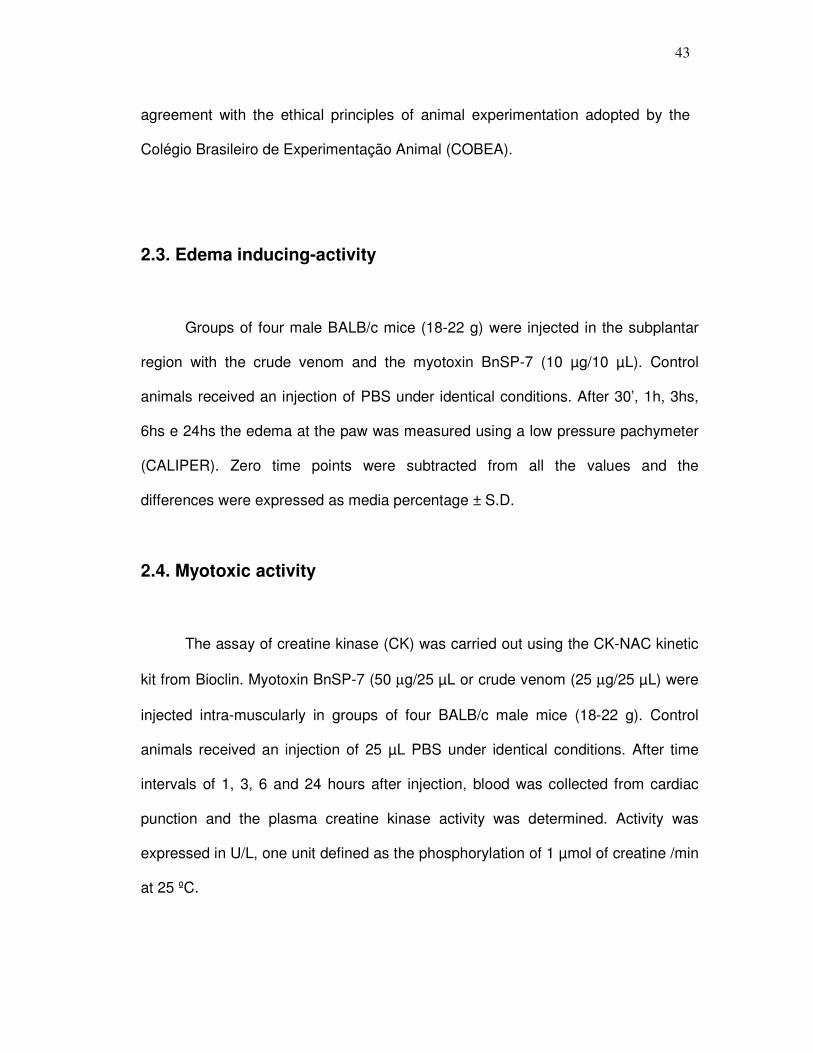
43
agreement with the ethical principles of animal experimentation adopted by the
Colégio Brasileiro de Experimentação Animal (COBEA).
2.3. Edema inducing-activity
Groups of four male BALB/c mice (18-22 g) were injected in the subplantar
region with the crude venom and the myotoxin BnSP-7 (10 µg/10 µL). Control
animals received an injection of PBS under identical conditions. After 30’, 1h, 3hs,
6hs e 24hs the edema at the paw was measured using a low pressure pachymeter
(CALIPER). Zero time points were subtracted from all the values and the
differences were expressed as media percentage ± S.D.
2.4. Myotoxic activity
The assay of creatine kinase (CK) was carried out using the CK-NAC kinetic
kit from Bioclin. Myotoxin BnSP-7 (50 µg/25 µL or crude venom (25 µg/25 µL) were
injected intra-muscularly in groups of four BALB/c male mice (18-22 g). Control
animals received an injection of 25 µL PBS under identical conditions. After time
intervals of 1, 3, 6 and 24 hours after injection, blood was collected from cardiac
punction and the plasma creatine kinase activity was determined. Activity was
expressed in U/L, one unit defined as the phosphorylation of 1 µmol of creatine /min
at 25 ºC.

44
2.5. Histological Analysis
Myotoxic activity was also evaluated on the basis of morphologic alterations
induced by i.m. injections of the myotoxin BnSP-7 (50 µg/25 µL) or crude venom
(25 µg/25 µL) in the left gastrocnemius skeletal muscle of BALB/c male mice (18-
22 g, n = 3). Control animals received an injection of 25 µL PBS under identical
conditions. After time intervals of 1, 3, 6, 24 and 72 hours, 1 and 2 weeks of
injection the animals were sacrificed in CO2 chamber and a small section of the
central region of the muscle was excised. The material was then soaked in fixing
solution (10% formaldehyde in PBS, v/v) and dehydrated by increasing
concentrations of ethanol and processed for inclusion in paraffin. The resulting
blocks were sliced in 6.0 µm thick sections, stained with 0.25% (w/v) hematoxin-
eosin (HE) or Masson and examined under a light microscope.
2.6. RNA extraction
Total RNA was extracted from gastrocnemius muscle by Tri-Reagent
(Sigma) method following the manufacture’s instructions, after i.m. injection of 50
µg of BnSP-7 or 50 µL of PBS at different time intervals (1, 3, 6, 24, 72 hours and
1 and 2 weeks). RNA extraction was carried out in an RNAse-free environment.
RNA was quantified by reading the absorbance at 260 nm according to the
method described by Sambrook et al. (1989).
45
2.7. Semi-quantitative RT-PCR analysis:
These experiments were accomplished according to Baldo, C. (2004) with
modifications. The reverse transcription of 1 µg RNA was carried out using M-MLV
RT reverse transcriptase (2 U/µL) (Invitrogen), oligo (dT)15, primers (6.3 pmol/ µL),
dNTP (0.2 mM), MgCl2 (2.5 mM) and 0.5 U RNAseOUT Ribonuclease inhibitor
(Recombinant).
For the polymerase chain reaction (PCR), the cDNA (2 µL) obtained was
incubated with 0.05 U/µL Platinum® Taq DNA Polymerase (Invitrogen), 0.5 mol/µL
of 3’ and 5’ primers, 0.2 mM dNTP, 200 mM, 10x Tris-HCl buffer and 2 mM MgCl2.
The RT-PCR conditions for the MMP-9 were: 37°C for 1 hour (RT-reaction),
95°C for 5 min (hot start) followed by 36 cycles at 94°C for 40 sec (denaturing),
58°C for 40 sec (annealing temperature), and 72°C for 50 sec (extension). PCR
conditions for the MMP-2 were: 37°C for 1 hour (RT-reaction), 95°C for 5 min (hot
start) followed by 38 cycles at 94°C for 40 sec (denaturing), 60°C for 40 sec
(anneling temperature), and 72°C for 50 sec (extension). PCR conditions for the β
actin were: 37°C for 1 hour (RT-reaction), 95°C for 5 min (hot start) followed by 28
cycles at 94°C for 40 sec (denaturing), 58°C for 40 sec (anneling temperature),
and 72°C for 50 sec (extension). Semi quantitative RT-PCR was established by
terminating reactions at intervals of 28, 30, 33, 36, 38 and 40 cycles for each
primer pair to ensure that the PCR amplicons were within the linear portion of the
amplification curve. PCR products were detected on 2% agarose electrophoresis
using as nucleotide size markers the 100 bp Ladder (Invitrogen). The primers used
were β-Actin (forward 3’ AACCCTAAGGCCAACCGTGA; reverse 5’
ATGCCACAGGATTCCATACC; 490 bp), MMP-2 (forward 3’
46
GTTGGCAGTGCAATACCTGA; reverse 5’ GGTATCCATCTCCATGCTCC; 404
bp) and MMP-9 (forward 3’ CATCGAACTTCGACACTGAC; reverse 5’
GCCACGACCATACAGATACT; 212 bp). The primer for the protein β-actina was
positive control of the reaction since this protein is expressed constitutively by the
cells.
The relative abundance of the genes was obtained by the reason among
the reading of optical densitometry of genes MMP-9 or MMP-2 for the constitutive
gene.
2.8. Peritoneal cells and culture
The MPAC (murine peritoneal adherent cells) experiments were
accomplished according to Clissa et al. (2001). The production of peritoneal
exudates cells from 18 to 22 g male BALB/c mice was elicited by intraperitoneal
injection of 1 mL Brewer's thioglycollate medium (Difco Laboratories). Five days
later, the cells were collected from the mouse peritoneal cavity in 5 mL of complete
RPMI medium (Gibco) containing 2 IU/mL of Heparin (Roche). The pooled cells
were then washed twice with phosphate-buffered saline (PBS) and then
resuspended in complete RPMI containing 5% heat-inactivated Fetal Calf Serum
(FCS) (Bio-Whittaker). The cells were cultured in 24 well (5 x 10⁵ cells/well) cell
culture plates, incubated at 37ºC in the presence of CO2 for 2 h. After incubation,
they were washed three times with PBS, to remove non-adherent cells, and RPMI
containing 5% FCS was added to each well. Approximately 95% of the remaining
adherent cells were macrophages as determined by morphology.
47
Viable cells were quantified by the presence in live cells of mitochondrial
succinyl dehydrogenase, by adding the enzyme substrate 3-(4,5-dimethylthiazol-2-
yl) 2.5 diphenyl-tetrazolium bromide (MTT) (Sigma) to the adhered cells. After 24
hours from stimulation with 2.5, 5.0, and 10.0 µg/mL of BnSP-7, BnSP-7-BPB or CV
culture supernatants were discarded and the cells remaining on the plate were
incubated with 200 µL of MTT (5 mg/mL in PBS) for 90 min at 37ºC. The plates
were then washed with PBS, and 200 µL/well of dimethyl sulfoxide (Sigma) were
added. One hour later, the optical density was determined at 490 nm. The
absorbance obtained using non-treated cells was used as 100% viability.
2.9. Cytokine measurement
For this assay MPAC were stimulated with 5 µg/ml of crude venom, BnSP-7
or BnSP-7-BPB in duplicate. After 2, 4, 6, 8 and 24 hours the supernatants were
collected and stored at -20º C for subsequent measurement by ELISA. Treatments
with PBS were used at zero time as control.
We also tested the induction of cytokine on gastrocnemius muscle. 10 µg/10
µl of crude venom, BnSP-7 or BnSP-7-BPB were injected in the left gastrocnemius
muscle of BALB-c male mice (18-22 g, n = 3). Control animals received an injection
of 10 µl PBS under identical conditions. After 2, 4, 6, 8 and 24 hours the animals
were sacrificed, the muscle was extracted, added to 200µl of PBS, perforated and
homogenized with a glass stem in the own tube eppendorf. Then, more 300µl of
PBS was added and samples were centrifuged 12000xg, 5 minutes
(Laborzentrifugem 2K 15-Sigma). The supernatants were collected and stored at -
20º C for subsequent measurement by ELISA.

48
Cytokine levels in the cells supernatants and gastrocnemius muscle
supernatants were measured by ELISA, according to the manufacturer's
instructions (R&D Systems). The monoclonal and polyclonal antibodies against IL-
1β, IL-6, TNF-α and KC (the mice cytokine corresponding to the human cytokine IL-
8) were from R&D Systems. Cytokine concentrations were calculated by
interpolation of the regression curve of known amounts of recombinant cytokines
(rIL-6, rIL-1β, rTNF- α and rKC from R&D System) and reported as ng/ml.
2.10. Statistical Analysis
The experiments accomplished in triplicate were analyzed by one way
ANOVA followed by the test of Dunnett, while experiments made in duplicate were
analyzed by one way ANOVA followed by the test of Tukey. Differences were
considered statistically significant at p<0.05. The results are expressed as the
average of the triplicate or duplicate plus the standard deviation.
3. Results
3.1. Edematogenic activity
There was a significant increase of mice paws inoculated with CV or BnSP-
7, evidencing the presence of edema (Table I). The maximum edema percentage

49
obtained by the CV was of 79%, 1 hour after inoculation, while the myotoxin
BnSP-7 presented a maximum increase after 30 minutes (77%).
3.2. Myotoxic activity
Table I also shows a significant increase in the release of creatine kinase
induced by BnSP-7 when compared to CV and PBS control. The maximum of
release was observed 3 hours after inoculation of CV and BnSP-7, corresponding to
515.4 and 4,147.5 U/L, respectively. The activity of CK for control group was of
approximately 47 U/L.
3.3. Histological analysis
Figures 1 and 2 show histological sections of mice gastrocnemius muscle
extracted 1, 3, 6, 24, 72 hours, 1 and 2 weeks after the inoculation with PBS, 25
µg CV or 50 µg BnSP-7.
In comparison with control PBS, the muscles inoculated with CV presented
swollen and with great extravasation of erythrocytes, characterizing a hemorrhagic
effect. After 3 hours, it was observed an inflammatory infiltrate, composed mainly
of neutrophils that increased progressively up to 72 hours. The tissue necrosis
was evidenced mainly between 24 and 72 hours. Myocytes in regeneration were
observed after one week of CV inoculation. In this period, it can be noticed a
decrease of edema, hemorrhage, necrosis and leukocyte infiltrate, which became
composed mainly by macrophages. The muscle cells in regeneration showed a
reduced size with large and central nuclei, with prominent nucleolus. The process

50
of repair of the muscle fibers is followed by formation of fibrous connective tissue,
evidenced by the presence of granulation tissue in slides stained with Masson
(Figures 1G, 1H).
In comparison with the control PBS, in the first 3 hours after the inoculation
of myotoxin BnSP-7 the muscle presented swollen (spaced fibers), with a lot of
necrotic cells and discreet inflammatory infiltrate constituted predominantly by
neutrophils. The edema decreased progressively until 6 hours, while the
inflammatory exudate increased progressively up to 72 hours, being located
mainly in the endomisium. After 24 hours, the number of necrotic cells decreased
(Figures 2E, 2F, 2G). From 72 hours to 2 weeks the inflammatory infiltrate became
multifocal, being characterized practically by macrophages and the edema and
necrosis have disappeared. In that period, were visualized myocytes in
regeneration and the presence of granulation tissue (Masson-Figure 2E),
composed of inflammatory cells, fibroblasts and newly formed and forming
vessels.
3.4. Expression of MMPs
The RT-PCR reactions showed that MMP-9 expression was gradually
increased from 72 hours to one week and started to decrease two weeks after
inoculation of BnSP-7 in the gastrocnemius muscle, reaching the highest level of
expression at one week. No significant levels of MMP-9 induced by PBS were
detected (Figura 3B). MMP-2 expression was verified in all times tested, with a
decrease at 3, 6 and 24 hours after stimulus by both BnSP-7 and PBS (Figura 3C).
51
3.5. Effect on cytokines
Exposure of MPAC and gastrocnemius muscle to toxins took to an increase
of all cytokines tested, however the release of TNF-α wasn’t significative (results not
showed).
MPAC levels of IL-6 reached peak 8 hours after stimuli by BnSP-7, while for
muscle the peak was at 2 hours. BnSP-7-BPB decreased successfully IL-6 levels
when compared to BnSP-7 in both experimental models (Figures 4A and 5A,
respectively). MPAC levels of IL-8 reached peak 6 hours after stimuli by BnSP-7,
while for muscle the peak was at 2 hours. BnSP-7-BPB also decreased IL-8 levels
when compared to BnSP-7 (Figures 4B and 5B, respectively). MPAC levels of IL-1β
reached peak 6 hours after stimuli by BnSP-7, while for muscle the peak was at 4
hours. BnSP-7-BPB wasn’t so successful to diminish levels of IL-1β when
compared to BnSP-7 since it was observed at only one time interval of each
experiment (Figures 4C and 5C, respectively). BnSP-7 induced higher cytokines
release in MPAC while in gastrocnemius muscle the CV was able to induce higher
levels of all cytokines.
4. Discussion
Bothropics myotoxins induce serious muscle damage soon after the
intramuscular injection, characterized mainly by edema, inflammation and
mionecrose. (Gutiérrez et al., 1989; Nishioka et al., 1992; Gutiérrez & Rucavado,

52
2000). Thus, the pathogenesis of these effects is being very studied through the
morphologic and biochemical alterations in the affected muscle (Gutiérrez &
Lomonte, 1995; Rodrigues et al., 2001; Harris, 2003; Rucavado et al., 2002;
Rodrigues et al., 2007).
In this work it was verified that both crude venom and myotoxin BnSP-7
were able to induce significant increase of mice paw, characterizing the edema.
For Zuliani et al. (2004) and Landucci et al. (1998) fosfolipases A2 increase the
microvascular permeability and the blood flow in mice and rat paw causing the
edema. The edema formation can also be broadly linked to mast cells activation
(Cirino et al., 1989; Landucci et al., 1998). Mast cells are inflammatory cells
located close to blood vessels, nerves, epithelium and smooth muscles and they
express and release type II fosfolipases A2, that have the function of modulating
it’s degranulation (Zuliani et al., 2004). The discovery that mast cells activation by
bothropstoxin-I, a fosfolipase A2 Lys-49 isolated from Bothrops jararacussu snake
venom (Homsi-Brandeburgo et al., 1988), was inhibited by heparin suggests that
the positive charge of this toxin is the major responsible factor for its
pharmacological properties (Landucci et al., 1998). Other studies showed that the
initial edema (30 minutes) is inhibited by pretreatment with ciproheptadin,
indicating the participation of histamine or serotonin in the process, while the late
edema (5 hours) is inhibited with pretreatment with aspirin, suggesting the
prostaglandins participation (Gutiérrez et al., 1986). Besides, Landucci et al.
(1998) suggest that phospholipids hydrolysis by phospholipases A2 is not essential
for edema formation, in other words, the phospholipases may have a
pharmacological domain independent from catalytic site (Kini & Evans, 1989; Kini,
2003), explaining then the role of BnSP-7 Lys-49 in the induction of this effect.

53
The muscle damage showed that first intervals of time after inoculation of
crude venom or BnSP-7 were characterized by edema, necrosis and inflammation
and, just for CV, hemorrhage. This bleeding is due to the action of hemorrhagic
toxins, like metalloproteinases, present in the crude venom (Gutiérrez et al., 1995)
that cause lesions on the walls of the small blood vessels (Oshaka et al., 1979;
Ownby et al., 1990). The mechanism for which hemorrhagic metalloproteinases
causes systemic or local tissue damage is due to their proteolytic action in proteins
of the basal membrane (Bjarnason and Fox, 1994; Escalante et al., 2006). This
action weakens the mechanical stability and hemodynamic biophysical forces that
usually operate in the circulation, provoking a distention in the capillary wall until the
integrity is broken and extravasation occurs in an explosive way, leading to tissue
hypoxia and, consequently, to cellular death (Gutiérrez and Rucavado, 2000;
Gutiérrez et al., 2005; Gutiérrez et al., 2006). So, it is suggested that this distention
and abrupt rupture result in the loss of the integrity not only of endothelial cells, but
also of the components of the basal membrane (Moreira et al., 1994; Gutiérrez et
al., 2006). Bleeding injuries can also be provoked by the inhibitory effect on platelet
aggregation by SVMPs (Bjarnason and Fox, 1994, Kamiguti, et al., 2003). Besides,
SVMPs induce the separation of endothelial cells from extracellular matrix proteins
and its apoptosis (Tanjoni et al., 2005; Diaz et al., 2005; You et al., 2003).
The mionecrosis caused by CV and BnSP-7 was assayed by levels of
creatine kinase in the serum of mice injected with these toxins. The enzyme
creatine kinase is found in high concentrations in the heart, skeletal muscles and
in the brain. Thus, when the muscle tissue is damaged in some way the levels of
CK in the blood increase. The myotoxin BnSP-7 induced a larger release of the
enzyme creatine kinase than crude venom.
54
The myotoxicity induced by snake venoms can be caused by the indirect
action of hemorrhagins, that cause injury to blood vessels leading to ischemia and,
consequently, to cellular death; or it can be caused by the direct action of
phospholipases A2 by rupture of plasma membranes leading to alteration on
permeability and this way they provoke cellular damage. The mionecrosis induced
by CV and BnSP-7 begins 1 hour after the inoculation. This effect observed in the
treated muscles with CV can also be due to the direct action of PLA2s Lys49 on
plasma membrane. It is believed that it’s action in the muscle plasma membrane is
due to areas of its structure that recognize acceptor sites negatively charged, both
for low as high affinity in the muscle membrane, and to areas that interact and
penetrate in the lipid bilayer promoting destabilization of the membrane through an
independent mechanism of the catalytic action, practically absent in those
proteins. This action results in an influx of calcium, that begins a variety of
degenerative mechanisms as alterations in the cytoskeleton, mitochondrial
damages, proteases and calcium-dependent phospholipases activation and
hypercontraction of myofibers, that consequently cause more cellular damages,
taking to necrosis of muscular fibers (Harris, 2003; Gutiérrez and Ownby, 2003).
The use of synthetic peptides evidenced the C-terminal region (115-129) as
responsible for the toxic activities in some myotoxic PLA2 Lys49 (Núñez et al.,
2001; Lomonte et al., 2003a). This region combines cationic and hydrophobic
amino acids responsible for the cellular membranes damage mechanism. The
cationic residues would interact electrostatically with anionic groups of an aceptor
site possibly membrane phospholipids negatively loaded (Lomonte et al., 2003b),
while the hydrophobic residues, especially the aromatic ones, would interact with
and possibly penetrate the phospholipidic bilayer, resulting in its destabilisation

55
(Núñez et al., 2001). However, the fact of a single peptide plays all the main toxic
activities of the origin molecule, does not prevent the existence of other motify that
can participate or complement the action of toxic effector site (Lomonte et al.,
2003(b)). In contrast, C-terminal peptides of some myotoxic PLA2 Asp49 showed
no activity of membrane direct damage, suggesting that the toxic mechanism
exercised by these proteins, that probably involves its catalytic activity as an
important step, is different from that used by myotoxic PLA2 Lys49 (Núñez et al.,
2001).
The results obtained by histological analyses confirm the high levels of CK
liberated by gastrocnemius muscle. Soares et al. (2000) also verified an increase
in CK plasma levels induced by BnSP-7. Rodrigues et al. (1998) showed necrosis
of gastrocnemius muscle 24 hours after inoculation of BnSP-7, while BnSP-7
modified with BPB did not produced any visible necrosis.
Our histological analysis, as well as in other studies (Gutiérrez et al., 1991;
Gutiérrez & Lomonte, 1989; Harris, 1991, Harris, 2003), also showed that
mionecrosis is followed by the invasion of inflammatory infiltrate in abundance and
by regeneration in the affected muscle.
The efficiency of local circulation is essential for complete muscle
regeneration, since it allows the arrival in the damaged region of macrophages
that phagocyte necrotic cells, as well as nutrients and O2 to supply the intense
metabolic activity of the regeneration phase (Harris, 2003). Since CV caused
intense hemorrhage, as shown previously, the reinervation and the regeneration of
the blood vessels in the muscle are essential for the regeneration process
(Vignaud et al., 2005; Harris, 2003). Besides, in this slower process of
regeneration it is common the emergence of many fibroblasts and fibrosis

56
(Gutiérrez & Lomonte, 1995). This dense organization of ECM difficults further cell
migration and regeneration, thus resulting in a terminal residual fibrotic state within
the muscle (Bedair et al., 2007).
The myotoxin BnSP-7 did not cause damage to blood vessels, nerves or
basal lamina, witch stays intact in the periphery of the muscle cells, therefore
favoring a fast and efficient regenerative process also evidenced in this work. The
muscle tissue inoculated with CV or the myotoxin BnSP-7 has the capacity of
regeneration of its fibers after 72 hours before the inoculation of these samples. It
is possible that the venom and toxins themselves begin a vigorous regenerative
process of the muscular fibers due to their chemotactic properties, recruiting
macrophages and activating satellites cells (Harris, 2003). Although the
morphological structure of muscle injected with BnSP-7 or CV showed in
regeneration, its function was not verified in the present work. Vignaud et al.
(2005) verified that some slow-twitch muscle contractile properties were not fully
recovered even by 12 weeks after treatment by snake venom Notechis scutatus
scutatus.
The satellites cells, precursory of the regenerate muscle cells, are located
between the plasma membrane and the basal lamina of muscle fibers and they
are surrounded by ECM components including laminins, fibronectin and type-IV
collagen. These cells are very resistant to aggressions of several different natures,
remaining intact to the attack, for instance of phospholipases A2, and they are
activated and enter into division during the degenerative processes (Harris, 2003).
Being activated they are known to express MMPs, especially gelatinases witch
target collagen-IV and laminins. An elevated level of MMPs production is thought
to result in cell migration into injured areas with subsequent differentiation and

57
fusion of satellite cells into myofibers, characterizing the regeneration (Kherif et al.,
1999, Fahime et al., 2000). Our data showed an increase in expression of MMP-9
from 72 hours to two weeks after inoculation of BnSP-7 demonstrating that MMP-9
may have a role on regeneration process, since we see regenerating muscle fibers
at these same times in our histological analyses.
Kherif et al. (1999) demonstrated by in situ hybridization that MMP-9 mRNA
is produced by inflammatory cells, predominantly polymorphonuclear leukocytes,
and by putative activated satellite cells. Besides other cells usually found in
histological studies of injured muscles like macrophages and mast cells are know
to store and produce MMP-9 in response to different stimuli from necrotic tissue,
representing one important source of this MMP.
We also observed expression of MMP-2 in our experiments. Many types of
cells are able to produce MMP-2 such as skeletal muscle, endothelial and
fibroblast cells (Guérin and Holland, 1995). The decrease at 3, 6 and 24 hours
after stimulus by both BnSP-7 and PBS is being investigated. Both PBS and
BnSP-7 expressed MMP-2 in all times tested. Kherif et al. (1999) hypothesize that
a constant MMP-2 activation may intervene during muscle regeneration.
Although the expression of MMPs was evidenced during tissue
degeneration and regeneration process induced by BnSP-7, the respective active
or latent forms were not detected in the present study. Kherif et al. (1999) showed
that skeletal muscle produce both MMP-2 and MMP-9 gelatinases as latent and/or
active forms, but with differential patterns related to the time course of muscle
necrosis and regeneration. The same way, Rucavado et al. (2002) found by
zymography and a specific enzymatic assay that MMPs in muscle homogenates
corresponded predominantly to the latent forms, therefore lacking activity.

58
The MMPs can also contribute to inflammatory processes regulating
physical barriers, modulating inflammatory mediators such as cytokines and
chemokines, and establishing chemokine gradients in inflamed tissues that
regulate the movement of leukocytes at sites of infection or injury. MMPs can
either promote or repress inflammation by the direct proteolytic processing of
inflammatory cytokines and chemokines to activate, inactivate or antagonize
chemokine function (Manicone and McGuire, 2008). Thus, our findings of
increased levels of IL-1β, IL-6 and IL-8 on muscle homogenate and MPAC
suggest an involvement of these cytokines with MMPs and regeneration. MMP-2
and MMP-9 can activate the IL-1β precursor to the active form (Schonbeck et al.,
1998) leading then to stimulation of macrophages essential for regenerating
process, once they remove the necrotic cells allowing occupation of new
myofibers. Interestingly, IL-1β up-regulates MMP-9 (Brenner et al., 1989). MMP-9
process IL-8 in a product that have more potent chemoattractant activities (Van
den Steen et al., 2000) promoting then activation of neutrophils. In addition these
cytokines also induce the expression of MMPs (Sarén et al., 1996).
Raised levels of pro-inflammatory cytokines have been detected in the
serum of human victims or in experimental models following Bothrops envenoming
(Barravieira et al., 1995; Barros et al., 1998; Cardoso et al., 2001; Clissa et al.,
2001; Carneiro et al., 2002; Rucavado et al., 2002; Cruz et al., 2005; Chaves et al,
2005; Fernandes et al., 2006; Zuliani et al., 2004; Chacur et al., 2004; Zamuner et
al., 2005). Our present findings showed an increase of IL-6, IL-8, IL-1β for both
experimental models, MPAC and gastrocnemius muscle, induced by toxins and
crude venom, although these values were higher for gastrocnemius muscle. These

59
cytokines are important in the regeneration processes since they activate many
inflammatory cells.
The modification of BnSP-7 with BPB decreased significantly the release of
all cytokines in almost all times tested for both MPAC and muscle. These results
reaffirm the conclusion made by Soares et al. (2000) that the alkylation of BnSP-7
His 48 with BPB leads to a reduction of pharmacological effects, suggesting that
this modification induces conformational changes in molecule regions different
from the catalytic site, since Lys49 PLA2 lack enzymatic activity.
We conclude that both experimental models used in this work succeed on
showing the profile of pro-inflammatory cytokine release of all toxins tested. We
have demonstrated that BnSP-7 induces an increase in edema, release of CK,
cytokines (IL-6, IL-8, IL-1-1β) and expression of MMP-2 and MMP-9. This way, this
myotoxin can be acting as pro-inflammatory stimulus, inducing cytokine production
and liberation from the inflammatory and satellite cells, which present important
role in the inflammatory process and, consequently, in the evolution of local tissue
damage and regeneration. Future studies using antibodies anti-cytokine or anti-
inflammatories are of extreme importance for the understanding of inflammatory
mechanisms induced by this toxin. The MMPs must also be more studied since
they seem to play an important role on cell migration and regeneration process. In
addition, studies of the pathogenesis of local tissue damage on envenomation can
take to a better understanding of the mechanisms that lead to tissue necrosis and
later regeneration, what could inspire the creation of new therapeutic methods.
Thus, the understanding of the mechanism of action of snake venom toxins can
reveal mechanisms of cellular and tissue injury that are common to other
60
pathological conditions, like degenerative diseases (Duchennes’s muscular
dystrophy), allowing the development of alternative therapies
5. Acknowledgements
The authors wish to thank Prof. Dr. Luiz Ricardo Goulart Filho for technical
assistance and FAPEMIG, CNPq and FAPESP of Brazil for financial suport. This
work was performed as partial requirement for the MsC degree of Carolina de
Freitas Oliveira at the Universidade Federal de Uberlândia.
6. References
Baldo, C., 2004. Lesão tecidual e inflamação induzidas pela Neuwiedase: Uma
metaloprotease isolada da peçonha da serpente Bothrops neuwiedi pauloensis.
(manuscript in preparation).
Barravieira, B., Lomonte, B., Tarkowski, A., Hanson, l.A., Maira, D.A., 1995.
Acute-phase reactions, including cytokines, in patients bitten by Bothrops and
Crotalus snakes in Brazil. J. Venom. Anim. Toxins. 1, 11-22.

61
Barros, S.F., Friedlanskaia, I., Petricevich, V.L., Kipnis, T.L., 1998. Local
inflammation, lethality and cytokine release in mice injected with Bothrops atrox
venom. Mediators of Inflammation. 7, 339-346.
Bedair, H., Liu, T.T., Kaar, J.L., Badlani, S., Russell, A.J., Li, Y., Huard, J., 2007.
Matrix metalloproteinase-1 therapy improves muscle healing. J Appl Physiol. 102,
2338-2345.
Bjarnason, J.B., Fox, J.W., 1994. Hemorrhagic metalloproteinase from snake
venoms. Pharmac. Ther. 62, 325-372.
Bradford, M.M., 1976. A rapid and sensitive method for the quantitation of
microgram quantities of protein utilizing the principle of protein-dye binding. Anal.
Biochemistr. 72, 248-254.
Brenner, D.A., O’Hara, M., Angel, P., Chojjkier, M., Karin, M. L., 1989. Prolonged
activation of jun and collagenase genes by tumour necrosis factor-α. Nature. 337,
661-663.
Cardoso, D.F., Lopes-Ferreira, M., Faquim-Mauro, E., Macedo, M. S., Farsky,
S.H.P., 2001. Role of crotoxin, a phospholipase A2 isolated from Crotalus durissus
terrificus snake venom, on inflammatory and immune reactions. Mediators of
Inflammation. 10, 125-133.

62
Carneiro, A.S., Ribeiro, O.G., Franco, M., Cabrera, W.H.K., Vorraro, F., Siqueira,
M., Ibañez, O.M., Starobinas, N., 2002. Local inflammatory reaction induced by
Bothrops jararaca venom differs in mice selected for acute inflammatory response.
Toxicon. 40, 1571-1579.
Chacur, M., Milligan, E.D., Sloan, E.M., Wieseler-frank, J., Barrientos, R.M.,
Martin, D., Poole, S., Lomonte, B., Gutiérrez, J. M., Maier, S. F., Cury, Y., Watkins,
L. R., 2004. Snake venom phospholipase A2 (Asp49 and Lys49) induce
mechanical allodynia upon peri-sciatic administration: involviment of spinal cord
glia, proinflammatory cytokines and nitric oxide. Pain. 108, 180-191.
Chaves, F., Teixeira, C.F.P., Gutiérrez, J.M., 2005. Role of TNF-α, IL-1β and IL-6
in the local tissue damage induced by Bothrops asper snake venom: an
experimental assessment in mice. Toxicon. 45, 171-178.
Cirino, G., Peers, S.H., Wallace, J.L., Flower, R.J., 1989 A study of phospholipase
A2 induced oedema in rat paw. Eur. J. Pharmacol. 166, 505-510.
Clissa, P.B., Laing, G.D., David, R., Theakston, G., Mota, I., Taylor, M. J., Moura-
Da-Silva, A.M., 2001. The effect of jararhagin, a metalloproteinase from Bothrops
jararaca venom, on pro-inflammatory cytokines released by murine peritoneal
adherent cells. Toxicon. 39, 1567-1573.

63
Cruz, A.H., Mendonça, R.Z., Petricevich, V.L., 2005. Crotalus durissus terrificus
venom interfers with morphological, functional, and biochemical changes in murine
macrophage. Mediators of Inflammation. 6, 349-359.
Díaz, C., Valverde, L., Brenes, O., Rucavado, A., Gutiérrez, J.M., 2005.
Characterization of events associated with apoptosis/anoikis induced by snake
venom metalloproteinase BaP1 on human endothelial cells. J. Cell. Biochem. 94,
520-528.
Escalante, T., Shannon, J., Moura-Da-Silva, A.M., Gutiérrez, J.M., Fox, J.W.,
2006. Novel insight sinto capillary vessel basement membrane damage by snake
venom hemorrhagic metalloproteinases: A biochemical and immunohistochemical
study. Arch. Biochem. Biophys. 455, 144-153.
Fashime, E. EL., Torrente, Y., Caron, N.J., Bresolin, M.D., Tremblay, J.P., 2000. In
vivo migration of transplanted myoblasts requires matrix metalloproteinase activity.
Exp Cell Res. 258, 279-287.
Fernandes, C.M., Zamuner, S.R., Zuliani, J.P., Rucavado, A., Gutiérrez, J.M.,
Teixeira, C.F.P., 2006. Inflammatory effects of BaP1 a metalloproteinase isolated
from Bothrops asper snake venom: Leukocyte recruitment and release of
cytokines. Toxicon. 47, 549-559.
Gasche, Y., Fujimura, M., Morita-Fujimura, Y., Copin, J.C., Kawase, M.,
Massengale, J., Chan, P.H., 1999. Early appearance of activated matrix

64
metalloproteinase-9 after focal cerebral ischemia in mice: a possible role in blood-
brain barrier dysfunction. J. Cerebral Blood Flow Metab. 19, 1020-1028.
Guérin, C.W., Holland, P.C., 1995. Synthesis and secretion of matrix-degrading
metaloproteases by human skeletal muscle satellite cells. Dev. Dyn. 202, 91-99.
Gutiérrez, J.M., Lomonte, B., Chaves, F., Moreno, E., Cerdas, L., 1986.
Pharmacological activities of a toxic phospholipase A2 isolated from the venom of
the snake Bothrops asper. Comp. Biochem. Physiol. 84, 159-164.
Gutiérrez, J.M., Chaves, F., Gene, J.A., Lomonte, B., Camacho, Z., Schosinsky,
K., 1989. Myonecrosis induced in mice by basic myotoxin isolated from the venom
of snake Bothrops nummifer (jumping viper) from Costa Rica. Toxicon. 277, 35-
45.
Gutiérrez, J.M., Lomonte B., 1989. Local tissue damage induced by Bothrops
snake venoms. A review. Mem. Inst. Butantan. 51, 211-223.
Gutiérrez, J.M., Núnez, J., Díaz, C., Cintra, A.C.O., Homsi-Brandeburgo, M.I.,
Giglio, J.R., 1991. Skeletal muscle degeneration and regeneration after injection
of bothropstoxin-II, a phospholipase A2 isolated from the venom of the snake
Bothrops jararacussu. Exp. Mol. Pathol. 55, 217-229.
Gutiérrez, J.M., Lomonte, B., 1995. Local pathological effects induced by
Bothrops snake venom, Mem. Inst. Butantan. 33, 1405-1474.
65
Gutiérrez, J.M., Romero, M., Núñez, J., Chaves, F., Borkow, G., Ovadia, M., 1995.
Skeletal muscle necrosis and regeneration after injection of BaH1, a hemorrhagic
metalloproteinase isolated from the venom of the snake Bothrops asper
(Terciopelo). Exp Mol Pathol. 62, 28-41.
Gutiérrez, J.M., Rucavado, A., 2000. Snake venom metalloproteinases: Their role
the pathogenesis of local tissue damage. Biochemie. 82, 841-850.
Gutiérrez, J.M., Ownby, C.L., 2003. Skeletal muscle degeneration induced by
venom phospholipases A2: insights into the mechanisms of local and systemic
myotoxicity. Toxicon. 42, 915-931.
Gutiérrez, J.M., Rucavado, A., Escalante, T., Díaz, C., 2005. Hemorrhage induced
by snake venom metalloproteinases: biochemical and biophysical mechanisms
involved in microvessel damage. Toxicon. 45(8), 997-1011.
Gutiérrez, J.M., Núñez, J., Escalante, T., Rucavado, A., 2006. Blood flow is
required for rapid endothelial cell damage induced by a snake venom
hemorrhagic metalloproteinase. Microvasc Res. 71, 55-63.
Harris, J.B., 2003. Myotoxic phospholipases A2 and regeneration of skeletal
muscles. Toxicon. 42, 933-945.
Harris, J.B., 1991. Phospholipases in snake venoms and their effects on nerve
and muscle. Pharmacol Ther. 31, 79-102.
66
Homsi-Brandeburgo, M.I., Queiroz, L.S., Santo-Neto, H., Simioni, L.R., Giglio,
J.R., 1988. Fractionation of Bothrops jararacussu snake venom: Partial chemical
characterization and biological activity of bothropstoxin. Toxicon. 26, 615-627.
Kamiguti, A.S., Gallagher, P., Marcinkiewicz, C., Theakston, R.D., Zuzel, M.,
2003. Identification of sites in the cysteine-rich domain of the class P-III snake
venom metalloproteinases responsible for inhibition of platelet function. FEBS
Lett. 549, 129-134.
Kherif, S., Lafuma, C., Dehaupas, M., Lachkar, S., Fournier, J.G., Verdiere-
Sahuqué, M., Fardeau, M., Alameddine, H.S., 1999. Expresion of matrix
metalloproteinases 2 and 9 in regenerating skeletal muscle: a study in
experimentally injured and mdx muscles. Dev Biol. 205, 158-170.
Kini, R.M., Evans, H.J., 1989. A model to explain the pharmacological effects of
snake venom phospholipases A2. Toxicon. 27, 613-635.
Kini, R.M., 2003. Excitement ahead: structure, function and mechanism of action
of snake venom phospholipase A2 enzymes. Toxicon. 24, 827-840.
Landucci, E.C.T., Castro, R.C., Pereira, M,F., Cintra, A .C.O., Giglio, J.R.,
Marangoni, S., Oliveira, B., Cirino, G., Antunes, E., De Nucci, G., 1998. Mast cell
degranulation induced by two phospholipase A2 homologues: dissociation
between enzymatic and biological activities. Eur. J. Pharmacol. 343, 257-263.

67
Leib, S.L., Leppert, D., Clements, J., Tauber, M.G., 2000. Matrix
metalloproteinases contribute to brain damage in experimental pneumococcal
meningitis. Infect Immun. v. 68 p. 615-620.
Lomonte, B., Angulo, Y., Calderón, L., 2003a. Na overview of lysine-49
phospholipase A2 myotoxins from croalid snake venoms and their structural
determinants of myotoxic action. Toxicon. 42, 885-901.
Lomonte, B., Angulo, Y., Santamaría, C., 2003b. Comparative study of synthetic
peptides corresponding to region 115-129 in Lys49 myotoxic phospholipases A2
from snake venoms. Toxicon. 42, 307-312.
Manicone, A.M., McGuire, J.K., 2008. Matrix metalloproteinases as modulators of
inflammation. Semin Cel Devl Biol. 19, 34-41.
Moreira, L., Borkow, G., Ovadia, M., Gutiérrez, J.M., 1994. Pathological changes
induced by BaH1, a hemorrhagic proteinase isolated from Bothrops asper
(Terciopelo) snake venom, on mouse capillary blood vessels. Toxicon. 32, 976-
87.
Nagase, H., Woessner, J.F., 1999. Matrix metalloproteinases. J. Biol. Chem. 274,
21491-21494.

68
Nishioka, S.A., Silvera, P.V.P., 1992. A clinical and epidemiologic study of 292
cases od lance-headed viper bite in a Brazilian teaching hospital. Am. J. Trop.
Med. Hyg. 47, 805-810.
Núñez, C.E., Angulo, Y., Lomonte, B., 2001. Identification of the myotoxic site of
the Lys49 phospholipase A2 from Agkistrodon piscivorus snake venom: synthetic
C-terminal peptides from Lys49, but not from Asp49 myotoxins, exert membrane-
damaging activities. Toxicon. 39, 1587-1594.
Oshaka, A., 1979. Hemorrhagic, necrotizing and edema-forming effects of snake
venoms. Hand. Exp. Pharmac. 52, 480-546.
Ownby, C.L., Nikai, T., Imai, K., Sugihara, H., 1990. Patoghenesis of
haemorrhage induced by bilitoxin, a hemorrhagic toxin isolated from the venom of
the comon cantil (Agkistrondon bilineatus bilineatus). Toxicon. 23, 837-846.
Rodrigues, R.S., Izidoro, L.F., Teixeira, S.S., Silveira, L.B., Hamaguchi, A.,
Homsi-Brandeburgo, M.I., Selistre-de-Araújo, H.S., Giglio, J.R., Fuly, A.L.,
Soares, A.M., Rodrigues, V.M. 2007. Isolation and functional characterization of a
new myotoxic acidic phospholipase A(2) from Bothrops pauloensis snake venom.
Toxicon. 50(1), 153-65.
Rodrigues, V.M., Soares, A.M., Mancin, A.C., Fontes, M.R.M., Homsi-
Brandeburgo, M.I., Giglio, J.R., 1998. Geographic variations in the composition of

69
myotoxins from Bothrops neuwied snake venoms: biochemical characterization
and biological activity. Comparative Biochemistry Physiology. 121, 215-222.
Rodrigues, V.M., Soares, A.M., Andrião-Escarso, S.H., Franceschi, A.M.,
Rucavado, A., Gutiérrez, J.M., Giglio, J.R., 2001. Pathological alterations induced
by neuwiedase, a metalloproteinase isolated from Bothrops neuwiedi snake
venom. Biochimie. 83(6), 471-479.
Rosenberg, G.A., Navratil, M., 1997. Metalloproteinase inhibition blocks edema in
intracerebral hemorrhage in the rat. Neurology. 48, 921-926.
Rucavado, A., Escalante, T., Teixeira, C.F.P., Fernándes, C.M., Díaz, C.,
Gutiérrez, J.M., 2002. Increments in cytokines and matrix metalloproteinases in
skeletal muscle after injection of tissue-damaging toxins from the venom of the
snake Bothrops asper. Mediators of Inflammation. 11, 121-128.
Sambrook, J., Fritsch, E.F., Maniatis, T., 1989. Molecular Cloning Laboratory
Manual 2.ed. USA: Cold Spring Harbor Laboratory Press.
Sarén, P., Welgus, H.G., Kovanen, P.T., 1996. TNF-α and IL-1β selectively induce
expression of 92-kDa gelatinase by human macrophages. J. Immunol. 157, 4159-
4165.

70
Schonbeck, U., Mach, F., Libby, P., 1998. Generation of biologically active IL-1
beta by matix metalloproteinases: a novel caspase-1-independent pathway of IL-1
beta processing. J Immunol. 161, 3340-3346.
Shapiro, S.D., 1998. Matrix metalloproteinase degradation of extracellular matrix:
biological consequences. Curr Opin Cell Biol. 10, 602-608.
Soares, A.M., Guerr-Sá, R., Borja-Oliveira, C.R., Rodrigues, V.M., Rodrigues-
Simioni, L., Rodrigues, V., Fontes, M.R.M., Lomonte, B., Gutiérrez, J.M., Giglio,
J.R. Structural and functional characterization of BnSP-7, a Lys49 myotoxic
phospholipase A2 homologue from Bothrops neuwiedi pauloensis venom. Arch
Biochem Biophys. 378, 201-209.
Tanjoni, I., Weinlich, R., Della-Casa, M.S., Clissa, P.B., Saldanha-Gama, R.F.,
Freitas, M.S., Barja-Fidalgo, C., Amarante-Mendes, G.P., Moura-DA-Silva, A.M.,
2005. Jararhagin, a snake venom toxin, induces a specialized form of apoptosis
(anoikis) selective to endothelial cells. Apoptosis. 10, 851-861.
Trengove, N.J., Stacey, M.C., Macauley, S., Bennet, N., Gibson, J., Burslem, F.,
Murphy, G., Schultz, G., 1999. Analysis of the acute and chronic wound
environments: the role of proteases and their inhibitors. Wound Rep Reg. 7, 442-
452.
Van Den Steen, P.E., Proost, P., Wuyts, A., Van Damme, J., Opdenakker, G.,
2000. Neutrophil gelatinase B potentiates interleukin-8 tenfold by aminoterminal

71
processing, whereas it degrades CTAP-III, PF-4, and GRO-alpha and leaves
RANTES and MCP-2 intact. Blood. 96, 2673-2681.
Vignaud, A., Hourdé, C., Torres, S., Caruelle, J.P., Martelly, I., Keller, A., Ferry,
A., 2005. Functional, cellular and molecular aspects of skeletal muscle recovery
after injury induced by snake venom form Notechis scutatus scutatus. Toxicon.
45, 789-801.
Voronov, E., Apte, R.N., Soler, S., 1999. The systemic inflammatory response
syndrome related to the release of cytokines following severe envenomation, J.
Venom. Anim. Toxins. 5, 5-33.
You, W.K., Sco, H.J., Chung, K.H., Kim, D.S., 2003. A novel metalloprotease from
Gloydius halys venom induces endothelial cell apoptosis through its protease and
disintegrin-like domains. J. Biochem. 134, 739-749.
Zamuner, S.R., Teixeira, C.F.P., 2002. Cell adhesion molecules involved in the
leukocyte recruitment induced by venom of the snake Bothrops jararaca.
Mediators of Inflammation. 11, 351-357.
Zamuner, S.R., Zulliani, J.P., Fernandes, C.M., Gutiérrez, J.M., Teixeira, C.F.P.,
2005. Inflammation induced by Bothrops asper venom: release of proinflammatory
cytokines and eicosanoids, and role of adhesion molecules in leukocyte
infiltration. Toxicon. 46, 806-813.

72
Zuliani, J.P., Fernandes, C.M., Zamuner, S.R., Gutiérrez, J.M., Teixeira, C.F.P.,
2004. Inflammatory events induced by Lys-49 and Asp-49 phospholipases A2
isolated from Bothrops asper snake venom: role of catalytic activity. Toxicon. 45,
335-346.

73
Figure Captions Figure 1: Photomicrography of fine cross cuts with 6,0 µm of thickness of left mice
gastrocnemius muscle, after 1, 3, 6, 24 and 72 hours, 1 and 2 weeks. The
alterations were induced by 25µg of crude venom (CV) of Bothrops pauloensis
dissolved in 50µl of PBS.
A: PBS. (200 x), B, C, D, E, F, H: CV � 1, 3, 6, 24 and 72 hours, respectively.
(400 x), G, H: CV � 1 and 2 weeks, respectively. (200x)
I = Inflammatory infiltrate, H = Hemorrhage, N = Necrosis, E = Edema, CN =
Central Nuclei, GT = Granulation Tissue, HE = Hematoxilin and Eosin, M =
Masson.
Figure 2: Photomicrography of fine cross cuts with 6,0 µm of thickness of left mice
gastrocnemius muscle, after 1, 3, 6, 24 and 72 hours, 1 and 2 weeks. The
alterations were induced by 50 µg of the myotoxin BnSP-7 isolated from Bothrops
pauloensis crude venom dissolved in 50µl of PBS.
A, B, C, D, F: BnSP-7 � 1, 3, 6, 24 hours and 2 weeks, respectively. (200 x), E,
G: BnSP-7 � 72 hours and 1 week, respectively (400 x).
I = Inflammatory infiltrate, N = Necrosis, E = Edema, CN = Central Nuclei, GT =
Granulation Tissue, HE = Hematoxilin and Eosin, M = Masson
Figure 3: Semi-quantitative expression of genes MMP-9 and MMP-2 in the
gastrocnemius muscle of mice BALB/c inoculated with 25 µg of BnSP-7. A-
Standard electrophoretic on agarose gel 2% of fragments obtained by RT-PCR. B-
Optical densitometry of gene MMP-9 obtained by RT-PCR. C- Optical densitometry
of gene MMP-2 obtained by RT-PCR. * The relative abundance of the genes was
obtained by the reason among the reading of optical densitometry of genes MMP-9
or MMP-2 for the constitutive gene (β-actin).
P1, P3, P6, P24, P72, P1w, P2w: Control animals injected with 25 µL of PBS, and
sacrificed after 1, 3, 6, 24 and 72 hours, 1 and 2 weeks respectively.
B1, B3, B6, B24, B72, B1w, B2w: Animals injected with 25 µg of BnSP-7, and
sacrificed after 1, 3, 6, 24 and 72 hours, 1 and 2 weeks respectively.
M: 100-pb ladder marker

74
Figure 4: Release of cytokines by MPAC. MPAC were stimulated with 5 µg/ml of
crude venom (CV), BnSP-7 or BnSP-7-BPB. After 2, 4, 6, 8 and 24 hours the
supernatants were collected and stored at -20º C for subsequent measurement by
ELISA: A = IL-6, B = IL-8, C = IL-1β. Treatments with PBS were used at zero time
as control. The experiments were accomplished in duplicate and analyzed
statistically by ANOVA followed by the test of Tukey. The symbols represents
significant difference (p<0.05) between: BnSP-7 and BPB (*), BnSP-7 and CV (#),
BPB and CV (O). Results are presented by the average ± SD.
Figure 5: Release of cytokines by gastrocnemius skeletal muscle. Gastrocnemius
muscle was injected with 10 µg/10 µL of crude venom, BnSP-7 or BnSP-7-BPB.
Control animals received an injection of 10 µl PBS under identical conditions. After
2, 4, 6, 8 and 24 hours the animals were sacrificed and the muscle was extracted,
processed and the supernatants were collected and stored at -20º C for subsequent
measurement by ELISA: A = IL-6, B = IL-8, C = IL-1β. The experiments were
accomplished in triplicate and analyzed statistically by ANOVA followed by the test
of Dunnett, where the symbol * represents a significant difference (p <0,05) among
the experimental groups (BnSP-7, BPB, CV) and the group control (PBS), the
symbol # represents a significant difference (p <0,05) among BnSP-7 and BPB.
Results are presented by the average ± SD.

75
Figure 1
A PBS
EEEE HHHH
B 1h HE
IIII
EEEE
D 6h HE
NNNN
IIII
EEEE
E 24h HE
NNNN
IIII
F 72h HE
GTGTGTGT CCCCNNNN
H 2W M
CNCNCNCN
GTGTGTGT
G 1W M
C 3h HE
HHHH
EEEE

76
Figure 2
GGGGTTTT
E 72h M
CCCCNNNN
F 1W HE
NNNN
EEEE
A 1h HE
NNNN
EEEE
B 3h HE
IIII
NNNN
EEEE
C 6h HE
NNNN
IIII
D 24h HE
G 2W HE
CCCCNNNN

77
Figure 3
B
0,0
0,2
0,4
0,6
0,8
1,0
1,2
Re
lati
ve
ab
un
da
nc
e o
f M
MP
-9
(*)
1h 3hs 6hs 24hs 72hs 1w 2w
Time
PBS
BnSP-7
*
*
*
C
0,0
0,2
0,4
0,6
0,8
1,0
1,2
Re
lativ
e ab
und
anc
e of
MM
P-
2 (*
)
1h 3hs 6hs 24hs 72hs 1w 2w
Time
PBS
BnSP-7
* *
*
MMP-9
Β actina MMP-2
500 bp 400 bp
200 bp
A
M P1 B1 P3 B3 P6 B6 P24 B24 P72 B72 P1w B1w P2w B2w M
M P1 B1 P3 B3 P6 B6 P24 B24 P72 B72 P1w B1w P2w B2w M
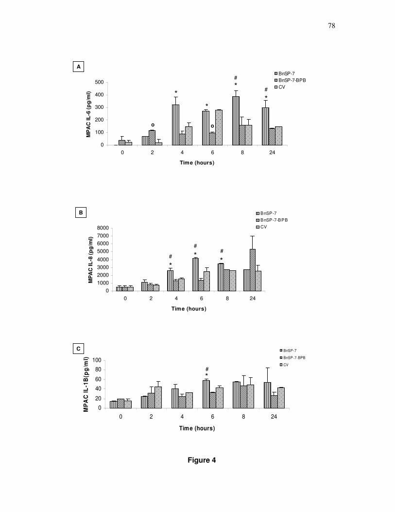
78
Figure 4
0
1000
2000
3000
4000
5000
6000
7000
8000
0 2 4 6 8 24
Time (hours)
MP
AC
IL-8
(p
g/m
l)
BnSP-7
BnSP-7-BPB
CV
* #
# #
* *
B
0
100
200
300
400
500
0 2 4 6 8 24
Time (hours)
MP
AC
IL-6
(p
g/m
l)
BnSP-7BnSP-7-BPBCV
o
*
*
o
* #
#
*
A
0
20
40
60
80
100
0 2 4 6 8 24
Time (hours)
MP
AC
IL-1
B(p
g/m
l)
BnSP-7
BnSP-7-BPB
CV
* #
C

79
Figure 5
0
2000
4000
6000
8000
2 4 6 8 24
Time (hours)
Gas
tro
cnem
ius
mu
scle
IL-8
(p
g/m
l)
BnSP-7
BnSP-7-BPB
CV
PBS
*
*
*
*
* *
*
*
B
#
#
0
5000
10000
15000
20000
25000
30000
2 4 6 8 24
Time (hours)
Gas
tro
cnem
ius
mu
scle
IL-1β
(p
g/m
l)
BnSP-7
BnSP-7-BPB
CV
PBS
* *
* *
*
* *
*
*
*
*
C
#
0
5000
10000
15000
20000
2 4 6 8 24
Time (hours)
Gas
tro
cnem
ius
mu
scle
IL-6
(p
g/m
l)
BnSP-7
BnSP-7-BPB
CV
PBS
* *
* * *
*
*
A
# *
# #

80
Tables
Table I: Induction of time-dependent edemaa and creatine kinase releaseb.
EDEMA (%) CK (U/mL)
0,5h 1h 3h 6h 24hs 1h 3h 6h 24hs*
PBS 2.5 ± 0.7 16.5 ± 7.8 7.0 ± 1.1 14.7 ± 5.8 5.9 ± 0.7 170 ± 11,4 32,5 ± 11,6 22,6 ± 9,3 70,2 ± 5,7
BnSP-7 77.1 ± 1,3 70.4 ± 8.8 66.2 ± 2.3 48.3 ± 10.2 33.1 ± 4.2 3747,3 ± 56,6 4147,5 ± 113,3 2833,3 ± 572,4 149,0 ± 73,3
CV 76.0 ± 3.5 79.0 ± 12.7 62.5 ± 7.8 49.0 ± 1.4 30.5 ± 1.7 361,6 ± 116,8 515,4 ± 125,9 469,5 ± 11,4 264,4 ± 68,7
a: After inoculation of 25µg/50µL of CV from Bothrops pauloensis or 50µg/50 µL of myotoxin BnSP-7 in mice paw, the increase of paw area was measured with a low pressure pachymeter (0.5, 1,
3, 6 and 24 hours) and expressed in percentage of induced edema. b: Mice were injected with 25 µg of CV from Bothrops pauloensis or 50µg of myotoxin BnSP-7 in gastrocnemius muscle,
sacrificed after 1, 3, 6 and 24 hours. The activity was expressed in U/L. Animals injected with 50 µL of PBS were used as control.
The experiments were analyzed statistically by ANOVA followed by the test of Dunnett. All results represented significant difference (p <0,05) among the experimental groups (BnSP-7 and CV)
and the group control (PBS). The results are presented by the average ± SD (n=3). *Differences of the time 24hs were not significative.
80

81
Anexos

82
Anexo I
TOXICON
(http://www.elsevier.com/wps/find/journaldescription.cws_home/259/authorinstructi
ons)
An Interdisciplinary Journal on the Toxins Derived from Animals, Plants and
Microorganisms - Official Journal of The International Society on Toxinology
Guide for Authors
Manuscript Preparation
General: Manuscripts must be typewritten, double-spaced with wide margins on
one side of white paper. Good quality printouts with a font size of 12 or 10 pt are
required. The corresponding author should be identified (include a Fax number
and E-mail address). Full postal addresses must be given for all co-authors. All
numbers should be numbered consecutively. Authors should consult a recent
issue of the journal for style if possible. An electronic copy of the paper should
accompany the final version. The Editors reserve the right to adjust style to certain
standards of uniformity. Authors should retain a copy of their manuscript since we
cannot accept responsibility for damage or loss of papers. Original manuscripts
are discarded one month after publication unless the Publisher is asked to return
original material after use.

83
Paper length: Toxicon has set no standard length for papers, but the Editors insist
upon a clear presentation of data in as concise a form as is consistent with good
reporting. The fragmentation of a report into several short papers is discouraged.
Abstracts: There should be an abstract of no more than 200 words.
Text: Follow this order when typing manuscripts: Title, Authors, Affiliations,
Abstract, Keywords, Main text (introduction, materials and methods, results and
discussions), Acknowledgements, Appendix, References, Vitae, Figure Captions
and then Tables. Do not import the Figures or Tables into your text. The
corresponding author should be identified with an asterisk and footnote. All other
footnotes (except for table footnotes) should be identified with superscript Arabic
numbers.
Units: Units of measure must be clearly indicated.
Symbols: The Latin name must be given for all animal and plant species. Trade
names or abbreviations of chemicals may be used only when preceded by the
chemical or scientific name. Thereafter, trade names, common names or
abbreviations should be used.
Mathematical equations: Compound numbers should be in bold face Arabic
numerals or underscored.

84
Conflict of interest : Toxicon requires full disclosure of all potential conflicts of
interest. All sources of funding supporting the work are to be declared. At the end
of the manuscript text (and in the cover letter of the manuscript), under a
subheading "Conflict of Interest statement", all authors must disclose any financial
and personal relationships with other people or organisations that could
inappropriately influence (bias) their work. If there are no conflicts of interest, the
authors should state, "The authors declare that there are no conflicts of interest."
Signed copies of the Toxicon Conflict of Interest policy form are required upon
submission. The Conflict of Interest policy form can be downloaded here. In order
to minimize delays, we strongly advise that the signed copies of these statements
are prepared before you submit your manuscript. The corresponding author is
responsible for sharing this document with all co-authors. Each and every co-
author must sign an individual disclosure form. The corresponding author is
responsible for uploading their form and those of their co-authors.
References: All publications cited in the text should be presented in a list of
references following the text of the manuscript. In the text refer to the author's
name (without initials) and year of publication (e.g. "Since Peterson (1993) has
shown that?" or "This is in the agreement with results obtained later (Kramer,
1994)"). For three or more authors use the first author followed by "et al.", in the
text. The list of references should be arranged alphabetically by authors' names.
The manuscript should be carefully checked to ensure that the spelling of authors'
names and dates are exactly the same in the text as in the reference list.

85
Illustrations: All illustrations should be provided in camera-ready form, suitable
for reproduction (which may include reduction) without retouching. Photographs,
charts and diagrams are all to be referred to as "Figure(s)" and should be
numbered consecutively in the order to which they are referred. They should
accompany the manuscript, but should not be included within the text. All
illustrations should be clearly marked on the back with the figure number and the
author's name. All figures are to have a caption. Captions should be supplied on a
separate sheet.
Line drawings: Good quality printouts on white paper produced in black ink are
required. All lettering, graph lines and points on graphs should be sufficiently large
and bold to permit reproduction when the diagram has been reduced to a size
suitable for inclusion in the journal. Dye-line prints or photocopies are not suitable
for reproduction. Do not use any type of shading on computer-generated
illustrations.
Photographs: Original photographs must be supplied as they are to be
reproduced (e.g. black and white or colour). If necessary, a scale should be
marked on the photograph. Please note that photocopies of photographs are not
acceptable. Photographs must be kept to a minimum.
Colour: Where colour printing is required the author will be charged for colour
printing at the current colour printing costs.
Tables: Tables should be numbered consecutively and given a suitable caption
and each table typed on a separate sheet. Footnotes to tables should be typed

86
below the table and should be referred to by superscript lowercase letters. No
vertical rules should be used. Tables should not duplicate results presented
elsewhere in the manuscript, (e.g. in graphs).