FACULDADE DE ODONTOLOGIA
RESPOSTA TECIDUAL EM RATOS SUBMETIDOS À INJEÇÃO SUBMUCOSA DE DOIS MATERIAIS DE PREENCHIMENTO COM FINALIDADE ESTÉTICA:
ANÁLISES CLÍNICA E HISTOLÓGICA
SABRINA POZATTI MOURE
2011
PONTIFÍCIA UNIVERSIDADE CATÓLICA DO RIO GRANDE DO SUL FACULDADE DE ODONTOLOGIA
PROGRAMA DE PÓS-GRADUAÇÃO EM ODONTOLOGIA DOUTORADO EM ODONTOLOGIA
Sabrina Pozatti Moure
Resposta Tecidual em Ratos Submetidos à Injeção Submucosa
de Dois Materiais de Preenchimento com Finalidade Estética:
Análises Clínica e Histológica
Porto Alegre
2011

Sabrina Pozatti Moure
Resposta Tecidual em Ratos Submetidos à Injeção Submucosa
de Dois Materiais de Preenchimento com Finalidade Estética:
Análises Clínica e Histológica
Tese apresentada como parte dos requisitos para obtenção do título de Doutor em Odontologia,
área de concentração em Estomatologia Clínica.
Orientadora: Profa. Dra. Maria Antonia Zancanaro de Figueiredo
Porto Alegre
2011

M931r Moure, Sabrina Pozatti
Resposta tecidual em ratos submetidos à injeção submucosa
de dois materiais de preenchimento com finalidade estética :
análises clínica e histológica / Sabrina Pozatti Moure. – Porto
Alegre, 2011.
88 f.
Tese (Doutorado) – Faculdade de Odontologia, PUCRS.
Orientadora: Profa. Dra. Maria Antonia Zancanaro de
Figueiredo
1. Odontologia. 2. Estomatologia Clínica. 3. Materiais
Biocompatíveis. 4. Ácido Hialurônico. 5. Histologia.
I. Figueiredo, Maria Antonia Zancanaro de. II. Título.
CDD 617.695
Ficha catalográfica elaborada pela
Bibliotecária Salete Maria Sartori, CRB 10/1363

EEppííggrraaffee

“Se eu vi mais longe, foi por estar de pé sobre ombros de gigantes.”
Isaac Newton

DDeeddiiccaattóórriiaa

Aos meus pais, Telmo (in memoriam) e Arléte
que com sabedoria me instigaram a ver mais longe
e por vidas de trabalho, esforço e amor
me possibilitaram chegar até aqui.

AAggrraaddeecciimmeennttooss

AGRADECIMENTOS
Agradeço a ti, mãe, exemplo de força e caráter nos inúmeros papéis que
desempenhas em minha vida: tua existência é fundamental para a minha.
Ao meu pai, agradeço pelas lindas memórias deixadas que hoje se
manifestam em tanta saudade.
Ao Juliano, grande amigo que a vida me presenteou como irmão: obrigada
pela lealdade e por toda a alegria que trazes aos meus dias.
Agradeço ao Alexandre pelo carinho, estímulo e real interesse nesse trabalho
desde o seu início.
Expresso minha gratidão a pessoas também muito especiais que contribuíram
de diversas formas durante essa trajetória: Betina, Déia, Dinda, Paulo e Vó.
A minha orientadora, professora Dra. Maria Antonia Zancanaro de
Figueiredo, o meu sincero agradecimento pela confiança depositada em mim. Ao
ser orientada por ti, reforcei conceitos do que é um exemplo de docente e aprendi
que se pode exigir com doçura.
Às professoras Dra. Fernanda Gonçalves Salum, Dra. Karen Cherubini e
Dra. Liliane Soares Yurgel agradeço pela valiosa oportunidade de participar do
Serviço de Estomatologia da PUCRS. Convivendo com a seriedade e o
conhecimento admiráveis de vocês, compreendi os motivos que lhe confere tanto
renome.
Ao professor Dr. José Antonio Poli de Figueiredo pela disponibilidade e
pelas enriquecedoras sugestões: muito obrigada.
Agradeço imensamente ao professor Dr. Vinicius Duval da Silva pela
paciência e incansável ajuda.

Às queridas amigas que o Curso me concedeu agradeço pelo apoio, cuidado
e pelos momentos de descontração essenciais: Maria Noel Marzano Rodrigues
Petruzzi e Soraya de Azambuja Berti Couto.
Aos professores Dr. Carlos Luiz Reichel e Dr. Fernando Tettamanzy
expresso meu agradecimento pelo auxílio na interpretação de histologia geral desse
trabalho.
Agradeço à professora Dra. Maria Martha Campos pelo espaço cedido para
execução dos experimentos e pelas contribuições metodológicas.
Aos professores Dr. Lauro Gilberto Nunes da Rosa e Dra. Maria Cristina
Munerato que muito me estimularam e também possibilitaram o início desse
doutoramento: serei sempre grata.
Obrigada, também, a todos os mestres da Disciplina de Patologia Bucal da
FO-UFRGS com quem muito aprendi.
Agradeço aos demais colegas do Curso pela agradável convivência,
especialmente à Ana Carolina Uchoa Vasconcellos e Márcia Fava. Também sou
agradecida àqueles que comigo dividiram trabalho, expectativas e me auxiliaram no
manejo dos animais durante a execução dessa pesquisa: Karlon Fróes de Vargas
e Ruchielli Loureiro Borghetti.
À Natália Athayde Porto pelos inúmeros favores prestados: obrigada.
Ao técnico de laboratório, Tiago Giuliani, agradeço pela prontidão e pelo
auxílio.
À CAPES por financiar meu Curso.

RReessuummoo

RESUMO
Os materiais de preenchimento são produtos injetáveis utilizados
frequentemente na medicina estética com a intenção de atenuar as rugas da face e
aumentar o volume dos lábios. Essa modalidade de tratamento, que muitas vezes
substitui procedimentos cirúrgicos tradicionais, como a ritidoplastia, apresenta
resultados estéticos satisfatórios, embora se saiba da possibilidade de alguns efeitos
indesejados ocorrerem no local da implantação do produto ou mesmo à distância. A
literatura odontológica relata inúmeros casos de lesões buco faciais decorrentes do
uso de materiais de preenchimento, refletindo uma nova realidade na prática do
cirurgião dentista. Nessa pesquisa, 2 dos materiais com finalidade estética mais
utilizados por dermatologistas e cirurgiões plásticos foram injetados em línguas de
ratos: polimetilmetacrilato 10% (n=16) e ácido hialurônico 20mg/mL (n=18), além de
uma solução inerte para controle (n=16). Após 7, 60 e 90 dias, avaliaram-se as
alterações locais clínicas e histológicas de cada produto. Foi verificada a intensidade
da resposta inflamatória na área da injeção (hematoxilina e eosina), a quantidade de
vasos sanguíneos neoformados e de macrófagos (imunoistoquímica) e a densidade
de fibras colágenas (picrosírius). Nos mesmos tempos experimentais, a migração
sistêmica das substâncias injetadas foi verificada por meio do exame microscópico
de órgãos de metabolismo e excreção (fígado e rim) pela coloração de hematoxilina
e eosina. Os resultados mostraram que os 2 materiais de preenchimento
desencadearam, em maior ou menor grau, algum tipo de resposta inflamatória local.
Na análise comparativa, constatou-se que o polimetilmetacrilato demonstrou casos
de reação a corpo estranho, enquanto o ácido hialurônico apresentou características
que sugerem biocompatibilidade. Essa pesquisa faz parte de um conjunto de
experimentos vinculados ao uso de materiais de preenchimento facial com finalidade
estética e reforça a necessidade do cirurgião dentista identificar e manejar as
reações adversas que podem decorrer desse tipo de tratamento amplamente
utilizado na atualidade.
Palavras-chave: Biocompatibilidade. Polimetil Metacrilato. Ácido Hialurônico.
Histologia.

AAbbssttrraacctt

ABSTRACT
Dermal fillers are injectable products commonly used in aesthetic medicine
with the intention of alleviating face wrinkles and increasing lip volume. This type of
treatment, which often replaces traditional surgical procedures such as
rhytidectomies, provides satisfactory cosmetic results. It is known, however, that
there is a risk of undesired effects at the site of injection of the product or even at a
distance. Dental literature reports numerous cases of orofacial injuries caused by the
use of dermal fillers, reflecting a new reality in dental surgeons' practice. In this
research, 2 of the most commonly used materials for aesthetic purposes by
dermatologists and plastic surgeons were injected in rat tongues: 10%
polymethylmethacrylate (n=16) and 20mg/mL hyaluronic acid (n=18), compared to an
inert solution for control (n=16). After 7, 60 and 90 days, local clinical and histological
alterations of each product were analyzed. The following factors were verified:
intensity of the inflammatory response in the injection area (hematoxylin and eosin),
the amount of newly formed blood vessels and macrophages
(immunohistochemistry) and the density of collagen fibers (picrosirius). At the same
experimental times, systemic migration of injected substances was observed through
microscopic examination of organs of metabolism and excretion (liver and kidney)
stained by hematoxylin and eosin. Results showed that both filling materials triggered
some kind of local inflammatory response to a greater or lesser degree. In the
comparative analysis, it was found that polymethylmethacrylate showed cases of
foreign body reaction, while hyaluronic acid demonstrated characteristics suggesting
biocompatibility. This research is part of a set of experiments related to the use of
facial dermal fillers for aesthetic purposes and it reinforces the need for dental
surgeons to identify and manage adverse reactions which may result from such
currently widely used treatment.
Key-words: Biocompatibility. Polymethyl Methacrylate. Hyaluronic Acid. Histology.

Sumário

SUMÁRIO
1 INTRODUÇÃO .............................................................................................
2 ARTIGO 1 .....................................................................................................
2.1 Introduction ...................................................................................................
2.2 Dermal fillers .................................................................................................
2.3 Local inflammatory reaction ..........................................................................
2.4 Migration .......................................................................................................
2.5 Conclusion ....................................................................................................
2.6 References ...................................................................................................
3 ARTIGO 2 .....................................................................................................
3.1 Introduction ...................................................................................................
3.2 Material and methods ...................................................................................
3.3 Results ..........................................................................................................
3.4 Discussion ....................................................................................................
3.5 References ...................................................................................................
4 DISCUSSÃO GERAL ...................................................................................
REFERÊNCIAS ..................................................................................................
ANEXOS .............................................................................................................
APÊNDICES .......................................................................................................
15
19
23
23
25
29
30
31
35
39
40
43
50
54
57
63
69
85

IInnttrroodduuççããoo

16
1 INTRODUÇÃO
O processo de envelhecimento da face causa alterações estruturais e
funcionais nos tecidos orgânicos que independem dos fatores ambientais. A
epiderme se torna mais fina e a derme atrófica e menos elástica, relativamente
acelular e avascular (FENSKE; LOBER, 1986; ACHILLES, 2004). O colágeno, a
elastina e os glicosaminoglicanos sofrem alterações de estrutura e densidade e a
espessura do tecido subcutâneo se reduz (FENSKE; LOBER, 1986). As
manifestações clínicas desses fenômenos apresentam-se como sulcos profundos na
pele, denominados rugas, que comprometem a estética e promovem a busca por
procedimentos que venham a manter ou recuperar a aparência externa jovial
(PERENACK, 2005).
Historicamente, os tratamentos utilizados para mascarar o envelhecimento
facial eram focados exclusivamente na tração dos tecidos obtida por meio de
procedimentos cirúrgicos, como a ritidoplastia (VARGAS; AMORIM; PITANGUY,
2009). Nos últimos 40 anos, uma série de substâncias injetáveis passou a ser
desenvolvida com o intuito de preencher e suavizar as rugas na região buco facial.
Como exemplo temos o silicone líquido, o colágeno bovino, a gordura autóloga, a
hidroxiapatita, o ácido poli-L-láctico e, mais recentemente, o polimetilmetacrilato
(PMMA) e o ácido hialurônico (AH) (YOON; HAN; KIM, 2003; EPPLEY; DADVAND,
2006). Tais produtos receberam o nome de materiais de preenchimento e se
tornaram hoje um dos procedimentos mais comumente executados na medicina
estética (NIAMTU, 2006; SÁNCHEZ et al., 2011).
O preenchimento facial é uma alternativa, não só para correção de alterações
decorrentes do envelhecimento, mas também para produzir um aumento de volume
artificial com fins cosméticos de estruturas da face, usualmente dos lábios (HÖNIG;
BRINK; KORABIOWSKA, 2003). Os materiais de preenchimento tem ainda
aplicação na área dos procedimentos médicos reconstrutivos, sendo utilizados para
corrigir defeitos causados por trauma ou pela lipodistrofia facial secundária à terapia
antiretroviral - TARV (SÁNCHEZ et al., 2011). Atualmente, a escolha desse tipo de
tratamento tem crescido de forma expressiva, especialmente pela vantagem dessa
técnica ser minimamente invasiva e de custo acessível (NIAMTU, 2006).

17
A literatura classifica os materiais de preenchimento atuais em não
reabsorvíveis e reabsorvíveis, sendo os últimos aqueles que tem um efeito
temporário de permanência nos tecidos (ZIMMERMANN; CLERICI, 2004).
Dos produtos não reabsorvíveis, o polimetilmetacrilato é o mais utilizado, com
expressivo emprego especialmente nos casos de lipodistrofia facial em pacientes
HIV positivos sob TARV (JONES, 2005; SKEIE et al., 2009). As apresentações
comerciais variam de acordo com a concentração de PMMA empregada, que pode
ser de 2, 10 e 30%, sendo a intermediária a mais usada pelos profissionais da área
(ACHILLES, 2004).
O ácido hialurônico é o mais utilizado dentre os materiais de preenchimento
reabsorvíveis. É usual o emprego desse produto na concentração de 20mg/mL,
embora existam outras apresentações comercialmente disponíveis (5,5mg/mL e
25mg/mL). Tem efeito temporário, sendo enzimaticamente metabolizado ou
fagocitado gradualmente em um período de 3 a 24 meses, dependendo do volume
do agente implantado nos tecidos (ACHILLES, 2004). Um grande número de autores
relata que o AH tem um tempo médio de permanência tissular de 9 meses
(LUPTON; ALSTER, 2000; FERNÁNDEZ-ACEÑERO; ZAMORA; BORBUJO, 2003;
ACHILLES, 2004).
À medida em que são produzidos e utilizados, há uma busca constante pelo
material de preenchimento ideal (ZIMMERMANN; CLERICI, 2004). Teoricamente,
além de não apresentarem potencial carcinogênico ou teratogênico, essas
substâncias não deveriam ser migratórias, nem suscitar resposta inflamatória, sendo
quimicamente inertes (HÖNIG; BRINK; KORABIOWSKA, 2003; ACHILLES, 2004).
No entanto, nenhum dos produtos existentes até o momento demonstrou todas
essas características e a literatura atual tem apontado uma série de casos nos quais
ocorreram complicações buco faciais decorrentes da aplicação desses materiais
(LOMBARDI et al., 2004; ZIMMERMANN; CLERICI, 2004; EDWARDS; FANTASIA;
IOVINO, 2006; DA COSTA MIGUEL et al., 2009; JHAM et al., 2009).
Observa-se um crescimento no uso dos materiais de preenchimento facial
devido ao custo relativamente baixo e à fácil execução dos procedimentos,
impulsionados por uma busca incessante pela melhora da aparência física. Esse
tema tem feito parte da rotina na prática clínica do cirurgião dentista, que passou a

18
se deparar com reações adversas nos tecidos bucais e peribucais provocadas pela
injeção dessas substâncias.
Até pouco tempo, a literatura relacionada a esse assunto era baseada quase
que exclusivamente em relatos de casos clínicos. Pesquisas na área da odontologia
recentemente realizadas passaram a focar no entendimento das respostas causadas
pelos materiais de preenchimento com finalidade estética (LOUREIRO BORGHETTI
et al., 2011; VARGAS, 2011).
A presente tese foi desenvolvida para auxiliar na melhor compreensão do
comportamento biológico frente a esses produtos estéticos. A análise de
componentes envolvidos na resposta inflamatória buscou identificar, clínica e
histologicamente, as respostas teciduais locais e à distância vinculadas ao uso de
dois produtos mais utilizados na atualidade.

Artigo 1

20
2 ARTIGO 1
O artigo a seguir intitula-se “Dermal fillers: matter of interest in the
dentist’s current practice” e foi formatado e submetido de acordo com as normas
da revista Journal of Oral Pathology & Medicine (Anexos A e B).
21
Dermal fillers: matter of interest in the dentist’s current practice
Sabrina Pozatti Moure1, Karlon Fróes de Vargas1, Ruchielli Loureiro Borghetti1,
Karen Cherubini1, Maria Antonia Zancanaro de Figueiredo1
1 Division of Oral Medicine, School of Dentistry, Pontifical Catholic University of Rio
Grande do Sul (PUCRS), Porto Alegre, Brazil
Running tittle: Dermal fillers: interest to the dentist
Key-words: dermal fillers, diagnosis, foreign body reaction
Corresponding author:
Dra. Maria Antonia Zancanaro de Figueiredo
Address: Serviço de Estomatologia do Hospital São Lucas da PUCRS
Av. Ipiranga, 6690 – 2º andar/sala 231, Porto Alegre, RS. Brasil. 90610-000
Phone/Fax: +55 51 3320.3254
Email: [email protected]

22
Abstract
Currently, there has been a visible growth in demand for aesthetic procedures
which maintain or restore the beauty, especially of the face. This need brought about
the emergence of less invasive and more affordable techniques, when compared to
traditional surgical procedures. There is a number of substances that have been
developed and made available for use, although it is known that none of them is
harmless to the tissue, causing possible adverse reactions. The areas of application
are varied, being the perioral region one of the most targeted, by the need to reduce
the nasolabial fold and increase in the volume of the lips. Adverse reactions in these
locations are often detected in routine tests performed by the dental surgeon. They
are usually observed as papular or nodular lesions in the oral mucosa and may even
mimic other diseases. The objective of this paper is to review the existing literature
linked to the theme, emphasizing the importance of knowing these types of changes
which have been increasingly present in clinical dentistry.

23
Introduction
The search for the maintenance or achievement of facial beauty has always
been present in human history. Until recently, treatment for this type of need focused
on rhytidectomy through surgical techniques of tissue traction. Less invasive
alternatives, with quick results and being economically more accessible, such as the
injection of dermal fillers, have emerged in recent years aiming at the reduction of
wrinkles and skin defect correction. Those techniques have been widely used by
healthcare professionals, especially dermatologists and plastic surgeons (1, 2, 3).
However, filling materials have also been known by the chances of causing
unwanted effects (4, 5, 6). Once they can be used in anatomical regions closer to the
dental surgeon's field, this new reality is reflected in the routine of these
professionals, who have increasingly reported orofacial tissue damage resulting from
adverse effects of these products. Thus, it is necessary that the dental surgeon
becomes able to detect and identify these lesions establishing differential diagnosis
with other pathologies and treating the patient adequately.
Dermal fillers
The concept of beauty is often directly associated with youthfulness. A young
face is one which has no wrinkles or lines, caused by structural and functional
changes in organic tissues associated with the aging process. Over the years, the
epidermis becomes thinner, the dermis becomes less elastic (7, 8) and the
subcutaneous tissue is reduced because their constituents, such as collagen, elastin
and glycosaminoglycans are gradually changed (7, 9, 10).
In the intention to restore this lost volume in the aging process of the face, a
series of filling materials started to be developed and used in the last 40 years. Table
1 lists the currently most employed dermal fillers (2). Several other types of filling
agents were developed in the past such as liquid silicone, paraffin, bovine collagen,
autologous fat, hydroxyapatite, poly-L-lactic acid, but most fell into disuse due the
numerous foreign body reactions and hypersensitivity (11). The dermal fillers are
different with regard to chemical composition and length of permanence in tissues
(6). Some of the most used fillers, such as polymethylmethacrylate (PMMA), are

24
classified as non-resorbable, since their constituents cannot be degraded by the
body (12). Others are called resorbable, because they have a limited length of
permanence in the tissues, such as hyaluronic acid (HA) (3, 13).
Table 1. Types of filling materials. Adapted from Carruthers et al.(2).
Composition Brand name Distributor
Non-resorbable
Silicone MDX-4-4011 No longer available
PMMA microespheres and bovine collagen
Artecoll Rofil Medical International
Artefill Artes Medical
PMMA microespheres suspended in carboxygluconate/cellulose gel
Metacrill NewPlastic
Nutricel Laboratories Lebon Prod Quim Farm
Resorbable Bovine collagen Zyderm/Zyplast Allergan
Poly-L-lactic acid Sculpra Sanofi-Aventis
Pharmaceutical Calcium hydroxyl appatite Radiesse Bioform Medical, Inc Hyaluronic acid (bacterial origin) Restylane Medicis Aesthetics Puragen
Perlane
Mentor Corp
Trademarks of PMMA most commonly used (Metacrill®, NewPlastic®) are
composed of solid polymethylmethacrylate microspheres suspended in a colloid
medium (carboxygluconate and carboxymethylcellulose, respectively). The
microspheres are irregular in surface and have varying diameters ranging between
30 and 80μm (11, 12, 14). In the face, it is indicated and used for the filling of the
nasolabial folds, lip and malar augmentation, facial contour definition, as well as the
correction of lipodystrophy caused by antiretroviral therapies in HIV positive patients
(6, 12, 15). PMMA is available for use at concentrations of 2, 10 and 30% and the
indication of concentration varies proportionally according to the size of the defect to
be treated (12). Microscopic examination of this material shows the presence of
multiple translucent microspheres distributed in polymorphic cystic spaces (16).
The hyaluronic acid is found naturally in the connective tissue and several
other human tissues (16). HA used as filling material can be obtained in vitro
(through bacterial culture). This substance has been widely used for the correction of
fine wrinkles and mild to moderate depressions (17). Several brands have been

25
developed (Restylane®, Puragen™, Perlane®) and are commercially available in
different concentrations (5.5mg/mL, 20mg/mL and 25mg/mL), and the indication of
each also varies in accordance to the extent of the defect to be corrected (3). When
taken to the microscope, the material exhibits an amorphous and basophilic nature
(16).
For years, the ideal dermal filler has been searched without success (6, 13).
This should include specific criteria such as: not causing an inflammatory response;
neither migrating nor being immunogenic; being chemically inert; not having potential
to be carcinogenic or teratogenic and being biocompatible (4).
In the current literature, there are numerous reports of complications arising
from the use of these products, such as local inflammatory processes of the foreign
body reaction and migration of the substances (18-24).
Local inflammatory reaction
When an exogenous material is introduced in the body, there is a
concatenated sequence of adjacent tissue inflammatory events (25).
Irritation caused from the filling material application generates local
coagulation and activates the surrounding tissue. A series of chemical mediators
released at this time causes hemodynamic changes which include changes in size
and vascular flow, as well as increased permeability of blood vessels. Fibrinogen,
platelet-activating factor (PAF) and cytokines are some of these mediators which, by
causing vascular changes, facilitate the exudation of leukocytes (26).
Polymorphonuclear neutrophils accumulate at the site of injury and are responsible
for the effective first line of defense in the inflammatory process (3, 27).
The presence of neutrophils, in addition to hyperemia due to vascular
changes and edema caused by plasma exudation in the early stages after injection of
dermal fillers (3, 12), justify the clinical signs and symptoms that include pain,
erythema, ecchymosis and increase in regional volume (3, 28-31). The duration and
intensity of these effects depend on individual factors and product used, ranging from

26
a few days to a few weeks (13). Skin necrosis and infection can also occur
sometimes causing irreversible damage to tissues (12).
Once the materials are not readily reabsorbed, and the irritative stimulus
persists, the inflammatory process becomes chronic in the form of a mononuclear
infiltrate of lymphocytes and plasma cells (Figure 1) (3, 25). To enable the infiltration
of these cells into the site of injection, which takes place beginning to the end of the
inflammatory process, it is necessary to generate a blood vascular network.
Fibrinogen present in the initial coagulation turns into fibrin, which is a potent
vasodilator and pro-angiogenic factor. It is known that platelets also play this role.
Histamine released by mast cells present in the tissue stimulates neovascularization
and vessel permeability increases, allowing extravasation of leukocytes. Even
exudate leukocytes release cytokines which will provide a signal so that leukocytes
and angioblasts proliferate (27, 32, 33).
The progression of these inflammatory events requires extravasation and
migration of macrophages to the site of injection of the material for phagocytosis (25).
The attraction of phagocytes occurs mainly by the action of cytokines produced and
secreted by cells already present in inflammation, such as neutrophils, lymphocytes,
endothelial cells, fibroblasts, as well as the macrophages themselves. Examples are
granulocyte macrophage colony-stimulating factors (GM-CSF), which are produced
by T lymphocytes, endothelial cells, fibroblasts and macrophages (32). Interleukins
Figure 1 - Microscopic appearance of PMMA on connective tissue with presence of lymphoplasmacytic cells and macrophages in a section stained with hematoxylin and eosin (x400, original magnification). Note the similarity between the material´s microspheres (arrows) and adipocytes.

27
(IL) such as IL-2, which is produced by T lymphocytes (27) and transforming growth
factor beta (TGFβ) secreted by neutrophils, endothelial cells, fibroblasts and
macrophages themselves (34) can also illustrate this process. The recognition of
exogenous material by the macrophage occurs via receptors that seize material
surface molecules and induce the intake of these phagocytic cells enabled for later
degranulation and digestion of the encapsulated material (27).
In biocompatible materials such as HA (3, 14, 28, 29, 35), a granulation tissue
composed of newly formed blood vessels, macrophages and fibroblasts (25)
represents the reparative phase of the chronic inflammatory response. On the other
hand, when the material implanted in the tissue is immunogenic, the inflammatory
process follows another route: the foreign body reaction, by the presence of foreign
body giant cells (6, 25, 35, 36). These cells are formed from the fusion of
macrophages, with the help of interleukins IL-3 and IL-4, when they cannot
phagocytize the injected material (6, 25, 35, 36).
According to some authors, all filling materials can cause foreign body reaction
in some patients (6). The etiopathogenesis for that is uncertain and no prediction can
be made (14). Some possible causes include the injection of a large volume of
material, the presence of impurities in the dermal filler, as well as cross-reactivity
mediated by immune complement due to previous systemic infections. In addition,
physical and chemical characteristics of the products are important factors regarding
biocompatibility. Filling materials with irregular shape, porous surfaces and varied
diameter particles, with those larger than 20µm tend to trigger lasting inflammation
that can follow the path of the foreign body reaction (2, 25, 27, 37, 38).
The productive-reparative phase of the inflammatory process is characterized
by the release of matrix metalloproteinases (MMPs) by macrophages and giant cells,
to remodel the extracellular matrix (ECM) (25). In the case of resorbable materials in
general, this last stage of inflammation will exist until the final degradation of the
injected product. For non-resorbable materials, the reaction continues until one
capsule is formed around the implant (13, 27). This fibrous capsule around the
surface of the material is mainly caused by the production of TGFβ, predominantly
expressed by macrophages, which leads to the formation of collagen fibers (39).

28
The late clinical signs that occur at the site of injection of filling materials
appear in the form of painless papules or nodules, sometimes only noticeable by
digital palpation (Figure 2) (3, 16). These lesions tend to be located in the oral
submucosa (usually in the lips, gingivolabial sulcus and cheek mucosa) detected for
weeks to years after injection of the material and are usually confused with
pathologies of distinct etiology and behavior, such as cysts or tumors of the salivary
glands (24).
Literature available on the adverse effects of dermal fillers is almost
exclusively based on case reports (21, 40, 41). In 2004, a group of authors reported
11 cases of patients with nodules in the orofacial region, some of whom initially
thought they were cases of salivary cysts or lesions of neoplastic origin. After biopsy,
microscopic examination showed the presence of a foreign body reaction in all
samples. The patients had undergone subcutaneous injections of various filling
materials, including PMMA and HA (21). Several other authors also reported cases of
submucosal nodules that were presumed to be mucocele or benign neoplastic
lesions. However, after the histopathology of biopsied specimens, the diagnosis was
foreign body reaction caused by PMMA and HA (24, 40-42).
One of the first experimental studies on this subject have been recently
published. Loureiro Borghetti et al. (3) evaluated the clinical and histopathologic local
responses 7, 60 and 90 days after the injection of 5.5mg/mL HA and 25mg/mL HA in
rat tongues. The authors concluded that both concentrations of HA proved tolerable
by the tissue, suggesting that this material has characteristics of biocompatibility.
Figure 2 - Left lower lip submucosal nodule (arrow) noted on clinical examination in patient who had a cosmetic procedure with PMMA 10 years prior.

29
Sánchez et al. (6) used skin biopsies to investigate the activation of macrophages
and inflammatory response in patients with dermal fillers, including PMMA, and
observed that there is a strong relationship between non-resorbable filling material
and foreign body reaction.
Migration
The migration of the substance is another side effect reported due to the use
of dermal fillers, although it is a very controversial issue in the literature (37, 43).
Most researches mention the migration at a distance, without specifying its precise
location (12).
A study observed the presence of renal and hepatic inflammatory infiltrate in
rats subjected to injection of filling materials in the ears. Changes were interpreted as
a result of the systematization of drugs, which could occasionally act as chemotactic
substances, acting at a distance in organs of metabolism and excretion (44).
Other authors (38) analyzed microscopically structures such as lymph nodes,
lung, spleen and liver of animals that received cheek, armpit and groin injections of
various materials, most of which contained PMMA. The authors found no signs
of migration, except in lymph nodes, and pointed out that there are three ways in
which particles of materials can be transported: 1) hematogenous, if an inadvertent
injection reaches a vein, 2) lymphatic, just as the hematogenous mechanism, it is
necessary that a large lymphatic vessel be injured, and 3) by phagocytosis, when
material fragments are phagocyted at the injection site and macrophages move
through the lymphatic system.
Loureiro Borghetti et al. (3) injected different concentrations of HA in rat
tongues. After 7, 60 and 90 days of monitoring, the authors have found, through
histological analysis, the absence of any kidney alteration.
30
Conclusion
Day after day there has been a greater appeal for facial aesthetics and easy
access to the most varied types of treatment. In a direct relationship, it is expected
that dental surgeons have to increasingly deal with oral lesions arising from adverse
reactions to filling materials.
Therefore it is important that healthcare professionals be aware of possible
undesirable effects as well as how to handle patients undergoing cosmetic
procedures using filling materials. It is suggested that the dentist becomes aware of
papular or nodular lesions located in the oral mucosa, valuing all stages of
anamnesis and physical examination, with special attention to the history of previous
aesthetic treatments.

31
References
1. ALLEMANN IB, BAUMANN L. Hyaluronic acid gel (Juvéderm™) preparations in
the treatment of facial wrinkles and folds. Clin Interv in Aging 2008; 3: 629-34.
2. CARRUTHERS J, COHEN SR, JOSEPH JH, NARINS RS, RUBIN M. The
science and art of dermal fillers for soft-tissue augmentation. J Drug Dermatol
2009; 8: 335-50.
3. LOUREIRO BORGHETTI R, VARGAS KF, POZATTI MOURE S, GONÇALVES
SALUM F, de FIGUEIREDO MA. Clinical and histologic evaluation of effects of
hyaluronic acid in rat tongue. Oral Surg Oral Med Oral Pathol Oral Radiol Endod
(in press).
4. HÖNIG JF, BRINK U, KORABIOWSKA M. Severe granulomatous allergic tissue
reaction after hyaluronic acid injection in the treatment of facial lines and its
surgical correction. J Craniofac Surg 2003; 14: 197-200.
5. DOVER JS, CARRUTHERS A, CARRUTHERS J, ALAM M. Clinical use of
Restylane. Skin Therapy Lett 2005; 10: 5-7.
6. SÁNCHEZ et al. Study of biomaterial-induced macrophage activation, cell-
mediated immune response and molecular oxidative damage in patients with
dermal bioimplants. Immunobiology (in press).
7. FENSKE NA, LOBER CW. Structural and functional changes of normal aging
skin. J Am Acad Dermatol 1986; 42: 10-9.
8. LUPO MP. Hyaluronic acid fillers in facial rejuvenation. Semin Cutan Med Surg
2006; 25: 122-6.
9. DASTOOR SF, MISCH CE, WANG H. Dermal fillers for facial soft tissue
augmentation. J Oral Implantol 2007; 33: 191-204.
10. ATIYEH BS, DIBO SA. Nonsurgical nonablative treatment of aging skin:
radiofrequency technologies between aggressive marketing and evidence-based
efficacy. Aesth Plast Surg 2009; 33: 283-94.
11. MERCER SE, KLEINERMAN R, GOLDENBERG D, EMANUEL PO.
Histopathologic identification of dermal filler agents. J Drugs Dermatol 2010; 9:
1072-8.
12. VARGAS KF, BORGHETTI RL, MOURE SP, SALUM FG, CHERUBINI K, de
FIGUEIREDO MA. Use of polymethylmethacrylate as permanent filling agent in

32
the jaw, mouth and face regions - implications for dental practice. Gerodontology
(in press).
13. ZIMMERMANN US, CLERICI TJ. The histological aspects of fillers complications.
Semin Cutan Med Surg 2004; 23: 241-50.
14. LEMPERLE G, MORHENN V, CHARRIER U. Human histology and persistence of
various injectable filler substances for soft tissue augmentation. Aesth Plast Surg
2003; 27: 354-66.
15. SKEIE L, BUGGE H, NEGAARG A, BERGERSEN B. Large particle hyaluronic
acid for the treatment of facial lipoatrophy in HIV-positive patients: 3-year follow-
up study. HIV Med 2010; 11: 170-7.
16. DADZIE OE, MAHALINGAM M, PARADA M, HELOU TE, PHILIPS T, BHAWAN
J. Adverse cutaneous reactions to soft tissue fillers - a review of the histological
features. J Cutan Pathol 2008; 35: 536-48.
17. CHRISTENSEN L, BREITING V, JANSSEN M, VUUST J, HOGDALL E. Adverse
reactions to injectable soft tissue permanent fillers. Aesth Plast Surg 2005; 29:
34-48.
18. GHISLANZONI M, BIANCHI F, BARBARESCHI M, ALESSI E. Cutaneous
granulomatous reaction to injectable hyaluronic acid gel. Br J Dermatol 2006;
154: 755-8.
19. BONNEMA H, POPA ER, VAN TIMMEREN MM, VAN WACHEM PB, DE LEIJ
LFMH, VAN LUYN MJA. Distribution patterns of the membrane during the foreign-
body reaction biomaterial in rats and mice. J Biomed Mater Res A 2003; 64: 502-
8.
20. FRIEDLAND JA. Human histology and the persistence of various injectable filler
substances for soft tissue augmentation. Aesth Plast Surg 2003; 27: 367.
21. LOMBARDI T, SAMSON J, PLANTIER F, HUSSON C, KÜFFER R. Orofacial
granulomas after injection of cosmetic fillers. Histopathologic and clinical study of
11 cases. J Oral Pathol Med 2004; 33: 115-20.
22. ZIELKE H, WÖLBER L, WIEST L, RZANY B. Risk profiles of different injectable
fillers: results from the injectable filler safety study (IFS Study). Dermatol Surg
2008; 34: 326-35.
23. BRANDT FS, CAZZANIGA A. Hyaluronic acid gel fillers in the management of
facial aging. Clin Interv Aging 2008; 3: 153-9.

33
24. JHAM BC, NIKITAKIS NG, SCHEPER MA, PAPADIMITRIOU JC, LEVY BA,
RIVERA H. Granulomatous foreign-body reaction involving oral and perioral
tissues after injection of biomaterials: a series of 7 cases and review of the
literature. J Oral Maxillofac Surg 2009; 67: 280-5.
25. ANDERSON JM, RODRIGUEZ A, CHANG DT. Foreign body reaction to
biomaterials. Semin Immunol 2008; 20: 86-100.
26. HU W, EATON JW, TANG L. Molecular basis of biomaterial-mediated foreign
body reactions. Blood 2001; 98: 1231-8.
27. LUTTIKHUIZEN DT, HARMSEN MC, VAN LUYN MJA. Cellular and molecular
dynamics in the foreign body reaction. Tissue Eng 2006; 12: 1955-70.
28. NARINS RS, BRANDT F, LEYDEN J, LORENC ZP, RUBIN M, SMITH S. A
randomized, double-blind, multicenter comparison of the efficacy and tolerability
of Restylane versus Zyplast for the correction of nasolabial folds. Dermatol Surg
2003; 29: 588-95.
29. DELORENZI C, WEINBERG M, SOLISH N, SWIFT A. A multicenter study of the
efficacy and safety os subcutaneous non-animal-stabilized hyaluronic acid in
aesthetic facial contouring: interim report. Dermatol Surg 2006; 32: 205-11.
30. LEMPERLE G, RULLAN PP, GAUTHIER-HAZAN N. Avoiding and treating dermal
filler complications. Plast Reconstr Surg 2006; 118: 92S-107S.
31. LEVY PM, DE BOULLE K, RASPALDO H. Comparison if injection comfort of a
new category of cohesive hyaluronic acid filler with preincorporated lidocaine and
a hyaluronic acid filler alone. Dermatol Surg 2009; 35: 332-6.
32. HAMILTON JA. GM-CSF in inflammation and autoimmunity. Trends Immunol
2002; 23: 403-8.
33. MAURER M, VON STEBUT E. Macrophage inflammatory protein-1. Int J Biochem
Cell Biol 2004; 36: 1882-6.
34. SCHMIDT-WEBER CB, BLASER K. Regulation and role of transforming growth
factor-beta in immune tolerance induction and inflammation. Curr Opin Immunol
2004; 16: 709-16.
35. HAMILTON RG, STROBOS J, ADKINSON F. Immunogenicity studies of
cosmetically administered nonanimal-stabilized hyaluronic acid particles.
Dermatol Surg 2007; 33: S176-85.
36. VIGNERY A. Macrophage fusion: the making of osteoclasts and giant cells. J Exp
Med 2005; 202: 337-40.

34
37. LAESCHKE K. Biocompatibility of microparticles into soft tissue fillers. Semin
Cutan Med Surg 2004; 23: 214-7.
38. LEMPERLE G, MORHENN VB, PESTONJAMASP V, GALLO RL. Migration
studies and histology of injectable microspheres of different sizes in mice. Plast
Reconstr Surg 2004; 113: 1380-90.
39. QUAGLINO D, NANNEY LB, DITESHEIM JA, DAVIDSON JM. Transforming
growth factor-beta stimulates wound healing and modulates extracellular matrix
gene expression in pig skin: incisional wound model. J Invest Dermatol 1991; 97:
34-42.
40. EDWARDS PC, FANTASIA JE, IOVINO R. Foreign body reaction to hyaluronic
acid (Restylane): an adverse outcome of lip augmentation. J Oral Maxillofac Surg
2006; 64: 1296-9.
41. DA COSTA MIGUEL MC, NONAKA CFW, DOS SANTOS JN, GERMANO AR,
DE SOUZA LB. Oral foreign body granuloma: unusual presentation of a rare
adverse reaction to permanent injectable cosmetic filler. Int J Oral Maxillofac Surg
2009; 38: 385-7.
42. FERNÁNDEZ-ACEÑERO MJ, ZAMORA E, BORBUJO J. Granulomatous foreign
body reaction against hyaluronic acid: report of a case after lip augmentation.
Dermatol Surg 2003; 29: 1225-6.
43. GOLDMAN MP. Pressure-induced migration of a permanent soft tissue filler.
Dermatol Surg 2009; 35: 403-6.
44. ROSA SC, DE MACEDO JLS. Reações adversas a substâncias de
preenchimento subcutâneo. Rev Soc Bras Cir Plást 2005; 20: 248-52.
Conflict of interest: None declared.

Artigo 2

36
3 ARTIGO 2
O artigo a seguir intitula-se “Clinical and pathological characteristics of
polymethylmethacrylate and hialuronic acid in the rat tongue” e foi formatado e
submetido de acordo com as normas da revista International Journal of Oral &
Maxillofacial Surgery (Anexos C e D).

37
Clinical and pathological characteristics of
polymethylmethacrylate and hialuronic acid in the rat tongue
Sabrina Pozatti Moure1, Karlon Fróes de Vargas1, Ruchielli Loureiro Borghetti1,
Fernanda Gonçalves Salum1, Karen Cherubini1, Maria Antonia Zancanaro de
Figueiredo1
1 Division of Oral Medicine, School of Dentistry, Pontifical Catholic University of Rio
Grande do Sul (PUCRS), Porto Alegre, Brazil
Sources of support: Coordenação de Aperfeiçoamento de Pessoal de Nível
Superior (CAPES).
Key words: dermal fillers; inflammation; adverse effects.
Short title: Oral effects of dermal fillers.
Corresponding author:
Dra. Maria Antonia Zancanaro de Figueiredo
Address: Serviço de Estomatologia do Hospital São Lucas da PUCRS
Av. Ipiranga, 6690 – 2º andar/sala 231, Porto Alegre, RS. Brasil. 90610-000
Phone/Fax: +55 51 3320.3254
Email: [email protected]

38
Abstract
Adverse effects on the oral mucosa after the use of dermal fillers have been
increasingly reported due to the increment of their use for facial aesthetics. The
objective of this study was to evaluate both clinically and histologically the initial and
late reactions, locally and at long distance, against 2 types of product: 10%
polymethylmethacrylate and 20mg/mL hyaluronic acid. Each substance was
randomly and separately injected in the rat tongues (polymethylmethacrylate, n=16;
hyaluronic acid, n=18). They were compared with the control group (n=16) in 3
experimental times (7, 60 and 90 days) in the following analyses: clinical, intensity of
local inflammatory response (hematoxylin and eosin), amount of newly formed blood
vessels and macrophages (immunohistochemistry), density of collagen fibers
(picrosirius) and systemic migration of the product in the liver and kidney
(hematoxylin and eosin). The results showed inflammation triggered from the material
injection, suggesting that both substances cause responses in local tissue, although
there was biocompatibility when hyaluronic acid was used. This research highlights
the importance of experimental studies on the subject, since adverse reactions have
been observed routinely in the practice of dentists.

39
Introduction
Recently, dental surgeons have been dealing with nodular lesions
preferentially distributed in the labial or buccal submucosa resembling inflammatory
or neoplastic pathologies of minor salivary glands9. However, when biopsied and
having their specimens evaluated microscopically, these lesions show an exogenous
material associated with foreign body reaction. These products are dermal fillers
used by dermatologists and plastic surgeons to restore the volume of subcutaneous
tissue lost during the aging process or to enhance soft parts, such as the lips14, 25.
Reports of numerous cases of foreign body reaction in the orofacial region by
injecting filling materials draw attention to one important fact: it has become
increasingly common to use these cosmetic procedures for seeking or maintaining a
youthful appearance of the face. This occurs mainly due the fact that this technique is
minimally invasive and more affordable when compared to traditional surgical
traction14.
The reaction caused by the presence of an exogenous substance inside the
tissues occurs in the form of an inflammatory response that begins with the influx of
neutrophils, causing pain and exudation. Later, lymphocytes, plasma cells and
macrophages are detected at the site, with possible formation of giant cells. In this
case, a foreign body reaction takes place. On the periphery of the inflammation area
there are intense signs of fibroplasia and neovascularization27.
In addition to the reactions that occur at the site of injection of these
substances, there is also migration. This is a very controversial issue in literature,
and most papers on migration at a distance do not specify its precise location. A
study by Rosa and de Macedo20 noted the presence of hepatic and renal
inflammatory infiltrates in rats that received the application of different filling materials
in the ear. The authors suggest that these substances could act at a distance in
organs of metabolism and excretion.
The most widely used dermal fillers are polymethylmethacrylate (PMMA) and
hyaluronic acid (HA). PMMA is perennial in the tissues due to the body's difficulty to
degrade its constituents12. On the other hand, HA is resorbable, having a limited
length of permanence, ranging from 6 to 9 months13. Both are indicated for filling
wrinkles and correcting nasolabial folds, as well as soft tissue augmentation25. One of

40
PMMA's peculiarities is its use in cases of facial lipodystrophy in HIV patients treated
with antiretrovirals24.
Most studies have mentioned the reactions resulting from the use of facial
filling materials based almost exclusively on case reports5, 7, 13. Recent research
suggests the involvement of inflammatory cells such as lymphocytes, macrophages
and giant cells in the immune response to the dermal filler21. Therefore, an
experimental study was undertaken on the topic, evaluating and comparing clinical
and histopathologic responses at early and late study times using different materials.
Materials and methods
This longitudinal randomized experimental study used 50 Wistar female rats
(Rattus norvegicus) from the same breeder. All procedures were performed
according to institutional standards for the care and use of experimental animals after
the approval of local ethics committees.
The rats were kept in cages placed in ventilated shelves with the regular
temperature of 22 ± 1°C and light-dark cycles of 12 hours. They were fed with
Nuvilab-CR1 and filtered water ad libitum.
In the beginning of the experiment, each animal weighed about 200g and had
an average of 2 months of age. They were divided into 3 groups according to the
indicated treatment: PMMA (10% NewPlastic®; Lebon Chemicals and
Pharmaceuticals Ltd., Rio de Janeiro, Brazil) (n=16); HA (20mg/mL Puragen™;
Mentor Corporation, Santa Barbara, USA) (n=18) and control (NaCl 0.9%) (n=16).
Each group was subdivided into 3 experimental times of 7, 60 and 90 days,
according to the interval between treatment and euthanasia.
The animals were weighed prior to subsequent anesthesia using
intraperitoneal injection of xylazine hydrochloride 20mg/mL (0.05mL/100g) with
ketamine hydrochloride 50mg/mL (0.1mL/100g). Once anesthetized, the rats were
placed in supine position on the operating table for the injection of the substance to
be tested. The needle (26G ½, 13 x 4.5) was cautiously introduced in the tissues with
its bevel facing up and the long axis as parallel as possible to the mucosa. The

41
submucosa was dissected at 7mm and standardized with the use of an endodontic
cursor. Thus, 0.07mL of each filling material was introduced in the middle third of the
ventral tongue, 2mm to the left of the midline and 7mm to the front of the frenum.
Clinical alterations
In each experimental time, the animals were sedated for clinical evaluation
and the presence or absence of fundamental lesions such as plaques, papules and
ulcerations was observed.
After clinical evaluation, euthanasia was proceeded by asphyxiation with
isoflurane and subsequently the tongue, the right medial lobe of the liver and the right
kidney were removed. These fragments were fixed in neutral buffered formalin at
10% and processed for hematoxylin and eosin (H-E) staining. Immunohistochemistry
and picrosirius were also applied in tongue samples.
Training was conducted with an experienced pathologist aiming to standardize
the criteria for analysis. Before reading the slides, the examiner was calibrated and
blinded. The intra-examiner calibration was performed using the Intraclass
Correlation through re-analysis of 30 slides or fields with an interval of 7 days,
showing excellent correlation between the readings related to the intensity of
inflammatory response (p = 0.904), number of newly formed blood vessels and
macrophages (p = 0.967) and density of collagen fibers (p = 1.000).
For the microscopic analysis of the tongues, in the PMMA and HA groups,
slides where exogenous material could be identified were considered viable and
evaluation of the tissue near implant was made. In the control group, the area being
evaluated corresponded to the anatomic region where the material was applied.
Intensity of inflammatory response
Through the use of a biological microscope (Zeiss Axioskop 40, Zeiss,
Oberkochen, Germany), at x100, x200 and x400 magnifications, the examiner
qualitatively assessed the presence of inflammatory cells (neutrophils, eosinophils,

42
lymphocytes, plasma cells, macrophages) in the tongue sections stained by H-E.
Giant cell analysis was done by individual descriptive evaluation of the slides. From
the criteria already used by other studies8, 14, the observation of certain
characteristics defined the inflammation score, always bearing in mind the area
where there was more intense tissue response:
0- Absent: absence of inflammation;
1- Mild: presence of sparse mononuclear cells;
2- Moderate: presence of lymphoplasmocitary infiltrates and/or sparse
neutrophil and eosinophils;
3- Intense: presence of neutrophil and eosinophil infiltrates.
Number of newly formed blood vessels and macrophages
The immunohistochemical reactions in tongue sections were carried out using
streptavidin-biotin complex with the following primary antibodies: anti-CD34
(NovoCastra, Newcastle, UK, 1:250 dilution) and anti-CD68 (NovoCastra, Newcastle,
UK, 1:200 dilution). Positive controls were made through histological sections of the
rat's intestine (CD34) and lung (CD68). In the negative control, there was omission of
the primary antibody.
Using a Zeiss microscope (Axioskop 40, Carl Zeiss, Jena, Germany) coupled
to a camera (Cool Snap-Pro cf, Media Cybernetics, Bethesda, USA) connected to a
Dell computer (model Optiplex GX620, Round Rock, USA), for each rat, 10
microscopic fields were captured at a larger magnification (x400) for each marker.
The manual count of structures was performed in each field of 166µm2 considering
the following criteria of positivity: wall thickness and size of blood vessel in stained
slides with CD34 and cellular morphology of macrophage in slides stained with
CD68.
Density of collagen fibers
It was possible to capture 3 to 5 fields of slides with tongues stained by
picrosirius in a polarized light microscope (Zeiss Axioskop 40 Zeiss, Oberkochen,

43
Germany) coupled to a camera (Cool Snap-Pro cf, Media Cybernetics, Bethesda,
USA) connected to a Dell computer (Optiplex GX620 model, Round Rock, USA).
The chosen area should cover the largest amount of collagen fibers by using the
x100 magnification. Images were transported to Image-Pro Plus, version 4.5.1
(Media Cybernetics, Inc.; 2005) where the collagen percentage, represented by red
birefringent, was calculated in the total slide area (673µm2).
Migration
Migration was evaluated in the liver and kidney slides stained by H-E in a
biological microscope (Zeiss Axioskop 40 Zeiss, Oberkochen, Germany) at x100,
x200 and x400 magnifications, based on the presence or absence of inflammatory
response or traces of injected material.
All data was tabulated and analyzed through the SPSS 17 software (SPSS
Inc.), with the Kruskal-Wallis non-parametric test, complemented by its Multiple
Comparisons test at a significance level of 5%, since the distribution of data did not
adjust to the Normal Distribution.
Results
Clinical alterations
In clinical analysis at 7 days, all animals showed lesions in the form of ulcers
close to the PMMA injection site while no change was found in the HA group. At 60
days, 1 of 5 rats that received PMMA showed papules and 50% of those receiving
HA developed white plaques in the region. When assessed 90 days after the material
injection, both test groups showed white plaques: 2 animals from the PMMA and 1
from the HA group. The control group did not show any clinical alteration at the 3
experimental times.

44
Intensity of inflammatory response
At 7 days, inflammatory response at the site of injection of PMMA was intense
(Fig. 1a), differing significantly from the moderate response at other times (Fig. 1b).
Animals treated with HA presented a distinct behavior: in the first week, they showed
a response that ranged from moderate to intense (Fig. 2a), dropping significantly until
the 90-day period, when a mild to moderate inflammatory process was observed (Fig.
2b). No changes were observed in the control group (Table 1).
Table 1. Comparison of local inflammatory response intensity of each material throughout the different studied time spans.
Material Score Time
Day 7 Day 60 Day 90
PMMA
0- Absent 0 0 0
1- Mild 0 0 0
2- Moderate 2 5 5
3- Intense 4 0 0
Total (n) 6 5 5
Mean Rank 11.83A 6.50
B 6.50
B
HA
0- Absent 0 0 0
1- Mild 0 1 3
2- Moderate 3 5 3
3- Intense 3 0 0
Total (n) 6 6 6
Mean Rank 13.50A 8.75
AB 6.25
B
Control
0- Absent 6 6 4
1- Mild 0 0 0
2- Moderate 0 0 0
3- Intense 0 0 0
Total (n) 6 6 4
Mean Rank 8.50A 8.50
A 8.50
A
Kruskal-Wallis non-parametric test, p<0.05. Distinct letters in line differ significantly.
The test groups showed a significantly higher inflammatory response than the
control group at 7, 60 and 90 days. PMMA tended to present more intense
inflammatory scores than HA, though no statistically significant difference could be
seen (Table 2).

45
Table 2. Comparison of local inflammatory response intensity in each studied time span between the
different materials.
Time Score Material
PMMA HA Control
Day 7
0- Absent 0 0 6
1- Mild 0 0 0
2- Moderate 2 3 0
3- Intense 4 3 0
Total (n) 6 6 6
Mean Rank 13.00A 12.00
A 3.50
B
Day 60
0- Absent 0 0 6
1- Mild 0 1 0
2- Moderate 5 5 0
3- Intense 0 0 0
Total (n) 5 6 6
Mean Rank 12.50A 11.58
A 3.50
B
Day 90
0- Absent 0 0 4
1- Mild 0 3 0
2- Moderate 5 3 0
3- Intense 0 0 0
Total (n) 5 6 4
Mean Rank 11.50A 8.75
A 2.50
B
Kruskal-Wallis non-parametric test, p<0.05. Distinct letters in line differ significantly.
The evaluation of the presence of giant cells showed that this event was
remarkably present in the PMMA group at all experimental times (Table 3).
Table 3. Giant cells (GC) in the implantation site of materials in each studied time span.
Material Time
Day 7 Day 60 Day 90
PMMA Presence of GC (# rats) 5
3
4
Total (n) 6 5 5
HA Presence of GC (# rats) 1
0
0
Total (n) 6 6 6
Control Presence of GC (# rats) 0
0
0
Total (n) 6 6 4

46
Number of newly formed blood vessels and macrophages
The PMMA sites of injection showed a gradual increase, with statistically
different newly formed blood vessels at all times (Fig. 1c, 1d and 1e). HA showed a
predominance of these structures at 7 and 60 days (Fig. 2c and 2d), followed by a
significant decrease in the last experimental time (Fig. 2e). The control group showed
no difference in the number of newly formed vessels between the 3 study times
(Table 4). When comparing the behaviour of substances injected at each time, it was
observed that the test groups always presented more newly formed blood vessels
than the control group. During the first 7 days, the rats injected with HA had a
significantly higher number of that event than those which received PMMA. This
relationship reached the same level after 60 days and was reversed in the last
experimental period (Table 4).
With regard to the number of macrophages present in the tissue where the
substances were applied, no significant difference between the times in the PMMA
(Fig. 1f, 1g and 1h) and control groups was seen. HA, on the other hand, showed a
greater number of these cells at 7 and 60 days (Fig. 2f and 2g) (Table 4). The test
groups presented a statistically higher number of macrophages than the control
group at all experimental times. The only significant difference between the filling
materials occurred after 90 days, when PMMA had these cells in greater amounts
(Table 4).
Density of collagen fibers
The percentage of collagen fibers near the area where PMMA was present
was higher at 7 and 60 days (Fig. 1i and 1j). This value decreased significantly until
90 days of experiment (Fig. 1k). HA, however, reached a density peak of these fibers
at the intermediate time (Fig. 2j). The control group showed no differences between
the 3 study times (Table 4). When treatments were compared in the first 7 days, the
rats that received PMMA had a higher amount of collagen fibers in the area than the
other groups (with statistically significant difference only between the PMMA group
and control). At 60 days, both test groups had a higher density of collagen fibers
compared to the control group, therefore showing a statistical difference. In the last

47
experimental time, the amount of collagen fibers was higher in the HA group followed
by PMMA and control groups, with statistically significant difference only between HA
and control groups (Table 4).
Table 4. Median and Interquartile Interval distribution of materials in different time spans for each of
the analysis.
Analysis Material
Time
Day 7 Day 60 Day 90
Median (P25-P75) Median (P25-P75) Median (P25-P75)
Number of
newly formed
blood vessels
(#/166µm2)
PMMA 1.00Cb
(0.00-2.00) 2.00Ba
(1.00-2.00) 2,00Aa
(1,00-3,00)
HA 2.00Aa
(1.00-3.00) 2.00Aa
(1.00-3.00) 1.00Bb
(0.00-2.00)
Control 0.00Ac
(0.00-0.00) 0.00Ab
(0.00-1.00) 0.00Ac
(0.00-0.00)
Number of
macrophages
(#/166µm2)
PMMA 1.00Aa
(1.00-2.00) 1.00Aa
(0.00-3.00) 2.00Aa
(1.00-3.00)
HA 1.00Aa
(0.00-2.50) 1.00Aa
(0.00-2.00) 0.00Bb
(0.00-2.00)
Control 0.00Ab
(0.00-0.00)
0.00Ab
(0.00-0.00) 0.00Ac
(0.00-0.00)
Density of
collagen
fibers
(%/673µm2)
PMMA 3.99Aa
(3.17-4.88) 5.30Aa
(3.00-10.69) 1.41Bab
(1.09-1.69)
HA 2.56Bab
(1.59-3.38) 5.00Aa
(3.34-8.40) 1.57Ba
(1.22-3.57)
Control 0.43Ab
(0.21-0.91) 0.25Ab
(0.00-0.60) 0.76Ab
(0.34-0.80)
Kruskal-Wallis non-parametric test, p<0.05. Medians followed by distinct upper case letters in the line differ significantly. Medians followed by distinct lower case letters in the column differ significantly.
Migration
Neither alterations compatible with an inflammatory response nor the
presence of traces of the injected substances could be seen in the liver and kidney
samples.

c d e
f g h
a b
i j k
48
Fig. 1. PMMA local inflammatory response. Intense inflammation with infiltration of neutrophils
(arrows) at 7 days (a) and moderate inflammation with lymphoplasmocitary infiltrates at 90 days (b)with the presence of giant cells (arrow) (H-E, x400). CD34 expression on new-formed blood vessels at
7 (c), 60 (d) and 90 days (e) detected by immunohistochemistry (x400). CD68 expression onmacrophages at 7 (f), 60 (g) and 90 days (h) detected by immunohistochemistry (x400). Collagen
fibers stained with picrosirius under polarized microscopy at 7 (i), 60 (j) and 90 days (k) (picrosirius,x200).

Fig. 2. HA local inflammatory response. Intense inflammation with infi ltration of neutrophils (arrows) at
7 days (a) and mild inflammation with sparse lymphoplasmocitary cells at 90 days (b) (H-E, x400).
CD34 expression on new-formed blood vessels at 7 (c), 60 (d) and 90 days (e) detected by
immunohistochemistry (x400). CD68 expression on macrophages at 7 (f), 60 (g) and 90 days (h)
detected by immunohistochemistry (x400). Collagen fibers stained with picrosirius under polarized
microscopy at 7 (i), 60 (j) and 90 days (k) (picrosirius, x200).
a b
i j k
c d e
f g h
49
50
Discussion
In this research, 2 of the most commonly used filling materials by
dermatologists and plastic surgeons in the perioral region were used in order to
obtain a better understanding of local and distant responses that those materials can
cause to tissues. The ventral tongue of the rat was the structure chosen for the
injection of substances with the intent to eliminate any interference with the results,
since this region is free from trauma or chronic irritation14. During the experiment, the
control group showed no changes in any of the analysis, which allowed it to be
considered as a physiological standard for comparison.
Responses triggered by the injection of dermal fillers illustrated the
inflammatory process in all its stages. Generally speaking, the presence of
exogenous material initially generates a clot at the site, which serves as the
foundation for future extravasation of inflammatory cells15. There are synthesis and
accumulation of chemical mediators such as histamine, cytokines and
prostaglandins, which modify the vascular bed and blood flow to neutrophil
margination and diapedesis, the first exuded leukocytes15. In the analysis after 7
days, there was an intense inflammatory response that occurred at the expense of a
polymorphonuclear neutrophil infiltration in both materials, though PMMA has shown
a higher number of cases of intense inflammation than HA, possibly because it is
less tolerated by tissues14, 17, 25. Previous research using the same materials obtained
similar results14, 25. Although there are reports that HA is able to induce allergic
processes in some patients11, very few eosinophils have been seen in the tissues
surrounding the studied materials and this presence was considered compatible with
physiological conditions19.
So that the extravasation of these inflammatory cells can take place, it is
necessary to generate a network of blood vessels. In the beginning of coagulation
there is fibrinogen which is converted to fibrin - a potent vasodilator and pro-
angiogenic factor that promotes proliferation, migration and differentiation of
endothelial cells. In addition, histamine released by mast cells present in the tissue,
platelets and neutrophils recruited in the early stages of the inflammatory process
stimulate neovascularization15. In the first week after the material injection, the
animals from the test groups showed statistically larger numbers of newly formed
51
blood vessels when stained with CD34 and compared to the control. The same was
observed over the 60 and 90 days of experiment, reinforcing the idea that the
vascular supply is necessary to make the inflammatory process feasible from start to
finish.
Due to the inability of the initial responses to degrade the injected filling
material, inflammation became chronic at 60 and 90 days of experiment, represented
by the presence of a lymphoplasmocitary infiltrate, featuring a moderate intensity
response1, 2. Nevertheless, in the last monitoring period, half of the HA cases had a
mild response, with sparse presence of lymphocytes and plasma cells. This behavior
was previously described in other studies11, 14 and, besides corroborating its
resorbable nature, it suggests the compatibility of this product with the tissues.
Reinforcing that, dermal fillers showed that the intensity of the inflammatory
response was related to the number of macrophages. When stained with CD68, the
number of phagocytes in the test groups was statistically larger than in the control
group throughout the entire experiment, remaining stable in the PMMA group in the
3 evaluated periods and decreasing at 90 days in the HA group. The explanation lies
in the fact that in the PMMA group, the cells involved in chronic inflammatory process
that remained in place recruited even more macrophages15. Exuded lymphocytes
produce and release interleukins (IL), such as IL-2, which attract macrophages to the
injured site15. Endothelial cells stimulate the attraction of phagocytes by releasing
cytokines such as granulocyte-macrophage colony-stimulating factor (GM-CSF)16
and secrete transforming growth factor beta (TGFβ)22. Moreover, lymphocytes,
neutrophils and macrophages themselves also assist in this process16.
In biocompatible materials, the resolution of inflammatory response occurs
through a granulation tissue composed of newly formed blood vessels, macrophages
and fibroblasts1. If the material implanted in tissue is immunogenic, the inflammatory
response follows another pathway represented by the presence of giant cells
originated from the fusion of macrophages1, 21. Most rats injected with PMMA showed
giant cells in the vicinity of the material. In the HA group, however, a small number of
these cells was seen in just one animal and exclusively in the first week of the
experiment, reinforcing the idea that this material has biocompatibility
characteristics11, 14, 19.

52
The pathogenesis and the natural course of the foreign body reaction after the
injection of a filling agent remains unknown9, 11. Physicochemical properties of the
materials, including particle size, chemical composition and structure and surface
charge are the main factors that influence the inflammatory response pathway to be
followed10, 11, 21. PMMA is a synthetic material composed of particles of irregular
surface and varying diameters, ranging between 30 and 80μm, reasons why it
usually causes more intense inflammatory responses, such as foreign body reaction3,
11, 12. The attempt to inject a large volume in a single session and the presence of
impurities in the dermal filler may possibly contribute to the pathogenesis of this
reaction17.
In the rats treated with PMMA, density of collagen fibers was increased at 7
and 60 days, suffering a decrease at 90 days. A possible explanation for this finding
is that inflammatory cells remaining close to the injection site produce and release
matrix metalloproteinases and cytokines, inciting collagenase activity that leads to
loosely arranged collagen fibers18. Furthermore, implants with particles that are
irregular, porous and large in diameter, such as PMMA, tend to induce long-lasting
inflammatory responses with low deposition of collagen fibers, in the form of a
characteristically thin fibrous capsule around each microsphere3, 12, 15, 27. In the HA
group, the peak density of collagen fibers was at 60 days, followed by a decrease at
90. This phenomenon was interpreted as reflecting the transition between the
granulation tissue and the healing of the injured area associated with material
degradation, when there is wound remodelling23. In agreement with this finding, Yoon
et al.26 did not observe the proliferation of collagen fibers 4 months after applying
20mg/mL HA.
In inflammation, the histopathological behavior translates into clinical
manifestations. The ulcerated lesions in the tongue of animals that received PMMA
at 7 days were interpreted as a reflection of the acute inflammatory response14. The
edema coupled with the increase of volume caused by the injection of the material
may have distended and traumatized tissues, leading to loss of epithelium. At 60 and
90 days, the white plaques and papules observed in some rats in the test groups are
justified by keeping the increase in local volume, which may favor the injury and
result in thickening of the keratin fibers or hyperplasia of collagen fibers9, 14. Several

53
authors mention these clinical signs in patients treated with dermal fillers, which may
occur from weeks to years after injection4, 6, 27.
Literature does not clearly indicate the migration ability of these filling agents.
The absence of changes in the evaluated organs of metabolism and excretion agrees
with most previous studies12, 14, while Rosa and de Macedo20 have found marked
hepatic and renal inflammation in an experimental study in rats.
The results of this research showed that both filling materials caused
inflammatory responses and that HA seems to be better tolerated by the tissue.
PMMA had a distinct tissue response through the formation of a foreign body
reaction. This finding raises questions about the inflammatory response in the long
run, since the consequences can be quite damaging. These topics must be clarified
through future research in the medical and dental care areas, since these materials
have been widely used in facial and perioral tissues.

54
References
1. Anderson JM, Rodriguez A, Chang DT. Foreign body reaction to biomaterials.
Seminars in Immunology 2008: 20: 86-100.
2. Cakmak O, Turkoz HK, Polat S, Serin GM, Hizal E, Tanyeri H. Histopathologic
response to highly purified liquid silicone injected intradermally in rats´ skin. Aesth
Plast Surg 2011: 35: 538-544.
3. Carruthers J, Cohen SR, Joseph JH, Narins RS, Rubin M. The science and art of
dermal fillers for soft-tissue augmentation. J Drug Dermatol 2009: 8: 335-350.
4. Christensen L, Breiting V, Janssen M, Vuust J, Hogdall E. Adverse reactions to
injectable soft tissue permanent fillers. Aesthetic Plast Surg 2005: 29: 34-48.
5. da Costa Miguel MC, Nonaka CFW, dos Santos JN, Germano AR, de Souza LB.
Oral foreign body granuloma: unusual presentation of a rare adverse reaction to
permanent injectable cosmetic filler. Int J Oral Maxillofac Surg 2009: 38: 385-387.
6. Dadzie OE, Mahalingam M, Parada M, Helou TE, Philips T, Bhawan J. Adverse
cutaneous reactions to soft tissue fillers - a review of the histological features. J
Cutan Pathol 2008: 35: 536-548.
7. Edwards PC, Fantasia JE, Iovino R. Foreign body reaction to hyaluronic acid
(Restylane): an adverse outcome of lip augmentation. J Oral Maxillofac Surg
2006: 64: 1296-1299.
8. Gomes MS, Barletta FB, Della Bona A, Vanni JR, Pereira C da C, Figueiredo JAP
de. Microbial leakage and apical inflammatory response in dog´s teeth after root
canal filling with different sealers, post space preparation and exposure to the oral
environment. J Appl Oral Sci 2007: 15: 429-436.
9. Jham BC, Nikitakis GN, Scheper MA, Papadimitriou JC, Levy BA, Rivera H.
Granulomatous foreign-body reaction involving oral and perioral tissues after
injection of biomaterials: a series of 7 cases and review of the literature. J Oral
Maxillofac Surg 2009: 67: 280-285.
10. Laeschke K. Biocompatibility of microparticles into soft tissue fillers. Semin Cutan
Med Surg 2004: 23: 214-217.
11. Lemperle G, Morhenn V, Charrier U. Human histology and persistence of various
injectable filler substances for soft tissue augmentation. Aesth Plast Surg 2003:
27: 354-366.
55
12. Lemperle G, Morhenn VB, Pestonjamasp V, Gallo RL. Migration studies and
histology of injectable microspheres of different sizes in mice. Plast Reconstr Surg
2004: 113: 1380-1390.
13. Lombardi T, Samson J, Plantier F, Husson C, Küffer R. Orofacial granulomas
after injection of cosmetic fillers. Histopathologic and clinical study of 11 cases. J
Oral Pathol Med 2004: 33: 115-120.
14. Loureiro Borghetti R, Vargas KF, Pozatti Moure S, Gonçalves Salum F, de
Figueiredo MA. Clinical and histologic evaluation of effects of hyaluronic acid in
rat tongue. Oral Surg Oral Med Oral Pathol Oral Radiol Endod (in press).
15. Luttikhuizen DT, Harmsen MC, Van Luyn MJA. Cellular and molecular dynamics
in the foreign body reaction. Tissue Eng 2006: 12: 1955-1970.
16. Maurer M, Von Stebut E. Macrophage inflammatory protein-1. Int J Biochem Cell
Biol 2004: 36: 1882-1886.
17. Mercer SE, Kleinerman R, Goldenberg D, Emanuel PO. Histopathologic
identification of dermal filler agents. J Drugs Dermatol 2010: 9: 1072-1078.
18. Moure SP, Carrard VC, Lauxen IS, Manso PP, Oliveira MG, Martins MD,
Sant´Ana Filho M. Collagen and ellastic fibers in odontogenic entities: analysis
using light and confocal laser microscopic methods. Open Dent J 2011: 5: 116-
121.
19. Narins RS, Brandt F, Leyden J, Lorenc ZP, Rubin M, Smith S. A randomized,
double-blind, multicenter comparison of the efficacy and tolerability of Restylane
versus Zyplast for the correction of nasolabial folds. Dermatol Surg 2003: 29: 588-
595.
20. Rosa SC, de Macedo JLS. Reações adversas a substâncias de preenchimento
subcutâneo. Rev Soc Bras Cir Plást 2005: 20: 248-252.
21. Sánchez O, Rodríguez-Sureda V, Domínguez C, Fernández-Figueras T, Vilches
A, Llurba E, Alijotas-Reig J. Study of biomaterial-induced macrophage activation,
cell-mediated immune response and molecular oxidative damage in patients with
dermal bioimplants. Immunobiology (in press).
22. Schmidt-Weber CB, Blaser K. Regulation and role of transforming growth factor-
beta in immune tolerance induction and inflammation. Curr Opin Immunol 2004:
16: 709-716.
23. Singer AJ, Clark RAF. Cutaneous wound healing. N Engl J Med 1999: 341: 738-
746.

56
24. Skeie L, Bugge H, Negaarg A, Bergersen B. Large particle hyaluronic acid for the
treatment of facial lipoatrophy in HIV-positive patients: 3-year follow-up study. HIV
Med 2009: 11: 170-177.
25. Vargas KF, Borghetti RL, Moure SP, Salum FG, Cherubini K, de Figueiredo MA.
Use of polymethylmethacrylate as permanent filling agent in the jaw, mouth and
face regions - implications for dental practice. Gerodontology (in press).
26. Yoon ES, Han SK, Kim WK. Advantages of the presence of living dermal
fibroblasts within Restylane for soft tissue augmentation. Ann Plast Surg 2003:
51: 587-592.
27. Zimmermann US, Clerici TJ. The histological aspects of fillers complications.
Semin Cutan Med Surg 2004: 23: 241-250.
Aknowledgements
The authors thank FAPERGS PROAP. NUMBER 04/2005 Process 0401882 for
providing the equipment to capture images made at the School of Dentistry, Federal
University of Rio Grande do Sul (UFRGS).
Funding: None.
Competing interests: None declared.
Ethics: This study was approved by the Scientific and Ethic Committee (protocol
#0005/10) and by the Ethic Committee for Animal Use (protocol #10/00151) of the
Pontifical Catholic University of Rio Grande do Sul (PUCRS), Brazil. (Anexos E e F)

DDiissccuussssããoo GGeerraall
58
4 DISCUSSÃO GERAL
O apelo pela melhora ou manutenção da aparência facial tem aumentado nos
últimos anos, visando principalmente a retardar os sinais do envelhecimento
(PERENACK, 2005). Os recursos da medicina estética para essa necessidade eram
focados em técnicas cirúrgicas de tração dos tecidos, mas passaram a dividir espaço
com procedimentos menos invasivos e mais acessíveis economicamente, como a
injeção de materiais de preenchimento (YOON; HAN; KIM, 2003; EPPLEY;
DADVAND, 2006; VARGAS; AMORIM; PITANGUY, 2009). Muitos desses produtos
utilizados tem resultados satisfatórios e são seguros, apesar das literaturas médica e
odontológica relatarem um número considerável de complicações buco faciais
decorrentes da sua aplicação (LOMBARDI et al., 2004; ZIMMERMANN; CLERICI,
2004; EDWARDS; FANTASIA; IOVINO, 2006; DA COSTA MIGUEL et al., 2009;
JHAM et al., 2009). Os efeitos adversos refletem um novo problema na prática do
cirurgião dentista, impulsionando a realização de pesquisas que almejam a uma
melhor compreensão sobre o tema bem como ao manejo adequado dos pacientes
acometidos por essas complicações (LOUREIRO BORGHETTI et al., 2011;
VARGAS, 2011).
A opção por se trabalhar com ratos ocorreu pela carência de pesquisas
experimentais sobre o tema, já que a maioria das informações disponíveis na
literatura é baseada em relatos de casos (EDWARDS; FANTASIA; IOVINO, 2006;
DA COSTA MIGUEL et al., 2009). Estudos em animais costumam ser mais
fidedignos pela possibilidade de padronizar variáveis importantes como o operador e
as técnicas de aplicação dos materiais e de análise (GOMES et al., 2007).
O AH e o PMMA foram os materiais de preenchimento escolhidos por serem
os mais utilizados por dermatologistas e cirurgiões plásticos, sendo um deles
reabsorvível e o outro não (ZIMMERMANN; CLERICI, 2004). Elegeram-se as
concentrações intermediárias desses produtos porque são as mais frequentemente
indicadas para preenchimento de rugas suaves a moderadas dispostas na região
peribucal (SÁNCHEZ et al., 2011; VARGAS et al., 2011). O ventre posterior de
língua foi a região anatômica escolhida para injeção dos materiais pela menor
vulnerabilidade ao trauma ou à irritação crônica, visando a eliminar quaisquer fatores
59
que pudessem interferir nos resultados (LOUREIRO BORGHETTI et al., 2011;
VARGAS, 2011). Além disso, a aplicação do material em estruturas anatômicas da
pele, onde estão presentes folículos pilosos, glândulas sebáceas e sudoríparas,
poderia dificultar e confundir a análise histológica (FIGUEIREDO et al., 2001),
principalmente quando da utilização do PMMA. Os tempos entre as injeções dos
materiais e a eutanásia dos ratos foram pré-estabelecidos, para que se tivesse uma
resposta inicial (7 dias), usualmente relatada na literatura como complicações
agudas, e tardia (60 e 90 dias), momento em que o processo inflamatório inicia a
degradação do material reabsorvível e que lesões crônicas começam a ser relatadas
(GHISLANZONI et al., 2006; ZIELKE et al., 2008; LOUREIRO BORGHETTI et al.,
2011).
A utilização de escores na avaliação histológica local corada pela técnica de
rotina (HE) permitiu identificar e valorizar os tipos de células envolvidos nas
respostas inflamatórias iniciais e tardias, seguindo critérios utilizados previamente
em outros estudos (FIGUEIREDO et al., 2001; GOMES et al., 2007; LOUREIRO
BORGHETTI et al., 2011). Na técnica de imunoistoquímica, o marcador CD34 foi
usado para a identificação de vasos sanguíneos neoformados (IRION et al., 2008)
na intenção de analisar de que forma a angiogênese do processo inflamatório se
estruturou ao longo dos tempos experimentais. A escolha pelo marcador CD68
baseou-se em relatos da literatura que descrevem a marcação positiva desses
anticorpos em macrófagos, células com importante função nas reações a corpo
estranho por materiais de preenchimento (BONNEMA et al., 2003; DA COSTA
MIGUEL et al., 2009; JHAM et al., 2009; SÁNCHEZ et al., 2011). Complementando a
análise, a fase produtivo-reparativa da resposta inflamatória foi feita por meio do
método histoquímico de coloração com picrosírius sob luz polarizada, uma vez que
esta técnica é específica para detecção de fibras colágenas (ABRAHÃO et al.,
2006).
No que se refere às alterações clínicas, a análise inicial mostrou um
predomínio de lesões ulceradas na língua dos animais tratados com PMMA, como
descrito em estudos anteriores (ZIMMERMANN e CLERICI, 2004; CHRISTENSEN
et al., 2005; DADZIE et al., 2008). A exposição do tecido conjuntivo poderia deixar o
ambiente mais suscetível aos agentes externos, exacerbando a reação inflamatória
local nos animais desse grupo experimental. No entanto, ambos os materiais de

60
preenchimento utilizados promoveram uma resposta moderada aos 60 dias de
avaliação, o que sugere que as reações inflamatórias foram desencadeadas pela
presença do produto implantado. As alterações tardias observadas em alguns
animais dos grupos teste condizem com vários relatos da literatura e apresentaram-
se na forma de placas brancas e pápulas. Possivelmente se justificam pela
hiperplasia de fibras colágenas e pela manutenção do aumento de volume local
vinculado à presença do produto injetado (ZIMMERMANN; CLERICI, 2004;
CHRISTENSEN et al., 2005; EDWARDS; FANTASIA; IOVINO, 2006; DADZIE et al.,
2008; DA COSTA MIGUEL et al., 2009; LOUREIRO BORGHETTI et al., 2011).
Os resultados do presente estudo ilustraram as distintas fases do processo
inflamatório secundário à injeção de cada um dos materiais de preenchimento. Na
avaliação histológica da língua dos animais, os neutrófilos exsudados atingiram o
local nos momentos iniciais, representando a primeira linha de defesa. Mais tarde,
corroborando os achados descritos na literatura, linfócitos, plasmócitos, assim como
macrófagos marcados com CD68, fizeram a defesa efetiva contra a substância
exógena (CHRISTENSEN et al., 2005; ANDERSON et al., 2008; CAKMAK et al.,
2011). Quando o material estudado era o PMMA, confirmando relatos de vários
autores, um dado relevante encontrado foi a expressiva presença de células
gigantes (CAKMAK et al., 2011; VARGAS, 2011); ao contrário dos casos que
receberam injeção de AH em que essas células multinucleadas estiveram ausentes
ou se apresentaram de forma esporádica (LEMPERLE; MORHENN; CHARRIER,
2003). Essa fase exsudativa ocorreu por conta da geração de uma rede de vasos
sanguíneos presente do início ao fim do processo inflamatório, evidenciada pela
marcação imunoistoquímica com CD34. Ao longo do experimento, a fase produtivo-
reparativa tendeu à cicatrização, na medida em que o AH era degradado. Diversos
autores afirmam haver mínima resposta fibrosa nos tecidos decorrente da
implantação desse material (ZIMMERMANN; CLERICI, 2004; CHRISTENSEN et al.,
2007), embora em 2011 Loureiro Borghetti et al. tenha sugerido a presença de
fibroplasia circundando o produto. No que se refere ao PMMA, observou-se uma
característica peculiar com redução significativa da densidade de fibras colágenas
aos 90 dias de experimento. Esse resultado pode ser justificado pelo frouxo arranjo
das fibras colágenas causado pela permanência de células inflamatórias que, em
processos crônicos como esse, liberam mediadores químicos capazes de regular a

61
produção e a degradação da matriz extracelular (MEC), estimulando a atividade das
colagenases (MOURE et al., 2011). Ademais, é descrita a formação de uma cápsula
fibrosa caracteristicamente delgada ao redor das microesferas do PMMA
(LEMPERLE et al., 2004; ZIMMERMANN; CLERICI, 2004; LUTTIKHUIZEN;
HARMSEN; VAN LUYN, 2006; CARRUTHERS et al., 2009), o que levanta a
hipótese de que características físico-químicas peculiares do produto possam ter
determinado tal comportamento.
Interpretando os dados das análises histológicas locais, foi exequível reforçar
a ideia de que a inflamação é um processo concatenado de múltiplos eventos, no
qual uma resposta local, mesenquimal e complexa ocorre com o propósito de
debelar o efeito do material implantado. Os resultados obtidos no presente estudo, a
exemplo de outros autores, sugerem que o AH apresente características de
biocompatibilidade (DOVER et al., 2005; GHISLANZONI et al., 2006; LOUREIRO
BORGHETTI et al., 2011) e que o PMMA seja capaz de suscitar reação a corpo
estranho, possivelmente por suas características físico-químicas (LEMPERLE;
MORHENN; CHARRIER, 2003; LEMPERLE et al., 2004; CARRUTHERS et al.,
2009).
A avaliação histológica do fígado e rim dos animais buscou observar a
presença de migração sistêmica mencionada por diversos autores (LEMPERLE et
al., 2004; ROSA; DE MACEDO, 2005; LOUREIRO BORGHETTI et al., 2011), porém
relatada superficialmente na literatura. Um achado curioso foi o intenso processo
inflamatório nos órgãos de metabolismo e excreção referido em experimento
desenvolvido por Rosa em 2001. Contrapondo-se a esse autor e indo ao encontro
da maioria dos relatos prévios (LEMPERLE et al., 2004; LOUREIRO BORGHETTI et
al., 2011), não foram visualizadas quaisquer alterações nos tecidos hepático e renal
avaliados na presente pesquisa. Ainda assim, acredita-se ser necessário o uso de
metodologias que simulem a injeção inadvertida de materiais de preenchimento em
vasos venosos (LEMPERLE et al., 2004) com o objetivo de investigar a capacidade
de migração dos mesmos para outros órgãos, em especial o pulmão, devido ao risco
de complicações tromboembólicas.
O PMMA mostrou, nesta pesquisa, ser capaz de suscitar reação a corpo
estranho, confirmando achados de estudos anteriores (LOMBARDI et al., 2004;
62
ZIMMERMANN; CLERICI, 2004; EDWARDS; FANTASIA; IOVINO, 2006; DA COSTA
MIGUEL et al., 2009; JHAM et al., 2009). Essa característica do produto gera maior
preocupação em virtude do seu custo ser baixo e seu uso não ter qualquer restrição
e controle de venda. Isso permite que profissionais despreparados e tecnicamente
inaptos sejam tentados a utilizar este material de forma indiscriminada (VARGAS;
AMORIM; PITANGUY, 2009, MERCER et al., 2010). Entretanto, alguns autores
relatam que eventualmente todos os materiais de preenchimento, até mesmo o AH,
são potencialmente capazes de desencadear reações a corpo estranho
(LEMPERLE; MORHENN; CHARRIER, 2003; ZIMMERMANN; CLERICI, 2004;
CHRISTENSEN, 2007). Afirmações como essa reforçam a necessidade de que se
considere sempre a capacidade de resposta de cada paciente.
Estudos futuros, incluindo pesquisas na área de biomateriais, deverão
contribuir para um melhor entendimento do processo inflamatório causado pelo uso
de materiais de preenchimento. Analisar as características fisico-químicas dos
produtos poderá auxiliar na compreensão dos motivos que levam determinado
material a ser capaz de induzir alterações na resposta inflamatória a longo prazo,
como a reação a corpo estranho, ou mesmo de migrar sistemicamente. A clara
definição etiopatogênica acerca das complicações, por uso de materiais de
preenchimentos, poderá contribuir para o aprimoramento da manufatura bem como
auxiliar na escolha do produto a ser utilizado na área médica. Outrossim, norteará o
manejo dos pacientes acometidos pelas enfermidades, inclusive no campo de
atuação do cirurgião dentista, já que esses materiais tem sido largamente utilizados
na região peribucal.

RReeffeerrêênncciiaass

64
REFERÊNCIAS
ABRAHÃO, I. J. et al. Collagen analysis in human tooth germ papillae. Braz Dent J, São Paulo, v. 17, n. 3, p. 208-212, 2006. ACHILLES, R. Imunologia e Biocompatibilidade. In: MAIO, M. de. Tratado de Medicina Estética. São Paulo: Roca, 2004. p. 1215-1223. ALLEMANN, I. B.; BAUMANN, L. Hyaluronic acid gel (Juvéderm™) preparations in the treatment of facial wrinkles and folds. Clin Interv in Aging, Miami Beach, v. 3, n. 4, p. 629-634, 2008. ANDERSON, J. M.; RODRIGUEZ, A.; CHANG, D.T. Foreign body reaction to biomaterials. Seminars in Immunology, Cleveland, v. 20, n. 2, p. 86-100, Dec. 2008. ATIYEH, B. S.; DIBO, S. A. Nonsurgical nonablative treatment of aging skin: radiofrequency technologies between aggressive marketing and evidence-based efficacy. Aesth Plast Surg, Beirut, v. 33, n. 3, p. 283-294, May. 2009. BONNEMA, H. et al. Distribution patterns of the membrane during the foreign-body reaction biomaterial in rats and mice. J Biomed Mater Res A, Groningen, v. 64, n. 3, p. 502-508, Mar. 2003. BRANDT, F. S.; CAZZANIGA, A. Hyaluronic acid gel fillers in the management of facial aging. Clin Interv Aging, Coral Gables, v. 3, n. 1, p. 153-159, 2008. CAKMAK, O. et al. Histopathologic response to highly purified liquid silicone injected intradermally in rats´ skin. Aesth Plast Surg, Istanbul, v. 35, n. 4, p. 538-544, Feb. 2011. CARRUTHERS, J. et al. The science and art of dermal fillers for soft-tissue augmentation. J Drug Dermatol, Vancouver, v. 8, n. 4, p. 335-350, Apr. 2009. CHRISTENSEN, L. et al. Adverse reactions to injectable soft tissue permanent fillers. Aesth Plast Surg, Copenhagen, v. 29, n. 1, p. 34-48, Jan-Feb. 2005. CHRISTENSEN, L. Normal and pathologic tissue reactions to soft tissue gel fillers. Dermatol Surg, Copenhagen, v. 33, n. S2, p. S168-S175, Dec. 2007. DA COSTA MIGUEL, M. C. et al. Oral foreign body granuloma: unusual presentation of a rare adverse reaction to permanent injectable cosmetic filler. Int J Oral Maxillofac Surg, Natal, v. 38, n. 4, p. 385-387, Apr. 2009. DADZIE, O. E. et al. Adverse cutaneous reactions to soft tissue fillers - a review of the histological features. J Cutan Pathol, Boston, v. 35, n. 6, p. 536-548, Jun. 2008.

65
DASTOOR, S. F.; MISCH, C. E.; WANG, H. Dermal fillers for facial soft tissue augmentation. J Oral Implantol, Ann Arbor, v. 33, n. 4, p. 191-204, 2007. DELORENZI, C. et al. A multicenter study of the efficacy and safety os subcutaneous non-animal-stabilized hyaluronic acid in aesthetic facial contouring: interim report. Dermatol Surg, Quebec, v. 32, n. 2, p. 205-211, Feb. 2006. DOVER, J. S. et al. Clinical use of Restylane. Skin Therapy Lett, Chestnut, v. 10, n. 1, p. 5-7, Feb. 2005. EDWARDS, P. C.; FANTASIA, J. E.; IOVINO, R. Foreign body reaction to hyaluronic acid (Restylane): an adverse outcome of lip augmentation. J Oral Maxillofac Surg, Omaha, v. 64, n. 8, p. 1296-1299, Aug. 2006. EPPLEY, B. L.; DADVAND, B. Injectable soft-tissue fillers: clinical overview. Plast Reconstr Surg, Indianapolis, v. 118, n. 4, p. 98e-106e, Sept. 2006. FENSKE, N. A.; LOBER, C. W. Structural and functional changes of normal aging skin. J Am Acad Dermatol, New York, v. 15, n. 4 Pt 1, p. 571-585, Oct. 1986. FERNÁNDEZ-ACEÑERO, M. J.; ZAMORA, E.; BORBUJO, J. Granulomatous foreign body reaction against hyaluronic acid: report of a case after lip augmentation. Dermatol Surg, Madrid, v. 29, n. 12, p. 1225-1226, Dec. 2003. FIGUEIREDO, J. A. et al. The histological effects of four endodontic sealers implanted in the oral mucosa: submucous injection versus implant in polyethylene tubes. Int Endod J, Porto Alegre, v. 34, n. 5, p. 377-385, July. 2001. FRIEDLAND, J. A. Human histology and the persistence of various injectable filler substances for soft tissue augmentation. Aesth Plast Surg, Phoenix, v. 27, n. 5, p. 367, Oct. 2003. GHISLANZONI, M. et al. Cutaneous granulomatous reaction to injectable hyaluronic acid gel. Br J Dermatol, Milan, v. 154, n. 4, p. 755-758, Apr. 2006. GOLDMAN, M. P. Pressure-induced migration of a permanent soft tissue filler. Dermatol Surg, San Diego, v. 35, n. S1, p. 403-406, Feb. 2009. GOMES, M. S. et al. Microbial leakage and apical inflammatory response in dog´s teeth after root canal filling with different sealers, post space preparation and exposure to the oral environment. J Appl Oral Sci, Porto Alegre, v. 15, n. 5, p. 429-436, Oct. 2007. HAMILTON, J. A. GM-CSF in inflammation and autoimmunity. Trends Immunol, Parkville, v. 23, n. 8, p. 403-408, Aug. 2002. HAMILTON, R. G.; STROBOS, J.; ADKINSON, F. Immunogenicity studies of cosmetically administered nonanimal-stabilized hyaluronic acid particles. Dermatol Surg, Scottsdale, v. 33, n. S2, p. S176-S185, Dec. 2007.

66
HÖNIG, J. F.; BRINK, U.; KORABIOWSKA, M. Severe granulomatous allergic tissue reaction after hyaluronic acid injection in the treatment of facial lines and its surgical correction. J Craniofac Surg, Göttingen, v. 14, n. 2, p. 197-200, Mar. 2003. HU, W.; EATON, J. W.; TANG, L. Molecular basis of biomaterial-mediated foreign body reactions. Blood, Houston, v. 98, n. 4, p. 1231-1238, Aug. 2001. IRION, L. C. D. et al. Angiogenesis in non-small cell lung cancer: microvessel area in needle biopsy in vascular tumor density. Anal Quant Cytol Histol, Manchester, v. 30, n. 2, p. 83-91, Apr. 2008. JHAM, B. C. et al. Granulomatous foreign-body reaction involving oral and perioral tissues after injection of biomaterials: a series of 7 cases and review of the literature. J Oral Maxillofac Surg, Baltimore, v. 67, n. 2, p. 280-285, Feb. 2009. JONES, D. HIV facial lipoatrophy: causes and treatment options. Dermatol Surg, Los Angeles, v. 31, n. 11 Pt 2, p. 1519-1529, Nov. 2005. LAESCHKE, K. Biocompatibility of microparticles into soft tissue fillers. Semin Cutan Med Surg, Siershahn, v. 23, n. 4, p. 214-217, Dec. 2004. LEMPERLE, G.; MORHENN, V.; CHARRIER, U. Human histology and persistence of various injectable filler substances for soft tissue augmentation. Aesth Plast Surg, San Diego, v. 27, n. 5, p. 354-366, Sept-Oct. 2003. LEMPERLE, G. et al. Migration studies and histology of injectable microspheres of different sizes in mice. Plast Reconstr Surg, San Diego, v. 113, n. 5, p. 1380-1390, Apr. 2004. LEMPERLE, G.; RULLAN, P. P.; GAUTHIER-HAZAN, N. Avoiding and treating dermal filler complications. Plast Reconstr Surg, San Diego, v. 118, n. 3S, p. 92S-107S, Sept. 2006. LEVY, P. M.; DE BOULLE, K.; RASPALDO, H. Comparison if injection comfort of a new category of cohesive hyaluronic acid filler with preincorporated lidocaine and a hyaluronic acid filler alone. Dermatol Surg, Geneva, v. 35, n. S1, p. 332-336, Feb. 2009. LOMBARDI, T. et al. Orofacial granulomas after injection of cosmetic fillers. Histopathologic and clinical study of 11 cases. J Oral Pathol Med, Paris, v. 33, n. 2, p. 115-120, Feb. 2004. LOUREIRO BORGHETTI, R. et al. Clinical and histologic evaluation of effects of hyaluronic acid in rat tongue. Oral Surg Oral Med Oral Pathol Oral Radiol Endod, in press, 2011. LUPO, M. P. Hyaluronic acid fillers in facial rejuvenation. Semin Cutan Med Surg, New Orleans, v. 25, n. 3, p. 122-126, Sept. 2006.
67
LUPTON, J. R.; ALSTER, T. S. Cutaneous hypersensitivity reaction to injectable hyaluronic acid gel. Dermatol Surg, Washington, v. 26, n. 2, p. 135-137, Feb. 2000. LUTTIKHUIZEN, D. T.; HARMSEN, M. C.; VAN LUYN, M. J. A. Cellular and molecular dynamics in the foreign body reaction. Tissue Eng, Groningen, v. 12, n. 7, p. 1955-1970, July. 2006. MAURER, M.; VON STEBUT, E. Macrophage inflammatory protein-1. Int J Biochem Cell Biol, Mainz, v. 36, n. 10, p. 1882-1886, Oct. 2004. MERCER, S. E. et al. Histopathologic identification of dermal filler agents. J Drugs Dermatol, New York, v. 9, n. 9, p. 1072-1078, Sept. 2010. MOURE, S. P. et al., Collagen and ellastic fibers in odontogenic entities: analysis using light and confocal laser microscopic methods. Open Dent J, Porto Alegre, v. 5, p. 116-121, July. 2011. NARINS, R. S. et al. A randomized, double-blind, multicenter comparison of the efficacy and tolerability of Restylane versus Zyplast for the correction of nasolabial folds. Dermatol Surg, New York, v. 29, n. 6, p. 588-595, June. 2003. NIAMTU, J. The use of Restylane in cosmetic facial surgery. J Oral Maxillofac Surg, Richmond, v. 64, n. 2, p. 317-325, Feb. 2006. PERENACK, J. Treatment options to optimize display of anterior dental esthetics in the patient with the aged lip. J Oral Maxillofac Surg, New Orleans, v. 63, n. 11, p. 1634-1641, Nov. 2005. QUAGLINO, D. et al. Transforming growth factor-beta stimulates wound healing and modulates extracellular matrix gene expression in pig skin: incisional wound model. J Invest Dermatol, Nashville, v. 97, n. 1, p. 34-42, July. 1991. ROSA, S. C. Estudo experimental da reação tissular frente a substâncias remodeladoras cutâneas utilizadas em cirurgia plástica estética reparadora. 2001. 130f. Dissertação (Mestrado em Ciências da Saúde). Faculdade de Ciências da Saúde, Universidade de Brasília, Brasília, 2001. ROSA, S. C.; DE MACEDO, J. L. S. Reações adversas a substâncias de preenchimento subcutâneo. Rev Soc Bras Cir Plást, Brasília, v. 20, n. 4, p. 248-252, 2005. SÁNCHEZ, O. et al. Study of biomaterial-induced macrophage activation, cell-mediated immune response and molecular oxidative damage in patients with dermal bioimplants. Immunobiology, in press, 2011. SCHMIDT-WEBER, C. B.; BLASER, K. Regulation and role of transforming growth factor-beta in immune tolerance induction and inflammation. Curr Opin Immunol, Davos, v. 16, n. 6, p. 709-716, Dec. 2004.

68
SINGER, A. J.; CLARK, R. A. F. Cutaneous wound healing. N Engl J Med, New York, v. 341, n. 10, p. 738-746, Sept. 1999. SKEIE, L. et al. Large particle hyaluronic acid for the treatment of facial lipoatrophy in HIV-positive patients: 3-year follow-up study. HIV Med, Oslo, v. 11, n. 3, p. 170-177, Mar. 2010. VARGAS, A. F.; AMORIM, N. G.; PITANGUY, I. Complicações tardias dos preenchimentos permanentes. Rev Bras Cir Plást, Rio de Janeiro, v. 24, n. 1, p. 71-81, Jan-Mar. 2009. VARGAS, K. F. Resposta tecidual em ratas submetidas à injeção na língua de polimetilmetacrilato em distintas concentrações: avaliação clínica e histológica. 2011. 110f. Dissertação (Mestrado em Odontologia). Faculdade de Odontologia, Pontifícia Universidade Católica do Rio Grande do Sul, Porto Alegre, 2011. VARGAS, K. F. et al. Use of polymethylmethacrylate as permanent filling agent in the jaw, mouth and face regions - implications for dental practice. Gerodontology, in press, 2011. VIGNERY, A. Macrophage fusion: the making of osteoclasts and giant cells. J Exp Med, New Haven, v. 202, n. 3, p. 337-340, Aug. 2005. YOON, E.; HAN, S.; KIM, W. Advantages of the presence of living dermal fibroblasts within Restylane for soft tissue augmentation. Ann Plast Surg, Seoul, v. 51, n. 6, p. 587-592, Dec. 2003. ZIELKE, H. et al. Risk profiles of different injectable fillers: results from the injectable filler safety study (IFS Study). Dermatol Surg, Berlin, v. 34, n. 3, p. 326-335, Mar. 2008. ZIMMERMANN, U. S.; CLERICI T. J. The histological aspects of fillers complications. Semin Cutan Med Surg, Paris, v. 23, n. 4, p. 241-250, Dec. 2004.

Anexos

70
ANEXOS
ANEXO A – Confirmação de submissão (Artigo 1)

71
ANEXO B – Normas da revista escolhida para submissão (Artigo 1)

72

73

74

75

76

77
ANEXO C – Confirmação de submissão (Artigo 2)

78
ANEXO D – Normas da revista escolhida para submissão (Artigo 2)

79

80

81

82
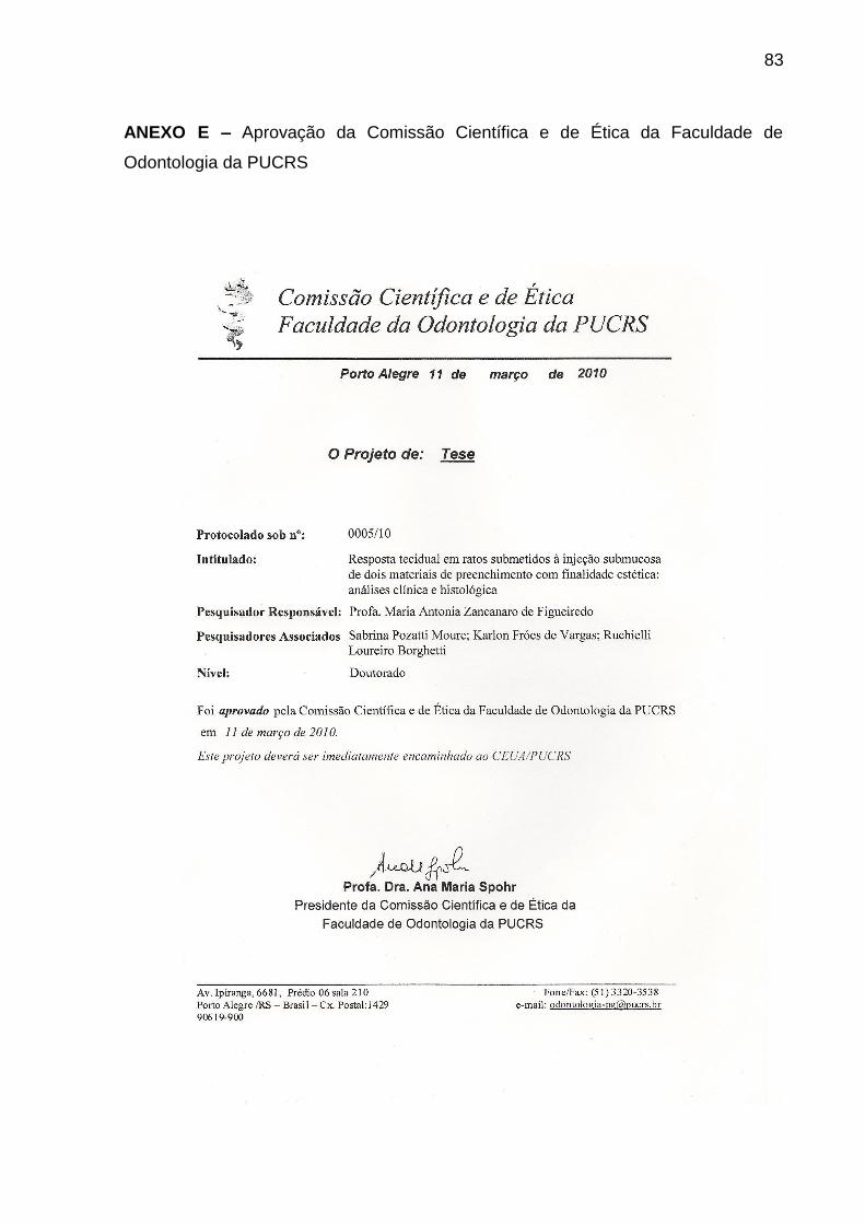
83
ANEXO E – Aprovação da Comissão Científica e de Ética da Faculdade de
Odontologia da PUCRS

84
ANEXO F – Aprovação da Comissão de Ética Para o Uso de Animais

Apêndices

86
APÊNDICES
Alterações clínicas presentes nas regiões próximas às implantações dos materiais.
U: lesão ulcerada; PB: placa branca; P: pápula.
Aspecto histológico dos materiais de preenchimento: polimetilmetacrilato (HE; 200x)
e ácido hialurônico (HE; 100x). PMMA: polimetilmetacrilato; AH: ácido hialurônico.
Aspecto histológico sem alterações do fígado e do rim, respectivamente, 60 dias
após a injeção de PMMA. Fígado: espaço porta (EP) (HE; 400x). Rim: glomérulos
(GR) e túbulos renais (TR) (HE; 200x).
PMMA
AH
U PB
P
GR
TR
EP

87
PONTIFÍCIA UNIVERSIDADE CATÓLICA DO RIO GRANDE DO SUL
FACULDADE DE ODONTOLOGIA
PROGRAMA DE PÓS-GRADUAÇÃO EM ODONTOLOGIA
ÁREA DE CONCENTRAÇÃO EM ESTOMATOLOGIA CLÍNICA
FICHA DE AVALIAÇÃO CLÍNICA
IDENTIFICAÇÃO
Rato nº: ____ Peso inicial: _____Kg
Peso final: _____Kg
Material:
□ PMMA 10%
□ AH 20mg/mL
□ Controle
Lâmina nº: _______
Tempo:
□ 7 dias
□ 60 dias
□ 90 dias
AVALIAÇÃO CLÍNICA LOCAL
□ Úlcera
□ Placa branca
□ Pápula
□ Outras formas: ________________________________________________________________
Sinais secundários:
□ sim □ não
Especifique (sangramento, supuração, abscesso):
______________________________________________________________________________
______________________________________________________________________________
______________________________________________________________________________
______________________________________________________________________________
______________________________________________________________________________
______________________________________________________________________________
______________________________________________________________________________
______________________________________________________________________________
______________________________________________________________________________
______________________________________________________________________________
______________________________________________________________________________
______________________________________________________________________________
______________________________________________________________________________
Fotos: ________________________________________________________________________
Data da avaliação: __/__/____.
Ficha de avaliação clínica utilizada durante o experimento.

88
PONTIFÍCIA UNIVERSIDADE CATÓLICA DO RIO GRANDE DO SUL
FACULDADE DE ODONTOLOGIA
PROGRAMA DE PÓS-GRADUAÇÃO EM ODONTOLOGIA
ÁREA DE CONCENTRAÇÃO EM ESTOMATOLOGIA CLÍNICA
FICHA DE AVALIAÇÃO HISTOLÓGICA
IDENTIFICAÇÃO
Rato nº: ____ Peso inicial: _____Kg
Peso final: _____Kg
Material:
□ PMMA 10%
□ AH 20mg/mL
□ Controle
Lâmina nº: _______
Tempo:
□ 7 dias
□ 60 dias
□ 90 dias
AVALIAÇÃO HISTOLÓGICA LOCAL
HE
Intensidade da resposta inflamatória
- Escores:
□ 0 - Ausente □ 1 - Leve □ 2 - Moderada □ 3 - Intensa
IMUNOISTOQUÍMICA
Vasos sanguíneos neoformados (CD34) Macrófagos (CD68)
- Nº vasos neoformados/campo (166µm2): - Nº macrófagos/campo (166µm
2):
Campo 1: ____ Campo 6: ____ Campo 1: ____ Campo 6: ____
Campo 2: ____ Campo 7: ____ Campo 2: ____ Campo 7: ____
Campo 3: ____ Campo 8: ____ Campo 3: ____ Campo 8: ____
Campo 4: ____ Campo 9: ____ Campo 4: ____ Campo 9: ____
Campo 5: ____ Campo 10: ___ Campo 5: ____ Campo 10: ___
PICROSÍRIUS
Densidade de fibras colágenas
- % fibras colágenas/campo (673µm2)
Campo 1: ____ Campo 2: ____ Campo 3: ____ Campo 4: _____ Campo 5: ____
MIGRAÇÃO
HE
Fígado Rim
□ presença de resposta inflamatória □ presença de resposta inflamatória
□ presença de resquícios do material □ presença de resquícios do material
Fotos: ________________________________________________________________________
Data da avaliação: __/__/____.
Ficha de avaliação histológica utilizada durante o experimento.
Recommended