Embed Size (px)
Citation preview
UNESP - UNIVERSIDADE ESTADUAL PAULISTA FACULDADE DE ODONTOLOGIA DE ARARAQUARA
Karina Eiras Dela Coleta Pizzol
ALTERAÇÕES DO TECIDO MOLE, ESPAÇO FARÍNGEO E
ESTABILIDADE APÓS AVANÇO MAXILO-MANDIBULAR COM ROTAÇÃO ANTI-HORÁRIA E PRÓTESE TOTAL
DE ATM
Araraquara 2008

UNESP - UNIVERSIDADE ESTADUAL PAULISTA FACULDADE DE ODONTOLOGIA DE ARARAQUARA
Karina Eiras Dela Coleta Pizzol
ALTERAÇÕES DO TECIDO MOLE, ESPAÇO FARÍNGEO E ESTABILIDADE APÓS AVANÇO MAXILO-MANDIBULAR COM ROTAÇÃO ANTI-HORÁRIA E PRÓTESE TOTAL DE ATM
Araraquara 2008
Tese apresentada ao Programa de Pós-
graduação em Ciências Odontológicas - Área
de Ortodontia, da Faculdade de Odontologia
de Araraquara, Universidade Estadual
Paulista, para obtenção do título de Doutor
em Ortodontia.
Orientador: Prof. Dr. João Roberto
Gonçalves
2
Pizzol, Karina Eiras Dela Coleta.
Alterações do tecido mole, espaço faringeano e estabilidade após avanço maxilo-mandibular com rotação anti-horária e prótese total de ATM / Karina Eiras Dela Coleta Pizzol. – Araraquara: [s.n.], 2008.
141 f. ; 30 cm.
Tese (Doutorado) – Universidade Estadual Paulista, Faculdade de Odontologia Orientador : Prof. Dr. João Roberto Gonçalves 1. Cirurgia 2. Prótese articular 3. Avaliação I. Título
Ficha catalográfica elaborada pela Bibliotecária Maria Helena Matsumoto Komasti Leves, CRB-8/2570
Serviço Técnico de Biblioteca e Documentação da Faculdade de Odontologia de Araraquara / UNESP
3
Karina Eiras Dela Coleta Pizzol
Alterações do tecido mole, espaço
faríngeo e estabilidade após avanço
maxilo-mandibular com rotação anti-
horária e prótese total de ATM
Comissão Julgadora
Tese para obtenção do grau de Doutor
Presidente e Orientador: Prof. Dr. João Roberto
Gonçalves
2º Examinador: Prof. Dr. Ary dos Santos-Pinto
3º Examinador: Prof. Dr. Roberto Henrique Barbeiro
4º Examinador: Prof. Dr. Darceny Zanetta Barbosa
5º Examinador: Profa. Dra. Terumi Okada
Araraquara, 23 de setembro de 2008

4
Dados Curriculares
Karina Eiras Dela Coleta Pizzol
Nascimento: 09/02/76- Torrinha_S.P.
Filiação: Roberto Dela Coleta
Laura Helena Eiras Dela Coleta
1995/1998: Curso de Graduação
Faculdade de Odontologia de
Araraquara “Júlio de Mesquita
Filho”(UNESP)
1999/2002: Curso de Pós-Graduação em
Ortodontia, nível Mestrado, no
Centro de Pesquisas Odontológicas
São Leopoldo Mandic
2005/2008: Curso de Pós-Graduação em
Ortodontia, nível Doutorado, na
Faculdade de Odontologia de
Araraquara (UNESP)
5
Dedicatória
A Deus,
Agradeço todas as dificuldades que enfrentei; não
fosse por elas, eu não teria saído do lugar.
"Minhas imperfeições e fracassos são como uma bênção de Deus, assim
como meus sucessos e meus talentos, e eu coloco ambos a seus pés."
( Mahatma Gandhi )
Aos meus Pais,
Roberto e Laura, por terem proporcionado o suporte
necessário para que eu pudesse vencer todas as etapas
da minha vida, por me ensinaram que caráter e
honestidade valem mais do que qualquer bem material.
Pelo amor incondicional, pela dedicação e pela vida.
“Teus pensamentos e vontades são a chave de teus atos e
atitudes....”
(Chico Xavier)

6
Ao meu amado Marido Nilton,
Por ser meu chão, minha luz e meu porto seguro. Pelo
amor incondicional, respeito, companheirismo e
cumplicidade em todos os dias de nossas vidas.
“Se um dia tiver que escolher entre o mundo e o amor...
Lembre-se:
Se escolher o mundo ficará sem o amor, mas se
escolher o amor, com ele conquistará o mundo.”
(Albert Einsten)
“O verdadeiro amor é exigente, implacável, e, ao
mesmo tempo, infinitamente delicado. "
À minha filha Letícia,
Que me ensinou a lutar por um sonho, a doar amor sem
esperar nada em troca e perceber que a vida pode ser
tão simples e bela quanto um olhar ou um sorriso.
"O dia mais importante não é o dia em que conhecemos
uma pessoa e sim quando ela passa a existir dentro de
nós."
( Autor Desconhecido )
7
Aos meus irmãos,
Flávia e Thiago, pela amizade, amor, carinho e
compreensão que sempre dedicaram a mim.
Aos meus avós,
Aparecida, Alice e Benedito por serem minha referência
de superação, de luta e de dedicação à família.
"Há quem diga que todas as noites são de sonhos. Más
há também quem garanta que nem todas, só as de verão.
No fundo, isso não tem importância. O que interessa
mesmo não é a noite em si, são os sonhos. Sonhos que
o homem sonha sempre, em todos os lugares, em todas
as épocas do ano, dormindo ou acordado."
( William Shakespeare )
.... dedico este trabalho.

8
Agradecimento Especial
Ao meu Mestre e Orientador,
João Roberto Gonçalves, que foi acima de tudo um grande amigo.
Obrigada pela confiança, pela contribuição em minha formação
profissional, pelo exemplo de dedicação e competência, pelo
respeito e por todas as oportunidades concedidas durante o
período de Pós-Graduação. Meu muito obrigado por tornar este
sonho realidade.
"Não há nada como o sonho para criar o futuro.
Utopia hoje, carne e osso amanhã."
( Victor Hugo )
Ao professor,
Ary dos Santos Pinto pela confiança, disponibilidade, pelo
respeito, pela dedicação, esmero e satisfação em ensinar,
pela valiosa contribuição científica na execução deste
trabalho.
"Grande professor é aquele que realiza o que ensina."
( Columbano )
Ao professor,
Lary M. Wolford meu muito obrigado por permitir a
elaboração deste trabalho, por sua dedicação e pela
valiosa contribuição científica na minha formação
profissional.
Aos colegas Daniel Serra Cassano e Daniela A. Godoy
Gonçalves pelo grandioso auxílio na execução deste
trabalho.
9
"Do mesmo modo que o campo, por mais fértil que seja, sem
cultivo não pode dar frutos, assim é o espírito sem
estudo."
( Cícero )
Meu muito Obrigado
Aos Professores,
Dirceu Barnabé Raveli, Luiz Gonzaga Gandini Jr., Lídia
Parzekian Martins, Maurício T. Sakima, Rita Cordeiro e
Lourdes dos Santos-Pinto pela disponibilidade e
ensinamentos que contribuíram fundamentalmente para a
minha formação e crescimento profissional.
“Nunca um desejo lhe é dado sem que também lhe seja dado o
poder de realizá-lo. Entretanto, você pode ter que se esforçar
por ele."
( Richard Bach )
Élcio Marcantonio e Roberto Henrique Barbeiro pelo
incentivo hoje e sempre, por despertarem meu
interesse na área de cirurgia ortognática e por terem
sido fundamentais na minha formação profissional.
10
"A vida é em parte o que nós fazemos dela, e em parte
o que é feito pelos amigos que nós escolhemos."
( Tennessee Williams )
Aos amigos de Turma,
Paulo (Beca), Marcus Vinícius, Ricardo e Renato pelo
incentivo, amizade, e por todos os momentos que
passamos juntos.
"Seja cortês com todos, mas íntimo de poucos, e deixe estes
poucos serem bem testados antes que você dê a eles a sua
confiança. A verdadeira amizade é uma planta de crescimento
lento, e deve experiementar e resistir os choques da
adversidade antes de ser receber o nome de amizade."
( George Washington )
Aos amigos conquistados na Pós-Graduação,
Adriano, André, Luana, Savana, Amanda, Cecília, Rafael,
Luiz Guilherme, Helder, Nancy, Luciana e Michele pela
amizade e apoio durante todos os momentos.
"Amizade, palavra que designa vários sentimentos, que
não pode ser trocada por meras coisas materiais. Deve
ser guardada e conservada no coração."
( Autor Desconhecido )
11
Os meus sinceros agradecimentos......
À Faculdade de Odontologia de Araraquara na pessoa do
Prof. Dr. José Cláudio Martins Segala, pela oportunidade
de crescimento profissional junto à UNESP.
Aos Funcionários do Departamento de Clínica Infantil,
secretaria de Pós-graduação e secretaria da
Diretoria, pela disponibilidade, respeito e dedicação
sempre.
Aos funcionários da biblioteca, em especial à Maria
Helena e à Ceres, que muito colaboraram para a
concretização deste trabalho.

12
13
Sumário
14
Sumário
Resumo 15
Abstract 17
1 Introdução 19
2 Proposição 23
3 Capítulos 25
3.1 Capítulo 1 27
3.2 Capítulo 2 62
3.3 Capítulo 3 89
4 Considerações finais 120
5 Referências 129
6 Anexos 136

15
Pizzol KEDC. Alterações do tecido mole, espaço faríngeo e estabilidade
após avanço maxilo-mandibular com rotação anti-horária e prótese total
de ATM [tese doutorado]. Araraquara: Faculdade de Odontologia da
UNESP; 2008
Resumo
Este estudo avaliou a resposta do tecido mole, do espaço faríngeo e a
estabilidade após avanço maxilo-mandibular com rotação anti-horária e
reconstrução da ATM com próteses totais articulares do tipo TMJ
Concepts system®. As mudanças cirúrgicas e pós-cirúrgicas foram
analisadas utilizando-se telerradiografias laterais. Com o movimento
cirúrgico, houve redução do ângulo do plano oclusal (14,9 ± 8,0°) e
aumento do espaço aéreo faríngeo - PASnar (4,9mm). A região anterior
da maxila moveu-se para a frente e para cima enquanto a porção
posterior, para a frente e para baixo. A mandíbula avançou, e sofreu
rotação no sentido anti-horário. No período pós-cirúrgico, a maxila
apresentou alterações mínimas no plano horizontal, enquanto todas as
medidas mandibulares permaneceram estáveis. A postura da cabeça
(OPT/NS) mostrou flexão imediatamente após a cirurgia e extensão em
longo prazo, enquanto a curvatura cervical (OPT/CVT) não apresentou
mudanças. Os resultados cirúrgicos mostraram ainda aumento das
distâncias entre a terceira vértebra cervical (C3) e o mento e desta com
16
o hióide, permanecendo estáveis durante o período de observação. A
distância entre o osso hióide e o plano mandibular reduziu durante e
após a cirurgia. Já a resposta do tecido mole evidenciou diferentes
razões entre tecido duro/mole nos pacientes com e sem genioplastia. As
mudanças horizontais na morfologia do lábio superior após avanço,
impacção da maxila, sutura em VY e sutura da base alar mostraram
maior movimento do que as mudanças observadas em tecido duro. O
avanço maxilo-mandibular com rotação anti-horária do plano oclusal
associado a próteses totais de ATM (TMJ Concepts system®) mostrou-
se estável durante o período de observação. O espaço aéreo faríngeo
aumentou significativamente, tendo sido influenciado pela posição da
cabeça após a cirurgia. A resposta dos tecidos moles ante os
movimentos esqueléticos realizados mostrou-se previsível.
Palavras-chave: Cirurgia; prótese articular; avaliação
17
Pizzol KEDC. Stability, soft tissue response and oropharyngeal airway
space changes after maxillo-mandibular advancement and counter-
clockwise rotation with total joint TMJ prostheses [tese doutorado].
Araraquara: Faculdade de Odontologia da UNESP; 2008
Abstract
This study evaluated stability, soft tissue response and oropharyngeal
airway space changes after maxillo-mandibular advancement and
counter-clockwise rotation with TMJ reconstruction using TMJ Concepts
system® total joint prostheses. Lateral cephalograms were analyzed to
estimate surgical and post surgical changes. During surgery, the
occlusal plane angle decreased 14.9 ± 8.0° and the retroglossal airway
space (PASnar) increased 4.9mm. The anterior region of maxilla moved
forward and upward while the posterior nasal spine moved downward
and forward. The mandible changed forward and rotated in a counter-
clockwise direction. At long-term follow-up evaluation the maxilla
showed minor horizontal changes, while all mandibular measurements
remained stable. Head posture (OPT/NS) showed flexure immediately
after surgery and extension long-term post surgery, while cervical
curvature (OPT/CVT) had no significant changes. Surgery increased the
distances between the third cervical vertebrae (C3) and menton, and C3
and hyoid, remaining stable afterwards. The distance from the hyoid to
the mandibular plane decreased during surgery and in the longest
18
follow-up. Soft tissue response indicated different hard/soft tissue ratios
between patients with or without genioplasties. Horizontal changes in
upper lip morphology after maxillary advancement/impaction, VY
closure, and alar base cinch sutures showed greater movement, than
observed in hard tissue. TMJ Concepts total joint prostheses associated
with maxillo-mandibular advancement and counter-clockwise rotation
showed to be stable during the follow-up observation period. Immediate
increase in oropharyngeal airway dimension, was influenced by post-
surgical changes in head posture but remained stable over the follow-up
period. Soft tissue changes showed a known predictable response.
Keywords: Surgery; joint prosthesis; evaluation

19
Introdução
20
1 Introdução
Certas patologias associadas à articulação temporo-
mandibular podem gerar alterações clínicas envolvendo oclusão,
músculos, respiração e estética facial18. Sintomas comuns da disfunção
temporo-mandibular incluem sons/estalidos na ATM, dores, limitação de
movimentos mandibulares, mudanças na oclusão, dificuldade
mastigatória entre outros. Embora a grande maioria dessas disfunções
articulares possa ser tratada com terapias não invasivas, existe um
grupo restrito de pacientes com degenerações articulares irreversíveis
que requerem reparo ou reconstrução cirúrgica, tradicionalmente
realizada com tecido autógeno. Entretanto, quando esses enxertos
autógenos são associados a avanços mandibulares de grande
amplitude, observam-se resultados pouco previsíveis com freqüente
reabsorção e recidiva. Algumas condições articulares específicas
podem predispor a comportamento semelhante. Exemplos destas
condições são: 1) ATMs previamente operadas (duas ou mais cirurgias
anteriores); 2) colocação prévia de implantes aloplásticos de ATM
contendo Proplast/Teflon, Silastic, acrílico ou cimentos ósseos; 3)
patologias inflamatórias, infecciosas ou reabsorções condilares em
adultos; 4) doenças auto-imunes ou dos ligamentos; 5) fibrose ou
21
anquilose; 6) ausência de ATM decorrente de patologia, trauma ou
deformidade congênita e 7) tumores envolvendo a região da fossa e/ou
côndilo e ramo mandibular33. Nesses casos, as próteses totais de ATM
produzidas pelo processo de prototipagem são a melhor opção. Por
meio da tecnologia CAD/CAM (computer assisted design/computer
assisted manufacture) é possível obter detalhes anatômicos que
permitem uma adaptação precisa para cada caso em particular.
As patologias de ATM podem afetar pacientes de qualquer
idade e de ambos os gêneros. Quando essas condições (ex: artrite
reumatóide, lupus, doenças auto-imunes do tecido conjuntivo,
reabsorção condilar idiopática entre outros) ocorrem em pacientes
jovens, podem ocasionar alterações no crescimento maxilo-mandibular,
deformidades dentofaciais, além de distúrbios respiratórios. Em
pacientes adultos, processos degenerativos da ATM podem também
alterar a morfologia crânio-facial com restrição importante do espaço
aéreo faríngeo, exigindo reconstrução articular associada à cirurgia
ortognática com finalidade de otimizar os resultados estéticos e
funcionais5,25.
Quando a cirurgia ortognática é realizada em associação à
colocação de próteses articulares, é possível obter-se resultados mais
estáveis31 para a correção da deformidade facial do que quando essa
cirurgia é realizada sem levar em consideração o estado das ATMs20,26.
Entretanto, a estabilidade dos resultados não se restringe somente aos
Introdução
22
fatores relacionados ao movimento cirúrgico ou à intervenção ou não
nas ATMs, mas também a fatores que contribuem para o sucesso ou
falha das próteses articulares como: hipersensibilidade ao metal32,
micromovimento da prótese, perda de componentes da prótese, fratura
ou corrosão do material2,4,10, biocompatibilidade2-4, contaminação por
bactéria11,12 e desenvolvimento de osso heterotópico ao redor da
prótese30.
Não existem trabalhos na literatura que se propõem avaliar
a estabilidade do movimento ortodôntico-cirúrgico, bem como as
mudanças respiratórias e de tecido mole, quando próteses totais
articulares são colocadas concomitantemente ao ato cirúrgico. A grande
maioria das pesquisas restringe-se a relatos da colocação isolada de
próteses articulares sem correção da má oclusão esquelética associada
ou propõe-se a avaliar a sintomatologia e os índices de qualidade de
vida após o emprego dessa técnica.
Introdução
23
Proposição
24
2 Proposição
2.1 Proposição geral
O objetivo deste estudo foi avaliar a estabilidade do
avanço maxilo-mandibular com rotação anti-horária do plano oclusal,
associado à prótese total de ATM (TMJ Concepts system®) sobre o
comportamento esquelético, do tecido mole e do espaço aéreo faríngeo.
2.2 Proposições específicas
Este estudo tem como propósitos:
1. Avaliar a estabilidade do avanço com rotação anti-
horária maxilo-mandibular associada à colocação de prótese total de
ATM (TMJ Concepts system®);
2. Avaliar as mudanças e estabilidade no espaço aéreo
faríngeo promovidas pela técnica cirúrgica;
3. Determinar se existe correlação entre a quantidade de
movimento cirúrgico e as mudanças no tecido mole e determinar sua
razão de correspondência;
4. Avaliar a influência da genioplastia nas mudanças do
tecido mole.

25
Capítulos
26
Esta tese de Doutoramento foi redigida em capítulos
correspondentes a artigos de periódicos para publicação.
Capítulo 1 Maxillo-Mandibular Counter-Clockwise Rotation and
Mandibular Advancement with TMJ Concepts® Total Joint Prostheses:
Part I - Skeletal and Dental Stability
Karina E. Dela Coleta, Larry M. Wolford, João Roberto Gonçalves, Ary
dos Santos Pinto, Lécio Pitombeira Pinto, Daniel Serra Cassano Artigo submetido à publicação no periódico International Journal of Oral
Maxillofacial Surgery.
Capítulo 2 Maxillo-Mandibular Counter-Clockwise Rotation and
Mandibular Advancement with TMJ Concepts® Total Joint Prostheses:
Part II – Airway Changes and Stability
Karina E. Dela Coleta, Larry M. Wolford, João Roberto Gonçalves, Ary
dos Santos Pinto, Daniel Serra Cassano, Daniela A. Godoy Gonçalves Artigo submetido à publicação no periódico International Journal of Oral
Maxillofacial Surgery.
Capítulo 3 Maxillo-Mandibular Counter-Clockwise Rotation and
Mandibular Advancement with TMJ Concepts® Total Joint Prostheses:
Part IV - Soft Tissue Response Karina E. Dela Coleta, Larry M. Wolford, João Roberto Gonçalves, Ary
dos Santos Pinto, Daniel Serra Cassano, Daniela A. Godoy Gonçalves Artigo submetido à publicação no periódico International Journal of Oral
Maxillofacial Surgery.
Maxillo-Mandibular Counter-Clockwise Rotation and Mandibular
Advancement with TMJ Concepts® Total Joint Prostheses:
Part I - Skeletal and Dental Stability
1Karina E. Dela Coleta, 2Larry M. Wolford, 1Joao Roberto Gonçalves, 1Ary dos Santos Pinto, 3Lécio Pitombeira Pinto, 2Daniel Serra Cassano,
1Pediatric Dentistry Department - Araraquara Dental School, Sao Paulo State University, Brazil 2Department of Oral and Maxillofacial Surgery, Texas A&M University Health Science Center, Baylor College of Dentistry, Baylor University Medical Center, Dallas, TX, USA 3Department of Restorative Dentistry, Pharmacology, Dental and Nursing School, Federal University of Ceará, Fortaleza, Brazil Address correspondence and reprint requests to: Larry M. Wolford, DMD: 3409 Worth St, Suite 400 Dallas, TX 75246 Phone: 214-828-9115 Phone: 214-828-1714 E-mail: [email protected]
28
Abstract
The purpose of this study was to evaluate skeletal and dental stability in patients that
had TMJ reconstruction and mandibular counter-clockwise advancement using TMJ
Concepts total joint prostheses (TMJ Concepts Inc. Ventura, CA) with maxillary
osteotomies being performed at the same operation. Forty-seven females (14 to 57
years old) met the criteria for inclusion with an average post surgical follow-up of 40.6
months (range 12 to 143 months). Lateral cephalograms were analyzed to estimate
surgical and post surgical changes. During surgery, the occlusal plane angle decreased
14.9 ± 8.0°. The maxilla moved forward at ANS 1.3 ± 2.4mm, point A 2.5 ± 2.2mm,
and upper incisor tip (U1T) 5.6 ± 3.0mm and upward at ANS -0.6 ± 1.9mm, point A
-1 ± 1.9mm, and U1T -1.3 ± 1.9mm. The posterior nasal spine moved downward and
forward 5.5 ± 4.2 mm and 2.9 ± 3.1 mm respectively. The mandible advanced 7.9 ± 3.5
mm at the lower incisor tips, 12.4 ± 5.4 mm at point B, 17.3 ± 7.0 mm at menton, 18.4
±8.5 mm at pogonion, and 11.0 ± 5.3 mm at gonion. Vertically the lower incisors
moved upward -2.9 ± 4.0 mm, B point and Pog remained unchanged but Me moved
downward 2.6 ± 3.9 mm and Go 18.4 ± 9.2 mm. At longest follow-up post surgery, the
maxilla showed minor horizontal changes while all mandibular measurements remained
stable. TMJ reconstruction and mandibular advancement with TMJ Concepts total joint
prosthesis in conjunction with maxillary osteotomies for counter-clockwise rotation of
the maxillo-mandibular complex was a stable procedure for these patients at the longest
follow-up.
Key words: Orthognathic Surgery; TMJ prostheses; Stability
29
Introduction
Temporomandibular joint (TMJ) pathology can create clinical problems
including masticatory musculature, jaws, occlusion, and other associated structures
resulting in pain and jaw dysfunction. Although many cases of TMJ dysfunction and
symptoms can usually be managed with non-surgical therapies, there remains a group
of patients with irreversible TMJ damage, requiring surgical repair or reconstruction;
traditionally with autogenous tissues. However, certain specific TMJ conditions and
pathology can have adverse outcomes using autogenous grafts, producing a significant
failure rate with their use7,23. These conditions include: 1) multiply operated TMJs (2 or
more previous operations); 2) previous TMJ alloplastic implants containing
Proplast/Teflon (PT, Vitek Inc., Houston, TX), Silastic (Dow Corning Inc, Midland,
MO), acrylic, bone cements, metal-on-metal articulations or failed prostheses; 3)
inflammatory, infective, reactive, or resorptive TMJ pathologies; 4) connective tissue
and autoimmune diseases; 5) fibrous and bony ankylosis; 6) absence of TMJ structures
due to pathology, trauma, or congenital deformity; and 7) tumors involving the fossa
and/or condyle and mandibular ramus region28. In these cases a custom-made total joint
prostheses may be the best option. Using CAD/CAM (computer assisted
design/computer assisted manufacture) technology, prostheses are designed and
manufactured to fit the specific anatomical requirements for each patient.
TMJ pathology can affect patients of any age and both genders but, when these
conditions occur in young patients, maxillo-mandibular growth alterations commonly
occur resulting in dentofacial deformities and associated malocclusions. In adults, TMJ
pathology (i.e., rheumatoid arthritis, psoriatic arthritis, reactive arthritis, condylar
fractures, etc) can also cause dentofacial deformities. Degenerative pathological
30
processes of the condyles may require TMJ reconstruction and orthognathic surgery to
achieve optimal functional and esthetic results.
Consideration should be given for surgical correction of co-existing TMJ
pathology as part of the orthognathic surgical correction plan. Wolford et al.27,
routinely perform concomitant TMJ and orthognathic surgery for correction of patients
with co-existing dentofacial deformities and TMJ internal derangement, with a high
success rate. There have been variable success rates reported for TMJ prostheses,
ranging from 60% to 100%16. There are risks and complications that can occur with the
use of TMJ total joint prostheses. A common problem in patients with previous PT and
Silastic implants as well as bone cements, acrylic, or metal-on-metal articulations, is the
recurrent development of foreign body giant-cell reaction (FBGCR) and reactive bone
that can cause limited jaw function as well as pain, fibrous and/or bony ankylosis.
When reconstructing these patients with the TMJ Concepts total joint prostheses,
packing autologous fat grafts around the articulating area of the prostheses has been
shown to decrease the FBGCR and minimize the occurrence of excessive joint fibrosis
and heterotopic calcification, consequently providing improved range of motion in
prosthetic TMJ reconstruction and decreased pain25.
There are many known factors which influence the success or failure of the total
joint prostheses. The challenge is to minimize these factors such as metal
hypersensitivity26, prosthesis micro movement, loosening of the prosthetic components,
material wear, break-down, and corrosion7, biocompatible and functionally compatible
materials2, FBGCR7,11, prosthesis failure11,19, bacterial contamination, and development
of heterotopic/reactive bone around the prostheses25.
31
It is considered a surgical success at long-term follow-up when the total joint
prostheses provide TMJ and occlusal stability, improve function, decrease pain, and a
long functional lifetime. Previous studies9-13,23,24,28,29 have showed that TMJ
reconstruction with total joint prostheses resulted in a significant decrease in pain, and
improvement in jaw function, diet and maximal interincisal opening. The present study
has the specific purpose of evaluating skeletal and dental stability of TMJ
reconstruction and mandibular advancement in a counter-clockwise direction using
TMJ total joint prostheses with maxillary osteotomies being performed at the same
operation.
Patients and Methods
This retrospective study evaluated records of 50 consecutive patients from a
single private practice, from 1990 through 2003, who underwent TMJ reconstruction
and counter-clockwise rotation of the maxillo-mandibular complex. Criteria for study
inclusion were: 1. End-stage bilateral or unilateral TMJ reconstruction and mandibular
advancement using custom-made TMJ total joint prostheses (TMJ Concepts system®),
and maxillary osteotomies for counter-clockwise rotation of the maxillo-mandibular
complex and occlusal plane angle; 2. All surgical procedures performed by one surgeon
(L.M.W.) at Baylor University Medical Center, Dallas, TX, USA; 3. Use of maxillary
and mandibular rigid fixation; 4. Females at least 14 years of age and males at least 17
years of age; 5. Absence of post surgical trauma; and 6. Minimum of 12-month post
surgery follow-up. Patients were rejected based on the following criteria: 1.
Craniofacial syndromes; and 2. Records (radiographs) inadequate or poor quality.
There were 49 patients (47 females, 2 males) meeting the criteria, with one female
32
patient excluded because of less than 12 month follow-up. The two males were
excluded from the study to make a homogeneous sample of 47 females (Table 1).
The custom-made total joint prostheses used in this study, were originally
developed in 1989 by Techmedica Inc., Camarillo, CA, USA, and since 1997, have
been manufactured by TMJ Concepts, Inc., Ventura, CA, USA. These prostheses are
CAD/CAM devices (computer assisted design/computer assisted manufacture),
designed to fit the specific anatomical requirements for each patient.
Forty-three patients had bilateral TMJ reconstruction and 4 patients had a
unilateral prosthesis with a sagittal split osteotomy on the contra-lateral side. All
patients had Le Fort I maxillary osteotomies, most with segmentation. All patients had
coronoidectomies on the prosthesis side(s) at the reconstruction surgery or at a previous
surgery. Mean patient age at the time of surgery was 34.5 years (range, 14 years to 57
years). Presurgical (T1) records were taken 1 day (range, 1 to 6 days) before surgery;
immediate post surgical (T2) records were taken 5 days (range, 2 to 16 days) after
surgery; and longest follow-up (T3) records were taken at a mean of 40.6 months
(range, 12 to 143 months) after surgery.
Imaging evaluation
Two examiners were calibrated by repetition of the process until the method
was considered adequate by a third examiner. Standardized lateral cephalometric
radiographs (Quint Sectograph; American Dental Co, Hawthorne, CA) were randomly
traced by one and digitized twice by other of the investigators approximately 1 week

33
apart. Average values between the 2 replicates were used to decrease landmark
technical errors.
There were 16 landmarks identified by one examiner and digitized using
DFPlus software (Dentofacial Software Inc, Toronto, Canada). The following
landmarks were used to compute 25 measurements (Tab 2, Fig 1): Nasion, Sella, Point
A, Anterior nasal spine, Posterior nasal spine, Point B, Pogonion, Menton, Gonion, and
dental points. S-N minus 7° was used as the horizontal reference plane (HRP) and a
line perpendicular through sella as the vertical reference plane (VRP). Horizontal and
vertical changes for each landmark were evaluated. Surgical changes were computed as
the differences between T2 and T1 values and post surgical changes between T3 and T2
values.
Null hypothesis
Mandibular advancement with counter-clockwise rotation of the occlusal plane
with total joint TMJ prostheses is an unstable procedure.
Statistical method
All data were transferred to SPSS (release 9.0; SPSS Chicago, IL) for statistical
analysis. The skewness and kurtosis statistics showed normal distributions for all
variables. Paired t tests were performed to evaluate the surgical (T2-T1) and post
surgical changes (T3-T2). A significance level of p < .05 was applied. The reliability of
tracing, landmark identification, and analytical measurements had an intraclass
correlation coefficient greater than 0.94.
34
Patients who received bilateral TMJ prostheses (n=43) and unilateral (n=4) were
compared as separate groups. Because there were no statistically significant differences
between those groups in post surgical changes, all the patients were analyzed as a single
group. Patients were also divided into two groups with group 1 having 12 to 24 months
post surgical follow-up (n = 18) and group 2 having 25 to 143 months follow-up (n =
29). There were no statistically significant differences in any of the parameters
evaluated between the two groups. Therefore, all 47 patients were analyzed as a single
group.
Surgical Technique
Seven patients required a preliminary surgical stage to remove previously
placed, failed total joint prostheses that contained metal (i.e. Vitek total joint
prostheses, Vitek Inc., Houston, TX; Christensen total joint prostheses, TMJ Implants
Inc., Golden, CO) , so that an accurate CT scan could be taken. Metal in the TMJ
and/or ramus can interfere with the CT scan imaging data, and significantly distort the
3-dimensional (3-D) plastic model on which the custom-made total joint prostheses are
made. CT scans were taken on all patients extending from supero-posterior to the TMJ
to anterior to the chin, maxilla and nasal bones. The 3-D plastic model was then
created using stereolithography technology (Figure 3 A). A surgical prediction tracing
was developed from a presurgical lateral cephalometric radiograph to determine the
desired final position of the maxilla and mandible. The 3-D model was mounted on an
anatomical articulator and precise model surgery performed to reposition the mandible
to the desired post surgical position relative to the maxilla that remained in its original
position on the model. Once the mandibular position was achieved, the mandible was
secured to the maxilla by placing quick-cure acrylic inter-occlusally to lock-in the

35
position of the mandible. The condyles were cut off and if indicated, bony
recontouring of the fosse and lateral aspect of the rami was completed. Any
recontouring on the 3-D model had to be accurately duplicated on the patient at the time
of surgery.
The custom-made total joint prostheses were then manufactured using
CAD/CAM technology on the 3-D model to fit the patient’s specific anatomical
requirements (Figure 3 B). Immediately prior to surgery, the mandibular movements
done on the 3-D model were accurately duplicated on anatomically mounted dental
models, and an intermediate splint constructed to aid in repositioning the mandible.
The maxillary model was then repositioned and sectioned if necessary to achieve the
best occlusal relationship. A final splint was constructed when indicated.
At surgery, an endaural or preauricular approach was used to perform the
condylectomy, joint debridement, coronoidectomy to release the temporalis muscle,
and if indicated, accurate bony recontouring of the fossa as dictated by the recontouring
done on the 3-D model. Through a submandibular approach, the medial pterygoid and
masseter muscles were reflected off the mandibular ramus and lateral recontouring
completed as indicated from the 3-D model. The mandible was then mobilized and
repositioned using the intermediate splint and inter-maxillary fixation applied. The
fossa component was inserted through the endaural / preauricular incision and
stabilized to the zygomatic arch with 3 to 4, 2 mm diameter bone screws. The
mandibular prosthetic component was inserted through the submandibular incision and
stabilized to the ramus with 8 to 12, 2 mm diameter bone screws. Following
stabilization of the prostheses, most patients had fat grafts (usually harvested from the
36
abdomen) packed around the articulating area of the prostheses to help prevent fibrosis
and heterotopic/reactive bone formation post surgery. The incisions were closed.
Multiple maxillary osteotomies were then performed to establish the best
possible functional and esthetic result, since presurgery the maxilla was usually AP
retruded as well as had anterior vertical maxillary excess and/or posterior vertical
maxillary deficiency with a high occlusal plane angulation. The maxilla was stabilized
with bone plates and porous block hydroxyapatite grafts (PBHA, Interpore 200,
Interpore Inc., Irvine, CA).
When indicated, genioplasty, turbinectomies, nasoseptoplasty, rhinoplasty, etc.,
were performed at the same surgery. Many of these patients, particularly those with
significant retrognathia, had moderate to severe presurgical sleep apnea symptoms
because of the decreased oropharyngeal airway. The suprahyoid muscles were not
deliberately detached from the genial tubercles in any of the cases. Alloplastic
materials such as PBHA or HTR (Hard Tissue Replacement, Walter Lorenz Inc,
Jacksonville, FL) were used for augmentation genioplasties although some patients had
osseous genioplasties.
Post surgery, no maxillo-mandibular fixation was used in any cases, but light
interarch elastics were routinely applied to help support the mandible, since the muscles
of mastication were reflected from the mandible and were initially non-functional. Post
operative elastics were generally discontinued following adequate functional return of
the pterygomasseteric musculature (usually 2 to 4 weeks post surgery), unless required
for finishing orthodontic mechanics. Passive physical therapy was used on all patients
37
beginning approximately 6 to 8 weeks post surgery. Patients were instructed to open
and close their jaws and begin shifting their jaws from side to side 4 to 5 sessions per
day for 10 to 15 minutes each session. Patients were maintained on a puree to soft diet
for 4 months post surgery to allow the maxilla to complete the initial bone healing
phase. Patients were then encouraged to begin working up to a normal diet.
Orthodontic appliances were usually maintained for at least 6 months post surgery and
then removed at the discretion of the orthodontist.
Results
Surgical changes (T2-T1)
Initial values, surgical and longest follow-up changes are listed in Table 3. The
mean surgical changes showed upward and forward movement of the maxillary anterior
region (Fig 2). The horizontal movement of anterior nasal spine (ANS) was 1.3 mm
(range -7.3 to 7.1mm), and point A was 2.5 mm (range -6.0 to 6.8 mm). In the
horizontal direction, positive values mean forward movement, negative values mean
posterior movement. The vertical movement (positive values mean downward
movement, negative values mean upward movement) of the ANS was -0.6 mm (range -
4.0 to 3.4 mm) and point A was -1.0 mm (range -4.2 to 3.3 mm). Posterior nasal spine
(PNS) was displaced downward 5.5 mm (range 1.8 to 14.3 mm) and forward 2.9 mm
(range -3.9 to 10.9 mm). The upper incisor tip (U1T) moved forward 5.6mm (range -
0.6 to 11.3 mm) and vertically -1.3 mm (range -5.7 to 2.6 mm).
All the anterior mandibular measurements were advanced in a horizontal
direction with lower incisor tip (L1T) 7.9 mm (range 0.9 to 14.3 mm), point B 12.4 mm
(range 1.7 to 22.5 mm), Pogonion (Pog) 18.4 mm (range 2.1 to 42.1 mm), and Menton
38
(Me) 17.3 mm (range 2.6 to 32.8 mm). In the vertical direction, L1T showed a superior
movement of -2.9 mm (range -16.5 to 2.3 mm), while B point and Pog showed no
movement. However, Me showed an inferior movement of 2.6 mm (range -6.7 to 12.5
mm). Gonion (Go) moved downward 18.4 mm (range -1.5 to 43.4 mm) and forward
11.0 mm (range 2.8 to 25.6 mm). The occlusal plane angle (OPA) relative to HRP
decreased a mean -14.9º (range -37.0 to -2.3º) and SNPog angle increased 9.1º (range
1.0 to 20.1º).
There was a surgical increase in the SNA angle of 2.3º (range -6.5 to 6.4º) and
SNB of 6.9º (range 1.0 to 12.8º). The ANB angle decreased -4.6º (range -10.5 to 2.2º)
because of the greater increase of the SNB value compared to SNA. Overjet (OJ)
decreased -2.2 mm (range -7.4 to 1.8 mm). Overbite (OB) increased 1.6 mm (range -14.7
to 3.9 mm).
Post surgical stability (T3-T2)
Point A and posterior nasal spine (PNS), in their horizontal direction only,
showed a change backwards of -0.4 mm (range -2.8 to 5.0 mm) and -0.8 mm (range
-8.4 to 3.9 mm) respectively, that were considered statistically significant (p<0.05). The
remaining maxillary landmarks remained stable. All the anterior mandible
measurements (L1T, B, Pog, Me) showed no statistically significant change at long-
term follow-up (p<0.05). Neither OPA nor SNPog angles had significant changes long
term post surgery. Therefore, all horizontal and vertical mandibular measurements
remained stable during the follow-up period (Table 3). There was a mean decrease in
the ANB angle of -0.4º (range -2.6 to 5.4º), while SNA, SNB and overjet (OJ) showed
39
no statistically significant change (T3-T2). Overbite (OB) increased 0.7 mm (range -0.7
to 2.1 mm).
Case 1: (CT patient #41) This 28 year old female presented 4 years post trauma
that involved multiple mandibular fractures including bilateral subcondylar fractures,
comminution of the right condyle, symphysis fracture with loss of a central incisor, as
well as fracture of the anterior maxilla resulting in the loss of 7 teeth from the left
lateral incisor through the right 2nd bicuspid. The missing teeth were previously
replaced with 5 osseo-integrated dental implants and a prosthesis. She had one previous
surgery on her right TMJ with no improvement. Her diagnoses included: 1) Right
TMJ severe arthritis, 2) Anterior open bite, 3) Transverse facial asymmetry, and 4)
Retruded maxilla and mandible (Fig 4 A and B; 5 A-C; 6 A). She had severe right TMJ
pain, headaches, myofascial pain, and difficulty eating and chewing.
Following orthodontic preparation, surgery was performed (Figure 6 B) in one
operation including: 1) Right TMJ reconstruction and mandibular counter-clockwise
advancement (right ramus was lengthened and advanced 26 mm) with a custom made
TMJ total joint prosthesis (TMJ Concepts system®), 2) Right coronoidectomy, 3) Left
mandibular ramus sagittal split osteotomy, and 4) Multiple maxillary osteotomies to
down graft the posterior aspect, advance it, and transversely level the occlusal plane.
The A-P occlusal plane was decreased 16 degrees. The patient’s longest follow-up at
completion of the study was 79 months post surgery showing good stability (Figures 4
C and D; 5 D-F), with elimination of TMJ pain, headaches, and myofascial pain;
improved jaw function, occlusion, and facial esthetics. The patient recently returned
for a follow-up evaluation at 18 years post surgery (Figures 4 E and F, 5 G-I, 6 C). She
40
remains pain free with good jaw stability (Figure 6 D), esthetics, and function with an
incisal opening of 42 mm.
Case 2: (TW, patient # 47) This 25 year old female was referred after failed
previous bilateral TMJ surgery, maxillary osteotomies, and genioplasty (Fig. 7 A and
B; 8 A-C; 9 A). She reported problems with other joints and a rheumatology evaluation
diagnosed a non-specific CTAD. MRI showed severe condylar resorption and a
reactive pannus surrounding the TMJ articular discs. Her surgical diagnoses included:
1) Severe bilateral condylar resorption, 2) Maxillary A-P and posterior vertical
hypoplasia, 3) Severe A-P mandibular hypoplasia, 4) Class II occlusion with severe
apertognathia (7 mm), 5) Decreased oropharyngeal airway (A-P dimension of 2 mm,
where normal is 11 + 2 mm) causing severe sleep apnea, 6) Severe masticatory
dysfunction, and 7) Severe TMJ and myofascial pain.
The surgical procedures performed (Figure 9 B) included: 1) Bilateral TMJ
reconstruction and mandibular advancement in a counter-clockwise direction utilizing
the TMJ Concepts/Techmedica custom-made total joint prostheses with the rami
lengthened 17 mm and the chin (pogonion) advanced 24 mm, 2) Bilateral
coronoidectomies, 3) Multiple maxillary osteotomies with the maxillary incisor tips
advanced 7 mm and the posterior maxilla inferiorly positioned 5 mm stabilized with
bone plates and PBHA grafts, and 4) Osseous chin augmentation (Figure 9 B). The
mandibular occlusal plane was surgically decreased 19 degrees. At 6 years post
surgery, the patient maintained a stable facial balance and occlusion (Figure 7 C and D,
8 D-F, 9 C and D). Incisal opening improved from 24 mm presurgery to 42 mm post
41
surgery. Pain levels decreased from 9 at T1 to 1 at T3. The sleep apnea was resolved,
and the patient could eat relatively normally.
Discussion
TMJ reconstruction with total joint prostheses is indicated in specific TMJ
conditions and pathology with irreversible joint damage. Some of those progressive
TMJ disease conditions (i.e., rheumatoid arthritis, psoriatic arthritis, reactive arthritis,
idiopathic condylar resorption, etc.) are predominantly found in females and can result
in malocclusion, facial disfigurement, TMJ dysfunction, and pain7,9-13,23,29.
The demographic data from our study revealed that the need for maxillo-
mandibular surgery with total joint TMJ prostheses reconstruction involves a relatively
younger patient population (many under the age of 40 years including teenagers), which
means that the longevity of the prosthesis is an important variable. Longevity and
stability of any implanted joint prosthesis is based on the proper indication for its use,
correct placement and maintenance of the prosthesis, the properties and
biocompatibility of the materials used, recipient’s biological acceptance of the device,
the implant's stability in situ, and the ability of the recipient to understand the
limitations involved with having a prostheses in place. TMJ Concepts custom-made
total joint prosthesis system was designed with these factors in mind9.
Previous studies have shown that TMJ reconstruction with this specific total
joint prosthesis system resulted in a significant improvement in pain, function, diet and
increase in maximum interincisal opening9-13,23,28,29. There are only a few studies
currently available in the literature concerning stability of maxillo-mandibular surgery
42
associated with total joint prostheses23,29. This present study evaluated this aspect of
TMJ prostheses using the TMJ Concepts custom-made total joint prostheses.
In this study, surgical changes showed upward and forward movement of the
anterior region of the maxilla, while the posterior region was displaced downward and
forward; thus the palatal plane angle also rotated in a counter-clockwise direction. The
amount and direction of the surgical movement of the maxilla was directly related to
the mandibular movement.
The post surgical stability of upward maxillary repositioning by Le Fort I
osteotomy, was shown to be relatively stable by many authors3,17,18. According to the
literature the stability of the surgical movement of the maxilla (in the vertical and
horizontal planes) was stable with counter-clockwise rotation of the maxillo-
mandibular complex in the presence of healthy TMJs4,22. In our study, point A showed
a post surgical mean change of -0.4mm in the horizontal plane, and although clinically
insignificant, it was statistically significant. This alteration can be explained in part by
post surgical bone remodeling or by post surgical orthodontic movement. Point A is
considered a dento-alveolar point, being subject to alteration of incisor position. With
retraction of maxillary incisors, Point A can move posteriorly, and soft tissue tension
created by maxillary advancement can also cause remodeling of point A. The posterior
nasal spine (PNS) showed a clinically minimal post surgical mean horizontal movement
of -0.8 mm, but statistically significant. This change can be associated with bone
remodeling also. Most of the cases studied received three pieces maxillary
segmentation with a midline split in the palate that could affect the posterior nasal spine
anatomy with consequential bony remodeling.
43
In reference to the counter-clockwise rotation and advancement of the mandible,
all of the anterior points of the mandible remained stable in the post surgical long-term
follow-up period. The mean mandibular advancement at the incisor tips was 7.9 mm,
Point B 12.4 mm, and pogonion substantially greater with 18.4 mm as a result of the
counter-clockwise rotation of the maxillo-mandibular complex. The counter-clockwise
rotation resulted in pogonion advancing 6.0 mm more than point B and 10.5 mm more
than the lower incisor tips. This demonstrates the advantage of counter-clockwise
rotation in advancing the mandible and chin in the high occlusal plane angle facial type
patient. Decrease in the occlusal plane and mandibular plane angles were directly
correlated with the anterior movement of the mandible. Our clinical results, confirm
Wolford et al.22 previous supportive research and philosophy that maxillo-mandibular
counter-clockwise rotation, in high occlusal plane facial types, may improve function
and esthetics with a stable occlusion4,22.
In the vertical plane, the counter-clockwise rotation of the mandible resulted in
an upward movement of L1T and no movement at B point and Pog. The mean Menton
surgical movement was in a downward direction as a result on geometrically up-
righting the anterior aspect of the mandible (L1T to Me) causing Menton to rotate
downward and forward compared to the lower incisor tips. Gonion showed a major
downward surgical movement, due to reorientation of the mandibular ramus in that
direction.
The long-term post surgical mandibular stability in this study was found to be
comparable to the results of Chemello et al.4, with counter-clockwise rotation of the
44
maxillo-mandibular complex and occlusal plane in patients with healthy TMJs. These
results are significantly better than those found by other authors such as Moore et al.14;
Arnett & Tamborello1, in which mandibular surgical advancement (without counter-
clockwise rotation) was performed on patients without regard to the presence or
absence of TMJ pathology, nor was any appropriate TMJ surgical intervention provided
for any of the patients with TMJ pathology in those studies. This allowed post surgical
relapse related to condylar remodeling and resorption to occur is some of their patients.
One of biggest changes with the surgery in our study occurred in the occlusal
plane angle. According to Ricketts15, the normal occlusal plane angle is 8 + 4 degrees
and is defined as: A line tangent to the lower bicuspid cusp tips through the second
molar buccal groove and the angle formed with the Frankfort horizontal plane. In our
study, the T1 occlusal plane angle was a mean of 25.1o and was surgically decreased at
T2 to a mean of 10.2o with a mean change of 14.9o. The alteration of this angle is
significantly influential on the horizontal and vertical menton position. With a decrease
in the occlusal plane angulation, there is an increase of the horizontal projection of
Menton compared to the lower incisor tips. Counter-clockwise rotation of maxillo-
mandibular complex with mandibular advancement has inherent risks to the healthy as
well as the TMJ with untreated pathology. The mandibular lever arm is lengthened so
the soft tissues including skin, muscles, periostium, etc., are stretched increasing the
load to the TMJs. This can create or exacerbate TMJ problems.
Post surgical increased loading of the joints occurs until the TMJs, soft tissues,
muscles, skeletal structures, and occlusion reach a state of equilibrium and adaptation
to the new position, which could take several months. Although advancement of the
45
maxillo-mandibular complex in a counter-clockwise direction may further increase the
loading of the TMJ by stretching the associated soft tissues, it is a very stable procedure
in the presence of healthy TMJs4,6,22,27. According to Wolford et al.6,30,31, patients with
co-existing TMJ dysfunction undergoing mandibular advancement without surgical
correction of the TMJ pathology, are likely to have significantly increased signs and
symptoms of TMJ dysfunction and pain.
Several studies have noted that at some period after surgery, the condyles tend
to move posteriorly and superior1y in the fossa following mandibular advancement20,21.
Van Sickels et al.20 noted this phenomenon with both wire osteosynthesis and rigid
fixation from 8 weeks to 2 years after surgery. This posterior movement may be an
adaptive response to mandibular advancement and change in the fulcrum arm length of
the mandible, related to TMJ disc position change pre and post surgery, and/or soft
tissue tension related to the advancement.
In our study, the joints were replaced by TMJ total joint prostheses (TMJ
Concepts system), making it possible to get highly predictable functional, esthetic and
stable results, since the TMJ prostheses are not affected by muscle adaptation, disc
position, or physiological loading of the joints.
The Techmedica custom-made total joint prostheses (now manufactured by
TMJ Concepts) were previously evaluated by Henry &Wolford7, to determine the
outcomes in patients with a history of Proplast-Teflon (PT) TMJ implants. Twenty-six
patients (43 joints) were evaluated, with a follow-up from 4 to 24 months. The
prosthesis provided an 86% success rate relative to stability and function, with a level
46
of residual pain rated as good in 46%, fair 38%, and poor in 16% of the patients. The
residual pain for the most part was related to the multiply operated patients, pre surgical
irreversible pain, and continued foreign body giant cell reaction from failed previous
TMJ alloplastic implants and prostheses containing PT.
The main problems associated with TMJ total joint reconstruction is related to
wear at the articular surfaces, foreign body reaction, mobility of the implant with
displacement, and implant fracture, caused by the use of inappropriate alloplastic
materials5. Wolford & Karras25 conducted a comparative study on patients who had
Techmedica total joint prostheses placed. A total of 22 patients had fat grafts placed
and were compared with 37 patients without fat grafts. Statistically significant
improvement was found for MIO and excursive movements in the fat-grafted joints
compared with the non-grafted joints. In addition, 35% of the non-grafted joints
required additional surgery for the removal of heterotopic/reactive bone or severe
fibrosis, whereas none of the fat-grafted joints required secondary joint surgery.
Because TMJ patients are often relatively young (mean age in this study was 35
years), a total TMJ prosthesis must have a very long lifetime and once the prosthesis is
implanted, there is no way to go back to the previous anatomy19. Our follow-up period
ranged from 12 to 143 months, with a median of 40.6 months. Only 10 patients had
been followed for five years or more. It will be important to continue to monitor
groups of patients such as ours over the coming years, particularly the younger patients.
Speculand et al.16 stated that it is not possible to determine the lifetime of this type of
TMJ prostheses. Wolford24, demonstrated that custom-made total joint prostheses,
constructed with materials used as the gold standard for orthopedic joint devices, work
47
very well for TMJ reconstruction. Total joint prostheses with use of appropriate
materials are the only predictable alternative for many patients. During the past 19
years that these prostheses have been available, the senior author (LMW) has placed
over 540 prostheses and has not replaced any because of wearing out. The longevity of
the prostheses remains unknown.
The current study demonstrates that the TMJ Concepts total joint prostheses
work well with good stability at longest follow-up (12 to 143 months), and is a viable
technique for TMJ reconstruction, with mandibular advancement and counter-
clockwise rotation of the maxillo-mandibular complex and occlusal plane, when
indicated for patients with irreversible end-stage TMJ pathology and co-existing
dentofacial deformity.
References
1. Arnett GW, Tamborello JA. Progressive Class II development. Female idiopathic
condylar resorption. Oral Maxillofac Surg Clin North Am 1990; 2:699-716.
2. Baird DN, Rea WJ. The temporomandibular joint implant controversy: A review of
autogenous/alloplastic materials and their complications. J Nutr Environ Med 1998;
8:289-300.
3. Brammer J, Finn R, Bell WH, Sinn D, Reisch J, Dana K. Stability after bimaxillary
surgery to correct vertical maxillary excess and mandibular deficiency. J Oral Surg
1980; 38:664-670.
4. Chemello PD, Wolford LM, Buschang MS. Occlusal plane alteration in orthognathic
surgery, Part II: Long-term stability of results. Am J Orthod Dentofacial Orthop
1994; 106:434-440.
48
5. Fontenot MG. Temporomandibular joint devices: past present and future. In: Sessle
BI, Bryant PS, Dionne RA, eds. Temporomandibular Disorders and Related Pain
Conditions. Seattle: IASP Press, 1995: 309–322.
6. Goncalves JR, Cassano DS, Wolford LM, Santos-Pinto A, Marquez IM. Postsurgical
Stability of Counterclockwise Maxillomandibular Advancement Surgery: Affect of
Articular Disc Repositioning. J Oral Maxillofac Surg 2008;66:724-738.
7. Henry CH, Wolford LM. Treatment outcomes for temporomandibular joint
reconstruction after Proplast-Teflon implant failure. J Oral Maxillofac Surg 1993;
51:352-360.
8. Henry CH, Hughes CV, Gerard HC, Hudson AP, Wolford LM. Reactive arthritis:
Preliminary microbiologic analysis of the human temporomandibular joint. J Oral
Maxillofac Surg 2000; 58:1137-1144.
9. Mercuri LG, Wolford LM, Sanders B, White RD, Hurder A, Henderson W. Custom
CAD/CAM Total Temporomandibular Joint Reconstruction System: Preliminary
Multicenter Report. J Oral Maxillofac Surg 1995;53:106-115.
10. Mercuri, LG. Subjective and objective outcomes in patients reconstructed with a
custom-fitted alloplastic temporomandibular joint prosthesis. J Oral Maxillofac
Surg 1999;57:1427-1430.
11. Mercuri LG, Giobbie-Hurder A. Long-term outcomes after total alloplastic
temporomandibular joint reconstruction following exposure to failed materials. J
Oral Maxillofac Surg 2004;62:1088-1096.
12. Mercuri LG, Wolford LM, Sanders B, White RD, Giobbie-Hurder A. Long-term
follow-up of the CAD/CAM patient fitted total temporomandibular joint
reconstruction system. J Oral Maxillofac Surg 2002;60:1440-1448.
49
13. Mercuri LG. The use of alloplastic prostheses for tmeporomandibular joint
reconstruction. J Oral Maxillofac Surg 2000;58:70-75.
14. Moore KE, Gooris PJJ, Stoelinga PJ. The contributing role of condylar resorption to
skeletal relapse following mandibular advancement surgery. Report of five cases. J
Oral Maxillofac Surg 1991; 49:448-460.
15. Ricketts, RM. Cephalometric analysis synthesis. Angle Orthod 1961; 31:141-156.
16. Speculand B, Hensher R, Powell D. Total prosthetic replacement of the TMJ:
experience with two systems 1988–1997. Br J Oral Maxillofac Surg 2000; 38:360–
369.
17. Stoker NG, Epker BN. The posterior maxillary osteotomy: A retrospective study of
treatment results. Int J Oral Surg 1974; 3:153-157.
18. Turvey TA, Phillips C, Zaytoun HSJr, Profitt WR. Simultaneous superior
repositioning of the maxilla and mandibular advancement. Am J Orthod Dentofacial
Orthop 1988; 94:372-381.
19. Van Loon JP, De Bont L, Boering G. Evaluation of temporomandibular joint
prostheses: Review of literature. J Oral Maxillofac Surg 1995;53:984-997.
20. Van Sickels JE, Tiner BD, Keeling S, Clark G, Bays R, Rugh J. Condylar position
with rigid fixation versus wire osteosynthesis of a sagittal split osteotomy. J Oral
Maxillofac Surg 1996; 54 (Suppl 3):105-106.
21. Will LA, Joondeph DR, Hohl TH, West RA. Condylar position following
mandibular advancement. J Oral Maxillofac Surg 1984; 47:578-588.
22. Wolford LM, Chemello PD, Hilliard F. Occlusal plane alteration in orthognathic
surgery- Part I: Effects on function and esthetics. Am J Orthod Dentofacial Orthop
1994; 106:304-316.
50
23. Wolford LM, Cottrell DA, Henry CH. Temporomandibular joint reconstruction of
the complex patient with the Techmedica custom-made total joint prosthesis. J Oral
Maxillofac Surg 1994; 52:2-10.
24. Wolford LM. Temporomandibular joint devices: Treatment factors and Outcomes.
Oral Surg Oral Med Oral Pathol Oral Radiol Endod 1997; 83:143-149.
25. Wolford LM, Karras SC. Autologous fat transplantation around temporomandibular
joint total joint prostheses: Preliminary treatment outcomes. J Oral Maxillofac Surg
1997; 55:245-251.
26. Wolford LM, Mehra P, Rea W. Metal hypersensitivity in patients with total joint
prosthesis. J Oral Maxillofac Surg 2000; 58:29 (suppl 1) (abstr).
27. Wolford LM, Karras SC, Mehra P. Concomitant temporomandibular joint and
orthognathic surgery: A preliminary report. J Oral Maxillofac Surg 2002; 60:356-
363.
28. Wolford LM, Dingwerth DJ, Talwar RM, Pitta MC. Comparison of 2
Temporomandibular Joint Total Joint Prosthesis Systems. J Oral Maxillofac Surg
2003; 61:685-690.
29. Wolford LM, Pitta MC, Reiche-Fischel O, Franco PF. TMJ Concepts/Techmedica
custom-made TMJ total joint prosthesis: 5-year follow-up study. Int J Oral
Maxillofac Surg 2003; 32: 268–274.
30. Wolford LM, Reiche-Fischel O, Mehra P. Changes in temporomandibular joint
dysfunction after orthognathic surgery. J Oral Maxillofac Surg 2003; 61:655-660.
31. Wolford LM, Reich-Fischel O, Mehra P. Changes in TMJ Dysfunction after
Orthognathic Surgery. J Oral Maxillofac Surg 2003;61:670-678.

51
Legends
Table 1. Demographics of the 47 female patients included in the study.
Table 2. Cephalometric landmarks used for analysis.
Table 3. Initial values, surgical and post surgical changes.
Figure 1. Reference landmarks and lines measured on a lateral cephalogram. The
horizontal reference plane (HRP) was constructed at 7o to the SN plane through
sella (S). The vertical reference plane (VRP) was constructed perpendicular to HRP
through sella (S).
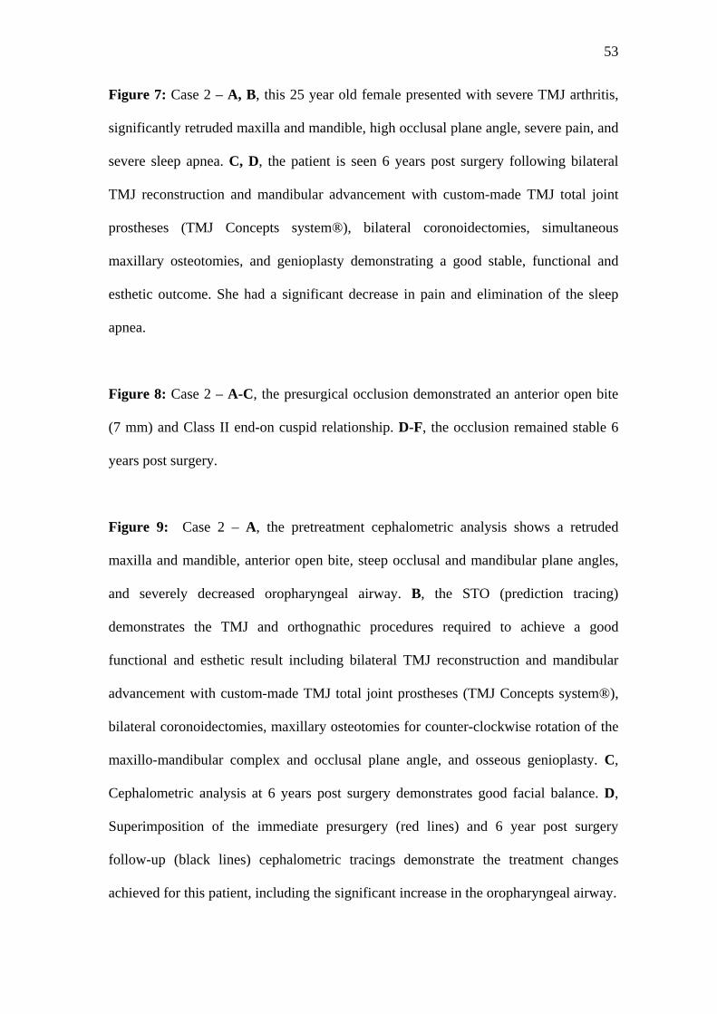
Figure 2. Superimposition of pre and post surgical lateral cephalograms demonstrate
the surgical changes achieved.
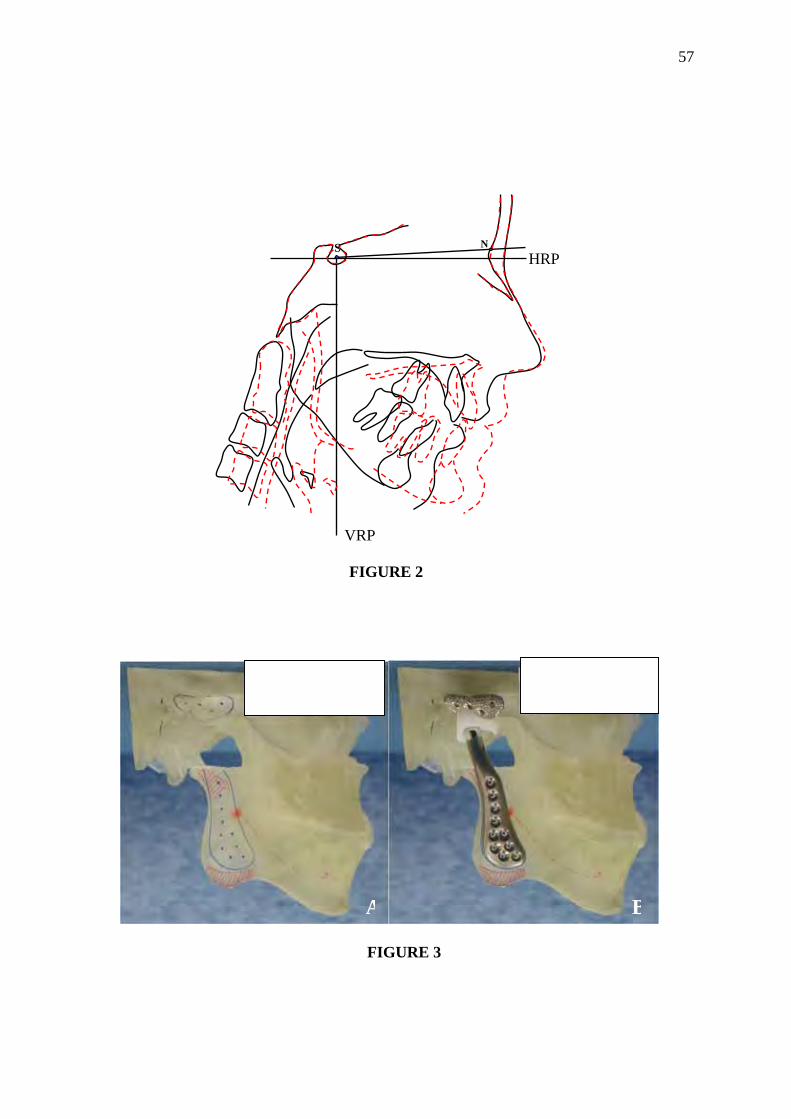
Figure 3: A, A 3-D stereolithography model of the patient’s jaws and jaw joints was
constructed from CT scan data. The mandible was repositioned on the model. The red
marks indicate areas of bony recontouring to facilitate the fit of the prosthesis. B,
Custom-fitted total joint prostheses are constructed to fit the specific anatomical
requirements for each patient.
Figure 4: Case 1 (CT, patient # 41) A, B, This 28 year old female is seen 4 years post
trauma with multiple mandibular fractures and loss of 8 maxillary teeth. She presents
with right TMJ severe arthritis and pain. The mandible and maxilla are significantly
retruded with a high occlusal plane angle and transverse asymmetry. C, D, the patient is
52
seen 79 months post surgery following right TMJ reconstruction and mandibular
advancement with custom-made TMJ total joint prostheses, left mandibular ramus
sagittal split for advancement, and maxillary osteotomies. E, F, The patient was
recently evaluated at 18 years post surgery showing the maintenance of good facial
balance.
Figure 5: Case 1 – A-C, the presurgical occlusion demonstrates anterior open bite and
the replacement of 7 teeth (left lateral incisor through the right 2nd bicuspid) with osseo-
integrated implants and prosthesis. D-F, the occlusion remained stable 79 months post
surgery. G-I, at 18 years post surgery, she maintained a stable occlusion.
Figure 6: Case 1 – A, the pretreatment cephalometric analysis shows a retruded
maxilla and mandible, anterior open bite, steep occlusal and mandibular plane angles,
vertical facial asymmetry, and significant degenerative changes in the right condyle . B,
the STO (prediction tracing) demonstrates the TMJ and orthognathic procedures
required to achieve a good functional and esthetic result including right TMJ
reconstruction and mandibular advancement with custom made TMJ total joint
prostheses, left mandibular ramus sagittal osteotomy, right coronoidectomy, and
maxillary osteotomies for counter-clockwise rotation and transverse leveling of the
maxillo-mandibular complex. C, Cephalometric analysis at 18 years post surgery
demonstrates good facial balance. D, Superimposition of the immediate post surgery
(red lines) and 18 year follow-up (black lines) cephalometric tracings demonstrate the
treatment stability achieved for this patient.
53
Figure 7: Case 2 – A, B, this 25 year old female presented with severe TMJ arthritis,
significantly retruded maxilla and mandible, high occlusal plane angle, severe pain, and
severe sleep apnea. C, D, the patient is seen 6 years post surgery following bilateral
TMJ reconstruction and mandibular advancement with custom-made TMJ total joint
prostheses (TMJ Concepts system®), bilateral coronoidectomies, simultaneous
maxillary osteotomies, and genioplasty demonstrating a good stable, functional and
esthetic outcome. She had a significant decrease in pain and elimination of the sleep
apnea.
Figure 8: Case 2 – A-C, the presurgical occlusion demonstrated an anterior open bite
(7 mm) and Class II end-on cuspid relationship. D-F, the occlusion remained stable 6
years post surgery.
Figure 9: Case 2 – A, the pretreatment cephalometric analysis shows a retruded
maxilla and mandible, anterior open bite, steep occlusal and mandibular plane angles,
and severely decreased oropharyngeal airway. B, the STO (prediction tracing)
demonstrates the TMJ and orthognathic procedures required to achieve a good
functional and esthetic result including bilateral TMJ reconstruction and mandibular
advancement with custom-made TMJ total joint prostheses (TMJ Concepts system®),
bilateral coronoidectomies, maxillary osteotomies for counter-clockwise rotation of the
maxillo-mandibular complex and occlusal plane angle, and osseous genioplasty. C,
Cephalometric analysis at 6 years post surgery demonstrates good facial balance. D,
Superimposition of the immediate presurgery (red lines) and 6 year post surgery
follow-up (black lines) cephalometric tracings demonstrate the treatment changes
achieved for this patient, including the significant increase in the oropharyngeal airway.
54
Age Previous TMJ Surg Follow-up n
Name Gender (years) TMJ Diagnoses
Right Left (months) 1 PA F 29 (B) A, (B) FA, (B) SI, (B) FBGCR 3 3 16 2 JA F 34 (B) A, (B) PTI, (B) FBGCR 1 2 25 3 JB F 50 (B) A, (B) PTI, (B) FBGCR 3 3 61 4 GB F 38 (B) A 0 0 46 5 SB F 21 (B) A 0 0 37 6 SBu F 52 (B) A 0 0 26 7 JD F 38 (B) A 0 0 37 8 CE F 14 (B) A 0 0 13 9 DE F 42 (B) A 0 0 32 10 MF F 36 (B) A 1 1 48 11 JF F 42 (B) A 0 0 12 12 KG F 39 (B) A 0 0 14 13 CG F 27 (B) A, (B) FA, (B) PTI, (B) FBGCR 3 3 32 14 EG F 43 (B) PsA 1 1 22 15 CGa F 44 (L) A 0 0 51 16 PG F 32 (B) BA, (B) SI, (B) FBGCR 2 2 12 17 SG F 45 (B) A, (B) FA, (B) F Vitek TJP, (B) FBGCR 6 3 14 18 KH F 42 (B) BA, (B) PTI, (B) FRG, (B) FBGCR 4 5 46 19 VH F 37 (B) A 0 0 22 20 KHo F 20 (B) ICR 0 0 12 21 KHa F 36 (B) A, (B) PTI, (B) SI, (B) FBGCR 3 3 35 22 CH F 57 (B) A 0 0 14 23 SJ F 18 (B) ICR 0 0 19 24 SL F 17 (B) JRA 0 0 25 25 EL F 42 (B) F Christ TJP, (B) FBGCR 1 1 14 26 NL F 46 (B) A 3 3 14 27 LL F 26 (L) A, (L) PTI, (L) FBGCR 0 1 62 28 EM F 44 (L) A 0 4 53 29 LM F 21 (B) ICR 0 0 13 30 MM F 34 (B) A, (B) PTI, (B) FBGCR 2 2 24 31 CM F 22 (B) ICR 2 2 51 32 RN F 28 (B) A 0 4 35 33 BO F 43 (B) A, (B) PTI, (B) FBGCR 1 1 49 34 DP F 30 (B) A, (B) PTI, (B) F Vitek TJP, (B) FBGCR 6 6 108 35 BP F 41 (B) PTI, (B) SI, (B) F Christ TJP, (B) FBGCR 8 8 16 36 LR F 21 (B) A 2 2 43 37 DS F 37 (B) A, (B) PTI, (B) FBGCR 2 2 91 38 LS F 41 (B) A 0 0 99 39 KS F 15 (B) JRA 0 0 86 40 ES F 35 (B) F Vitek TJP, (B) FBGCR 3 3 56 41 CT F 28 (R) A, (R) SbCoFx 0 0 79 42 GT F 21 (B) ICR 2 1 18 43 CTu F 51 (B) F Vitek TJP, (B) FBGCR 2 2 35 44 KV F 37 (B) SI, (B) F Vitek TJP, (B) FBGCR 5 4 143 45 CW F 36 (B) PTI, (B) FRG, (B) BA 4 4 51 46 CWe F 46 (B) A 1 0 24 47 TW F 25 (B) RA 1 1 72
Abreviations
(B) - Bilateral
(L) - Left side
(R) - Right side
A - Arthritis
RA - Rheumatoid Arthritis
JRA - Juvenal Rheumatoid Arthritis
PsA - Psoriatic Arthritis
ICR - Idiopatic Condylar Resorption
PTI - Proplast/Teflon Implant
SI - Silastic Implant
FRG - Failed Rib Graft
BA - Bony Ankylosis
FA - Fibrous Ankylosis
SbCoFx - Sub-condylar Fracture
F Vitek TJP - Failed Vitek-Kent Total Joint Prosthesis
F Christ TJP - Failed Christensen Total Joint Prosthesis
FBGCR - Foreign Body Giant Cell Reaction
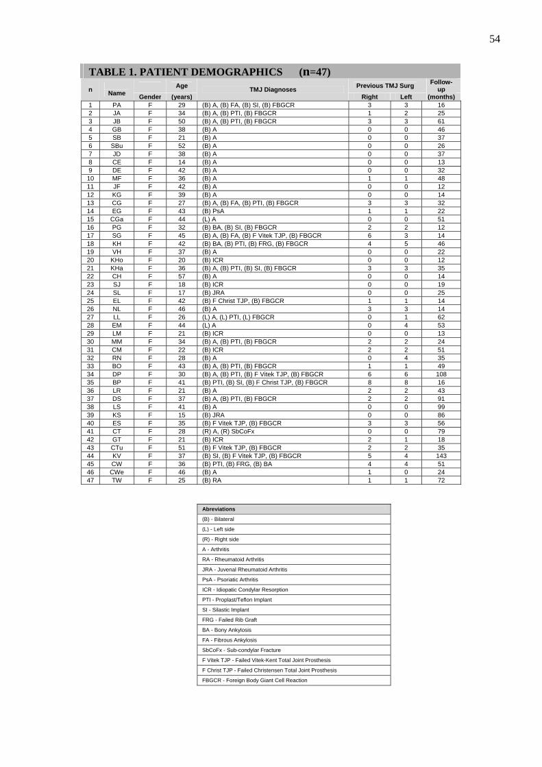
TABLE 1. PATIENT DEMOGRAPHICS (n=47)
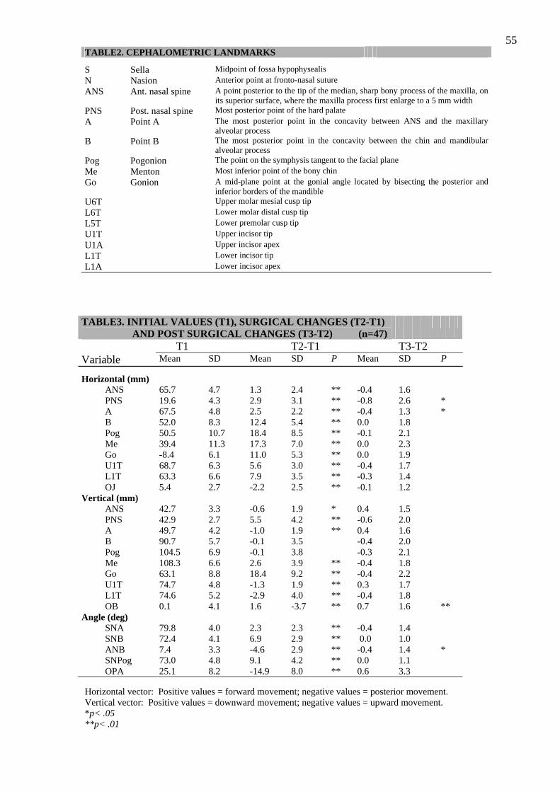
55TABLE2. CEPHALOMETRIC LANDMARKS
S
Sella Midpoint of fossa hypophysealis
N Nasion Anterior point at fronto-nasal suture ANS Ant. nasal spine A point posterior to the tip of the median, sharp bony process of the maxilla, on
its superior surface, where the maxilla process first enlarge to a 5 mm width PNS Post. nasal spine Most posterior point of the hard palate A Point A The most posterior point in the concavity between ANS and the maxillary
alveolar process B Point B The most posterior point in the concavity between the chin and mandibular
alveolar process Pog Pogonion The point on the symphysis tangent to the facial plane Me Menton Most inferior point of the bony chin Go Gonion A mid-plane point at the gonial angle located by bisecting the posterior and
inferior borders of the mandible U6T Upper molar mesial cusp tip L6T Lower molar distal cusp tip L5T Lower premolar cusp tip U1T Upper incisor tip U1A Upper incisor apex L1T Lower incisor tip L1A Lower incisor apex
Horizontal vector: Positive values = forward movement; negative values = posterior movement. Vertical vector: Positive values = downward movement; negative values = upward movement. *p< .05 **p< .01
TABLE3. INITIAL VALUES (T1), SURGICAL CHANGES (T2-T1) AND POST SURGICAL CHANGES (T3-T2) (n=47)
T1 T2-T1 T3-T2
Variable Mean SD Mean SD P Mean SD P
Horizontal (mm) ANS 65.7 4.7 1.3 2.4 ** -0.4 1.6 PNS 19.6 4.3 2.9 3.1 ** -0.8 2.6 * A 67.5 4.8 2.5 2.2 ** -0.4 1.3 * B 52.0 8.3 12.4 5.4 ** 0.0 1.8 Pog 50.5 10.7 18.4 8.5 ** -0.1 2.1 Me 39.4 11.3 17.3 7.0 ** 0.0 2.3 Go -8.4 6.1 11.0 5.3 ** 0.0 1.9 U1T 68.7 6.3 5.6 3.0 ** -0.4 1.7 L1T 63.3 6.6 7.9 3.5 ** -0.3 1.4 OJ 5.4 2.7 -2.2 2.5 ** -0.1 1.2 Vertical (mm) ANS 42.7 3.3 -0.6 1.9 * 0.4 1.5 PNS 42.9 2.7 5.5 4.2 ** -0.6 2.0 A 49.7 4.2 -1.0 1.9 ** 0.4 1.6 B 90.7 5.7 -0.1 3.5 -0.4 2.0 Pog 104.5 6.9 -0.1 3.8 -0.3 2.1 Me 108.3 6.6 2.6 3.9 ** -0.4 1.8 Go 63.1 8.8 18.4 9.2 ** -0.4 2.2 U1T 74.7 4.8 -1.3 1.9 ** 0.3 1.7 L1T 74.6 5.2 -2.9 4.0 ** -0.4 1.8 OB 0.1 4.1 1.6 -3.7 ** 0.7 1.6 ** Angle (deg) SNA 79.8 4.0 2.3 2.3 ** -0.4 1.4 SNB 72.4 4.1 6.9 2.9 ** 0.0 1.0 ANB 7.4 3.3 -4.6 2.9 ** -0.4 1.4 * SNPog 73.0 4.8 9.1 4.2 ** 0.0 1.1 OPA 25.1 8.2 -14.9 8.0 ** 0.6 3.3

56
FIGURE 1
S
Go
Me Pog
B
N
ANS
A
L1T L5T
U6T L6T
HRP
VRP
PNS
U1A
L1A
U1T
57
FIGURE 2
FIGURE 3
S NHRP
VRP
A B

58
FIGURE 4
FIGURE 5
A
B
C
D
E
F
A
B
C
D
E
F
G
H
I
59
FIGURE 6
B
A C
D

60
FIGURE 7
FIGURE 8
A B C
D E F
A
B
C
D
61
FIGURE 9
B
A C
D
Maxillo-Mandibular Counter-Clockwise Rotation and Mandibular
Advancement with TMJ Concepts® Total Joint Prostheses:
Part II – Airway Changes and Stability
1Karina E. Dela Coleta, 2Larry M. Wolford, 1João R. Gonçalves, 1Ary dos Santos Pinto, 2Daniel Serra Cassano, 3Daniela A. Godoy Gonçalves,
1Pediatric Dentistry Department - Araraquara Dental School, Sao Paulo State University, Brazil 2Department of Oral and Maxillofacial Surgery, Texas A&M University Health Care Center, Baylor College of Dentistry, and Baylor University Medical Center. Dallas, TX. 3Department of Prosthodontics - Araraquara Dental School, Sao Paulo State University, Brazil Address correspondence and reprint requests to: Larry M. Wolford, DMD: 3409 Worth St, Suite 400 Dallas, TX 75246 Phone: 214-828-9115 Phone: 214-828-1714 E-mail: [email protected]
63
Abstract
The purpose of this study was to evaluate the anatomical changes and stability of the
oropharyngeal airway and head posture following TMJ reconstruction and mandibular
advancement with TMJ Concepts custom-made total joint prostheses and maxillary
osteotomies with counter-clockwise rotation of the maxillo-mandibular complex.
Forty-seven females (14 to 57 years old) were included in the study with an average
post surgical follow-up was 40.6 months (range 12 to 143 months). The patients’
lateral cephalograms were traced, and analyzed to determine surgical (T2-T1) and post
surgical changes (T3-T2) of the oropharyngeal airway, hyoid bone, and head posture.
The skeletal and dental changes for this same patient population were presented in Part
I of this series of studies. The results of this study showed that surgery increased the
narrowest retroglossal airway space (PASnar) 4.9 mm (range -3.5 to 15.7 mm). Head
posture (OPT/NS) showed flexure immediately after surgery (-5.6 ± 6.7o) and
extension long-term post surgery (1.8 ± 6.7o), while cervical curvature (OPT/CVT) had
no significant change. Surgery increased the distances between the third cervical
vertebrae and menton (C3-Me) 11.7 ± 9.1 mm and the third cervical vertebrae and
hyoid (Hy-C3) 3.2 ± 3.9 mm, and remained stable. The distance from the hyoid to the
mandibular plane (MP-Hy) decreased during surgery (-3.8 ± 5.8 mm) and after surgery
(-2.5 ± 5.2 mm). Maxillo-mandibular advancement with counter-clockwise rotation
and TMJ reconstruction with total joint prostheses produced immediate increase in
oropharyngeal airway dimension, which was influenced by long-term changes in head
posture but remained stable over the follow-up period.
Key words: Airway Changes; Stability; Orthognathic surgery; TMJ prostheses

64
Introduction
Important relationships exist between the pharyngeal structures and the
development of the face and occlusion. The oropharyngeal airway can influence the
growth of craniofacial structures, by creating postural changes capable of affecting the
relationship of teeth as well as the direction of jaw growth which may develop in a
downward and backward direction17. On the other hand, skeletal features in patients
with high occlusal plane angle facial morphologies (HOP) may be related to the
etiology of a narrower dimension of the airway10.
HOP patients commonly exhibit increased anterior lower face height, retruded
mandible and maxilla, Class II malocclusion, high occlusal plane angle and a
decreased oropharyngeal airway16. Also, those patients frequently have TMJ problems
with some cases resulting in progressing condylar resorption. This can decrease ramus
height and increase mandibular retrusion and respiratory disturbances due to upper
airway obstruction14.
Correction of these deformities in adolescents and adults to achieve optimal
functional and esthetic results requires orthognathic surgery, with counter-clockwise
rotation of the maxillo-mandibular complex and decrease of the occlusal plane angle25.
In cases of TMJ irreversible damage, it may be necessary to reconstruct the TMJ and
advance the mandible using a total joint prosthesis26.
According to Mehra et al.13 maxillo-mandibular advancement surgery
(mandibular advancement of 7.5 mm) with counter-clockwise rotation increased the
oropharyngeal airway spaces 3.5 mm in the retropalatal region and 5.7 mm in the
65
retroglossal region, representing a 76% increase in the retroglossal oropharyngeal
airway dimension relative to the amount of mandibular advancement. Other studies
have reported airway increases ranging from 42% to 51%5,12.
However, the oropharyngeal airway is not the only structure improved with
the mandibular advancement and/or counter-clockwise rotation. Previous
studies1,7,9,11,22,24 have shown that there are also changes in hyoid bone position due to
mandibular advancement. Post surgical hyoid bone position may reflect the stretching
of the suprahyoid musculature and occupy an important role in the maintenance of the
oropharyngeal airway space. Potential muscle tension increase is thought, by some
authors, to be related to skeletal relapse1,22-24. Schendel and Epker22 reported that the
hyoid bone tends to return almost to its original presurgical position after a certain post
surgical period following mandibular advancement with inter-maxillary fixation.
LaBanc and Epker11 reported immediate post surgical movement of the hyoid bone in
an anterior direction, but, at the same time, they emphasized the ‘‘highly variable’’
nature of the post surgical position of the hyoid. Most studies describe changes in
hyoid bone position and pharyngeal airway size 1 to 2 years post surgery1,7,9,11,23,24.
Mandibular advancement surgery has been previously shown to increase
oropharyngeal airway space5, however data on this procedure associated to TMJ total
joint reconstruction and large mandibular advancements is not available. Although
stability using total joint prostheses has been established, changes in airway space and
its stability must be studied.
66
To help resolve the issue concerning post surgical stability and to better
understand counter-clockwise rotation of the occlusal plane and the affect on the
oropharyngeal airway, the present study tests the following null hypotheses:
1) There is no increase in the oropharyngeal airway space with TMJ total joint
reconstruction, maxillo-mandibular advancement surgery and counter-clockwise
rotation of the occlusal plane.
2) The oropharyngeal airway space does not remain stable over the post surgical
period.
Patients and methods
This retrospective study evaluated treatment records from a single private
practice, from 1990 through 2003, of patients with end-stage TMJ pathology, retruded
maxilla and mandible, and high occlusal plane angle. All patients were operated by
one of the authors (LMW) at Baylor University Medical Center, Dallas, TX, USA.
Patients were selected according to the inclusion and exclusion criteria as presented in
Part I2. The patient sample was the same as Part I2 and included 47 female patients,
who underwent TMJ reconstruction and mandibular advancement with total joint
prostheses and simultaneous maxillary osteotomies with counter-clockwise rotation of
the maxillo-mandibular complex and occlusal plane. The study demographics are
presented in Table 1, Part I2 of this series of studies. There were 43 patients treated
with bilateral TMJ total joint prostheses and 4 patients had unilateral prosthesis and
sagittal split osteotomy on the contralateral side. The occlusal plane angle was
decreased in all subjects by posterior down grafting the maxilla and/or anterior
maxillary upward positioning with counter-clockwise rotation of the maxillo-
mandibular complex. Mean patient age at the time of surgery was 34.5 years (range 14
67
to 57 years). The surgical technique and post operative management are presented in
Part I2 of this series. All osteotomies were rigidly stabilized using bone plates and
screws without using post surgical maxillo-mandibular fixation, but light force vertical
elastics were used on most patients for a minimum of 2 to 4 weeks to control the
occlusion and provide vertical support to the mandible until the muscles of mastication
reattached to the mandible and regained function.
The custom-made total joint prostheses used in this study, were originally
developed in 1989 by Techmedica Inc., Camarillo, CA, USA, and since 1997, have
been manufactured by TMJ Concepts, Inc., Ventura, CA, USA. These prostheses are
CAD/CAM devices (computer assisted design/computer assisted manufacture),
designed to fit the specific anatomical requirements for each patient.
Imaging evaluation
For all patients, lateral cephalometric radiographs were taken using a standard
radiographic technique (centric relation with Frankfort horizontal parallel to the floor)
at the following intervals: T1- immediately before surgery (range 1 to 6 days), T2-
immediate post surgery (range 2 to 16 days), and T3- long-term follow-up (range 12 to
143 months). The radiographs were randomly traced by 1 of the examiners and
digitized twice by another examiner approximately 1 week apart. There were 16
landmarks (Table 1) identified and digitized by using DFPlus software (Dentofacial
Software Inc, Toronto, Canada). The landmarks were used to compute 28
measurements describing airway dimensions, head position, cervical curvature, hyoid
position, and maxillo-mandibular relationships. S-N minus 7° was used as the
horizontal reference plane (HRP), and a perpendicular line to HRP through sella was
68
used as the vertical reference plane (VRP). The horizontal and vertical changes for
each landmark were evaluated (Fig 1). Surgical change (T2-T1) and long-term stability
(T3-T2) were calculated and statistically analyzed.
Error of measurement
To determine the consistency of the method, the two examiners were
previously calibrated by repetition of the process until the method was considered
adequate by a third examiner. Each lateral cephalogram was traced twice to evaluate
for errors in landmark localization (“random error”) during tracing and each lateral
cephalometric radiograph got a medium of the measurements. The intra-examiner
consistency (ICC) was calculated for reliability of tracing, landmark identification, and
analytical measurements showing a correlation coefficient always greater than 0.94.
Statistical method
All data were transferred to SPSS (release 9.0; SPSS Chicago, IL) for statistical
analysis. The skewness and kurtosis statistics showed normal distributions for all
variables. Unilateral and bilateral TMJ prostheses were compared. Because there were
no statistically significant differences between these groups in post surgical changes,
all the patients were analyzed as a single group. Paired t tests were performed to
evaluate the surgical (T2-T1) and post surgical changes (T3-T2). A significance level
of p <.05 was applied. Pearson product-moment correlations were used to determine
the relationships between changes of specific anatomical measurements and
oropharyngeal airway space changes. Correlations were also used to assess the
association between surgical and post surgical changes in the oropharyngeal airway
space.
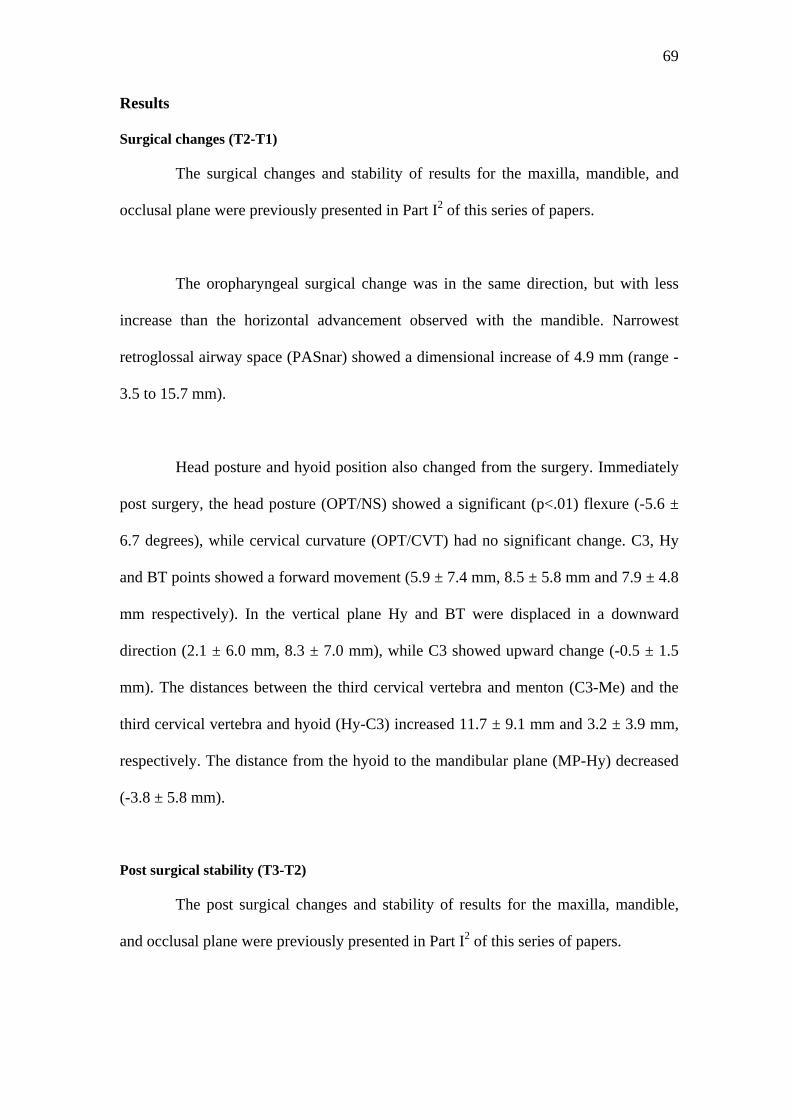
69
Results
Surgical changes (T2-T1)
The surgical changes and stability of results for the maxilla, mandible, and
occlusal plane were previously presented in Part I2 of this series of papers.
The oropharyngeal surgical change was in the same direction, but with less
increase than the horizontal advancement observed with the mandible. Narrowest
retroglossal airway space (PASnar) showed a dimensional increase of 4.9 mm (range -
3.5 to 15.7 mm).
Head posture and hyoid position also changed from the surgery. Immediately
post surgery, the head posture (OPT/NS) showed a significant (p<.01) flexure (-5.6 ±
6.7 degrees), while cervical curvature (OPT/CVT) had no significant change. C3, Hy
and BT points showed a forward movement (5.9 ± 7.4 mm, 8.5 ± 5.8 mm and 7.9 ± 4.8
mm respectively). In the vertical plane Hy and BT were displaced in a downward
direction (2.1 ± 6.0 mm, 8.3 ± 7.0 mm), while C3 showed upward change (-0.5 ± 1.5
mm). The distances between the third cervical vertebra and menton (C3-Me) and the
third cervical vertebra and hyoid (Hy-C3) increased 11.7 ± 9.1 mm and 3.2 ± 3.9 mm,
respectively. The distance from the hyoid to the mandibular plane (MP-Hy) decreased
(-3.8 ± 5.8 mm).
Post surgical stability (T3-T2)
The post surgical changes and stability of results for the maxilla, mandible,
and occlusal plane were previously presented in Part I2 of this series of papers.
70
The narrowest retroglossal airway space (PASnar) remained stable post
surgery, although BT point showed a postero-superior movement. The hyoid moved
superiorly, thereby decreasing its distance from the mandibular plane (-2.5 ± 5.2 mm).
However, there was no significant change in the distance between the hyoid and the
third cervical vertebra (Hy-C3) and in the distance between the third cervical vertebra
and menton (C3-Me). Head posture (OPT/NS) showed 1.8 ± 6.7 degrees of extension
while cervical curvature (OPT/CVT) had no change.
Correlations
The correlations showed that surgical increase in oropharyngeal airway space
was associated with a variety of other changes (Table 3). All the mandibular horizontal
measurements (B, Pog, Me, Go) showed positive correlation with the oropharyngeal
measurement (PASnar). The further the mandible was advanced, the greater the
dimensional increase of PASnar. This correlation was also found at A point in the
horizontal direction, showing the influence of the maxillary position on the airway
space. In vertical direction, only Go showed positive correlation with the increase of
oropharyngeal airway space. Similarly, patients with greater increases in the distances
between C3 and menton (C3-Me) and between C3 and hyoid (Hy-C3) showed greater
increases in PASnar immediately post surgery. This correlation was also true relative
to the tongue (BT) in the horizontal direction.
The head position was correlated with the oropharyngeal airway space.
Patients with greater head extension (OPT/NS) showed greater increases of PASnar.
Although the oropharyngeal airway space showed a positive correlation with a variety
71
of other changes, the occlusal and the mandibular plane angles, and C3-horizontal
showed a negative correlation with the increase of PASnar.
Positive correlations were identified for the mandibular and head position
measurements with Me horizontal and C3-Me as well as Me horizontal and Hy-C3
(Table 4). However, there were negative correlations between the occlusal plane angle
(OPA) and C3-Me, OPA and HY-C3 as well as Me vertical and MP-Hy. The greater
the mandibular counter-clockwise rotation (decrease OPA and MPA), the greater the
increase of the distances between C3-Me and Hy-C3.
Long-term post surgery (Table 3), the strongest correlations were found
between changes in oropharyngeal dimension and head position (OPT/NS), mandibular
position (C3-Me), hyoid position (Hy horizontal and Hy-C3) and C3 position (C3
horizontal and vertical). Patients who extended their heads (OPT/NS) more over the
post surgical period showed greater increase in PASnar dimension. The oropharyngeal
airway measurement showed greater increase for patients who increased the distances
between C3 and Me and between C3 and Hy in the follow-up period. The long-term
post surgical changes in Me related with the head and hyoid positions, showed a
positive correlation between Hy-C3 and Me in the horizontal direction, and between
OPT/NS and Me-vertical. Therefore, the greater extension of head position and C3
change resulted in greater anterior and superior movement of menton post surgery.
Discussion
The high occlusal plane facial type (HOP) patient, also called long face
syndrome, dolicocephalic or hyperdivergent, has known morphological characteristics
72
including maxillo-mandibular clockwise growth pattern, decreased oropharyngeal
airway space, and TMJ problems10,13,25,26. The comprehensive treatment for those
patients includes maxillo-mandibular counter-clockwise rotation that will improve
function and facial balance as well as permanently increase the oropharyngeal airway
space. Patients with end-stage TMJ pathology such as connective tissue autoimmune
diseases, idiopathic condylar resorption, ankylosis, severe trauma, more than two failed
TMJ surgeries, and so on, may benefit from TMJ reconstruction (using TMJ Concepts
total joint prostheses), simultaneously with maxillo-mandibular counter-clockwise
rotation, in order to provide the best stability, improve function and esthetics, as well
as decrease TMJ associated pain and other symptoms.
There are a variety of ways to evaluate the oropharyngeal airway space changes
associated with maxillo-mandibular orthognathic surgery including lateral
cephalometry, computed tomography, polysomnography, and nasopharyngoscopy12,20.
In our study, the PAS was measured on lateral cephalometric films that were taken
with patients in a sitting position. This technique has the advantage of being expedient
and convenient, since it is a conventional document required for orthognathic surgery
during planning and follow-up evaluation. Riley et al.20 found a good correlation
between the posterior airway space and the pharyngeal space measured by
cephalometric radiography and computed tomography.
According to Fitzpatrick et al.6, the upper airway dimensions are also
influenced by the patient’s position, either sitting or supine. This positional influence
of upper airway dimensions can be related to the effects of gravity on upper airway
structures and to the lung volume dependency of upper airway patency. In our sample

73
all lateral cephalometric films were obtained in the same sitting position, so this
variable had a small impact on the method used to determine the surgical (T2-T1) and
post surgical (T3-T2) changes. Landmark definition and digitalization were tested and
found to have high intra-class correlation coefficient (greater than 0.94). This
minimized bias and increased the dependability of the study.
The methodology applied in our research used only one measurement to
evaluate the narrowest retroglossal airway (PAS), differing from other studies4,8,13,14
where 2 or more measurements were used to show airway changes due to the surgery.
This fact can be justified by the impossible task of identifying some landmarks after
surgery (T2 and T3), since the mandibular component of the TMJ total joint prostheses
overlapped and obscured the mandibular ramus and portions of the oropharyngeal soft
tissue structures.
The surgical skeletal and dental changes (T2-T1) and longest follow-up
stability results (T3-T2) are presented in Part I2 of this series of papers. The counter-
clockwise rotation of the maxillo-mandibular complex advanced the mandible an
average of 12.4 mm at B point, 18.4 mm at Pogonion, and 17.3 mm at Menton.
Mandibular advancement measured at menton was substantially greater than the
incisor edge as a result of the counter-clockwise rotation, which demonstrates the
advantage of this movement in high occlusal plane angle facial type patients. All
surgical movements (T2-T1) remained stable during the follow-up period (T3-T2),
except for minimal horizontal changes that occurred at point A and PNS.
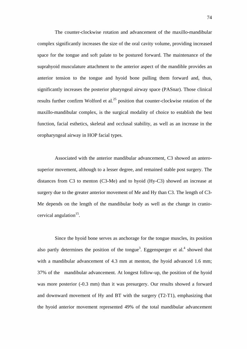
74
The counter-clockwise rotation and advancement of the maxillo-mandibular
complex significantly increases the size of the oral cavity volume, providing increased
space for the tongue and soft palate to be postured forward. The maintenance of the
suprahyoid musculature attachment to the anterior aspect of the mandible provides an
anterior tension to the tongue and hyoid bone pulling them forward and, thus,
significantly increases the posterior pharyngeal airway space (PASnar). Those clinical
results further confirm Wolford et al.25 position that counter-clockwise rotation of the
maxillo-mandibular complex, is the surgical modality of choice to establish the best
function, facial esthetics, skeletal and occlusal stability, as well as an increase in the
oropharyngeal airway in HOP facial types.
Associated with the anterior mandibular advancement, C3 showed an antero-
superior movement, although to a lesser degree, and remained stable post surgery. The
distances from C3 to menton (C3-Me) and to hyoid (Hy-C3) showed an increase at
surgery due to the greater anterior movement of Me and Hy than C3. The length of C3-
Me depends on the length of the mandibular body as well as the change in cranio-
cervical angulation15.
Since the hyoid bone serves as anchorage for the tongue muscles, its position
also partly determines the position of the tongue3. Eggensperger et al.4 showed that
with a mandibular advancement of 4.3 mm at menton, the hyoid advanced 1.6 mm;
37% of the mandibular advancement. At longest follow-up, the position of the hyoid
was more posterior (-0.3 mm) than it was presurgery. Our results showed a forward
and downward movement of Hy and BT with the surgery (T2-T1), emphasizing that
the hyoid anterior movement represented 49% of the total mandibular advancement
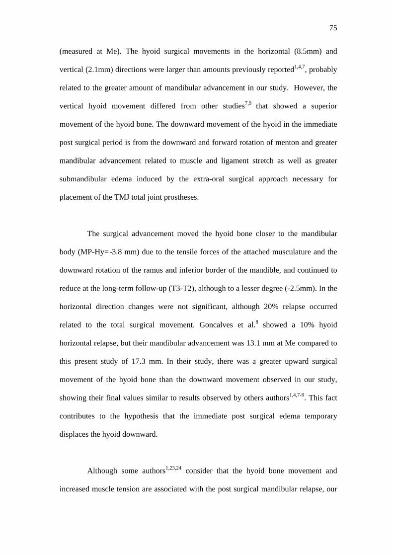
75
(measured at Me). The hyoid surgical movements in the horizontal (8.5mm) and
vertical (2.1mm) directions were larger than amounts previously reported1,4,7, probably
related to the greater amount of mandibular advancement in our study. However, the
vertical hyoid movement differed from other studies7,9 that showed a superior
movement of the hyoid bone. The downward movement of the hyoid in the immediate
post surgical period is from the downward and forward rotation of menton and greater
mandibular advancement related to muscle and ligament stretch as well as greater
submandibular edema induced by the extra-oral surgical approach necessary for
placement of the TMJ total joint prostheses.
The surgical advancement moved the hyoid bone closer to the mandibular
body (MP-Hy= -3.8 mm) due to the tensile forces of the attached musculature and the
downward rotation of the ramus and inferior border of the mandible, and continued to
reduce at the long-term follow-up (T3-T2), although to a lesser degree (-2.5mm). In the
horizontal direction changes were not significant, although 20% relapse occurred
related to the total surgical movement. Goncalves et al.8 showed a 10% hyoid
horizontal relapse, but their mandibular advancement was 13.1 mm at Me compared to
this present study of 17.3 mm. In their study, there was a greater upward surgical
movement of the hyoid bone than the downward movement observed in our study,
showing their final values similar to results observed by others authors1,4,7-9. This fact
contributes to the hypothesis that the immediate post surgical edema temporary
displaces the hyoid downward.
Although some authors1,23,24 consider that the hyoid bone movement and
increased muscle tension are associated with the post surgical mandibular relapse, our
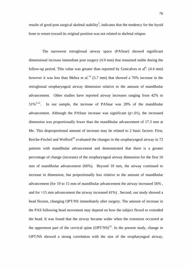
76
results of good post surgical skeletal stability2, indicates that the tendency for the hyoid
bone to return toward its original position was not related to skeletal relapse.
The narrowest retroglossal airway space (PASnar) showed significant
dimensional increase immediate post surgery (4.9 mm) that remained stable during the
follow-up period. This value was greater than reported by Goncalves et al8. (4.4 mm)
however it was less than Mehra et al.13 (5.7 mm) that showed a 76% increase in the
retroglossal oropharyngeal airway dimension relative to the amount of mandibular
advancement. Other studies have reported airway increases ranging from 42% to
51%5,12. In our sample, the increase of PASnar was 28% of the mandibular
advancement. Although the PASnar increase was significant (p<.01), the increased
dimension was proportionally lower than the mandibular advancement of 17.3 mm at
Me. This disproportional amount of increase may be related to 2 basic factors: First,
Reiche-Fischel and Wolford19 evaluated the changes in the oropharyngeal airway in 72
patients with mandibular advancement and demonstrated that there is a greater
percentage of change (increase) of the oropharyngeal airway dimension for the first 10
mm of mandibular advancement (66%). Beyond 10 mm, the airway continued to
increase in dimension, but proportionally less relative to the amount of mandibular
advancement (for 10 to 15 mm of mandibular advancement the airway increased 56% ,
and for >15 mm advancement the airway increased 41%) . Second, our study showed a
head flexion, changing OPT/NS immediately after surgery. The amount of increase in
the PAS following head movement may depend on how the subject flexed or extended
the head. It was found that the airway became wider when the extension occurred at
the uppermost part of the cervical spine (OPT/NS)15. In the present study, change in
OPT/NS showed a strong correlation with the size of the oropharyngeal airway,
77
however, OPT/CVT in the lower part of the cervical spine showed no change or only a
weak correlation to the size of the PAS. Considering that the normal value for PAS
based on lateral cephalometric radiograph is 11 ± 2 mm21, our results showed an
immediate post surgical PASnar average value of 12.2 mm that stabilized at 11.1 mm
at the longest follow-up period, demonstrating a final PAS measurement within normal
limits.
Head posture affects the pre and post surgery PAS dimension. Muto et al.15
observed a positive correlation between airway space and head posture of 0.807 (PAS-
OPT/NS). The present study showed a significant positive correlation between airway
increase and head posture of 0.456 with surgical changes and of 0.603 during the
follow-up period.
This cranio-cervical adaptation (OPT/NS) is influenced by the post surgical
supra-hyoid muscle tension, the direction and distance of surgical movement
(mandibular advancement or setback) and the changes in PAS. There is a strong
tendency toward head flexion after most orthognathic surgical procedures. It was
previously reported18 that patients with high occlusal plane facial morphology treated
by maxillary intrusion (no counter-clockwise rotation of the maxillo-mandibular
complex) had a somewhat extended head posture presurgically. Their head flexion post
surgery brought them toward the center of the normal range, but only temporarily.
According to the authors, patients who undergo mandibular advancement are in the
middle of the normal range for head posture prior to treatment, but they have about the
same amount of transient flexion. In our study, patients flexed their heads after surgery
(average, 5.6o) and extended it 1.8o during the long term follow-up. Previous studies
78
have reported 0.65o to 3.4o of head flexure following advancement surgery7,18,22. The
amount of head flexion observed in our study may be due to the immediate surgical
counter-clockwise rotation of the maxillo-mandibular complex, promoting a greater
amount of mandibular advancement with subsequent increased tension of the supra and
infra-hyoid musculature. The slight extension of the head post surgery (T3-T2) was
probably a result of the supra- and infra-hyoid musculature adapting to the surgical
changes.
PASnar showed a significant negative correlation (- 0.726) between the
surgical movement (T2-T1) and the post surgical changes (T3-T2). The father the
PASnar was changed surgically, the greater the chance of instability of this area.
Considering that OPT/NS didn’t show significant movement after surgery, we can’t
attribute this negative correlation to the post surgical head extension.
The correlation of maxillo-mandibular bone movement with PASnar showed a
positive correlation with A point, B point, Pog and Me points in a horizontal direction,
and Go in the horizontal and vertical directions. The further the maxillo-mandibular
complex was advanced and Go was moved inferiorly, the greater the increase of PAS.
Although those correlations were statistically significant, the values were low probably
due to the lack of standardized head posture resulting in an increase of the variability
of OPT/NS at different times (T2- OPT/NS= 105.6 ± 7.1 degrees; T3-OPT/NS= 107.4
± 8.1 degrees). Also, immediately post surgery, the tongue (BT) advancement showed
a positive correlation with the PASnar.
79
Occlusal and mandibular plane angle changes showed a negative correlation
with PASnar. Those angulations were reduced relative to the HRP line by the
mandibular counter-clockwise rotation, increasing the airway space. This same
negative correlation was observed at the third vertebra (C3) position in vertical and
horizontal directions during the follow-up period.
C3-Me and Hy-C3, measurements showed a positive correlation with PASnar
in the immediate and long-term evaluations. The anterior movement of Me was
positively correlated to increases in C3-Me which was directly correlated to the
PASnar measurement. These correlations were stronger than observed by Goncalves et
al.8 The data indicated that oropharyngeal airway improvement due to the counter-
clockwise rotation of the maxillo-mandibular complex would be significantly greater if
the patients had maintained the same head position during the 3 different times of
evaluation. The lack of head and neck positional during radiographic acquisition, was
directly related to the variability observed for OPT/NS and OPT/CVT at T1, T2, T3
and to the lesser increase than expected for the oropharyngeal airway dimension.
Changes in head and neck posture immediately post surgery constrained the
immediate oropharyngeal airway improvement. Muto et al.15 showed that a change of
10° in OPT/NS produced about 4 mm of change in the PAS. They also showed that the
distance between C3 to Me was related to the oropharyngeal airway improvement,
which was also observed in the present study. However, it is important to emphasize
that although the majority of measurements showed a statistically significant
correlation with PASnar, they were low (mostly less than 0.5). This could be related in
part to the influence of head posture in the airway dimension.
80
The results of this study show that: (1) maxillo-mandibular advancement
surgery with counter-clockwise occlusal plane rotation improved the oropharyngeal
airway dimensions; (2) changes of C3, Hy and BT distances to mandibular
advancement were correlated with oropharyngeal airway changes; (3) head position
influenced the amount of increase in oropharyngeal airway dimensions that occurred
after maxillo-mandibular advancement surgery, (4) the oropharyngeal airway space
remained stable over the post surgical follow-up period; (5) total joint prostheses
provide stability of maxillo-mandibular counter-clockwise rotation and consequently
of the narrowest retroglossal airway space (PASnar).
References
1. Chung DH, Hatch JP, Dolce C, Van Sickels JE, Bays RA, Rugh JD. Positional
change of the hyoid bone after bilateral sagittal split osteotomy with rigid and wire
fixation. Am J Orthod Dentofacial Orthop 2001;119:382-389.
2. Coleta KED, Wolford LM, Goncalves JR, Santos-Pinto A, Pinto LP, Cassano DS.
Maxillo-Mandibular Counter-Clockwise Rotation and Mandibular Advancement
with TMJ Concepts ® Total Joint Prostheses: Part I Skeletal and Dental Stability –
IJOMS-D-07-00531.
3. Djupesland G, Lyberg T, Krogstad O. Cephalometric analysis and surgical treatment
of patients with obstructive sleep apnea syndrome. Acta Otolaryngol (Stockh)
1987;103:551-557.
4. Eggensperger N, Smolka K, Johner A, Rahal A, Thüer U, Iizuka T. Long-term
changes of hyoid bone and pharyngeal airway size following advancement of the
mandible. Oral Surg Oral Med Oral Pathol Oral Radiol Endod 2005;99:404-410.
81
5. Farole A, Mundenar MJ, Braitman LE. Posterior airway changes associated with
mandibular advancement surgery: implications for patients with obstructive sleep
apnea. Int J Adult Orthodont Orthognath Surg 1990;5:255-258.
6. Fitzpatrick MF, Yildirim Y, Jalleh R, Wighman AJA, Douglas NJ. Posture affects
cephalometric measurements in patients with obstructive sleep apnea [abstract].
Am Rev Respir Dis 1990;141:A858.
7. Gale A, Kilpeläinen PV, Laine-Alava MT. Hyoid bone position after surgical
mandibular advancement. Eur J Orthod 2001;23: 695-701.
8. Goncalves JR, Buschang P, Gonçalves DG, Wolford LM. Postsurgical stability of
oropharyngeal airway changes following counter-clockwise maxillo-mandibular
advancement surgery. J Oral Maxillofac Surg 2006;64:755-762.
9. Hayes RJ, Sarver DM, Jacobson A. The quantification of soft tissue cervicomental
changes after mandibular advancement surgery. Am J Orthod Dentofacial Orthop
1994;105:383-391.
10. Joseph AA, Elbaum J, Cisneros GJ, Eisig SB. A cephalometric comparative study
of the soft tissue airway dimensions in persons with hyperdivergent and
normodivergent facial patterns. J Oral Maxillofac Surg 1998;56:135-139.
11. LaBanc JP, Epker BN. Changes of the hyoid bone and tongue following
advancement of the mandible. Oral Surg Oral Med Oral Pathol Oral Radiol Endod
1984;57:351-356.
12. Li KK, Guilleminault C, Riley RW, Powell NB. Obstructive sleep apnea and
maxillomandibular advancement: An assessment of airway changes using
radiographic and nasopharyngoscopic examinations. J Oral Maxillofac Surg
2002;60:526-530.
82
13. Mehra P, Downie M, Pita M, Wolford LM. Pharyngeal airway space changes after
counterclockwise rotation of the maxillomandibular complex. Am J Orthod
Dentofacial Orthop 2001;120:154-159.
14. Mishima K, Yamada T, Sugahara T. Evaluation of respiratory status and
mandibular movement after total temporomandibular joint replacement in patients
with rheumatoid arthritis. Int J Oral Maxillofac Surg 2003;32:275–279.
15. Muto T, Takeda S, Kanazawa M, Yamazaki A, Fujiwara Y, Mizoguchi I. The
effect of head posture on the pharyngeal airway space (PAS). Int J Oral Maxillofac
Surg 2002;31:579-583.
16. O’Ryan FS, Gallagher DM, La Banc JP, Epker BN. The relation between
nasorespiratory function and dentofacial deformity: a review. Am J Orthod
1982;82:403-410.
17. Oulis CJ, Vadiakas GP, Ekonomides J, Dratsa J. The effect of hypertrophic
adenoids and tonsils on the development of posterior crossbite and oral habits. J
Clin Pediatr Dent 1994;18:197–201.
18. Phillips C, Snow MD, Turvey TA, Proffit WR. The effect of orthognathic surgery
on head posture. Eur J Orthodont 1991;13:397-403.
19. Reiche-Fischel O, Wolford LM. Posterior airway space changes after double jaw
surgery with counter-clockwise rotation. AAOMS 78th Annual Meeting and
Scientific Sessions. J Oral Maxillofac Surg 1996;54:96.
20. Riley RW, Powell NB, Guilleminault C. Cephalometric roentgenograms and
computerized tomographic scans in obstructive sleep apnea. Sleep 1986;9:514-
515.
83
21. Riley RW, Powell NB, Guilleminault C. Obstructive sleep apnea syndrome: a
surgical protocol for dynamic airway reconstruction. J Oral Maxillofac Surg
1993;51:742-747.
22. Schendel SA, Epker BN. Results after mandibular advancement surgery: an
analysis of 87 cases. J Oral Surg 1980;38:265-281.
23. Tan JL, Marshall SD, Kallal RH, Monahan R, Jacobson RS. The relationship
between presurgical hyoid bone position and 1-year postsurgical change in
mandibular position following mandibular advancement surgery. Int J Adult
Orthod Orthognath Surg 1995; 10:263-268.
24. Wessberg GA, Schendel SA, Epker BN. The role of suprahyoid myotomy in
surgical advancement of the mandible via sagittal split ramus osteotomies. J Oral
Surg 1982;40:273-277.
25. Wolford LM, Chemello PD, Hilliard FW. Occlusal plane alteration in orthognathic
surgery – Part I: effects on function and esthetics. Am J Orthod Dentofac Orthop
1994;106:304-316.
26. Wolford LM, Cottrell DA, Henry CH. Temporomandibular joint reconstruction of
the complex patient with the Techmedica custom-made total joint prosthesis. J Oral
Maxillofac Surg 1994;52:2-10.
84
Legends
Figure 1. Landmarks, distances and planes used to define linear and angular
measurements. HRP means horizontal reference plane and VRP means vertical
reference plane. Linear measurements: PASnar; narrowest retroglossal airway space
(the narrowest distance between the base of the tongue and the posterior pharyngeal
wall, measured by a perpendicular line from the posterior pharyngeal wall). C3-Me;
distance from C3 to Me. MP–Hy; distance from Hy to mandibular plane measured by a
perpendicular line from MP to Hy. Hy–C3; distance from hyoid to C3. Angular
measurements: OPT/NS; angle of odontoid process/head posture. OPT/CVT; cervical
curvature. OPA; angle of occlusion plane to N-S line. MPA; angle of mandibular plane
to N-S line.
Figure 2. Superimposition of pre and post surgical lateral cephalograms demonstrating
surgical changes.
Table 1. Cephalometric landmarks.
Table 2. Initial values, surgical changes and post surgical stability.
Table 3. Pearson correlation coeficients between surgical and post surgical landmarks
and oropharyngeal airway changes.
Table 4. Pearson correlation coeficients between surgical and post surgical changes.
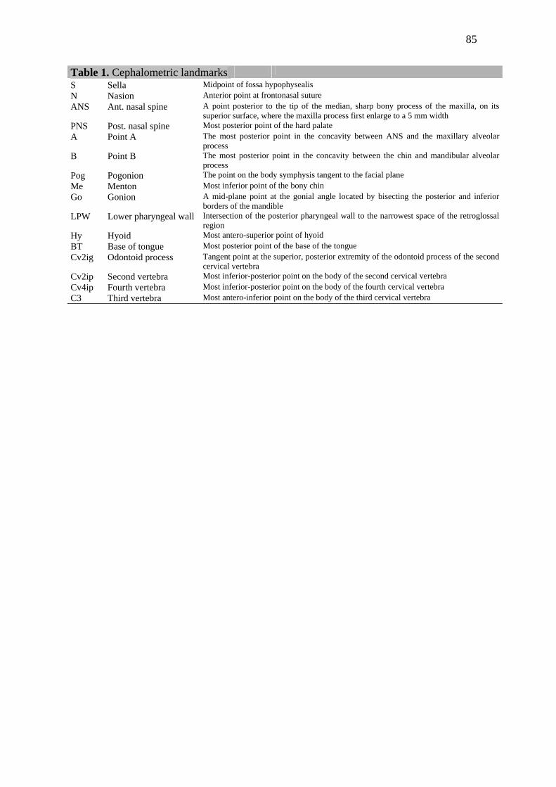
85
Table 1. Cephalometric landmarks
S Sella Midpoint of fossa hypophysealis N Nasion Anterior point at frontonasal suture ANS Ant. nasal spine A point posterior to the tip of the median, sharp bony process of the maxilla, on its
superior surface, where the maxilla process first enlarge to a 5 mm width PNS Post. nasal spine Most posterior point of the hard palate A Point A The most posterior point in the concavity between ANS and the maxillary alveolar
process B Point B The most posterior point in the concavity between the chin and mandibular alveolar
process Pog Pogonion The point on the body symphysis tangent to the facial plane Me Menton Most inferior point of the bony chin Go Gonion A mid-plane point at the gonial angle located by bisecting the posterior and inferior
borders of the mandible LPW Lower pharyngeal wall Intersection of the posterior pharyngeal wall to the narrowest space of the retroglossal
region Hy Hyoid Most antero-superior point of hyoid BT Base of tongue Most posterior point of the base of the tongue Cv2ig Odontoid process Tangent point at the superior, posterior extremity of the odontoid process of the second
cervical vertebra Cv2ip Second vertebra Most inferior-posterior point on the body of the second cervical vertebra Cv4ip Fourth vertebra Most inferior-posterior point on the body of the fourth cervical vertebra C3 Third vertebra Most antero-inferior point on the body of the third cervical vertebra
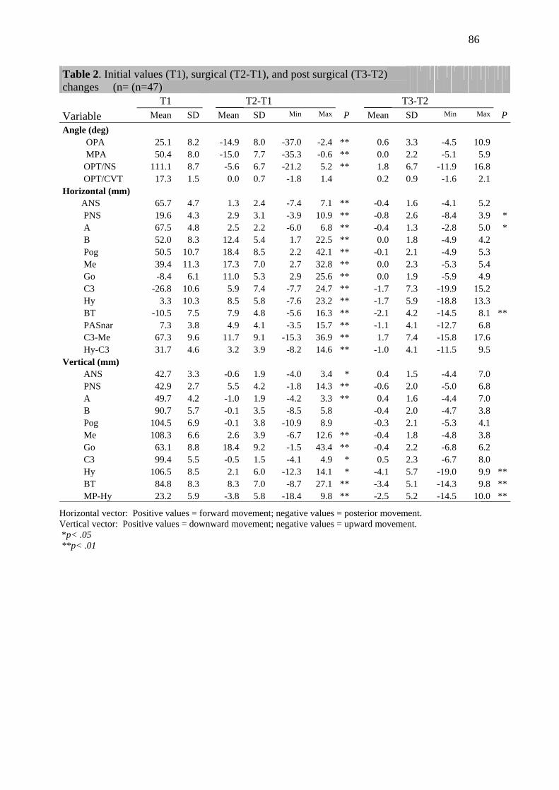
86
Table 2. Initial values (T1), surgical (T2-T1), and post surgical (T3-T2) changes (n= (n=47)
T1 T2-T1 T3-T2 Variable Mean SD Mean SD Min Max P Mean SD Min Max PAngle (deg) OPA 25.1 8.2 -14.9 8.0 -37.0 -2.4 ** 0.6 3.3 -4.5 10.9 MPA 50.4 8.0 -15.0 7.7 -35.3 -0.6 ** 0.0 2.2 -5.1 5.9 OPT/NS 111.1 8.7 -5.6 6.7 -21.2 5.2 ** 1.8 6.7 -11.9 16.8 OPT/CVT 17.3 1.5 0.0 0.7 -1.8 1.4 0.2 0.9 -1.6 2.1Horizontal (mm)
ANS 65.7 4.7 1.3 2.4 -7.4 7.1 ** -0.4 1.6 -4.1 5.2 PNS 19.6 4.3 2.9 3.1 -3.9 10.9 ** -0.8 2.6 -8.4 3.9 * A 67.5 4.8 2.5 2.2 -6.0 6.8 ** -0.4 1.3 -2.8 5.0 * B 52.0 8.3 12.4 5.4 1.7 22.5 ** 0.0 1.8 -4.9 4.2 Pog 50.5 10.7 18.4 8.5 2.2 42.1 ** -0.1 2.1 -4.9 5.3 Me 39.4 11.3 17.3 7.0 2.7 32.8 ** 0.0 2.3 -5.3 5.4 Go -8.4 6.1 11.0 5.3 2.9 25.6 ** 0.0 1.9 -5.9 4.9 C3 -26.8 10.6 5.9 7.4 -7.7 24.7 ** -1.7 7.3 -19.9 15.2 Hy 3.3 10.3 8.5 5.8 -7.6 23.2 ** -1.7 5.9 -18.8 13.3 BT -10.5 7.5 7.9 4.8 -5.6 16.3 ** -2.1 4.2 -14.5 8.1 ** PASnar 7.3 3.8 4.9 4.1 -3.5 15.7 ** -1.1 4.1 -12.7 6.8 C3-Me 67.3 9.6 11.7 9.1 -15.3 36.9 ** 1.7 7.4 -15.8 17.6 Hy-C3 31.7 4.6 3.2 3.9 -8.2 14.6 ** -1.0 4.1 -11.5 9.5Vertical (mm) ANS 42.7 3.3 -0.6 1.9 -4.0 3.4 * 0.4 1.5 -4.4 7.0 PNS 42.9 2.7 5.5 4.2 -1.8 14.3 ** -0.6 2.0 -5.0 6.8 A 49.7 4.2 -1.0 1.9 -4.2 3.3 ** 0.4 1.6 -4.4 7.0 B 90.7 5.7 -0.1 3.5 -8.5 5.8 -0.4 2.0 -4.7 3.8 Pog 104.5 6.9 -0.1 3.8 -10.9 8.9 -0.3 2.1 -5.3 4.1 Me 108.3 6.6 2.6 3.9 -6.7 12.6 ** -0.4 1.8 -4.8 3.8 Go 63.1 8.8 18.4 9.2 -1.5 43.4 ** -0.4 2.2 -6.8 6.2 C3 99.4 5.5 -0.5 1.5 -4.1 4.9 * 0.5 2.3 -6.7 8.0 Hy 106.5 8.5 2.1 6.0 -12.3 14.1 * -4.1 5.7 -19.0 9.9 ** BT 84.8 8.3 8.3 7.0 -8.7 27.1 ** -3.4 5.1 -14.3 9.8 ** MP-Hy 23.2 5.9 -3.8 5.8 -18.4 9.8 ** -2.5 5.2 -14.5 10.0 **
Horizontal vector: Positive values = forward movement; negative values = posterior movement. Vertical vector: Positive values = downward movement; negative values = upward movement. *p< .05 **p< .01

87
Table 3. Pearson correlation coeficients between surgical and post surgical landmarks and oropharyngeal airway changes
Variable Surgical Changes Post surgical Changes (T1-T2)/(T2-T3) PASnar PASnar
Angle (deg) OPA -0.396 ** -0.038 MPA -0.377 ** -0.221 OPT/NS 0.456 ** 0.603 ** OPT/CVT -0.028 -0.133 Horizontal (mm) ANS 0.284 -0.081 PNS 0.114 -0.051 A 0.352 * -0.004 B 0.420 ** 0.243 Pog 0.344 * 0.274 Me 0.409 ** 0.277 Go 0.385 ** 0.033 C3 -0.475 ** -0.657 ** Hy -0.125 -0.394 ** BT 0.394 ** 0.60 C3-Me 0.697 ** 0.707 ** Hy-C3 0.650 ** 0.603 ** Vertical (mm) ANS -0.154 -0.091 PNS 0.271 0.110 A -0.177 -0.095 B -0.046 0.058 Pog -0.115 0.065 Me 0.079 0.083 Go 0.426 ** 0.182 C3 0.140 0.540 ** Hy 0.021 -0.068 BT -0.111 -0.170 MP-Hy 0.058 0.251
*p< .05 **p< .01
Table 4. Pearson correlation coeficients between surgical and post surgical Variable Surgical Changes Post surgical Changes
(T1-T2)/(T2-T3) Me-HT Me-VT OPA Me-HT Me-VT OPA
C3-Me 0.623 ** 0.105 - 0.633 ** 0.260 0.151 - 0.118 MP-Hy - 0.198 - 0.319 * 0.061 0.140 0.132 - 0.189 Hy-C3 0.338 * - 0.025 - 0.445 ** 0.354 * - 0.061 - 0.151 OPT/NS - 0.229 - 0.188 0.030 - 0.075 0.332 * 0.065 OPT/CVT - 0.024 0.001 0.147 - 0.210 0.180 - 0.010
*p< .05 **p< .01

88
FIGURE 1
FIGURE 2
S NHRP
VRP
Hy
S
Go
Me Pog
B
N
ANS
A
HRP
VRP
PNS
Cv2ig
Cv2ip LPW
BT C3
Cv4ip
OPTCVT
OPA
Maxillo-Mandibular Counter-Clockwise Rotation and Mandibular
Advancement with TMJ Concepts® Total Joint Prostheses:
Part IV - Soft Tissue Response
1Karina E. Dela Coleta, 2Larry M. Wolford, 1João R. Gonçalves, 1Ary dos Santos Pinto, 2Daniel Serra Cassano, 3Daniela A. Godoy Gonçalves,
1Department of Pediatric Dentistry - Araraquara Dental School, Sao Paulo State University, Brazil 2Department of Oral and Maxillofacial Surgery, Baylor College of Dentistry, Texas A&M University System. 3Department of Prosthodontics - Araraquara Dental School, Sao Paulo State University, Brazil Address correspondence and reprint requests to: Larry M. Wolford, DMD: 3409 Worth St, Suite 400 Dallas, TX 75246 Phone: 214-828-9115 Phone: 214-828-1714 E-mail: [email protected]
90
Abstract
The purpose of this study was to evaluate soft tissue response to maxillo-mandibular
counter-clockwise rotation, with TMJ reconstruction and mandibular advancement using
TMJ Concepts system® total joint prostheses, and maxillary osteotomies in forty-four
females (14 to 57 years of age). Eighteen patients had genioplasties with either Porous
Block Hydroxyapatite or HTR implants. Lateral cephalograms were taken 1 week before
surgery and at least 12 months (mean 40.8 months) post surgery to evaluate soft to hard
tissue changes. Two groups were analyzed: Group 1, no genioplasty (n = 26) and Group
2, with genioplasty (n = 18). Surgically, the maxilla moved forward and upward by
counter-clockwise mandibular rotation with greater horizontal movement in Group 2.
Vertically, both groups showed diversity of maxillo-mandibular mean movement. Hard
and soft tissue ratios were different between groups. Group 1 showed a consistent 1 : 0.97
ratio of hard to soft tissue advancement at Pogonion and 1 : 1.01 ratio (range 1 : 0.99 to
1 : 1.61) at B point and LMf. Group 2 results were less consistent, with ratios between 1 :
0.84 and 1 : 1.02. Horizontal changes in upper lip morphology after maxillary
advancement/impaction, VY closure, and alar base cinch sutures showed almost greater
movement in both groups, than observed in hard tissue. When comparing the percentage
movement of soft tissue to underlying bone, the upper lip moved anteriorly at subnasale
145% (Group 1) and 180% (Group 2), and at labrale superius 100% (Group 1) and 107%
(Group 2). There was a slight upper lip lengthening of 0.4 and 0.5 mm. Counter-
clockwise rotation of the maxilla-mandibular complex using TMJ Concepts total joint
prostheses resulted in similar soft tissue response as previously reported for traditional
maxillo-mandibular advancement without counter-clockwise rotation of the occlusal plane.
The association of chin implants, in the present sample, showed higher variability of soft tissue response.
Key words: Soft tissue; Orthognathic surgery; TMJ prostheses

91
Introduction
Dentofacial deformities are the result of variations in skeletal and dental
alveolar morphology that affect function as well as facial appearance and balance.
Orthognathic surgery for these patients can successfully improve these aspects. The key
to achieving improved function and facial esthetics is to carefully and systematically
analyze functional and facial balance, establish esthetic priorities, and then coordinate
and implement correction through the use of cephalometric planning and occlusal
studies1.
Since the improvement of facial appearance is often an important motivating
factor in seeking treatment16, the ability to predict the outcome of treatment is essential.
The predictability of treatment depends on the relationship between the hard and soft
tissues26. Orthognathic surgery moves the skeletal elements in a planned and controlled
manner, but the soft tissue drape is not as precisely controlled during surgery11,17. There
are a number of techniques available for the planning of orthognathic treatment and these
have become increasingly sophisticated over the years. Cephalometric prediction tracings
and computer imaging software19,21,27 are examples.
According to Dolce et al.8 the percentage change of soft tissue points for a given
hard tissue advancement depends on both treatment method and time. Bone movement in
orthognathic surgery gives rise to changes in the positions of the adjacent soft tissues,
with such change varying according to the location, direction, and degree of movement.
The behavior of the soft tissues, especially the labial tissues, can be influenced by aspects
such as lip thickness, length, and taper, surgical change in palatal plane, and soft tissue
manipulation techniques (ie, VY closure and alar base cinch suture)3,22. The balance of
92
the soft tissue contours of the nose and chin must also be evaluated3,4. Although the soft
tissue horizontal response can be relatively accurately determined, the vertical plane isn’t
as precise. To obtain the final soft tissue results, a minimum of 6-12 months follow-up is
required11.
In planning cases for double jaw surgery, the vertical and horizontal predicted
surgical positioning of the maxilla will affect the amount of mandibular repositioning and
the need for mandibular osteotomies and/or adjunctive genioplasty. The simultaneous
repositioning of the maxilla, mandible, and chin can dramatically alter the facial soft
tissue contour and proportions to afford the clinician much greater latitude in treatment
outcomes2.
Although the addition of other adjunctive surgical procedures such as
genioplasty, rhinoplasty, cheek augmentation, etc., to the clockwise or counter-clockwise
rotation of the maxillo-mandibular complex can improve esthetic results, they may make
it more difficult to determine with certainty the changes in the soft tissue profile26. In
studies that examined the hard to soft tissue response with bilateral mandibular ramus
sagittal split osteotomy (no genioplasty), most authors reported a ratio of 1:1 for
pogonion9,17,20. A similar ratio is generally accepted for B-point. However, Ewing &
Ross9 concluded that when an advancement genioplasty is included in the surgical
movement, the soft tissue of the chin tends to move downward and proportionally less
than the hard tissue movement in the horizontal direction, making the soft tissues thinner
in this area. The soft tissue response shows considerable individual variation,
contributing to inaccuracy of soft tissue outcome predictions10.
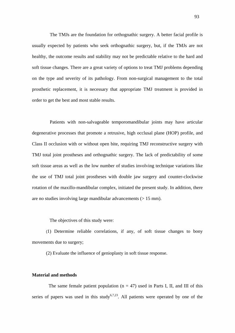
93
The TMJs are the foundation for orthognathic surgery. A better facial profile is
usually expected by patients who seek orthognathic surgery, but, if the TMJs are not
healthy, the outcome results and stability may not be predictable relative to the hard and
soft tissue changes. There are a great variety of options to treat TMJ problems depending
on the type and severity of its pathology. From non-surgical management to the total
prosthetic replacement, it is necessary that appropriate TMJ treatment is provided in
order to get the best and most stable results.
Patients with non-salvageable temporomandibular joints may have articular
degenerative processes that promote a retrusive, high occlusal plane (HOP) profile, and
Class II occlusion with or without open bite, requiring TMJ reconstructive surgery with
TMJ total joint prostheses and orthognathic surgery. The lack of predictability of some
soft tissue areas as well as the low number of studies involving technique variations like
the use of TMJ total joint prostheses with double jaw surgery and counter-clockwise
rotation of the maxillo-mandibular complex, initiated the present study. In addition, there
are no studies involving large mandibular advancements (> 15 mm).
The objectives of this study were:
(1) Determine reliable correlations, if any, of soft tissue changes to bony
movements due to surgery;
(2) Evaluate the influence of genioplasty in soft tissue response.
Material and methods
The same female patient population (n = 47) used in Parts I, II, and III of this
series of papers was used in this study6,7,23. All patients were operated by one of the
94
authors (LMW) at Baylor University Medical Center, Dallas, TX, USA. Patients were
selected according to the inclusion and exclusion criteria as presented in Part I6.
However, 3 patients had lateral cephalograms where the soft tissue profile could not be
identified. Therefore, longitudinal records were available for 44 of the 47 female patients.
All patients had TMJ reconstruction and mandibular advancement using total joint
prostheses with simultaneous maxillary osteotomies and counter-clockwise rotation of
the occlusal plane6. Eighteen patients had an augmentation genioplasty in the same
operation with Porous Block Hydroxyapatite (PBHA, Interpore 200, Interpore Inc, Irvine,
CA) or Hard Tissue Replacement (HTR) polymer implant, (Walter Lorenz CO.
Jacksonville, FL). Mean patient age at surgery was 35 years (range, 14 to 57 years). All
maxillary osteotomies were rigidly stabilized using bone plates and screws, synthetic
bone grafting when indicated, and no maxillo-mandibular fixation. Details of the surgical
techniques are presented in Part I6. An alar cinch suture and VY vestibular closure were
performed in all cases in the study12. Post surgery, light force elastics were used on most
patients for 2 to 4 weeks to control the occlusion.
The sample was divided into 2 groups based on the presence or absence of a
genioplasty: Group 1, no genioplasty (n = 26); Group 2, with genioplasty (n = 18).
Patients were offered a genioplasty if the surgeon recommended it for improved esthetic
outcome. Most genioplasties were performed for horizontal augmentation of the chin
with minimal vertical change. Correlation coefficients were calculated between hard
tissue advancement and corresponding soft tissue movement.
The custom-made total joint prostheses used in this study, were originally
developed in 1989 by Techmedica Inc., Camarillo, CA, USA, and since 1997, have been

95
manufactured by TMJ Concepts, Inc., Ventura, CA, USA. These prostheses are
CAD/CAM devices (computer assisted design/computer assisted manufacture), designed
to fit the specific anatomical requirements for each patient.
Imaging evaluation
Lateral cephalometric radiographs were taken using a standard radiographic
technique (centric relation, Frankfort horizontal plane parallel to the floor and lips
relaxed). Radiographs had to be of good quality with all hard and soft tissue landmarks
clearly identifiable. Three patients were disqualified from an initial sample of 47 cases,
because of poor quality records where soft tissue profile could not be determined.
Radiographs were taken at the following intervals: T1- immediately before surgery
(range 1 to 6 days), and T2- long-term follow-up with an average 40.8 months (range, 12
to 143 months) post surgery. Each patient’s lateral cephalograms were traced, digitized
twice, and averaged to estimate hard and soft tissue surgical changes in the 2 groups
analyzed: Group 1, no genioplasty (n = 26), and Group 2, with genioplasty (n = 18). The
landmarks used for measurements are presented in Fig. 1, Table 1. The landmarks were
digitized by using Dentofacial Planner Plus version 2.02 (Dentofacial Software Inc,
Toronto, Canada) and analyzed to describe hard and soft tissues surgical movements as
well as the correlation between them. Stable reference lines were established from S-N
minus 7° for the horizontal reference plane (HRP) and a perpendicular line to HRP
through sella for the vertical reference plane (VRP). Hard and soft tissue changes due to
surgical movement were evaluated in relation to these reference lines (Fig. 1).
96
Method error
To determine the consistency of the method, two examiners were previously
calibrated by repetition of the process until the method was considered adequate by a
third examiner. Random errors in landmark localization were decreased by tracing twice
each lateral cephalogram and using the medium values of each measurement. The intra-
examiner consistency (ICC) was calculated for reliability of tracing, landmark
identification, and analytical measurement showing a correlation coefficient always
greater than 0.94.
Statistical method
All data were transferred to SPSS (release 9.0; SPSS Chicago, IL) for statistical
analysis. The skewness and kurtosis statistics showed normal distributions for all
variables. Differences were compared between patients in Groups 1 and 2. Because there
were statistically significant differences between those groups in post surgical changes,
the patients were analyzed in two distinct groups: Group 1 - no genioplasty and Group 2 -
with genioplasty. Paired t tests were performed to evaluate the surgical changes (T2-T1).
A significance level of p < .05 was applied. To compare the results with existing studies,
linear regression analyses were performed that evaluated the relationships between hard
tissue variables and their soft tissue counterparts. Pearson product-moment correlations
were also used to assess the association between the soft and hard tissues.
Results
Group 1 (no genioplasty, n = 26)
The maxilla advanced 1.1 ± 2.3 mm at anterior nasal spine (ANS), 2.2 ± 2.3 mm
at A point and 2.0 ± 2.9 mm at posterior nasal spine (PNS), while vertically, just PNS
showed statistically significant downward movement (5.0 ± 4.0 mm). The supra-dental
97
(Sd) and upper incisor tip (U1T) landmarks showed the greatest changes in the maxillary
area (horizontal 3.8 ± 2.3 mm and 5.4 ± 3.0 mm; vertical -1.0 ± 1.9 mm and -0.8 ± 1.9
mm, respectively). For vertical movement, negative numbers indicate upward and
positive numbers downward movement. For horizontal movement, positive numbers
indicate forward movement and negative numbers indicate backward movement.
Horizontal soft tissue changes were very similar in amount and direction to hard tissue
surgical movements. However, it was observed that areas Sn and Sls showed greater
advancement than their respective hard tissue points (ANS and A point). Upper lip soft
tissue forward movements at Sn was 1.6 ± 1.8 mm, Sls 3.2 ± 1.9 mm, Ls 3.8 ± 2.2 mm,
and Sts 5.0 ± 2.8 mm. Sn (-0.6 ± 1.0 mm) was the only upper lip landmark that showed
significant vertical change (Table 2).
The mandibular hard tissue changed significantly in all horizontal
measurements as observed in the following landmarks: L1T 6.9 ± 3.8 mm, Id 9.5 ± 4.4
mm, B point 11.4 ± 4.8 mm, Pog 14.7 ± 6.1 mm, Me 16.3 ± 7.0 mm, and Go 11.1 ± 5.2
mm. Vertically, the mandible showed no statistically significant movement at B point and
Me, but an inferior change of 16.7 ± 8.9 mm at Go point and an upward movement at
L1T (-3.7 ± 4.4 mm), Id (-2.8 ± 3.9 mm) and Pog (-1.5 ± 3.5 mm). Soft tissue moved
forward and upward in all mandibular landmarks respectively (Sti 7.4 ± 3.4 mm and -3.5
± 4.2 mm; Li 8.7 ± 3.8 mm and -5.1 ± 4.5 mm; LMf 11.6 ± 5.0 mm and -3.6 ± 4.0 mm;
Pog’14.4 ± 6.0 mm and -2.5 ± 3.8 mm; Gn’ 15.8 ± 6.6 mm and -1.7 ± 3.4 mm) as
recorded in Table 2.
98
In the middle third of the face, soft tissues advanced at point Pn (0.4 ± 1.0 mm)
and at Cm showed forward (1.4 ± 1.6 mm) and upward (-0.8 ± 1.2 mm) movement. N’
and Nd showed no change.
Maxillary and mandibular angular measurements (SNA, SNB, ANB and
SNPog) confirmed linear movements as recorded in Table 4. Occlusal plane and
mandibular plane angles decreased a mean of 13.8 ± 7.9 degrees and 14.0 ± 7.6 degrees
respectively, during the maxillo-mandibular counter-clockwise rotation. Surgical changes
associated with the mandibular counter-clockwise advancement are seen in Table 4 and
Fig 2.
Group 2 (with genioplasty, n = 18)
In group 2, the maxilla showed significant movement at A point horizontally
(1.5 ± 2.6 mm) and at PNS, in both directions (H 2.3 ± 3.5 mm and V 4.9 ± 3.3 mm). The
supra-dental and upper incisor tip advanced 2.8 ± 2.6 mm and 5.0 ± 3.2 mm respectively,
while in the vertical plane both points showed upward movement (-1.7 ± 2.4 mm and -1.7
± 2.5 mm). The upper lip soft tissue changes showed progressive increased advancement
at points: Sn 0.9 ± 1.5 mm, Sls 2.5 ± 2.2 mm, Ls 3.0 ± 2.6 mm, and Sts 3.5 ± 3.3 mm.
Vertically, there was no significant movement (Table 3).
The mandibular hard tissue changes were significant in all horizontal
measurements, being greater than observed in Group 1 (L1T 8.2 ± 3.1 mm, Id 11.0 ± 3.7
mm, B 13.9 ± 4.8 mm, Pog 22.6 ± 6.9 mm, Me 18.6 ± 5.4 mm, and Go 11.1 ± 5.1 mm). In
the vertical plane, the mandible showed an inferior movement at Me of 3.3 ± 4.0 mm and
99
Go 19.8 ± 8.6 mm, while L1T and Id showed upward movement. Soft tissue moved
forward in all mandibular points: Sti 7.4 ± 4.9 mm, Li 9.8 ± 5.1 mm, LMf 14.2 ± 5.7 mm,
Pog’ 19.0 ± 5.8 mm and Gn’ 21.5 ± 6.7 mm, and upward at Sti -4.1 ± 3.4 mm; Li -5.8 ±
3.9 mm; LMf -3.1 ± 3.7 mm and Pog’ -1.1 ± 5. mm recorded in Table 3. The middle third
of the face did not show significant movement, except at Cm point (horizontal 1.2 ± 1.5
mm and vertical -0.8 ± 1.1 mm).
Maxillary and mandibular angular measurements (SNA, SNB, ANB and
SNPog) confirm linear movements as recorded in Table 4. Occlusal plane and
mandibular plane angles decreased a mean 14.9 ± 5.7 degrees and 16.5 ± 6.0 degrees
respectively, being greater than observed in Group 1.
Correlation
1. Horizontal correlations. The high correlation of hard tissue landmarks and
their soft tissue counterparts can be seen in Table 7. Many statistically significant and
reliable correlations were found among hard and soft tissue landmarks in both groups.
Anterior nasal spine had significant correlations (although not strong) with superior labial
sulcus and subnasale in Groups 1 (r=0.51) and 2 (r=0.69); and with labrale superius,
stomion superius, and inferius only in the Group 1 (Table 5). The ratios from ANS to Sn
were 1:1.45 in Group 1 and 1:1.80 in Group 2. Soft tissue landmarks appeared to follow
A point more closely than anterior nasal spine, with ratios from A to Sls of 1:1.45 (Group
1) and 1:1.66 (Group 2). A surgical movement of A point could predict long-term
movement of superior labial sulcus with correlations of 0.76 (Group 1) and 0.68 (Group 2).
That is, a 1.0 mm hard tissue movement would result in a 1.45 mm and a 1.66 mm soft tissue
movement for each. The Sd landmark showed stronger correlations than observed in ANS
100
and A point with correlation (Sd-Ls) of 0.83 (Group 1) and 0.88 (Group 2). The
mandibular hard tissue measurements (B point, Pog, and Id) showed high correlation with
all inferior soft tissue points (Sti, Li, Mlf, Pog’) in both groups. Also, there was a
significant correlation of B point with Sn in Group 2. Ratios between hard and soft
tissues after horizontal advancement were: B point:LMf 1 : 1.01 (Group 1) and 1 : 1.02
(Group 2); Pog:Pog’ 1 : 0.97 (Group 1) and 1 : 0.84 (Group 2); Id:Li 1 : 0.91 (Group 1)
and 1 : 0.89 (Group 2). Dental lardmarks (U1T and L1T) correlated quite highly with the
soft tissue measurements in both groups, demonstrating a close relationship between
lower incisor tip and stomion inferius (Table 9). These changes can be seen in Figure 2
that shows the percentage of soft tissue movement in relation to hard tissue surgical
change in the horizontal plane.
2. Vertical correlations. A number of reliable correlations for vertical
movements could be found, but far less than for horizontal movements. Table 8 showed
high correlation (r>0.75) of hard tissue landmarks and their respective soft tissue
reference points, only in the mandibular region. In Pearson product-moment correlations
(Table 6), maxillary landmarks (A point, ANS and Sd) showed good correlation (r>0.6)
with few soft tissue landmarks, only in Group 2. None of the maxillary landmarks
showed correlation greater than 0.6 in Group 1. Mandibular landmarks correlations were
stronger than observed in the maxillary region, nevertheless the values were higher in
Group 1. Dental landmarks (U1T and L1T) showed a different behavior. While the upper
incisor tip showed few correlations, the inferior tooth (L1T) reveal good correlations with
all mandibular soft tissue landmarks in both groups. Figure 3 shows the percentage of
soft tissue movement in relation to hard tissue surgical change in the vertical plane.
101
Discussion
The prediction of the soft tissue response that will follow surgical hard tissue
advancement appears to be a straight forward procedure in general. After surgery,
patients show straighter facial profiles, more harmonious lip balance, and labial folds
more defined. Those changes can be predicted in millimeters in most cases, however
when some soft tissue manipulation techniques are added (ie alar base cinch suture, V-Y
vestibular incision closure) or other surgical procedures are incorporate, such as a
genioplasty, this generalization does not apply.
The influence of different surgical procedures on soft tissue results was
considered in our study, since more than 40% of the patient sample required an
augmentation genioplasty. So, the results were compared between the two groups (Group
1, no genioplasty and Group 2, with genioplasty) and were found to have statistically
significant differences. This standardization of the sample optimized the reliability of soft
tissue prediction.
Predicting the soft tissue profile from orthognathic surgery was first described
in 1972 by McNeill et al18. Since then, studies have reported on soft tissue responses to
hard tissue changes8. However, special considerations needed to be taken in reference to
the follow-up period. Transient soft tissue changes that result from different stages of
healing and resolution of edema were eliminated by requiring post surgery cephalograms
be taken at least 12 months post surgery, although soft tissues overlying maxillary
structures may take several years to reach their final equilibrium. An analysis of stability
data revealed that most horizontal and vertical soft tissue change after Le Fort I surgery

102
stabilize in the first year after surgery13. Based on this fact our study used at least 12
months of follow-up with the purpose to obtain soft tissue equilibrium.
The final soft tissue profile of the lips is of particular importance because there is
generally more emphasis on post surgical changes in the lips than on the nose or chin,
when evaluating esthetics of life-sized lateral photographs5.
Powers24 found that the upper lip and labiomental fold horizontally were very
predictable, with only a few patients (3% and 8%, respectively) exhibiting clinically
significant differences. The predictability of maxillary surgery is influenced by the ability
of the surgeon to accurately position and provide adequate stability of the maxilla in its
new location as well as the variability of the soft tissue response15.
Our study evaluated the relation of the maxilla and upper lip in the horizontal
and vertical planes. The results showed a mean movement forward and upward of the
anterior maxilla (ANS, A point, Sd and U1T) and downward movement of PNS in both
groups, although some of these changes was not statistically significant. This counter-
clockwise rotation of the palatal plane and occlusal plane promoted increased
advancement in a graduated fashion from the upper to the lower part of the maxilla,
allowing greater projection of the upper incisor while the anterior nasal spine was only
slightly advanced. Vertically, the upper incisor showed greater upward movement than
the maxilla superior area (ANS and A point), which may be a favorable movement in
high-angle cases to reduce the upper incisor exposure. However, it was observed that the
upper incisor tip (U1T) showed equal or less upward movement than the supra-dental
103
landmark, probably by the post surgical orthodontic treatment that can influence the
incisor vertical position.
The soft tissue response of the upper lip was similar to the maxilla, but with
different intensity. While the maxillary hard tissue advanced an average 1.1 to 5.4 mm in
Group 1 and 0.5 to 5.0 mm in Group 2, the respective soft tissue landmarks showed a
forward mean movement of 1.6 to 5.0 mm (Group 1) and 0.9 to 3.5 mm (Group 2). The
landmarks Ls and Sts showed the greatest mean anterior change, while Sn and Sls
showed slightly lower amounts of mean anterior movement. In the vertical plane, the soft
tissue showed upward movement, but in a decreased scale from the uppermost (Sn) to
lowermost (Sts) landmarks. Thus, the stomion superius showed minimal vertical change,
maintaining the lip length.
The use of the alar base cinch suture and VY vestibular incision closure
techniques directly affected upper lip results. The alar base suture prevents flaring of the
alar base of the nose and thickens the lip while the VY closure helps minimize shortening
of the upper lip and maintains lip thickness12. These techniques14 improve the surgeon’s
ability to control the post surgery morphologic features and esthetics of the upper lip4.
Comparing hard and soft tissues changes, we observed that the uppermost points
of the lip showed greater advancement than their hard tissue counterparts. The same fact
did not occur at the lip vermilion. Most previous studies showed the higher region of the
upper lip (Sn) presented less change than the inferior part of the lip (Sts) when the
maxilla was advanced. Gregoret12 reported Sn followed just 30% of hard tissue
movement and Sts, 80%. Our results showed that the upper landmarks of the lip (Sn and
104
Sls) moved more than 100% of the osseous advancement in both groups, with 70-100%
of soft to hard tissue advancement in the lower part (Sl and Sts), as observed in Figure 2.
This result was expected considering that an alar base cinch suture and VY vestibular
closure were performed in all cases of the study, promoting a greater projection of the
subnasal area. The amount of hard tissue movement at ANS and A point was small with
only 0.5 to 2.2 mm of advancement. Also, orthodontic brackets in place for the
presurgical cephalogram were removed in most instances at longest follow-up. This could
introduce soft tissue discrepancies affecting predictions and lip thickness.
Nary Filho et al.22 reported that it was possible to detect a tendency toward more
posterior positioning of the upper lip with maxillary surgery, which can be compensated
by the V-Y suture technique. Alterations in the vertical position of the soft tissues were
not significant.
In our study, the Pearson product-moment analysis showed good correlation of
maxillary hard and soft tissues horizontally (Table 5). Previous studies16,21 have reported
slightly lower correlations between the two tissue movements, with the average
correlation ratio being approximately 0.75, and ranged between 0.45 and 0.97. The
average correlation for upper lip landmarks in the present study ranged between 0.51 to
0.90 and ratios from 0.92 - 1.45 (Group 1) and 0.70 - 1.80 (Group 2). Similar to our
results, others have reported maxillary advancements that used specialized soft tissue
reconstruction techniques, such as VY closure, demonstrating higher soft/hard tissue
ratios that range from 0.78 to 1.0014. Vertically, this correlation was not strong although
the behavior of both tissues showed movement in the same direction.
105
The significant correlations between hard and soft tissue response seen in the
present study may be due to a variety of factors. Homogeneity of the sample, the
accuracy of the surgical method, the technical skills of the surgeon and the standardized
surgical technique of alar base cinch suture and VY closure, may explain the
improvement in correlations.
In the mandibular area, our results showed an advancement of all landmarks
associated with a counter-clockwise rotation and decrease of occlusal and mandibular
plane angles. Our results showed that the amount of advancement was greater than
previous studies9,25,26 with a mean movement of 11.4 mm at B point, 14.7 mm at Pog,
16.3 mm at Me, and 11.1 mm at Go (Group 1). In the Group 2, the amount of hard tissue
advancement was slightly more at B point, Me and Go, but significantly greater at Pog
(22.6 mm). The more pronounced mandibular advancement observed in our study is
justified by the patients previous history of TMJ pathology or irreversible damages with
condylar resorption, significant mandibular retrusion, and high occlusal plane angle that
required greater movements to obtain optimal functional and esthetic results.
The mandibular dental landmarks (L1T, Id) showed less significant movement
in both groups as compared to the lower areas of the mandible (Pog, Me). The anterior
maxillary upward movement and posterior downward movement promoted a mandibular
counter-clockwise rotation with greater horizontal projection in the inferior area of the
mandible (Pog, Me). Therefore, it was possible to obtain a significant mandibular
projection without as great of maxillary and dental advancement. Proffit & Phillips25
considered that the soft tissues are relaxed and the lip pressure is decreased by the
106
surgical treatment when the mandible rotates upward and forward following maxillary
intrusion.
The mandibular soft tissues in both groups, showed gradual increased
advancement similar to the hard tissues. The lower lip landmarks movement was greater
than their respective osseous counterpoints only at Sti (7.4 mm in Group 1) and at LMf
(11.6 mm in Group 1; 14.2 mm in Group 2). Vertically, all soft tissue landmarks showed
upward movement with Li elevating more than the inferior stomion (in both groups),
probably related to the labial seal or thinning of the lower lip.
In studies that examined the hard to soft tissue response with bilateral sagittal
split osteotomies, most reported a ratio of 1:1 at pogonion and B point9,18,21. However,
these studies demonstrated great variability in soft tissue response of the lower lip18,21.
The soft tissue in Group 1 of our study showed similar changes to that obtained
in other studies, with 97% at pogonion and 101% at B point. The lower lip predictably
moved anteriorly in a graduated fashion from 91% at Li to 107% at Sti relative to the
underlying hard tissue movement. The Pearson product-moment analysis showed high
correlations of mandibular hard and soft tissues horizontally, but not so consistent
vertically. In the present study, the horizontal average correlation for inferior soft tissue
landmarks and their osseous counterparts ranged from 0.84 to 0.98 in Group 1. This data
points out high levels of soft tissue predictability based on the skeletal hard tissue
movements.
107
However, when a genioplasty is performed, the soft tissue response shows
considerable individual variation and is also dependent on whether the procedure is
performed alone or in combination with other surgery10. In our study, Group 2 soft tissue
behavior was similar to Group 1 but to a lesser degree. The highest correlation of hard to
soft tissue change was at Pog (0.93) horizontally, ranging from 0.81 to 0.93. Previous
studies9,26 showed a lower correlation (0.34 - 0.49) between hard and soft tissue
advancement at pogonion. Vertically, the mandible had less significant correlations than
in the horizontal plane, although almost all were greater than 0.6 (Tables 6).
The ratios obtained for Group 2 was 1 : 1.02 (B : LMf) and 1 : 0.84 (Pog :
Pog’), being greater than shown by other genioplasty studies that ranged from 1 : 0.6 to
1:1.9,11 Shaughnessy et al.26, suggested that a ratio of 1 : 0.9 can be used to predict hard to
soft tissue movements in osseous genioplasty, although attention has been paid to the
importance of maintaining as much soft tissue attachment as possible to the repositioned
bony segment to obtain predictable soft tissue changes. Similar results were found by
Ewing & Ross9 although the average difference between hard and soft tissue movement
was ± 2.6 mm.
In mandibular advancement with genioplasty the soft to hard tissue correlations
are much less consistent1 then observed in cases of advancement without genioplasty. The
cases requiring genioplasty were often the more severe cases, and soft tissue drape in
severe retrognathia is usually abnormal. Individual assessment is essential in such cases.
Also, minor variations in the surgical management of tissues occur from patient to patient
and differs from surgeon to surgeon, so that variation in results in the chin area are not
surprising9.

108
According to Ewing & Ross9, genioplasty cases develop a thinning or unfurling of
the lower lip at the vermilion border. This factor was also observed in our results where
the lower anterior movement of stomion inferior compared to the hard tissue change,
promoted a thinning of the lower lip in genioplasty cases. This relation was not seen in
Group 1. The authors9 recommend that surgeons should evaluate their own genioplasty
cases to establish the soft tissue response to their particular surgical technique.
The lower lip did not advance as much as the lower incisor. Instead tended to
become thinner or fall back, with a change of 91% in Group 1 and 89% in Group 2
relative to the incisor movement. This is illustrated in Figures 2 and 4.
Our study results showed better soft tissue response to hard tissue movements in
most areas compared to previous studies probably related to the maxillo-mandibular
counter-clockwise rotation and advancement with TMJ reconstruction using total joint
prostheses that permits greater mandibular advancement to optimizing facial profiles as
well as soft tissue management techniques (ie alar base cinch suture, V-Y vestibular
closure). Upper lip soft tissue horizontal response was greater than the hard tissue
movement related to the relatively small amount of maxillary advancement and the use of
the alar base cinch suture and the V-Y closure of the vestibular incision which tends to
thicken the lip. There was less soft tissue horizontal movement of the lower lip and
pogonion with genioplasty as compared to no genioplasty.
References
1. Bell WH. Correction of the short face syndrome- Vertical maxillary deficiency: a
preliminare report. J Oral Surg 1977;35:110-120.

109
2. Bell WH, Jacobs JD, Quejada JG. Simultaneous repositioning of the maxilla, mandible
and chin. Treatment planning and analysis of soft tissues. Am J Orthod 1986;89:28-
50.
3. Betts NJ, Vig KW, Vig P. Changes in the nasal and labial soft tissues after surgical
repositioning of the maxilla. Int J Adult Orthod Orthognath Surg 1993;8:7-23.
4. Brooks BW, Buschang PH, Bates JD, Adams TB, English JD. Predicting upper lip
response to 4-piece maxillary LeFort I osteotomy. Am J Orthod Dentofacial Orthop
2001;120:124-133.
5. Burcal RG, Laskin DM, Sperry TP. Recognition of profile change after simulated
orthognathic surgery. J Oral Maxillafc Surg 1987;45:666-670.
6. Coleta KED, Wolford LM, Goncalves JR, Santos-Pinto A, Pinto LP, Cassano DS.
Maxillo-Mandibular Counter-Clockwise Rotation and Mandibular Advancement with
TMJ Concepts® Total Joint Prostheses: Part I – Skeletal and Dental Stability –
IJOMS-D-07-00531
7. Coleta KED, Wolford LM, Goncalves JR, Santos-Pinto A, Pinto LP, Cassano DS.
Maxillo-Mandibular Counter-Clockwise Rotation and Mandibular Advancement with
TMJ Concepts ® Total Joint Prostheses: Part II - Airway Changes and Stability –
IJOMS-D-07-00532.
8. Dolce C, Hatch JP, Van Sickels JE, Rugh JD. Five-year outcome and predictability of
soft tissue profiles when wire or rigid fixation is used in mandibular advancement
surgery. Am J Orthod Dentofacial Orthop 2003;124:249-256.
9. Ewing M, Ross RB. Soft tissue response to mandibular advancement and genioplasty.
Am J Orthod Dentofacial Orthop 1992;101:550-555.

110
10. Gallagher DM, Bell WH, Storum KA. Soft tissue changes associated with
advancement genioplasty performed concomitantly with superior positioning of the
maxilla. J Oral Maxillofac Surg 1984;42:238-242.
11. Gregoret J. Ortodontia e cirurgia ortognatica_diagnóstico e planejamento. São Paulo:
Ed Santos; 1999. 520p.
12. Guymon M, Crosby DR, Wolford, LM. The alar base cinch suture to control nasal
width in maxillary osteotomies. Int. J. Adult Orthod and Orthognath Surg 1988; 2:
89-95.
13. Hack GA, de Mol van Otterloo JJ, Nanda R. Long-term stability and prediction of
soft tissue changes after LeFort I surgery. Am J Orthod Dentofac Orthop
1993;104:544-555.
14. Hackney FL, Nishioka GJ, Van Sickels JE. Frontal soft tissue morphology with
double VY closure following Le Fort I osteotomy. J Oral Maxillofac Surg
1988;46:850-855.
15. Jacobson R, Sarver DM. The predictability of maxillaryAm J Orthod Dentofacial
Orthop 2002;122:142-154.
16. Kiyak HA, Vitaliano PP, Crinean J. Patients’ expectations as predictors of
orthognathic surgery outcomes. Health Psychology 1988;7:251-268.
17. Lines PA, Steinhauser EW. Soft tissue changes in relationship to movement of hard
structures in orthognathic surgery:a preliminare report. J Oral Surg 1974;32:891-896.
18. McNeill RW, Proffit WR, White RP. Cephalometric prediction for orthodontic
surgery. Angle Orthod 1972;42:154-164.
19. Miller L, Morris DO, Berry E. Visualizing three-dimensional facial soft tissue
changes following orthognathic surgery. Eur J Orthod 2007;29:14-20.
111
20. Mobarak KA, Espeland L, Krogstad O, Lyberg T. Soft tissue profile changes
following mandibular advancement surgery: predictability and long-term outcome.
Am J Orthod Dentofacial Orthop 2001;119:353-367.
21. Moss JP, McCance AM, Fright WR, Linney AD, James DR. A three-dimensional soft
tissue analysis of fifteen patients with Class II Division 1 malocclusions after
bimaxillary surgery. Am J Orthod Dentofacial Orthop 1994;105:430-437.
22. Nary Filho H, Goncales ES, Berrentin-Felix G, de Souza Cesar U, Achja GL.
Evaluation of the facial soft tissues following surgically assisted maxillary expansion
associated with the simple V-Y suture. Int J Adult Orthod Orthognath Surg
2002;17:89-97.
23. Pinto LP, Wolford LM, Buschang PH, Bernardi FH, Goncalves JR, Cassano DS.
Maxillo-Mandibular Counter-Clockwise Rotation and Mandibular Advancement with
TMJ Concepts ® Total Joint Prostheses: Part III Pain and Dysfunction Outcomes –
IJOMS-D-07-00535.
24. Powers B. The accuracy of computer generated profile predictions associated with
mandibular advancement surgery [thesis]. Chicago: Northwestern University; 1998.
25. Proffit WR, Phillips C. Adaptations in lip posture and pressure following
orthognathic surgery. Am J Orthod Dentofac Orthop 1988;93:294-302.
26. Shaughnessy S, Mobarak KA, Høgevold HE, Espeland L. Long-term skeletal and
soft-tissue responses after advancement genioplasty. Am J Orthod Dentofacial Orthop
2006;130:8-17.
27. Tollefson TT, Sykes JM. Computer imaging software for profile photograph analysis.
Arch Facial Plast Surg 2007;9:113-119.
112
Legends Figure 1. Hard and soft tissue landmarks used in cephalometric analysis (definition in
Table 1)
Figure 2. Percentage of soft tissue movement in relation to hard tissue surgical change in
horizontal plane
Figure 3. Percentage of soft tissue movement in relation to hard tissue surgical change in
vertical plane
Figure 4. Schematic representation of hard and soft tissue changes
Table 1. Cephalometric landmarks and some measurement definitions
Table 2. Group 1 (no genioplasty) horizontal and vertical movements (millimeters) of
hard and soft tissue landmarks (T1-T2)
Table 3. Group 2 (genioplasty) horizontal and vertical movements (millimeters) of hard
and soft tissue landmarks (T1-T2)
Table 4. . Group 1 (no genioplasty) and Group 2 (with genioplasty) angular movements
(degrees) of hard tissue landmarks (T1-T2)
Table 5. Pearson product-moment correlations for horizontal landmark movement
Table 6. Pearson product-moment correlations for vertical landmark movement
Table 7. Linear regression for horizontal landmark movement
Table 8. Linear regression for vertical landmark movement
Table 9. Ratios between hard and soft tissues after horizontal advancement
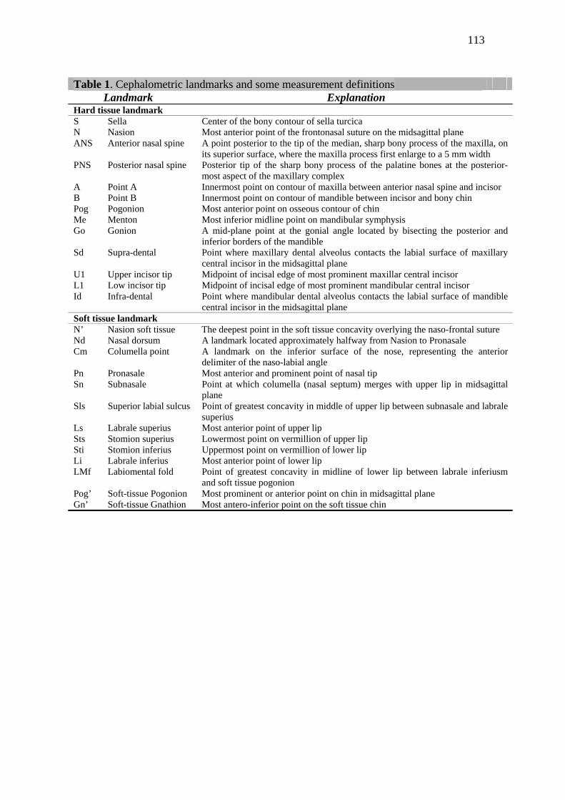
113
Table 1. Cephalometric landmarks and some measurement definitions Landmark Explanation
Hard tissue landmark S Sella Center of the bony contour of sella turcica N Nasion Most anterior point of the frontonasal suture on the midsagittal plane ANS Anterior nasal spine A point posterior to the tip of the median, sharp bony process of the maxilla, on
its superior surface, where the maxilla process first enlarge to a 5 mm width PNS Posterior nasal spine Posterior tip of the sharp bony process of the palatine bones at the posterior-
most aspect of the maxillary complex A Point A Innermost point on contour of maxilla between anterior nasal spine and incisor B Point B Innermost point on contour of mandible between incisor and bony chin Pog Pogonion Most anterior point on osseous contour of chin Me Menton Most inferior midline point on mandibular symphysis Go Gonion A mid-plane point at the gonial angle located by bisecting the posterior and
inferior borders of the mandible Sd Supra-dental Point where maxillary dental alveolus contacts the labial surface of maxillary
central incisor in the midsagittal plane U1 Upper incisor tip Midpoint of incisal edge of most prominent maxillar central incisor L1 Low incisor tip Midpoint of incisal edge of most prominent mandibular central incisor Id Infra-dental Point where mandibular dental alveolus contacts the labial surface of mandible
central incisor in the midsagittal plane Soft tissue landmark N’ Nasion soft tissue The deepest point in the soft tissue concavity overlying the naso-frontal suture Nd Nasal dorsum A landmark located approximately halfway from Nasion to Pronasale Cm Columella point A landmark on the inferior surface of the nose, representing the anterior
delimiter of the naso-labial angle Pn Pronasale Most anterior and prominent point of nasal tip Sn Subnasale Point at which columella (nasal septum) merges with upper lip in midsagittal
plane Sls Superior labial sulcus Point of greatest concavity in middle of upper lip between subnasale and labrale
superius Ls Labrale superius Most anterior point of upper lip Sts Stomion superius Lowermost point on vermillion of upper lip Sti Stomion inferius Uppermost point on vermillion of lower lip Li Labrale inferius Most anterior point of lower lip LMf Labiomental fold Point of greatest concavity in midline of lower lip between labrale inferiusm
and soft tissue pogonion Pog’ Soft-tissue Pogonion Most prominent or anterior point on chin in midsagittal plane Gn’ Soft-tissue Gnathion Most antero-inferior point on the soft tissue chin

114
*p<.05 **p<.01
Table 2. Group 1 (no genioplasty, n = 26) horizontal and vertical movements (millimeters) of hard and soft tissue landmarks (T1-T2)
Horizontal Vertical Landmark Mean SD p Mean SD p
Hard tissue (mm) ANS 1.1 2.3 * -0.3 1.5 PNS 2.0 2.9 ** 5.0 4.0 ** A 2.2 2.3 ** -0.6 1.6 Sd 3.8 2.3 ** -1.0 1.9 * U1 5.4 3.0 ** -0.8 1.9 * L1 6.9 3.8 ** -3.7 4.4 ** Id 9.5 4.4 ** -2.8 3.9 ** B 11.4 4.8 ** -1.3 3.4 Pog 14.7 6.1 ** -1.5 3.5 * Me 16.3 7.0 ** 1.0 3.3 Go 11.1 5.2 ** 16.7 8.9 ** Soft Tissue (mm) N’ 0.3 1.0 0.0 0.1 Nd 0.0 1.2 -0.2 1.0 Pn 0.4 1.0 * -0.6 1.7 Cm 1.4 1.6 ** -0.8 1.2 ** Sn 1.6 1.8 ** -0.6 1.0 ** Sls 3.2 1.9 ** -0.3 1.3 Ls 3.8 2.2 ** -0.6 1.8 Sts 5.0 2.8 ** -0.1 1.5 Sti 7.4 3.4 ** -3.5 4.2 ** Li 8.7 3.8 ** -5.1 4.5 ** LMf 11.6 5.0 ** -3.6 4.0 ** Pog’ 14.4 6.0 ** -2.5 3.8 ** Gn’ 15.8 6.6 ** -1.7 3.4 *
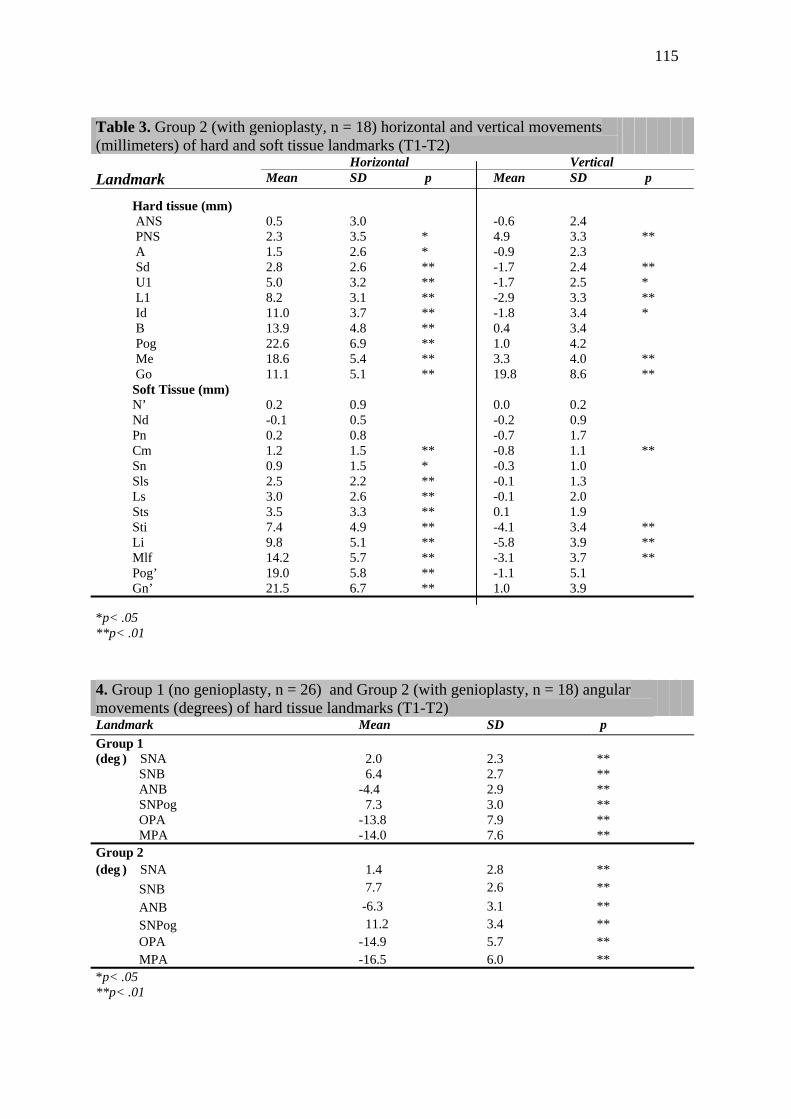
115
*p< .05 **p< .01
*p< .05 **p< .01
Table 3. Group 2 (with genioplasty, n = 18) horizontal and vertical movements (millimeters) of hard and soft tissue landmarks (T1-T2)
Horizontal Vertical Landmark Mean SD p Mean SD p
Hard tissue (mm) ANS 0.5 3.0 -0.6 2.4 PNS 2.3 3.5 * 4.9 3.3 ** A 1.5 2.6 * -0.9 2.3 Sd 2.8 2.6 ** -1.7 2.4 ** U1 5.0 3.2 ** -1.7 2.5 * L1 8.2 3.1 ** -2.9 3.3 ** Id 11.0 3.7 ** -1.8 3.4 * B 13.9 4.8 ** 0.4 3.4 Pog 22.6 6.9 ** 1.0 4.2 Me 18.6 5.4 ** 3.3 4.0 ** Go 11.1 5.1 ** 19.8 8.6 ** Soft Tissue (mm) N’ 0.2 0.9 0.0 0.2 Nd -0.1 0.5 -0.2 0.9 Pn 0.2 0.8 -0.7 1.7 Cm 1.2 1.5 ** -0.8 1.1 ** Sn 0.9 1.5 * -0.3 1.0 Sls 2.5 2.2 ** -0.1 1.3 Ls 3.0 2.6 ** -0.1 2.0 Sts 3.5 3.3 ** 0.1 1.9 Sti 7.4 4.9 ** -4.1 3.4 ** Li 9.8 5.1 ** -5.8 3.9 ** Mlf 14.2 5.7 ** -3.1 3.7 ** Pog’ 19.0 5.8 ** -1.1 5.1 Gn’ 21.5 6.7 ** 1.0 3.9
4. Group 1 (no genioplasty, n = 26) and Group 2 (with genioplasty, n = 18) angular movements (degrees) of hard tissue landmarks (T1-T2)
Landmark Mean SD p Group 1 (deg ) SNA SNB ANB SNPog OPA MPA
2.0 6.4 -4.4 7.3 -13.8 -14.0
2.3 2.7 2.9 3.0 7.9 7.6
** ** ** ** ** **
Group 2 (deg ) SNA 1.4 2.8 ** SNB 7.7 2.6 ** ANB -6.3 3.1 ** SNPog 11.2 3.4 ** OPA -14.9 5.7 ** MPA -16.5 6.0 **
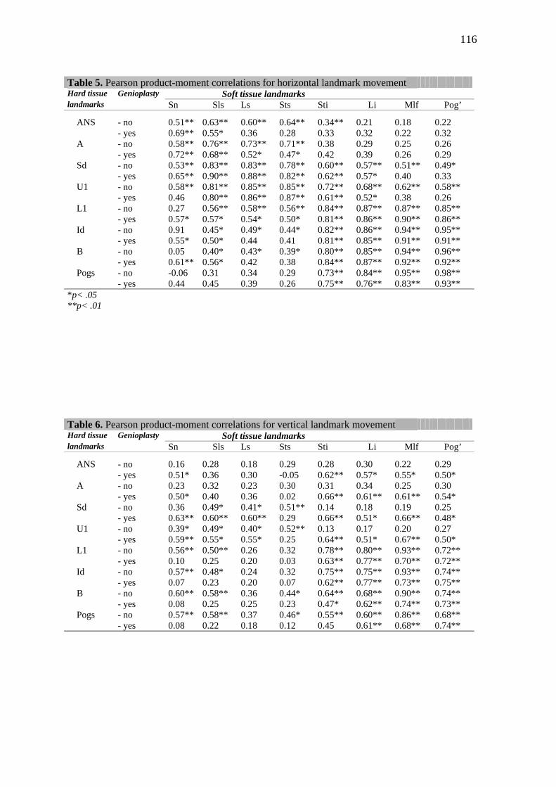
116
Table 5. Pearson product-moment correlations for horizontal landmark movement Hard tissue Genioplasty Soft tissue landmarks landmarks Sn Sls Ls Sts Sti Li Mlf Pog’
ANS - no - yes
0.51** 0.69**
0.63** 0.55*
0.60** 0.36
0.64** 0.28
0.34** 0.33
0.21 0.32
0.18 0.22
0.22 0.32
A - no - yes
0.58** 0.72**
0.76** 0.68**
0.73** 0.52*
0.71** 0.47*
0.38 0.42
0.29 0.39
0.25 0.26
0.26 0.29
Sd - no - yes
0.53** 0.65**
0.83** 0.90**
0.83** 0.88**
0.78** 0.82**
0.60** 0.62**
0.57** 0.57*
0.51** 0.40
0.49* 0.33
U1 - no - yes
0.58** 0.46
0.81** 0.80**
0.85** 0.86**
0.85** 0.87**
0.72** 0.61**
0.68** 0.52*
0.62** 0.38
0.58** 0.26
L1 - no - yes
0.27 0.57*
0.56** 0.57*
0.58** 0.54*
0.56** 0.50*
0.84** 0.81**
0.87** 0.86**
0.87** 0.90**
0.85** 0.86**
Id - no - yes
0.91 0.55*
0.45* 0.50*
0.49* 0.44
0.44* 0.41
0.82** 0.81**
0.86** 0.85**
0.94** 0.91**
0.95** 0.91**
B - no - yes
0.05 0.61**
0.40* 0.56*
0.43* 0.42
0.39* 0.38
0.80** 0.84**
0.85** 0.87**
0.94** 0.92**
0.96** 0.92**
Pogs - no - yes
-0.06 0.44
0.31 0.45
0.34 0.39
0.29 0.26
0.73** 0.75**
0.84** 0.76**
0.95** 0.83**
0.98** 0.93**
*p< .05 **p< .01 Table 6. Pearson product-moment correlations for vertical landmark movement Hard tissue Genioplasty Soft tissue landmarks landmarks Sn Sls Ls Sts Sti Li Mlf Pog’
ANS - no - yes
0.16 0.51*
0.28 0.36
0.18 0.30
0.29 -0.05
0.28 0.62**
0.30 0.57*
0.22 0.55*
0.29 0.50*
A - no - yes
0.23 0.50*
0.32 0.40
0.23 0.36
0.30 0.02
0.31 0.66**
0.34 0.61**
0.25 0.61**
0.30 0.54*
Sd - no - yes
0.36 0.63**
0.49* 0.60**
0.41* 0.60**
0.51** 0.29
0.14 0.66**
0.18 0.51*
0.19 0.66**
0.25 0.48*
U1 - no - yes
0.39* 0.59**
0.49* 0.55*
0.40* 0.55*
0.52** 0.25
0.13 0.64**
0.17 0.51*
0.20 0.67**
0.27 0.50*
L1 - no - yes
0.56** 0.10
0.50** 0.25
0.26 0.20
0.32 0.03
0.78** 0.63**
0.80** 0.77**
0.93** 0.70**
0.72** 0.72**
Id - no - yes
0.57** 0.07
0.48* 0.23
0.24 0.20
0.32 0.07
0.75** 0.62**
0.75** 0.77**
0.93** 0.73**
0.74** 0.75**
B - no - yes
0.60** 0.08
0.58** 0.25
0.36 0.25
0.44* 0.23
0.64** 0.47*
0.68** 0.62**
0.90** 0.74**
0.74** 0.73**
Pogs - no - yes
0.57** 0.08
0.58** 0.22
0.37 0.18
0.46* 0.12
0.55** 0.45
0.60** 0.61**
0.86** 0.68**
0.68** 0.74**

117
Table 7. Linear regression for horizontal landmark movement Dependent
variable Independent
variable Genioplasty Coefficient Intersection F p r
Sn ANS
-no -yes
0.41 0.34
1.17 0.72
8.26 14.36
0.008 0.001
0.51 0.69
Sls A
-no -yes
0.63 0.59
1.76 1.63
32.74 14.07
0.000 0.001
0.76 0.68
Ls Sd
-no -yes
0.80 0.86
0.75 0.59
50.98 53.09
0.000 0.000
0.82 0.88
Sts U1
-no -yes
0.79 0.91
0.72 - 0.97
61.59 44.43
0.000 0.000
0.85 0.86
Sti L1
-no -yes
0.76 1.27
2.17 - 3.04
57.22 31.21
0.000 0.000
0.84 0.81
Li Id
-no -yes
0.75 1.18
1.55 - 3.18
70.37 41.70
0.000 0.000
0.86 0.85
LMf B
-no -yes
0.97 1.10
0.51 - 0.99
195.18 87.97
0.000 0.000
0.94 0.92
Pog’ Pog
-no -yes
0.95 0.77
0.35 1.52
562.95 104.04
0.000 0.000
0.98 0.93
Table 8. Linear regression for vertical landmark movement Dependent
variable Independent
variable Genioplasty Coefficient Intersection F p r
Sn ANS -no -yes
0.11 0.22
- 0.56 - 0.13
0.63 5.48
0.433 0.032
0.16 0.51
Sls A -no -yes
0.27 0.23
- 0.11 0.09
2.75 3.04
0.110 0.100
0.32 0.40
Ls Sd -no -yes
0.38 0.49
- 0.21 0.75
4.80 8.78
0.038 0.009
0.60 0.52
Sts U1 -no -yes
0.41 0.19
0.24 0.43
8.99 1.08
0.006 0.313
0.52 0.25
Sti L1 -no -yes
0.75 0.65
- 0.76 - 2.18
36.29 10.37
0.000 0.005
0.78 0.63
Li Id -no -yes
0.87 0.91
- 2.63 - 4.16
31.64 23.72
0.000 0.000
0.75 0.77
LMf B -no -yes
1.06 0.79
- 2.22 - 3.40
104.65 19.57
0.000 0.000
0.90 0.74
Pog’ Pog -no -yes
0.73 0.90
- 1.38 - 2.00
20.13 18.79
0.000 0.000
0.68 0.73
Table 9. Ratios and percentage changes between hard and soft tissues after horizontal advancement
Ratios No genioplasty Percentage Changes
Genioplasty Percentage Changes
ANS: Sn A: Sls Sd: Ls U1: Sts L1: Sti Id: Li B: LMf Pog: Pog’
1: 1.45 1:1.45
1:1 1:0.92 1:1.07 1:0.91 1:1.01 1:0.97
145 145 100 92
102 91
101 97
1:1.80 1:1.66 1:1.07 1:0.70 1:0.90 1:0.89 1:1.02 1:0.84
180 166 107 70 90 89 102 84

118

121
4 Considerações Finais
A ciência evolui em meio a erros e acertos. Avanços
consistentes e embasados cientificamente, só ocorrem mediante
observação criteriosa e analítica dos fatos; sempre desprovida de
tendenciosidades.
Historicamente, cirurgias de ATM evoluíram de maneira
consistente, porém controvertida. Muito antes de Costen6 o advento do
raio X no final do século XIX, ofereceu indícios para uma possível
correlação entre diminuição do espaço intra-articular e dor facial. Essa
constatação bastou para que uma série de condilectomias
conservadoras ou radicais fosse realizada na tentativa de restabelecer o
espaço diminuído. Claramente equivocada, essa técnica gerou
inúmeros efeitos-colaterais sempre acompanhados de mordida aberta
anterior e dor.
Com o surgimento da ressonância magnética, viabilizou-se
um diagnóstico mais minucioso e preciso dos componentes internos da
ATM, principalmente do disco articular e de seus ligamentos e
inserções. Assim, abriu-se caminho à interpretação de que os
problemas relacionados à ATM originavam-se de posições anômalas
dos discos articulares, perfurações ou outras alterações morfológicas
que, de maneira simplista, foram tratados com menissectomias,
122
reposicionamentos de disco, ou substituição dos discos
morfologicamente alterados por implantes aloplásticos ou retalhos
musculares pediculados. Novamente a instituição generalizada dessas
técnicas não trouxe os resultados esperados para muitos pacientes.
Embora os resultados iniciais fossem promissores, com índice de
sucesso em torno de 91%27, o acompanhamento em longo prazo
mostrou que pacientes submetidos a implantes de Proplast/Teflon (PT,
Vitek Inc., Houston, TX) na sua maioria, desenvolviam reação de células
gigantes por corpo estranho que os levavam a quadros dramáticos,
irreversíveis e de solução paliativa10,14,17,24.
Paralelamente ao desenvolvimento das técnicas cirúrgicas
para tratamento da ATM e suas conseqüências negativas, desenvolveu-
se também técnicas conhecidas como de invasão mínima (artroscopia e
artrocentese). O advento dessas técnicas derrubou a idéia de que um
disco deslocado seria o causador da dor, uma vez que, com simples
lavagem ou remoção de aderências da ATM ocorria melhora
significativa dos sintomas1,13,15,16. Desse modo, assumiu-se que as
alterações morfológicas seriam na verdade, conseqüências dos eventos
bioquímicos que antecederam essa alteração, e não sua causa como
acreditavam os seguidores de terapias intervencionistas.
Outra contribuição importante que a artroscopia e a
artrocentese trouxeram foi o desenvolvimento de pesquisas sobre os
eventos biológicos moleculares que ocorriam na articulação doente. A
Considerações finais
123
partir disso, observou-se uma seqüência de eventos químicos que
denunciavam a transição de um metabolismo anabólico (adaptativo)
para um metabolismo catabólico (degenerativo) na articulação
comprometida7,22.
Embora a ATM tenha um grande potencial adaptativo devido à
sua fibrocartilagem, essa adaptação depende da variabilidade biológica
de cada indivíduo ante o agente agressor. Um exemplo disso são os
pacientes que exercem apertamento ou outra parafunção, e pelo
estímulo existente, podem sair de uma condição metabólica adaptativa
para uma condição metabólica degenerativa. Somado ao processo
degenerativo, é comum observar-se uma sintomatologia exacerbada
nos pacientes portadores de DTM. Frequentemente acompanhados de
dor crônica, os sintomas são normalmente desencadeados pelos
sucessivos anos de processo patológico na ATM somado ao
comprometimento emocional muitas vezes presente23. Os portadores de
dor crônica sofrem de sensibilização central pela estimulação
continuada das vias aferentes associada a alterações no sistema
inibitório descendente da dor19,21. O resultado importante dessas
alterações é que a dor não reflete simplesmente a presença,
intensidade ou duração de um estímulo específico na periferia, mas
também mudanças na função do Sistema Nervoso Central34,35. Dessa
forma, muitos pacientes sofredores de dor crônica não respondem de
maneira previsível ao trauma cirúrgico, ainda que este venha a eliminar
Considerações finais
124
o fator etiológico que iniciou o estímulo doloroso. Como uma entidade
própria, a dor ou a falha dos mecanismos fisiológicos de sua inibição,
perpetua-se ou agrava-se independentemente da intervenção
realizada28. Cirurgias em ATM ou outras cirurgias realizadas nesses
pacientes participam, de maneira negativa, no sistema de
retroalimentação de seus problemas físicos e emocionais. Portanto, o
tratamento cirúrgico para portadores de dor crônica não é a melhor
escolha35.
Felizmente as cirurgias em ATM não são rotina em nosso
meio, como acontece com a cirurgia ortognática convencional. Nos
Estados Unidos da América do Norte essa situação é bem diferente. O
sistema privativo de saúde desse país teve participação direta no
estímulo às intervenções complexas, principalmente as de caráter
cirúrgico. A expectativa de que a complexa, cara e ousada intervenção
possa trazer a solução para seu sofrimento e a posterior frustração por
não atingir os resultados esperados, leva pacientes a repetidas cirurgias
em ATM. Pacientes pagantes dos caros planos existentes sentem-se
indulgentes com intervenções dispendiosas na tentativa subconsciente
de sentir que esses procedimentos fazem jus ao dinheiro empregado.
Esta linha de pensamento é agravada quando intervenções cirúrgicas
estão envolvidas. Intervenções cirúrgicas, no entendimento do paciente
abalado emocionalmente pela DTM com dor crônica, têm um caráter de
socorro extremo, externo e totalmente desvinculado da participação do
Considerações finais
125
paciente ou mesmo de uma equipe multidisciplinar. É como se uma
intervenção isolada fosse capaz de solucionar problemas que se
desenvolveram ao longo de décadas e provavelmente por etiologias
multifatoriais.
A evidenciação científica de que DTMs de origem articular
são um evento multifatorial com respostas biológicas variáveis fez com
que protocolos de tratamento generalizados e invasivos fossem
questionados. Criou-se então um consensual preconceito sobre a
execução de cirurgias nas ATMs pela comunidade científica baseado
nos insucessos obtidos e na melhor compreensão dos mecanismos
envolvidos na dor crônica da ATM, gerando uma rejeição às
intervenções cirúrgicas nessa articulação como forma previsível de
tratamento. Assim, de um extremo passou-se a outro. E novos
equívocos se somam resultando em mais dificuldade em se estabelecer
bons protocolos de tratamento.
Esse preconceito generalizado não estaria presente caso
terapias intervencionistas fossem aplicadas diante de condições
específicas, o que reduziria em muito, os altos índices de insucesso
acumulados no passado. Existem várias situações clínicas bem
definidas em que a intervenção cirúrgica na ATM assume indicação
indiscutível. São elas, as anquiloses, as microssomias hemifaciais tipo
III, as reabsorções condilares idiopáticas totais, as reabsorções de
origem reumatóide ou auto-imune, os traumatismos com destruição
Considerações finais
126
condilar, os tumores e os pacientes previamente submetidos a múltiplas
cirurgias, em especial as de implantes de Proplast/Teflon. Pacientes
portadores dessas condições sofrem com o preconceito citado.
A rejeição à intervenção cirúrgica das ATMs, faz com que
inúmeros pacientes portadores de reabsorção condilar idiopática sejam
submetidos a sucessivas cirurgias ortognáticas na expectativa de se
obter a estabilidade não conseguida nas cirurgias anteriores devido à
patologia da ATM9. Pacientes com dramática restrição do espaço aéreo
buco ou nasofaríngeo devido a patologias diversas da ATM que
afetaram o crescimento mandibular são condenados a uma vida de
limitações já que seus côndilos mandibulares não suportariam a
amplitude do avanço mandibular necessário para atender suas
necessidades. A mesma situação ocorre quando processos auto-imunes
acometem as ATMs em idade precoce. O comprometimento da
articulação afeta o crescimento, gerando um padrão dolicofacial com
severa retrusão mandibular onde a cirurgia ortognática isolada não é
uma opção previsível29. Essas condições estão presentes na amostra
estudada nesta série de artigos. Graças à superação de preconceitos e
a uma convicção obstinada, a técnica desenvolvida pelo Dr. Larry
Wolford mudou para melhor a vida de muitos pacientes. O
desenvolvimento de uma prótese individualizada por meio da tecnologia
CAD/CAM e com materiais biocompatíveis viabilizou um alto índice de
sucesso para esses casos. Embora ainda não se saiba ao certo a
Considerações finais
127
longevidade dessa prótese, já é possível afirmar, com base nos
resultados desta série de artigos, que o índice de estabilidade cirúrgica
é extremamente satisfatório. Os 19 anos de experiência do Dr. Wolford
com a prótese estudada, somados a mais de 540 substituições de ATM
com sucesso, faz com que esse tipo de prótese customizada represente
uma opção real para inúmeros pacientes carentes de soluções
definitivas para seus problemas.
Não há como ignorar, que em alguns raros casos da
amostra estudada, a melhora nas funções e na sintomatologia dolorosa
foi limitada*. Mas são esses mesmos casos na qual há uma história de
sucessivas e fracassadas tentativas de intervenção cirúrgica somada ao
desenvolvimento de sensibilização central. Esses casos merecem
estudos complementares. O que se observou na grande maioria dos
casos, foi justamente o oposto. Os resultados mostraram que as
próteses totais de ATM (TMJ Concepts system®) apresentam
estabilidade em longo prazo (1 a 11,9 anos de acompanhamento),
sendo uma técnica viável para casos de reconstrução de ATM na qual
se faz necessário grandes avanços mandibulares com rotação anti-
horária do plano oclusal. Pacientes com deformidades dentofaciais
decorrentes de patologias da ATM são os grandes beneficiados pela
técnica. Porém, esse não foi o único benefício observado. O aumento
significativo do espaço aéreo bucofaríngeo incrementou a qualidade de
vida a esses pacientes. Enquanto a cirurgia ortognática isolada em
Considerações finais
*Pinto LP, Wolford LM, Buschang; Bernardi FH, Goncalves JR, Cassano DS. Maxillo-MandibularCounter-Clockwise Rotation and Mandibular Advancement with TMJ Concepts®Total JointProstheses: Part III - Pain and Dysfunction Outcomes. IJOMS-D-07-00535R1 .
128
pacientes com ATMs saudáveis viabiliza, segundo Gonçalves8 (2006),
um aumento médio de 4,4 mm nas dimensões faringeanas, a
incorporação da cirurgia de substituição das ATMs em pacientes com
comprometimento irreversível possibilitou resultados semelhantes
(média de 4,9 mm). As mudanças em tecido mole comportaram-se de
forma previsível diante da movimentação esquelética e otimizaram a
estética facial que, de maneira nenhuma, poderia ser alcançada pelos
métodos convencionais de cirurgia ortognática.
Assim, é preciso que se olhe para as cirurgias de ATM
sem uma visão mercadológica e livre de preconceitos. A substituição
das ATMs por prótese, embora rara, tem se mostrado uma ótima opção
quando bem indicada, e algumas vezes, é a única opção de tratamento
presente. Para que se reverta essa visão preestabelecida sobre as
intervenções cirúrgicas faz-se necessário que estudos científicos
imparciais, com metodologia meticulosa e amostra significante, e com
um longo período de acompanhamento sejam realizados, assim como
este trabalho se propôs a fazer. As mudanças nesse conceito foram
iniciadas, mas ainda há um grande caminho a ser percorrido
principalmente em relação às indicações da técnica discutida no
presente trabalho.
Considerações finais
129
Referências
130
5 Referências *
1. Alpaslan GH, Alpaslan C. Efficacy of temporomandibular joint
arthrocentesis with and without injection of sodium hyaluronate in
treatment of internal derangements. J Oral Maxillofac Surg. 2001; 59:
613-8.
2. Amstutz HC, Campbell P, Kossovsky N, Clarke IC. Mechanism and
clinical significance of wear debris-induced osteolysis. Clin Orthop.
1992; 276: 7-18.
3. Baird DN, Rea WJ. The temporomandibular joint implant controversy:
A review of autogenous/alloplastic materials and their complications.
J Nutr Environ Med.1998; 8: 289-300.
4. Blunn GW, Walker PS, Joshi A, Hardinge K. The dominance of cyclic
sliding in producing wear in total knee replacements. Clin Orthop.
1991; 273: 253-60.
5. Chuong R, Piper MA, Boland TJ. Recurrent giant cell reaction to
residual Proplast in the temporomandibular joint. Oral Surg Oral Med
Oral Pathol.1993; 76: 16-9.
6. Costen JB. The present status of the mandibular joint syndrome in
otolaryngology. Trans Am Acad Ophthalmol Otolaryngol. 1951; 55:
809-23.
* De acordo com o estilo Vancouver. Disponível em:http://www.nlm.nih.gov/bsd/uniform_requirements.html
131
7. De Bont LGM. Temporomandibular joint degenerative diseases:
pathogenesis. In: Stegenga B, de Bont LGM, editors. Management of
temporomandibular joint degenerative diseases: biologic basis and
treatment outcome. Basel: Birkhauser; 1996. p. 3-11.
8. Gonçalves JR, Buschang P, Gonçalves DG, Wolford LM. Postsurgical
stability of oropharyngeal airway changes following counter-clockwise
maxillo-mandibular advancement surgery. J Oral Maxillofac Surg.
2006; 64: 755-62.
9. Goncalves JR, Cassano DS, Wolford LM, Santos-Pinto A, Marquez
IM. Postsurgical Stability of Counterclockwise Maxillomandibular
Advancement Surgery: Affect of Articular Disc Repositioning. J Oral
Maxillofac Surg. 2008; 66: 724-38.
10.Henry CH, Wolford LM. Treatment outcomes for temporomandibular
joint reconstruction after Proplast-Teflon implant failure. J Oral
Maxillofac Surg.1993; 51: 352-8.
11.Henry CH, Hudson AP, Gerard HC, Franco PF, Wolford LM.
Identification of chlamydia trachomatis in the human
temporomandibular joint. J Oral Maxillofac Surg. 1999; 57: 683-9.
12.Henry CH, Hughes CV, Gerard HC, Hudson AP, Wolford LM.
Reactive arthritis: preliminary microbiologic analysis of the human
temporomandibular joint. J Oral Maxillofac Surg. 2000; 58: 1137-44.
Referências
132
13.Hepguler S, Akkoc YS, Pehlivan M, Ozturk C, Celebi G, Saracoglu A,
Ozpinar B. The efficacy of intra-articular sodium hyaluronate in
patients with recucing displaced disc. J Oral Rehabil. 2002; 29: 80-6.
14.Homsy CA. Important information on temporomandibular joint
surgery_ Letter of 1986. Vitek, Inc, October 31, 1986.
15.Kopp S, Akerman S, Nilner M. Short-term effects of intra-articular
sodium hyaluronate, glucocorticoid, and saline injections on
rheumatoid arthritis. J Craniomandib Disord. 1991; 5: 231-8.
16. Kopp S, Wenneberg B, Haraldson T, Carlsson GE. The short-term
effect of intra-articular injections of sodium hyaluronate and
corticosteroid on temporomandibular joint pain and dysfunction. J
Oral Maxillofac Surg. 1985; 43: 429-35.
17.Mosby EL, Wagner JD, Robinson RC. Assessment of Teflon-Proplast
implants in temporomandibular joint reconstruction following
menisectomy [abstract]. Presented at the AAOMS 70th Annual
Meeting, Boston, MA, September1988. J Oral Maxillofac Surg. 1988;
46(Suppl) :M16.
18.Okesson JP. Orofacial pain. Guidelines for assessment, diagnosis
and management. Carol Stream: Quintessence Publishing; 1996.
19.Ren K, Dubner R. Descending modulation in persistent pain: an
update. Pain. 2002; 100: 1–6.
Referências
133
20. Satrom KD, Sinclair PM, Wolford LM. The stability of double jaw
surgery: A comparison of rigid versus wire fixation. Am J Orthod
Dentofacial Orthop. 1991; 99: 550-63.
21.Scholz J, Broom DC, Youn DH, Mills CD, Kohno T, Suter MR, et al.
Blocking caspase activity prevents transsynaptic neuronal apoptosis
and the loss of inhibition in lamina II of the dorsal horn after
peripheral nerve injury. J Neurosci. 2005; 25: 7317-23.
22.Stegenga B, de Bont LGM, Boering G, van Willigen JD. Tissue
responses to degenerative changes in the temporomandibular joint. J
Oral Maxillofac Surg. 1991; 49: 1079-88.
23.Suvinen TI, Reade PC, Kemppainem P, Könönen M, Dworkin SF.
Review of a etiological concepts of temporomandibular pain
disorders: towards a bio psychosocial model for integration of
physical disorder factors with psychological and psychosocial illness
impact factors. Eur J Pain. 2005; 9: 613-3.
24. Turlington EG, Welch SR. Foreign-body reaction to Teflon-Proplast
fossa implant in TMJ arthroplasty [abstract]. Presented at the
AAOMS 68th Annual Meeting, New Orleans, LA, September1986
(suppl abstr).
25. Van Loon JP, De Bont L, Boering G. Evaluation of
temporomandibular joint prostheses: review of literature. J Oral
Maxillofac Surg. 1995; 53: 984-96.
Referências

134
26. Van Sickels JE: Stability in orthognathic surgery. In: Booth PW,
Schendel SA, Hausaman JE, editors. Maxillofacial surgery. London:
Churchill Livingstone; 1999. v. 2, p. 1297- 1304.
27.Vitek. Surgery of TMJ surgical results. Houston: Vitek; 1986.
28.Von Korff M, Leresche L, Dworkin SF. First onset of common pain
symptoms: a prospective study of depression as a risk factor. Pain.
1993;55: 251-8.
29.Wolford LM. Clinical indications for simultaneous TMJ and
orthognathic surgery. Cranio.2007; 25: 273-82.
30.Wolford LM, Karras SC. Autologous fat transplantation around
temporomandibular joint total joint prostheses: preliminary treatment
outcomes. J Oral Maxillofac Surg. 1997; 55: 245-51.
31. Wolford LM, Karras S, Mehra P. Concomitant temporomandibular
joint and orthognathic surgery: a preliminary report. J Oral Maxillofac
Surg. 2002; 60: 356-62.
32. Wolford LM, Mehra P, Rea W. Metal hypersensitivity in patients with
total joint prosthesis [abstract]. J Oral Maxillofac Surg. 2000;
58(Suppl 1): 29.
33.Wolford LM, Pitta MC, Reiche-Fischel O, Franco PF. TMJ
Concepts/Techmedica custom-made TMJ total joint prosthesis: 5-
year follow-up study. Int J Oral Maxillofac Surg. 2003; 32: 268-74.
34.Woolf CJ. Evidence for a central component of post-injury pain
hypersensitivity. Nature. 1983; 306: 686-8.
Referências

135
35.Woolf CJ. Central sensitization: uncovering the relation between pain
and plasticity. Anesthesiology. 2007; 106: 864-7.
Referências
136
Anexos

137
ANEXO 1

1
38
Tab
ela
A1-
Val
ores
inic
iais
e m
udan
ças c
irúrg
icas
e p
ós-c
irúrg
icas
T1
T2-T
1
T3
-T2
Varia
ble
Mea
n SD
M
ean
SD
Min
M
ax
P M
ean
SD
Min
M
ax
P
(deg
) S
NA
79
.8
4.0
2.3
2.3
-6.5
6.
4 **
-0
.4
1.4
-2.4
5.
9
S
NB
72
.4
4.1
6.9
2.9
1.0
12.8
**
0.
0 1.
0 -3
.1
2.2
AN
B
7.4
3.3
-4.6
2.
9 -1
0.5
2.2
**
-0.4
1.
4 -2
.6
5.4
*
SN
.Pog
73
.0
4.8
9.1
4.2
1.0
20.1
**
0.
0 1.
1 -2
.8
2.5
OP
A
25.1
8.
2 -1
4.9
8.0
-37.
0 -2
.3
**
0.6
3.3
-4.5
10
.9
MP
A
50.4
8.
0 -1
5.0
7.7
-35.
3 -0
.6
**
0.0
2.2
-5.1
5.
9
O
PT/
NS
11
1.1
8.7
-5.6
6.
7 -2
1.2
5.2
**
1.8
6.7
-11.
9 16
.8
OP
T/C
VT
17.3
1.
5 0.
0 0.
7 -1
.8
1.4
0.
2 0.
9 -1
.6
2.1
(m
m) A
NS
-hor
izon
tal
65.7
4.
7 1.
3 2.
4 -7
.3
7.1
**
-0.4
1.
6 -4
.0
5.2
AN
S-v
ertic
al
42.7
3.
3 -0
.6
1.9
-4.0
3.
4 *
0.4
1.5
-2.2
7.
0
P
NS
-hor
izon
tal
19.6
4.
3 2.
9 3.
1 -3
.9
10.9
**
-0
.8
2.6
-8.4
3.
9 *
P
NS
-ver
tical
42
.9
2.7
5.5
4.2
-1.8
14
.3
**
-0.6
2.
0 -5
.0
6.8
A-h
oriz
onta
l 67
.5
4.8
2.5
2.2
-6.0
6.
8 **
-0
.4
1.3
-2.8
5.
0 *
A
-ver
tical
49
.7
4.2
-1.0
1.
9 -4
.2
3.3
**
0.4
1.6
-2.5
7.
0
B
-hor
izon
tal
52.0
8.
3 12
.4
5.4
1.7
22.5
**
0.
0 1.
8 -4
.9
4.2
B-v
ertic
al
90.7
5.
7 -0
.1
3.5
-8.4
5.
8
-0.4
2.
0 -4
.7
3.8
Pog
- hor
izon
tal
50.5
10
.7
18.4
8.
5 2.
1 42
.1
**
-0.1
2.
1 -4
.9
5.3
Pog
- ver
tical
10
4.5
6.9
-0.1
3.
8 -1
0.9
8.8
-0
.3
2.1
-5.3
4.
0
M
e- h
oriz
onta
l 39
.4
11.3
17
.3
7.0
2.6
32.8
**
0.
0 2.
3 -5
.3
5.3
Me-
ver
tical
10
8.3
6.6
2.6
3.9
-6.7
12
.5
**
-0.4
1.
8 -4
.8
3.8
Go-
hor
izon
tal
-8.4
6.
1 11
.0
5.3
2.8
25.6
**
0.
0 1.
9 -5
.9
4.9
Go-
ver
tical
63
.1
8.8
18.4
9.
2 -1
.5
43.4
**
-0
.4
2.2
-6.8
6.
2
U
1T-h
oriz
onta
l 68
.7
6.3
5.6
3.0
-0.6
11
.3
**
-0.4
1.
7 -3
.9
4.8
U1T
-ver
tical
74
.7
4.8
-1.3
1.
9 -5
.7
2.6
**
0.3
1.7
-3.8
5.
7
L
1T-h
oriz
onta
l 63
.3
6.6
7.9
3.5
0.9
14.3
**
-0
.3
1.4
-2.8
3.
9
L
1T-v
ertic
al
74.6
5.
2 -2
.9
4.0
-16.
5 2.
3 **
-0
.4
1.8
-4.3
3.
8
C
3- h
oriz
onta
l -2
6.8
10.6
5.
9 7.
4 -7
.7
24.7
**
-1
.7
7.3
-19.
9 15
.2
C3-
ver
tical
99
.4
5.5
-0.5
1.
5 -4
.1
4.9
* 0.
5 2.
3 -6
.7
8.0
Hy-
hor
izon
tal
3.3
10.3
8.
5 5.
8 -7
.6
23.2
**
-1
.7
5.9
-18.
8 13
.3
Hy-
ver
tical
10
6.5
8.5
2.1
6.0
-12.
3 14
.1
* -4
.1
5.7
-19.
0 9.
9 **
BT-
hor
izon
tal
-10.
5 7.
5 7.
9 4.
8 -5
.6
16.3
**
-2
.1
4.2
-14.
5 8.
1 **
BT-
ver
tical
84
.8
8.3
8.3
7.0
-8.7
27
.1
**
-3.4
5.
1 -1
4.3
9.8
**
P
AS
nar
7.3
3.8
4.9
4.1
-3.5
15
.7
**
-1.1
4.
1 -1
2.7
6.8
C3-
Me
67.3
9.
6 11
.7
9.1
-15.
3 36
.9
**
1.7
7.4
-15.
8 17
.6
Hy-
C3
31.7
4.
6 3.
2 3.
9 -8
.2
14.6
**
-1
.0
4.1
-11.
5 9.
5
M
P-H
y 23
.2
5.9
-3.8
5.
8 -1
8.4
9.8
**
-2.5
5.
2 -1
4.5
10.0
**
OJ
5.
4 2.
7 -2
.2
2.5
-7.4
1.
8 **
-0
.1
1.2
-3.5
3.
8
O
B
-0.1
4.
1 -1
.6
3.7
-14.
7 3.
9 **
-0
.7
1.6
-7.5
2.
1 **
*p<
.05
**p<
.01

139
ANEXO 2
140
Autorizo a reprodução deste trabalho (Direitos de publicação reservado ao autor)
Araraquara, 23 de setembro de 2008
Karina Eiras Dela Coleta Pizzol