Embed Size (px)
Citation preview

UNIVERSIDADE FEDERAL DO RIO GRANDE DO SUL INSTITUTO DE CIÊNCIAS BÁSICAS DA SAÚDE
DEPARTAMENTO DE BIOQUÍMICA PROF. TUISKON DICK PROGRAMA DE PÓS-GRADUAÇÃO EM CIÊNCIAS BIOLÓGICAS: BIOQUÍMICA
Diagnóstico de anormalidades cromossômicas em fetos
com múltiplas malformações no HCPA:
Experiência com o uso exclusivo de cariótipo e avaliação
da contribuição da análise molecular
Tese de Doutorado
REJANE GUS KESSLER
Porto Alegre, maio de 2009

UNIVERSIDADE FEDERAL DO RIO GRANDE DO SUL INSTITUTO DE CIÊNCIAS BÀSICAS DA SAÚDE
DEPARTAMENTO DE BIOQUÍMICA PROF. TUISKON DICK PROGRAMA DE PÓS-GRADUAÇÃO EM CIÊNCIAS BIOLÓGICAS: BIOQUÍMICA
Diagnóstico de anormalidades cromossômicas em fetos com
múltiplas malformações no HCPA:
Experiência com o uso exclusivo de cariótipo e avaliação da
contribuição da análise molecular
REJANE GUS KESSLER
Orientador: Prof. Dr. Roberto Giugliani Co-orientadora: Dra. Sandra Leistner-Segal
Tese de Doutorado defendida como requisito parcial para obtenção do título de Doutor.
BANCA EXAMINADORA:
Profa. Dra. Vera Maria Treis Trindade (Departamento de Bioquímica, ICBS,Universidade Federal do Rio Grande do Sul)
Profa. Dra. Mariluce Riegel (Instituto de Genética Médica, Universidade de Zurich)
Dr. Sharbel Weidner Maluf (Serviço de Genética Médica, Hospital de Clínicas de Porto Alegre) Porto Alegre, maio de 2009

II
Às minhas filhas, Bruna e Ariela, verdadeiras
fontes de inspiração, e que são a razão de tudo…

III
AGRADECIMENTOS
Gostaria de iniciar minha lista de agradecimentos com meu caro
Roberto Giugliani, não porque de praxe devemos fazê-lo ao Orientador, mas sim, porque foi ele quem primeiro acreditou em mim desde os meus primórdios aqui na Genética (há mais de 25 anos). Sempre me incentivou a crescer profissionalmente, e também como pessoa, me ajudando em momentos difíceis, sendo por diversas vezes mais “amigo” do que “chefe”. Certamente, se não fosse por ele, eu não estaria aqui, defendendo meu doutorado hoje. A sua sabedoria, liderança e competência serviram de exemplo para que eu pudesse construir minha caminhada, tornando-me uma profissional segura e confiante, e fazendo das minhas próprias conquistas uma batalha gratificante. A ele, minha eterna gratidão e admiração. - À minha querida amiga, confidente e co-orientadora (nesta ordem), Sandra Segal, que não poupou esforços para que eu chegasse até aqui, sempre me incentivando, tentando me passar seus conhecimentos, extremamente prestativa e disponível, tanto na bancada como nas correções dos artigos e tese; incansável e pacienciosa, a quem devo grande parte deste trabalho. - À minha querida amiga e eterna incentivadora Maria Teresa Sanseverino, por suas constantes e sábias intervenções e sugestões, mas principalmente, por ser uma otimista incurável e ter sempre uma palavra de consolo na hora certa, uma verdadeira Madre Teresa de Calcutá. -À minha amiga de “priscas eras” Mariluce Riegel, que bravamente atravessou os mares, conquistando os Alpes suíços, chegando até a Universidade de Zurich, e que me abriu as portas do seu laboratório para as análises de QF-PCR e MLPA, sem as quais este trabalho nunca teria se concretizado. - À minha querida colega de sala, Fernanda Timm, por ser esta amiga tão leal, confidente de todas as horas, sempre pronta para ouvir e ajudar, deixando constantemente à disposição seu ombro consolador para repousar nas horas de desespero. Não poderia deixar de agradecer também, a sua paciência em me ajudar, e muito, com suas valiosas dicas na informática, tema que ela domina tão melhor do que eu.

IV
- Às tão queridas e prestativas Ingrid, Lili Cossio, Patricia Prolla e Patricia Koehler por suas sugestões e por terem colocado à minha disposição o laboratório de Medicina Genômica. - Aos bolsistas da Biologia Molecular do SGM Thaís, Ana, Marcela, Camila e Rafael por me receberem com tanto carinho e paciência no laboratório. - Às “amigníficas” Cristina Netto e Maira Burin (Sandra e Teresa também “amigníficas”), por tantos momentos relaxantes nas nossas “happy hours” e/ou congressos, que tornaram muito mais leve este período tão pesado que todos nós passamos nesta etapa final de doutorado. - Aos bolsistas do Laboratório de Cultura de Tecidos, por nunca se negarem a me emprestar reagentes ou materiais que por ventura faltassem para a rotina (fato muito comum); em especial ao Alexandre, por agüentar meu mau humor matinal (outro fato bastante comum). - Ao colega e amigo Sharbel por dividir algumas angústias citogenéticas, e cobrir sempre minhas férias com tanta dedicação. E também a suas bolsistas, principalmente a Maiana Fahler, sempre prontas para ajudar e ceder o que estivesse ao seu alcance, como boas colegas e vizinhas. - Aos meus “velhos” amigos e companheiros de “guerra” desde a antiga Unidade de Genética Médica: Julio, Têmis, Laura (agora chefe), Maria Luiza, Janice e Ricardo, por terem dado um colorido especial ao meu cotidiano durante tantos anos. - Aos jovens companheiros da sala de cafezinho do atual SGM por tantos momentos descontraídos, em especial o Hugo, por se colocar à disposição caso necessitasse de sua ajuda e conhecimento para as análises de MLPA. - À Jacira, Zeniara, Marilda, Cléia, Priscila e todo o pessoal da secretaria, pelo apoio técnico e alegria do convívio. - Ao tão eficiente Célio, por não medir esforços para tentar conseguir tudo que estava ao seu alcance, mesmo o que não estava, sempre com muita dedicação e prestatividade. - À professora Vera Maria Treis Trindade, por suas valiosas sugestões como relatora.

V
- Ao Curso de Pós-Graduação em Bioquímica da UFRGS, por aceitar tantas solicitações minhas, oportunizando o término deste trabalho. - Ao FIPE HCPA, que aprovou o projeto e deu o apoio financeiro necessário. E finalmente àquelas pessoas, que não me ajudaram diretamente nem contribuíram cientificamente com o trabalho, mas são aquelas que realmente dão sentido à minha vida: - Às minhas filhas Bruna e Ariela, que são a razão de tudo que faço, que dão o real valor para minhas conquistas, e a valiosa oportunidade de realizar-me como mãe, me possibilitando sentir um amor único, verdadeiro e incondicional. - Aos meus pais, eternos incentivadores, os alicerces da minha vida, formadores da minha personalidade, exemplos a serem seguidos, que possuem a lucidez e a solidez que eu necessitava para concretizar meus sonhos. - Ao meu irmão Miguel, que sempre foi um exemplo de profissional sério e competente, por suas sábias sugestões e por acreditar que eu poderia chegar lá (ou aqui), “mesmo sendo bióloga”. - À minha irmã, não de sangue, mas de escolha, Anne Bryk, uma eterna “Pollyana”, que sempre tentou me mostrar que para tudo na vida existe um lado brilhante e positivo, mesmo no lado mais obscuro da lua. - Ao Marcelo, pai das minhas filhas, que por muitos anos esteve ao meu lado, segurando “as pontas”, e as meninas também, e que continua uma pessoa muito especial, com a qual sei que sempre poderei contar. - Ao meu querido Nestor, que esteve constantemente ao meu lado nestes últimos três anos, e muito me incentivou para chegar até aqui, acreditou em mim, me apoiou e me deu a força necessária para seguir adiante, mesmo quando a vontade era de desistir em momentos de fraqueza e desânimo. A sua constante dedicação, preocupação e carinho me fizeram descobrir um amor diferente… A TODOS, O MEU ETERNO E INESQUECÍVEL “MUITO OBRIGADA”!!!
VI
RESUMO
Com o desenvolvimento da citogenética convencional, a partir da metade do século passado, houve um enorme crescimento no entendimento da etiologia das síndromes malformativas, sendo as anomalias cromossômicas reconhecidas como a causa genética mais freqüente dos defeitos congênitos. Assim sendo, a investigação de aberrações cromossômicas através do cariótipo se tornou uma rotina na investigação das malformações e de outras condições. Desde os anos 70, quando o diagnóstico pré-natal para a detecção de anomalias cromossômicas no feto foi estabelecido, este tem se tornado uma prática usual em muitos países, e uma importante ferramenta para o aconselhamento genético. A introdução da ultra-sonografia pré-natal na obstetrícia permitiu a identificação precoce de fetos com malformações, os quais têm grande probabilidade de serem portadores de alguma anormalidade cromossômica. O conhecimento da etiologia de sua doença é fundamental para diminuir a ansiedade da família e para tomada de decisões sobre a gestação em curso e sobre gestações futuras. Foi realizado um estudo retrospectivo de 18 anos (1989-2007) que teve como objetivos gerais: estimar a freqüência das anormalidades cromossômicas mais comuns através do cariótipo convencional e suas indicações para as gestantes do Hospital de Clínicas de Porto Alegre (artigo 1). Embora o cariótipo seja considerado o exame padrão ouro para este tipo de investigação, o mesmo possui algumas limitações. Entre elas se destaca a dependência de uma cultura de células, o que significa uma espera de 10 a 14 dias para se obter um resultado, tempo este precioso para a família e para a gestante. Além disto, corre-se o risco de perda da cultura por contaminação ou falta de crescimento celular, e com isso nenhum resultado é atingido, permanecendo a família sem um esclarecimento sobre a situação. Na tentativa de solucionar estes problemas, nesta tese, também foi proposta uma continuação do estudo (de 2006 a 2008) em fetos com múltiplas malformações, para testar a aplicação de técnicas moleculares, como QF-PCR e MLPA em diferentes materiais fetais (artigo 2). Ainda, quando a coleta de materiais tradicionais como líquido amniótico, ou biópsia de vilosidades coriônicas, eram impossíveis de se coletar por alguma dificuldade, incluindo os problemas morfológicos do feto, materiais alternativos, como urina, foram obtidos para se testar sua utilidade para se atingir o diagnóstico citogenético. Os achados citogenéticos relatados no artigo 1 foram compatíveis com os da literatura, sendo a trissomia do 21 a anormalidade cromossômica mais freqüentemente detectada no pré-natal. Entretanto, como o risco de recorrência desta síndrome foi zero neste estudo, um filho anterior com síndrome de Down foi considerado uma indicação fraca, sendo priorizadas para a realização do exame outras famílias carentes de maior risco. Por outro lado, com a técnica de MLPA (artigo 2) não foram obtidos resultados satisfatórios, provavelmente porque as amostras não permitiam obter DNA na qualidade necessária para esse procedimento. Com a técnica de QF-PCR pode-se obter 30 resultados (alguns parciais) nas 50 amostras coletadas. O estudo citogenético convencional de 13 amostras em que materiais diversos dos usuais foram empregados teve 100% de sucesso. Com a aplicação desse arsenal de técnicas, a chance de se obter uma informação citogenética ficou bem maior, diminuindo a ansiedade do casal e auxiliando nas suas decisões reprodutivas.
VII
ABSTRACT
With the development of conventional cytogenetic techniques, since the second half of the last century, there was an enormous growth in knowledge of the etiology of malformed syndromes, being the chromosomal abnormalities considered the most common genetic causes of congenital defects. Therefore, the investigation of chromosomal aberrations by karyotype became a routine in the investigation of the malformations and many other conditions. Since the 70’s, when prenatal diagnosis to detect chromosomal abnormalities in the fetus was set up, this became usual practice in many countries, and an important tool for genetic counseling. The introduction of the ultrasound in the obstetrician practice allowed the early identification of the malformed fetus, which has a great probability of being a carrier of some chromosomal aberration. The knowledge of the etiology of the disease is essential to reduce the anxiety of the family and to plan future pregnancies. From 1989 to 2007, 905 pregnant women from Hospital de Clinicas de Porto Alegre had an investigation for their fetus conditions by conventional karyotypes. With this information, a study was made which the main objectives were: to estimate the frequency of the most common chromosomal abnormalities and to estimate the most frequent indications that lead those women to make this invasive procedure (manuscript 1). Although the karyotype is considered the gold standard test for this type of research, it has some limitations, among them: the necessity of a cell culture, which means an expected time between 10 to 14 days to obtain the desired result, and time is a very precious aspect in pregnancy. Moreover, there is a risk of loss of culture because of contamination or lack of cell growth, leaving the family without a result. In an attempt to solve these problems, we proposed another study (from 2006 to 2008) to check the application of molecular techniques such as MLPA and QF-PCR in different malformed fetal materials (manuscript 2). Still, when the collection of traditional materials such as amniotic fluid, blood cord or chorionic villus biopsy were impossible to collect because of morphological problems on the malformed fetus, other alternative materials such as urine or intraperitoneal fluid were collected in an attempt to test these materials as a manner to reach the cytogenetic diagnosis. Our cytogenetics findings were similar to the literature, being trisomy 21 the most frequent chromosomal abnormalities. On the other hand, we did not find any recurrent case of this syndrome, so the indication for doing the exam, as having a previous child with Down syndrome, stayed as a weak indication that comes after others priorities, like pregnancies with greater risks. When we tried to introduce the MLPA technique, we did not achieve satisfactory results, probably because the samples were not suitable for good quality of DNA extraction. We then applied QF-PCR, and obtained 30 results (some partials) from 50 samples collected. The karyotype results of 13 samples from additional materials, not commonly used elsewhere, achieved 100% of success. With all these alternatives techniques, the chance to achieve a diagnosis was much higher, reducing the anxiety of the couple and helping the family to make decisions for futures pregnancies.

VIII
LISTA DE ABREVIATURAS
BVC- Biópsia de Vilosidades Coriônicas
EIM- Erros Inatos do Metabolismo
FISH- Fluorescence In Situ Hybridization
LA- Líquido Amniótico
MLPA- Multiplex ligation-dependent probe amplification
QF-PCR- Quantitative Fluorescence Ploymerase Chain Reaction
RAD - Rapid Aneuploidy Diagnosis
STR- Small Tandem Repeats
TN- Translucência Nucal

IX
LISTA DE FIGURAS
FIGURA 1
Punção de vilosidades coriônicas..........................................................................04
FIGURA 2
Cultura de amniócitos.............................................................................................06
FIGURA 3
Eletroferograma de uma Trissomia do 21…………………………………………… 11
FIGURA 4
QF-PCR do caso nº 2, trissomia do 13..................................................................56
FIGURA 5
QF-PCR do caso nº 47, trissomia do 18................................................................57
FIGURA 6
QF-PCR do caso nº 52, trissomia do 18................................................................58

X
SUMÁRIO
I. INTRODUÇÃO………………………………………………………………………….01
I.1. Histórico .......................................................................................................02
I.2. Tipos de procedimentos invasivos..............................................................04
I.2.1. Amniocentese...............................................................................................04
I.2.2. Biópsia de Vilosidades Coriônicas...............................................................05
I.2.3. Cordocentese…..……………………………………………………………… . 06
I.2.4. Coleta de Material Fetal Alternativo………..……………………..……………07
I.3. Cuidados Básicos com a Cultura de Células.............................................07
I.4. Técnicas Moleculares Alternativas…..……….............................................09
I.4.1. FISH………………………………………………..…………….........................09
I.4.2. QF-PCR………………..................................................................................10
I.4.3. MLPA………………......................................................................................12
I.5. Critérios para suspeitar de Aberrações Cromossômicas em fetos com
múltiplas malformações…………………........................................................... 13
1.6. Justificativa .................................................................................................15
II. OBJETIVOS.............................................................................................................16
II.1. Objetivos Gerais............................................................................................17
II.2. Objetivos Específicos ..................................................................................17

XI
III. RESULTADOS.........................................................................................................18
III.1. Artigo 1 ................................................................................................................19
Artigo 1 em PDF....................................................................................................20
III.2. Artigo 2 ............................................................................................................... 25
Artigo 2 submetido para revista.............................................................................26
IV. DISCUSSÃO ...........................................................................................................48
V. CONCLUSÕES………………………………..............................................................60
VI. REFERÊNCIAS.......................................................................................................64
VII. ANEXOS………………………………………………………………………………….71
VII.1. Parecer do Comitê de Ética do HCPA........................................................72
VII.2. Termo de Consentimento Livre e Esclarecido (pré-natal).......................73
VII.3. Termo de Consentimento Livre e Esclarecido (pós-natal)……………….74
VII.4. Confirmação da submissão do artigo 2.....................................................75
VII.5. Comentário do Editor ………………………………………………..…………76

1
I. INTRODUÇÃO

2
I. INTRODUÇÃO
I.1. Histórico:
O começo da ciência chamada Citognética Humana foi atribuído a Walther
Flemming, um anatomista alemão, pioneiro na descrição da cariocinese ou mitose
(1882) e considerado o fundador da ciência da citogenética, o estudo do material
celular hereditário. Ele foi o primeiro a observar e descrever sistematicamente o
comportamento dos cromossomos no núcleo celular, durante a mitose (Steven,
1999). Durante os anos seguintes, vários trabalhos foram publicados
apresentando diferentes estimativas quanto ao número de cromossomos
presentes na espécie humana. Um dos trabalhos de maior impacto foi o de
Theophilus Schickel Painter, no início da década de 20, em células obtidas de
testículos humanos, o qual descreveu que o número de cromossomos era 46 ou
48. Painter acabou decidindo a favor de 48 cromossomos, em sua última
publicação (Painter, 1923). Mais de 30 anos se passaram até o descobrimento do
verdadeiro número cromossômico humano, quando em 1956, Tjio e Levan
aperfeiçoaram o método da colchicina e hipotonia, chegando finalmente aos reais
46 cromossomos. Inicia-se então, a era da citogenética clínica, principalmente
depois que Lejeune e colaboradores (1959), estudando os cromossomos de
fibroblastos de um paciente com Síndrome de Down, descobriram e descreveram
a existência de um cromossomo extra, referindo-se pela primeira vez a uma
trissomia, a trissomia do cromossomo 21. Desde os anos 50, com o
desenvolvimento das técnicas da citogenética convencional, e principalmente com
a introdução da técnica de bandeamento G (Drets e Shaw, 1971), houve um

3
enorme avanço no conhecimento das síndromes cromossômicas, a mais comum
das causas das doenças genéticas (Pena, 1998).
A citogenética tornou-se então uma ferramenta fundamental para o
diagnóstico clínico pré e pós-natal. De todos os recém-nascidos vivos, 2-3%
podem apresentar alguma malformação congênita, e provavelmente, metade de
todos os conceptos, humanos pode ter algum tipo de defeito cromossômico (Boué
e Boué, 1973). Além disso, um em cada 200 nascidos vivos pode ter alguma
anormalidade cromossômica, sendo estas responsáveis pela maior causa de
retardo metal (Shaffer e Lupski, 2000). Isto reforça a idéia de que a análise
citogenética tornou-se fundamental para a investigação destes casos nos últimos
anos, demonstrando ser uma técnica indispensável para o manejo do diagnóstico
pré-natal.
Desde a década de 70, o diagnóstico pré-natal para a detecção de
anormalidades cromossômicas tem se tornado um procedimento rotineiro em
muitos países desenvolvidos (Magalhães e Magalhães, 2001). Este procedimento
invasivo tem sido amplamente reconhecido como um método confiável, com riscos
aceitáveis para casais que possuem probabilidade elevada de gerarem uma
criança com anormalidades cromossômicas significativas. Portanto, este
procedimento tornou-se uma ferramenta fundamental para o aconselhamento
genético, podendo evitar, tanto o nascimento, como a recorrência de crianças
afetadas com estas doenças (Milunsky e Milunsky, 1998).
A análise cromossômica microscópica de células cultivadas (cariótipo), tem
sido considerada uma técnica padrão ouro para o diagnóstico pré-natal, desde sua
primeira aplicação, por Steele e Breg, em 1966. A coleta direta de material fetal

4
para a análise em laboratório permite a realização de diversos outros exames.
Além do cariótipo fetal, a coleta permite a análise de ensaios enzimáticos para
erros inatos do metabolismo (EIM) e análise molecular de diversas doenças
gênicas. Atualmente, a obtenção do material fetal para qualquer uma destas
análises, é realizada através de procedimentos invasivos, ou seja, através de uma
punção transabdominal, realizada por um ginecologista ou radiologista experiente
e guiada por ultra-sonografia (figura 1).
Figura 1. Punção de vilosidades coriônicas guiada por ultra-sonografia
Fonte: www.ncbi.nlm.nih.gov/books/bv.fcgi?rid=eurekah
I.2. Tipos de Procedimentos Invasivos:
Os principais procedimentos invasivos realizados para obtenção de material
fetal, cujo crescimento celular vai resultar na análise do cariótipo são:
I.2.1. Amniocentese:
A amniocentese é a coleta de aproximadamente 20ml de líquido amniótico
(LA), no segundo trimestre, entre 15 e 16 semanas de gestação. Desde que a
amniocentese foi introduzida em meados da década de 60, juntamente com a

5
análise citogenética, esta tem sido reconhecida como uma técnica muito segura e
confiável para ajudar casais com risco de gerarem uma criança com anormalidade
cromossômica clinicamente significativa (Caron et al., 1999).
I.2.2 Biópsia de Vilosidades Coriônicas (BVC):
Passados alguns anos, no início da década de 80, a Biópsia de Vilosidades
Coriônicas (BVC) foi introduzida como uma alternativa bastante segura, porém
mais precoce do que a amniocentese (Brambati et al. 1998; Jenkins e Wapner,
1999). A BVC, realizada entre 12 e 14 semanas de gestação, é também por via
transabdominal com a coleta de 10 a 15mg de vilosidades da placenta, sendo que
este material é o “espelho genético” do feto. As vilosidades, depois de limpas e
dissecadas, podem ser analisadas diretamente e também após cultivo. Neste
procedimento, o fator crítico é a limpeza do material, pois deve ser retirada
minuciosamente toda decídua para ser descartada qualquer possibilidade de
contaminação com células maternas.
Tanto as células do LA como as de BVC, depois de obtidas e lançadas em
cultura, crescem aderidas na superfície de um frasco (figura 2), utilizado
especificamente para este fim, e posteriormente coletado para a análise do
cariótipo.

6
Figura 2. Cultura de amniócitos
Fonte: gentilmente cedido por Dra.Fernanda Timm (tese de doutorado)
Apesar de o cariótipo ter demonstrado ser um exame altamente confiável
para o diagnóstico de anormalidades cromossômicas fetais, tanto numéricas como
estruturais, estes procedimentos invasivos oferecem riscos para a gestação que
variam nos diferentes centros, sendo que o mais tardio (amniocentese) oferece
menor risco que o mais precoce (BVC), respectivamente em torno de 0.5% e 1%.
A acuracidade do cariótipo fetal de células cultivadas do LA tem sido considerada
entre 99.4%-99.8%, e do cariótipo da BVC está entre 97.5%-99.6% (Magalhães e
Magalhães, 2001).
I.2.3. Cordocentese:
No início dos anos 80, com uma melhora na imagem da ultra-sonografia, o
acesso ao sangue fetal ficou mais fácil, este podendo ser obtido através da
punção de um vaso umbilical, na idade gestacional entre 18 a 20 semanas. Este
procedimento, chamado cordocentese (Daffos et al., 1983), é utilizado na ausência
de LA, ou quando se faz necessário o esclarecimento de um diagnóstico prévio

7
duvidoso. Entretanto, mesmo que a cultura de sangue permita um resultado de
cariótipo em 72 horas, a idade gestacional já está bastante avançada e em muitos
casos muito tarde para uma provável interrupção. Além disto, este procedimento
está associado a um risco maior para complicações na gestação, portanto só deve
ser utilizado em casos muito bem selecionados (Dugoff e Hobbins, 2002).
I.2.4. Coleta de Material Fetal Alternativo:
Em diversas situações, a coleta de LA, BVC ou cordocentese, torna-se
impossível de ser realizada, devido a condição anatômica do feto (que muitas
vezes possui líquido amniótico totalmente ausente). Mas exatamente por ele
possuir múltiplas malformações, há necessidade de se obter o exame do cariótipo.
Nestes casos, tenta-se realizar o exame em materiais alternativos, que muitas
vezes são coletados como medidas terapêuticas (como drenagem cerebral em
hidrocefalias), e cujo material é aproveitado para a realização do cariótipo. Estes
possíveis materiais fetais alternativos podem ser: urina, fluidos intraperitoneal e
cérebro-espinhal, fluidos de rins displásicos ou de higromas císticos. Esta prática
não é muito usual na maioria dos laboratórios (Gole et al. 1997; Donnenfeld et al.,
2001), porém foi utilizada no primeiro artigo desta tese com um sucesso de 100%
(item III.1).
I.3. Cuidados básicos com a Cultura de Células
Apesar de o cariótipo ter permanecido como padrão ouro para detecção de
aneuploidias, este método apresenta algumas limitações, sendo que a mais
importante é a dependência do cultivo das células (Bui, 2007). Mesmo com o

8
advento de técnicas modernas, a falha no crescimento das células ainda é um
grande obstáculo a ser vencido. O laboratório de cultura celular depende de vários
fatores para obter sucesso no crescimento e obtenção de células viáveis. Os mais
importantes estão listados a seguir:
a) a assepsia é um dos aspectos fundamentais a ser cuidado, e iniciando
na coleta do material (o coletador, tanto obstetra como radiologista, deve ser muito
bem treinado neste aspecto), e seguindo regras rigorosas no laboratório, como a
lavagem constante das mãos com detergentes especiais, utilização de luvas,
toucas, máscaras e propés. Um jaleco apropriado deve ser utilizado apenas dentro
da sala de cultura.
b) utilização de material permanente (vidraria) estéril e descartável, sem a
reutilização de nenhum material, evitando assim problemas com lavagem e/ou
esterilização.
c) utilização de um meio de cultura apropriado para cada tipo de tecido.
Obviamente, este deve ser estéril e dentro do prazo de validade.
d) os equipamentos devem ser mantidos constantemente limpos e sob
manutenção. No caso das incubadoras, o ajuste da temperatura (37°C), umidade
e quantidade de CO2 (0,5%) deve ser permanentemente controlado. Mesmo com
todos estes cuidados, infelizmente, algumas vezes, pode haver falha em algum
destes itens acima citados, ocasionando a falta do resultado esperado pela
família.
Outra importante limitação do cariótipo reside no fato das análises
dependerem de uma pessoa muito experiente e bem treinada na observação ao
microscópio. Ao contrário de muitas análises bioquímicas, esta não é uma análise

9
automatizada, e depende exclusivamente da capacidade do olho humano bem
treinado (Pereira et al. 2000).
Como uma opção para o procedimento invasivo, surgiu um teste ultra-
sonográfico de rastreamento de risco gestacional para cromossomopatias
(13,18,21), denominado Translucência Nucal (Nicolaides et al., 1992). Mediante a
medida do tecido subcutâneo da nuca do feto, entre 11-14 semanas de idade
gestacional, pode-se selecionar pacientes para os exames invasivos. Se a medida
for baixa, estas pacientes podem evitar a punção. Mas o resultado é emitido como
uma probabilidade, e não como um teste diagnóstico ( Magalhães, 2001).
I.4. Técnicas Moleculares Alternativas
I.4.1. FISH (Fluorescence in situ hybridization):
Apesar do cariótipo ser um exame de muita precisão, pesquisadores
buscaram alternativas para suprir outra de suas grandes limitações: o tempo que a
cultura de células leva para atingir o crescimento celular desejado (em torno de 10
a 14 dias) e conseqüentemente, a demora do resultado. Neste tipo de exame, a
ansiedade da espera do resultado aumenta a cada dia que passa, principalmente
se a gestante é informada de que seu filho tem alguma malformação. Nestes
casos, a ansiedade é maior ainda, e a descoberta da etiologia da malformação é
fundamental, tornando a questão “tempo” uma prioridade. O mais importante
avanço nos últimos 20 anos foi o desenvolvimento da técnica citogenética
molecular chamada de FISH (Klinger et al., 1992; Munnè et al., 1998). Através de
uma hibridização do DNA alvo com sondas fluorescentes na própria lâmina, esta
técnica pode detectar os principais cromossomos envolvidos em aneuploidias (13,

10
18, 21, X e Y), que representam 90% de todas estas anormalidades
cromossômicas (Munnè et al., 1998). Isto pode ser realizado em até 48hs, e não
depende da cultura de células, ou seja, esta hibridização pode ocorrer em núcleos
interfásicos de LA ou BVC, conseqüentemente abreviando o resultado. No
diagnóstico pré-natal, o FISH é bastante utilizado como uma resposta rápida e
inicial das principais aneuploidias (triagem), necessitando a confirmação
complementar através do cariótipo. O FISH em núcleos interfásicos é utilizado
através de kits comercialmente disponíveis, e tem demonstrado, em múltiplos
estudos, ser um método altamente sensível e específico para a detecção de
aneuploidias (Shaffer e Bui, 2007). Obviamente a capacidade diagnóstica do FISH
é limitada pelas sondas que são escolhidas; conseqüentemente anormalidades
cromossômicas estruturais, aberrações cromossômicas numéricas incomuns, ou
marcadores, não ficarão evidenciados, necessitando do cariótipo para detectar ou
descartar estas raras, porém possíveis, alterações.
I.4.2. QF-PCR (Quantitative Fluorescent Polymerase Chain Reaction):
Esta outra técnica alternativa para o FISH foi primeiramente desenvolvida
por Mansfield em 1993. Trata-se de um ensaio multiplex QF-PCR, que também
permite a detecção um grande número de alterações numéricas cromossômicas
em 24-48 horas. Seqüências cromossômicas repetidas e altamente polimórficas
(short tandem repeats, ou STRs), que podem variar de comprimento entre os
indivíduos, são amplificadas por PCR utilizando-se iniciadores (primers) que são
marcados com fluorocromos. Os produtos da amplificação (amplicons) são
separados por eletroforese em capilar, podendo ser visualizados e quantificados

11
utilizando-se um analisador automático com um software apropriado. Para cada
sonda utilizada e informativa, dois picos serão produzidos com uma altura ou área
na razão de 1:1, que representa um feto normal heterozigoto (dialélico normal).
Amostras de fetos trissômicos irão demonstrar 3 picos na razão de 1:1:1
(trissômico trialélico), ou 2 picos na razão de 2:1 (trissômico dialélico)(figura 3). Se
quatro ou mais marcadores polimórficos de STRs forem utilizados para cada
cromossomo analisado, poucas amostras ficarão não informativas devido a
homozigose em alguns loci (Pena, 1998).
Figura 3. Eletroferograma da sonda D21S11 amplificada com dois tipos de trissomia 21.
No painel superior, um eletroferograma com um padrão trialélico, na proporção 1:1:1.
No meio, o painel mostra a trissomia do 21 com um padrão dialélico na proporção 2:1.
No painel inferior, um feto normal, heterozigoto na proporção 1:1. (Fonte: Tóth, 1998)
Por mais de 10 anos, QF-PCR tem sido aplicado com confiabilidade e muito
sucesso no diagnóstico pré-natal, tanto em LA como em BVC (Shaffer e Bui,
2007). Kits para QF-PCR estão comercialmente disponíveis e a sua acuracidade
para não mosaicismo e aneuploidias comuns é similar ao FISH interfásico. QF-

12
PCR pode detectar 20-30% de mosaicismo (Donaghue et al., 2005) e é superior
tanto ao FISH como ao cariótipo tradicional, no que diz respeito à detecção de
contaminação materna (Stojilkovic-Mikic et al., 2005). A maior vantagem do QF-
PCR sobre o FISH é seu custo-benefício, principalmente quando se processa um
grande número de amostras. Conseqüentemente, esta tecnologia está
substituindo o FISH na maioria dos laboratórios, principalmente na Europa.
Entretanto, ambas as tecnologias têm a mesma limitação no que diz respeito a
anomalias cromossômicas desbalanceadas e incomuns. Mas, como estas são
relativamente raras e quase sempre associadas a malformações fetais graves, o
médico poderá detectá-las através do ultra-som, e solicitar a busca de supostas
aberrações cromossômicas incomuns através de um cariótipo tradicional.
I.4.3. MLPA (Multiplex Ligation-dependent Probe Amplification):
Outra técnica de citogenética molecular que tem sido muito utilizada
recentemente é a MLPA, que é uma análise quantitativa baseada na PCR
(Schouten et al., 2002). Possui muitas vantagens, como a alta eficiência, facilidade
de operacionalizar, e necessidade de pequenas quantidades de DNA. Com
apenas 20ng de DNA, e somente um par de iniciadores, esta nova tecnologia
tanto é capaz de detectar um número anormal seqüências do DNA genômico, bem
como uma mutação de ponto. Comparada com a reação controle, a área do pico
de cada produto de amplificação reflete o número de cópias da seqüência alvo
que está sendo analisada na amostra. Um número aberrante de cópias de uma ou
mais seqüências detectadas por MLPA pode ser evidenciado por um aumento ou
diminuição da área do pico do produto da amplificação das sondas destas

13
seqüências. Nesta técnica, as sondas é que são amplificadas adicionadas às
amostras, e são capazes de discriminar seqüências que diferem em apenas um
único nucleotídeo (Zhou e Ren, 2009). Os produtos gerados pela PCR são
separados por eletroforese capilar adaptada em seqüenciador automatizado
(normalmente o ABI 3130), e analisadas através de um software (Coffalyser V9.4).
Tanto o termociclador como o seqüenciador são equipamentos fundamentais e
estão presentes hoje em dia em quase todos os laboratórios de biologia molecular.
Até 96 amostras podem ser analisadas simultaneamente, e os resultados podem
ser obtidos em 24-48 horas. O kit SALSA MLPA P095 MCR-Holland, Amsterdam
está comercialmente disponível e é específico para a detecção de aneuploidias.
Este kit contém oito sondas independentes para cada cromossomo 13, 18, 21, e X
mais quatro sondas específicas para o cromossomo Y, e é muito utilizado para a
detecção rápida de aberrações numéricas destes cromossomos no diagnóstico
pré-natal. Embora a aplicação da técnica de MLPA seja fácil, a implantação de um
novo protocolo deste ensaio é muito complexa e demanda muito tempo para se
conseguir determinar as condições ideais para a sua utilização. Os resultados
dependem basicamente da qualidade da extração de DNA (Roeder, et al.2009).
I. 5. Critérios para suspeitar de Aberrações Cromossômicas em fetos com
múltiplas malformações
Aberrações cromossômicas autossômicas são caracterizadas por quatro
critérios básicos: retardo de crescimento intra-uterino e pós-natal; um padrão de
sinais dismórficos, especialmente na face, genitália e membros distais;
malformações (geralmente múltiplas); e retardo mental (Schinzel, 2001). Embora

14
nenhum dos quatro critérios seja obrigatório, a deficiência mental é a característica
mais consistente, porém é a única que não pode ser detectada precocemente.
História familiar de rearranjos cromossômicos freqüentemente resulta em
história de perda fetal, redução da fertilidade, baixo peso e/ou prematuridade do
neonato com muitas malformações que vai a óbito logo depois do nascimento.
Nestes casos, há fortes evidências de rearranjos desbalanceados no feto, cujo
cariótipo se torna fundamental.
No quadro abaixo, listamos as malformações mais comuns em fetos que
são prováveis portadores de aberrações cromossômicas, e que foram utilizados
no trabalho como critérios para selecionar os casos suspeitos de
cromossomopatias (Schinzel, 2001).
Malformações comuns em aberrações cromossômicas autossômicas
___________________________________________________________ Pálato fendido, lábio fendido, ou ambos
Atresia de esôfago; fistula traqueoesofágica; atresia anal com fístula
Má rotação do intestino, mesentérico comum; onfalocele
Malformação do coração e grandes vasos
Malformação do rim e trato urinário
Alguma malformação cerebral, particularmente holoprosencefalia e
agenesia do corpo caloso
Ausência ou hipoplasia do radio e polegar
Hexadactilia Postaxial
Microftalmia, coloboma ocular
Espinha bífida (ocipital ou lombar)
_________________________________________________________
Fonte: Schinzel, 2001

15
I.6. Justificativa
Quando múltiplas malformações são detectadas por ultra-sonografia fetal,
embora as possibilidades etiológicas sejam amplas, a probabilidade de uma
anomalia cromossômica estar presente é muito alta. O cariótipo se torna
fundamental nesses casos, mas nem sempre seu resultado é obtido. Algumas
vezes esse insucesso decorre dos fatores técnicos já comentados, em outras, isso
se dá porque o feto vai a óbito antes que uma investigação seja possível, não se
chegando a nenhum diagnóstico pela indisponibilidade dos materiais necessários
à análise. Em qualquer dos casos, é muito difícil oferecer um aconselhamento
genético apropriado, e a família permanece sem nenhuma informação sobre a
condição do feto ou sobre a probabilidade ter um outro filho com o mesmo
problema. Como esta é uma situação muito difícil de manejar, na tentativa de
encontrar uma alternativa para superá-la propusemos este estudo, buscando
contribuir para diminuir a ansiedade destas famílias e facilitando o planejamento
de futuras gestações baseado em informações precisas.

16
II. OBJETIVOS

17
II. OBJETIVOS
II.1 - Objetivos Gerais
Este trabalho teve como objetivos gerais: estimar a freqüência das
anormalidades cromossômicas mais comuns e suas indicações no Hospital de
Clínicas de Porto Alegre, através do cariótipo convencional; e avaliar o uso de
técnicas alternativas para ampliar a possibilidade de se obter uma informação
citogenética pré-natal em fetos com múltiplas malformações de etiologia
desconhecida.
II. 2. Objetivos Específicos
1. Avaliar a contribuição das técnicas de citogenética molecular (MLPA ou
QF-PCR), aplicadas em diferentes tecidos, para detectar possíveis aneuploidias
dos cromossomos 13, 18, 21, X e Y em fetos com múltiplas malformações e sem
diagnóstico;
2. Avaliar a utilidade da extração de DNA das células do LA que não
aderiram no fundo do frasco de cultura para possível emprego em análises
moleculares e assim permitir testes adicionais;
3. Avaliar a possibilidade de obter o cariótipo em materiais alternativos
(urina, fluido do higroma cístico, fluido intraperitoneal, ou fluido cérebroespinal), o
que seria útil quando se torna impossível a coleta dos tradicionais (LA, BVC, ou
cordocentese);
4. Comparar as técnicas de citogenética molecular com a citogenética
tradicional em relação à identificação da etiologia nos fetos com múltiplas
malformações.

18
III. RESULTADOS
19
III. RESULTADOS
Os resultados desta tese estão organizados na forma de um artigo
publicado e outro submetido à publicação.
III.1. Artigo 1
Prenatal Diagnosis for Fetal Chromosomal Abnormalities: report of 18-year
experience in a Brazilian public hospital
Publicado na revista “Genetics and Molecular Biology”, 2008; 31 (4): 829-833.

Prenatal diagnosis of fetal chromosomal abnormalities:Report of an 18-year experience in a Brazilian public hospital
Rejane G. Kessler, Maria Teresa V. Sanseverino, Sandra Leistner-Segal, José A.A. Magalhães
and Roberto Giugliani
Serviço de Genética Medica, Hospital de Clínicas de Porto Alegre, Porto Alegre, RS, Brazil.
Abstract
The study of the fetal karyotype became an important tool for the fetal diagnosis of genetic diseases in the 1970s. Al-though application of this test has remained very restricted in Brazil, we had 905 referrals for prenatal fetalkaryotyping between 1989 and 2007. In 879 cases, a fetal karyotype was obtained. We detected 74 abnormal karyo-types (8.4%), the majority being found when the prior indication was fetal malformation. When obtaining amnioticfluid or chorionic villus samples was difficult, alternative fetal materials (urine, cystic hygroma, cystic lung, intre-peritoneal and cerebrospinal fluids) were collected and we had success in obtaining karyotypes in all 13 cases. Al-though, the option of terminating abnormal pregnancies does not legally exist in Brazil, the information gained inassessing the prognosis of on-going pregnancies or estimating recurrence risks justifies prenatal diagnosis of chro-mosome abnormalities. We conclude that, in keeping with the policy in most other countries, prenatal cytogeneticanalysis is strongly recommended in high-risk pregnancies for fetal abnormalities. However, the unique aspect of thistype of study is not its rarity in world terms, but its rarity in Brazil. This argues that Brazilian health policy on prenataldiagnosis requires reforming to make it much more widely available within the public health care sector.
Key words: prenatal diagnosis, chromosomal abnormalities, fetal malformations.
Received: January 31, 2008; Accepted: August 25, 2008.
Introduction
During the last decades the study of fetal karyotypes
has become a very important tool for genetic counseling on
recurrence risk and/or fetal chromosome diagnosis of at-
risk pregnancies (Magalhães, 2001). Invasive prenatal di-
agnosis continues to be the standard method for searching
for chromosomal aneuploidies or other genetic diseases
(Bui, 2007). Prenatal diagnosis of cytogenetic abnormali-
ties is now widely recognized as a reliable method with an
acceptable risk for couples at high risk of giving birth to a
child with clinically significant chromosome abnormalities
(Caron et al., 1999). Despite the fact that in Brazil amnio-
centesis and CVS were first introduced by Nazareth et al.
(1981) and Gollop et al. (1988) respectively, there is still no
public health care policy for application of cytogenetic pre-
natal diagnosis. As in other developing countries, this test is
mostly confined to expensive private clinics, which means
that it is rarely available for the great majority of pregnant
women who depend on public medical services.
Nevertheless, we have been offering this test in our
public hospital since 1989. Prenatal diagnosis is a very re-
stricted test in Brazil, mainly because induced abortion,
even indicated by fetal genetic disease, is not legally al-
lowed. Despite this, we have had 905 referrals for fetal
karyotyping since it was first offered by our clinic in 1989.
In the first four years, we had an average of 80 cases/year
and this number decreased in the following ten years to 45
cases/year. In the last four years this number decreased
even further, to 35 cases/year. This will be discussed later.
Even with the development of modern techniques,
cell culture failure remains one of the main obstacles to be
overcome. In order to improve the chance of getting a
karyotype result, alternative fetal samples, such as urine or
cystic hygroma fluid were used for chromosome analysis
when malformations were found in the fetus and availabil-
ity of conventional tissues was limited. The purposes of this
study were: 1) to describe the most frequent indications for
karyotyping the fetus in our socio-economic conditions; 2)
to estimate the frequency of the most common prenatal
chromosome abnormalities in patients from the Hospital de
Clinicas de Porto Alegre; 3) to assess the cytogenetic re-
sults obtained with alternative tissue samples compared to
amniocytes and chorionic villi.
Genetics and Molecular Biology, 31, 4, 829-833 (2008)
Copyright © 2008, Sociedade Brasileira de Genética. Printed in Brazil
www.sbg.org.br
Send correspondence to Rejane Gus Kessler. Serviço de GenéticaMedica, Hospital de Clinicas de Porto Alegre, Rua Ramiro Barcelos2350, 90035-903 Porto Alegre, RS, Brazil. E-mail:[email protected].
Research Article

Materials and Methods
Cytogenetic findings were retrospectively reviewed
from 1989 to 2007 in 905 pregnant women, with a mean
maternal age of 32.7 years, and mean gestational age of
22.7 weeks. Those women underwent prenatal cytogenetic
evaluation only after a genetic counseling session, which
means that risks, methods and indications were explained
to the family. All samples were collected by a single gyne-
cologist. The method used for sample collection was trans-
abdominal punction guided by ultrasound. Samples were
obtained for all patients, even in cases of lack of amniotic
fluid, when alternative fluids were collected. Amniotic
fluid, or any other fetal sample collected, were cultivated in
long-term cell cultures, with Amniomax medium, at 37 °C
in CO2 incubator. Cordocentesis followed the standard
blood culture that means, short-term culture (72 h) at 37 °C,
and no requirement for a CO2 incubator. We used standard
Giemsa-banding staining technique for all chromosome
analyses.
Results
The most frequent indications for prenatal cyto-
genetic diagnosis were advanced maternal age (with an av-
erage of 39.9 years old and mean gestational age of 18.7
weeks), abnormal findings on fetal ultrasound, a previous
child with chromosomal abnormalities, and increased
nuchal translucency (Table 1). Despite advanced maternal
age being the most frequent indication for prenatal diagno-
sis, the majority of aberrant karyotypes were found when
the indication was a fetal malformation detected by ultra-
sound. On the other hand, although the history of a previous
child with Down syndrome was a relatively frequent indi-
cation, we did not find any positive cases in this group.
From the 905 prenatal cytogenetic analysis per-
formed, we failed to obtain results in 26 (2.8%). Among the
879 karyotypes obtained, 74 (8.4%) were abnormal.
(Table 1). Numerical abnormalities were found in 64 cases
(7.3%), and structural aberrations in 10 cases (1.1%). The
majority of numerical chromosomal abnormalities were
autosomal trisomies. Trisomy 21 was the most frequent
(28; 3.2%), and the second most frequent was trisomy 18
(24; 2.7%). Interestingly, trisomy 18 was almost entirely
restricted to the group of “fetal abnormalities detected by
ultrasound” and none was detected in the “increased nuchal
translucency” group (p < 0.001). On the other hand, the dif-
ference in the frequencies of trisomy 21 between these two
types of ultrasound prescreening was not statistically sig-
nificant (p = 0.096). Trisomy 13 was found in six cases
(0.7%), monosomy X in one (0.6%) and one case showed
triploidy. Among structural chromosomal aberrations,
translocations were the most frequent, and were detected in
four out of the 879 cases analyzed (0.45%): reciprocal
translocations in two cases and Robertsonian translocations
in two others. Marker chromosomes were found in three
cases, deletions in two cases and an inversion was present
in one case.
In 13 cases alternative fluid samples were obtained
(Table 2). The reasons for collecting alternative materials
were lack of amniotic fluid in seven cases of kidney pathol-
830 Kessler et al.
Table 1 - Indications for invasive prenatal diagnosis and abnormal karyotypes.
Primary indication Total number of cases (%) Karyotypes obtained Abnormal karyotypes (%) Type of abnormalities (n)
Advanced maternal age 235(25.9) 227 13(5.7) Trisomy 21 (10)
Trisomy 18 (3)
Fetal malformation at ultrasound
other than increased nuchal
translucency
177(19.5) 169 38(22.5) Trisomy 18 (19)
Trisomy 21 (9)
Trisomy 13 (4)
47,__,+mar (2)
45,X (1)
Triploidy (1)
46,XX+13,der(13;14)(q10;q10) (1)
46,XY,del(18)(p?) (1)
Previous child with trisomy 125(13.8) 123 0 0
Increased nuchal translucency 65 (7.1) 63 9 (14.3) Trisomy 21 (8)
47, XY,+mar(1)
Non immune Fetal hydrops 54 (5.9) 50 10 (20) 45,X (4)
Trisomy 13(2)
Trisomy 18(2)
Trisomy 21(1)
46,XY,+14,der(14;21)(q10;q10)(1)
Others 249(27.5) 247 4 (1.6) 46,XX,+der(18)add(18)(p11)(1)
46,XX, t(15;16)(q21;p12)(1)
46,XX,inv(12)(q13q23)(1)
46,XY, t(7;10)(p21;q21)(1)
Total 905 879 74 (8.6%)

ogies, therapeutic drainage to facilitate delivery in six cases
due to ascitis (n = 2), abdominal cyst (n = 2), pulmonary
cyst (n = 1) and hydrocephaly (n = 1). The gestational age
varied from 18th to 36th weeks with a mean age of 27.3
weeks. We had success in culturing these materials and in
obtaining karyotypes in all cases (Table 2).
Discussion
Prenatal diagnosis has become a major aid to genetic
counseling and for this, several important areas of technol-
ogy have evolved, especially cytogenetic prenatal diagno-
sis, using analysis of cultured cells from the amniotic fluid
at mid-trimester. Because of its high reliability and safety
record with the lowest fetal loss and embryonic damage,
amniocentesis has become the most common practice for
prenatal diagnosis (Park et al., 2001). However, CVS (cho-
rionic villus sample) has gained popularity as a successful
first trimester prenatal diagnostic technique since the mid
1980s (Brambati et al., 1998), probably because of the ad-
vantage of establishing a diagnosis some weeks earlier in
the pregnancy. Cordocentesis is a procedure used to obtain
a sample from fetal blood directly from the umbilical cord
in cases where amniocentesis is not possible or is used to
give a quick result only in high-risk cases since procedure
related pregnancy loss is high (Costa et al., 1998).
Prenatal cytogenetic diagnosis using the above tech-
niques was established in many countries, including Brazil
(Gollop et al., 1993; Pinto Jr, 2002), and has been per-
formed for more than 18 years at the Hospital de Clínicas de
Porto Alegre. During this period, the number of cytogenetic
analyses has decreased by almost 50% per year in the Hos-
pital and this can be explained by two facts: the introduc-
tion of nuchal translucency (NT) as a reliable screening
method, and in the last four years medical insurance has
provided payment for this exam, making it more accessible
for the population. We would question whether NT alone is
reliable to detect all forms of cytogenetic abnormality,
since no cases of trisomy 18 were found in our NT sample
(n = 65). On the other hand, when other forms of fetal ab-
normality detected by ultrasound were considered, then a
frequency of trisomy 18 emerged which was even higher
than trisomy 21 within this group. Intriguingly, our results
suggest that prior diagnosis of fetal malformations using ul-
trasound is particularly efficacious for detecting trisomy 21
with the nuchal translucency test and, for trisomy 18, when
other types of malformation are detected. However, Cheng
et al. (2003) detected five cases of trisomy 18 among 171
instances of increased NT. This discrepancy might be due
to our small size sample. Anyway, our results indicate that
although ultrasound for nuchal translucency is strongly ad-
vised, any ultrasound prescreening should not be restricted
to nuchal translucency, but should include also more gener-
alized types of malformation, such as heart abnormalities,
which are claimed to be present in almost all trisomy 18 fe-
tuses. However, we feel that NT measurement used as a
routine screening has decreased the number of referrals due
to advanced maternal age, which has a low specificity, and
has increased relatively the number of referrals for fetal ab-
normalities with a higher specificity. However, it has to be
realized that tests such as nuchal translucency are not re-
placements for cytogenetic analysis, but provide strong in-
dications for performing cytogenetic analysis in abnormal
cases. The same arguments apply to serum screening in
pregnant women. In some countries, such as the United
Kingdom, increased maternal age is no longer applied as
the sole referral indication for chromosome prenatal diag-
nosis; it is the combination of maternal age, serum screen-
ing and nuchal translucency and detection of other abnor-
malities by ultrasound which determines the validity of
performing subsequent expensive cytogenetic analysis.
However, all this is predicated on having all methods sup-
ported under the public health care system.
In a preliminary genetic counseling session, the ap-
proaches, methods and correct indications, are discussed
with the family. In our sample, the history of a previous
child with Down syndrome is the third more frequent indi-
cation. Although the risk of a recurrent trisomy is well es-
tablished (Warburton et al, 2004), the risk is low and, not
surprisingly, we did not find any recurrent case. Con-
sidering the current economic limitations to offer prenatal
tests in our country, we propose that higher priority for the
indication of prenatal diagnosis should be given to preg-
nancies where a malformation is detected on ultrasound
scan than for couples who had a previous Down child, un-
less Down syndrome was caused by a Robertsonian trans-
location carried by one of the parents. This latter also
assumes that post natal cytogenetic screening of all Down
patients and, where necessary, their parents has occurred
Prenatal diagnosis of fetal chromosomal abnormalities 831
Table 2 - Source of fetal material for karyotyping and success rate of cell cultures.
Fetus sample Number of cases (n) Culture success (n) Success rate (%) Gestational age in weeks (average)
Amniotic fluid 777 755 97.1 28.8
CVS 61 57 93.4 12.9
Cord blood 54 54 100 26.4
Alternative fluidsa 13 13 100 31.2
Total 905 879 97.2 28.4
aBladder (6), cystic hygroma (2), intraperitoneal (2), displastic kidney (1), cystic lung (1), cerebrospinal (1) fluids.

already to identify those families with a high recurrence
risk due to one of the parents being a carrier of a
translocation involving chromosome 21. It is such families
that will derive the most benefit from prenatal diagnosis.
With such a directed policy we, and other centers, would be
able to provide more opportunity for poor families with
higher risks for fetal abnormalities to be assisted by prena-
tal diagnosis within the public health care system in Brazil.
The results of fetal cytogenetic abnormalities in our
study are similar to those reported in the literature (Caron et
al., 1999; Carothers et al., 1999; Quintana et al., 1999).
Several studies have shown that Down syndrome is the
most common and clinically significant cytogenetic abnor-
malities detected in prenatal cytogenetic studies (Mathews
et al., 1992; Carothers et al., 1999), followed by Edwards
Syndrome (Song et al., 1997; Han et al., 2000). This was
also found to be the case in our own series. The frequency
of chromosomal abnormalities in the general population is
estimated to be 0.5% of live births, but the frequency within
the high-risk population is higher (around 5%, as observed
in newborns with malformation by Nazer et al., 2003, in
Chile). The frequency of chromosomal abnormalities in our
sample was even higher (8.5%) than other studies (Park et
al., 2001), probably because our Medical Genetic Service,
as a reference center, receives patients who have been
screened already by physicians in other Centers (without
Genetic Services available) and are, therefore, more prone
to having a chromosomal abnormality due to ultrasound al-
terations or familial history.
Karyotyping unconventional fetal samples, when it is
difficult to obtain the traditional ones, is not a very common
approach in most laboratories (Donnenfeld et al., 2001;
Gole et al., 1997). We used this alternative when necessary
and achieved a 100% success rate on an admittedly limited
sample of 13 cases; however, the success rate is higher than
that observed in other studies (Teoh et al., 1996; Don-
nenfeld et al., 2001).
Although, the option of terminating genetically ab-
normal pregnancies does not legally exist in Brazil, the in-
formation gained in assessing the prognosis of on-going
pregnancies or estimating recurrence risks for future family
planning justifies prenatal diagnosis of chromosome abnor-
malities. In our sample the three most frequent indications
were advanced maternal age, fetal malformation at ultra-
sound and a previous child with trisomy. However, the ma-
jority of aberrant karyotypes were found in the group with a
fetal malformation detected by ultrasound and, as argued
above, this opens up the possibility of triaging the initial re-
ferral group and being more efficient in deriving the maxi-
mum benefit to the maximum number of patients under
limited resources.
Although, the benefit of using “alternative” fetal sam-
ples for karyotyping is marginal in terms of numbers this
approach can provide a karyotype result to high-risk fami-
lies in situations where it has proven impossible to derive
traditional tissues for analysis, even in advanced gesta-
tional age.
In general the analysis of our data supports the con-
tention that the wide practice performed in many other
countries of prenatal cytogenetic analysis being made
available to the whole population and performed routinely
in high-risk pregnancies, should also take place in Brazil
within the public health care sector and not be almost en-
tirely confined to the private care sector, as at present.
However, a solid public health care policy for prenatal di-
agnosis needs to be established in which the distribution of
facilities and reasonable coverage of expenditures has to be
evaluated.
References
Brambati B, Tului L, Cislaghi C and Alberti E (1998) First 10000
chorionic villus samplings performed on singleton pregnan-
cies by a single operator. Prenat Diagn 18:255-266.
Bui TH (2007) Prenatal cytogenetic diagnosis: Gone FISHing,
BAC soon. Ultrasound Obstet Gynecol 30:247-251.
Caron L, Tihy F and Dallaire L (1999) Frequencies of chromo-
somal abnormalities at amniocentesis: Over 20 years of
cytogenetic analysis. Am J Med Genet 82:149-154.
Carothers AD, Boyd E, Lowther G, Ellis PM, Couzin DA, Faed
MJW and Robb A (1999) Trends in prenatal diagnosis of
Down syndrome and other autosomal trisomies in Scotland
1990 to 1994, with associated cytogenetic and epidemiolog-
ical findings. Genet Epidemiol 16:179-190.
Cheng PJ, Liu CM, Chueh HY, Lin CM and Shoong YK (2003)
First-trimester nuchal translucency measurement and echo-
cardiography at 16 to 18 weeks of gestation in prenatal de-
tection for trisomy 18. Prenat Diagn 23:248-251.
Costa D, Borrell A, Soler A, Carrio A, Margarita E, Ballesta F,
Puerto B, Caballin MR and Fortuny A (1998) Cytogenetic
studies in fetal blood. Fetal Diagn Ther 13:169-175.
Donnenfeld AE, Lockwood D and Lamb AN (2001) Prenatal di-
agnosis from cystic hygroma fluid: The value of fluores-
cence in situ hybridization. Am J Obstet Gynecol
185:1004-1008.
Gole LA, Anandakumar C, Bongso A, Chua TM, Wong YC and
Ratnam SS (1997) Analysis of cystic hygroma, ascitic and
pleural fluids by conventional lymphocyte culture and fluo-
rescent in situ hybridization. Prenat Diagn 17:1151-1157.
Gollop TR, Eigier A, Naccache N, Bittencourt EA and Hauschild
D (1988) Amostra de vilo corial por via transabdominal:
Nota preliminar. Femina 16:767-768.
Gollop TR, Naccache NF, Campos IMA and Pieri PC (1993)
Amostra de vilo corial: 1290 casos / Chorionic villus sam-
pling: 1290 cases. Rev Bras Ginecol Obstet 15:84-87.
Han JR, Kim MY, Ahn HK, Cho JH, Ryu HM, Kim JM, Kim YM,
Park SY, Han HK and Yang JH (2000) Comparison of the
contribution rate of various prenatal screening methods for
Down syndrome. Korean J Obstet Gynecol 43:1780-1785.
Magalhães JAA (2001) Medicina fetal. In: Freitas F, Martins
Costa SH, Ramos JGL and Magalhães JAA (Eds) Rotinas
em Obstetrícia. 4th edition. ArtMed, Porto Alegre, pp 38-47.
Mathews T, Navsaria D and Verma RS (1992) Prenatal diagnosis
of 1,400 consecutive amniocentesis. Gynecol Obstet Invest
34:122-123.
832 Kessler et al.

Nazareth HRS, Pinto Jr W and Andrade JAD (1981) Diagnóstico
pré-natal de aberrações cromossômicas. Primeira experiên-
cia brasileira. Rev Bras Genet 3:459-470. (Abstract in Eng-
lish).
Nazer J, Antolini M, Juárez ME, Cifuentes L, Hubner ME, Pardo
A and Castillo S (2003) Prevalence of chromosomal aberra-
tions at birth in the Clinical Hospital of Universidad de
Chile, 1990-2001. Rev Med Chil 131:651-8.
Park SY, Kim JW, Kim YM, Lee MH, Han JY, Kim YM, Yang JH
and Ryu HM (2001) Frequencies of fetal chromosomal ab-
normalities at prenatal diagnosis: 10 years experiences in a
single institution. J Korean Med Sci 16:290-293.
Pinto Jr W (2002) Diagnóstico pré-natal. Ciênc Saúde Coletiva
7:139-157. (Abstract in English).
Quintana JA, Quiñones OM, Méndez LA, Lavista MG, González
CE and Hernández GP (1999) Resultados del diagnóstico
prenatal cromosómico en Ciudad Habana. Rev Cuba Obstet
Ginecol 25:153-158.
Song HK, Ryu HM, Kim MY, Kim ES, Yoo SJ, Lee YH, Choi SK
and Han HW (1997) Prenatal diagnosis of down syndrome.
Korean J Obstet Gynecol 40:2826-2832.
Teoh TG, Ryan G, Johnson J and Winsor EJ (1996) The role of fe-
tal karyotyping from unconventional sources. Am J Obstet
Gynecol 175:873-877.
Warburton D, Dallaire L, Thangavelu M, Ross L, Levin B and
Kline J (2004) Trisomy recurrence: A reconsideration based
on North American data. Am J Hum Genet 75:376-385.
Associate Editor: Peter L. Pearson
License information: This is an open-access article distributed under the terms of theCreative Commons Attribution License, which permits unrestricted use, distribution, andreproduction in any medium, provided the original work is properly cited.
Prenatal diagnosis of fetal chromosomal abnormalities 833

25
III.2. Artigo 2
(submetido em 13/03/2009 ao Journal of Biomedicine and Biotechnology)
Molecular cytogenetics: the contribution of new techniques to the etiologic diagnosis in fetus with multiple malformations Rejane Gus Kessler1,5, Sandra Leistner-Segal1, Maria Teresa Sanseverino1, José Antônio de Azevedo Magalhães2, Marcelle Cerski3, Patricia Barrios4 and Roberto Giugliani1,5
1Medical Genetics Service, Hospital de Clinicas de Porto Alegre, Brazil 2Obstetrics and Gynecology Service, Hospital de Clinicas de Porto Alegre, Brazil 3Pathology Service, Hospital de Clinicas de Porto Alegre, Brazil 4Cardiology Service, Hospital de Clinicas de Porto Alegre, Brazil 5Post-Graduation Program in Biological Sciences: Biochemistry; Federal University of Rio Grande do Sul, Brazil
Corresponding autor: Rejane Gus Kessler Medical Genetics Service Hospital de Clinicas de Porto Alegre Ramiro Barcelos, 2350 90035-903 - Porto Alegre - RS Brazil Tel + 55 51 21018011 Fax + 55 51 21018010 e-mail: [email protected]
Co-authors e-mails: [email protected] [email protected] [email protected] [email protected] [email protected] [email protected] Running Title: Molecular cytogenetics in fetus with malformations

26
Abstract
Chromosomal anomalies are reported as the most common genetic condition in
humans, indicating that cytogenetic analysis is fundamental for the investigation of
malformation syndromes. Prenatal diagnosis, for detecting fetus chromosomal
aberration, has become routinely applied. A fetus with multiple malformations has a
great probability of having abnormal chromosomes. Although karyotyping has
proved to be a highly reliable test, it has some limitations, mainly time consuming
and culture failure. Trying to overcome these aspects, we propose to apply
molecular techniques, such as MLPA and QF-PCR, in different fetus sources. The
importance of this study remains in the alternatives we proposed to give a final
diagnosis to a multiple malformation fetus. With these alternatives methods, we
have more possibility to obtain cytogenetic information, which is very important for
genetic counseling and reproductive decisions on the family.
INTRODUCTION
The development of conventional cytogenetic technique in the 50’s leaded
to a rapid increase on the knowledge about the etiology of malformation
syndromes, being chromosomal anomalies reported as the most common genetic
condition in human beings [1]. Around 2-3% of newborns may have congenital
malformations, and from those, just 20% have an established etiology (genetic or
environmental), being 80% of these multifactorial or unknown [2]. But this is only
the tip of the iceberg, as probably half of the human concepts may have some kind
of chromosomal defect [3], indicating that cytogenetic analysis is fundamental for
the investigation of these cases. Since the 70’s, prenatal diagnosis for detecting

27
cytogenetic abnormalities has become a routine procedure in many countries, and
an important tool for the prevention of birth of handicapped children [4].
Cytogenetic analysis is an important component of invasive prenatal
diagnosis as chromosomal abnormalities are detected in about 1 in 200 newborns
and constitute a major cause of mental retardation and congenital malformations
[5]. Microscopic chromosome analysis of cultured cells has been regarded as the
gold standard method for prenatal diagnosis, since its first application to prenatal
testing in 1966 by Steele and Breg [6] and the routine use of chromosome banding
analysis in 1970s. Karyotyping has proved to be highly reliable for diagnosis of
numerical chromosome abnormalities and structural rearrangements in fetal cells
obtained invasively by either amniocentesis in the second trimester of pregnancy,
or chorionic villus sampling (CVS) in the first trimester, since the early 1980s. The
diagnostic accuracy of karyotyping fetal cells from cultured amniotic fluid (AF) has
been found to be 99.4%-99.8%, and that of CVS 97.5-99.6%. However, the main
limitation of karyotyping remains the requirement of a cell culture, resulting in a
delay of 10-14 days [7]. Furthermore, the success of cell culture depends on many
factors: very good laboratory conditions and tissue culture materials, technician’s
experience, satisfactory cell growth with good quality of metaphases.
Unfortunately, due to a failure in one of the steps of this process some families
may remain without karyotype results.
In the early 1980s, as better ultrasonographic imaging became available, the
access to fetal blood became easier, as it could be obtained at about 18-20 weeks’
gestation from umbilical cord (cordocentesis) [8]. Although blood sample allows
rapid karyotyping within 72 hours, the gestational age at collection is already

28
advanced, and in positive cases it would be too late for interruption. Besides, this
procedure is associated with higher risk of complications than other prenatal
diagnostic and, hence, has been performed only in selected cases [9].
When a fetus with multiple malformations is detected by ultrasound, the list
of possible etiologies is very large, but the possibility of a chromosomal anomaly is
high. However, the result of a karyotype, so important on the evaluation, is not
always achieved. In some cases this is caused by the factors explained above, in
other cases, because the fetus dyes before any diagnostic investigation was
started. In any case, it is very difficult to provide an appropriate genetic counseling
without a karyotype, and the family stays with no information about the fetus
condition nor about the risk for future pregnancies. This is a very hard situation,
and trying to find an alternative to diminish the anxiety of those families, we
proposed this study, which had the following objectives:
1) to assess the contribution of molecular cytogenetic techniques (MLPA or
QF-PCR) in different tissues for detecting aneuploidies of chromosomes 13, 18,
21, X and Y in fetus with multiple malformations;
2) to assess the feasibility of extracting DNA from cells that did not adhere to
the flask and were still floating on the medium, to avoid a potential loss of a cell
culture of amniotic fluid;

29
3) to assess the feasibility of performing karyotype in alternative materials
(urine, cystic hygroma fluid, intraperitoneal or cerebrospinal fluids) of the fetus,
whenever it was impossible to obtain AF, CVS or blood;
4) to compare both molecular cytogenetic techniques with conventional
cytogenetics regarding the identification of the etiology of multiple malformations;.
MATERIAL AND METHODS
For testing molecular techniques, we obtained different tissues from 50
multiple malformations fetus distributed as: umbilical cord (15), lung (7),
amniocytes (14) and paraffin embedded tissues (14). For traditional karyotypes, we
had 115 fetus, also with multiple malformations, and the materials tested were: AF,
UC and alternative materials. The criteria for including the fetus as multiple
malformations with indication for chromosomal aberrations were based on the
“Catalogue of Unbalanced Chromosome Aberration in Man” [10]. They are
summarized in Table 1.
1. DNA extraction:
The DNA from fresh tissues was extracted as described by Miller et al [11]
with slights modifications. Tissue especimens were grinded before addition of the
nuclei lysis buffer and 1/10 of the reagent’s volume used for blood extraction was
used. In a few cases (n=8) a commercial kit was used for DNA extraction.
(NucleoSpin®Tissue from Macherey-Nagel).

30
2. DNA extraction from paraffin embedded tissue:
This technique was adapted from Andreassen [12] and Coura [13] as
following: Paraffin block was sliced in small pieces between 5-10µ and 5 slices
were placed into an ependorff. 1,5ml of Xylol was added and incubated for 30
minutes at 37°C. Tubes were centrifuged at 14.000 rpm for 3 minutes. The
supernatant was removed and since the addition of Xylol all the steps were
repeated once. After removing the last supernatant, samples were washed with
70% Ethanol and centrifuged for 3 minutes at 7.200 rpm. This step was repeated
twice and samples were left at room temperature for at least 30 minutes. After
completely removal of the paraffin, 300 µl of Nuclei Lysis buffer, 20µl of SDS and
20µl of proteinase K were added. Samples were incubated for 3 days at 60°C and
on the third day an extra volume of 5µl of proteinase K was added. The remaining
steps for DNA precipitation were the same ones as described above for tissue
DNA extraction.
3. Multiplex ligation-dependent probe amplification (MLPA):
Multiplex ligation-dependent probe amplification (MLPA) is a
semiquantitatitive analysis based on polymerase chain reaction (PCR). It
possesses many advantages such as high efficiency, simple operation, low cost
and has been wildly applied in researches of diseases associated with copy
number variation, point mutation and methylation [14].

31
This new multiplex method is able to detect abnormal copy numbers of
genomic DNA sequences requiring a minimum of 20ng of human DNA[15]. In this
technique, it is not the nucleic acid, but the probes added to the samples that are
amplified. MLPA allows discrimination of sequences that differ only in a single
nucleotide, therefore MLPA can be used for detection of known mutation. It is
basically a method to make a nuclei acid sample suitable for multiplex polymerase
chain reaction (PCR) with the use of only one pair of primers. In the currently
available kits, the products generated by PCR are separated by sequence-type
eletrophoresis. The thermocycler and sequencing-type eletrophoresis equipment
that are required, are present in most DNA diagnostic laboratories. Up to 96
samples can be handled simultaneously, 45 DNA sequences, and results can be
obtained within 24 hours. One of the currently MLPA kits (P095, MRC-Holland,
Amsterdam) is commercially available and contains eight independent probes for
each of the chromosomes involved in almost frequent aneupolidies: 13, 18, 21, and
X, and four Y-specific probes; and it is used as a rapid prenatal test by several
medical centers on a large scale [15]. MLPA profiles must be compared with a
similar profile obtained from a control DNA sample. Compared with a control
reaction, the relative peak area of each amplification product reflects the relative
copy number of the target sequence of that probe in the analyzed sample. An
aberrant copy number of one or more of the sequences detected by MLPA probes
can therefore be detected by a decrease or increase in relative peak area of the
amplification products of the probes detecting those sequences.
The length of the amplification product of each probe is different, and ranges
in size between 130 and 480 nucleotides. This provides an optimal separation and

32
low background on sequencing type eletrophoresis gels. Although performing a
MLPA reaction is easy, the development of new MLPA assays is complex and
time-consuming, and the success of the results depends basically on the quality of
the DNA extraction.
Briefly the protocol is:
Denature 20-500 ng of DNA by heating to 98°C in a thermocycler; add the MLPA
probes and leave overnight at 60°C for hybridization; add the ligase and ligase
buffer at 54°C for 15 min.(ligation of the two probe parts); inactivate the ligase by
heating to 98°C; add PCR primers, dNTPs, and polymerase and start the PCR
(amplification of probes); separation of amplification products by capillary
sequencer (analyze the products by eletrophoresis).
4. Quantitative Fluorescent-Polymerase Chain Reaction (QF-PCR)
This method uses PCR amplification and fluorescent dye labelled primers
target highly polymorphic regions of DNA sequence called short tandem repeats
(STRs) that are located on the chromosomes of interest [16]. Each target marker is
specific to the chromosome on which it is located, thus the copy number of the
STR marker can be diagnostic of the copy number of the chromosome. Informative
STR markers have been selected that exhibit a high heterogeneity so that copy
number can be easily determined. A normal diploid sample has the normal
complement of two of each of the somatic chromosomes, thus two alleles of a
chromosome specific STR are determine by the QF-PCR technique as two peaks
in a 1:1 ratio. The observation of an extra STR allele as either a three peak pattern

33
in a 1:1:1 ratio or two peak pattern in a 2:1 ratio is diagnostic of a presence of an
additional sequence which in turn may represent an additional chromosome, as in
the case of a trisomy.
Amplified products of the QF-PCR technique are analyzed quantitatively on
a capillary Genetic Analyzer (ABI 3100) to determine the copy number of the
analyzed STRs markers.
The kit used in the study was from ELUCIGENE. The ELUCIGENE
QST∗R™ range of products are DNA based multiplexed assays for the rapid
prenatal determination of aneuploidy status for the three most common autosomal
trisomies and the sex chromosomes X and Y. PCR products are observed as a 5
dye labelled system using filter set G5. Filter set G5 detects the 6-FAM (blue), VIC
(green), NED (yellow) and PET (red) labelled fragments plus the Size Standard
marker labelled with LIZ (orange) on an electrophoretogram in the Genotyper
program. The markers used are described in Table 2 and 3.
5. PCR Set Up
PCR was performed according to the manufacturer’s instruction as
described briefly as following: the termal cycler is programmed for a single step
cycle to activate the DNA polymerase at 95°C, for 15 minutes, linked to an
amplification cycling program of 30 seconds at 95°C (denaturation), 1 minute and
30 seconds at 59°C (annealing) and 1 minute and 30 seconds at 72°C (extension)
for 26 cycles. This should be linked to a 30 minutes time-delay file at 72°C

34
(extension) on the final cycle; sufficient vials should be separated to pre-aliquoted
QSTR reaction mix for a number of samples and controls to be run. The vials are
centrifuged at 12,000g for 10 seconds; 2.5µl is added of test DNA to a sample vial
containing QSTR reaction mix; the 95°C activation program is initiated (step 1). On
completion of the amplification program the samples may be stored at room
temperature overnight or at 2-8°C up to 7 days before analysis be capillary
eletrophoresis.
Optimal results can be obtained using an ABI 3100 Genetic Analyzer
We also obtained 115 samples from pregnant women with multiple
malformations fetus, for traditional karyotype analysis. The materials obtained
were: AF, UC and alternative materials, such as urine, cystic hygroma fluid,
intraperitoneal or cerebrospinal fluids. AF and alternative materials were cultivated
as long term culture, with Amniomax medium, at 37°C, in CO2 incubator. The blood
culture (UC) was processed as short term culture (72hs) following the standard
procedure for this material.
RESULTS
We had 50 samples from different fetal materials for molecular techniques
analysis, and 115 for traditional karyotyping (AF, UC, or alternative materials). All
samples were from multiple malformations fetus with no diagnosis.
For the molecular techniques, we first tested all the samples with the MLPA
kit P095. Obtaining genetic profiles from samples containing minimal amounts of
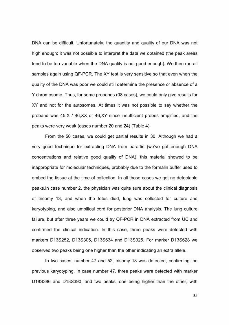
35
DNA can be difficult. Unfortunately, the quantity and quality of our DNA was not
high enough: it was not possible to interpret the data we obtained (the peak areas
tend to be too variable when the DNA quality is not good enough). We then ran all
samples again using QF-PCR. The XY test is very sensitive so that even when the
quality of the DNA was poor we could still determine the presence or absence of a
Y chromosome. Thus, for some probands (08 cases), we could only give results for
XY and not for the autosomes. At times it was not possible to say whether the
proband was 45,X / 46,XX or 46,XY since insufficient probes amplified, and the
peaks were very weak (cases number 20 and 24) (Table 4).
From the 50 cases, we could get partial results in 30. Although we had a
very good technique for extracting DNA from paraffin (we’ve got enough DNA
concentrations and relative good quality of DNA), this material showed to be
inappropriate for molecular techniques, probably due to the formalin buffer used to
embed the tissue at the time of collection. In all those cases we got no detectable
peaks.In case number 2, the physician was quite sure about the clinical diagnosis
of trisomy 13, and when the fetus died, lung was collected for culture and
karyotyping, and also umbilical cord for posterior DNA analysis. The lung culture
failure, but after three years we could try QF-PCR in DNA extracted from UC and
confirmed the clinical indication. In this case, three peaks were detected with
markers D13S252, D13S305, D13S634 and D13S325. For marker D13S628 we
observed two peaks being one higher than the other indicating an extra allele.
In two cases, number 47 and 52, trisomy 18 was detected, confirming the
previous karyotyping. In case number 47, three peaks were detected with marker
D18S386 and D18S390, and two peaks, one being higher than the other, with

36
markers D18S535 and D18S391. In case number 52, three peaks were detected
with marker D18S386, and two peaks detected, again one being higher than the
other, with markers D18S391 and D18S978.
From the floating amniocytes that did not adhere to the flask, and were
colleted at the first medium change (14 cases), we succeeded in extracting DNA
and performed QF-PCR. This material would be normally discharged. From those,
we were able to obtain eight molecular results from which two did not have a
previous successful karyotype (case number 4, and a partial result in case number
6).
The best results obtained for molecular analysis were from DNA extracted
using a commercial kit (samples 47 to 54) as described before (Material and
Methods).
From 6 cases of aneuploidies that had previous karyotype (31, 39, 47, 50,
51 and 52), we got only 2 confirmations (cases number 47 and 52). On the other
hand, one case (number 2) that we did not get any karyotype result, we obtained a
positive result through QF-PCR. Case number 53 could not be confirmed by
molecular analysis because we did not know the origin of the extra chromosome
marker, which did not hybridize with the probes used. Two previous normal
karyotypes (cases number 48 and 54) were confirmed with QF-PCR. We had no
discordant results between the molecular and traditional techniques.
In Table 5, we described the results from different sources of fetal material
for karyotyping in 115 multiple malformations fetus. When, for some anatomical
reason, AF CVS or UC could not be collected, the obstetrician tried other fetal
material, sometimes for therapeutic reasons, such as bladder drainage, and those

37
materials were used also for karyotying. Although they were few cases, we had
100% of culture success and karyotyping.
DISCUSSION
Cytogenetic analysis is an important tool for detecting chromosome
abnormalities, once this is the cause of most common genetic disease in man [1]. It
has been very useful for prenatal diagnosis, and also in clinical genetics. However
the traditional technique has some limitations, and in order to overcome these
problems some new molecular and rapid techniques have been developed, such
as QF-PCR and MLPA. In our study, we tried to use all the resources we could to
give diagnosis for a multiple malformations fetus that could evolve to death, or had
already died without any information about his condition. The main limitation we
wanted to overcome was to avoid the situation of leaving a multiple malformations
fetus without a final diagnosis or karyotype. For genetic counseling and for the
family this is very important information. So we tried to apply those molecular
techniques in postmortem or paraffin-embedded fetal tissues, or even amniocytes
that would be discharged.
Karyotyping unconventional fetal samples, when it is difficult to obtain the
traditional ones, is not a very common approach in most laboratories [17,18].
Nevertheless, we used this alternative whenever necessary, and achieved 100%
success rate on a limited sample of 13 cases [19], however this rate is much higher
than other studies [17,20].

38
There is an ongoing debate whether Rapid Aneuploidy Diagnosis (RAD)
should be employed as an adjunct to karyotyping or whether it could be used as a
stand-alone test in selected groups of women [21,22]. The controversy is due to
residual probability of a chromosome abnormality (both balanced and unbalanced)
when RAD demonstrates a normal result. Few studies have estimated the residual
risk of a clinically significant chromosome aberration for different indications when
RAD results are normal. In a meta-analysis of 12 studies involving invasive tests,
the risk of having a chromosome aberration that was not expected to be detected
by RAD methods, was estimated to be 0.9%[23]. In our results, from 6
aneuploidies already diagnosed by karyotyping, only two cases (33.3%) were
detected by QF-PCR, probably because of the poor quality of the DNA (mainly in
paraffin). On the other hand, a third case detected by QF-PCR, waited for 3 years
to receive a result (case number 2), once the tissue culture failure at the time of the
karyotyping. Little is known about the patients’ preference: weather QF-PCR alone
or together with full karyotype, however in Sweden a research was made in 6000
women, and 70% chose QF-PCR analysis [7]. But, for sure, both techniques
together are much more secure, once all molecular techniques have some
limitations.
MLPA has the same inherent limitations as those of QF-PCR in that it will
not detect most structural chromosome aberrations, or balanced rearrangements
such as translocations and inversions. Moreover, maternal cell contamination and
69,XXX triploidy will not be diagnosed by MLPA [7]. In our study, for example, in
case 53, a marker chromosome was detected by the traditional karyotype,

39
however, the molecular techniques were not able to detect it, because we did not
know the origin of the marker, so no specific probe could be applied.
Although we developed a good protocol for extracting DNA from paraffin
block wax, this material showed to be inappropriate for those molecular
techniques, as demonstrated in other studies which also needed smaller fragments
of DNA [24]. Another study in postmortem tissues embedded in paraffin succeeded
to obtain longer amplifications fragments of around 300 bp using a specific
treatment called pre-PCR restoration [25], thus achieving better results.
Whenever AF was usually set up, not all amniocytes adhered to the bottom
of the flask. After few days, by the first medium change, we observed that there
were still a considerable number of floating cells, which could be used to obtain a
good amount of DNA. We proposed to extract DNA from those cells in order to
guarantee a result independently to the cell culture, and also to abbreviate the
result with molecular techniques. We developed this protocol, and although we did
not succeed as expected, we could obtain results from two cases when cell culture
had failure. We could probably improve the success of these analysis by extracting
DNA with commercial kits rather than in-house techniques in orther to obtain better
quality DNA, which is essencial for the molecular analysis used here. The need of
more rapid testing methods which do not require cell culture has been recognized
by the scientific community to improve pregnancy management and alleviate
parental anxiety [26].
The best results obtained were from the last 8 cases, which the DNA was
extracted using a commercial kit. This is a very important information and

40
corroborates the fact that high quality DNA is necessary, from which we can obtain
results even with degraded DNA and background [27].
In conclusion, for follow up diagnostic testing, karyotyping has provided the
gold standard method. This technology has remained essentially unchanged over
30 years, as no new technology has proven to be superior in terms of being able to
detect such a wide range of abnormalities with the necessary precision [28].
Nevertheless, molecular testing, such as QF-PCR or MLPA, have their importance
in terms of giving a rapid result, of being practical, and low costing.
The importance of this study remains in the alternatives we proposed to give
a final diagnosis to a multiple malformation fetus. We suggested some ways to
achieve a result and give to the family the information they need to rebuild their
lives, and make plans for their future, with the help of more rapid and efficient
technology (RAD) with an appropriate genetic counseling.

41
REFERENCES [1] S.D.J. Pena, “Molecular Cytogenetics II: PCR-based diagnosis of chromosomal deletions and microdeletion syndromes”, Genetics and Molecular Biology, vol.21, no. 4, pp. 453-460, 1998. [2] R. Stevenson, and J.G. Hall, (2006) Human Malformation and related Anomalies, Oxford University Press, New York. [3] A. Boué, and J.Boué, “Evaluation des erreurs chromosomiques au moment de la conception”, Biomedicine, vol. 18, pp.372-377, 1973. [4] A. Milunsky, and J. Milunsky, (1998) Genetic Counseling: Preconception, Prenatal and Perinatal, In: Milunsky, A. (Ed.) Genetic Disorders and the Fetus. Diagnosis, Prevention and Treatment,The Jonh Hopkins University Press,Boston. pp.1-52. [5] L.G. Shaffer, and J.R. Lupski, “ Molecular mechanisms for constitutional chromosomal rearrangements in humans”, Annual Review of Genetics, vol. 34, pp.297-329, 2000. [6] M.W. Steele, and W.R.J. Breg, “Chromosome analysis of human amniotic-fluid cells”, Lancet, vol. 19, no.1, pp.383-385, 1966. [7] T.H. Bui, “Prenatal cytogenetic diagnosis: gone FISHing, BAC soon”,Ultrasound in Obstetrics and Gynecology, vol. 30, pp.247-251, 2007. [8] F. Daffos, M. Capella-Pavlovsky, and F. Forestier, “A new procedure for fetal blood sampling in utero: preliminary results in fifty-three cases”, American Journal of Obstetrics and Gynecology, vol. 146, pp. 985-987, 1983. [9] L. Dugoff, and J.C. Hobbins, “Invasive procedures to evaluate the fetus”. Clin Obstet Gynecol, vol. 45, pp.1039-1053, 2002. [10] A. Schinzel, (2001) Notes on Clinical Findings in Autosomal Chromosome Aberrations, In: Schinzel, A. (Ed.) Catalogue of Unbalanced Chromosome Aberrations in Man, Walter de Gruyter Press, Berlin, New York, pp.28-34.
[11] A.S. Miller, D.D. Dykes, and H.F. Polesky, “A simple salting out procedure for extracting DNA from human nucleated cells”, Nucleic Acids Research, vol.16, no.3, pp.1215, 1988.
[12] C.N. Andreassen, F.B. Sorensen, J. Overgaard, and J. Alsner, “Optimisation and validation of methods to access single nucleotide polymorphisms (SNPs) in Archival histological material”, Radiotherapy and Oncology, vol. 72, pp.351-356, 2004.

42
[13] R. Coura, J.C. Prolla, L. Meurer, and P. Ashton-Prolla, “An alternative protocol for DNA extraction from formalin fixed and paraffin wax embedded tissue”, Journal of Clinical Pathology, vol. 58, pp.894-895, 2005. [14] D. Zhou, and Z. Ren, “Multiplex ligation-dependent probe amplification and its application”, Zhonghua Yi Xue Yi Chuan Xue Za Zhi, vol. 26, no. 1, pp. 45-49, 2009. [15] J.P. Schouten, C.J. McElgunn, R. Waaijer, D. Zwijnenburg, F. Diepvens, and G. Pals, “Relative quantification of 40 nuclei acid sequences by multiplex ligation-dependent probe amplification”, Nucleic Acids Research, vol. 30, pp. 57, 2002. [16] E.S. Mansfield, “Diagnosis of Down Syndrome and other aneuploidies using quantitative polymerase chain reaction and small tandem repeat polymorphisms”, Human Molecular Genetics, vol. 2, no.1, pp.43-50, 1993. [17] A.E. Donnenfeld, D. Lockwood, and A.N. Lamb, “Prenatal diagnosis from cystic hygroma fluid : The value of fluorescence in situ hybridization”, American Journal of Obstetrics and Gynecology , vol.185 , pp.1004-1008, 2001. [18] L.A. Gole, C. Anandakumar, A. Bongso, T.M. Chua, Y.C. Wong and S.S. Ratnam,. “Analysis of cystic hygroma, ascitic and pleural fluids by conventional lymphocyte culture and fluorescent in situ hybridization”, Prenatal Diagnosis, vol. 17, pp.1151-1157, 1997. [19] R.G. Kessler, M.T.V. Sanseverino, S. Leistner-Segal, J.A.A. Magalhães and R. Giugliani, “Prenatal diagnosis of fetal chromosomal abnormalities: Report of an 18-year experience in a Brazilian public hospital”, Genetics and Molecular Biology, vol.31, no.4, pp.829-833, 2008. [20] T.G. Teoh, G. Ryan, J. Johnson, and E.J. Winsor, “The role of fetal karyotyping from unconventional sources”, American Journal of Obstetrics and Gynecology , vol.175, pp. 873-877, 1996. [21] W.C. Leung, E.T. Lau, T.T. Lao, and M.H. Tang, “Can amnio-polymerase chain reaction alone replace conventional cytogenetic study for women with positive biochemical screening for fetal Down syndrome ? ” Obstetric and Gynecology, vol. 101, pp. 865-861, 2003. [22] V. Cirigliano , G. Voglino, A. Marongiu, M.P. Canadas, E. Ordonez, E. Lioveras, A. Plaja, C. Fuster, and M. Adinolfi, “ Rapid prenatal diagnosis by QF-PCR:evaluation of 30,000 consecutive clinical samples and future applications”, Annual NY Academy of Sience, vol. 1075, pp. 288-298, 2006.

43
[23] W.C. Leung, and T.T Lao TT. ,“ Rapid aneuploidy testing, traditional karyotyping, or both?” Lancet, vol. 366, pp. 97-98, 2005. [24] B. Halvarsson, A. Lindblom, E. Rambech, K. Lagerstedt, and M. Nilbert, “Microsatellite instability and/or immunostaining for diagnosis of hereditary nonpolyposis colorectal cancer ? ” Virchows Archiv , vol. 444, pp. 134-141, 2004. [25] S. Bonin, F. Petrera, B. Niccolini B, and G. Stanta, “PCR analysis in archival postmortem tissues”, Journal of Clinical Patholology: Molecular Pathology, vol. 56, pp.184-186, 2003. [26] U. Nicolini , F. Lalatta, F. Nattacci, C. Curcio, and T.H. Bui “The introduction of Q-F PCR in prenatal diagnosis of fetal aneuploidies: time of reconsideration ”, Human Reproduction Update, vol.10, pp. 541-548, 2004. [27] A.D. Roeder, P. Elsmore, M. Greenhalgh ,and A. McDonald,“Maximization DNA profiling success from sub-optimal quantities of DNA: a staged approach ”, Forensic Science International Genetics, vol. 3, no.2, pp.128-137, 2009. [28] H.R. Slater, D.L. Bruno, H. Ren, M. Pertile, J.P. Schouten, K.H.A Choo, “Rapid, high throughput prenatal detection of aneuploidy using a novel quantity method (MLPA)”, Journal of Medical Genetics , vol. 40, pp.907-912, 2009.

44
Table 1. Malformations common in autosomal chromosomal aberrations __________________________________________________________ Cleft palate, cleft lip, or both Esophageal atresia, TE fistula; anal atresia with fistula Malrotation of the gut, common mesentery; omphalocele Malformation of the heart and the great vessels Malformation of the kidney and urinary tract Certain brain malformation, particularly holoprosencephaly and agenesis of corpus callosum Absence or hypoplasia of radius and thumb Postaxial hexadactyly Microphtalmia, ocular coloboma Spina bifida (occipital or lumbar)
_________________________________________________________
Table 2. 13, 18 and 21 markers in ELUCIGENE QSTRs
Marker Location Observed
Heterozygosity
Allele Size
Range (bp)
Marker Dye
Colour
D13S252 13q12.2 0.85 260-330 red
D13S305 13q13.3 0.75 418-470 green
D13S628 13q31.1 0.69 425-472 yellow
D13S634 13q21.33 0.81 355-440 blue
D13S325 13q14.11 0.86 235-320 green
D18S386 18q22.1 0.88 320-407 green
D18S390 18q22.3 0.75 345-400 yellow
D18S391 18q11.31 0.75 196-230 green
D18S535 18q12.3 0.92 450-500 Blue
D18S819 18q11.2 0.70 370-450 Red
D18S978 18q12.3 0.67 180-230 Yellow
D21S11 21q21.1 0.90 220-283 Blue
D21S1437 21q21.1 0.84 283-350 Blue
D21S1409 21q21.2 0.81 160-220 Red
D21S1411 21q22.3 0.93 256-345 Yellow
D21S1435 21q21.3 0.75 152-210 Blue

45
Table 3. X and Y markers in ELUCIGENE QSTRs
Marker Location Observed
Heterozygosity
Allele Size
Range (bp)
Marker Dye
Colour
DXS981 Xq13.1 0.86 225-260 Blue
DXS1187 Xq26.2 0.72 122-170 Green
HPRT Xq26.2 0.78 265-300 Green
DXS7423 Xq28 0.74 372-388 Green
DXYS267 Xq21.3/Yp11.2 0.87 240-280 Red
AMEL Xp22.22/Yp11.2 - 104-110 Yellow
DXS6807 Xp22.32 0.70 331-351 Blue
DXS1283E Xp22.31 0.89 292-340 Yellow
SRY Yp11.31 - 244-251 Yellow
DYS448 Yq11.223 - 323-381 Red

46
Table 4. Results of 50 samples from different tissues analyzes by QF-PCR
Legend: UC- Umbilical cord; Amnio-amnocytes; Paraff- paraffin
Obs. Cases number 27, 34,35 and 36 were not included in the study because the was not enough DNA
Case
number
Chr.
21
Chr.
18
Chr.
13
X / Y Interpretation/comments Traditional
Karyotype
Fetal
Material
DNA
[ng/µl]
01 2 2 2 XX No T13, 18, 21 detected Lung 10
02 2 2 3 XY Trisomy 13 - UC 35
03 2 2 2 XY No T13, 18, 21 detected UC 40
04 2 2 2 XY No T13, 18, 21 detected Amnio 0.275
05 - - - - Not enough DNA Amnio 5.37
06 - - - XY Very weak peaks Amnio 10.7
07 - - - XX No DNA left Paraff 34.5
08 2 2 2 XX No T13, 18, 21 detected UC 53
09 2 2 2 XY No T13, 18, 21 detected UC 54
10 2 2 2 XX No T13, 18, 21 detected UC 49.4
11 2 2 2 XY No T13, 18, 21 detected UC 49.1
12 2 2 2 XY No T13, 18, 21 detected Lung 45.4
13 - - - XY Very weak peaks Lung 0.94
14 2 2 2 XX No T13, 18, 21 detected Lung 43.85
15 2 2 2 XY No T13, 18, 21 detected Lung 52
16 - - - XX Very weak peaks UC 1.94
17 - - - XX Very weak peaks 46,XX UC 49.5
18 2 2 2 XY No T13, 18, 21 detected Lung 60
19 2 2 2 XX No T13, 18, 21 detected UC 96.5
20 - - - X/XX? Very weak peaks UC 15.2
21 2 2 2 XX Very weak peaks UC 27.1
22 - - - XX Very weak peaks UC 0.26
23 - - - - No peaks UC 0.61
24 - - - XY? Very weak peaks UC 13.3
25 2 2 2 XX Very weak peaks Lung 2.45
26 - - - - No peaks Paraff 11.4
28 - - - - No peaks Amnio 42.6
29 - - - - No peaks 46,XX Amnio 33.4
30 - - - - No peaks 46,XY Amnio 19.5
31 - - - - No peaks 47,XX+21 Amnio 39.9
32 - - - - No peaks Paraff 230
33 - - - - No peaks Paraff 6.98
37 - - - - No peaks Paraff 35.9
38 - - - - No peaks 46,XX Paraff 10.6
39 - - - - No peaks 45,X Paraff 6.47
40 - - - - No peaks 46,XY Paraff 40.9
41 - - - - No peaks Paraff 10.2
42 - - - - No peaks Paraff 1.87
43 - - - - No peaks Paraff 36.4
44 - - - - No peaks Paraff 43.8
45 - - - - No peaks Paraff 42.2
46 - - - - No peaks Paraff 14.3
47 2 3 2 XY Trisomy 18 47,XY,+18 Amnio 1.32
48 2 2 2 XX No T13, 18, 21 detected 46,XX Amnio 3.31
49 - - - - No peaks 46,XX Amnio 4.19
50 - - - XY Very weak peaks 47,XY+21 Amnio 4.81
51 - - - XY Very weak peaks 47,XY,+18 Amnio 0,34
52 2 3 2 XY Trisomy 18 47,XY,+18 Amnio 10.5
53 2 2 2 XX No T13, 18, 21 detected 47,XX,+mar Amnio 0.98
54 2 2 2 XX No T13, 18, 21 detected 46,XX UC 49.3

47
Table 5. Different materials from multiple malformations fetus and success rate of cell cultures
Fetus
sample
Number of
cases (n)
Culture
success (n)
Success
rate (%)
AF 87 83 95.5
UC 15 14 93.3
Alternative
fluidsa
13 13 100
TOTAL 115 110 95.6 aBladder (6), cystic hygroma (2), intraperitoneal (2), displastic kidney (1), cystic lung (1), cerebrospinal (1) fluids.

48
IV. DISCUSSÃO

49
IV. DISCUSSÃO
A análise citogenética é uma ferramenta fundamental para a detecção de
anormalidades cromossômicas, sendo esta a causa mais comum de doenças
genéticas no homem (Pena, 1998). A citogenética tornou-se uma ferramenta
crescentemente utilizada para o diagnóstico pré-natal, desde sua primeira
aplicação por Steele e Breg em 1966. Atualmente o diagnóstico pré-natal de
anormalidades cromossômicas é amplamente reconhecido como um método
confiável para casais com risco alto de gerarem uma criança clinicamente afetada
devido a aberrações cromossômicas (Caron e colaboradores, 1999). Os métodos
tradicionais de coleta invasiva de LA, BVC ou cordocentese têm sido utilizados no
Hospital de Clínicas de Porto Alegre desde 1989, e 905 gestantes tiveram acesso
a este tipo de exame em 18 anos, mesmo com as condições sócio-econômicas
limitadas de um país em desenvolvimento. Entretanto, durante todo este período,
o número de coletas vem diminuindo a cada ano, provavelmente devido à
introdução de um eficiente método de triagem, a medida da Translucência Nucal
(TN). Em uma sessão de aconselhamento genético, os métodos invasivos ou não,
riscos e indicações são discutidos com a família. No nosso estudo, as três
indicações de maior freqüência para a realização do exame foram: idade materna
avançada, malformação fetal detectada na ultra-sonografia e filho anterior com
trissomia do 21, nesta ordem (Tabela 1, artigo1). Entretanto, a maioria dos
cariótipos anormais foi encontrada no grupo cuja indicação era a detecção de
malformações no feto através da ultra-sonografia, e devido a importância deste
grupo é que nos dedicamos a ele na continuação deste estudo (artigo 2). Mesmo

50
sendo a terceira mais freqüente indicação do exame, casais que tiveram um filho
prévio com síndrome de Down, não encontramos nenhum caso de recorrência. A
recorrência para síndrome de Down é realmente baixa, conforme Warburton e
colaboradores (2004), portanto não deve ser priorizada no nosso meio, onde as
condições econômicas são limitadas, devendo-se oportunizar o exame para casais
de maior risco. Os achados citogenéticos no nosso estudo foram similares aos da
literatura (Caron et al., 1999; Carothers et al., 1999; Quintana et al., 1999),
mostrando que a síndrome de Down é a mais comum e clinicamente significativa
das anormalidades cromossômicas, seguida da síndrome de Edwards (trissomia
do 18). A freqüência das anormalidades cromossômicas na população de risco é
de 5% (Park et al., 2001), entretanto no nosso estudo esta freqüência chegou a
8,6%, provavelmente por ser o nosso serviço um centro de referência Nacional,
para onde são drenados muitos casos de alto risco. Todos os resultados relativos
às freqüências das anormalidades cromossômicas foram obtidos através da
citogenética tradicional.
No entanto, a técnica tradicional tem algumas limitações, principalmente por
depender do cultivo de células (Bui, 2007): o tempo de cultivo ao redor de duas
semanas, que é extenso para uma paciente gestante, e a probabilidade de um
insucesso no crescimento celular. No intuito de superar estes problemas, algumas
técnicas moleculares de resposta rápida têm sido desenvolvidas, tais como QF-
PCR e MLPA. Nos países desenvolvidos o custo do pessoal técnico é muito mais
elevado do que o custo das técnicas moleculares automatizadas (Tóth et al. 1998),
situação diferente da que ocorre em países como o Brasil, onde as despesas com
mão de obra especializada em citogenética são menores do que as relacionadas

51
com a aquisição de sondas fluorescentes e outros insumos importados. Em nosso
estudo procuramos utilizar todos os recursos que estavam ao nosso alcance para
concluir o diagnóstico de um feto com múltiplas malformações e que poderia
evoluir para o óbito sem qualquer informação sobre sua condição. O diagnóstico
definitivo nesta situação é uma informação muito valiosa, e de extrema
importância tanto para a definição do caso em si quanto para o aconselhamento
genético. Para tanto, incluímos no nosso protocolo análises moleculares, que
independem de cultivo celular e potencialmente permitiriam se chegar a um
diagnóstico em algumas horas apenas. As técnicas moleculares que se mostraram
mais adequadas à nossa amostra e às nossas necessidades e condições foram as
técnicas de QF-PCR e MLPA. Ambas as técnicas são de resposta rápida (24 a 48
h) e podem detectar através de sondas e marcadores fluorescentes os
cromossomos envolvidos nas mais freqüentes aneuploidias (13, 18, 21, X e Y),
que representam 90% das aberrações cromossômicas na espécie humana
(Munné, 1998). A técnica de MLPA pode até detectar mosaicismos (porém apenas
aqueles de altas freqüências). Entretanto, nenhuma delas é capaz de detectar
aberrações estruturais ou rearranjos balanceados (inversões ou translocações),
tampouco triploidias (Bui, 2007). Devido a estas limitações, é ainda discutível a
aplicação destas técnicas de detecção rápida de aneuploidias (conhecidas como
RAD - Rapid Aneuploidy Diagnosis). Discute-se especialmente se elas poderiam
ser aplicadas sozinhas, ou deveriam ser acompanhadas sempre da confirmação
pelo cariótipo (Leung, et al. 2003; Cirigliano, et al. 2006). A controvérsia é devido
a probabilidade residual de alguma aberração cromossômica existir mesmo
quando RAD indicar um resultado normal, que na verdade seria um falso negativo,

52
uma vez que a técnica, em função das suas peculiaridades, deixa de detectar
algumas alterações. Poucos estudos estimaram o risco residual para diferentes
indicações quando RAD apresenta resultados normais. Em uma meta-análise de
12 estudos envolvendo testes invasivos, o risco de uma aberração cromossômica
não ser detectada por RAD foi de 0,9% (Leung e Lao, 2005). Também pouco se
sabe da preferência das pacientes, se preferem utilizar unicamente as técnicas
moleculares rápidas, como QF-PCR ou MLPA, ou ter também o cariótipo
completo. Num estudo realizado na Suécia em 6000 mulheres, 70% escolheram
apenas fazer QF-PCR (Bui, 2007). Parece não haver dúvida de que as técnicas
(citogenética tradicional e molecular) se complementam e, portanto, juntas podem
oferecer melhores resultados (Leung et al., 2004).
Por exemplo, no caso 53 apresentado à Tabela 4 do nosso artigo 2, foi
realizado cariótipo convencional e QF-PCR, mas apenas com o cariótipo foi
possível detectar a presença de um cromossomo marcador, uma vez que a QF-
PCR se restringe aos marcadores específicos dos 5 cromossomos de maior
interesse. Esta Tabela 4 se refere apenas aos resultados de QF-PCR, pois a
técnica de MLPA não gerou resultados possíveis de serem analisados,
provavelmente porque a qualidade e/ou a quantidade de DNA era inadequada ou
insuficiente. Esta técnica é muito sensível, e se o DNA contém impurezas, as
áreas dos picos tendem a variar muito, não sendo possível se chegar a nenhuma
conclusão. Isso limita a sua aplicação em amostras nas quais o material é escasso
e com condições de conservação usualmente não ideais, como é o caso das
amostras de material fetal, especialmente as não convencionais ou aquelas
conservadas em parafina.

53
Nesta mesma Tabela 4, do mesmo artigo, pudemos observar os diferentes
materiais fetais utilizados para extração e análise molecular de aneuploidias,
totalizando 50 casos de fetos com múltiplas malformações e sem diagnóstico. O
material fetal utilizado para extrair DNA e aplicar estas técnicas moleculares foi
muito variado, desde tecido fetal fresco de necropsia como pulmão, ou cordão
umbilical, até material fetal fixado em blocos de parafina. Também foi extraído
DNA dos amniócitos (células fetais) que não ficaram aderidos ao fundo do frasco,
e que seriam de qualquer maneira desprezados na primeira troca de meio de
cultura. Do total de 50 amostras, obtivemos resultados em 30, sendo que em
alguns destes, só foi possível detectar os cromossomos sexuais. Embora esta
proporção de obtenção de resultados esteja abaixo da relatada em outros estudos,
(Adinolfi, 2003 et al., Leung et al., 2004, Ogilvie et al., 2005), é compreensível uma
vez que 13 dessas amostras (26%) eram de material fetal fixado em parafina
(casos 26, e de 32 a 45). A técnica para extração de DNA dos blocos de parafina
foi adaptada de Andreassen e colaboradores 2004, e testes já demonstraram que
o material obtido muitas vezes não tinha qualidade adequada para amplificação.
Provavelmente, variáveis pré-analíticas como processo de fixação, utilização de
formalina não tamponada, temperatura e qualidade da parafina utilizada
influenciam na pouca qualidade da extração, já que a qualidade do processamento
e preparo das amostras parafinadas em todas as etapas até a inclusão em
parafina é um ponto crítico para garantir o sucesso das análises (Halvarsson et al.,
2004). É importante salientar que nesta técnica de QF-PCR, a qualidade do DNA é
fundamental, podendo ser até mais determinante do que a quantidade. Este fato
pode ser exemplificado pelos casos nº4 (46,XY) e 47 (trissomia 18), onde com

54
pequenas concentrações de DNA (0.275 e 1.32 ng/µl, respectivamente) foi
possível se chegar a um resultado; enquanto que nos casos 28 e 29, apesar da
grande concentração de DNA (42.6 e 33.4 ng/µl respectivamente), não foi possível
obter nenhum resultado (Tabela 4, artigo 2).
Não obtivemos resultados em todas as nossas amostras, conforme seria de
se esperar, mas o sucesso em alguns justificou a nossa abordagem. Um exemplo
emblemático foi de um recém-nascido que faleceu logo após o nascimento e cujo
cariótipo no sangue de cordão ficou inviabilizado pois a cultura dos linfócitos não
teve sucesso, trazendo desalento adicional à família, que ficaria sem um
diagnóstico. Uma alíquota do cordão umbilical do feto ficou congelada a -20°C, por
3 anos aguardando algum desenvolvimento técnico que permitisse sua análise.
Incluído no presente estudo, conseguimos extrair DNA da amostra e aplicar a
técnica de QF-PCR, com a qual obtivemos o resultado de trissomia 13, o que
permitiu esclarecer o caso e orientar a família (caso 2, Figura 4). Nos poucos
casos em que houve material suficiente para poder comparar a citogenética
convencional com a molecular (QF-PCR), eles foram concordantes, como por
exemplo, nos dois casos de trissomia 18 (casos 47 e 52; Figuras 5 e 6
respectivamente). Não houve nenhum caso discordante, e mesmo quando os
picos eram fracos no QF-PCR, foi possível determinar os cromossomos sexuais
(resultado parcial). Tóth e colaboradores (1998) encontraram concordância em
todos os casos de diagnóstico pré-natal nas 212 mulheres grávidas testadas,
quando compararam a citogenética tradicional com a técnica de QF-PCR. Schmidt
e colaboradores (2000) encontraram, também, uma excelente concordância entre
a citogenética tradicional e a molecular, quando examinaram o LA de 662

55
gestantes, com apenas um caso não informativo. Cabe aqui salientar que ele
utilizou apenas dois a três marcadores STRs. Na nossa amostra, para cada
cromossomo em análise foram utilizados 5 a 6 marcadores, diminuindo a
probabilidade de homozigose, situação esta que torna o resultado não informativo.
Segundo Pena (1998), se quatro ou mais marcadores polimórficos de STRs forem
utilizados para cada cromossomo analisado, poucas amostras ficarão não
informativas devido à homozigose em alguns loci.
Ainda como uma outra tentativa de diminuir o tempo de resposta,
garantindo um resultado citogenético pré-natal, e não dependendo unicamente
das células que estão em cultivo (que podem não crescer, ou até contaminar),
estabelecemos um protocolo inovador para aproveitar os amniócitos que flutuam
no meio de cultura e que ainda não aderiram ao fundo do frasco na primeira troca
de meio (4-5 dias depois do lançamento da cultura). Estabelecemos um protocolo
de aproveitamento destas células que seriam descartadas, juntando-as em um
tubo cônico e extraindo das mesmas o DNA, para posteriormente aplicar a técnica
de QF-PCR. Obtivemos sucesso com esta estratégia, uma vez que dos 14 casos
de amniócitos aproveitados para extração de DNA apenas seis casos ficaram sem
diagnóstico. E dentre os 8 casos nos quais obtivemos resultados com essa
técnica, dois deles (casos 4 e 6) estavam sem diagnóstico citogenético devido à
falha na cultura. Os melhores resultados foram obtidos quando a extração DNA foi
realizada com kits comercialmente disponíveis (casos 47, 48, 52, 53, 54). Huang e
colaboradores (2008) isolando células fetais da circulação materna também
compararam as duas técnicas de extração de DNA (in house X kits) e

56
conseguiram melhores resultados com os kits, tanto na quantidade como na
qualidade dos DNAs.
Figura 4. QF-PCR do caso nº2, trissomia do 13.

57
Figura 5. QF-PCR do caso nº 47, trissomia do 18.
58
Figura 6. QF-PCR do caso nº52, trissomia do 18.

59
Como mais uma opção para garantir uma informação sobre a constituição
cromossômica em um feto com múltiplas malformações, utilizamos materiais
alternativos, outros que não os tradicionais (LA, BVC ou sangue de cordão),
quando estes eram impossíveis de se coletar devido às malformações fetais e
gestações complicadas, especialmente em casos de higromas císticos ou
hidropsia fetal. Estes materiais fetais eram usualmente coletados para fins
terapêuticos, como por exemplo, em casos de drenagem do fluido cerebral
(hidrocefalias) para diminuir o volume cerebral na tentativa de parto por via baixa.
Também a urina era utilizada quando havia necessidade de esvaziamento da
bexiga nos casos de rins displásicos. Uma vez coletados, estes fluidos eram
aproveitados para a tentativa de cultivo celular e posterior análise do cariótipo. Em
todos os materiais alternativos coletados (n=13) foi obtido com sucesso o cariótipo
convencional (Tabela 5, do artigo 2). Esta não é uma prática comum na maioria
dos laboratórios (Gole et al. 1997; Donnenfeld et al., 2001), porém a utilizamos
como um recurso adicional e obtivemos 100% de sucesso na cultura e análise do
cariótipo (Tabela 2, artigo 1), porcentagem esta superior a observada em outros
estudos. Donnenfeld e colaboradores (2001) atingiram apenas 76% de sucesso na
análise citogenética de 72 amostras de fluidos de higroma cístico, enquanto Teoh
e colaboradores (1996) conseguiram 87% de sucesso nas análises citogenéticas
de 39 amostras de urina, fluido pleural, ascite pericárdica e fluidos de higroma
cístico. Gole e colaboradores (1997) realizaram o cariótipo tradicional em 14
amostras de fluidos de higroma cístico, pleural e ascite, e obtiveram 85% de
sucesso.

60
V. CONCLUSÕES

61
V. CONCLUSÕES:
Esta tese permitiu diversas conclusões:
- Os nossos achados citogenéticos são muito similares aos da literatura,
sendo a trissomia 21 a mais freqüente das anormalidades cromossômicas,
seguida da trissomia do 18 (Síndrome de Edwards). A freqüência de todas as
anormalidades cromossômicas no nosso meio (8,6%) é ainda maior do que a
esperada na população de alto risco (5%), provavelmente por ser o Serviço de
Genética Médica um Centro de Referência Nacional.
- Um filho anterior com Síndrome de Down passou a ser considerada uma
indicação de menor peso, uma vez que não encontramos nenhum caso de
recorrência. Portanto, outras indicações de maior gravidade devem ser priorizadas
numa população carente e de alto risco.
- O número de análises citogenéticas diminuiu quase 50% ao ano,
basicamente com a introdução da medida da Translucência Nucal (TN).
Entretanto, este é um teste de triagem e não diagnóstico, portanto não pode
substituir o cariótipo.
- Os fetos com múltiplas malformações podem ter seu diagnóstico
citogenético garantido através da aplicação simultânea de várias técnicas. As
técnicas de MLPA ou QF-PCR podem ser aplicadas com sucesso em diferentes

62
tecidos fetais, como uma alternativa para uma resposta rápida, sem ter que
depender de uma cultura de células, que pode falhar por vários motivos, não
fornecendo o resultado esperado.
- É possível extrair DNA de amniócitos que ainda estão flutuando no meio
de cultura e que seriam descartados na primeira troca de meio. Esta alternativa
diminui em muito o tempo de espera pela cultura de líquido amniótico e evita a
perda de um diagnóstico por contaminação das células ou falta de crescimento
das mesmas.
- A utilização de amostras de fluidos alternativos do feto para a realização
do cariótipo é uma opção a ser considerada, quando o líquido amniótico ou
sangue de cordão umbilical são difíceis de serem obtidos. Em casos bem
selecionados, esta prática pode evitar o risco adicional de se realizar um outro
procedimento invasivo apenas para a obtenção do cariótipo.
- O sucesso na extração de DNA de blocos de parafina parece depender de
um processo controlado de fixação do tecido, incluindo o uso de reagentes de alta
qualidade. Para garantir o sucesso das análises moleculares, também é
fundamental que o DNA seja extraído de forma integral (sem fragmentação) e
com uma quantidade razoável.
- Para que se obtenha um resultado satisfatório nas técnicas moleculares,
especialmente MLPA, é fundamental que o processo de extração de DNA seja

63
realizado da melhor forma possível, sendo que a utilização de kits comerciais teve
melhor desempenho que a técnica usual in house.
- Embora QF-PCR seja uma técnica bem estabelecida e preencha os
requisitos de uma metodologia de resposta rápida tão importante no diagnóstico
pré-natal, ela é limitada a apenas alguns cromossomos. Isso, aliado a uma
pequena taxa de insucesso, indica que ela não pode ainda substituir totalmente as
técnicas de citogenética tradicional.
- A utilização simultânea de diferentes técnicas (convencionais e
moleculares) e diferentes tecidos (tradicionais e alternativos) pode ser decisiva
para que, pelo menos em alguns casos, um diagnóstico citogenético seja
alcançado, permitindo o esclarecimento do caso e o aconselhamento genético.

64
VI. REFERÊNCIAS

65
VI. REFERÊNCIAS
Adinolfi M, Sherlock J, Cirigliano V, Pertl B. Prenatal screening for aneuploidies by
Quantitative Fluorescente Polymerase Chain Reaction. Comm Genet, 2003; 3:50-
60.
Andreassen CN, Sorensen FB, Overgaard J, Alsner J. Optimisation and validation
of methods to access single nucleotide polymorphisms (SNPs) in Archival
histological material. Radiother and Oncol, 2004; 72:351-356.
Brambati B, Tului L, Cislaghi C, Albertti E. First 10000 chorionic villus samplings
performed on singleton pregnancies by a single operator. Prenat Diagn, 1998; 18:
255-266.
Boué A, Boué, J. Evaluation des erreurs chromosomiques au moment de la
conception. Biomedicine, 1973; 18:372-377.
Bui TH. Prenatal cytogenetic diagnosis: gone FISHing, BAC soon. Ultrasound
Obstet Gynecol, 2007; 30:247-251.
Caron L, Tihy F, Dallaire L. Frequencies of chromosomal abnormalities at
amniocentesis : over 20 years of cytogenetic analysis. Am J Med Genet, 1999 ;
82 :149-154.
Carothers AD, Boyd E, Lowther G, Ellis PM, Couzin DA, Faed MJW , Robb A.
Trends in prenatal diagnosis of Down syndrome and other autosomal trisomies in
Scotland 1990 to 1994, with associated cytogenetic and epidemiological findings.
Genet Epidemiol, 1999; 16:179-190.

66
Cirigliano V, Voglino G, Marongiu A, Canadas MP, Ordonez E, Lioveras E, Plaja A,
Fuster C, Adinolfi M. Rapid prenatal diagnosis by QF-PCR:evaluation of 30,000
consecutive clinical samples and future applications. Annual NY Academy of
Sience, 2006; 1075: 288-298.
Daffos F, Capella-Pavlovsky M, Forestier F. A new procedure for fetal blood
sampling in utero : preliminary results in fifty-three cases. Am J Obstet Gynecol,
1983 ; 146 :985-987.
Donaghue C, Mann K, Docherty Z, Ogilvie CM. Detection of mosaicism for primary
trisomies in prenatal samples by QF-PCR and karyotype analysis. Prenat Diagn,
2005; 25:65-72.
Donnenfeld AE, Lockwood D, Lamb AN. Prenatal diagnosis from cystic hygroma
fluid : The value of fluorescence in situ hybridization. Am J of Obstet Gynecol,
2001; 185:1004-1008.
Drets ME, Shaw MW. Specific banding patterns of human chromosomes. Proc.
Natl Acad Sci USA, 1971 ; 68(9) :2073-2077.
Dugoff L, Hobbins JC. Invasive procedures to evaluate the fetus. Clin Obstet
Gynecol, 2002; 45:1039-1053.
Gole LA, Anandakumar C, Bongso A, Chua TM, Wong YC, Ratnam SS. Analysis
of cystic hygroma, ascitic and pleural fluids by conventional lymphocyte culture and
fluorescent in situ hybridization. Prenatal Diagnosis, 1997; (17):1151-1157.
Halvarsson B, Lindblom A, Rambech E, Lagerstedt K, Nilbert M. Microsatellite
instability and/or immunostaining for diagnosis of hereditary nonpolyposis
colorectal cancer ? Virchows Archiv , 2004 ; 444 : 134-141.

67
Huang DJ, Mergenthaler-Gatfield s, Hahn S, Holzgreve W, Zhong XY. Isolation of
cell-free DNA from maternal plasma using manual and automated systems.
Methods Mol Biol, 2008 ; 444 :203-208.
Jenkins TM, Wapner RJ. First trimester prenatal diagnosis: chorionic villus
sampling. Semin Perinatol , 1999; 23:403-413.
Klinger K, Landes G, Shook D, Harvey R, Lopez L, Locke P, Lerner T, et al. Rapid
detection of chromosome aneuploidies in uncultured amniocytes by using
fluorescence in situ hybridization (FISH). Am J Hum Genet, 1992; 51: 55-65.
Lejeune J, Gautier M, Turpin R. Étude des chromosomes somatiques de neuf
enfants mongoliens. Compt. Rend. Acad. Sci, 1959; 248:1721-1722.
Leung WC, Lau ET, Lao TT, Tang MH. Can amnio-polymerase chain reaction
alone replace conventional cytogenetic study for women with positive biochemical
screening for fetal Down syndrome ? Obstetric and Gynecology, 2003; 101: 865-
861.
Leung WC, Waters JJ, Chitty L. Prenatal Diagnosis by rapid aneuploidy detection
and karyotyping : a prospective study of the role of ultrasound in 1589 second-
trimester amniocentesis. Prenat Diagn, 2004 ; 24(10):790-5.
Leung WC, Lao TT. Rapid aneuploidy testing, traditional karyotyping, or
both? Lancet, 2005; 366: 97-98.
Magalhães, JAA, Magalhães OA. Medicina Fetal. In: Rotinas em Obstetrícia, 4ª ed.
Porto Alegre, Artes Médicas, 2001; cap 3, p.46-57.

68
Mansfield ES. Diagnosis of Down Syndrome and other aneuploidies using
quantitative polymerase chain reaction and small tandem repeat polymorphism.
Hum Mol Genet, 1993; 2:43-50.
Milunsky A., Milunsky J. Genetic Counseling: Preconception, Prenatal and
Perinatal. In: Genetic Disorders and the Fetus. Diagnosis, Prevention and
Treatment, 4th ed. The Jonh Hopkins University Press, Baltimore and London,
1998; cap 1, p.1-52.
Munné S, Magli C, Bahçe M, Fung J, Legator M, Morrison L, Cohert J, Gianaroli L.
Preimplantation diagnosis of the aneuploidies most commonly found in
spontaneous abortions and live births: X,Y, 13, 14, 15, 16, 18, 21, 22. Prenat
Diagn, 1998; 18 (13):1459-1466.
Nicolaides KH, Snijders RJ, Gosden CM, Berry C, Campbell S.
Ultrasonographically detectable markers of fetal chromosomal abnormalities.
Lancet, 1992; 340 (8821):704-707.
Ogilvie CM, Donaghue C, Fox SP, Docherty Z, Mann K. Rapid Prenatal Diagnosis
of Aneuploidy using Quantitative Fluorescence-PCR (QF-PCR). J Histochem &
Cytichem, 2005; 53(3):285-288.
Painter TS. Further observations on sex chromosomes of mammals. Science,
1923; Sep 28, 58 (1500): 247-248.
Park SY, Kim JW, Kim YM, Lee MH, Lee MH, Han JY, Kim YM, Yang JH, Ryu HM.
Frequencies of Fetal Chromosomal Abnormalities at Prenatal Diagnosis: 10 years
experiences in a single institution. J Korean Med Sci , 2001; 16:290-293.
Pena SDJ. Molecular Cytogenetics I: PCR-based diagnosis of human trisomies
using computer-assisted laser densitometry. Genet Mol Biol, 1998; 21(3): 317-322.

69
Pereira RW, Sturzeneker R, Pena SDJ. Screening fetal losses for monossomy X
with a simple PCR-based procedure. Genet and Mol Biol, 2000; 23 (1):11-14.
Quintana Aguilar J, Quiñones Maza O, Méndez Rosado LA, Lavista González M,
González Noa CE, Hernández Pérez G. Resultados del diagnóstico prenatal
cromosómico en Ciudad Habana. Rev Cuba Obstet Ginecol, 1999; 25:153-158.
Roeder AD, Elsmore P, Greenhalgh M, McDonald A. Maximization DNA profiling
success from sub-optimal quantities of DNA: a staged approach. Forensic Scienc
Intern Genet, 2009; 3 (2):128-137.
Schinzel, A. Notes on Clinical Findings in Autosomal Chromosome Aberrations. In:
Catalogue of Unbalanced Chromosome Aberrations in Man, Walter de Gruyter
Press, 2001, Berlin, New York; p.28-34.
Schmidt W, Jendery J, Hecker K, Hackeloer BJ, Kerber S, Kochhan L, Held KR.
Detection of aneuploidy in chromosomes X, Y, 13, 18 and 21 by QF-PCR in662
selected pregnancies at risk. Mol Hum Reprod, 2000; 6(9): 855-860.
Schouten JP, McElgunn CJ, Waaijer R, Zwijnenburg D, Diepvens F, Pals G.
Relative quantification of 40 nuclei acid sequences by multiplex ligation-dependent
probe amplification. Nucleic Acids Res, 2002; 30:57.
Shaffer LG, Bui TH. Molecular cytogenetic and rapid aneuploidy detection methods
in prenatal diagnosis. Am J Med Genet C Semin Med Genet, 2007; 145: 87-98.
Shaffer LG, Lupski JR. Molecular mechanisms for constitutional chromosomal
rearrangenments in humans. Ann Rev Genet, 2000; 34:297-329.
Steele MW, Breg WR Jr. Chromosome analysis of human amniotic-fluid cells.
Lancet, 1966; 19 (1):383-385.

70
Steven L. History of Clinical Cytogenetics. In: The Principles of Clinical
Cytogenetics, 10th ed. NJ, Humana Press Inc. Toowa, 1999; cap.1., p. 3-8.
Stojilkovic-Mikic T, Mann K, Docherty Z, Ogilvie CM. Maternal cell contamination of
prenatal samples assessed by QF-PCR genotyping. Prenat Diagn, 2005; 25:79-83.
Teoh TG, Ryan G, Johnson J, Winsor EJ. The role of fetal karyotyping from
unconventional sources. Am J Obstet Gynecol, 1996; 175 :873-877.
Tóth T, Findlay I, Papp C, Tóth-Pál E, Marton T, Nagy B, Quirke P, Papp Z.
Prenatal detection of trisomy 21 and 18 from amniotic fluid by quantitative
fluorescent polymerase chain reaction. J Med Genet, 1998; 35:126-129.
Tjio HJ, Levan A.The chromosome numbers of man. Hereditas,1956; 42:1-6.
Warburton D, Dallaire L, Thangavelu M, Ross L, Levin B , Kline J Trisomy recurrence: a reconsideration based on North American data. Am J Hum Genet, 2004; 75:376-385.
Zhou D, Ren Z. Multiplex ligation-dependent probe amplification and its application.
Zhonghua Yi Xue Yi Chuan Xue Za Zhi, 2009; 26(1):45-49.

71
VII. ANEXOS

72
VII.1. Parecer do Comitê de Ética do HCPA

73
VII.2. Termo de Consentimento Livre e Esclarecido (Pré-Natal)

74
VII.3.Termo de Consentimento Livre e Esclarecido (Pós-Natal)

75
VII.4. Resposta ao artigo submetido à revista Journal of Biomedicine and
Biotechnology
De: Rasha Magdy [mailto:[email protected]]
Enviada em: sexta-feira, 13 de março de 2009 16:44
Para: Rejane Gus Kessler
Cc: [email protected]; Sandra Leistner Segal; Maria Teresa Vieira
Sanseverino; Jose Antonio De Azevedo Magalhaes; Marcelle Reesink
Cerski; Patricia Martins Moura Barrios; Roberto Giugliani
Assunto: JRejane Gus KesslerBB/631931: Acknowledging Receipt
Dear Mrs. Gus Kessler, The Research Article titled "Molecular cytogenetics: the contribution of new techniques to the etiologic diagnosis in fetus with multiple malformations," by Rejane Gus Kessler, Sandra Leistner-Segal, Maria Teresa Sanseverino, Jose Antonio Magalhaes, Marcelle Cerski, Patricia Barrios, and Roberto Giugliani has been received and assigned the number JBB/631931. An editor will be assigned to handle the review process of your manuscript, and he/she will inform you as soon as a decision is reached. All authors will receive a copy of all the correspondences regarding this manuscript. However, only the submitting author will be able to upload any revisions to the journal's manuscript tracking system. Thank you for submitting your work to Journal of Biomedicine and Biotechnology. Best regards, Rasha Magdy Journal Publishing Editor Journal of Biomedicine and Biotechnology Hindawi Publishing Corporation

76
VII. 5. Comentário do editor da revista Genetics and Molecular Biology

Editorial commentary on the state of clinical genetics in Brazil exemplifiedby the article on prenatal diagnosis by Kessler et al. Genetics and MolecularBiology (this issue)
Peter Pearson1
Departamento de Genética e Biologia Evolutiva, Instituto de Biociências, Universidade de São Paulo,
São Paulo, SP, Brazil.
As a relatively recent immigrant from Northern Eu-
rope, United Kingdom via the Netherlands, two countries
both benefitting from well organised clinical genetic ser-
vices, it is a great puzzle and worry to me why clinical ge-
netic services, particularly in the public sector, are so badly
organised in Brazil. Of course, the short answer is, the field
is grossly underfunded. But underlying that simplistic con-
clusion is the undeniable fact that the Brazilian Ministry of
Health at the federal level and various other bodies at the
state level have failed to recognise the public health care
value of establishing well-organised genetic screening and
counselling facilities. Although Brazil can be justly proud
of its health care position and initiatives in combating HIV
infection and poliomyelitis, amongst others, its record on
establishing well-organised and regulated genetic services
with appropriate attention to training and professional rec-
ognition of genetic counsellors, cytogeneticists, molecular
geneticists etc., falls far, far short of what is required and
would be acceptable in most other countries.
The de facto situation is that anybody with a medical
degree can put up a shingle claiming to carry out genetic di-
agnosis irrespective of training or specialist background.
This is a legal loophole, which does not do justice to the
technical requirements for genetic diagnosis and counsel-
ing or give professional recognition to the people really re-
quired to carry out the analyses, who are not usually not
medically qualified and learnt their trade in Biology and re-
lated areas. Further, the current situation does not require
diagnostic centres to provide evidence of proficiency, to
submit annual returns on productivity or participate in qual-
ity control trials. The rich, particularly in large conurba-
tions such as São Paulo, Porto Alegre, Rio de Janeiro etc.,
are well catered for by private clinics, albeit without any
traceable quality control by governmental agencies. Al-
though a few of the private laboratories take part in interna-
tional quality control programs, this is commercial window
dressing and at a national level undesirable. What self-
respecting Ministry of Health leaves it to foreign organisa-
tions to carry out the quality control work that they should
be performing themselves? In Brazil, the basic maxim is, if
you can pay for it, then you can get it. The vast majority of
the population are either excluded financially from making
use of private clinics, or are unaware of the health risks en-
suing from genetic disease or find great difficulty in finding
a centre prepared to undertake genetic investigation free of
charge. And even in situations where the government have
taken steps to provide genetic testing within the public sec-
tor, such as post-natal chromosome diagnosis, the estab-
lished level of remuneration is so low, that the diagnostic
laboratories cannot afford to carry out cytogenetic analyses
without a further source of income to cover the costs. Most
or all genetic diagnoses carried out in the public sector in
Brazil occur in public universities by people who are al-
ready overburdened by teaching and trying to do research,
let alone run a genetic service as well. Most receive no re-
muneration for this and their limited time is donated freely
to keep the services running. The bottom line is that the
available capacity is far too small for the number of re-
quests and only a small proportion of all applicants actually
receive attention within a short period of time. In short, ge-
netic testing within the public health care sector cannot and
never will take place adequately without extensive govern-
mental planning, reorganisation and introduction of ade-
quate financial incentives.
Chromosome prenatal diagnosis is now a well-
established procedure and passed out of the realms of being
experimental more than 15 years ago in most of the devel-
oped world. Why then should the GMB choose to publish
an article on this subject at this juncture (Kessler et al.,
Genetics and Molecular Biology, 31, 4, 834-835 (2008)
Copyright © 2008, Sociedade Brasileira de Genética. Printed in Brazil
www.sbg.org.br
Send correspondence to Peter L. Pearson. Departamento de Ge-nética e Biologia Evolutiva, Instituto de Biociências, Universidadede São Paulo, Caixa Posta. 11461, 05422-970 São Paulo, SP,Brazil. E-mail: [email protected].
Editorial Comment
1 The writer is one of only two genetic diagnosticians to have been certified for both molecular genetic and cytogenetic diagnosis in the Netherlands. He was the chairman
of the Departments of Human Genetics within the medical schools of Leiden and subsequently Utrecht in the Netherlands and was a member of the Dutch ministry of
health committee that formulated the genetic health care plan practised in the Netherlands today. He helped negotiate the logistics of financing genetic services with the
health care insurers. He is not medically qualified.

2008)? There are two reasons for doing so: through lack of
any official avenue for cytogenetic centres to disclose pro-
ductivity and efficiency rates, the article permits this partic-
ular group from Porto Alegre to present its results on
carrying out chromosome prenatal diagnosis in the public
health-care sector and for the results to be evaluated by oth-
ers; secondly the article points to many of the inadequacies
in Brazilian genetic screening services, not from the point
of view of the efficiency of the Porto Alegre group itself,
which succeeded remarkably well under the circumstances,
but with the problems encountered on the way. Firstly,
there are very few centres performing chromosome prena-
tal diagnosis in the public sector and many of the patients
have to travel large distances. The work is specialised and
labour intensive. This group was fortunate in that their hos-
pital was prepared to fund the screening, probably account-
ing why the group could maintain its activities for the 18
year period described in the article. The lack of governmen-
tal financial support is one of the main reasons why so few
groups are active with chromosome prenatal screening,
despite an ever increasing need as physicians and the Bra-
zilian public at large become increasingly aware of the di-
agnostic possibilities. Of course this situation begs the
question of why one would even bother to carry out chro-
mosome prenatal diagnosis when induced abortion is still
illegal in Brazil and likely to remain so for a long period of
time, but not for ever. Even the prototypically Roman Cath-
olic country Italy legalised abortion 30 years ago despite
ongoing controversies and attempts to reverse the law to
this day. However, the straight answer for the current Bra-
zilian situation is that knowing before a baby is born that it
carries a chromosome abnormality, mentally prepares the
parents beforehand on what they can expect at either mis-
carriage or birth of their child. In countries where induced
abortion is legal, prenatal diagnosis is rarely refused even
when the parents announce before hand that whatever the
outcome, they will not have the child aborted: the parent’s
right to know what is in store for them is regarded as suffi-
cient grounds for carrying out chromosome prenatal diag-
nosis.
How can the Brazilian situation be improved? Since
2004 a working group, composed of members from the two
Brazilian Society of Genetics and Medical Genetics, re-
spectively, and technical staff from the Ministry of Health
have evaluated the potential need for genetic services na-
tion-wide and drawn up guidelines on minimal require-
ments for infrastructure and financial commitments. The
group also formulated a proposal for setting up a national
policy for clinical genetics in the public sector, which in-
cludes publicity campaigns on prevention and education di-
rected not only to the general public but also to health
professionals. The Ministry of Health should be responsi-
ble for setting up such policy as well as monitoring the
quality through annual reports. However, 4 years is a long
period of time and the crucial question is when will the
Ministry of Health finally start making use of the recom-
mendations of the committee and legalise implementation
of a Brazilian national program in genetic health care? I
would suggest also that, following implementation of the
plan the derived criteria of quality control and reporting
also be extended to the private sector and make them oblig-
atory for all private centres to stay in business. Only then,
will the Brazilian public at large start receiving the quality
of genetic health care that it is entitled to.
ReferencesKessler RG, Sanseverino MTV, Leistner-Segal S, Magalhães
JAA and Giugliani R (2008). Prenatal diagnosis of fetal
chromosomal abnormalities: Report of an 18-year experi-
ence in a Brazilian public hospital. Genet Mol Biol 31:836-
840.
Pearson 835