Embed Size (px)
Citation preview

8/11/2019 Lectura Sentidos II
http://slidepdf.com/reader/full/lectura-sentidos-ii 1/16
Early Development of the Vertebrate
Inner EarMARTA MAGARIN ˜ OS,1,2,3 JULIO CONTRERAS,1,2,4 MARI A R. ABURTO,1,2
AND ISABEL VARELA-NIETO1,2,5*1Instituto de Investigaciones Biomedicas ‘‘Alberto Sols’’, CSIC-UAM, Madrid, Spain
2Unit 761, Centro de Investigacion Biomedica en Red de Enfermedades Raras (CIBERER),Instituto de Salud Carlos III, Madrid, Spain
3Departamento de Biologıa, Universidad Autonoma de Madrid, Spain4Facultad de Veterinaria, Universidad Complutense de Madrid, Spain
5Instituto de Investigacion Sanitaria Hospital, Universitario La Paz, IdiPAZ, Madrid, Spain
ABSTRACTThis is a review of the biological processes and the main signalingpathways required to generate the different otic cell types, with particu-lar emphasis on the actions of insulin-like growth factor I. The sensoryorgans responsible of hearing and balance have a common embryonic ori-gin in the otic placode. Lineages of neural, sensory, and support cells aregenerated from common otic neuroepithelial progenitors. The sequentialgeneration of the cell types that will form the adult inner ear requiresthe coordination of cell proliferation with cell differentiation programs,the strict regulation of cell survival, and the metabolic homeostasis of oticprecursors. A network of intracellular signals operates to coordinate thetranscriptional response to the extracellular input. Understanding themolecular clues that direct otic development is fundamental for thedesign of novel treatments for the protection and repair of hearing lossand balance disorders. Anat Rec, 295:1775–1790, 2012. VC 2012 WileyPeriodicals, Inc.
Key words: apoptosis; autophagy; deafness; IGF-I; miRNA;notch signaling; otic precursors; SOX2
AN INTRODUCTION TO THE ANATOMY OFTHE ADULT INNER EAR
The inner ear is a structurally complex sensory sys-tem formed by the organs responsible for hearing andbalance. The inner ear is located inside the temporalbone and it is formed by soft canals and cavities, namedthe membranous labyrinth, filled by endolymph and
encased by the bony labyrinth. Between the membra-nous labyrinth and the bony labyrinth there is a spacefilled by a different fluid the perilymph.
In mammals, a coiled structure, the cochlea, is respon-sible for hearing and contains the organ of Corti, thesensory receptor, where the mechanosensory hair cellstransduce the sound stimuli and generate the electro-chemical signal response that otic neurons will transmitto the brain (Raphael and Altschuler, 2003; Fig. 1). Thecochlea is a complex and integrated system, the damageof a specific cell type can lead to the damage of othercochlear elements and to hearing loss. Genetic and envi-ronmental factors can damage cochlear cells causing
deafness (Eisen and Ryugo, 2007; Liu and Yan, 2007;Raviv et al., 2010; Fetoni et al., 2011). The cochlea hasthree major functional parts: the lateral wall, the organof Corti, and the spiral ganglion (Fig. 1). The lateralwall, with the spiral ligament and the stria vascularis,is essential to the normal physiology of hearing (Jinet al., 2007). The stria vascularis is responsible for endo-lymph production (Takeuchi et al., 2000), a specialized
extracellular fluid with intracellular characteristics like
Grant sponsor: MICINN, Spain; Grant number: SAF2008 andSAF2011; Grant sponsor: CSIC I3P
*Correspondence to: Prof. Isabel Varela-Nieto, Instituto deInvestigaciones Biomedicas ‘‘Alberto Sols,’’ Consejo Superior deInvestigaciones Cientıficas (CSIC), Universidad Autonoma deMadrid (UAM), Arturo Duperier 4, 28029 Madrid, Spain. Fax:þ34 915854401. E-mail: [email protected]
Received 24 July 2012; Accepted 24 July 2012.
DOI 10.1002/ar.22575Published online 8 October 2012 in Wiley Online Library(wileyonlinelibrary.com).
THE ANATOMICAL RECORD 295:1775–1790 (2012)
VVC 2012 WILEY PERIODICALS, INC.

8/11/2019 Lectura Sentidos II
http://slidepdf.com/reader/full/lectura-sentidos-ii 2/16

8/11/2019 Lectura Sentidos II
http://slidepdf.com/reader/full/lectura-sentidos-ii 3/16
of Corti. The stereocilia of OHC are embedded in theTectorial Membrane, an extracellular component thatcovers the organ of Corti throughout the cochlea. Sup-port cells, Deiter’s cells, Hensen’s cells, Claudius’s cells,participate in regulating the ionic and nutrients homeo-stasis (Forge and Wright, 2002; Chang et al., 2008).
Alterations in their functions are a frequent cause of hearing impairment (Lefebvre and Van de Water, 2000).The spiral ganglion is located within the cochlear
modiolus and it is formed by the cell bodies of bipolarneurons that connect the hair cells of the organ of Cortiwith the brain (Nayagam et al., 2011). The dendriticends of spiral neurons innervate the hair cells, type Ineurons are the most abundant subtype (95%) and in-nervate the IHC. Type II neurons innervate severalOHC. The axons of spiral neurons leave the spiral gan-glion and pass through the basis of the modiolus to formthe cochlear division of the cochleo-vestibular nervetowards the cochlear nuclei at the brainstem. Sound infor-mation progresses in a complex multisynaptic, parallel,and ascendant pathway from the cochlea through thebrainstem nuclei to the auditory cortex (Webster et al.,
1992; Fig. 2). The cochlear nuclei, olivar complex, nucleusof lateral lemniscus, inferior colliculus, and medial genic-ulate complex are part of the rely nuclei that transmit theinformation to the cortex, where the auditory informationis processed in multiple areas. The tonotopic organizationpresent at the cochlea is maintained along the pathway.In addition, neurons from the brainstem (superior olivarycomplex) also contact hair cells in a centrifugal controlmechanism of the auditory pathway.
The vestibular system contains the balance receptorswith highly specialized mechanoreceptor hair cells (Gold-berg, 1991; Fig. 1). Three sensory organs called ‘‘cristae’’located at the base of the semicircular canals are respon-sible of balance perception, whereas the two ‘‘maculae’’ of the sacculus and utriculus detect linear and angular
acceleration (Highstein and Fay, 2004).
To sum up, the mammalian inner ear contains six sen-sory patches: the coiled cochlear duct or organ of Corti,which is the auditory receptor, the maculae, and the threecristae corresponding to the three semicircular canals areresponsible of balance perception. Sensory patches are con-nected to the brain nuclei by the fibers of the spiral and
vestibular ganglions that form the eighth cranial nerve.
DEVELOPMENT OF THE VERTEBRATEINNER EAR
The sensory organs of the inner ear have a commonembryonic origin at the otic placode. The cells that consti-tute the adult inner ear originate from the embryonic oticplacode with the exceptions of the melanocytes of the striavascularis and the ganglionar Schwann cells that are of neural crest origin (D’Amico-Martel and Noden, 1983;Rubel and Fritzsch, 2002; Fritzsch et al., 2011). Cranialplacodes are regions of the ectoderm that generate a widevariety of cell types, including elements of the senseorgans and most of the sensory neurons of the cranialnervous system. The different placodes derive from a com-
mon preplacodal region, which surrounds the neural plate(Streit, 2007). Before the otic placode becomes visible, theectodermal cells that will form it undergo a genetic pro-gram to express preplacodal transcription factors as the
Dlx family, Sox9a and Foxi1 (Ekker et al., 1992; Grovesand Bronner-Fraser, 2000; Solomon and Fritz, 2002; Liuet al., 2003). There is also induction from the underlyingmesenchyme that produces growth factors of the fibro-blast growth factor family (FGF10, FGF19, or FGF15depending on the species) and from the hindbrain, whichsecretes FGF3 (Maroon et al., 2002; Leger and Brand,2002; Wright and Mansour, 2003).
After induction, the otic placode invaginates beneaththe surface ectoderm to form the otic cup, which pinchesoff in birds and mice, or cavitates in fish to produce the
otic vesicle or otocyst (Fig. 3; Haddon and Lewis, 1996;
Fig. 1. Anatomy of the inner ear. A : Schematic view of the inner ear
showing the cochlear and vestibular parts and the sensory areas,
crista, macula, and organ of Corti (shadowed). B: Whole mount prepa-
ration of the cochlea showing the basal turn, the apex, the round win-
dow (RW), and the oval window (OW). Note the pigmented stria
vascularis through the lateral wall (melanin granules at intermediate
cell layers). Scale bar 0.5 mm. C: Low magnification of a midmodiolar
section from a mouse cochlea. Note the scala vestibule (SV) limiting
by the Reissner’s membrane (RS) with the scala media (SM) where the
auditory receptor is located (red boxes), the scala tympani (ST) with
the basilar membrane (BM), and the spiral ganglion (SG, in the osse-
ous Rosenthal’s canal). The lateral wall (LW) is directly in contact with
the osseous otic capsule. Scale bar 0.5 mm. D: The organ of Corti
containing the neurosensorial cells (inner hair cell, IHC; outer hair cells,OHC), the nonsensorial cells (DC: Deiter cells; HC: Hensen cells; CC:
Claudius cells; PC: pillar cells), the tectorial membrane (TM), the spiral
limbus (SL), the basilar membrane (BM), the tunnel of Corti (asterisk),
and the myelinated cochlear nerves fibers (CNF). Scale bar 50 lm ( E
and F ) Synaptophysin (Syn, in E) labeling of presynapses in the IHC
and efferent fibers arriving at the IHC and OHC, Neurofilament 200 kD
(NF, in F) labeling of the afferent fibers of the organ of Corti and the
synapse region. Scale bar 50 lm. ( G ) Phalloidin histochemistry of the
organ of Corti, labeling F-actin in viable sensory epithelium (stereocilia
and cuticular plate of hair cells, reticular lamina, and pillar cells). Scale
bar 50 lm. H: Semithin section showing the cytoarchitecture of the
spiral ganglion (SG). The inset shows electron microphotograph of the
ganglionar cells, surrounded by the Schwann cells (SC). The most
abundant neurons in the SG, the type I cells, present a myelin sheath,
with external compact myelin (CM) and internal loose myelin (LM).
Scale bar 5–0.1 lm (inset) and 30 lm. (I) Detail of the mouse lateral
wall showing the stria vascularis with the marginal cells (MC) close to
the scala media, the intermediate cells (IC), and the basal cells (BC).
The spiral ligament (SpL) is the most lateral part that is close to the
otic capsule of the cochlea. Scale bar 50 lm. J: Kir4.1 (KCNJ10, an
inwardly rectifying Kþ channel) expression in the stria vascularis of an
aging mouse. Note the relative loss of expression in some patches in
the stria (asterisks). This channel is related with the production of the
endocochlear potential, thus with an important role in auditory physiol-
ogy. Scale bar 50 lm. K : Naþ-Kþ-ATPase expression in the stria vas-
cularis, another functional marker of striatal healthiness that are
related with ion homeostasis and auditory physiology (Patuzzi, 2011).Scale bar 50 lm. L and M: The sensory epithelium of the vestibular
inner ear: the macula and the cristae gross anatomy. L: Cytoarchitec-
ture of a semithin section of the macula (L) and cristae ampullaris (M)
showing the morphological characteristics of these vestibular recep-
tors. Note the arrangement of the hair cells (HC) with the stereocilia
(asterisks), the supporting cells (SC), the basement membrane
(arrows), the otoconial membrane (OM) with otolites, and the vestibu-
lar nerve fibers (VNF). Scale bar 50 lm. N and O: Detail of the macula
(N) and cristae ampullaris (O) showing the myosin VIIa expression
(red, labeling sensory hair cells) and neurofilament 200 kD expression
(green, labeling macula nerve fibers). Arrows show the afferent calyx
of type I hair cells.
SENSORY AND NEURONAL CELL FATE SPECIFICATION 1777

8/11/2019 Lectura Sentidos II
http://slidepdf.com/reader/full/lectura-sentidos-ii 4/16

8/11/2019 Lectura Sentidos II
http://slidepdf.com/reader/full/lectura-sentidos-ii 5/16
conditional activation of the Notch pathway in oticregions at early mouse developmental stages inducesprosensory markers in the whole otic epithelium (Hart-man et al., 2010). These results support a model whereearly activation of Notch promotes the prosensory char-acter in specific regions of the developing otocyst.Nonetheless, there is still some controversy. Basch et al.,(2011) showed that ectopic activation of Notch signalingdid not induce ectopic sensory patches in nonsensoryregions of the cochlea suggesting that Notch signaling is
not sufficient for prosensory specification in the mousecochlea. In addition, they show that conditionally inacti-vation of RBPjk, the mediator of Notch signaling, is notfollowed by a reduction on the prosensory markers, butinstead by a shorter life of hair and supporting cells.
Bone Morphogenetic Protein 4 (BMP4)
BMP4 is a member of the TGFb superfamily of secreted signaling molecules that has been proposed to
Fig. 3. Inner ear development of vertebrates. A: Scheme of the early
development of the vertebrate inner ear. The inner ear originates from
the otic placode, a thickening of the head ectoderm that invaginates to
form the otic cup and separates from the ectoderm to form the otocyst
or otic vesicle, an ellipsoid-shaped structure able to generate most of
the cell types of the adult inner ear. In parallel to otic vesicle shaping, the
neuronal precursors (yellow) delaminate from the otic cup ventral region,
migrate and differentiate to produce the postmitotic otic neurons (red)
that form the acoustic-vestibular ganglion. (Modified from Ref. Varela-
Nieto et al., 2004) B: Schematic drawing of the inner ear of adult verte-
brates. From left to right: zebrafish, birds, and mouse. The origin and
early developmental stages of the inner ear are very similar in verte-
brates, although ear development in mammals is delayed with respect to
that of birds and, in zebrafish, the otic cup cavitates to form the otocyst.
All vertebrates show the general vestibular (light purple) and auditory
sensory components (dark purple), but with considerable differences, of
which the most notable is that the auditory region is a coiled cochlea in
mammals that contain the organ of Corti, and a straight cochlear duct in
birds called the basilar papilla. Zebrafish auditory function is carried out
by the saccule and the lagena. Abbreviations: A: anterior; ac: anterior
crista; bp: basilar papilla; co: cochlea; D: dorsal; l: lagena; lc: lateral
crista; M: medial pc: posterior crista; s: saccule; u: utricle.
SENSORY AND NEURONAL CELL FATE SPECIFICATION 1779

8/11/2019 Lectura Sentidos II
http://slidepdf.com/reader/full/lectura-sentidos-ii 6/16
play a key role in the specification of prosensory patches(Oh et al., 1996; Wu and Oh, 1996; Cole et al., 2000).
Although BMP4 expression is in agreement with an in-ductive sensory role, functional in vitro studies haveprovided opposing results (Li et al., 2005; Pujades et al.,2006). This controversy evidences the complexity of BMP4 actions in different developmental scenarios.
The Fibroblast Growth Factor (FGF) Family
The FGF family is composed by a large number of ligands and four receptors (FGFR) implicated in the reg-ulation of cell differentiation, proliferation, growth,motility, and survival (Wright and Mansour, 2003). FGF
signaling plays a key role during vertebrate inner eardevelopment and participates in the early induction of the otocyst (Schimmang, 2007), in prosensory specifica-tion (Pirvola et al., 2002; Sanchez-Calderon et al., 2007;Millimaki et al., 2007), otic neurogenesis and neurito-genesis (Nicholl et al., 2005; Wei et al., 2007), and inpillar cell differentiation (Doetzlhofer et al., 2009; San-chez-Calderon et al., 2010). FGF ligands presentredundant actions and complex interactions, therefore acombination of experimental approaches and the studyof mouse, chicken and zebrafish animal models are beingused to further understand and unravel FGF actions ininner ear development (Kelly and Chen, 2009).
The HMG-Box Transcription Factor SOX2
The high mobility group (HMG)-box transcription fac-tor SOX2 is a marker of the prosensory region, as wellas of the otocyst proneural domain (Fig. 5). Studies in thechicken embryo evidenced that inductive signals regulatedirectly SOX2 expression in the neural tube and thatSOX2 is responsible for neural fate acquisition in prolifer-ating precursors (Rex et al., 1997; Bylund et al., 2003;Graham et al., 2003). Early in development during otic ves-icle stages, SOX2 and other SOX proteins such as SOX3are expressed by proliferating cells in the prosensory do-main. SOX2 wide expression in the prosensory domainbecomes restricted to the supporting cells as developmentcontinues (Kiernan et al., 2005; Neves et al., 2007). Indeed,
several genes that are initially expressed in the prosensorydomain are afterwards restricted to the supporting cells( Jag1, Lfng, and p27 kip). These spatiotemporal patternsemphasize the different and even opposing functions thatcertain factors may have at different developmental stages,which greatly difficult the identification of specific prosen-sory markers (Kelley, 2007).
Two mutant mice deficient for Sox2 have beendescribed, Light coat and circling ( Lcc) and Yellow sub-marine (Ysb). Both mutants show defects in hearing andbalance (Kiernan et al., 2005), the cochleae of Lcc micelack neurons in the spiral ganglion (Puligilla et al.,2010) and neither hair cells nor supporting cells
Fig. 4. Cell specification in the otic vesicle. Three cell lineages are
generated from the otic vesicle: prosensory, neural, and nonsensory.
Auditory and vestibular neurons are generated from a common neuro-
nal progenitor. Hair and supporting cells derive from the prosensory
progenitors. The drawing summarizes some of the factors required to
produce cellular diversity. Among others to be identified, SOX2, Islet-
1, the Notch pathway, Eya1, BMP4, and the miR-200 miRNA family
are required to generate the pool of prosensory cells. The Notch sig-
naling pathway is also playing a role in the differentiation of hair cells.
Ids release the interacting partners of Atoh1, and the Ahoh1 positive
cells will express JAG2 and Dll1, which by activating the Notch signaling
targets Hes1 and Hes5 in the surrounding cells, direct the supporting
cell fate. The expression of Neurog1, Islet-1, and SOX2 lead the neural
cell fate. Tbx1 is required to delimit the neurogenic domain in the oto-
cyst. The sequential activation of the proneural genes NeuroM and Neu-
roD will promote the formation of the vestibular and auditory neurons.
IGF-I/IGF1R signaling modulates cell survival and proliferation, although
a direct role in cell fate specification has not been shown yet.
1780 MAGARINOS ET AL.

8/11/2019 Lectura Sentidos II
http://slidepdf.com/reader/full/lectura-sentidos-ii 7/16
differentiate (Kiernan et al., 2005). In addition, muta-tions in human SOX2 cause sensorineural deafness(Hagstrom et al., 2005). Due to the missing expression of Sox2 in Jag1 mouse mutants, an interesting hypothesisis that the Notch pathway element Jag1 could inducethe expression of Sox2 (Dabdoub et al., 2008).
Insulin-Like Growth Factor I
IGF-I belongs to the family of polypeptides of insulinthat plays a central role in embryonic development and
adult nervous system homeostasis by endocrine, auto-crine, and paracrine mechanisms (Murillo-Cuesta et al.,2011, 2012). IGF-I is secreted by the developing chickenotocysts and it is expressed in the mouse inner earthroughout development (Camarero et al., 2002; San-chez-Calderon et al., 2010). Igf1r is expressed in thesensory patches of otocysts from HH19 chicken (Aburtoet al., 2012) and E15.5 mouse (Sanchez-Calderon et al.,2010) embryos. One of the earliest cell fate determina-tion steps in the specification of the proneurosensoryfield is the expression of the bHLH proneural gene
Fig. 5. Spatiotemporal expression patterns of SOX2 and B-RAF
during vertebrate inner ear development. In early development, SOX2
expression (red throughout the figure) marks, in chicken and mouse,
the prosensory and the sensory epithelia. SOX2 is initially expressed
in all prosensory and sensory regions ( A –D, A 0 and B0 ), later the
expression in hair cells diminishes (C0 and D0 ) and is restricted to sup-
porting cells (boxed area in E0 ). B-RAF (green throughout the figure)
although shows a basal expression at very early developmental stages
in most tissues, is expressed in abundance in sensory regions of
chicken and mouse (A–D, A 0 and B0 ). B-RAF is also highly expressed
in hair cells ( E and F, C0 and D0 asterisk and arrows) although the
expression in the mouse OHC diminishes at the postnatal P30 stage
(boxed area in E0, asterisk and arrows). While B-RAF is intensively
expressed in otic neurons of the AVG and the SG, SOX2 is expressed
in nonoverlapping regions of glial cells in chicken (B and D) and
mouse (F0, arrows in the boxed area). Both SOX2 and B-RAF are also
expressed in the vestibular component in nonoverlapping regions (B-
RAF higher expression is found in hair cells, while SOX2 is abundantly
expressed in supporting cells. The boxed areas show higher magnifi-
cations of the selected regions. The bracket in B0 indicates the sen-
sory region. The bracket in G0–J0 indicates the differential expression
of B-RAF and SOX2 in the macula. Abbreviations: AVG: acoustic-ves-
tibular ganglion; bp: basilar papilla; IHC: inner hair cells; OHC: outer
hair cells; ms, macula sacculi; mu: macula utriculi; sc: supporting
cells; sg: spiral ganglion. Orientation: D, dorsal; M, media. Scale bars:
200 lm applies to A 0 and C0; 100 lm applies to A–E, E0, and G0–I0: 50
lm applies to B0 and F0; 25 lm applies to D0 and to E0 inset. (Partially
reproduced from Ref. Magari~nos et al., 2010).
SENSORY AND NEURONAL CELL FATE SPECIFICATION 1781

8/11/2019 Lectura Sentidos II
http://slidepdf.com/reader/full/lectura-sentidos-ii 8/16
Neurog1 (Adam et al., 1998; Fritzsch et al., 2010). Neu-rog1 activates the proneural genes NeuroD and NeuroM ,all have the potential to activate the expression of Igf1and Igf1r genes. An appealing hypothesis is that uponprosensory specification by other growth factors andmorphogenetic proteins, Igf1 expression is upregulated
by proneural genes to master early neurogenesis. IGF-Iactions and IGF1R expression are mediated by an intra-cellular signaling network (Murillo-Cuesta et al., 2011,2012; Fig. 6), and precedes that of neurotrophins andTrkA expression in neurons (Li et al., 2009). Indeed, oticneurons gradually reduce their expression of the Igf1r,while they increase the neurotrophin receptor TrkC(Aburto et al., 2012), suggesting that IGF-I is a keytrophic factor during the otic neuronal progenitor phaseof early inner ear development.
BIRTH AND DIFFERENTIATION OF HAIR ANDSUPPORTING CELLS
Hair and supporting cells are originated after thespecification of the prosensory domain (Driver and Kel-ley, 2009). During early development, it seems to be adefault program by which the first cells specified are thehair cells that in turn, by lateral inhibition, block theirfate to neighbor cells. Atoh1, a basic helix–loop–helix(bHLH) transcription factor is a key player in hair cellproduction and development. Although its expressionpattern is somehow controversial (Bermingham et al.,1999; Lanford et al., 2000; Chen et al., 2002), loss andgain of function studies have proven that Atoh1 is neces-sary and sufficient to induce hair cell formation insensory and nonsensory cells (Bermingham et al., 1999;Zheng and Gao, 2000; Kawamoto et al., 2003; Woodset al., 2004; Jones et al., 2006). It is interesting to notethat the solely expression of Atoh1 is able to generatehair cells in nonsensory patches, suggesting that the
sensorial competence is not restricted to certain regionsof the otic epithelium (Kelley, 2006).
Before hair cell formation starts there is a broadexpression of a family of proteins, the Ids (Jones et al.,2006) that prevent hair cell differentiation by the inacti-vation of Atoh1. Ids are inhibitors of differentiation thatcontain a HLH domain that competes with Atoh1 for itsdimerization partners E47 and E2A. The Ids do not havea DNA binding domain, but they can sequester the fac-tors required by Atoh1 to form a functional heterodimer(Norton, 2000). The expression of the Id family isreduced in hair cells as development continues, conse-quently releasing the factors necessary for Atoh1activity, and thus allowing the differentiation of haircells (Jones et al., 2006). Once hair cells have differenti-
ated, the surrounding cells become supporting cells by alateral inhibition mechanism. The Notch pathway thathad had a previous role in prosensory specification isalso implicated in individual cell differentiation. The
Atoh1 positive cells begin to express two Notch ligands Jagged2 ( Jag2), and Dll1 (Lanford et al., 1999; Morrisonet al., 1999; Hartman et al., 2007). This leads to Notch1activation and to intracellular expression of Notch tar-gets such as Hes1, Hes5, Hey1, and Hey2 in the futuresupporting cells (Lanford et al., 2000; Zheng et al., 2000;Zine et al., 2001; Murata et al., 2006; Hayashi et al.,2008; Li et al., 2008; Doetzlhofer et al., 2009). There arealso other factors that act as Atoh1 antagonist such as
SOX2 and Prox1 (Zheng et al., 2000; Dabdoub et al.,2008; Doetzlhofer et al., 2009).
In summary, Notch activity through evolution isrequired for sensory development to specify the prosen-sory regions in the otic cup and otocyst, and later topromote cell type differentiation to hair and supporting
cells (Eddison et al., 2000; Daudet and Lewis, 2005).
The Role of MicroRNAs in Inner EarDevelopment
MicroRNAs (miRNA) are small noncoding RNA, whichbind to the three untranslated region of target mRNA tosuppress their translation (Kloosterman and Plasterk,2006). miRNA have been implicated in multiple biologi-cal processes across phyla including development anddisease (Bartel, 2009; Lewis and Steel, 2010). miRNA started to be an inner ear topic when the expression of the miR-183 family (composed by miR-182, miR-96, andmiR-183) was reported in the inner ear of zebrafishembryos (Wienholds et al., 2005). This family is highlyconserved throughout evolution and it is expressed inciliated neurosensorial organs (Pierce et al., 2008). ThesemiRNA are expressed in hair cells in chicken, andmouse, and in lower levels in mouse sensory neurons of acoustic and vestibular structures (Darnell et al., 2006;Weston et al., 2006; Li and Fekete, 2010). Indeed, muta-tions in miR-96 cause hearing loss in mice and men(Lewis et al., 2009; Mencıa et al., 2009). The dynamicexpression of the miR-183 family in the mouse begins atthe otocyst stage and has the higher expression in differ-entiating hair cells, suggesting that this family isinvolved in hair cell differentiation and maturation(Sacheli et al., 2009). Another miRNA family, the miR-200 (miR-200a, miR-200b, miR-200c, miR-141, and miR-429) has been associated to inner ear prosensory specifi-cation. The miR-200 family is expressed in the inner ear
epithelia of zebrafish, chicken, and mouse (Wienholdset al., 2005; Darnell et al., 2006; Weston et al., 2006).Because of their known targets and interacting path-ways, it has been suggested that it plays a key role inestablishing the prosensory epithelial domains (Soukup,2009).
The importance of miRNAs in the inner ear is furtherevidenced by the conditional deletion of the enzymeDicer specifically in the otic placode. These mutantsshow a severe phenotype similar to that found in theSox2 null mouse, with profound defects in inner ear neu-rogenesis and sensory epithelial histogenesis (Kiernanet al., 2005; Friedman et al., 2009; Soukup et al., 2009).The emerging role of miRNAs in inner ear developmentsuggests that they may be of use in novel therapeutic
strategies aimed to hair cell regeneration (Beisel et al.,2008).
DEVELOPMENTAL OTIC NEUROGENESIS AND INNERVATION
The AVG contains cells derived from the otic epitheliathat will transit through different stages to become post-mitotic neurons finally. Otic neurogenesis can thereforebe separated into different cellular stages that are char-acterized by the distinct expression of a combination of molecular markers. After the regionalization of the oticcup/vesicle by the expression of a defined set of genes
1782 MAGARINOS ET AL.

8/11/2019 Lectura Sentidos II
http://slidepdf.com/reader/full/lectura-sentidos-ii 9/16

8/11/2019 Lectura Sentidos II
http://slidepdf.com/reader/full/lectura-sentidos-ii 10/16
including among others, Neurog1, LFng, Dl1, Sox2, andSox3 that specify the neural fate, neuroblasts migratefrom the proneural region of the otic cup toward theclosely mesenchymal space. The expression of the pro-neural genes NeuroD and NeuroM define the followingstage in neural differentiation: the epithelial neuroblast
population (Chae et al., 2004; Sanchez-Calderon et al.,2007). This population will migrate in a process that hasbeen intensely studied in chicken and mouse embryos(D’Amico-Martel and Noden, 1983; Alvarez et al., 1989;Hemond and Morest 1991; Davies, 2007, 2011). Migra-tion begins at the otic cup stage and reaches itsmaximal rate in the early otic vesicle stage (Figs. 3A,7A). The new formed ganglionic neuroblast populationcan be identified by their location, round shape, and bythe expression of a defined set of transcription factors,neurofilaments, and neurotrophins receptors. Includingthe transcription factor Islet-1 (Hobert and Westphal,2000), which begins to be expressed before migration byepithelial neuroblasts (Adam et al., 1998; Camareroet al., 2003; Li et al., 2004). Ganglionic neuroblastsretain the capacity to undergo cell division to expandtheir population and, accordingly, express receptors forIGF-I and cell cycle activators as FoxM1. Once neuro-blasts exit from the cell cycle, they differentiate intopostmitotic neurons that begin to project their processestoward their peripheral and central targets (Whiteheadand Morest, 1985; Fekete and Campero, 2007). The imma-ture post-mitotic neurons show a reduced expression of early neural markers, and start to gradually expressanother set of genes related to neuritogenesis and cellcycle exit, as type III tubulin (TuJ1), G4, and the cyclinsinhibitor p27Kip (Sanchez-Calderon et al., 2007, 2010).Finally, the mature otic neurons generate action poten-tials, express synaptic receptors and neurotransmitters,and reach the most differentiated and mature state(Raphael and Altschuler, 2003). Mature neurons express
a whole set of genes related to neuronal trophic supportas the neurotrophins receptors TrkB and TrkC (Pirvolaet al., 1997; Brumwell et al., 2000; Kim et al., 2001).
Autophagy in the Developing Inner Ear
Autophagy is a self-degradative process of the cellularcytosolic constituents that is crucial for balancing sour-ces of energy in response to different extracellularstimuli and for preventing the accumulation of misfoldedproteins or damaged organelles (Qu et al., 2007; Glicket al., 2010; Ravikumar et al., 2010). Autophagy in ver-tebrates has been shown to have key roles duringdevelopment (Levine and Klionsky 2004; Mizushima andLevine 2010; Montero and Hurle 2010). In the inner ear,
autophagy genes are essential for the vestibular functionin mice (Mari~no et al., 2010) and dead cells with auto-phagic features have been observed in the damagedcochlea (Taylor et al., 2008). Autophagy plays a role dur-ing normal inner ear development (Aburto et al., inpress) and its inhibition by treatment with 3-methylade-nine (3-MA; Rubinsztein et al., 2007) dramaticallyimpairs neurogenesis (Fig. 6). An appealing hypothesisis that the degradation by autophagy of the otic cellsowns components provide the energy required for themigration of the neuronal precursors. In this context, itwould be very interesting to examine early inner ear de-velopment in Atg4b/ and Atg5/ deficient mice,
which show different degrees of vestibular defects (Mar-i~no et al., 2010).
Vestibular Versus Auditory Neuronal Cell Fate
Otic neuroblasts are the common progenitors of audi-
tory and vestibular neurons that will have defined traitsand innervate different brain centers and establish pre-cise connections with their peripheral sensory targets.There are no precise molecular markers of this specifica-tion process. The sequential expression of NeuroM andNeuroD by migrating neuroblasts could determine spe-cific stages of maturation; in the chicken embryo,evidence suggests that these genes do not identify differ-ent neuron identities but rather a temporal appearanceof vestibular and auditory neurons (Bell et al., 2008).Studies in mouse null mutants have also shown the tem-porally regulated expression of Neurog1 to first generatevestibular neurons and secondly cochlear neurons(Koundakjian et al., 2007).
Neuritogenesis and Axonal Pathfinding
The innervation of specific sensory structures by oticneurons is built in a stereotyped fashion (Fekete andCampero, 2007; Koundakjian et al., 2007; Appler andGoodrich, 2011). It has been suggested that otic neuronssend their projections by retracing back to the originaldelamination place (Carney and Silver, 1983; Bruceet al., 1997). However, there are some long distance con-nections that would require the release of molecularcues. Though the mechanisms and factors affecting axonguidance in the inner ear are still under study and dis-cussion, its basis are probably similar to those behindthe wiring of the central nervous system. Indeed, mostof the molecules that have been reported to play a role
in ear innervation are well-known chemoattractants, asbrain-derived neurotrophic factor, neurotrophin-3 (NT-3),Shh, and the FGF family (Fari~nas et al., 2001a,b;Fritzsch et al., 2004; Fantetti and Fekete, 2011), or elsechemorepellent cues such as Semaphorin3/Npn1, Eph/ Ephrins, and Slit/Robo (Webber and Raz, 2006; Feketeand Campero, 2007; Battisti and Fekete, 2008). It is alsointeresting to underline that molecules that reorganizethe cytoskeleton such as the integrins (Davies, 2007,2011), autophagy and pathways traditionally implicatedin axon outgrowth such as the RAF-MEK-ERK pathway,have been also reported to act in early otic neuritogene-sis (Zhong et al., 2007; Magari~nos et al., 2010; Fig. 7).
Autophagy is emerging as a new player in neurite gener-ation and degeneration (Yang et al., 2007; Koike et al.,
2008; Plowey et al., 2008). Unc-51, the homolog for Atg1in C. elegans, is required for axonal guidance (Oguraand Goshima, 2006) and it has been recently demon-strated that neural soma survival is required foradequate axonal maintenance and regeneration (Rodrı-guez-Muela et al., 2011).
Currently, the best available therapy for treatinghearing loss is the electrical stimulation of the spiralganglion neurons with cochlear implants. Therefore,there is a need to improve our knowledge on how audi-tory neurons survive, differentiate, and interact withtheir targets to provide new avenues for the therapy of inner ear disorders.
1784 MAGARINOS ET AL.

8/11/2019 Lectura Sentidos II
http://slidepdf.com/reader/full/lectura-sentidos-ii 11/16
Fig. 7. Early otic neurogenesis. A : Neurogenic markers of otic neu-
rogenesis. Schematic representation of stages of development of otic
neurons. Postmitotic neurons depend on neurotrophins, but the fac-
tors expanding neuroblast populations are still to be fully defined.
Abbreviations: Del, delaminating; IN, immature neuron; M P, multipotent
progenitor; NBe epithelial neuroblast; NBg, ganglionar neuroblast; ON,otic neuron. Adapted from Sanchez-Calderon et al., 2007. B: In vivo
early otic neurogenesis. (a) Cryostat section of a HH19 chicken
embryo inmunostained for the transcription factor Islet-1 (green) and
the neuron marker G4 (magenta). Panel b corresponds to the boxed
area in a, showing a higher magnification of the AVG. C: Schematic
representation of the AVG ex vivo culture. The AVG can be explanted
from the embryo at HH19þ. The figure shows a schematic drawing of
a HH19þ chicken embryo showing the otic vesicle and the AVG loca-
tion and of an AVG immediately after dissection (0 hr) and after 24 hr
in culture. Factors and drugs can be added to the serum-free culture
medium to study their effects on AVG neuritogenesis. D: Signaling dur-
ing early otic neuritogenesis. (a and b) Inhibition of both IGF-I signaling
pathways, RAF-MERK-ERK and PI3K/AKT impaired otic neuritogene-
sis. (a) AVG explants were cultured in the 0S medium, or with Sor (2.5
lM) and immunostained for G4 (red) and Islet-1 (green). Sor-treated
AVG have shorter processes without affecting the size of the AVG
soma. Reproduced from Ref. Magari~nos et al., 2010. (b) AVG cultured
in 0S or in the presence of LY (25 lM) and immunostained for TuJ-1(magenta) and Islet-1 (green) showed that both the neuronal soma
area and the length of the neurites of the LY culture are smaller.
Reproduced from Ref. Aburto et al., 2010. Representative images are
shown from at least six otic vesicles per condition obtained in at least
three independent experiments. Compiled confocal microscopy projec-
tions of AVG are shown. (c) Inhibition of autophagy alters AVG neuro-
genesis. AVG explants cultured in the 0S condition or with 3-MA were
immunostained for G4 (green). Scale bar: 300 lm. Fluorescence images
were obtained from the compiled projections of confocal images of otic
vesicles and acoustic-vestibular ganglia. Abbreviations: B AVG: Acous-
tic-vestibular ganglion; LY: LY294002 (PI3K/AKT inhibitor); Sor: Sorafe-
nib (RAF-MEK-ERK inhibitor); 3-MA: (3-methyladenine).
SENSORY AND NEURONAL CELL FATE SPECIFICATION 1785
8/11/2019 Lectura Sentidos II
http://slidepdf.com/reader/full/lectura-sentidos-ii 12/16
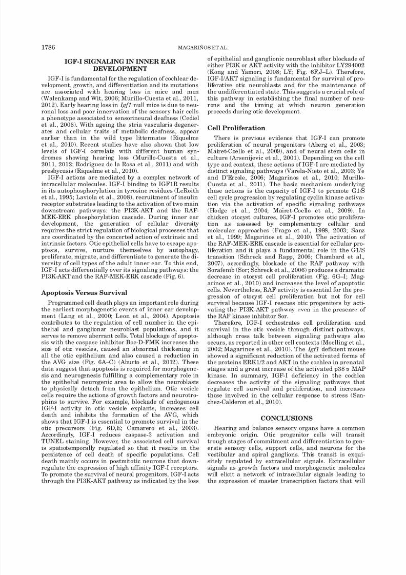
IGF-I SIGNALING IN INNER EAR DEVELOPMENT
IGF-I is fundamental for the regulation of cochlear de-velopment, growth, and differentiation and its mutationsare associated with hearing loss in mice and men
(Walenkamp and Wit, 2006; Murillo-Cuesta et al., 2011,2012). Early hearing loss in Igf1 null mice is due to neu-ronal loss and poor innervation of the sensory hair cellsa phenotype associated to sensorineural deafness (Cedielet al., 2006). With ageing the stria vascularis degener-ates and cellular traits of metabolic deafness, appearearlier than in the wild type littermates (Riquelmeet al., 2010). Recent studies have also shown that lowlevels of IGF-I correlate with different human syn-dromes showing hearing loss (Murillo-Cuesta et al.,2011, 2012; Rodriguez de la Rosa et al., 2011) and withpresbycusis (Riquelme et al., 2010).
IGF-I actions are mediated by a complex network of intracellular molecules. IGF-I binding to IGF1R resultsin its autophosphorylation in tyrosine residues (LeRoithet al., 1995; Laviola et al., 2008), recruitment of insulinreceptor substrates leading to the activation of two maindownstream pathways: the PI3K-AKT and the RAF-MEK-ERK phosphorylation cascade. During inner eardevelopment, the generation of cellular diversityrequires the strict regulation of biological processes thatare coordinated by the concerted action of extrinsic andintrinsic factors. Otic epithelial cells have to escape apo-ptosis, survive, nurture themselves by autophagy,proliferate, migrate, and differentiate to generate the di-versity of cell types of the adult inner ear. To this end,IGF-I acts differentially over its signaling pathways: thePI3K-AKT and the RAF-MEK-ERK cascade (Fig. 6).
Apoptosis Versus Survival
Programmed cell death plays an important role duringthe earliest morphogenetic events of inner ear develop-ment (Lang et al., 2000; Leon et al., 2004). Apoptosiscontributes to the regulation of cell number in the epi-thelial and ganglionar neuroblast populations, and itserves to remove aberrant cells. Total blockage of apopto-sis with the caspase inhibitor Boc-D-FMK increases thesize of otic vesicles, caused an abnormal thickening inall the otic epithelium and also caused a reduction inthe AVG size (Fig. 6A–C) (Aburto et al., 2012). Thesedata suggest that apoptosis is required for morphogene-sis and neurogenesis fulfilling a complementary role inthe epithelial neurogenic area to allow the neuroblaststo physically detach from the epithelium. Otic vesiclecells require the actions of growth factors and neurotro-
phins to survive. For example, blockade of endogenousIGF-I activity in otic vesicle explants, increases celldeath and inhibits the formation of the AVG, whichshows that IGF-I is essential to promote survival in theotic precursors (Fig. 6D,E; Camarero et al., 2003).
Accordingly, IGF-I reduces caspase-3 activation andTUNEL staining. However, the associated cell survivalis spatiotemporally regulated so that it results in thepersistence of cell death of specific populations. Celldeath mainly occurs in postmitotic neurons that down-regulate the expression of high affinity IGF-I receptors.To promote the survival of neural progenitors, IGF-I actsthrough the PI3K-AKT pathway as indicated by the loss
of epithelial and ganglionic neuroblast after blockade of either PI3K or AKT activity with the inhibitor LY294002(Kong and Yamori, 2008; LY; Fig. 6F,J–L). Therefore,IGF-I/AKT signaling is fundamental for survival of pro-liferative otic neuroblasts and for the maintenance of the undifferentiated state. This suggests a crucial role of
this pathway in establishing the final number of neu-rons and the timing at which neuron generationproceeds during otic development.
Cell Proliferation
There is previous evidence that IGF-I can promoteproliferation of neural progenitors (Aberg et al., 2003;Mairet-Coello et al., 2009), and of neural stem cells inculture (Arsenijevic et al., 2001). Depending on the celltype and context, these actions of IGF-I are mediated bydistinct signaling pathways (Varela-Nieto et al., 2003; Yeand D’Ercole, 2006; Magari~nos et al., 2010; Murillo-Cuesta et al., 2011). The basic mechanism underlyingthese actions is the capacity of IGF-I to promote G1/Scell cycle progression by regulating cyclin kinase activa-tion via the activation of specific signaling pathways(Hodge et al., 2004; Mairet-Coello et al., 2009). Inchicken otocyst cultures, IGF-I promotes otic prolifera-tion as assessed by complementary cellular andmolecular approaches (Frago et al., 1998, 2003; Sanzet al., 1999; Magari~nos et al., 2010). The activation of the RAF-MEK-ERK cascade is essential for cellular pro-liferation and it plays a fundamental role in the G1/Stransition (Schreck and Rapp, 2006; Chambard et al.,2007), accordingly, blockade of the RAF pathway withSorafenib (Sor; Schreck et al., 2006) produces a dramaticdecrease in otocyst cell proliferation (Fig. 6G–I; Mag-ari~nos et al., 2010) and increases the level of apoptoticcells. Nevertheless, RAF activity is essential for the pro-gression of otocyst cell proliferation but not for cell
survival because IGF-I rescues otic progenitors by acti-vating the PI3K-AKT pathway even in the presence of the RAF kinase inhibitor Sor.
Therefore, IGF-I orchestrates cell proliferation andsurvival in the otic vesicle through distinct pathways,although cross talk between signaling pathways alsooccurs, as reported in other cell contexts (Moelling et al.,2002; Magari~nos et al., 2010). The Igf1 deficient mouseshowed a significant reduction of the activated forms of the proteins ERK1/2 and AKT in the cochlea in prenatalstages and a great increase of the activated p38a MAPkinase. In summary, IGF-I deficiency in the cochleadecreases the activity of the signaling pathways thatregulate cell survival and proliferation, and increasesthose involved in the cellular response to stress (San-
chez-Calderon et al., 2010).
CONCLUSIONS
Hearing and balance sensory organs have a commonembryonic origin. Otic progenitor cells will transittrough stages of commitment and differentiation to gen-erate sensory cells, support cells, and neurons for thevestibular and spiral ganglions. This transit is exqui-sitely regulated by extracellular signals. Extracellularsignals as growth factors and morphogenetic moleculeswill elicit a network of intracellular signals leading tothe expression of master transcription factors that will
1786 MAGARINOS ET AL.
8/11/2019 Lectura Sentidos II
http://slidepdf.com/reader/full/lectura-sentidos-ii 13/16
generate gene expression spatiotemporal patterns.Future therapies for hearing loss and balance disorderswill benefit from the increasing knowledge on the molec-ular clues that direct otic development.
ACKNOWLEDGEMENTS
The authors thank the Image Unit (IIBM, Madrid) fortheir technical support. The anti-Islet-1 monoclonal anti-body was developed by Drs T.M. Jessel and J. Dodd andit was obtained from the Developmental Studies Hybrid-oma Bank, maintained at the Iowa University, Depart-ment of Biological Sciences. They warmly thank thecritical comments and generous sharing of results of ourcolleagues at the Neurobiology of Hearing group.
LITERATURE CITED
Aberg MAI, Aberg ND, Palmer TD, Alborn A-M, Carlsson-Skwirut
C, Bang P, Rosengren LE, Olsson T, Gage FH, Eriksson PS. 2003.IGF-I has a direct proliferative effect in adult hippocampal pro-
genitor cells. Mol Cell Neurosci 24:23–40. Aburto MR, Magari~nos M, Leon Y, Varela-Nieto I, Sanchez-
Calderon H. 2012. AKT signaling mediates IGF-I survival actionson otic neural progenitors. PLoS ONE. e 30790.
Aburto MR, Sanchez-Calderon H, Hurle JM, Varela-Nieto I, Mag-
ari~nos M. In press. Early otic development depends on autophagyfor apoptotic cell clearance and neural differentiation. Cell Death
Di. Adam J, Myat A, Le Roux I, Eddison M, Henrique D, Ish-Horowicz
D, Lewis J. 1998. Cell fate choices and the expression of Notch,Delta and Serrate homologues in the chick inner ear: parallels
with Drosophila sense-organ development. Development 125:4645–4654.
Alvarez IS, Martın-Partido G, Rodrıguez-Gallardo L, Gonzalez-
Ramos C, Navascues J. 1989. Cell proliferation during early de-velopment of the chick embryo otic anlage: quantitative compari-son of migratory and non-migratory regions of the otic
epithelium. J Comp Neurol 290:278–288. Anniko M. 1983. Cytodifferentiation of cochlear hair cells. Am J
Otolaryngol 4:375–388. Appler JM, Goodrich LV. 2011. Connecting the ear to the brain: Mo-
lecular mechanisms of auditory circuit assembly. Prog Neurobiol93:488–508.
Arsenijevic Y, Weiss S, Schneider B, Aebischer P. 2001. Insulin-likegrowth factor-I is necessary for neural stem cell proliferation anddemonstrates distinct actions of epidermal growth factor and
fibroblast growth factor-2. J Neurosci 21:7194–7202.Bartel DP. 2009. MicroRNAs: Target recognition and regulatory
functions. Cell 136:215–233.Basch ML, Ohyama T, Segil N, Groves AK. 2011. Canonical Notch
signaling is not necessary for prosensory induction in the mousecochlea: Insights from a conditional mutant of RBPjkappa. J Neu-rosci 31:8046–8058.
Battisti AC, Fekete DM. 2008. Slits and Robos in the developing
chicken inner ear. Dev Dyn 237:476–484.Bell D, Streit A, Gorospe I, Varela-Nieto I, Alsina B, Giraldez F.
2008. Spatial and temporal segregation of auditory and vestibularneurons in the otic placode. Dev Biol 322:109–120.
Bermingham NA, Hassan BA, Price SD, Vollrath MA, Ben-Arie N,
Eatock RA, Bellen HJ, Lysakowski A, Zoghbi HY. 1999. Math1: An essential gene for the generation of inner ear hair cells. Sci-ence 284:1837–1841.
Bruce LL, Kingsley J, Nichols DH, Fritzsch B. 1997. The develop-ment of vestibulocochlear efferents and cochlear afferents in mice.Int J Dev Neurosci 15:671–692.
Brumwell CL, Hossain WA, Morest DK, Bernd P. 2000. Role for ba-sic fibroblast growth factor (FGF-2) in tyrosine kinase (TrkB)expression in the early development and innervation of the audi-
tory receptor: In vitro and in situ studies. Exp Neurol 162:
121–145.Bylund M, Andersson E, Novitch BG, Muhr J. 2003. Vertebrate
neurogenesis is counteracted by Sox1-3 activity. Nat Neurosci 6:1162–1168.
Camarero G, Leon Y, Gorospe I, De Pablo F, Alsina B, Giraldez F, Varela-Nieto I. 2003. Insulin-like growth factor 1 is required for
survival of transit-amplifying neuroblasts and differentiation of otic neurons. Dev Biol 262:242–253.
Camarero G, Villar MA, Contreras J, Fernandez-Moreno C, Pichel
JG, Avenda~no C, Varela-Nieto I. 2002. Cochlear abnormalities ininsulin-like growth factor-1 mouse mutants. Hear Res 170:2–11.
Carney PR, Silver J. 1983. Studies on cell migration and axon guid-ance in the developing distal auditory system of the mouse. J
Comp Neurol 215:359–369.Cediel R, Riquelme R, Contreras J, Dıaz A, Varela-Nieto I. 2006. Sen-
sorineural hearing loss in insulin-like growth factor I-null mice: A
new model of human deafness. Eur J Neurosci 23:587–590.Chae JH, Stein GH, Lee JE. 2004. NeuroD: The predicted and the
surprising. Mol Cells 18:271–288.Chambard J-C, Lefloch R, Pouyssegur J, Lenormand P. 2007. ERK
implication in cell cycle regulation. Biochim Biophys Acta 1773:1299–1310.
Chang Q, Tang W, Ahmad S, Zhou B, Lin X. 2008. Gap junction
mediated intercellular metabolite transfer in the cochlea is com-promised in connexin30 null mice. PLoS ONE 3:e4088.
Chang W, ten Dijke P, Wu DK. 2002. BMP pathways are involvedin otic capsule formation and epithelial-mesenchymal signaling inthe developing chicken inner ear. Dev Biol 251:380–394.
Chatterjee S, Kraus P, Lufkin T. 2010. A symphony of inner ear de-velopmental control genes. BMC Genetics 11:68.
Chen P, Johnson JE, Zoghbi HY, Segil N. 2002. The role of Math1in inner ear development: Uncoupling the establishment of the
sensory primordium from hair cell fate determination. Develop-ment 129:2495–2505.
Cohen-Salmon M, Regnault B, Cayet N, Caille D, Demuth K, Har-delin JP, Janel N, Meda P, Petit C. 2007. Connexin30 deficiencycauses instrastrial fluid-blood barrier disruption within the coch-
lear stria vascularis. Proc Natl Acad Sci USA 104:6229–6234.Cole LK, Le Roux I, Nunes F, Laufer E, Lewis J, Wu DK. 2000. Sen-
sory organ generation in the chicken inner ear: Contributions of
bone morphogenetic protein 4, serrate1, and lunatic fringe. JComp Neurol 424:509–520.
D’Amico-Martel A, Noden DM. 1983. Contributions of placodal andneural crest cells to avian cranial peripheral ganglia. Am J Anat
166:445–468.Dabdoub A, Puligilla C, Jones JM, Fritzsch B, Cheah KSE, Pevny
LH, Kelley MW. 2008. Sox2 signaling in prosensory domain speci-fication and subsequent hair cell differentiation in the developingcochlea. Proc Natl Acad Sci USA 105:18396–18401.
Darnell DK, Kaur S, Stanislaw S, Konieczka JH, Yatskievych TA,
Antin PB. 2006. MicroRNA expression during chick embryo devel-opment. Dev Dyn 235:3156–3165.
Daudet N, Lewis J. 2005. Two contrasting roles for Notch activity
in chick inner ear development: Specification of prosensorypatches and lateral inhibition of hair-cell differentiation. Develop-ment 132:541–551.
Davies D. 2007. Temporal and spatial regulation of alpha6 integrin
expression during the development of the cochlear-vestibular gan-glion J Comp Neurol 502:673–682.
Davies D. 2011. Cell-extracellular matrix versus cell-cell interac-
tions during the development of the cochlear-vestibular ganglion.J Neurosci Res 89:1375–1387.
Doetzlhofer A, Basch ML, Ohyama T, Gessler M, Groves AK, SegilN. 2009. Hey2 regulation by FGF provides a Notch-independentmechanism for maintaining pillar cell fate in the organ of Corti.
Dev Cell 16:58–69.Driver EC, Kelley MK. 2009. Specification of cell fate in the mam-
malian cochlea. Birth Defects Res C Embryo Today 87:212–221.
Eddison M, Le Roux I, Lewis J. 2000. Notch signaling in the devel-opment of the inner ear: Lessons from Drosophila. Proc Natl AcadSci USA 97:11692–11699.
SENSORY AND NEURONAL CELL FATE SPECIFICATION 1787

8/11/2019 Lectura Sentidos II
http://slidepdf.com/reader/full/lectura-sentidos-ii 14/16
Eisen MD, Ryugo DK. 2007. Hearing molecules: Contributions from
genetic deafness. Cell Mol Life Sci 64:566–580.Ekker M, Akimenko MA, Bremiller R, Westerfield M. 1992. Re-
gional expression of three homeobox transcripts in the inner earof zebrafish embryos. Neuron 9:27–35.
Fantetti KN, Fekete DM. 2011. Members of the BMP, Shh and FGFmorphogen families promote chicken statoacoustic ganglion neu-
rite outgrowth and neuron survival in vitro. Dev Neurobiol 72:1213–1228.
Fari~nas I, Jones KR, Tessarollo L, Vigers AJ, Huang E, Kirstein
M, de Caprona DC, Coppola V, Backus C, Reichardt LF,Fritzsch B. 2001a. Spatial shaping of cochlear innervation bytemporally regulated neurotrophin expression. J Neurosci 21:6170–6180.
Fari~nas I, Jones KR, Tessarollo L, Vigers AJ, Huang E, Kirstein M,de Caprona DC, Coppola V, Backus C, Reichardt LF, Fritzsch B.2001b. Spatial shaping of cochlear innervation by temporally
regulated neurotrophin expression. J Neurosci 21:6170–6180.Fekete DM, Campero AM. 2007. Axon guidance in the inner ear.
Int J Dev Biol 51:549–556.Fetoni AR, Picciotti PM, Paludetti G, Troiani D. 2011. Pathogenesis
of presbycusis in animal models: A review. Exp Gerontol 46:413–425.
Forge A, Wright T. 2002. The molecular architecture of the inner
ear. BMB 63:5–24.Frago LM, Ca~non S, de la Rosa EJ, Leon Y, Varela-Nieto I. 2003.
Programmed cell death in the developing inner ear is balanced bynerve growth factor and insulin-like growth factor I. J Cell Sci116:475–486.
Frago LM, Leon Y, de la Rosa EJ, Gomez-Mu~noz A, Varela-Nieto I.1998. Nerve growth factor and ceramides modulate cell death inthe early developing inner ear. J Cell Sci 111 (Pt 5):549–556.
Friedman LM, Dror AA, Mor E, Tenne T, Toren G, Satoh T, Biese-
meier DJ, Shomron N, Fekete DM, Hornstein E, Avraham KB.2009. MicroRNAs are essential for development and function of
inner ear hair cells in vertebrates. Proc Natl Acad Sci USA 106:7915–7920.
Fritzsch B, Eberl DF, Beisel KW. 2010. The role of bHLH genes in
ear development and evolution: Revisiting a 10-year-old hypothe-sis. Cell Mol Life Sci 67:3089–3099.
Fritzsch B, Jahan I, Pan N, Kersigo J, Duncan J, Kopecky B. 2011.
Dissecting the molecular basis of organ of Corti development:Where are we now? Hear Res 276:16–26.
Fritzsch B, Tessarollo L, Coppola E, Reichardt LF. 2004. Neurotro-phins in the ear: Their roles in sensory neuron survival and fiber
guidance. Prog Brain Res 146:265–278.Glick D, Barth S, Macleod KF. 2010. Autophagy: Cellular and mo-
lecular mechanisms. J Pathol 221:3–12.Goldberg JM. 1991. The vestibular end organs: morphological and
physiological diversity of afferents. Curr Opin Neurobiol 1:
229–235.Graham V, Khudyakov J, Ellis P, Pevny L. 2003. SOX2 functions to
maintain neural progenitor identity. Neuron 39:749–765.Groves AK, Bronner-Fraser M. 2000. Competence, specification and
commitment in otic placode induction. Development 127:3489–3499.
Haddon C, Lewis J. 1996. Early ear development in the embryo of
the zebrafish, Danio rerio. J Comp Neurol 365:113–128.
Hartman BH, Hayashi T, Nelson BR, Bermingham-McDonogh O,Reh TA. 2007. Dll3 is expressed in developing hair cells in themammalian cochlea. Dev Dyn 236:2875–2883.
Hartman BH, Reh TA, Bermingham-McDonogh O. 2010. Notch sig-naling specifies prosensory domains via lateral induction in the
developing mammalian inner ear. Proc Natl Acad Sci USA 107:15792–15797.
Hayashi T, Kokubo H, Hartman BH, Ray CA, Reh TA, Berming-
ham-McDonogh O. 2008. Hesr1 and Hesr2 may act as early effec-tors of Notch signaling in the developing cochlea. Dev Biol 316:87–99.
Hemond SG, Morest DK. 1991. Ganglion formation from the oticplacode and the otic crest in the chick embryo: Mitosis, migration,and the basal lamina. Anat Embryol 184:1–13.
Highstein SM, Fay RR. 2004. The vestibular system. Springer
handbook of auditory research. Vol. 19. New York: Springer.Hobert O, Westphal H. 2000. Functions of LIM-homeobox genes.
Trends Genet 16:75–83.Hodge RD, D’Ercole AJ, O’Kusky JR. 2004. Insulin-like growth fac-
tor-I accelerates the cell cycle by decreasing G1 phase length andincreases cell cycle reentry in the embryonic cerebral cortex. J
Neurosci 24:10201–10210.Hudspeth AJ. 2008. Making an effort to listen: Mechanical amplifi-
cation in the ear. Neuron 59:530–545.
Jin Z, Mannstr€on P, J€arlebark L, Ulfendahl M. 2007. Malformationof stria vascularis in the developing inner ear of the Germanwaltzing guinea pig. Cell Tissue Res 328:257–270.
Jin Z, Ulfendahl M, J€arlebark L. 2008. Spatiotemporal loss of K þ
transport proteins in the developing cochlear lateral wall of guinea pigs with hereditary deafness. Eur J Neurosci 27:145–154.
Jones JM, Montcouquiol M, Dabdoub A, Woods C, Kelley MW. 2006.Inhibitors of differentiation and DNA binding (Ids) regulate
Math1 and hair cell formation during the development of theorgan of Corti. J Neurosci 26:550–558.
Kawamoto K, Ishimoto S-I, Minoda R, Brough DE, Raphael Y. 2003.Math1 gene transfer generates new cochlear hair cells in mature
guinea pigs in vivo. J Neurosci 23:4395–4400.
Kelley MW. 2006. Regulation of cell fate in the sensory epithelia of the inner ear. Nat Rev Neurosci 7:837–849.
Kelley MW. 2007. Cellular commitment and differentiation in theorgan of Corti. Int J Dev Biol 51:571–583.
Kelly MC, Chen P. 2009. Development of form and function in the
mammalian cochlea. Curr Opin Neurobiol 19:395–401.Kiernan AE, Pelling AL, Leung KKH, Tang ASP, Bell DM, Tease C,
Lovell-Badge R, Steel KP, Cheah KS. 2005. Sox2 is required forsensory organ development in the mammalian inner ear. Nature
434:1031–1035.Kim WY, Fritzsch B, Serls A, Bakel LA, Huang EJ, Reichardt LF,
Barth DS, Lee JE. 2001. NeuroD-null mice are deaf due to asevere loss of the inner ear sensory neurons during development.Development 128:417–426.
Kimura RS. 1975. The ultrastructure of the organ of Corti. Int RevCytol 42:173–222.
Kloosterman WP, Plasterk RHA. 2006. The diverse functions of
microRNAs in animal development and disease. Dev Cell 11:441–450.
Koike M, Shibata M, Tadakoshi M, Gotoh K, Komatsu M, Waguri S,Kawahara N, Kuida K, Nagata S, Kominami E, Tanaka K,
Uchiyama Y. 2008. Inhibition of autophagy prevents hippocampalpyramidal neuron death after hypoxic-ischemic injury. Am J
Pathol 172:454–469.Kong D, Yamori T. 2008. Phosphatidylinositol 3-kinase inhibitors:
Promising drug candidates for cancer therapy. Cancer Sci 99:1734–1740.
Koundakjian EJ, Appler JL, Goodrich LV. 2007. Auditory neuronsmake stereotyped wiring decisions before maturation of their tar-gets. J Neurosci 27:14078–14088.
Kwan T, White PM, Segil N. 2009. Development and regenerationof the inner ear. Ann N Y Acad Sci 1170:28–33.
Lanford PJ, Lan Y, Jiang R, Lindsell C, Weinmaster G, Gridley T,
Kelley MK. 1999. Notch signalling pathway mediates hair cell de-
velopment in mammalian cochlea. Nat Genet 21:289–292.Lanford PJ, Shailam R, Norton CR, Gridley T, Kelley MW. 2000.
Expression of Math1 and HES5 in the cochleae of wildtype and
Jag2 mutant mice. J Assoc Res Otolaryngol 1:161–171.Lang H, Bever MM, Fekete DM. 2000. Cell proliferation and cell
death in the developing chick inner ear: Spatial and temporal pat-terns. J Comp Neurol 417:205–220.
Laviola L, Natalicchio A, Perrini S, Giorgino F. 2008. Abnormalities
of IGF-I signaling in the pathogenesis of diseases of the bone,brain, and fetoplacental unit in humans. Am J Physiol EndocrinolMetab 295:E991–999.
Lefebvre PP, Van de Water TR. 2000. Connexins, hearing and deaf-ness: Clinical aspects of mutations in the connexin 26 gene. BrainRes 32:159–162.
1788 MAGARINOS ET AL.

8/11/2019 Lectura Sentidos II
http://slidepdf.com/reader/full/lectura-sentidos-ii 15/16
Leger S, Brand M. 2002. Fgf8 and Fgf3 are required for zebrafish
ear placode induction, maintenance and inner ear patterning.Mech Dev 119:91–108.
Leon Y, Sanchez-Galiano S, Gorospe I. 2004. Programmed cell deathin the development of the vertebrate inner ear. Apoptosis 9:
255–264.LeRoith D, Werner H, Beitner-Johnson D, Roberts CT, Jr. 1995. Mo-
lecular and cellular aspects of the insulin-like growth factor I re-ceptor. Endocr Rev 16:143–163.
Levine B, Klionsky DJ. 2004. Development by self-digestion: Molec-
ular mechanisms and biological functions of autophagy. Dev Cell6:463–477.
Lewis MA, Quint E, Glazier AM, Fuchs H, De Angelis MH, Lang-ford C, van Dongen S, Abreu-Goodger C, Piipari M, Redshaw N,
Dalmay T, Moreno-Pelayo MA, Enright AJ, Steel KP. 2009. AnENU-induced mutation of miR-96 associated with progressivehearing loss in mice. Nat Genet 41:614–618.
Lewis MA, Steel KP. 2010. MicroRNAs in mouse development anddisease. Semin Cell Dev Biol 21:774–780.
Li H, Corrales CE, Wang Z, Zhao Y, Wang Y, Liu H, Heller S. 2005.BMP4 signaling is involved in the generation of inner ear sensoryepithelia. BMC Dev Biol 5:16.
Li H, Costantini C, Scrable H, Weindruch R, Puglielli L. 2009.
Egr-1 and Hipk2 are required for the TrkA to p75(NTR) switch
that occurs downstream of IGF-1-R. Neurobiol Aging. 30:2010-2020.
Li H, Fekete DM. 2010. MicroRNAs in hair cell development anddeafness. Curr Opin Otolaryngol Head Neck Surg 18:459–465.
Li H, Liu H, Sage C, Huang M, Chen Z, Heller S. 2004. Islet-1
expression in the developing chicken inner ear. J Comp Neurol477:1–10.
Li S, Mark S, Radde-Gallwitz K, Schlisner R, Chin MT, Chen P.2008. Hey2 functions in parallel with Hes1 and Hes5 for mamma-
lian auditory sensory organ development. BMC Dev Biol 8:20.Liu D, Chu H, Maves L, Yan YL, Morcos PA, Postlethwait JH,
Westerfield M. 2003. Fgf3 and Fgf8 dependent and independenttranscription factors are required for otic placode specification.Development 130:2213–2224.
Liu XZ, Yan D. 2007. Ageing and hearing loss. J Pathol 211:188–197.
Ma Q, Anderson DJ, Fritzsch B. 2000. Neurogenin 1 null mutant
ears develop fewer, morphologically normal hair cells in smallersensory epithelia devoid of innervation. J Assoc Res Otolaryngol1:129–143.
Magari~nos M, Aburto MR, Sanchez-Calderon H, Mu~noz-Agudo C,
Rapp UR, Varela-Nieto I. 2010. RAF kinase activity regulatesneuroepithelial cell proliferation and neuronal progenitor cell dif-
ferentiation during early inner ear development. PLoS ONE 5:e14435.
Mairet-Coello G, Tury A, DiCicco-Bloom E. 2009. Insulin-like
growth factor-1 promotes G(1)/S cell cycle progression throughbidirectional regulation of cyclins and cyclin-dependent kinaseinhibitors via the phosphatidylinositol 3-kinase/Akt pathway indeveloping rat cerebral cortex. J Neurosci 29:775–788.
Mari~no G, Fernandez AF, Cabrera S, Lundberg YW, Cabanillas R,Rodrıguez F, Salvador-Montoliu N, Vega JA, Germana A, Fueyo
A, Freije JM, Lopez-Otın C. 2010. Autophagy is essential for
mouse sense of balance. J Clin Invest 120:2331–2344.
Maroon H, Walshe J, Mahmood R, Kiefer P, Dickson C, Mason I.2002. Fgf3 and Fgf8 are required together for formation of theotic placode and vesicle. Development 129:2099–2108.
Mencıa A, Modamio-Høybjør S, Redshaw N, Morın M, Mayo-MerinoF, Olavarrieta L, Aguirre LA, del Castillo I, Steel KP, Dalmay T,
Moreno F, Moreno-Pelayo MA. 2009. Mutations in the seed regionof human miR-96 are responsible for nonsyndromic progressivehearing loss. Nat Genet 41:609–613.
Millimaki BB, Sweet EM, Dhason MS, Riley BB. 2007. Zebrafishatoh1 genes: Classic proneural activity in the inner ear and regu-lation by Fgf and Notch. Development 134:295–305.
Millimaki BB, Sweet EM, Riley BB. 2010. Sox2 is required formaintenance and regeneration, but not initial development, of hair cells in the zebrafish inner ear. Dev Biol 338:262–269.
Mizushima N, Levine B. 2010. Autophagy in mammalian develop-
ment and differentiation. Nat Cell Biol 12:823–830.Moelling K, Schad K, Bosse M, Zimmermann S, Schweneker M.
2002. Regulation of Raf-Akt Cross-talk. J Biol Chem 277:31099–31106.
Montero JA, Hurle JM. 2010. Sculpturing digit shape by cell death. Apoptosis 15:365–375.
Morrison A, Hodgetts C, Gossler A, Hrabe de Angelis M, Lewis J.
1999. Expression of Delta1 and Serrate1 (Jagged1) in the mouseinner ear. Mech Dev 84:169–172.
Murata J, Tokunaga A, Okano H, Kubo T. 2006. Mapping of notchactivation during cochlear development in mice: Implications fordetermination of prosensory domain and cell fate diversification.J Comp Neurol 497:502–518.
Murillo-Cuesta S, Camarero G, Gonzalez-Rodrıguez A, Rodrıguez-dela Rosa L, Burks DJ, Avenda~no C, Valverde A, Varela-Nieto I.2012. IRS2-deficient mice show sensorineural hearing loss before
the onset of diabetes that is delayed by concomitant PTP1B lossof function. Mol Med 18:260–269.
Murillo-Cuesta S, Contreras J, Zurita E, Cediel R, Cantero R, Var-ela-Nieto I, Montoliu L. 2010. Melanin precursors prevent prema-ture age-related and noise-induced hearing loss in albino mice.Pigment Cell Melanoma Res 23:72–83.
Murillo-Cuesta S, Rodrıguez-de la Rosa L, Cediel R, Lassaletta L,
Varela-Nieto I. 2011. The role of insulin-like growth factor-I inthe physiopathology of hearing. Front Mol Neurosci 4:11.
Nayagam BA, Muniak MA, Ryugo DK. 2011. The spiral ganglion:Connecting the peripheral and central auditory systems. HearRes 278:2–20.
Neves J, Kamaid A, Alsina B, Giraldez F. 2007. Differential expres-sion of Sox2 and Sox3 in neuronal and sensory progenitors of thedeveloping inner ear of the chick. J Comp Neurol 503:487–500.
Nicholl AJ, Kneebone A, Davies D, Cacciabue-Rivolta DI, Rivolta MN,
Coffey P, Holley MC. 2005. Differentiation of an auditory neuronalcellline suitablefor celltransplantation. Eur J Neurosci 22:343–353.
Norton JD. 2000. ID helix-loop-helix proteins in cell growth, differ-entiation and tumorigenesis. J Cell Sci 113 (Pt 22):3897–3905.
Ogura K-I, Goshima Y. 2006. The autophagy-related kinase UNC-51
and its binding partner UNC-14 regulate the subcellular localiza-tion of the Netrin receptor UNC-5 in Caenorhabditis elegans. De-velopment 133:3441–3450.
Oh SH, Johnson R, Wu DK. 1996. Differential expression of bonemorphogenetic proteins in the developing vestibular and auditorysensory organs. J Neurosci 16:6463–6475.
Patuzzi R. 2011. Ion flow in stria vascularis and the production and
regulation of cochlear endolymph and the endolymphatic poten-tial. Hear Res 277:4–19.
Perron M, Opdecamp K, Butler K, Harris WA, Bellefroid EJ. 1999. X-ngnr-1 and Xath3 promote ectopic expression of sensory neuronmarkers in the neurula ectoderm and have distinct inducingproperties in the retina. Proc Natl Acad Sci USA 96:14996–15001.
Pierce ML, Weston MD, Fritzsch B, Gabel HW, Ruvkun G, SoukupGA. 2008. MicroRNA-183 family conservation and ciliated neuro-sensory organ expression. Evol Dev 10:106–113.
Pirvola U, Hallb€o€ok F, Xing-Qun L, Virkkala J, Saarma M, YlikoskiJ. 1997. Expression of neurotrophins and Trk receptors in thedeveloping, adult, and regenerating avian cochlea. J Neurobiol
33:1019–1033.
Pirvola U, Ylikoski J, Trokovic R, Hebert JM, McConnell SK, Parta-
nen J. 2002. FGFR1 is required for the development of the audi-tory sensory epithelium. Neuron 35:671–680.
Plowey ED, Cherra SJ, III, Liu Y-J, Chu CT. 2008. Role of autoph-agy in G2019S-LRRK2-associated neurite shortening in differenti-
ated SH-SY5Y cells. J Neurochem 105:1048–1056.Pujades C, Kamaid A, Alsina B, Giraldez F. 2006. BMP-signaling
regulates the generation of hair-cells. Dev Biol 292:55–67.
Puligilla C, Dabdoub A, Brenowitz SD, Kelley MW. 2010. Sox2 indu-ces neuronal formation in the developing mammalian cochlea. JNeurosci 30:714–722.
Qu X, Zou Z, Sun Q, Luby-Phelps K, Cheng P, Hogan RN, Gilpin C,Levine B. 2007. Autophagy gene-dependent clearance of apoptoticcells during embryonic development. Cell 128:931–946.
SENSORY AND NEURONAL CELL FATE SPECIFICATION 1789

8/11/2019 Lectura Sentidos II
http://slidepdf.com/reader/full/lectura-sentidos-ii 16/16
Raft S, Koundakjian EJ, Quinones H, Jayasena CS, Goodrich LV,
Johnson JE, Segil N, Groves AK. 2007. Cross-regulation of Ngn1and Math1 coordinates the production of neurons and sensory haircells during inner ear development. Development 134:4405–4415.
Raphael Y, Altschuler RA. 2003. Structure and innervation of the
cochlea. Brain Res Bull 60:397–422.Ravikumar B, Sarkar S, Davies JE, Futter M, Garcia-Arencibia
M, Green-Thompson ZW, Jimenez-Sanchez M, Korolchuk VI,Lichtenberg M, Luo S, Massey DC, Menzies FM, Moreau K,Narayanan U, Renna M, Siddiqi FH, Underwood BR, Winslow
AR, Rubinsztein DC. 2010. Regulation of mammalian autoph-agy in physiology and pathophysiology. Physiol Rev 90:1383–1435.
Raviv D, Dror AA, Avraham KB. 2010. Hearing loss: A common dis-
order caused by many rare alleles. Ann N Y Acad Sci 1214:168–179.
Rex M, Orme A, Uwanogho D, Tointon K, Wigmore PM, Sharpe PT,
Scotting PJ. 1997. Dynamic expression of chicken Sox2 and Sox3genes in ectoderm induced to form neural tissue. Dev Dyn 209:
323–332.Riquelme R, Cediel R, Contreras J, la Rosa Lourdes R-de, Murillo-
Cuesta S, Hernandez-Sanchez C, Zubeldia JM, Cerdan S, Varela-Nieto I. 2010. A comparative study of age-related hearing loss in
wild type and insulin-like growth factor I deficient mice. Front
Neuroanat 4:27.Rodrıguez-Muela N, Germain F, Mari~no G, Fitze PS, Boya P. 2012.
Autophagy promotes survival of retinal ganglion cells after opticnerve axotomy in mice. Cell Death Differ 19:162–169.
Rubel EW, Fritzsch B. 2002. Auditory system development: primary
auditory neurons and their targets. Annu Rev Neurosci 25:51–101.Rubinsztein DC, Gestwicki JE, Murphy LO, Klionsky DJ. 2007.
Potential therapeutic applications of autophagy. Nat Rev DrugDiscov 6:304–312.
Sacheli R, Nguyen L, Borgs L, Vandenbosch R, Bodson M, Lefebvre P,Malgrange B. 2009. Expression patterns of miR-96, miR-182 and
miR-183 in the development inner ear. Gene Expr Patterns 9:364–370.
Sanchez-Calderon H, Milo M, Leon Y, Varela-Nieto I. 2007. A net-
work of growth and transcription factors controls neuronal differ-entation and survival in the developing ear. Int J Dev Biol 51:557–570.
Sanchez-Calderon H, Rodriguez-de la Rosa L, Milo M, Pichel JG,Holley M, Varela-Nieto I. 2010. RNA microarray analysis inprenatal mouse cochlea reveals novel IGF-I target genes: Impli-cation of MEF2 and FOXM1 transcription factors. PLoS ONE
5:e8699.Santi P, Tsuprun V. 2001. Cochlear microanatomy and ultrastruc-
ture. In: Physiology of the Ear. Jahn, AF, Santos-Sacchi J, editors.San Diego, CA: Singular Publishing. p 256–283.
Sanz C, Leon Y, Troppmair J, Rapp UR, Varela-Nieto I. 1999. Strict
regulation of c-Raf kinase levels is required for early organogene-sis of the vertebrate inner ear. Oncogene 18:429–437.
Satoh T, Fekete DM. 2005. Clonal analysis of the relationshipsbetween mechanosensory cells and the neurons that innervate
them in the chicken ear. Development 132:1687–1697.Schimmang T. 2007. Expression and functions of FGF ligands dur-
ing early otic development. Int J Dev Biol 51:473–481.
Schreck R, Rapp UR. 2006. Raf kinases: Oncogenesis and drug dis-
covery. Int J Cancer 119:2261–2271.Solomon KS, Fritz A. 2002. Concerted action of two dlx paral-
ogs in sensory placode formation. Development 129:3127–3136.Soukup GA. 2009. Little but loud: small RNAs have a resounding
affect on ear development. Brain Res 1277:104–114.
Soukup GA, Fritzsch B, Pierce ML, Weston MD, Jahan I, McManusMT, Harfe BD. 2009. Residual microRNA expression dictates theextent of inner ear development in conditional Dicer knockout
mice. Dev Biol 328:328–341.
Streit A. 2007. The preplacodal region: an ectodermal domain with
multipotential progenitors that contribute to sense organs andcranial sensory ganglia. Int J Dev Biol 51:447–461.
Takahashi K, Yamanaka S. 2006. Induction of pluripotent stem cellsfrom mouse embryonic and adult fibroblast cultures by defined
factors. Cell 126:663–676.Takeuchi S, Ando M, Kakigi A. 2000. Mechanism generating endo-
cochlear potential: Role played by intermediate cells in stria vas-cularis. Biophys J 79:2572–2582.
Taylor RR, Nevill G, Forge A. 2008. Rapid hair cell loss: A mouse model
for cochlear lesions. J Assoc Res Otolaryngol 9:44–64. Varela-Nieto I, de la Rosa EJ, Valenciano AI, Leon Y. 2003. Cell
death in the nervous system: Lessons from insulin and insulin-like growth factors. Mol Neurobiol 28:23–50.
Varela-Nieto I, Morales-Garcia JA, Vigil P, Diaz-Casares A, GorospeI, Sanchez-Galiano S, Ca~non S, Camarero G, Contreras J, CedielR, Leon Y. 2004. Trophic effects of insulin-like growth factor-I
(IGF-I) in the inner ear. Hear Res 196:19–25.Walenkamp MJE, Wit JM. 2006. Genetic disorders in the growth
hormone-insulin-like growth factor-I axis. Horm Res 66:221–230.Wangemann P. 2006. Supporting sensory t ransduction: Cochlear fluid
homeostasis and the endocochlear potential. J Physiol 576:11–21.Webber A, Raz Y. 2006. Axon guidance cues in auditory develop-
ment. Anat Rec A Discov Mol Cell Evol Biol 288:390–396.
Webster DB, Popper AN, Fay RR. 1992. The mammalian auditorypathway: Neuroanatomy. Springer handbook of auditory research.
Vol 1. New York: Springer.Wegner M, Stolt CC. 2005. From stem cells to neurons and glia: A
Soxist’s view of neural development. Trends Neurosci 28:583–588.
Wei D, Jin Z, J€arlebark L, Scarfone E, Ulfendahl M. 2007. Survival,synaptogenesis, and regeneration of adult mouse spiral ganglionneurons in vitro. Dev Neurobiol 67:108–122.
Weston MD, Pierce ML, Rocha-Sanchez S, Beisel KW, Soukup GA.
2006. MicroRNA gene expression in the mouse inner ear. BrainRes 1111:95–104.
Whitehead MC, Morest DK. 1985. The development of innerva-tion patterns in the avian cochlea. Neuroscience 14:255–276.
Wienholds E, Kloosterman WP, Miska E, Alvarez-Saavedra E, Bere-
zikov E, de Bruijn E, Horvitz HR, Kauppinen S, Plasterk RH.2005. MicroRNA expression in zebrafish embryonic development.Science 309:310–311.
Woods C, Montcouquiol M, Kelley MW. 2004. Math1 regulates de-velopment of the sensory epithelium in the mammalian cochlea.Nat Neurosci 7:1310–1318.
Wright TJ, Mansour SL. 2003. FGF signaling in ear development
and innervation. Curr Top Dev Biol 57:225–259.Wu DK, Oh SH. 1996. Sensory organ generation in the chick inner
ear. J Neurosci 16:6454–6462. Yang Y, Fukui K, Koike T, Zheng X. 2007. Induction of autophagy
in neurite degeneration of mouse superior cervical ganglion neu-rons. Eur J Neurosci 26:2979–2988.
Ye P, D’Ercole AJ. 2006. Insulin-like growth factor actions duringdevelopment of neural stem cells and progenitors in the centralnervous system. J Neurosci Res 83:1–6.
Zheng JL, Gao WQ. 2000. Overexpression of Math1 induces robustproduction of extra hair cells in postnatal rat inner ears. NatNeurosci 3:580–586.
Zheng JL, Shou J, Guillemot F, Kageyama R, Gao WQ. 2000. Hes1
is a negative regulator of inner ear hair cell differentiation. De-velopment 127:4551–4560.
Zhong J, Li X, McNamee C, Chen AP, Baccarini M, Snider WD.
2007. Raf kinase signaling functions in sensory neuron differen-tiation and axon growth in vivo. Nat Neurosci 10:598–607.
Zine A, Aubert A, Qiu J, Therianos S, Guillemot F, Kageyama R, deRibaupierre F. 2001. Hes1 and Hes5 activities are required forthe normal development of the hair cells in the mammalian inner
ear. J Neurosci 21:4712–4720.
1790 MAGARINOS ET AL.


![Ingles lectura[1]](https://img.document.onl/doc/110x75/55b91295bb61eb97578b474e/ingles-lectura1.jpg)