Embed Size (px)
Citation preview
Anais da Academia Brasileira de Ciências
ISSN: 0001-3765
Academia Brasileira de Ciências
Brasil
de Menezes, Nanuza L.
Rhizophores in Rhizophora mangle L: an alternative interpretation of so-called "aerial roots"
Anais da Academia Brasileira de Ciências, vol. 78, núm. 2, junho, 2006, pp. 213-226
Academia Brasileira de Ciências
Rio de Janeiro, Brasil
Available in: http://www.redalyc.org/articulo.oa?id=32778203
How to cite
Complete issue
More information about this article
Journal's homepage in redalyc.org
Scientific Information System
Network of Scientific Journals from Latin America, the Caribbean, Spain and Portugal
Non-profit academic project, developed under the open access initiative
Anais da Academia Brasileira de Ciências (2006) 78(2): 213-226(Annals of the Brazilian Academy of Sciences)ISSN 0001-3765www.scielo.br/aabc
Rhizophores in Rhizophora mangle L: an alternative interpretationof so-called “aerial roots”
NANUZA L. DE MENEZES*
Instituto de Biociências, Universidade de São Paulo, Caixa Postal 11461, 05422-970 São Paulo, SP, Brasil
Manuscript received on August 31, 2005; accepted for publication on September 6, 2005.
ABSTRACT
Rhizophora mangle L., one of the most common mangrove species, has an aerial structure system that gives it
stability in permanently swampy soils. In fact, these structures, known as “ aerial roots” or “ stilt roots”, have
proven to be peculiar branches with positive geotropism, which form a large number of roots when in contact
with swampy soils. These organs have a sympodial branching system, wide pith, slightly thickened cortex,
collateral vascular bundles, polyarch stele and endarch protoxylem, as in the stem, and a periderm produced
by a phellogen at the apex similar to a root cap. They also have the same type of trichosclereid that occurs in
the stem, with negative geotropism, unlike true Rhizophora roots, which do not form trichosclereids at all. On
the other hand, these branches do not form leaves and in this respect they are similar to roots. These peculiar
branches are rhizophores or special root-bearing branches, analogous to those found in Lepidodendrales and
other Carboniferous tree ferns that grew in swampy soils.
Key words: Rhizophora mangle L, rhizophore, “ aerial roots”, “ stilt roots”.
INTRODUCTION
One of the few tree species of the Brazilian
mangrove is Rhizophora mangle, belonging to
a widespread genus in the Americas, Africa, Asia,
Madagascar and Australia (Juncosa and Tomlinson
1988a). According to Juncosa and Tomlinson
(1988b), the generic epithet means “ root-bearer”.
However, according to Plumier (1703), the name
Rhizophora was attributed by G. Pisone to the fact
that in the propagule of the viviparous plant “ the
radicle is located at the extremity of an axis, the rhi-
zophore”. In fact, Pisone considered the rhizophore
to be the exposed hypocotyl of the viviparous pro-
pagule itself.
One of the most striking features of this species
*Member Academia Brasileira de CiênciasE-mail: [email protected]
is the presence of structures that expand its support-
ing base. These structures are defined as aerial roots
by most authors, including Warming (1883), Hou
(1958), Gill and Tomlinson (1969, 1971a, b, 1977),
Sporne (1974), Chapman (1976), Hallé et al. (1978),
Ellmore et al. (1983), Tomlinson (1986), Juncosa
and Tomlinson (1988a, b), Mauseth (1988), Huang
and Huang (1990) and Raven et al. (1992). Some
of these authors have carried out extensive anatom-
ical studies on these structures (Gill and Tomlinson
1971a, Chapman 1976, Ellmore et al. 1983), and
concluded that they are roots, although they men-
tion that they have detected stem-like characteristics
in these organs. They also mention a strong char-
acteristic of roots, which is the presence of a root
cap.
Pitot (1951, 1958), in studies on Rhizophora
racemosa, not only always placed the term “ stilt
An Acad Bras Cienc (2006) 78 (2)
214 NANUZA L. DE MENEZES
root” in inverted commas, (“ racine échasse”), but
also referred to these structures as rhizophores, and
advocated that they are part stem and part root.
According to Pitot (1951, 1958), the rhizophore
grows with a stem-like structure (sometimes as
much as several meters) until it reaches the swampy
soil, the apex of the rhizophore transforming itself
into a root upon contact with the water. According
to this author, the submerged portion is, therefore, a
root, and all the aerial part, a stem.
On the other hand, Huang and Huang (1990),
working with several mangrove species, refer to
the fact that “ the structure of the aerial root in
Rhizophora mangle resembles that of the stem”,
although they do not explicitly refer to the structure
as a stem.
The presence of H-trichosclereids in the cor-
tex was observed, in both the stem and root of Rhi-
zophora mangle, by Gill and Tomlinson (1971a, b)
and in the stem and aerial root, by Warming (1883)
and Karsten (1891). The last two authors demon-
strate that there is a system of brachiform cells in
the submerged root cortex, with a special thicken-
ing in the cell walls, which prevents the collapse of
the cell due to the large air spaces within it. Refer-
ring to these cells observed by Warming (1883), Gill
and Tomlinson (1971a) propose that this was a tech-
nical flaw, once Warming’s observations were made
on pickled specimens. Gill and Tomlinson (1971a,
p. 63) emphasize that, as their laboratory was di-
rectly in front of the mangrove, they were working
with recently collected material, and were able to
affirm that the root does have H-trichosclereids.
The present study offers an alternative inter-
pretation for the aerial structure system that provides
stability to Rhizophora mangle in swampy soils, and
seeks to understand this structure by comparing it
with rhizophores, which are root-bearing organs of
the Carboniferous Lepidodendron (Stewart 1983), a
plant which also grows in swampy soils. In relation
to Lepidodendrales, Stewart (1983, p. 103) writes
that: “ these plants have a main axis that grows and
branches at both ends. The branches of the aerial
part form a three-dimensional system of dichoto-
mous or pseudomonopodial branches, with spirally
arranged leaves and terminal cones. The basal end
also branches dichotomously to form the anchoring
and water-absorption system, which is comprised of
rhizophores bearing spirally-arranged roots”.
Rhizophores have also been described in Se-
laginella as a sui generis organ (Nägeli and Leitgeb
1868 apud Jernstedt and Mansfield 1985, Goebel
1905, Jernstedt et al. 1994). Working with Diosco-
reaceae, Goebel (1905) refers to the “ relations be-
tween the thickened organ of Dioscorea and the root-
forming organ of Selaginella”. According to Goebel
(1905), this organ is neither root nor stem, but a
sui generis organ, half way between stem and root.
Ogura (1938), also working with Dioscoreaceae,
calls these thickened organs rhizophores.
MATERIALS AND METHODS
The plant material used was collected from the
mangrove on the Rio-Santos highway, at Km 197,
in the Municipal District of Bertioga, next to the
Guaratuba River . (Menezes s.n., SPF 124.080) on
02/25/1997.
Free-hand cross-sections were taken from the
stem, root and rhizophore of Rhizophora mangle L.,
and stained with astra blue and fuchsin, according
to the method described by Roeser (1962). The sec-
tions, mounted in 66% glycerine, were photogra-
phed using a VANOX model Olympus photomi-
croscope.
RESULTS
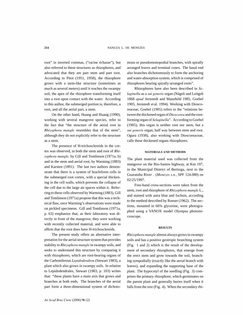
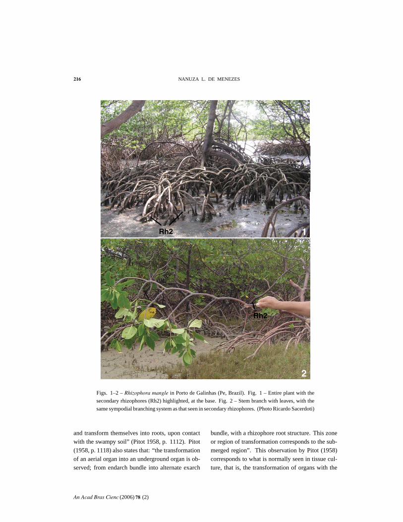
Rhizophora mangle almost always grows in swampy
soils and has a positive geotropic branching system
(Fig. 1 and 2) which is the result of the develop-
ment of secondary rhizophores, that emerge from
the erect stem and grow towards the soil, branch-
ing sympodially (exactly like the aerial branch with
leaves), and expanding the supporting base of the
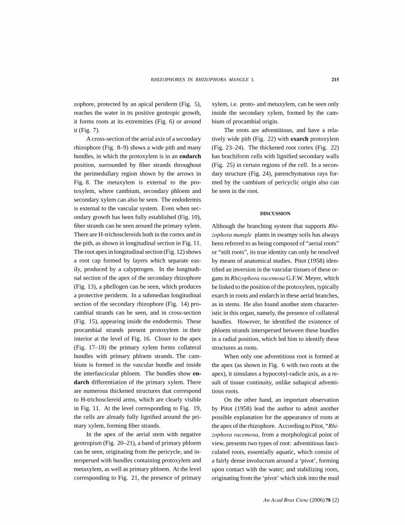
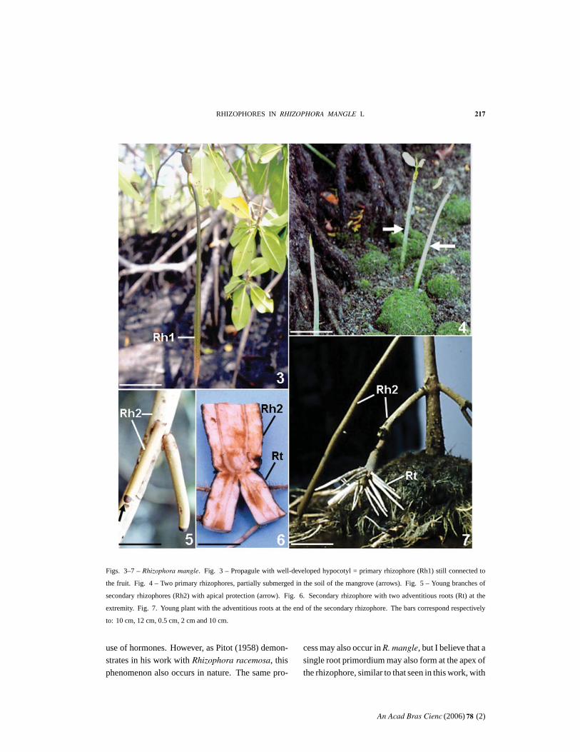
plant. The hypocotyl of the seedling (Fig. 3) com-
prises the primary rhizophore, which germinates on
the parent plant and generally buries itself when it
falls from the tree (Fig. 4). When the secondary rhi-
An Acad Bras Cienc (2006) 78 (2)
RHIZOPHORES IN RHIZOPHORA MANGLE L 215
zophore, protected by an apical periderm (Fig. 5),
reaches the water in its positive geotropic growth,
it forms roots at its extremities (Fig. 6) or around
it (Fig. 7).
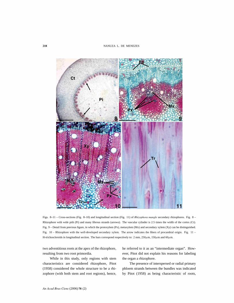
A cross-section of the aerial axis of a secondary
rhizophore (Fig. 8– 9) shows a wide pith and many
bundles, in which the protoxylem is in an endarchposition, surrounded by fiber strands throughout
the perimedullary region shown by the arrows in
Fig. 8. The metaxylem is external to the pro-
toxylem, where cambium, secondary phloem and
secondary xylem can also be seen. The endodermis
is external to the vascular system. Even when sec-
ondary growth has been fully established (Fig. 10),
fiber strands can be seen around the primary xylem.
There are H-trichosclereids both in the cortex and in
the pith, as shown in longitudinal section in Fig. 11.
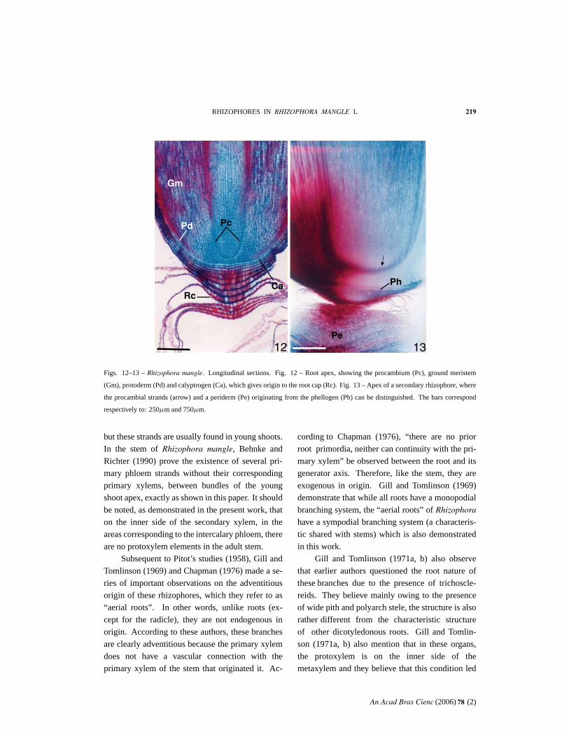
The root apex in longitudinal section (Fig. 12) shows
a root cap formed by layers which separate eas-
ily, produced by a calyptrogen. In the longitudi-
nal section of the apex of the secondary rhizophore
(Fig. 13), a phellogen can be seen, which produces
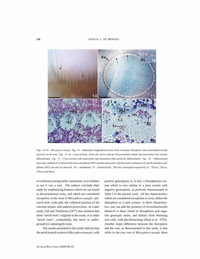
a protective periderm. In a submedian longitudinal
section of the secondary rhizophore (Fig. 14) pro-
cambial strands can be seen, and in cross-section
(Fig. 15), appearing inside the endodermis. These
procambial strands present protoxylem in their
interior at the level of Fig. 16. Closer to the apex
(Fig. 17– 18) the primary xylem forms collateral
bundles with primary phloem strands. The cam-
bium is formed in the vascular bundle and inside
the interfascicular phloem. The bundles show en-darch differentiation of the primary xylem. There
are numerous thickened structures that correspond
to H-trichosclereid arms, which are clearly visible
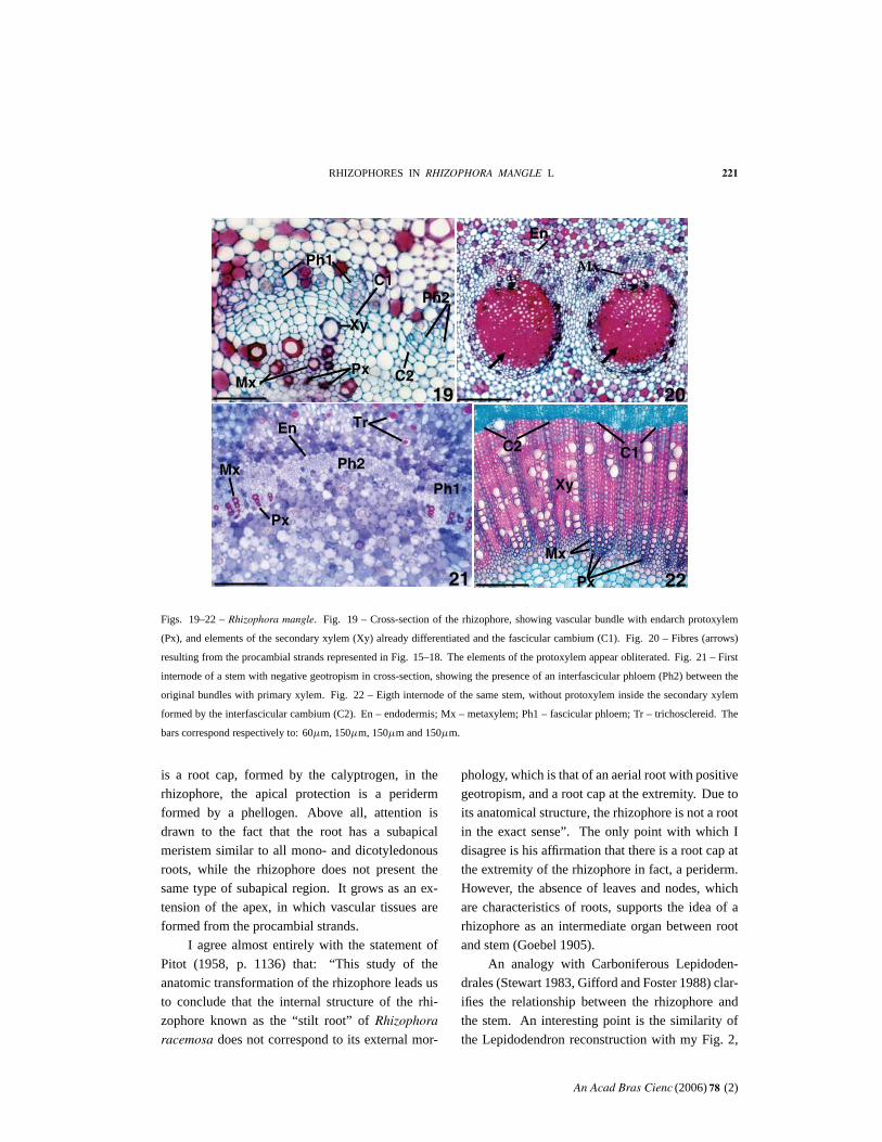
in Fig. 11. At the level corresponding to Fig. 19,
the cells are already fully lignified around the pri-
mary xylem, forming fiber strands.
In the apex of the aerial stem with negative
geotropism (Fig. 20– 21), a band of primary phloem
can be seen, originating from the pericycle, and in-
terspersed with bundles containing protoxylem and
metaxylem, as well as primary phloem. At the level
corresponding to Fig. 21, the presence of primary
xylem, i.e. proto- and metaxylem, can be seen only
inside the secondary xylem, formed by the cam-
bium of procambial origin.
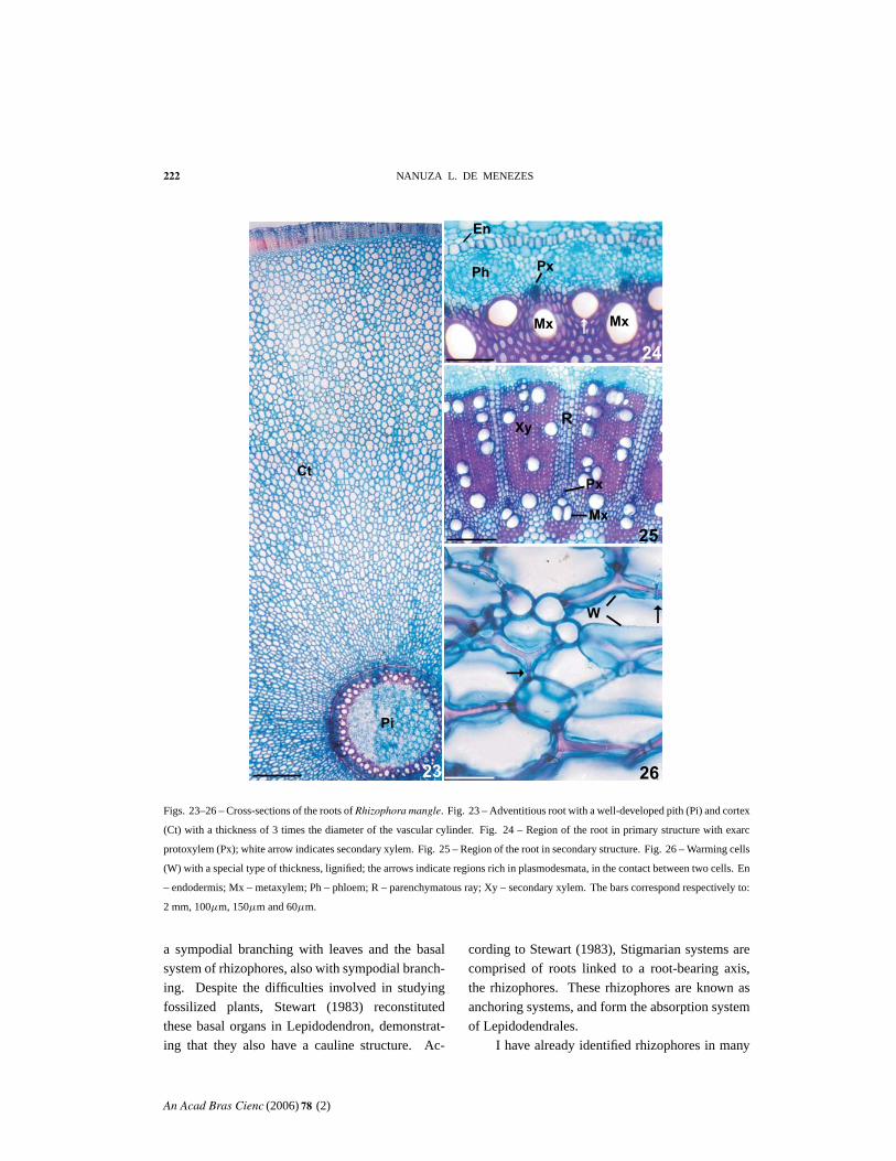
The roots are adventitious, and have a rela-
tively wide pith (Fig. 22) with exarch protoxylem
(Fig. 23– 24). The thickened root cortex (Fig. 22)
has brachiform cells with lignified secondary walls
(Fig. 25) in certain regions of the cell. In a secon-
dary structure (Fig. 24), parenchymatous rays for-
med by the cambium of pericyclic origin also can
be seen in the root.
DISCUSSION
Although the branching system that supports Rhi-
zophora mangle plants in swampy soils has always
been referred to as being composed of “ aerial roots”
or “ stilt roots”, its true identity can only be resolved
by means of anatomical studies. Pitot (1958) iden-
tified an inversion in the vascular tissues of these or-
gans in Rhizophora racemosa G.F.W. Meyer, which
he linked to the position of the protoxylem, typically
exarch in roots and endarch in these aerial branches,
as in stems. He also found another stem character-
istic in this organ, namely, the presence of collateral
bundles. However, he identified the existence of
phloem strands interspersed between these bundles
in a radial position, which led him to identify these
structures as roots.
When only one adventitious root is formed at
the apex (as shown in Fig. 6 with two roots at the
apex), it simulates a hypocotyl-radicle axis, as a re-
sult of tissue continuity, unlike subapical adventi-
tious roots.
On the other hand, an important observation
by Pitot (1958) lead the author to admit another
possible explanation for the appearance of roots at
the apex of the rhizophore. According to Pitot, “ Rhi-
zophora racemosa, from a morphological point of
view, presents two types of root: adventitious fasci-
culated roots, essentially aquatic, which consist of
a fairly dense involucrum around a ‘pivot’, forming
upon contact with the water; and stabilizing roots,
originating from the ‘pivot’ which sink into the mud
An Acad Bras Cienc (2006) 78 (2)
216 NANUZA L. DE MENEZES
Rh2 1
2
Rh2
Figs. 1– 2 – Rhizophora mangle in Porto de Galinhas (Pe, Brazil). Fig. 1 – Entire plant with the
secondary rhizophores (Rh2) highlighted, at the base. Fig. 2 – Stem branch with leaves, with the
same sympodial branching system as that seen in secondary rhizophores. (Photo Ricardo Sacerdoti)
and transform themselves into roots, upon contact
with the swampy soil” (Pitot 1958, p. 1112). Pitot
(1958, p. 1118) also states that: “ the transformation
of an aerial organ into an underground organ is ob-
served; from endarch bundle into alternate exarch
bundle, with a rhizophore root structure. This zone
or region of transformation corresponds to the sub-
merged region”. This observation by Pitot (1958)
corresponds to what is normally seen in tissue cul-
ture, that is, the transformation of organs with the
An Acad Bras Cienc (2006) 78 (2)
RHIZOPHORES IN RHIZOPHORA MANGLE L 217
Figs. 3– 7 – Rhizophora mangle. Fig. 3 – Propagule with well-developed hypocotyl = primary rhizophore (Rh1) still connected to
the fruit. Fig. 4 – Two primary rhizophores, partially submerged in the soil of the mangrove (arrows). Fig. 5 – Young branches of
secondary rhizophores (Rh2) with apical protection (arrow). Fig. 6. Secondary rhizophore with two adventitious roots (Rt) at the
extremity. Fig. 7. Young plant with the adventitious roots at the end of the secondary rhizophore. The bars correspond respectively
to: 10 cm, 12 cm, 0.5 cm, 2 cm and 10 cm.
use of hormones. However, as Pitot (1958) demon-
strates in his work with Rhizophora racemosa, this
phenomenon also occurs in nature. The same pro-
cess may also occur in R. mangle, but I believe that a
single root primordium may also form at the apex of
the rhizophore, similar to that seen in this work, with
An Acad Bras Cienc (2006) 78 (2)
218 NANUZA L. DE MENEZES
8
1110
Ct
Pi
Xy
Tr
Tr
PxMx
Xy
En
9
Px
Mx
Figs. 8– 11 – Cross-sections (Fig. 8– 10) and longitudinal section (Fig. 11) of Rhizophora mangle secondary rhizophores. Fig. 8 –
Rhizophore with wide pith (Pi) and many fibrous strands (arrows). The vascular cylinder is 2.5 times the width of the cortex (Ct).
Fig. 9 – Detail from previous figure, in which the protoxylem (Px), metaxylem (Mx) and secondary xylem (Xy) can be distinguished.
Fig. 10 – Rhizophore with the well-developed secondary xylem. The arrow indicates the fibers of procambial origin. Fig. 11 –
H-trichosclereids in longitudinal section. The bars correspond respectively to: 2 mm, 250µm, 150µm and 60µm.
two adventitious roots at the apex of the rhizophore,
resulting from two root primordia.
While in this study, only regions with stem
characteristics are considered rhizophore, Pitot
(1958) considered the whole structure to be a rhi-
zophore (with both stem and root regions), hence,
he referred to it as an “ intermediate organ”. How-
ever, Pitot did not explain his reasons for labeling
the organ a rhizophore.
The presence of interspersed or radial primary
phloem strands between the bundles was indicated
by Pitot (1958) as being characteristic of roots,
An Acad Bras Cienc (2006) 78 (2)
RHIZOPHORES IN RHIZOPHORA MANGLE L 219
Ca Ph
Pc
Rc
Pd
Gm
Pe12 13
Figs. 12– 13 – Rhizophora mangle. Longitudinal sections. Fig. 12 – Root apex, showing the procambium (Pc), ground meristem
(Gm), protoderm (Pd) and calyptrogen (Ca), which gives origin to the root cap (Rc). Fig. 13 – Apex of a secondary rhizophore, where
the procambial strands (arrow) and a periderm (Pe) originating from the phellogen (Ph) can be distinguished. The bars correspond
respectively to: 250µm and 750µm.
but these strands are usually found in young shoots.
In the stem of Rhizophora mangle, Behnke and
Richter (1990) prove the existence of several pri-
mary phloem strands without their corresponding
primary xylems, between bundles of the young
shoot apex, exactly as shown in this paper. It should
be noted, as demonstrated in the present work, that
on the inner side of the secondary xylem, in the
areas corresponding to the intercalary phloem, there
are no protoxylem elements in the adult stem.
Subsequent to Pitot’s studies (1958), Gill and
Tomlinson (1969) and Chapman (1976) made a se-
ries of important observations on the adventitious
origin of these rhizophores, which they refer to as
“ aerial roots”. In other words, unlike roots (ex-
cept for the radicle), they are not endogenous in
origin. According to these authors, these branches
are clearly adventitious because the primary xylem
does not have a vascular connection with the
primary xylem of the stem that originated it. Ac-
cording to Chapman (1976), “ there are no prior
root primordia, neither can continuity with the pri-
mary xylem” be observed between the root and its
generator axis. Therefore, like the stem, they are
exogenous in origin. Gill and Tomlinson (1969)
demonstrate that while all roots have a monopodial
branching system, the “ aerial roots” of Rhizophora
have a sympodial branching system (a characteris-
tic shared with stems) which is also demonstrated
in this work.
Gill and Tomlinson (1971a, b) also observe
that earlier authors questioned the root nature of
these branches due to the presence of trichoscle-
reids. They believe mainly owing to the presence
of wide pith and polyarch stele, the structure is also
rather different from the characteristic structure
of other dicotyledonous roots. Gill and Tomlin-
son (1971a, b) also mention that in these organs,
the protoxylem is on the inner side of the
metaxylem and they believe that this condition led
An Acad Bras Cienc (2006) 78 (2)
220 NANUZA L. DE MENEZES
Tr
Px
MxMx
Px
C1
C2
14 15
17 18
EnPx
Ph2
Tr
Ph1
16
Figs. 14– 18 – Rhizophora mangle. Fig. 14 – Submedian longitudinal section of the secondary rhizophore where procambial strands
(arrows) can be seen. Fig. 15– 16. Cross-sections, where the arrows indicate the procambial strands and protoxylem (Px) already
differentiated. Fig. 17 – Cross-section with protoxylem and metaxylem (Mx) perfectly differentiated. Fig. 18 – Differentiated
fascicular cambium (C1) between the fascicular phloem (Ph1) and the metaxylem; interfascicular cambium (C2) and the interfascicular
phloem (Ph2) can also be observed. En – endodermis; Tr – trichosclereid. The bars correspond respectively to: 750µm, 750µm,
150µm and 60µm.
to confusion among earlier anatomists, as to whether
or not it was a root. The authors conclude their
study by emphasizing features which are not found
in dicotyledonous roots, and which are considered
exceptions in the roots of Rhizophora mangle: pol-
yarch stele, wide pith, the collateral position of the
vascular tissues, and endarch protoxylem. In a later
study, Gill and Tomlinson (1977) also mention that
these “ aerial roots”originate in the trunk, or in other
“ aerial roots”, sympodially, but never in under-
ground (or submerged) roots.
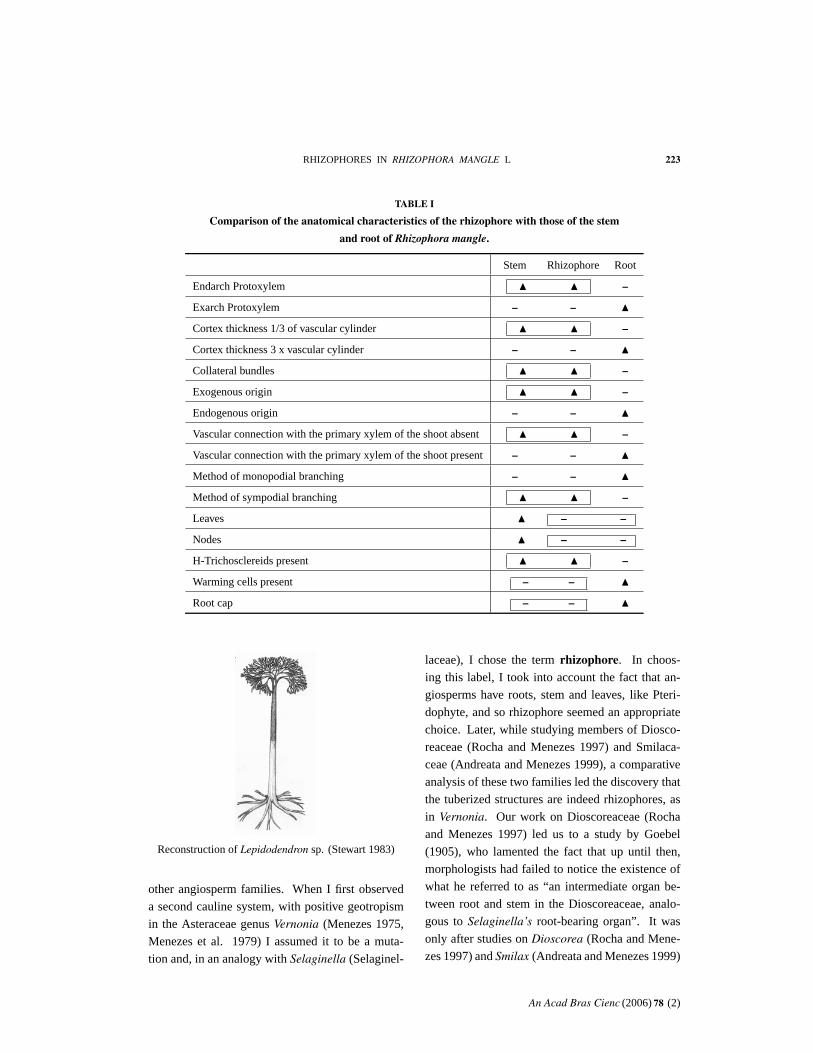
The results presented in this study indicate that
the aerial branch system of Rhizophora mangle, with
positive geotropism, is, in fact, a rhizophorous sys-
tem which is very similar to a stem system with
negative geotropism, as perfectly demonstrated in
Table I of the present work. All the characteristics
which are considered exceptions in roots, define the
rhizophore as a stem system: to these characteris-
tics, one can add the presence of H-trichosclereids
identical to those found in rhizophores and nega-
tive geotropic stems, and distinct from Warming
root cells, with phi-thickenings (Haas et al. 1976).
Another major difference between the rhizophore
and the root, as demonstrated in this work, is that
while in the true root of Rhizophora mangle there
An Acad Bras Cienc (2006) 78 (2)
RHIZOPHORES IN RHIZOPHORA MANGLE L 221
19
Ph1C1
Ph2
PxMx
Xy
C2
En
Tr
Mx Ph2
Px
En
Mx
Px
Xy
20
21 22
Ph1
C2 C1
Mx
Figs. 19– 22 – Rhizophora mangle. Fig. 19 – Cross-section of the rhizophore, showing vascular bundle with endarch protoxylem
(Px), and elements of the secondary xylem (Xy) already differentiated and the fascicular cambium (C1). Fig. 20 – Fibres (arrows)
resulting from the procambial strands represented in Fig. 15– 18. The elements of the protoxylem appear obliterated. Fig. 21 – First
internode of a stem with negative geotropism in cross-section, showing the presence of an interfascicular phloem (Ph2) between the
original bundles with primary xylem. Fig. 22 – Eigth internode of the same stem, without protoxylem inside the secondary xylem
formed by the interfascicular cambium (C2). En – endodermis; Mx – metaxylem; Ph1 – fascicular phloem; Tr – trichosclereid. The
bars correspond respectively to: 60µm, 150µm, 150µm and 150µm.
is a root cap, formed by the calyptrogen, in the
rhizophore, the apical protection is a periderm
formed by a phellogen. Above all, attention is
drawn to the fact that the root has a subapical
meristem similar to all mono- and dicotyledonous
roots, while the rhizophore does not present the
same type of subapical region. It grows as an ex-
tension of the apex, in which vascular tissues are
formed from the procambial strands.
I agree almost entirely with the statement of
Pitot (1958, p. 1136) that: “ This study of the
anatomic transformation of the rhizophore leads us
to conclude that the internal structure of the rhi-
zophore known as the “ stilt root” of Rhizophora
racemosa does not correspond to its external mor-
phology, which is that of an aerial root with positive
geotropism, and a root cap at the extremity. Due to
its anatomical structure, the rhizophore is not a root
in the exact sense”. The only point with which I
disagree is his affirmation that there is a root cap at
the extremity of the rhizophore in fact, a periderm.
However, the absence of leaves and nodes, which
are characteristics of roots, supports the idea of a
rhizophore as an intermediate organ between root
and stem (Goebel 1905).
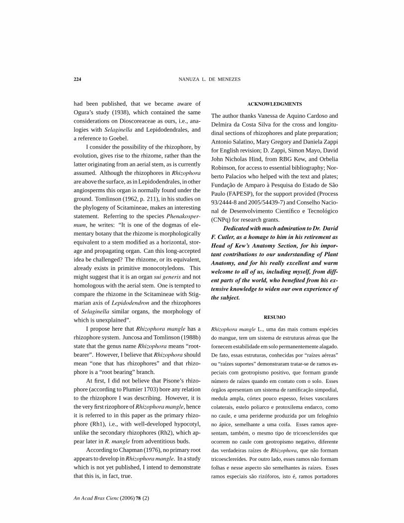
An analogy with Carboniferous Lepidoden-
drales (Stewart 1983, Gifford and Foster 1988) clar-
ifies the relationship between the rhizophore and
the stem. An interesting point is the similarity of
the Lepidodendron reconstruction with my Fig. 2,
An Acad Bras Cienc (2006) 78 (2)
222 NANUZA L. DE MENEZES
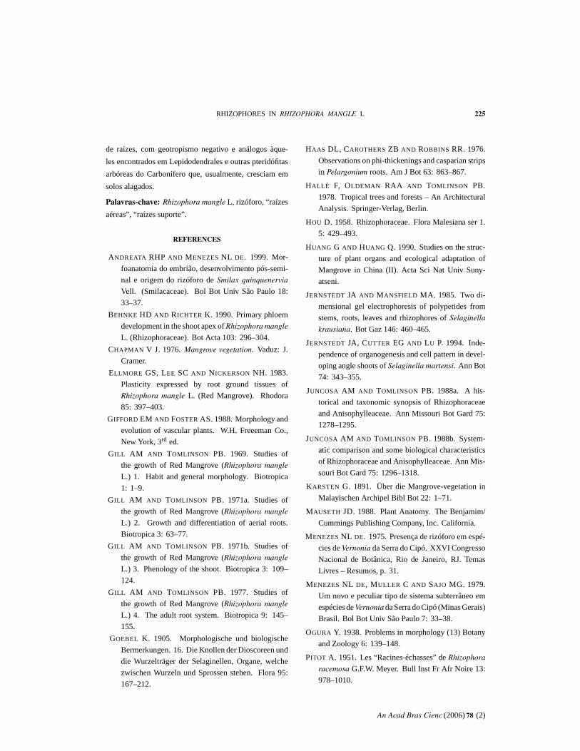
Figs. 23– 26 – Cross-sections of the roots of Rhizophora mangle. Fig. 23 – Adventitious root with a well-developed pith (Pi) and cortex
(Ct) with a thickness of 3 times the diameter of the vascular cylinder. Fig. 24 – Region of the root in primary structure with exarc
protoxylem (Px); white arrow indicates secondary xylem. Fig. 25 – Region of the root in secondary structure. Fig. 26 – Warming cells
(W) with a special type of thickness, lignified; the arrows indicate regions rich in plasmodesmata, in the contact between two cells. En
– endodermis; Mx – metaxylem; Ph – phloem; R – parenchymatous ray; Xy – secondary xylem. The bars correspond respectively to:
2 mm, 100µm, 150µm and 60µm.
a sympodial branching with leaves and the basal
system of rhizophores, also with sympodial branch-
ing. Despite the difficulties involved in studying
fossilized plants, Stewart (1983) reconstituted
these basal organs in Lepidodendron, demonstrat-
ing that they also have a cauline structure. Ac-
cording to Stewart (1983), Stigmarian systems are
comprised of roots linked to a root-bearing axis,
the rhizophores. These rhizophores are known as
anchoring systems, and form the absorption system
of Lepidodendrales.
I have already identified rhizophores in many
An Acad Bras Cienc (2006) 78 (2)
RHIZOPHORES IN RHIZOPHORA MANGLE L 223
TABLE I
Comparison of the anatomical characteristics of the rhizophore with those of the stem
and root of Rhizophora mangle.
Stem Rhizophore Root
Endarch Protoxylem � � –
Exarch Protoxylem – – �Cortex thickness 1/3 of vascular cylinder � � –
Cortex thickness 3 x vascular cylinder – – �Collateral bundles � � –
Exogenous origin � � –
Endogenous origin – – �Vascular connection with the primary xylem of the shoot absent � � –
Vascular connection with the primary xylem of the shoot present – – �Method of monopodial branching – – �Method of sympodial branching � � –
Leaves � – –
Nodes � – –
H-Trichosclereids present � � –
Warming cells present – – �Root cap – – �
Reconstruction of Lepidodendron sp. (Stewart 1983)
other angiosperm families. When I first observed
a second cauline system, with positive geotropism
in the Asteraceae genus Vernonia (Menezes 1975,
Menezes et al. 1979) I assumed it to be a muta-
tion and, in an analogy with Selaginella (Selaginel-
laceae), I chose the term rhizophore. In choos-
ing this label, I took into account the fact that an-
giosperms have roots, stem and leaves, like Pteri-
dophyte, and so rhizophore seemed an appropriate
choice. Later, while studying members of Diosco-
reaceae (Rocha and Menezes 1997) and Smilaca-
ceae (Andreata and Menezes 1999), a comparative
analysis of these two families led the discovery that
the tuberized structures are indeed rhizophores, as
in Vernonia. Our work on Dioscoreaceae (Rocha
and Menezes 1997) led us to a study by Goebel
(1905), who lamented the fact that up until then,
morphologists had failed to notice the existence of
what he referred to as “ an intermediate organ be-
tween root and stem in the Dioscoreaceae, analo-
gous to Selaginella’s root-bearing organ”. It was
only after studies on Dioscorea (Rocha and Mene-
zes 1997) and Smilax (Andreata and Menezes 1999)
An Acad Bras Cienc (2006) 78 (2)
224 NANUZA L. DE MENEZES
had been published, that we became aware of
Ogura’s study (1938), which contained the same
considerations on Dioscoreaceae as ours, i.e., ana-
logies with Selaginella and Lepidodendrales, and
a reference to Goebel.
I consider the possibility of the rhizophore, by
evolution, gives rise to the rhizome, rather than the
latter originating from an aerial stem, as is currently
assumed. Although the rhizophores in Rhizophora
are above the surface, as in Lepidodendrales, in other
angiosperms this organ is normally found under the
ground. Tomlinson (1962, p. 211), in his studies on
the phylogeny of Scitamineae, makes an interesting
statement. Referring to the species Phenakosper-
mum, he writes: “ It is one of the dogmas of ele-
mentary botany that the rhizome is morphologically
equivalent to a stem modified as a horizontal, stor-
age and propagating organ. Can this long-accepted
idea be challenged? The rhizome, or its equivalent,
already exists in primitive monocotyledons. This
might suggest that it is an organ sui generis and not
homologous with the aerial stem. One is tempted to
compare the rhizome in the Scitamineae with Stig-
marian axis of Lepidodendron and the rhizophores
of Selaginella similar organs, the morphology of
which is unexplained”.
I propose here that Rhizophora mangle has a
rhizophore system. Juncosa and Tomlinson (1988b)
state that the genus name Rhizophora means “ root-
bearer”. However, I believe that Rhizophora should
mean “ one that has rhizophores” and that rhizo-
phore is a “ root bearing” branch.
At first, I did not believe that Pisone’s rhizo-
phore (according to Plumier 1703) bore any relation
to the rhizophore I was describing. However, it is
the very first rizophore of Rhizophora mangle, hence
it is referred to in this paper as the primary rhizo-
phore (Rh1), i.e., with well-developed hypocotyl,
unlike the secondary rhizophores (Rh2), which ap-
pear later in R. mangle from adventitious buds.
According to Chapman (1976), no primary root
appears to develop in Rhizophora mangle. In a study
which is not yet published, I intend to demonstrate
that this is, in fact, true.
ACKNOWLEDGMENTS
The author thanks Vanessa de Aquino Cardoso and
Delmira da Costa Silva for the cross and longitu-
dinal sections of rhizophores and plate preparation;
Antonio Salatino, Mary Gregory and Daniela Zappi
for English revision; D. Zappi, Simon Mayo, David
John Nicholas Hind, from RBG Kew, and Orbelia
Robinson, for access to essential bibliography; Nor-
berto Palacios who helped with the text and plates;
Fundação de Amparo à Pesquisa do Estado de São
Paulo (FAPESP), for the support provided (Process
93/2444-8 and 2005/54439-7) and Conselho Nacio-
nal de Desenvolvimento Científico e Tecnológico
(CNPq) for research grants.
Dedicated with much admiration to Dr. DavidF. Cutler, as a homage to him in his retirement asHead of Kew’s Anatomy Section, for his impor-tant contributions to our understanding of PlantAnatomy, and for his really excellent and warmwelcome to all of us, including myself, from diff-ent parts of the world, who benefited from his ex-tensive knowledge to widen our own experience ofthe subject.
RESUMO
Rhizophora mangle L., uma das mais comuns espécies
do mangue, tem um sistema de estruturas aéreas que lhe
fornecem estabilidade em solo permanentemente alagado.
De fato, essas estruturas, conhecidas por “ raízes aéreas”
ou “ raízes suportes” demonstraram tratar-se de ramos es-
peciais com geotropismo positivo, que formam grande
número de raízes quando em contato com o solo. Esses
órgãos apresentam um sistema de ramificação simpodial,
medula ampla, córtex pouco espesso, feixes vasculares
colaterais, estelo poliarco e protoxilema endarco, como
no caule, e uma periderme produzida por um felogênio
no ápice, semelhante a uma coifa. Esses ramos apre-
sentam, também, o mesmo tipo de tricoesclereídes que
ocorrem no caule com geotropismo negativo, diferente
das verdadeiras raízes de Rhizophora, que não formam
tricoesclereídes. Por outro lado, esses ramos não formam
folhas e nesse aspecto são semelhantes às raízes. Esses
ramos especiais são rizóforos, isto é, ramos portadores
An Acad Bras Cienc (2006) 78 (2)
RHIZOPHORES IN RHIZOPHORA MANGLE L 225
de raízes, com geotropismo negativo e análogos àque-
les encontrados em Lepidodendrales e outras pteridófitas
arbóreas do Carbonífero que, usualmente, cresciam em
solos alagados.
Palavras-chave: Rhizophora mangle L, rizóforo, “ raízes
aéreas”, “ raízes suporte”.
REFERENCES
ANDREATA RHP AND MENEZES NL DE. 1999. Mor-
foanatomia do embrião, desenvolvimento pós-semi-
nal e origem do rizóforo de Smilax quinquenervia
Vell. (Smilacaceae). Bol Bot Univ São Paulo 18:
33– 37.
BEHNKE HD AND RICHTER K. 1990. Primary phloem
development in the shoot apex of Rhizophora mangle
L. (Rhizophoraceae). Bot Acta 103: 296– 304.
CHAPMAN V J. 1976. Mangrove vegetation. Vaduz: J.
Cramer.
ELLMORE GS, LEE SC AND NICKERSON NH. 1983.
Plasticity expressed by root ground tissues of
Rhizophora mangle L. (Red Mangrove). Rhodora
85: 397– 403.
GIFFORD EM AND FOSTER AS. 1988. Morphology and
evolution of vascular plants. W.H. Freeeman Co.,
New York, 3rd ed.
GILL AM AND TOMLINSON PB. 1969. Studies of
the growth of Red Mangrove (Rhizophora mangle
L.) 1. Habit and general morphology. Biotropica
1: 1– 9.
GILL AM AND TOMLINSON PB. 1971a. Studies of
the growth of Red Mangrove (Rhizophora mangle
L.) 2. Growth and differentiation of aerial roots.
Biotropica 3: 63– 77.
GILL AM AND TOMLINSON PB. 1971b. Studies of
the growth of Red Mangrove (Rhizophora mangle
L.) 3. Phenology of the shoot. Biotropica 3: 109–
124.
GILL AM AND TOMLINSON PB. 1977. Studies of
the growth of Red Mangrove (Rhizophora mangle
L.) 4. The adult root system. Biotropica 9: 145–
155.
GOEBEL K. 1905. Morphologische und biologische
Bermerkungen. 16. Die Knollen der Dioscoreen und
die Wurzelträger der Selaginellen, Organe, welche
zwischen Wurzeln und Sprossen stehen. Flora 95:
167– 212.
HAAS DL, CAROTHERS ZB AND ROBBINS RR. 1976.
Observations on phi-thickenings and casparian strips
in Pelargonium roots. Am J Bot 63: 863– 867.
HALLÉ F, OLDEMAN RAA AND TOMLINSON PB.
1978. Tropical trees and forests – An Architectural
Analysis. Springer-Verlag, Berlin.
HOU D. 1958. Rhizophoraceae. Flora Malesiana ser 1.
5: 429– 493.
HUANG G AND HUANG Q. 1990. Studies on the struc-
ture of plant organs and ecological adaptation of
Mangrove in China (II). Acta Sci Nat Univ Suny-
atseni.
JERNSTEDT JA AND MANSFIELD MA. 1985. Two di-
mensional gel electrophoresis of polypetides from
stems, roots, leaves and rhizophores of Selaginella
krausiana. Bot Gaz 146: 460– 465.
JERNSTEDT JA, CUTTER EG AND LU P. 1994. Inde-
pendence of organogenesis and cell pattern in devel-
oping angle shoots of Selaginella martensi. Ann Bot
74: 343– 355.
JUNCOSA AM AND TOMLINSON PB. 1988a. A his-
torical and taxonomic synopsis of Rhizophoraceae
and Anisophylleaceae. Ann Missouri Bot Gard 75:
1278– 1295.
JUNCOSA AM AND TOMLINSON PB. 1988b. System-
atic comparison and some biological characteristics
of Rhizophoraceae and Anisophylleaceae. Ann Mis-
souri Bot Gard 75: 1296– 1318.
KARSTEN G. 1891. Über die Mangrove-vegetation in
Malayischen Archipel Bibl Bot 22: 1– 71.
MAUSETH JD. 1988. Plant Anatomy. The Benjamim/
Cummings Publishing Company, Inc. California.
MENEZES NL DE. 1975. Presença de rizóforo em espé-
cies de Vernonia da Serra do Cipó. XXVI Congresso
Nacional de Botânica, Rio de Janeiro, RJ. Temas
Livres – Resumos, p. 31.
MENEZES NL DE, MULLER C AND SAJO MG. 1979.
Um novo e peculiar tipo de sistema subterrâneo em
espécies de Vernonia da Serra do Cipó (Minas Gerais)
Brasil. Bol Bot Univ São Paulo 7: 33– 38.
OGURA Y. 1938. Problems in morphology (13) Botany
and Zoology 6: 139– 148.
PITOT A. 1951. Les “ Racines-échasses” de Rhizophora
racemosa G.F.W. Meyer. Bull Inst Fr Afr Noire 13:
978– 1010.
An Acad Bras Cienc (2006) 78 (2)
226 NANUZA L. DE MENEZES
PITOT A. 1958. Rhizophores et racines chez Rhizophora
sp. Bull Inst Fr Afr Noire 20: 1103– 1138.
PLUMIER C. 1703. Nova Plantarum americanarum Gen-
era. Paris II.
RAVEN PH, EVERT RF AND EICHHORN SE. 1992.
Biology of Plants. Worth Publishers, Inc. New York.
ROCHA DC AND MENEZES NL DE. 1997. O sistema
subterrâneo em Dioscorea kunthiana Uline and R.
Knuth (Dioscoreaceae). Bol Bot Univ São Paulo
16: 1– 11.
ROESER KR. 1962. Die Nadel der Schwarzkiefer –
Massenprodukt und Kunstwert der Natur. Mikro-
kosmos 61: 33– 36.
SPORNE KR. 1974. The Morphology of Angiosperms.
Hutchinson and Co. (Publishers) Ltd., London,
England.
STEWART WN. 1983. Paleobotany and the evolution of
plants. Cambridge University Press, London, Eng-
land.
TOMLINSON PB. 1962. Phylogeny of the Scitamineae
– Morphological and Anatomical Considerations.
Evolution 16: 192– 213.
TOMLINSON PB. 1986. The botany of Mangroves. Cam-
bridge University Press, Cambridge.
WARMING E. 1883. Tropische Fragment II. Rhizophora
mangle L. Bot Jahrb 4: 519– 548.
An Acad Bras Cienc (2006) 78 (2)