Embed Size (px)
Citation preview

1
PONTIFÍCIA UNIVERSIDADE CATÓLICA DO RIO GRANDE DO SUL FACULDADE DE BIOCIÊNCIAS
PROGRAMA DE PÓS-GRADUAÇÃO EM BIOLOGIA CELULAR E MOLECULAR
IDENTIFICAÇÃO DE LIGANTES DA CHIQUIMATO QUINASE DE Mycobacterium tuberculosis POR DOCKING MOLECULAR
Carolina Pasa Vianna
Porto Alegre 2011

2
PONTIFÍCIA UNIVERSIDADE CATÓLICA DO RIO GRANDE DO SUL FACULDADE DE BIOCIÊNCIAS
PROGRAMA DE PÓS-GRADUAÇÃO EM BIOLOGIA CELULAR E MOLECULAR
IDENTIFICAÇÃO DE LIGANTES DA CHIQUIMATO QUINASE DE Mycobacterium tuberculosis POR DOCKING MOLECULAR
Pós-Graduando: Carolina Pasa Vianna Orientador: Prof. Dr. Walter Filgueira de Azevedo Junior
Dissertação apresentada ao Programa de Pós-Graduação em Biologia Celular e Molecular como requisito para obtenção do grau de Mestre.

3
A minha mãe Marlene Pasa Vianna e
ao meu pai Julio de Oliveira Vianna (in
memoriam) que tanto apoiaram e
incentivaram o meu crescimento
profissional.

4
AGRADECIMENTOS
Ao meu orientador, Prof. Dr. Walter Filgueira de Azevedo Junior, ao me receber em
seu laboratório e por acreditar na minha capacidade.
Às minhas amigas, Cristiane Bereta e Andréia Valim, por me acompanharem nesta
caminhada, sempre com palavras de coragem e incentivo.
Aos amigos que fiz ao longo do mestrado, em especial Mariel, Patrícia e Ivani.
Aos colegas do LabioQuest, em especial Rafael e Luis, pela ajuda e paciência
durante a realização do projeto.
A minha prima Vivian Pasa e ao meu amigo Maiko Andrade, por me receber em sua
casa, nos momentos que precisei de estadia para ficar em Porto Alegre.
A equipe diretiva da Escola Manoel Prestes, por me apoiar e me incentivar em todos
os momentos.
5
SUMÁRIO
LISTA DE FIGURAS vi
LISTA DE SÍMBOLOS E ABREVIATURAS vii
RESUMO viii
ABSTRACT ix
Capítulo 1: INTRODUÇÃO............................. .......................................................... 1
1.1) Tuberculose................................... ................................................................... 1
1.2) Via do Ácido Chíquimico ....................... .......................................................... 2
1.3) Chiquimato Quinase ( SK) ..................... ......................................................... 3
1.4) Planejamento racional de fármacos in silico................................................. 5
1.5) Propriedades físico-químicas .................. ....................................................... 7
1.6) OBJETIVO...................................... ................................................................... 9
1.6.1)Objetivo Geral............................... .................................................................. 9
1.6.2) Objetivos específicos ....................... ............................................................ 9
Capítulo 2: Artigo Científico ...................... ........................................................... 10
Capítulo 3: CONSIDERAÇÕES FINAIS................... .............................................. 39
REFERÊNCIAS BIBLIOGRÁFICAS......................... .............................................. 40

6
LISTA DE FIGURAS
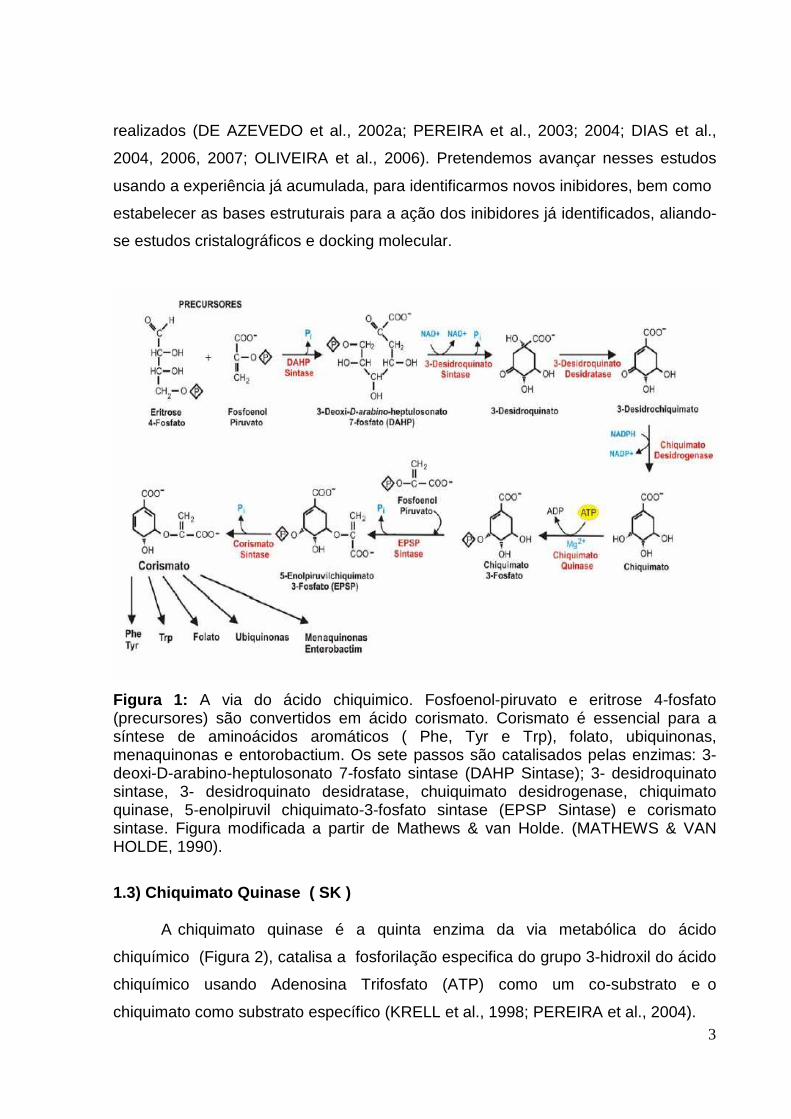
Figura 1 : A via do ácido chiquimico. Fosfoenol-piruvato e eritrose 4-fosfato
(precursores) são convertidos em ácido corismato. Corismato é essencial para a
síntese de aminoácidos aromáticos ( Phe, Tyr e Trp), folato, ubiquinonas,
menaquinonas e entorobactium. Os sete passos são catalisados pelas enzimas: 3-
deoxi-D-arabino-heptulosonato 7-fosfato sintase (DAHP Sintase); 3- desidroquinato
sintase, 3- desidroquinato desidratase, chuiquimato desidrogenase, chiquimato
quinase, 5-enolpiruvil chiquimato-3-fosfato sintase (EPSP Sintase) e corismato
sintase. Figura modificada a partir de Mathews & van Holde. (MATHEWS & VAN
HOLDE, 1990).
Figura 2 : Reação catalisada pela chiquimato quinase.
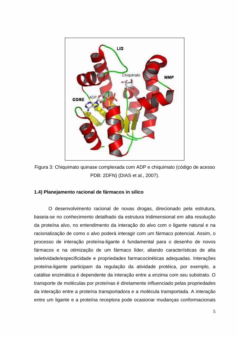
Figura 3 : Chiquimato quinase complexada com ADP e chiquimato (código de acesso
PDB: 2DFN) (DIAS et al., 2007).
vi

7
LISTA DE SÍMBOLOS E ABREVIATURAS AIDS Acquired immune deficiency syndrome ou Síndrome da
Imunodeficiência Adquirida
ADMET Absorção, distribuição, metabolismo, excreção e toxicidade
ADP Adenosina - difosfato
ATP Adenosine - triphosphate
BCG Bacille Calmette-Guérin ou vacina contra a tuberculose
HIV Human immunodeficiency virus ou Vírus da imunodeficiência
humana
MOLDOCK Molegro Virtual Docker
MtSK Mycobacterium Tuberculosis Shikimate Kinase
PDB Protein Data Bank ou Banco de Dados de Proteínas
RMSD Raiz do desvio médio quadrático
SK Shikimate kinase
SKM Shikimate
VS Virtual Screening ou Triagem virtual
WHO World Health Organization ou Organização Mundial da Saúde
vii
8
RESUMO
Entre as doenças infecciosas, a tuberculose se destaca, como sendo a principal
causa de morte humana, principalmente em países em desenvolvimento. Entre os
alvos identificados no genoma de Mycobacterium tuberculosis, as enzimas da via do
chiquimato merecem atenção especial, pois são essenciais para a sobrevivência dos
microorganismos e ausente em mamíferos. O objeto do nosso estudo é a quinta
enzima desta via, a Chiquimato Quinase (SK). Foram aplicados métodos de virtual
screening, a fim de identificar novos potenciais inibidores para esta enzima. Neste
trabalho nós empregamos o programa MOLDOCK em todas as simulações de
docking molecular. A precisão do docking enzima-ligante foi validada em um
conjunto de 12 complexos de SK- ligante, para aquelas estruturas cristalográficas
que estavam disponíveis, gerando um RMSD abaixo de 2,0 Å. A aplicação deste
protocolo em um banco de dados comercialmente disponível permitiu a identificação
de novas moléculas com potencial para se tornar drogas contra tuberculose. Além
disso, foram identificados os resíduos da cavidade de ligação que são essenciais
para as interações intermoleculares desta enzima.
Palavras chave: Tuberculose - Via do Chiquimato - Chiquimato Quinase - Docking
Molecular - Moldock - Virtual Screening
viii

9
ABSTRACT
Tuberculosis (TB) is the major cause of human mortality from a curable infectious
disease, attacking mainly in developing countries. Among targets identified in
Mycobacterium tuberculosis genome, enzymes of the shikimate pathway deserve
special attention, since they are essential to the survival of the microorganism and
absent in mammals. The object of our study is shikimate kinase (SK), the fifth
enzyme of this pathway. We applied virtual screening methods in order to identify
new potential inhibitors for this enzyme. In this work we employed MOLDOCK
program in all molecular docking simulations. Accuracy of enzyme-ligand docking
was validated on a set of 12 SK-ligand complexes for which crystallographic
structures were available, generating root-mean square deviations below 2.0 Å.
Application of this protocol against a commercially available database allowed
identification of new molecules with potential to become drugs against TB. Besides,
we have identified the binding cavity residues that are essential to intermolecular
interactions of this enzyme.
Keywords: Tuberculosis - Shikimate pathway - Shikimate Kinase - Docking
Molecular - Moldock - Virtual Screening
ix


1
Capítulo 1 INTRODUÇÃO
1.1 ) Tuberculose
As doenças infecciosas são a causa de sofrimento e morte de centenas de
milhões de pessoas, especialmente em áreas tropicais e subtropicais do mundo,
local onde ocorre cerca de 90% das mortes causadas por esse tipo de enfermidade.
Entre as doenças infecciosas a tuberculose destaca-se, sendo a principal causa
infecciosa de morte em humanos e está rapidamente tornando-se uma epidemia
global. É estimado que em todo o mundo cem milhões de pessoas são infectadas
anualmente. Aproximadamente dez milhões desenvolvem a doença, com cinco
milhões destes progredindo para um estágio infeccioso, culminando com três
milhões de mortes. De acordo com a Organização Mundial de Saúde (WHO Report
2009), a taxa global de incidência da tuberculose vem crescendo aproximadamente
0,3% ao ano.
A incidência de tuberculose teve um declínio rápido no início do século vinte nos
países em desenvolvimento, devido à melhora nas condições sanitárias e de
moradias. Essa tendência foi acelerada inicialmente pela introdução da vacinação
BCG e da descoberta de antibióticos como a estreptomicina e, posteriormente, com
a descoberta do ácido p-aminosalisílico (1946), isoniazida (1952) e rifampicina
(1965). Mas está ocorrendo um ressurgimento dos casos de tuberculose nos países
em desenvolvimento. O aumento no número de casos de tuberculose atualmente
está relacionado a dois principais fatores. O primeiro é a ocorrência da tuberculose
em pacientes co-infectados com o vírus HIV. Pacientes com AIDS, cujo sistema
imunológico enfraquecido não pode controlar o crescimento do bacilo, apresentam
um risco cem vezes maior de desenvolver a doença. O outro fator é a emergência
de cepas resistentes aos antimicrobianos de primeira linha (isoniazida e rifampicina)
utilizados no tratamento, devido às terapias inadequadas e o uso indiscriminado
destes antibióticos (BASSO et al., 2005).
Os mecanismos de resistência identificados até o momento são resultantes de
mutações pontuais em genes codificadores das proteínas que são os alvos destes

2
agentes anti-tuberculose (BASSO et al,1998). Cepas de M. tuberculosis resistentes
às drogas anti-tuberculose de primeira linha têm sido identificadas globalmente. Um
aspecto preocupante da situação brasileira é que taxas superiores a 45% dos
pacientes, previamente tratados, apresentam multi-resistência (definida como
resistente à isoniazida e rifampicina) adquirida.
1.2) Via da Ácido chíquimico
Uma via sintética que tem levado a um grande interesse como alvo para
desenho de novas drogas contra várias bactérias, outros organismos patogênicos e
herbicidas, é a via do ácido chiquímico (ALIBHAI & STALLINGS, 2001;
SCHÖNBRUNN et al., 2001; PARK et al., 2004). Esta via leva à síntese de
aminoácidos aromáticos e outros compostos desta classe em bactérias, fungos,
algas e plantas, porém ela é ausente em mamíferos. Já existem alguns herbicidas
(Roundup® - MONSANTO - ALIBHAI & STALLINGS, 2001) que inibem fortemente,
de maneira reversível, enzimas desta via sintética, o que a torna ainda mais
interessante para o desenho de fármacos direcionados a inibí-la (BENTLEY, 1990).
A via sintética do ácido chiquímico foi identificada em M. tuberculosis (RATLEDGE,
1982; ROBERTS et al., 1998; COLE, 1998), sendo composta por sete passos
enzimáticos que catalisam sequencialmente a conversão da eritrose-4-fosfato e
fosfoenolpiruvato em corismato (Figura 1). O corismato é o principal precursor para a
biossíntese de PABA (ácido p-aminobenzóico, precursor do tetrahidrofolato), ácido
p-hidroxibenzóico (precursor da coenzima Q ou ubiquinona), micobactinas e dos
aminoácidos aromáticos essenciais (HERRMANN & WEAVER, 1999). Os
intermediários da via do ácido chiquímico são ainda considerados como pontos de
ramificação para outras vias metabólicas (HERRMANN & WEAVER, 1999). Algumas
dessas enzimas de M. tuberculosis, como a 3-deoxi-D-arabino-heptulosonato 7-
fosfato sintase (MtDAHPS); corismato sintase (MtCS); a 5-enolpiruvilchiquimato-3-
fosfato sintase (MtEPSPS) e chiquimato quinase (MtSK), estão com seus cDNAs
clonados, as proteínas foram expressas e purificadas com as respectivas atividades
(OLIVEIRA et al., 2001; 2003; RIZZI et al., 2005). Algumas destas enzimas de M.
tuberculosis já foram cristalizadas e as estruturas em alta resolução determinadas;
estudos de bioinformática para modelagem estrutural por homologia também foram
3
realizados (DE AZEVEDO et al., 2002a; PEREIRA et al., 2003; 2004; DIAS et al.,
2004, 2006, 2007; OLIVEIRA et al., 2006). Pretendemos avançar nesses estudos
usando a experiência já acumulada, para identificarmos novos inibidores, bem como
estabelecer as bases estruturais para a ação dos inibidores já identificados, aliando-
se estudos cristalográficos e docking molecular.
Figura 1: A via do ácido chiquimico. Fosfoenol-piruvato e eritrose 4-fosfato (precursores) são convertidos em ácido corismato. Corismato é essencial para a síntese de aminoácidos aromáticos ( Phe, Tyr e Trp), folato, ubiquinonas, menaquinonas e entorobactium. Os sete passos são catalisados pelas enzimas: 3-deoxi-D-arabino-heptulosonato 7-fosfato sintase (DAHP Sintase); 3- desidroquinato sintase, 3- desidroquinato desidratase, chuiquimato desidrogenase, chiquimato quinase, 5-enolpiruvil chiquimato-3-fosfato sintase (EPSP Sintase) e corismato sintase. Figura modificada a partir de Mathews & van Holde. (MATHEWS & VAN HOLDE, 1990).
1.3) Chiquimato Quinase ( SK ) A chiquimato quinase é a quinta enzima da via metabólica do ácido
chiquímico (Figura 2), catalisa a fosforilação especifica do grupo 3-hidroxil do ácido
chiquímico usando Adenosina Trifosfato (ATP) como um co-substrato e o
chiquimato como substrato específico (KRELL et al., 1998; PEREIRA et al., 2004).

4
Figura 2: Reação catalisada pela chiquimato quinase.
A chiquimato quinase pertence à família estrutural das Nucleosídeo
Monofosfato Quinases (NMP). Ela é uma proteína da classe α/β, consistindo de uma
folha β central, formada por 5 fitas β paralelas circundadas por hélices-α, tendo uma
topologia geral muito semelhante à adenilato quinase (PEREIRA et al., 2004 ;
KRELL et al., 1998, 2001). Uma característica das NMP quinases é que elas sofrem
grandes mudanças conformacionais durante a catálise (VONRHEIN et al., 1995 ). As
NMP quinases são compostas por três domínios: 1) CORE, que contém um loop de
ligação ao fosfato altamente conservado (P-loop); 2) o domínio LID, que sofre
mudanças conformacionais substanciais devido a ligação ao substrato; e, 3) o
domínio de ligação NMP, responsável pelo reconhecimento e ligação de um
substrato específico (GU et al.,2002 ).
Com o objetivo de demonstrar a importância da via do ácido chiquímico em
Mycobacterium tuberculosis, foi realizado a disrrupção do gene aroK, que codifica a
chiquimato quinase. Os resultados obtidos através destes experimentos confirmaram
que esta enzima é essencial para viabilidade do bacilo (PARISH & STOKER, 2002).
5
Figura 3: Chiquimato quinase complexada com ADP e chiquimato (código de acesso
PDB: 2DFN) (DIAS et al., 2007).
1.4) Planejamento racional de fármacos in silico
O desenvolvimento racional de novas drogas, direcionado pela estrutura,
baseia-se no conhecimento detalhado da estrutura tridimensional em alta resolução
da proteína alvo, no entendimento da interação do alvo com o ligante natural e na
racionalização de como o alvo poderá interagir com um fármaco potencial. Assim, o
processo de interação proteína-ligante é fundamental para o desenho de novos
fármacos e na otimização de um fármaco líder, aliando características de alta
seletividade/especificidade e propriedades farmacocinéticas adequadas. Interações
proteína-ligante participam da regulação da atividade protéica, por exemplo, a
catálise enzimática é dependente da interação entre a enzima com seu substrato. O
transporte de moléculas por proteínas é diretamente influenciado pelas propriedades
da interação entre a proteína transportadora e a molécula transportada. A interação
entre um ligante e a proteína receptora pode ocasionar mudanças conformacionais

6
nesta que transporta a sinalização celular tanto por membranas quanto no meio
intracelular (SILVA, 2008). Portanto, é grande o interesse em compreender as forças
que estabilizam uma determinada interação proteína-ligante para o desenvolvimento
racional de novos fármacos. As principais forças moleculares que governam a
interação proteína-ligante são: ligações de hidrogênio, interações hidrofóbicas e de
Van der Walls.
O planejamento racional baseado em estrutura e no mecanismo de ação de
enzimas específicas é a estratégia mais eficiente no desenvolvimento de novos
fármacos, capaz de contribuir em todos os estágios do processo, desde a
descoberta de protótipos, sua otimização, até a elaboração de compostos
candidatos a testes clínicos. Esta estratégia é baseada no bloqueio ou estimulação
da atividade de macromoléculas, como proteínas ou ácidos nucléicos, associados a
diferentes processos patológicos. A informação estrutural do ligante permite a
descoberta e síntese de compostos com complementariedade geométrica,
hidrofóbica e eletrostática ao seu sítio de ligação, podendo vir a se tornar potenciais
inibidores e futuros fármacos (SILVA, 2008). Essa abordagem caracteriza o
planejamento racional de fármacos baseados em estrutura. O que a torna ainda
mais atrativa, quando utilizada em proteínas, é o conhecimento de que 78% dos
fármacos atuais têm como alvo receptor esse tipo de biomacromolécula
(MARSHALL, 2004)
Desde a concepção do alvo biológico até a descoberta de um novo fármaco,
um processo que pode levar em média 11 anos ou até mais, a bioinformática,
juntamente com a química computacional, vem oferecendo um excelente
direcionamento ao planejamento racional de fármacos, já que há inúmeros casos de
sucesso envolvendo o emprego de simulações computacionais (MARSHALL, 2004),
citando como exemplo os importantes fármacos: losartan, atorvastatina e celecoxib.
No planejamento racional de fármacos, destaca-se o docking molecular como
um dos métodos mais empregados. No docking, são investigadas as possíveis
orientações que determinada molécula assume no interior do sítio ativo de uma
proteína. Os métodos de docking, em geral, envolvem uma função de energia
contendo parâmetros eletrostáticos, van der Waals, ligações de hidrogênio, e
algumas vezes, hidrofóbicos, os quais geram modelos matemáticos que predizem as
7
melhores orientações do ligante, segundo uma lista escore de energia (SILVA,
2008).
O screening virtual vem complementar o processo de descoberta de novos
fármacos. Um dos mais recentes programas, Moldock (THOMSEN E
CHRISTENSEN, 2006) considera a flexibilidade do ligante e é baseado em evolução
diferencial dirigida e uma função escore baseada em campos de força. A partir
dessa estratégia é possível selecionar por screening virtual compostos de bases de
dados contendo tipicamente milhares de estruturas, eliminando assim ligantes não
promissores antes que sejam sintetizados.
Há diversos casos de sucesso com o uso da abordagem de screening virtual,
tal como a descoberta de isoflavonóides, como inibidores não-esteroidais da 5 –
redutase (BRENK et. al., 2003; CHEN et al., 2001). Porém o sucesso do screening
dito in silico, e em geral das técnicas de docking, depende do conhecimento de
detalhes estruturais do sítio de reconhecimento da biomacromolécula (CARLSON
et.al 1999).
1.5) Propriedades físico-químicas
Nos últimos anos, percebeu-se um avanço considerável no desenvolvimento
de técnicas de modelagem molecular que elevaram o poder de predição do
comportamento dos ligantes em sistemas biológicos,que são aplicadas no estudo de
diversas propriedades, como: absorção, distribuição, metabolismo, excreção e
toxicidade ( ERINS; ROSE, 2002).
Na busca de inovação e desenvolvimento de novos fármacos, é evidente a
pressão do mercado sobre a otimização dos recursos financeiros. Pode-se destacar
também a restrição, ou crescente dificuldade, com relação ao uso de animais para
utilização dos testes de toxicidade. A maioria dos fármacos retirados do mercado
ocorreram devido a estes fármacos causarem efeitos tóxicos indesejáveis como o
antiinflamatório Vioxx® (REVISTA ÉPOCA,2005). Dessa forma, os métodos de
predição de toxicidade in silico surgem como um importante e alternativa ferramenta
na seleção ou priorização de moléculas promissoras a serem avaliadas com maior
cautela em testes de toxicidade, reduzindo assim, os custos financeiros inerentes ao
processo, o uso indiscriminado de animais e satisfazendo as precauções em relação

8
à toxicidade desde a fases iniciais do processo de desenvolvimento de fármacos
(SILVA, 2008).
Para o desenvolvimento de novos fármacos, já é possível estimar
propriedades farmacocinéticas de ligantes com potencial a se tornar fármacos.
Como exemplo mais simples, a ‘ Regra dos Cinco’ (RO5), de Lipinski, preconiza que
os fármacos que apresentam biodisponibilidade por via oral, seguem, a saber: peso
molecular menor ou igual a 500, log P menor ou igual a 5, número de grupo de
doadores de ligações de hidrogênio menor ou igual 5 e número de grupos de
aceptores de ligações de hidrogênio menor ou igual a 10 (LIPINSKI et. al., 1997).
O Laboratório de Bioquímica Estrutural (LabBioqEst) da Faculdade de
Biociências-PUCRS tem como objeto de estudo a interação proteína-ligante em nível
molecular. A principal intenção é a determinação da estrutura tridimensional em alta
resolução de proteínas nativas e em complexos com ligantes, para o posterior
desenho racional de ligantes que possam interagir especificamente com as
proteínas alvo. Estas informações serão importantes para ampliar a capacidade do
grupo LabBioqEst-PUCRS em compreender o mecanismo de ação, a relação
estrutura-função das proteínas alvo e auxiliar no desenho racional de novos
fármacos.

9
1.6) OBJETIVO
1.6.1) Objetivo Geral
Este projeto de pesquisa pretende realizar estudos estruturais focados na interação
proteína-ligante, focado na Chiquimato Quinase de Mycobacterium tuberculosis.
1.6.2) Objetivos específicos
1) Simular a interação proteína-ligante por meio de algoritmos de docking
molecular;
2) Identificar aspectos estruturais determinantes para a especificidade do ligante
pela enzima;
3) Propor novos ligantes que apresentem indicativos estruturais que mostrem
aumento da especificidade deste pela enzima.

10
Capítulo 2 Artigo científico Este capítulo apresenta uma cópia do artigo científico submetido ao Journal of
Molecular Modeling, bem como cópia da confirmação da submissão.
>Dear Walter > >We have received the referee reports for your manuscript: "Identification of new potential Mycobacterium tuberculosis Shikimate Kinase inhibitors through molecular Docking simulations", which you submitted to Journal of Molecular Modeling > >TO VIEW REVIEWER ATTACHMENTS (should there be any), please login to the journal site as "Author" and access "Submission Needing Revision" from the Author Main Menu. On the next page display, navigate on the "Action Links" and select "View Reviewer Attachments" from the selection box. This will redirect you to the page that will allow you to "Download" and view the reviewer report attachments. Then, proceed with revising your manusript. > >As you can see, the referees have requested some revisions. >Please revise the manuscript taking the referees remarks into account. >To avoid misunderstandings it will be helpful if you provide a detailed response to the referee reports. > >I would appreciate a short receipt acknowledgement for this message. > >Thank you very much in advance. If you have further questions, please do not hesitate to contact me. The editorial office is looking forward to receiving the revised version of your manuscript. > >Kind regards, > >Andrzej Sokalski, Ph.D., D.Sc. >Editor in Chief > >COMMENTS FOR THE AUTHOR: > >Reviewer #1: The manuscript describes the development of a virtual screening study for the Mycobacterium tuberculosis Shikimate Kinase. >A lot of computational work has been done with technical diligence. The quality of the paper is quite good and it is acceptable for publication after minor changes: >* In order to further measure the reliability of the VS protocol, you should use an enriched database, adding known Shikimate Kinase inhibitors to the Acros Organics collection and evaluating the number of known compounds recognized as active by the VS protocol. >* The VS protocol reported in Figure 4, should be widely described in the text. >* The authors decided to apply the ADMET filter after docking calculations. In

11
order to reduce the number of compounds to be docked (for larger database), could it be better to apply this filter before the docking calculations? Please discuss it. >Reviewer #2: This manuscript reports a virtual screening study aimed to the identification of inhbitors of one of the enzymes of the shikimate pathway from Mycobacterium tuberculosis (shikimate kinase). Since the shikimate pathway is essential to M. tuberculosis but is absent in humans compounds inhibiting steps of this pathway are potential anti-tuberculosis drugs. >Therefore, the rationale of the work is sound and the results derived interesting and of potential use for other researchers. Authors are experienced not only in the field of virtual screening but also have published a molecular model of the enzyme and have been involved in the obtention of the 3D-structure of M. tuberculosis shikimate kinase (MT_SK). >Papers from other research groups have been previously published on the identification of MT_SK inhbitors as antitubercular drugs by similar procedures (Segura-Cabrera and Rodriguez-Perez (2008) Bioorg. Med. Chem. Lett. 18: 3152-3157 and Kumar et al. 2010 Chem. Biol. Drug. Des. 76: 277-284). Nevertheless, very limited information (experimental data) on the direct effect of these compounds on MT_SK is yet available. >The novelty of the submitted paper relays on the method used in the virtual screening and the selection of compounds according to their pharmacological properties. >Lead validation is a key issue in drug discovery and in my oppinion this is the missing point in this study. To increase the value of the submitted work and allow publication authors should give proof of the usefulness of the selected compounds as MT SK inhibitors. >Ideally this could be done by testing the inhibitory effect of the best compounds, on the enzyme in their lab (Pereira et al. 2004 Acta Cryst. D60: 2310-2319) since compounds belong to a collection of commercially available molecules (Acros Organics). Alternatively,authors could analyse by their screening procedure compounds which have been already tested directly on MT_SK. For instance, three MT SK inhbitors have been recently identified and tested (Mulabagal and Calderon 2010 Anal. Chem. 82: 3616-3621). Analysing these compounds would allow validation of the method and the results obtained. >Minor points: >Authors should modifiy the Introduction and the Results and Discussion sections to include the data reported in the references mentioned above (other virtual screening studies and identification of MT_SK inhibitors) > >References format should be checked and adapted to JMM guidelines

12
Identification of new potential Mycobacterium tuberculosis shikimate kinase
inhibitors through molecular docking simulations
Carolina Pasa Viannaa,b and Walter F. de Azevedo Jr.a,b*
a Faculdade de Biociências, Instituto Nacional de Ciência e Tecnologia em
Tuberculose (INCT-TB), Laboratório de Bioquímica Estrutural (LaBioQuest),
Pontifícia Universidade Católica do Rio Grande do Sul (PUCRS), Av. Ipiranga 6681,
Porto Alegre, RS 90619-900, Brazil
b Programa de Pós Graduação em Biologia Celular e Molecular, Pontifícia
Universidade Católica do Rio Grande do Sul, Porto Alegre, RS, Brazil
*To whom correspondence may be addressed: Walter Filgueira de Azevedo Jr.
Av. Ipiranga 6681 – Faculdade de Biociências – Prédio 12C, Porto Alegre RS 90619-
900, Brazil. Phone/Fax: +55 51 33204529; E-mail address: [email protected]

13
ABSTRACT
Tuberculosis (TB) is the major cause of human mortality from a curable infectious
disease, attacking mainly in developing countries. Among targets identified in
Mycobacterium tuberculosis genome, enzymes of the shikimate pathway deserve
special attention, since they are essential to the survival of the microorganism and
absent in mammals. The object of our study is shikimate kinase (SK), the fifth
enzyme of this pathway. We applied virtual screening methods in order to identify
new potential inhibitors for this enzyme. In this work we employed MOLDOCK
program in all molecular docking simulations. Accuracy of enzyme-ligand docking
was validated on a set of 12 SK-ligand complexes for which crystallographic
structures were available, generating root-mean square deviations below 2.0 Å.
Application of this protocol against a commercially available database allowed
identification of new molecules with potential to become drugs against TB. Besides,
we have identified the binding cavity residues that are essential to intermolecular
interactions of this enzyme.
Keywords: Tuberculosis; Shikimate pathway; Shikimate Kinase; Molecular Docking;
Virtual Screening

14
Introduction
Tuberculosis (TB) is the most important cause of human death from a
curable infectious disease. It is estimated that, worldwide, one hundred million people
are infected annually and about ten million develop the disease, with five million of
those progressing to an infectious stage, culminating with approximately three million
deaths. According to the World Health Organization [1], the overall incidence of TB
increases approximately 0.3% per year. The resurgence of this health problem
occurred mainly due to the proliferation of multi (MDR-TB), extensively (XDR-TB),
and recently, totally-drug (TDR-TB) resistant Mt strains. Besides, the high
susceptibility of HIV/AIDS infected patients to TB is also a health problem. Therefore,
there is an urgent need for the discovery and development of new and better drugs
for the TB treatment [2].
Enzymes of the shikimate pathway (SP) are promising targets for the
development of antimicrobial agents [3] and herbicides [4], because they are
essential to the survival of algae, higher plants, bacteria, fungi, apicomplexan
parasites and absent in mammals [5]. It is a seven-step biosynthetic route that
converts erythrose 4-phosphate to chorismate, a precursor of aromatic amino acids
and many other essential compounds [6].
The object of our study is the fifth enzyme of the SP, shikimate kinase (SK)
(EC 2.7.1.71), which catalyzes the specific phosphorylation of the 3-hydroxy group of
shikimate using ATP as a co-substrate resulting in shikimate-3-phosphate and ADP
[7-8]. This enzyme is an established target against Mt, since Parish and Stoeker
demonstrated that the SP is essential for the viability of Mt due to the disruption of
the aroK gene, which codes for the SK enzyme [9].
SK is a member of the nucleoside monophosphate kinases (NMP kinases)
family, which suffer large conformational changes during catalyses (Fig. 1) [10]. The
enzymes of this family are composed of three domains: the CORE, which contains a
highly conserved phosphate-binding loop (P-loop), the LID domain, which undergoes
substantial structural changes upon substrate binding, and the NMP-binding domain
which is responsible for the recognition and binding of a specific substrate [11].
Drugs are usually discovered by trial and error by means of high-throughput
screening approaches that use in vitro experiments to evaluate the activity of a large

15
number of compounds against a known target. This procedure is very costly and
time-consuming. If crystallographic information is available for the protein target, then
molecular docking simulations can be a helpful computational approach in the drug-
discovery process. [12]. Molecular docking is a simulation method that predicts the
conformation of a receptor-ligand complex, in which the receptor can be either a
protein or a nucleic acid, and the ligand is a small molecule. This computer
simulation can generate many possible positions for the ligand in the receptor-
binding pocket. Therefore, a criterion is necessary that will allow comparisons of all
possible positions of the ligand, and then a selection can be made for the best
position.
Our goal here is to find potential inhibitors against shikimate kinase from
Mycobacterium tuberculosis MtSK using virtual screening (VS). VS can decrease
costs and improve hits rates for lead discovery. For this, we used the MtSK structure
[13] as a target for the molecular docking simulations with MOLDOCK [14]. Our
docking protocol was validated against an ensemble of 12 crystallographic structures
available for complexes of MtSK. The VS was validated by inclusion of a known SK
inhibitor in the small-molecule database with over 4500 structures. We describe the
results obtained in terms of the MOLDOCK scores, modes of interaction and discuss
the importance of the active site residues in the ligand binding process.
Materials and Methods
Molecular docking simulations
One of the fundamental questions in structural biology is the study of protein-
ligand interactions, particularly considering the pharmacological applications of such
study in the design of drugs based on structure [15]. To simulate the interaction of
MtSK with a library of ligands, we used the MOLDOCK program [14], an
implementation of a variant of the evolutionary algorithm (EA). Recent evaluation of
MOLDOCK strongly indicates that it is capable of finding the right position of a ligand.
Furthermore, MOLDOCK exhibits better overall performance compared with
SURFLEX, FLEXX, and GOLD [14]. In the present work, all simulations were
performed in an iMac (Intel Processor Core 2 Duo, 2.66 GHz, 2 GB SDRAM DDR3
1066 MHz).

16
Re-docking and cross-docking
In molecular docking simulations, the best binary complex (protein-ligand) is
the one closer to the crystallographic structure. For that reason we must establish a
methodology that assesses the distance from the computer-generated solution
(pose) to the crystallographic structure. This distance can be calculated using the
root-mean-square deviation (RMSD), which is a measure of the differences between
values predicted by a model and the values actually observed from the object being
modeled or estimated (protein-ligand complex). The RMSD is calculated between
two sets of atomic coordinates, in this case, one for the crystallographic structure
(xctal, yctal, zctal; the object being modeled) and another for the atomic coordinates
obtained from the docking simulations (xpose, ypose, zpose; predicted model). A
summation is then taken over all N atoms being compared, using the following
equation:
∑=
−+−+−=N
1i
2i pose,i ctal,
2i pose,i ctal,
2i pose,i ctal, )z(z)y(y)x(x
N
1RMSD .
In docking simulations, it is expected that the best results generate RMSD values
less than 2.0 Å compared with crystallographic structures [16]. This procedure of
obtaining the crystallographic position of the ligand is often called “re-docking,” which
is fundamentally a validation method that determines whether the molecular docking
algorithm is able to recover the crystallographic position using computer simulation.
In this work, all RMSD calculations were calculated for non-hydrogen atoms.
In order to validate our docking protocol, we used the SK crystallographic
coordinates available at the protein data bank (PDB), under the access code 2DFN
[13]. We performed the docking simulation against the active site of MtSK and
compared the docked poses with the crystallographic structure. We used the
MOLDOCK default protocol with center at coordinates x=(-15.23), y=(-14.38), and
z=(14.88) Å, and a docking sphere radius of 9 Å. Fig. 2 shows the docking sphere
used in the simulations.
In the implementation of EA in MOLDOCK, computational approximations of
an evolution course, called genetic operators, are applied to simulate the
permanence of the most positive features. In a sample space, where there is a
problem or a search routine and many different possible solutions (candidates), each
option is ranked based on a set of parameters (scoring function or fitness function),

17
and only the best ranked solutions are kept for the next iteration. This cycle is
repeated until an optimal solution can be found. In the molecular docking simulations,
the optimal solution is the one with the best scoring function, which should be the
closest to the crystallographic structure. MOLDOCK presents two biological inspired
algorithms to perform positional searches in docking simulations. One is called the
optimizer search algorithm (MOLDOCK Optimizer), which is based on an EA [14].
The second is a guided differential evolution algorithm (GDEA) called MOLDOCK
SE. GDEA is based on an EA adjustment called differential evolution (DE), which
provides a distinct method for selecting and modifying candidate solutions
(individuals). We used MOLDOCK Optimizer as search algorithm.
In addition to re-docking, a procedure called “cross-docking” can also be used
to further validate a docking protocol. Considering that several crystallographic
structures are available for the same protein, cross-docking can be applied. This
procedure involves docking a number of ligands found in a variety of crystal
structures of a protein identical to a single rigid protein crystallographic conformation
[17]. When a protein target presents major conformational changes upon ligand
binding, a significant difference is expected between the crystallographic and docked
structures. We identified 12 MtSK structures in PDB with ligands in the shikimate-
binding site ( PDB access codes: 2DFN, 1U8A, 1WE2, 1ZYU, 2G1K, 2IYQ, 2IYR,
2IYS, 2IYX, 2IYY, 2IYZ, and 3BAF). This search was performed on February, 24th
2011. This validation procedures, re-docking and cross-docking, is the initial stage of
a virtual screening protocol (phase 1 ) described in the next sections.
Virtual screening
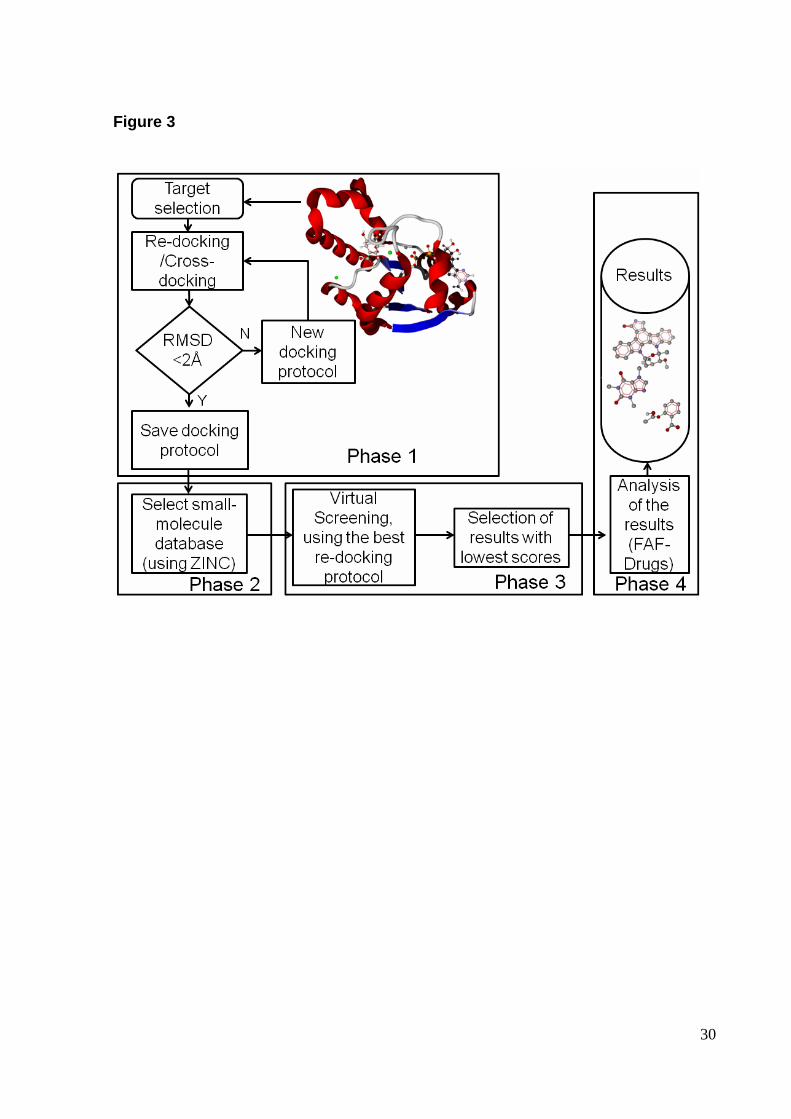
Our virtual screening (VS) protocol is divided in 4 phases as shown in Fig. 3.
Phase 1 is focused on selection and validation of a docking protocol, as described
earlier in the section re-docking and cross-docking. Phase 1 ends when an adequate
protocol is found (selection criterion RMSD < 2.0 Å). It should be pointed out that the
RMSD criterion is dependent on the number of torsion angles, and a less demanding
criterion may be adopted for re-docking of a ligand with a number of torsion angles
higher than 10 [14]. Once a docking protocol is chosen we select a small-molecule
database to be used in the screening (phase 2 ). Here we used a ligand library
commercially obtainable at Acros Organics. The ligands (mol2 format) were

18
downloaded from http://zinc.dock.org [18], with a total of 4579 small molecules. In
addition to commercially oriented databases the ZINC database also provides an
interface to build small-molecule databases based on molecular similarity, such as
Tanimoto coefficient [19, 20].
In phase 3 , we start docking simulations for each ligand present in the
selected database. MOLDOCK program is the workhorse of the present protocol. It
was used in all docking simulations described here. During a typical docking
simulation several orientations can be obtained for each ligand. Here we selected the
one with the lowest scoring function. The scoring function used by MOLDOCK
improves accuracy of scoring functions with a new hydrogen bonding term and new
charge schemes. Four scoring functions are implemented in the MOLDOCK,
including MOLDOCK score and PLANTS score [14, 21]. These two functions offer
grid-based versions, in which hydrogen bond directionality is not considered. In the
present protocol we employed grid-based MOLDOCK score since it offers
approximately four-fold greater speed by performing a precalculation of potential-
energy values on an equally spaced cubic grid.
The MOLDOCK score is based on the piecewise linear potential (PLP) scoring
functions developed by Yang et al. [22, 23]. The docking scoring function EMOLDOCK
SCORE is defined as the following:
intermolIntramolSCOREMOLDOCK E E E +=
where Eintermol is the intermolecular interaction energy:
∑ ∑∈ ∈
+=
ligandi proteinjijPLP
ij
jiintermol )(r E
Dr
qq332 E
All non-hydrogen atoms in the ligand and protein are taken in the summation. The
first term accounts for electrostatic interactions, in which the factor 332 is used to
obtain energy in kJ/mol. D represents the dielectric constant, which is the following:
ij4r D = . The second term (EPLP) is a PLP, described elsewhere [22, 23]. To ensure
that no energy term can be superior to the clash penalty, the electrostatic term is cut
off at a level equivalent to the distance of 2.0 Å for distances less than 2.0 Å.
Intramolecular energy is given by the following equation:
( )[ ]∑∑ ∑ ++=∈ ∈ bonds single
0ligand i ligand j
ijPLPpenaltyintramol φ - nφcos - 1A )(rE E E

19
The term Epenalty is a penalty energy to be added to Eintramol when two non-bonded
atoms are closer than 2 Å (for non-hydrogen atoms). This term avoids unrealistic
molecular topologies for the ligands. The second term is a PLP, already mentioned
[22, 23]. The last term accounts for torsion energy, which is expressed as a periodic
function. In this term, A, n, and φo are empirically determined [22, 23]. MOLDOCK
defines a limiting sphere where the search is focused. If a ligand non-hydrogen atom
is positioned outside this limiting sphere (the search space sphere), then a constant
penalty of 10000 is added to the total energy (implemented for the grid-based version
of the MOLDOCK score).
After identification of potential inhibitors by molecular docking simulations, the
best scored ligands were submitted to the web server FAF-Drugs [24], in order to
assess physical-chemical properties (phase 4 ). These are key properties that need
to be considered in early stages of the drug discovery process, and FAF-Drugs
allows users to filter molecules via simple rules such as molecular weight, polar
surface area, logP and number of rotatable bonds. The ligands were filtered following
the Lipinski’s rule of five (RO5). RO5 advocates that drugs which present oral
bioavailability, in general, follow: molecular weight less or equal to 500, LogP less or
equal to 5, number of hydrogen bond donor groups less or equal to 5 and number of
hydrogen bond acceptor groups less or equal to 10 [25].
In addition to the 4579 small molecules present in the Acros database we
added staurosporine (PubChem Compound Identification: CID 44259) to the
database to be used in the VS. This molecule has been already tested directly on
MtSK [26]. This addition allows testing whether this VS protocol is able to identify SK
inhibitors present in a database with over 4,500 ligands.
Results and Discussion
Docking and cross-docking
Re-docking simulations (phase 1 of the VS protocol) using the structure 2DFN
generated an RMSD of 1.6 Å. In addition, cross-docking simulations generated
RMSD ranging from 1 to 2 Å further validating the present docking protocol. These
two tests indicated that the docking simulation was successful, and that the protocol
is good enough to be used for the virtual screening process.

20
Virtual Screening
VS uses computational methodologies to identify biologically active molecules
against a specific protein target. Two main methodologies are used in VS. Methods
that search for similarity to validated ligands and molecular docking methods that
require the use of crystallographic information of the target. Here we made use of the
second approach. VS studies performed by other research groups have been
previously published on the identification of MtSK inhibitors as antitubercular drugs
by similar molecular docking procedures [27, 28]. Nevertheless, very limited
information (experimental data) on the direct effect of these compounds on MtSK is
yet available. The novelty of the present work relays on the method used in the VS
and the selection of compounds according to their pharmacological properties. The
VS simulations were carried out using the MOLDOCK program, having as target the
MtSK (PDB access code 2DFN). The ligand library comprises 4580 molecules
(Across database plus staurosporine). Addition of a known SK inhibitor allows testing
the accuracy of the present protocol.
After docking simulations, we selected 20 top-scoring compounds from the
initial set of 4580 compounds (selection based on MOLDOCK score). Staurosporine
was present in the 20 top-scoring compounds obtained in the VS, with MOLDOCK
score of -144.168. Identification of a known SK inhibitor among the best VS results
gives further validation for this VS protocol. These 20 potential inhibitors were
submitted to filter tests, available at the web server FAF-Drugs [24], to exclude those
compounds that have known undesirable physical-chemical features to oral
bioavailability. We could have applied this filter analysis previous to docking
simulations, since it would reduce simulation time. Nevertheless, we kept filtering
analysis after docking simulation, since the MOLDOCK protocol was fast enough to
be run in less than a week of CPU time of an iMac (Intel Processor Core 2 Duo, 2.66
GHz, 2 GB SDRAM DDR3 1066 MHz). In addition, application of filtering analysis
previous to docking simulations could eliminate candidates that fail to filtering
analysis but present promising MOLDOCK score, which could have toxicity reduced
by small modification in the structure.
Especially interesting is the fact that staurosporine is a well-known cyclin-
dependent kinase (CDK) inhibitor that has a plethora of structural and functional
studies [29-33]. Staurosporine is non-selective and too toxic for use in therapy, but

21
UCN-01, a hydroxylated form of staurosporine (7-hydroxystaurosporine), shows
greater selectivity for CDK and is currently undergoing clinical trials in the United
States and Japan [31]. This opens new possibilities to test new molecular moieties as
potential SK inhibitors, the CDK inhibitors that have already shown low toxic effects
make a promising dataset to be explored as potential SK inhibitors.
The FAF-Drugs parameters used were those of the Lipinski's role of five [25].
From the set with 20 selected molecules, 9 fit the Lipinski’s role of five, which
includes stauorosporine. Figures 4A-4I show the molecular structures for all 9
ligands. Since staurosporine is already a known SK inhibitor we excluded it from the
rest of the analysis. Staurosporine was included only to test the VS protocol. The
selected ligands are shown in Table 1. The MOLDOCK scores for these 8 molecules
ranging from -144.208 to -151.943. All 8 ligands show MOLDOCK scores better than
staurosporine.
Intermolecular interactions
In order to better understand the interactions of these 8 molecules with MtSK,
we used the program LIGPLOT [34] to access the atoms of both, the small molecules
and the protein ones that are responsible to make hydrogen bonds and van der
Waals contacts. A comparison among the MOLDOCK score values obtained for
these ligands, is not enough yet to predict activity, since in vitro assays are
necessary to conclude this. Therefore it is not possible to say that the selected
compounds, the ones with the best MOLDOCK scores, would be the most potent
ones. We could observe, only, that among the selected compounds the best scores
mean a greater potential to interact with the shikimate-binding cavity.
The docking simulation results corroborate the importance of some shikimate-
active site residues as responsible to establish intermolecular interactions with the
substrate as well as with the tested ligands. The binding of shikimate to its cavity,
presents pivotal residues that make protein-ligand interactions possible, as shown in
Fig. 5. These residues are essential to the ligand binding and, finally, to the reaction
catalyzed by the enzyme. The SK residues that perform intermolecular hydrogen
bonds (HB) with the shikimate are: Gly80, Arg136 and Arg58. SK makes van der
Waal contacts with residues: Ile45, Asp34, Pro11, Pro118, Gly79, Phe57, Leu119
and Gly81.

22
Information about intermolecular interactions for all 8 top-scoring compounds
is summarized in Table 2. Analysis of shikimate-binding site indicated that all top-
scoring compounds present interaction with residues Lys15, Ser16 and Arg117. Fig.
6 shows the intermolecular interactions for the top-scoring compound (ligand 1,
ZINC15707201). This figure is representative of the positioning of all top-scoring
compounds in the shikimate-binding pocket. Ligands 1, 2, 4, 5, 6 and 8 highlight the
presence of residue Val116, suggesting it is also relevant to intermolecular
interactions. Two previously published molecular docking studies focused on MtSK
were able to identify intermolecular molecular interactions with the same residues
[27, 28], further corroborating the pivotal importance of these residues for ligand-
binding affinity. Especially interesting is the fact that these previous molecular
docking studies analyzed completely different molecular moieties, such as dipeptides
(arginine-aspartate/lysine-aspartate) [28] and triazole/tetrazole heteroaromatic
systems [27]. These molecular structures were not present in the database used in
the present study.
Conclusions
Advanced molecular docking algorithms available nowadays make possible to
undertake larger virtual screening studies focused on small-molecules libraries up to
millions of compounds. Here we described an efficient molecular docking protocol,
which was able to recover crystallographic position of a ligand present in the active
site of the SK. Re-docking and cross-docking simulations generated RMSD results
below 2 Å. The virtual screening protocol was able to confirm a known SK inhibitor,
staurosporine, as a top-scoring compound. Furthermore, the present work indicates
new molecules with the potential to become drugs against TB. Besides, we identified
the MtSK binding-cavity residues that are essential to make possible the interactions
of this enzyme with a variety of molecules. Analysis of the top-scoring compounds
also indicates that MtSK has the ability to bind a variety of molecular moieties not
previously identified.
Acknowledgements
The authors would like to express their gratitude to the reviewers for their
valuable comments and suggestions. This work was supported by National Institute

23
of Science and Technology on Tuberculosis (Decit/SCTIE/MS-MCT-CNPq-
FNDCTCAPES). W.F.A. Jr. is research fellow of the National Council for Scientific
and Technological Development of Brazil (CNPq). C.P.V. acknowledges a
scholarship awarded by CAPES.
References
1. World Health Organization (2009) WHO Report. Geneva. Switzerland.
WHO/CDS/TB/2009. http://www.who.int/topics/tuberculosis/en/. Accessed in October
2010
2. Basso LA, Pereira Da Silva LH, Fett-Neto AG, De Azevedo Jr WF, Moreira IS,
Palma MS, Calixto JB, Astolfi Filho S, dos Santos RR, Soares MBP, Santos DS
(2005) Mem Inst Oswaldo Cruz 100(6): 475-506
3. Davies GM, Barrett-Bee KJ, Jude DA, Lehan M, Nichols WW, Pinder PE, Thain
JL, Watkins WJ, and Wilson RG (1994) Antimicrobial Agents and Chemotherapy 38:
403-406
4. Coggins JR (1989) The shikimate pathway as a target for herbicides. In :
A.D.Dodge (ed) Herbicides and Plant metabolism. Cambridge University Press,
Cambridge, UK, pp 97-112
5. Bentley R (1990) The shikimate pathway-a metabolic tree with many
branches. Crit Rev Biochem Mol Biol 25:307-384
6. Ratledge C (1982) Nutrition, growth and metabolism. In: C Ratledge, JL Stanford
(eds). The Biology of the Mycobacteria, vol 1. Academic Press, London, pp 185-271
7. Krell T, Coggins JR and Lapthorn AJ (1998) The three-dimensional structure of
shikimate kinase. J Mol Biol 278: 983-997
8. Pereira JH, Oliveira JS, Canduri F, Dias MVB, Palma MS, Basso LA, Santos DS, &
De Azevedo WF (2004) Acta Crystallogr Sect D-Biol Crystallogr 60: 2310-2319
9. Parish T, Stoker NG (2002) The common aromatic amino acid biosynthesis
pathway is essential in Mycobacterium tuberculosis. Microbiology 148: 3069-3077
10. Vonrhein C, Schlauderer GJ & Schulz GE (1995) Movie of the Structural
Changes During a Catalytic Cycle of Nucleoside Monophosphate Kinases. Structure
3: 483-490
11. Gu Y, Reshetnikova L, Li Y, Wu Y, Yan H, Singh S, Ji X (2002) Crystal Structure
of Shikimate Kinase from Mycobacterium tuberculosis Reveals the Dynamic Role of

24
the LID Domain in Catalysis. J Mol Biol 319: 779–789
12. Peitsch MC, (2004) Manuel Peitsch discusses knowledge management and
informatics in drug discovery. Drug Discovery Today: Biocilico, v 02, p 94-96
13. Dias MV, Vasconcelos IB, Prado AM, Fadel V, Basso LA, De Azevedo WF Jr,
Santos DS (2007) Acta Crystallogr Sect F Struct Biol Cryst Commun 63(Pt 1):1-6
14. Thomsen R, Christensen MH (2006) MolDock: A New Technique for High-
Accuracy Molecular Docking. J Med Chem 11: 3315-3321
15. De Azevedo Jr WF, Canduri F, Da Silveira NJF (2002) Biochem Biophys Res
Commun 293: 566-571
16. Friesner RA, Banks JL, Murphy RB, Halgren TA, Klicic JJ, Mainz DT, Repasky
MP, Knoll EH, Shaw DE, Shelley M, Perry JK, Francis P, Shenkin PS, Glide (2004) A
New Approach for Rapid, Accurate Docking and Scoring. 1. Method and Assessment
of Docking Accuracy. J Med Chem 47: 1739–1749
17. Thilagavathi R., Mancera RL (2010) Ligand-protein cross-docking with water
molecules. J Chem Inf Model 50: 415-21
18. Irwin JJ, Shoichet BK (2005). ZINC - A Free Database of Commercially Available
Compounds for Virtual Screening. J Chem Inf Model 45: 177-182
19. Timmers LFS, Pauli I, Caceres RA, de Azevedo Jr WF (2008) Drug-Binding
Databases. Curr Drug Targets 9(12): 1092-1099
20. De Azevedo WF Jr (2010) Structure-Based Virtual Screening. Curr Drug Targets
11(3): 261-263
21. De Azevedo WF Jr (2010) MolDock Applied to Structure-Based Virtual
Screening. Curr Drug Targets 11(3): 327-334
22. Yang JM, (2004) Development and evaluation of a generic evolutionary method
for protein-ligand docking. J Comput Chem 25: 843-57
23. Yang JM, Chen CC (2004) GEMDOCK: a generic evolutionary method for
molecular docking. Proteins 55: 288-304
24. Miteva MA, Violas S, Montes M, Gomez D, Tuffery P, Villoutreix BO (2006) FAF-
drugs: free adme/tox filtering of compound collections. Nucleic Acids Res 34: W738–
W744
25. Lipinski CA, Lombardo F, Dominy BW, Feeney PJ (1997) Experimental and
computational approaches to estimate solubility and permeability in drug discovery
and development settings. Advanced Drug Delivery Reviews 23: 3-25

25
26. Mulabagal V, Calderón AI (2010) Development of an ultrafiltration-liquid
chromatography/mass spectrometry (UF-LC/MS) based ligand-binding assay and an
LC/MS based functional assay for Mycobacterium tuberculosis shikimate kinase.
Anal Chem 82(9):3616-21
27. Segura-Cabrera A and MA Rodriguez-Perez (2008) Structure-based prediction of
Mycobacterium tuberculosis shikimate kinase inhibitors by high-throughput virtual
screening. Bioorg Med Chem Lett 18:3152-3157
28. Kumar et al (2010) Chem Biol. Drug Des 76: 277-284
29. Noble ME, Endicott JA, Johnson LN (2004) Protein kinase inhibitors: insights into
drug design from structure. Science 303:1800-1805
30. Canduri F, De Azevedo WF Jr (2005) Structural basis for interaction of inhibitors
with Cyclin-Dependent Kinase 2. Curr Computer-Aided Drug Design 1(1): 53-64
31. Lawrie AM, Noble ME, Tunnah P, Brown NR, Johnson LN, Endicott JA (1997)
Protein kinase inhibition by staurosporine revealed in details of the molecular
interaction with CDK2. Nat Struct Biol 4: 796-801
32. De Azevedo Jr WF, Mueller-Dieckmann H-J, Schulze-Gahmen U, Worland PJ,
Sausville E, Kim SH (1996) Structural basis for specificity and potency of a flavonoid
inhibitor of human CDK2, a cell cycle kinase. Proc Natl Acad Sci. USA 93(7): 2735-
2740
33. De Azevedo WF, Leclerc S, Meijer L, Havlicek L, Strnad M, Kim SH (1997)
Inhibition of cyclin-dependent kinases by purine analogues: crystal structure of
human cdk2 complexed with roscovitine. Eur J Biochem 243(1-2): 518-526
34. Wallace AC, Laskowski RA & Thornton JM (1995). LIGPLOT: A program to
generate schematic diagrams of protein-ligand interactions. Prot. Eng. 8: 127-134
26
Figure Legends
Fig. 1 Structure of shikimate kinase in complex with ADP and shikimate (PDB
access code: 2DFN).
Fig. 2 Search space sphere (green) defined for molecular docking simulations.
Fig. 3 Flowchart of virtual screening process.
Fig. 4 Molecular structures of the top-scoring compounds identified in the VS
protocol. A) Staurosporine. B) ZINC15707201. C) ZINC20462780. D)
ZINC15707234. E) ZINC15675581. F) ZINC15707188. G) ZINC22936889. H)
ZINC20464408. I) ZINC22936937.
Fig. 5 Shikimate-binding pocket with main residues found in intermolecular
interactions with shikimate.
Fig. 6 Shikimate-binding pocket with main residues found in intermolecular
interactions with the top-scoring compound (ZINC15707201).

27
Table Legends
Table 1 Physical-chemical properties of ligands that fitted the Lipinki's role of five
after analysis by FAF-Drugs.
Table 2 Detailed intermolecular interactions for the ligands selected in the VS
procedure. The presence of an X indicates that the interaction of a certain ligand with
another certain protein amino acid is observed. HB means hydrogen bonds and VDW
means van der Waals contacts.

28
Figure 1

29
Figure 2
30
Figure 3

31
Figure 4A
Figure 4B

32
Figure 4C
Figure 4D

33
Figure 4E
Figure 4F

34
Figure 4G
Figure 4H
Figure 4I

35
Figure 5

36
Figure 6

37
Table 1
Ligand ZINC Code Molecular
weight (Da) Number of HB
acceptors Number of HB
donnors LogP 1 15707201 423.3 10 1 0.82 2 20462780 485.3 10 4 -0.3 3 15707234 428.3 8 0 2.62 4 15675581 460.3 9 2 1.57 5 15707188 490.8 10 1 2.61 6 22936889 471.3 8 3 1.27 7 20464408 373.2 9 2 1.5 8 22936937 469.3 7 3 2.54

38
Table 2
Resídues Ligands
HB Zinc15675581 Zinc15707188 Zinc15707201 Zinc15707234 Zinc20462780 Zinc20464408 Zinc22936889 Zinc22936937
Gly12 X Ser13 X Gly14 X Lys15 X X X X X X Ser16 X X X
Thr17 X X X X
Asp32 X X Asp34 X X X
Leu78 X Arg117 X X X X X
Leu119 X X X X X
VDW
Leu10 X Gly12 X X X X X Gly14 X X X X X X X
Lys15 X X
Ser16 X X X X X Thr17 X X X X Val35 X Gly38 X X Phe49 X X X X X X
Arg58 X X
Gly80 X Val116 X X X X X X
Arg117 X X X

39
Capítulo 3
CONSIDERAÇÕES FINAIS
Há uma grande busca por terapias mais eficientes para as doenças que
acometem a humanidade. Sempre haverá a necessidade da introdução de novos
fármacos no arsenal terapêutico, seja pela falta de eficiência dos fármacos atuais,
seja pelo alto nível de toxicidade, pelo surgimento de novas doenças e de cepas
resistentes aos atuais fármacos utilizados. Aliás, talvez, não seja só a descoberta de
novas moléculas promissoras, mas sim a descobertas de propriedades e interações
de moléculas já existentes.
A tuberculose está em destaque no que tange ao desenvolvimento racional
de drogas, devido ao aumento de casos, em conjunto com o surgimento de cepas
resistentes aos medicamentos existentes e também ao surgimento da HIV/AIDS,
aumentando assim os casos de tuberculose devido a baixa imunidade destes
pacientes.
Com a descoberta da seqüência dos genes da Mycobacterium tuberculosis, o
bacilo causador da tuberculose, foram feitos nockout de alguns genes. Um deles foi
o aroK, gene codificador da enzima chiquimato quinase, e conforme estudos feitos
por Parisch et al., esta enzima mostrou-se de suma importância, pois a sua ausência
sozinha, é capaz de interromper a via metabólica do ácido chiquimico, inviabilizando
o bacilo, pois esta via é responsável por sintetizar aminoácidos aromáticos e outros
compostos essenciais para a sobrevivência do bacilo.
A aplicação da química computacional, aliada a bioinformática, tem oferecido
um excelente suporte para o desenvolvimento de novos fármacos. Com o poder
computacional e a tecnologia disponível atualmente pode ser realizado um
direcionamento nos estudos, facilitado pela capacidade de predição virtual de
interações e propriedades farmacológicas.
A realização deste trabalho foi de suma importância para a identificação de
novas moléculas com potencial de se tornarem fármacos contra a Tuberculose.
Além disso, identificamos resíduos do sítio ativo da Chiquimato Quinase que são
essenciais para a interação da proteína com estes tipos de ligantes. Esperamos em
trabalhos futuros realizar testes in vitro com estes ligantes afim de confirmar nossas
predições in silico.

40
REFERÊNCIAS BIBLIOGRÁFICAS ALIBHAI, M.F. & STALLINGS, W.C. (2001) Proc. Natl. Acad. Sci. U.S.A., 98, 2944-2946. BASSO, L.A., ZHENG, R., MUSSER, J.M., JACOBS, W.R. Jr. & BLANCHARD, J.S. (1998) J. Infect. Dis., 178, 769-775. BASSO, L. A., PEREIRA DA SILVA, L. H., FETT-NETO, A. G., DE AZEVEDO JR., W. F., MOREIRA, I. S., PALMA, M. S., CALIXTO, J. B., ASTOLFI FILHO, S., dos SANTOS, R. R., SOARES, M. B. P., SANTOS, D. S. (2005) Mem. Inst. Oswaldo Cruz, 100(6): 475-506. BENTLEY, R. (1990) Crit. Rev. Biochem. Mol. Biol., 25, 307-384. BRENK, R.; NAERUM,L.; GRAEDLER, U.; GERBER,H.; GARCIA,G.A. (2003).Virtual screening for submicromolar leads of tRNA-guanine transglycosylase based on a new unexpected binding mode detected by crystal structure analysis. Journal of Medicinal Chemistry, v. 46, p 1133 – 1143, COLE, S.T., BROSCH, R., PARKHILl, J. et al., (1998) Nature, 393, 537-544. CARLSON,H.; MASUKAWA,K.M.; McCAMMON,J. A. (1999). Method for including the dynamic fluctuations of a protein in a computer-aided drug desing. Journal of Physical Chemistry A, v. 103, p. 10213-10219, CHEN, G.S.; CHANG,C.S.; KAN,W.M.; CHANG, C.L.; WANG, K.C.; CHERN, J.N. (2001). Novel lead generation through hypothetical pharmacophore three-dimensional database searching: discovery of isoflavonoids as nonsteroidal inhibitors of rat 5�-reductase. Journal of Physical Chemistry, v.44, p. 3759 – 3763, DE AZEVEDO, W.F. Jr., CANDURi, F., SIMÕES DE OLIVEIRA, J., BASSO, L.A., Palma, M.S., Pereira, J.H. & Santos, D.S. (2002a) Biochem. Biophys. Res. Commun., 295, 142-148. DIAS, M.V.B., Ely, F., CANDURI, F., PEREIRA, J.H., FRAZZON, J., BASSO, L.A., Palma, M.S., DE AZEVEDO, W.F., & Santos, D.S. (2004). Acta Crystallogr. Sect. D.-Biol. Crystallogr. 60, 2003-2005. DIAS, M.V., BORGES, J.C., ELY, F., PEREIRA, J.H., CANDURI F., RAMOS CH, FRAZZON,J., PALMA M S, BASSO LA, SANTOS DS, DE AZEVEDO WF Jr. (2006) J. Struc. Biol. 154(2), 130-143. DIAS MV, VASCONCELOS IB, PRADO AM, FADEL V, BASSO LA, DE AZEVEDO WF Jr, SANTOS DS. (2007) Acta Crystallogr Sect F Struct Biol Cryst Commun. 63(Pt 1):1-6. ERINS, S.;ROSE, J. (2002). In silico ADME/Tox: the atate of the art. Journal of Molecular Graphics and Modelling, v. 20, p. 305 – 309.

41
GU, Y., RESHETNIKOVA,L., Li, Y., WU, Y,. YAN, H., SINGH, S. & JI, S. (2002). J Mol. Biol. 319, 779-789. HERRMANN, K.M. & WEAVER, L.M. (1999). Annu. Rev. Plant Physiol. Plant Mol. Biol. 50, 473-503. KRELL, T., COGGINS, J. R. & LAPTHORN, A. J. (1998) J. Mol. Biol. 278, 983-997. KRELL, T., MACLEAN,J. BOAM,D.J., COOPER, A., RESMINI,M., BROCKLEHURST,K., KELLY, S.M., PRICE, N.C., LAPTHORN, A. J. & COGGINS, J. (2001). Protein Sci.10,1137-1149. LIPINSKI, C. A.; LOMBARDO, F.; DOMINY, B. W.; FEENEY, P.J. (1997) Experimental and computational approaches to estimate solubility and permeability in drug discovery and development settings. Advanced Drug Delivery Reviews, v. 23, p. 3-25. MATHEWS, C.K. & VAN HOLDE, K.E. (1990) Biochemistry, Ed. Benjamin Cummings, 1130p., California, USA. MARSHALL,G.R. (2004) Introduction to chemoinformatics in drug discovery – A personal view. In: OPREA, T.I. Chemoinformatics in drug discovery. Weinheim: Wiley – VHC. p. 1-22. OLIVEIRA, J.S., PINTO, C.A., BASSO, L.A. & SANTOS, D.S. (2001) Protein Expr. Purif., 22, 430-435. OLIVEIRA, J.S., PEREIRA, J. H., CANDURI, F., RODRIGUES, N. C., DE SOUZA, O. N., DE AZEVEDO, W. F. Jr, BASSO, L. A., SANTOS, D. S. (2006) J. Mol. Biol. 359(3), 646-666. PARK, H., HILSENBECK, J.L., KIM, H.J., SHUTTLEWORTH, W.A., et al. (2004) Mol. Microbiol., 51, 963-971. PARISH, T.; STOKER, N. G. (2002) Microbiology, 148, 3069. PEREIRA, J.H., CANDURI, F., DE OLIVEIRA, J.S., DA SILVEIRA, N.J., BASSO, L.A., PALMA, M.S., DE AZEVEDO, W.F. Jr, & SANTOS, D.S. (2003) Biochem. Biophys. Res. Commun., 312, 608-614. PEREIRA, J.H., OLIVEIRA, J.S., CANDURI, F., DIAS, M.V.B., PALMA, M.S., BASSO, L.A., SANTOS, D.S., & DE AZEVEDO, W.F. (2004) Acta Crystallogr. Sect. D.-Biol. Crystallogr. 60, 2310-2319. RATLEDGE, C. (1982) Nutrition, growtl and metabolism, in: C. Ratlefge, J.L. Stanford (eds), The Biology of the Mycobacteria, vol. 1. Academic Press, London, pp. 185-271.

42
REVISTA ÉPOCA, v.380, Ago. 2005. RIZZI, C., FRAZZON, F., Ely, F., WEBER, P.G., FONSECA, I.O., GALLAS, M., Oliveira, J.S., MENDES, M.A., SOUZA, B.M., PALMA, M.S., SANTOS, D.S. & BASSO, L.A. (2005). Protein Expr. Purif., 40(1), 23-30. ROBERTS, F., ROBERTS, C.W., JOHNSON, J.J., KYLE, D.E., et al. (1998) Nature, 393, 801-805. SCHÖNBRUNN, E., ESCHENBURG, S., SHUTTLEWORTH, W.A., SCHLOSS, J.V., et al. (2001) Proc. Natl. Acad. Sci. U.S.A., 98, 1376-1380. SILVA, V. B. (2008) Estudos de modelagem molecular e relação estrutura atividade da oncoproteína hnRNP K e ligantes. USP- Ribeirão Preto. THOMSEN, R.; CHRISTENSEN, M.H. J. Med. Chem. (2006) 11: 315-3321. UCHOA, H.B., JORGE, G.E., DA SILVEIRA, N.J., CAMERA, J.C., CANDURI, F. & DE AZEVEDO, W.F. (2004) Biochem. Biophys. Res. Commun. 325, 1481-1486. VONRHEIN, C., SCHLAUDERER, G. J. & SCHULTZ, G. F. (1995). Struture, 3, 483-490. WORLD HEALTH ORGANIZATION (2009) WHO Report. Geneva. Switzerland.
WHO/CDS/TB/2009. http://www.who.int/topics/tuberculosis/en/. Acessado em
outubro de 2010.