Embed Size (px)
Citation preview
STRESS DISTRIBUTION AROUND ANGLED IMPLANTS WITH DIFFERENT LENGHTS: AN IN VITRO ANALYSIS
Joana Xavier (1), Tiago Borges (2), Marco Parente (3), Ricardo Faria-Almeida (1), João Manuel R.S. Tavares (3)
1. Faculdade de Medicina Dentária, Universidade do Porto; 2. Centro Médico Privado de Bragança; 3. Instituto de Ciência e Inovação em Engenharia Mecânica e
Engenharia Industrial, Faculdade de Engenharia, Universidade do Porto Introduction Severe bone atrophy cases often appear in the dental office. These situations are hard to rehabilitate with removable prosthesis, making dental implants the only truly effective option. However, in extreme cases, the correct placement of dental implants may also be difficult by the anatomical conditions of the area, with lower distance between the alveolar crest and the mandibular channel [1]. Currently, some authors claim that the atrophic jaws can be rehabilitated successfully through the use of short implants which seems to be a simpler, cheaper and faster option relatively to the placement of implants after bone increasing volume methods [1-4]. The main goal of this in vitro study was to evaluate based on the finite elements method the use of short dental implants in total mandibular rehabilitation and how the length of those implants can influence the stress distribution during the application of masticatory loads in mandibular rehabilitations, according with the All-on-4® concept. It was also intended to understand which are the regions of the bone/implant interface that suffer more stress and the role of the implant length in the observed stress. Material and methods The commercial implant studied was modeled in Solidworks®. Two different jaws were also modeled, one based on a Computed Tomography (CT) exam and using Mimics®, and a second one virtually modeled in SolidWorks®. Then, the implants were placed according with the All-on-4®. The anterior two implants, with a constant length of 8 mm, were placed vertically at the lower incisors zone. The two posterior implants were placed in premolar area with a constant distal angulation of 30°, and a variable length of 8, 6 and 4 mm. On the implants was placed a ferulized fixed bar that simulates the implant-supported rehabilitation. This assembly was placed both on the virtual made jaw and on the one obtained from the CT exam. Over the built structures bi and unilateral masticatory movements were simulated. The high stress values were then registered and compared between the models.
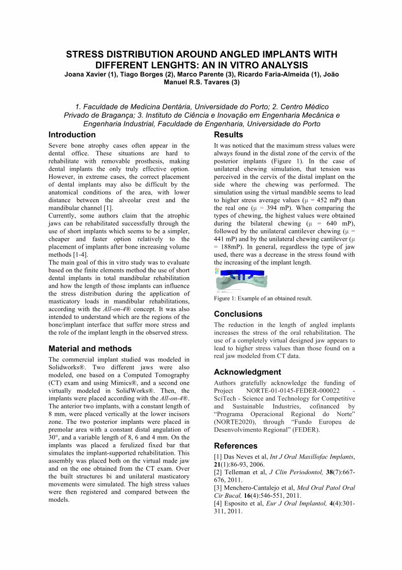
Results It was noticed that the maximum stress values were always found in the distal zone of the cervix of the posterior implants (Figure 1). In the case of unilateral chewing simulation, that tension was perceived in the cervix of the distal implant on the side where the chewing was performed. The simulation using the virtual mandible seems to lead to higher stress average values (µ = 452 mP) than the real one (µ = 394 mP). When comparing the types of chewing, the highest values were obtained during the bilateral chewing (µ = 640 mP), followed by the unilateral cantilever chewing (µ = 441 mP) and by the unilateral chewing cantilever (µ = 188mP). In general, regardless the type of jaw used, there was a decrease in the stress found with the increasing of the implant length.
Figure 1: Example of an obtained result. Conclusions The reduction in the length of angled implants increases the stress of the oral rehabilitation. The use of a completely virtual designed jaw appears to lead to higher stress values than those found on a real jaw modeled from CT data. Acknowledgment Authors gratefully acknowledge the funding of Project NORTE-01-0145-FEDER-000022 - SciTech - Science and Technology for Competitive and Sustainable Industries, cofinanced by “Programa Operacional Regional do Norte” (NORTE2020), through “Fundo Europeu de Desenvolvimento Regional” (FEDER). References [1] Das Neves et al, Int J Oral Maxillofac Implants, 21(1):86-93, 2006. [2] Telleman et al, J Clin Periodontol, 38(7):667-676, 2011. [3] Menchero-Cantalejo et al, Med Oral Patol Oral Cir Bucal, 16(4):546-551, 2011. [4] Esposito et al, Eur J Oral Implantol, 4(4):301-311, 2011.
(Avg: 75%)S, Mises
+2.272e−04+2.220e+01+4.441e+01+6.661e+01+8.882e+01+1.110e+02+1.332e+02+1.554e+02+1.776e+02+1.998e+02+2.220e+02+2.442e+02+2.665e+02
Step: Step−1, UntitledIncrement 1: Step Time = 1.000Primary Var: S, MisesDeformed Var: U Deformation Scale Factor: +1.000e+00
ODB: 8x6unipost26ab.odb Abaqus/Standard 6.12−3 Tue Apr 26 12:27:17 GMT Daylight Time 2016
X
Y
Z