Embed Size (px)
Citation preview
UNIVERSIDADE FEDERAL DO RIO GRANDE
PROGRAMA DE PÓS-GRADUAÇÃO EM AQUICULTURA
MARIA ANGÉLICA DOS REIS GARRIDO PEREIRA
USO DO PROBIÓTICO Bacillus spp. NA LARVICULTURA DO BIJUPIRÁ
Rachycentron canadum EM SISTEMA DE RECIRCULAÇÃO DE ÁGUA
RIO GRANDE, RS
2011

ii
UNIVERSIDADE FEDERAL DO RIO GRANDE
PROGRAMA DE PÓS-GRADUAÇÃO EM AQUICULTURA
USO DO PROBIÓTICO Bacillus spp. NA LARVICULTURA DO BIJUPIRÁ
Rachycentron canadum EM SISTEMA DE RECIRCULAÇÃO DE ÁGUA
MARIA ANGÉLICA DOS REIS GARRIDO PEREIRA
Dissertação apresentada como parte dos
requisitos para obtenção do grau de mestre
em Aqüicultura no Programa de Pós-
Graduação em Aqüicultura da Universidade
Federal do Rio Grande.
Orientador: Prof. Dr. Luís André Sampaio
Co-orientador: PhD. Michael Schwarz
RIO GRANDE, RS, BRASIL
JANEIRO, 2011

iii
AGRADECIMENTOS
Ao Prof. Dr. Luís André Sampaio pela orientação, pela oportunidade de
trabalhar na Virginia Tech, pela ajuda e ensinamentos.
Ao PhD. Michael Schwarz, querido Mike, por todo suporte técnico,
científico, e pessoal na estadia em Hampton, EUA.
Ao Prof. Dr. Luis Alberto Romano pela amizade e ajuda acadêmica. Ainda
devo mais de quinze mil bacalhaus!
À equipe VSAREC, principalmente Prof. Michael Jahncke, Brendan C.
Delbos e Helen Suton, mas também Abgail Villalba, Dan Kauffman e Bob Lane.
Ao MSc. Ricardo Rodrigues pela ajuda gigantesca no pré-projeto, projeto,
experimento, amostragem, análises estatísticas, chateações, enfim, por ter sido um
grande colega e amigo (meu „co-co-orientador‟).
Aos colegas e amigos da FURG que diretamente ou indiretamente me
ajudaram nesta caminhada acadêmica e de farras: André, Andréa, Alessandro Del Duca,
Alinca, Ana Velloso, Augusto, Beck, Cami Dal, Carlinhos, Cassiê, Cínthia, Cláudio,
Dariano, Diego, Diogão, Eduardo Martins, Faby, Fê Rosa, Gabi David Canabarro,
Gabriel, Getúlio, Iuri, Jana, Kassio, Kotoko, Leo, Luciano Jensen, Manuel, Martita,
Midori, Miguel, Okamoto, Prof. Klebinho, Prof. Luciano, Prof. Mano, Prof. Mineiro,
Prof. Paulo Abreu, Prof. Tesser, Renatão, Sabrina, Shakirita, Shay, Shei, Tati Fito,
Verônica, Vikaláctica,Vivi. Em ordem alfabética. Perdoem-me se esqueci de citar
alguém.
Aos professores do Programa de Pós-Graduação em Aquicultura.
Aos funcionários da Estação Marinha de Aquacultura.
Às minhas queridas amigas do triângulo, Fefê e Dani, e Ju pelo apoio.
Distante fisicamente, mas sempre presente no coração.
Ao William, por todos os momentos compartilhados, pelo companheirismo,
calma e paciência durante todo esse tempo.
Aos meus pais, Sônia e Gilberto, por todo apoio que tenho recebido, por
todo amor. Essa vitória é principalmente para vocês.
Aos meus irmãos, Malonta, Ti e Céci, muito obrigada por tudo.
À minha família, pela união inabalável: tias, tios, primas, primos, avós.
Principalmente, obrigado Ao Pai Celestial. Sem ele nada seria possível.

iv
ÍNDICE
Resumo v
Abstract vi
Introdução geral 1
Artigo anexo 11
“Probiotic effects of Bacillus spp. on cobia Rachycentron canadum larvae reared
in a recirculating aquaculture system”
Abstract 13
1. Introduction 14
2. Material and methods 17
3. Results 21
4. Discussion 22
5. Conclusion 27
Acknowledgements 27
References 27
Tables 35
Figures 36

v
RESUMO
O bijupirá (Rachycentron canadum) é um peixe marinho que apresenta bom potencial
para seu cultivo. Muitas pesquisas demonstram os efeitos benéficos do probiótico
Bacillus spp. na aquicultura, porém não há estudos sobre os efeitos de probióticos em
sistemas de recirculação de água (SRA). Este trabalho analisa os efeitos do probiótico
Bacillus spp. na performance de larvas de bijupirá criadas em SRA. O experimento foi
realizado na Universidade Virginia Tech (EUA). As larvas foram estocadas em dois
SRA independentes entre si - tratamento probiótico (TP) e tratamento controle (TC) - na
densidade de 15 larvas/L, durante 26 dias após a eclosão. Um dos sistemas (TP) recebeu
a adição de probiótico comercial composto por B. subtilis, B. licheniformis e B. pumilus
diretamente na água e via alimento vivo, conforme o fabricante. As análises de
microbiologia e qualidade de água foram feitas diariamente por medição de parâmetros
físico e químicos e plaqueamento em meio de cultura para contagem de Bacillus totais e
Vibrio totais. Ao final do experimento foram realizados biometria e contagem total dos
animais, teste de resistência ao estresse salino e fixação de larvas para
imunohistoquímica do timo para avaliar a expressão dos complexos multirreceptores de
antígenos CD3 e CD4 de linfócitos T e morfometria do rim cefálico. A sobrevivência
foi similar e de 14,9±2,4% e 15,1±0,9% para TP e TC respectivamente. O peso final foi
de 170±10 mg para TP e 160±10 mg TC, não havendo diferença estatística entre os
tratamentos. Na microbiologia da água não foram encontradas diferenças significativas
entre os tratamentos na contagem de víbrios totais (TP: 3.366±522 UFC/mL; TC:
4.964±902 UFC/mL). Na contagem de Bacillus totais os tratamentos diferiram
estatisticamente, sendo maior a concentração no TP pela adição dos Bacillus spp.
(901±88 UFC/mL e 61±24 UFC/mL). O teste de estresse por salinidade demonstrou
uma maior resistência ao estresse salino pelas larvas do tratamento probiótico. A análise
imunohistoquímica mostrou uma maior expressão de CD4 no timo em PT (TP: 43,7 ±
6,16% CD4 células/mm²; TC: 25,4 ± 3,5% CD4 células/mm²), o que pode indicar uma
melhor resposta imune contra patógenos. A análise morfométrica do rim cefálico indica
que este órgão é maior no TP (TP: 0,97 ± 0,33 milímetros; TC: 0,69 ± 0,22 milímetros),
provavelmente devido a um aumento na quantidade de linfócitos B. Estes resultados
indicam que o probiótico utilizado em um sistema de recirculação de água é efetivo
quanto à imunoestimulação e aumenta a resistência ao estresse salino.
Palavras-chave: microbiologia, teste de estresse, imunohistoquímica, piscicultura
marinha
vi
ABSTRACT
Cobia (Rachycentron canadum) is a marine finfish with good potential for mariculture.
The aim of this study was to analyze the effects of probiotic Bacillus spp. on the
performance of cobia larvae reared in a recirculating aquaculture system (RAS). Many
studies have demonstrated the beneficial effects of probiotic Bacillus spp. in
aquaculture, but there are no studies on the effects of probiotics in RAS, requiring
research on their effectiveness, since in open systems the beneficial effects are already
known. The trial was conducted at Virginia Tech VSAREC (USA). Larvae were
stocked into two independent RAS - probiotic treatment and control - at a density of 15
larvae L-1
, for 26 days after hatching. One of the systems (Probiotic treatment) received
the addition of a commercial probiotic consisting of Bacillus subtilis, Bacillus
licheniformis and Bacillus pumilus directly into the water and by live feed, according to
the manufacturer. At the end of the experiment we performed biometrics, larvae counts,
stress test and larvae were fixed for morphometric analysis of head kidney and for
immunohistochemistry of thymus to evaluate the phenotypic expression of complex
antigens CD3 and CD4 in T-lymphocytes. Survival and final weight had no significant
difference between groups. The analysis of water microbiology had no significant
differences between treatments in total counts of Vibrio, but in the total count of
Bacillus treatments differed statistically, with a higher concentration in Probiotic
treatment because of the addition of Bacillus spp. in this group. The salinity stress test
was expressed by stress sensitivity index (SSI), which demonstrated a greater resistance
to salt stress by larvae of probiotic treatment. Immunohistochemical analysis showed a
higher expression of CD4 in Probiotic treatment, which may indicate better immune
response against pathogens. The morphometric analysis of head kidney indicates that
this organ is larger in Probiotic treatment, probably due to an increase in the quantity of
vii
B-lymphocytes. These results indicate that Bacillus spp. probiotics used in RAS may
stimulate the immune system and increase the general stress resistance of cobia larvae.
Keywords: stress test, immunohistochemistry, marine fish farming

1
INTRODUÇÃO GERAL
O bijupirá Rachycentron canadum, internacionalmente conhecido por cobia, é a
única espécie da família Rachycentridae. É um peixe migratório e pelágico que ocorre
em todos os mares tropicais e subtropicais, com exceção da parte central e oriental do
Oceano Pacífico (Shaffer & Nakamura 1989). Os indivíduos desta espécie habitam
águas costeiras e plataformas continentais, podendo ser encontrados em baías e
estuários, assim como em profundidades de até 1.200 m (Shaffer & Nakamura 1989).
Espécimes já foram coletados em águas de salinidade entre 22 e 44 (Kaiser & Holt
2005), e têm sido cultivados com sucesso em sistemas com salinidade entre 5 e 30
(Resley et al. 2006).
Este peixe geralmente é encontrado sozinho ou em pequenos cardumes, e é
comum estarem perto de alguma estrutura flutuante ou mesmo na coluna d‟água (Kaiser
& Holt 2005). São carnívoros oportunistas que se alimentam de várias espécies de
peixes, caranguejos, camarões e lulas. O corpo do bijupirá é alongado e pode chegar a 2
metros de comprimento e 60 kg, sendo que as fêmeas crescem mais rápido que os
machos (FAO 2007). São amarronzados dorsalmente, possuem a superfície ventral
branca e apresentam listras que variam de claro a escuro nas laterais do corpo. A
longevidade é de aproximadamente 15 anos (Shaffer & Nakamura 1989).
Por não ser um peixe abundante no meio natural, o bijupirá não é um pescado
muito presente no comércio. Segundo a FAO (2010) a produção mundial do cultivo da
espécie no ano de 2009 foi de 24.860 t. É considerado um excelente alimento, mas
devido à baixa oferta do produto muitos consumidores desconhecem este peixe (Kaiser
& Holt 2005).
O bijupirá é uma das espécies de peixes marinhos com bom potencial para a
aquicultura. Entre seus atributos estão a facilidade de desova (Liao et al. 2004), taxas de
crescimento de até 6 kg/ano (Fraser & Davies 2009), altas taxas de sobrevivência em
tanques e gaiolas (estágio pós-larvicultura), adaptabilidade ao cativeiro (Arnold et al.
2002) e à ração (McLean et al. 2009). A espécie também demonstra alta resistência a
doenças (Holt et al. 2007), boa resposta à vacinação (Lin et al. 2006), aos probióticos
(Benetti et al. 2008) e aos imunoestimulantes (Leaño et al. 2003), e tolera ampla faixa
de salinidade (Resley et al. 2006). Além disso, possui alta qualidade como pescado e
demanda de mercado crescente (Faulk & Holt 2005).
2
Devido a essas características favoráveis à criação, a produção de bijupirá tem
crescido desde o início dos anos 1990 (Liao et al. 2004). A espécie tem sido cultivada
com sucesso na Ásia, sendo que aproximadamente 80% da sua produção ocorrem em
tanques-rede em alto-mar em Taiwan (Liao et al. 2004). Vietnã, China, Indonésia,
Filipinas, Irã e La Reunion já produzem bijupirá em tanques-rede e recentemente os
EUA, Brasil, Belize, República Dominicana, Martinica e México têm produzido e
desenvolvido pesquisas (Benetti et al. 2010). Em Taiwan, os peixes são frequentemente
criados em viveiros desde a eclosão até 30 g, quando são transferidos para viveiros
maiores ou tanques cobertos para crescimento. Após este período os peixes são criados
em tanques offshore até o final da fase de engorda, chegando ao peso de mercado de 6-8
Kg (para exportação) ou 8-10 Kg (para consumo interno em Taiwan) (Liao et al. 2004).
O uso de sistemas de recirculação de água (SRA) durante a larvicultura e
primeiros estágios de juvenil propicia aos produtores um controle ambiental maior,
permitindo a produção de alevinos durante todo ano e expandindo a produção para
regiões mais frias (Faulk et al. 2007). Segundo D‟Orbcastel et al. (2009), um SRA na
aquicultura é uma combinação de processos como remoção de sólidos (filtração
mecânica, decantação), controle de gases (oxigênio, gás carbônico) e processos
biológicos (nitrificação da amônia por biofiltro, desinfecção por UV).
Por propiciar um cultivo com maior biossegurança e manter a qualidade da água
em níveis adequados, o SRA pode proporcionar maiores taxas de sobrevivência (Ridha
& Cruz 2001). O aumento da produtividade, juntamente com o tratamento contínuo e
reuso da água, faz com que o SRA seja um sistema eficiente tanto para o produtor
quanto para o meio-ambiente (D‟Orbcastel et al. 2009).
O cultivo do bijupirá é relativamente recente, quando comparado a outras
culturas já estabelecidas de peixes marinhos, como salmão do Atlântico (Salmo salar),
robalo europeu (Dicentrarchus labrax), pargo (Pagrus auratus), linguados
(Pleuronectiformes) e outros. Por isso ainda há limitações significantes na sua produção,
como a produção viável de ovos e juvenis em quantidade, a ausência de dietas
específicas e o mercado ainda em desenvolvimento (Holt et al. 2007).
Ainda não está definido o pacote tecnológico do cultivo do bijupirá, porém
muitos estudos têm sido realizados no Brasil e no exterior, principalmente nas áreas de
nutrição, reprodução e fisiologia, como digestibilidade de nutrientes (Zhou et al. 2004),
alimentação larval (Faulk & Holt 2005), exigências nutricionais (Chou et al. 2001,
Fraser & Davies 2009), reprodução em cativeiro (Weirich et al. 2006, Faulk & Holt
3
2008), efeitos da salinidade (Denson et al. 2003), nitrogenados (Feeley et al. 2007,
Rodrigues et al. 2007) e fisiologia do estresse (Trushenski et al. 2010).
As pesquisas na área de imunologia são limitadas nesta espécie, sendo o único
trabalho publicado nesta área sobre eficácia de vacinas em bijupirás (Lin et al. 2006).
Pesquisas acerca do desenvolvimento e funcionamento do sistema imune de
peixes teleósteos tem tido cada vez mais importância (Press, 1999), uma vez que o uso
de quimioterápicos nas criações está sendo condenado (Gatesoupe 1999, Vine et al.
2006, Kesacordi-Watson et al. 2008, Wang et al. 2008). A intensificação da aqüicultura
no mundo, em resposta à crescente demanda por pescado, levou a um aumento do uso
inadequado de antibióticos e produtos químicos nas criações, resultando em problemas
na segurança alimentar e resistência bacteriana (Decamp & Moriarty 2007).
Muitas estratégias alternativas ao uso profilático de antibióticos no controle de
doenças foram propostas: instalação de manejos de biossegurança, vacinações efetivas,
estimulação imunológica não específica, assim como o uso de probióticos (Ringø et al.
2010). Gatesoupe (1999) define probiótico como microorganismos vivos, que ao serem
oferecidos como suplemento alimentar, melhoram a saúde geral do hospedeiro.
O uso de probióticos na produção de animais domésticos é conhecidamente
vantajoso e estes produtos têm grande potencial para a aquicultura marinha (Hansen &
Olafsen 1999). Em sistemas aquáticos os probióticos podem atuar de várias formas:
excluindo bactérias patogênicas por competição, melhorando a qualidade da água e a
condição nutricional e estimulando o sistema imune do organismo cultivado (Chen &
Chen 2001, Decamp & Moriarty 2006, Gatesoupe 1999, Kesarcodi-Watson et al. 2008,
Picchietti et al. 2009a, Velmurugan & Rajagopal 2009).
Os probióticos comumente usados em aquicultura abrangem vários taxa, como
bactérias lácticas, bacilares, nitrificantes e também leveduras (Decamp & Moriarty
2007). Em caso de bactérias, preferencialmente os probióticos são compostos por
organismos que formam esporos e, portanto, capazes de permanecer viáveis por muito
tempo no ambiente (Kesarcodi-Watson et al. 2008). Dentre as bactérias formadoras de
esporos utilizadas como probiótico na aquicultura destaca-se o gênero Bacillus.
Espécies de Bacillus formadoras de esporos estão presentes nos sedimentos
marinhos e são naturalmente ingeridas por animais aquáticos, sendo presentes em sua
microflora intestinal. Os Bacillus são usados como probióticos por sua ação inibitória
contra Vibrio e por sua ação no manejo de qualidade de água, degradando a matéria
orgânica (Gatesoupe 1999, Chen & Chen 2001 e Decamp & Moriarty 2006). Eles

4
também possuem habilidades na competição por locais de adesão, produzem
bacteriocinas (peptídeos antimicrobianos) e promovem imunoestimulação sistêmica
(Kesarcodi-Watson et al. 2008).
A adição de cepas selecionadas de Bacillus mostrou potencial de aprimorar a
produção, diminuindo a abundância de Vibrio na água e melhorando a taxa de
crescimento de Paralichthys olivaceus (Decamp et al. 2006 e Decamp et al. 2007). O
uso de Lactobacillus spp como probiótico na larvicultura de Sparus aurata,
administrado através do alimento vivo, promoveu aumento na sobrevivência e no
crescimento comparado aos grupos controle (Suzer et al. 2008).
A administração de probióticos durante a larvicultura propicia benefícios, uma
vez que as formas larvais da maioria dos peixes são liberadas no ambiente externo
começando a alimentação antes mesmo do trato digestório e o sistema imunológico
estarem completamente desenvolvidos (Picchietti et al. 2007).
Além disso, larvas de peixes marinhos normalmente não se alimentam de dietas
artificiais durante as fases iniciais de desenvolvimento, sendo necessário o uso de
alimento vivo. A dieta, seja ela composta por alimento inerte ou vivo, pode auxiliar no
transporte de microorganismos benéficos. Isso permite que a comunidade bacteriana do
hospedeiro seja modificada desde o início da larvicultura, através da adição de
probióticos (Picchietti al. 2009a)
A modulação do sistema imune é um dos benefícios mais importantes dos
probióticos. Sua potência abrange tanto a imunidade local (linfócitos associados à
mucosa intestinal) quanto a sistêmica (Nayak 2010). A estimulação da imunidade inata
de peixes gera um aumento da resposta imunológica rápida, ou seja, da imunidade
humoral. Este tipo de resposta, mediada por componentes inatos, pode ser benéfico para
os peixes sob as condições de cultivo, em particular na maricultura. Grandes perdas
podem ser observadas durante a larvicultura marinha, sendo essa mortalidade muitas
vezes atribuída à infecções por bactérias oportunistas presentes no sistema de cultivo
(Magnadóttir 2006).
A resposta imune é mediada principalmente pelos órgãos linfóides, que nos
peixes teleósteos incluem o rim cefálico, timo, baço e tecidos linfóides associados à
mucosa (Zapata et al. 2006). Nos peixes, o rim cefálico atua equivalentemente à medula
óssea de mamíferos, desempenhando um papel importante na função imunológica,
sendo o local para a maturação de linfócitos B e antígenos de captura (Laing et al.
5
2006). Já o timo é o órgão linfóide predominante, atuando como o local para a
produção, diferenciação e maturação de linfócitos T (Patel et al. 2009).
O timo desempenha um papel fundamental no desenvolvimento do sistema
imune adaptativo (Picchietti et al. 2009b). O complexo CD3 é um co-receptor que serve
como identificador geral de células T em peixes (Øvergård et al. 2009 ). Uma das
principais subclasses de linfócitos T são as chamadas células T auxiliares (ou T-helper),
que expressam o co-receptor CD4 (Buonocore et al. 2008). Este tipo de linfócito atua
estimulando a expressão e a secreção de citocinas, produzindo respostas via anticorpo,
através dos linfócitos B, ou fazendo ativação macrofágica (Buonocore et al. 2008).
Evidências demonstram que a administração suplementar de probióticos
estimula a imunidade de larvas e diminui a transcrição de genes pró-inflamatórios, além
de causar um aumento da quantidade de células-T (Picchietti et al. 2009a), eritrócitos e
leucócitos em geral (Irianto & Austin 2002) Em teoria, o aumento ou diminuição dos
co-receptores de linfócitos T, CD3 e CD4, pode estar associado à velocidade de
desenvolvimento do tecido linfóide e/ou à reatividade dos linfócitos em questão (Miceli
& Parnes 1993).
Como a larvicultura de peixes marinhos é um grande gargalo na produção, pode-
se utilizar sistemas total ou parcialmente fechados a fim de reduzir os riscos de
contaminação e influências ambientais, que são dois grandes problemas na aquicultura
(Lyndon, 1999).
Em sistemas abertos, como viveiros e tanques, probióticos são acrescentados a
cada renovação de água e seus efeitos são comprovadamente benéficos. Em sistemas
fechados de recirculação, contudo, não há muitos estudos sobre os efeitos da adição de
probióticos, principalmente porque a ação dos aparatos de recirculação sobre a
abundância de microorganismos na água de cultivo é pouco conhecido. Chen & Chen
(2001) e Taoka et al. (2006) relataram o uso de probióticos em SRA, mas nestes
trabalhos não há menção dos componentes do sistema.
Além da ausência de informação acerca do uso de probióticos em sistemas de
recirculação, não existem estudos publicados sobre o uso de probióticos no cultivo do
bijupirá. O objetivo deste estudo foi analisar os efeitos do probiótico Bacillus spp. na
larvicultura do bijupirá em sistema de recirculação de água.
6
REFERÊNCIAS BIBLIOGRÁFICAS
ARNOLD, CR, JB KAISER, GJ HOLT. 2002. Spawning of Cobia Rachycentron
canadum in Captivity. J. World Aquacult. Soc., 33:205-208.
BENETTI DD, R ORHUNM, B SARDENBERG, B O‟HANLON, A WELCH, R
HOENIG, I ZINK, JÁ RIVERA, B DENLINGER, D BACOAT, K PALMER, F
CAVALIN. 2008. Advances in hatchery and grow-out technology of cobia
Rachycentron canadum (Linnaeus). Aquacult. Res., 39:701-711.
BENETTI, DD, B O‟HANLON, JA RIVERA, AW WELCH, C MAXEY, MR
ORHUN. 2010. Growth rates of cobia (Rachycentron canadum) cultured in open
ocean submerged cages in the Caribbean. Aquaculture, 302:195-201.
BUONOCORE, F, E RANDELLI, D CASANI, L GUERRA, S PICCHIETTI, S
COSTANTINI, AM FACCHIANO, J ZOU, CJ SECOMBES, G SCAPIGLIATI.
2008. A CD4 homologue in sea bass (Dicentrarchus labrax): Molecular
characterization and structural analysis. Mol. Immunol., 45:3168–3177.
CHEN, CC, CHEN SN. 2001. Water Quality Management with Bacillus spp. in the
High-Density Culture of Red-Parrot Fish: Cichlasoma citrinellum X C.
Synspilum. N. Am. J. Aquacult., 63:66–73.
CHOU, R, MS SU, HY CHEN. 2001. Optimal dietary protein and lipid levels for
juvenile cobia (Rachycentron canadum). Aquaculture, 193, 81-89.
D‟ORBCASTEL, ER, JP BLANCHETON, A BELAUD. 2009. Water quality and
rainbow trout performance in a Danish Model Farm recirculating system:
Comparison with a flow through system. Aquacult. Eng., 40:135-143.
DECAMP, O, MORIARTY, D J W. 2006. Probiotics as alternative to antimicrobials:
limitations and potential. J. World Aquacult. Soc., 37:60-62.
DECAMP, O, MORIARTY, D J W. 2007. Aquaculture species profit from probiotics.
Feed Mix vol. 15, nº 1.
DENSON, M R, KR STUART, TIJ SMITH, CR WEIRLCH, A SEGARS. 2003. Effects
of Salinity on Growth, Survival, and Selected Hematological Parameters of
Juvenile Cobia Rachycentron canadum. J. World Aquacult. Soc., 34:496-504.
FAO. 2007. Cultured Aquatic Species Information Programme. Rachycentron canadum.
Cultured Aquatic Species Information Programme. Text by J.B. Kaiser, J.G.
Holt. In: FAO Fisheries and Aquaculture Department [online]. Rome. Updated
7
23 May 2007. [Cited 14 December 2010].
http://www.fao.org/fishery/culturedspecies/Rachycentron_canadum/en
FAO. 2010. Global Aquaculture Production (online query). Disponível em:
<http://www.fao.org/fishery/statistics/global-aquaculture-production/en>.
Acesso em: 20/10/2010.
FAULK, C, HOLT, G. 2008. Biochemical composition and quality of captive-spawned
cobia Rachycentron canadum eggs. Aquaculture, 279, 70-76.
FAULK, CK, HOLT, GJ. 2005. Advances in rearing cobia Rachycentron canadum
larvae in recirculating aquaculture systems: Live prey enrichment and
greenwater culture. Aquaculture, 249:231-243.
FAULK, CK, JB KAISER, GJ HOLT. 2007. Growth and survival of larval and juvenile
cobia Rachycentron canadum in a recirculating raceway system. Aquaculture,
270:149-157.
FEELEY, MW, DD BENETTI, JS AULT. 2007. Elevated oxygen uptake and high rates
of nitrogen excretion in early life stages of the cobia Rachycentron canadum
(L.), a fast-growing subtropical fish. J. Fish Biol., 71:1662-1678.
FRASER, TWK, DAVIES, SJ. 2009. Nutritional requirements of cobia, Rachycentron
canadum (Linnaeus): a review. Aquacult. Res., 40:1219-1234.
GATESOUPE, FJ. 1999. The use of probiotics in aquaculture. Aquaculture, 180:147–
165
HANSEN, GH, OLAFSEN, JA. 1999. Bacterial Interactions in Early Life Stages of
Marine Cold Water Fish. Microb. Ecol., 38:1–26.
HOLT, GJ, CK FAULK, MH SCHWARZ. 2007. A review of the larviculture of cobia
Rachycentron canadum, a warm water marine fish. Aquaculture, 268:181-187.
IRIANTO A & AUSTIN B. 2002 Probiotics in aquaculture. J. Fish Dis., 25: 633–642.
KAISER, JB, HOLT, GJ. 2005. Species Profile – Cobia. Southern Regional
Aquaculture Center Publication, 7202.
KESARCODI-WATSON, A, H KASPAR, MJ LATEGAN, L GIBSON. 2008.
Probiotics in aquaculture: The need, principles and mechanisms of action and
screening processes. Aquaculture, 274:1-14.
LAING, K J, J J ZOU, M K PURCELL, R PHILLIPS, C J SECOMBES, J D HANSEN.
2006. Evolution of the CD4 family: Teleost fish possess two divergent forms of
CD4 in Addition to Lymphocyte Activation Gene-3. J. Immunol., 177: 3939-
3951.
8
LEAÑO, E M, J J GUO, S L CHANG, I C LIAO. 2003. Levamisole enhances non-
specific immune response of cobia, Rachycentron canadum, fingerlings. J. Fish.
Soc. Taiwan, 30:321-330.
LIAO, IC, HM SU, EY CHANG. 2001. Techniques in finfish larviculture in Taiwan.
Aquaculture, 200:1-31.
LIAO, IC, TS HUANG, WS TSAI, CM HSUEH, SL CHANG, EM LEAÑO. 2004.
Cobia culture in Taiwan: current status and problems. Aquaculture, 237:155-
165.
LIN, JHY, TH CHEN, MS CHEN, HE CHEN, RL CHOU, TI CHEN, MS SU, HL
YANG. 2006. Vaccination with three inactivated pathogens of cobia
(Rachycentron canadum) stimulates protective immunity. Aquaculture,
255:125–132.
LYNDON, A.R. 1999. Fish Growth in Marine Culture Systems: A Challenge for
Biotechnology. Mar. Biotechnol., 1:376–379.
MAGNADÓTTIR, B. 2006. Innate immunity of fish (overview). Fish shellfish
immunol., 20:137-151.
MCLEAN, E, G SALZE, MH SCHWARZ & SR CRAIG. 2009. Cobia cultivation. In:
BURNELL, G & G ALLAN (eds.) New Technologies in aquaculture: Improving
production efficiency, quality and environmental management. CRC Press,
Estados Unidos, Chap. 25:804-821.
MICELI MC, PARNES JR. 1993. Role of CD4 and CD8 in T cell activation and
differentiation. Adv. Immunol.. 53:59–122.
NAYAK, SK. 2010. Probiotics and immunity: A fish perspective. Fish Shellfish
Immunol., doi: 10.1016/j.fsi.2010.02.017.
OLAFSEN, JA. 2001. Interactions between fish larvae and bacteria in marine
aquaculture. Aquaculture, 200:223-247.
ØVERGÅRD, AC, I HORDVIK, AH NERLAND, G EIKELAND, S PATEL. 2009.
Cloning and expression analysis of Atlantic halibut (Hippoglossus hippoglossus)
CD3 genes. Fish Shellfish Immunol., 27:707–713
PATEL, S, E SØRHUS, IU FIKSDAL, PG ESPEDAL, Ø BERGH, OM RØDSETH, C
MORTON, AH NERLAND. 2009. Ontogeny of lymphoid organs and
development of IgM-bearing cells in Atlantic halibut (Hippoglossus
hippoglossus L.). Fish Shellfish Immunol., 26:385–395.
9
PICCHIETTI, S, M MAZZINI, AR TADDEI, R RENNA, AM FAUSTO, V MULERO,
O CARNEVALI, A CRESCI, L ABELLI, 2007. Effects of administration of
probiotic strains on GALT of larval gilthead seabream: immunohistochemical
and ultrastructural studies. Fish Shellfish Immunol., 22:57-67.
PICCHIETTI, S, AM FAUSTO, E RANDELLI, O CARNEVALI, AR TADDEI, F
BUONOCORE, G SCAPGLIATI, L ABELLI. 2009a. Early treatment with
LactoBacillus delbrueckii strain induces an increase in intestinal T-cells and
granulocytes and modulates immune-related genes of larval Dicentrarchus
labrax (L.). Fish Shellfish Immunol., 26:368-376.
PICCHIETTI, S, L GUERRA, F BUONOCORE, E RANDELLI, AM FAUSTO, L
ABELLI. 2009b. Lymphocyte differentiation in sea bass thymus: CD4 and CD8-
a gene expression studies. Fish Shellfish Immunol., 27:50–56.
PRESS, C. 1999. The morphology of the immune system in teleost fishes. Fish Shellfish
Immunol., 9:309-318.
RESLEY M, K WEBBJR, G HOLT. 2006. Growth and survival of juvenile cobia,
Rachycentron canadum, at different salinities in a recirculating aquaculture
system. Aquaculture, 253:398–407.
RIDHA, MT, CRUZ, EM. 2001. Effect of biofilter media on water quality and
biological performance of the Nile tilapia Oreochromis niloticus L. reared in a
simple recirculating system. Aquacult. Eng., 24:157-166.
RINGØ, E, RE OLSEN, TØ GIFSTAD, RA DALMO, H AMLUND, GI HEMRE, AM
BAKKE. 2010. Prebiotics in aquaculture: a review. Aquacult. Nut., 16:117-136.
RODRIGUES, R, M SCHWARZ, B DELBOS, L SAMPAIO. 2007. Acute toxicity and
sublethal effects of ammonia and nitrite for juvenile cobia Rachycentron
canadum. Aquaculture, 271:553-557.
SHAFFER, RS, NAKAMURA, EL. 1989. Synopsis of Biological Data on the Cobia
Rachycentron canadum (Pisces: Rachycentridae). NOAA Technical Report
NMFS 82.
SUZER, C, D ÇOBAN, HO KAMACI, S SAKA, K FIRAT, O OTGUOGLU, H
KÜÇÜKSARI. 2008. Lactobacillus spp. bacteria as probiotics in gilthead sea
bream (Sparus aurata, L.) larvae: Effects on growth performance and digestive
enzyme activities. Aquaculture, 280:140-145.
TAOKA, Y, H MAEDA, J JO, M JEON, SC BAI, W LEE, K YUGE, S KOSHIO,
2006. Growth, stress tolerance and non-specific immune response of Japanese

10
flounder Paralichthys olivaceus to probiotics in a closed recirculating system.
Fisheries Sci., 72, 310-321.
TRUSHENSKI, J, M SCHWARZ, R TAKEUCHI, B DELBOS, LA SAMPAIO. 2010.
Physiological responses of cobia Rachycentron canadum following exposure to
low water and air exposure stress challenges. Aquaculture, 307:173-177.
VELMURUGAN, S, RAJAGOPAL, S. 2009. Beneficial uses of probiotics in mass
scale production of marine ornamental fish. Afr. J. Microbiol. Res., 3:185-190.
VERSCHUERE, L, G ROMBAUT, P SORGELOOS, W VERSTRAETE. 2000.
Probiotic Bacteria as Biological Control Agents in Aquaculture. Microbiol. Mol.
Biol. Rev., december, 655–671.
VINE, N G, WD LEUKES, H KAISER. 2006. Probiotics in marine larviculture. FEMS
Microbiol. Rev., 30:404-427.
WANG, Y, J LI, J LIN. 2008. Probiotics in aquaculture: Challenges and outlook.
Aquaculture, 281:1-4.
WEIRICH, CR, AD STOKES, TIJ SMITH, WE JENKINS, MR DENSON. 2006.
Outdoor Tank and Pond Spawning of Cobia in Coastal South Carolina.
Aquaculture, 18:1-17.
ZAPATA, A, B DIEZ, T CEJALVO, CG FRÍAZ, A CORTÉS. 2006. Ontogeny of the
immune system of fish. Fish Shellfish Immunol., 20:126-136.
ZHOU, QC, BP TAN, KS MAI, YJ LIU. (2004). Apparent digestibility of selected feed
ingredients for juvenile cobia Rachycentron canadum. Aquaculture, 241:441-
451.
11
ARTIGO ANEXO
Efeitos do probiótico Bacillus spp. na larvicultura do bijupirá
Rachycentron canadum em sistema de recirculação de água
Co-autores: Michael Schwarz, Brendan C. Delbos, Ricardo V. Rodrigues, Luis
Alberto Romano, Luís André Sampaio
Segundo as normas da revista Aquaculture (fator de impacto 1.925).
12
Probiotic effects of Bacillus spp. on cobia Rachycentron canadum larvae reared in a
recirculating aquaculture system
M. Angélica Garrido-Pereiraa, Michael H. Schwarz
b, Brendan C. Delbos
b, Ricardo V.
Rodriguesa, Luis A. Romano
c, Luís A. Sampaio
a,*
a Universidade Federal do Rio Grande — FURG, Instituto de Oceanografia,
Laboratório de Piscicultura Estuarina e Marinha, CP 474, Rio Grande, RS, 96201-900,
Brazil
b Virginia Tech — Virginia Seafood Agricultural Research and Extension Center, 102
S. King Street, Hampton, VA, 23669, USA
cUniversidade Federal do Rio Grande – FURG, Instituto de Oceanografia. Laboratório
de Patologia de Organismos Aquáticos, CP 474, Rio Grande, RS, 96201-900, Brazil
___________________________
*Corresponding author. Tel.: +55 53 32368131, Fax: + 55 53 32368042
E-mail address: [email protected] (LA Sampaio)

13
Abstract
Cobia (Rachycentron canadum) is a marine finfish with good potential for mariculture.
The aim of this study was to analyze the effects of probiotic Bacillus spp. on the
performance of cobia larvae reared in a recirculating aquaculture system (RAS). Many
studies have demonstrated the beneficial effects of probiotic Bacillus spp. in
aquaculture, but there are no studies on the effects of probiotics in RAS, requiring
research on their effectiveness, since in open systems the beneficial effects are already
known. The trial was conducted at Virginia Tech VSAREC (USA). Larvae were
stocked into two independent RAS - probiotic treatment and control - at a density of 15
larvae L-1
, for 26 days after hatching. One of the systems (Probiotic treatment) received
the addition of a commercial probiotic consisting of Bacillus subtilis, Bacillus
licheniformis and Bacillus pumilus directly into the water and by live feed, according to
the manufacturer. At the end of the experiment we performed biometrics, larvae counts,
stress test and larvae were fixed for morphometric analysis of head kidney and for
immunohistochemistry of thymus to evaluate the phenotypic expression of complex
antigens CD3 and CD4 in T-lymphocytes. Survival and final weight had no significant
difference between groups. The analysis of water microbiology had no significant
differences between treatments in total counts of Vibrio, but in the total count of
Bacillus treatments differed statistically, with a higher concentration in Probiotic
treatment because of the addition of Bacillus spp. in this group. The salinity stress test
was expressed by stress sensitivity index (SSI), which demonstrated a greater resistance
to salt stress by larvae of probiotic treatment. Immunohistochemical analysis showed a
higher expression of CD4 in Probiotic treatment, which may indicate better immune
response against pathogens. The morphometric analysis of head kidney indicates that
this organ is larger in Probiotic treatment, probably due to an increase in the quantity of
14
B-lymphocytes. These results indicate that Bacillus spp. probiotics used in RAS may
stimulate the immune system and increase the general stress resistance of cobia larvae.
Keywords: stress test, immunohistochemistry, marine fish farming
1. Introduction
Cobia (Rachycentron canadum), a marine finfish, is the only species in the
Rachycentridae family (Kaiser & Holt 2005). It has an emerging global potential for
mariculture, due to its extraordinary growth rate (Fraser & Davies 2009) and overall
aquaculture performance (Holt et al. 2007, Benetti et al. 2008,). Moreover, cobia have
demonstrated the capacity for induced and natural tank spawning and high fecundity
rates (Arnold et al. 2002, Liao et al. 2004), disease resistance (Holt et al. 2007), and
adaptability to commercially available aquafeeds (McLean et al. 2009).
Aquaculture environments can be stressful, triggering high mortality, especially
during larval rearing (Avella et al. 2010). Due to the interaction of microbes and their
effects on animals and their environment under typical intensive larviculture production
protocols, diseases caused by opportunistic pathogens like Vibrio anguillarum (Hansen
and Olafsen 1999), Photobacterium damselae (Lin et al. 2006), Streptococcus iniae
(Yanong and Francis-Floyd 2010) and many others (McLean et al. 2008), are usually
only treated from a clinical pathology perspective (Moriarty, 1998). Often, the diseases
are treated rather than the underlying causes, resulting in unnecessary drug use that may
have potentially negative environmental impacts (Gatesoupe 1999, Vine et al. 2006,
Wang et al. 2008, Kesacordi-Watson et al. 2008, Velmurugan and Rajagopal 2009).
Probiotics can be defined as live microbial feed supplements, which are
administered in such a way as to survive after entering the gastrointestinal tract. This
can be beneficial to the host animal by improving its intestinal microbial balance and in
15
turn its health (Gatesoupe 1999). In aquaculture, probiotics are inoculated into the
rearing water to improve culture conditions or incorporated into the feed (Taoka et al.
2006). The influence of the microbial community of the rearing water on the
gastrointestinal flora of the cultured animal is widely recognized (Gatesoupe 1999,
Verschuere et al. 2000).
Probiotic bacteria are commonly used in the animal industry to prevent the
occurrence of pathogenic microorganisms, thus minimizing the use of drugs to treat
diseases (Gatesoupe 1999, Vine et al. 2006, Kesacordi-Watson et al. 2008, Wang et al.
2008). They have been found to be effective in the control of pathogens in aquaculture
(Moriarty 1998, Silvi et al. 2008). The roles and effects of probiotics in aquaculture
have been examined over the past decade (Avella et al. 2010), in an ongoing search for
alternative preventive health strategies (Picchietti et al. 2007).
Several positive effects of probiotics in fish culture have been demonstrated,
such as higher survival (Velmurugan & Rajagopal 2009), faster growth (Venkat et al.
2004, Aly et al. 2008), improved stress tolerance (Rollo et al. 2006, Taoka et al. 2006),
immune system enhancement (Picchietti et al. 2007, Aly et al. 2008, Tinh et al. 2008,
Silvi et al. 2008, Picchietti et al. 2009a), and general welfare (Silvi et al. 2008).
The effects of probiotics on immune stimulatory functions include the
enhancement of non-specific immune responses (Taoka et al. 2006) and increase of T-
cells (Picchietti et al. 2009a). Some immunohistochemical markers can be used to study
leukocyte populations, like the T-cell co-receptors: CD3 and CD4. These receptors are
transmembrane glycoproteins, they belong to the Ig superfamily and are essential for
cell-mediated immunity (Laing et al. 2006). The CD4 co-receptor occurs in T-helper
cells, which play a central role in the immune system, stimulating the proliferation of B
16
lymphocytes and generating clones of memory cells for both cellular and humoral
immunity (Romano 2010).
Bacillus are gram-positive, spore-forming bacteria, used commercially as
probiotics (Chen & Chen 2001, Decamp & Moriarty 2006). Bacillus preparations are
resistant and have a long lasting shelf life and so they can be stored long-term in a
dehydrated form. Moreover, the beneficial roles of Bacillus spores applications in the
aquaculture field are well established (Gatesoupe, 1999, Avella et al. 2010).
Since marine fish larviculture is still a significant bottleneck toward industrial
expansion for many species, fully or partially closed, recirculating, onshore culture units
can be used to maximize biosecurity, reduce diseases and seasonal environmental
influences, important constraints on aquaculture in many areas (Lyndon, 1999). Marine
fish larvae can be reared in recirculating aquaculture systems (RAS), but little is known
on the effects of the equipment (i.e. filters and sterilizers) used to treat water in a RAS
on the probiotic bacteria. Chen & Chen (2001) and Taoka et al. (2006) reported the use
of probiotics in RAS, but they did not mention the system components.
Stress tests are useful tools to evaluate fry quality. They are based on the same
principle of exposing the organisms to a short but extreme stressful situation in which
the physiological condition of the animals determines their chances to survive (Dhert et
al. 1992b).
Thus, this study aims to examine the effects of a probiotic composed of three
Bacillus strains on survival, growth, resistance to a salinity stress challenge, and
imunostimulation in cobia larvae reared in a RAS.
17
2. Material and methods
2.1 Experimental design
The experiment was conducted at Virginia Tech's Virginia Seafood Agricultural
Research and Extension Center (VSAREC) (Hampton, VA, USA). Two independent
RAS were used in this experiment, each comprised of 3 tanks (300 L each), a
circulation pump (1/3 HP, AmpMaster 5600/4700, Pensacola, FL, USA), fluidized-bed
biofilter (Kaldness biological filtration media, R&B Aquatics, Waring, TX, USA),
bubble bead filter (BBF-XS4000, Aquaculture Systems Technologies, New Orleans,
LA, USA), 50 μm bag filter (X100 Convertible Filter Housing, FSI Filter Specialists,
Michigan City, IN, USA), UV sterilizer (025080, Emperor Aquatics, Pottstown, PA,
USA), immersion titanium heaters, protein skimmer (TF300, Top Fathom, Hudsonville,
MI, USA) for processing/removal of dissolved organics and small suspended solids, and
a diffusion aeration/degassing system. The two systems were maintained completely
independent of each other to ensure that no probiotics from the treatment system
contaminated the control system.
2.2 Larviculture
Fertilized cobia eggs were air transported from TroutLodge Marine Farms (Vero
Beach, FL, USA) to the VSAREC. Eggs were maintained in an incubator until hatching,
and then newly hatched larvae were stocked into the experimental tanks at a density of
15 larvae/L.
Larvae were produced according to standard larviculture production protocols
(McLean et al. 2009). L-type rotifers were enriched for 6 h with Protein Selco Plus
(INVE, Salt Lake City, UT, USA) and were fed from 2 through 9 dph, coinciding with
algal paste additions (Instant Algae Nanno 3600, Reed Mariculture, Campbell, CA,
USA) in a concentration of 106 cells/mL. Newly hatched AF Artemia (INVE, Salt Lake
18
City, UT, USA) were fed from 7 through 10 dph and enriched GSL Artemia (INVE,
Salt Lake City, UT, USA) were fed from10 through 23 dph. Artemia were enriched for
24 h with DC DHA Selco (INVE, Salt Lake City, UT, USA). Co-feeding of larvae with
Otohime Marine Larvae Weaning Diets (Reed Mariculture, Campbell, CA, USA) began
on 15 dph, weaning protocols began on 21 dph, and all fish were fully weaned and
Artemia discontinued by 23 dph.
Flow rate in the RAS was maintained at 60 L/hour from stocking through 9 dph.
On 10 dph flow increased to 90 L/hour; 12 dph flow was increased to 120 L/hour; 13
dph flow increased to 180 L/hour; 16 dph flow was increased to 240 L/hour and on 18
dph flow increased to 300 L/hour and remained there for the rest of the study.
At the end of the experiment (26 dph), 150 weanlings were randomly sampled
from each tank to determine final individual weight. All remaining larvae from the six
experimental tanks were counted to determine survival.
2.3 Probiotic strain
An experimental Bacillus mix provided by INVE Technologies (Belgium) was
used. It is composed of Bacillus subtilis, Bacillus pumilus and Bacillus licheniformis (1
x 1010
CFU/g). Each tank in Probiotic treatment received the probiotic daily dose of 5
g/m³ directly into the water; equally divided into four applications. Live feeds for this
treatment were also enriched with the probiotic, adding 0.5 g of commercial probiotic
per liter enrichment medium 4 hours before harvesting, according to the manufacturers
specifications.
2.4 Water Quality
Temperature, salinity, pH, dissolved oxygen, alkalinity, total ammonia–nitrogen
(TA-N), nitrite-nitrogen (NO2-N), and nitrate (NO3-N) of each experimental tank were
measured daily before the first feeding. Temperature and salinity were measured with
19
an YSI Model 30 (Yellow Springs Instruments, Yellow Springs, OH, USA). Dissolved
oxygen concentration was measured with an YSI Model 550A meter (Yellow Springs
Instruments, Yellow Springs, OH, USA) and pH was measured with an YSI Model
pH100 meter (Yellow Springs Instruments, Yellow Springs, OH, USA). Alkalinity was
determined using the Hach digital titration method 8203. TAN, NO2-N, and NO3-N
were determined via colorimetric assays, methods 10031, 8153, and 8039 using a D/R
2010 spectrophotometer (Hach, Loveland, CO, USA). The photoperiod was maintained
at 24 h light using fluorescent light banks.
2.5 Microbiology of Water
Water samples for microbial analysis were taken daily for abundance analysis of
total Vibrio and total Bacillus. Total Bacillus and Total Vibrio abundance were
determined by plating water samples from selected tanks. The culture medium TSA 2%
NaCl (Trypticase™ Soy Agar, Difco Laboratories, Detroit, MI, USA) was used for
total Bacillus abundance after sample pasteurization (Miskin et. al. 1998). For Total
Vibrio, the samples were plated on TCBS agar (thiosulfate citrate bile salt sucrose agar;
Difco Laboratories, Detroit, MI, USA).
The samples were plated in petri dishes mechanically through a WASP Spiral
Plater (Don Whitley Scientific Limited, Frederick, MD, USA). Abundance was
expressed in Colony-Forming Units per mL (CFU/mL) according to Banwart (1989).
2.6 Stress challenge
At the end of the experimental period, a stress challenge was carried out in
triplicate for each treatment, using 10 larvae per tank (n= 30 per treatment), according
to Dhert et al. (1992b). Fish from the Probiotic and Control treatment were transferred
with nets to 2L-beakers containing water at salinity 60, and survival was then monitored
every 5 minutes for 1 hour. The stress sensitivity index is the sum of the cumulative
20
mortalities in the consequent time intervals: the higher the numeric value of the index,
the more the larvae are stressed.
2.7 Histology and immunohistochemestry analysis
Larvae were sampled at 26 dph for histology and immunohistochemical analysis.
For morphometric analysis of head kidney, 30 larvae from each group were euthanized
with tricaine methanesulfonate (MS-222, Finquel®; Argent Chemical Laboratories,
Inc., Redmond, Washington, DC, USA). They were transferred to vials containing 10%
buffered formalin and stored until processing. Then larvae were dehydrated in a graded
series of ethanol, embedded with Paraplast®Plus (Paraplast ®Tissue Embedding Media,
Leica Microsystems Inc., Bannockburn, IL, USA). Serial longitudinal sections of 5 µm
were made until the whole head kidney was sectioned. Resulting sections were
subsequently stained with haematoxylin and eosin. Length and height of the head
kidney was measured using an optical microscope Olympus B201 with an ocular
micrometer (Carl Zeiss, Thornwood, NY, USA) (Romano et al. 1996).
An additional 30 larvae from each group were fixed in Bouin solution and
processed according to Prophet et al. (1992) for the immunohistochemistry evaluation
of the thymus. Immunohistochemistry was performed by ABC peroxidase (ABC,
Vectastain Elite, Vector) as described by Hsu et al. (1981). Serial sections of 5 µm
were made and they were incubated with a monoclonal anti CD3 and anti CD4 (Dako,
Argentina). Following rinses (0.1% diaminobenzidine solution), sections were
dehydrated, mounted and examined under bright-field illumination. The evaluation of
CD3 and CD4 receptors were calculated through quantitative analysis of the percentage
of phenotypic expression per square millimeter of tissue (Romano, 1996). The
phenotypic expression of CD3 and CD4 receptors were quantified using the software
Bioscan OPTIMAS ® 6.1 (Weibel, 1979).

21
2.8 Statistical analysis
All data were compared with Student‟s T test, using the software Statistica ®
7.0. The analyses were performed with significance level of P < 0.05. Percent data were
analyzed after they were transformed on arc-sine, but only original data are presented.
Results are shown as mean ± SD.
3. Results
There were no differences (P>0.05) in water quality between treatments:
measured values for TAN, NO2-N, NO3-N, temperature, alkalinity, pH, DO, and salinity
were: TAN: 0.13±0.04 mg/L; NO2-N: 0.05±0.02 mg/L; NO3-N: 2.96±0.76 mg/L;
temperature: 27.9±0.5 °C; alkalinity: 166±19 mg/L; pH: 7.95±0.1; DO: 6.38±0.18
mg/L; salinity was reduced from 35 to 22 from day 1 to 13 dph.
There were no significant differences (P>0.05) on survival and final weight
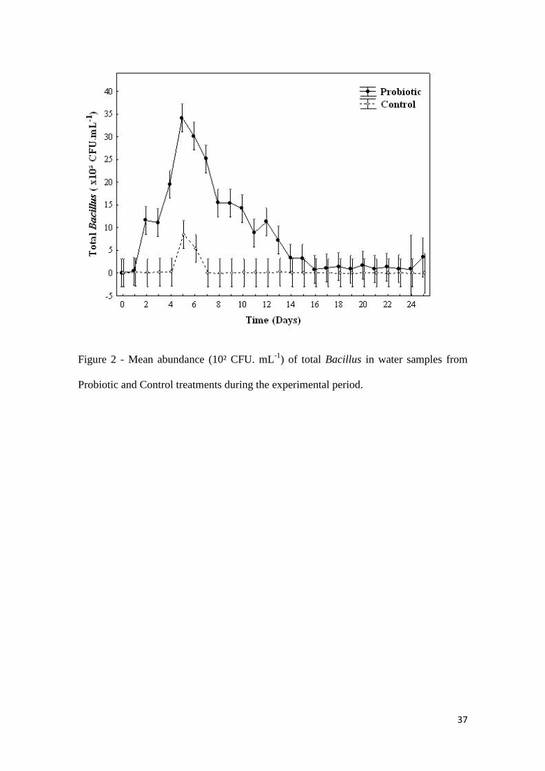
between treatments (Tab. 1). In the analysis of water microbiology Total Vibrio had no
statistical difference. Nevertheless, total count of Bacillus in the Probiotic treatment
differed statistically; with a higher concentration in the Probiotic treatment (P<0.05)
(Tab. 2). Variations in the abundance of microorganisms (CFU/mL) throughout the
experiment are shown in Fig. 1 and Fig. 2.
INSERT TABLE 1
INSERT TABLE 2
INSERT FIGURE 1
INSERT FIGURE 2
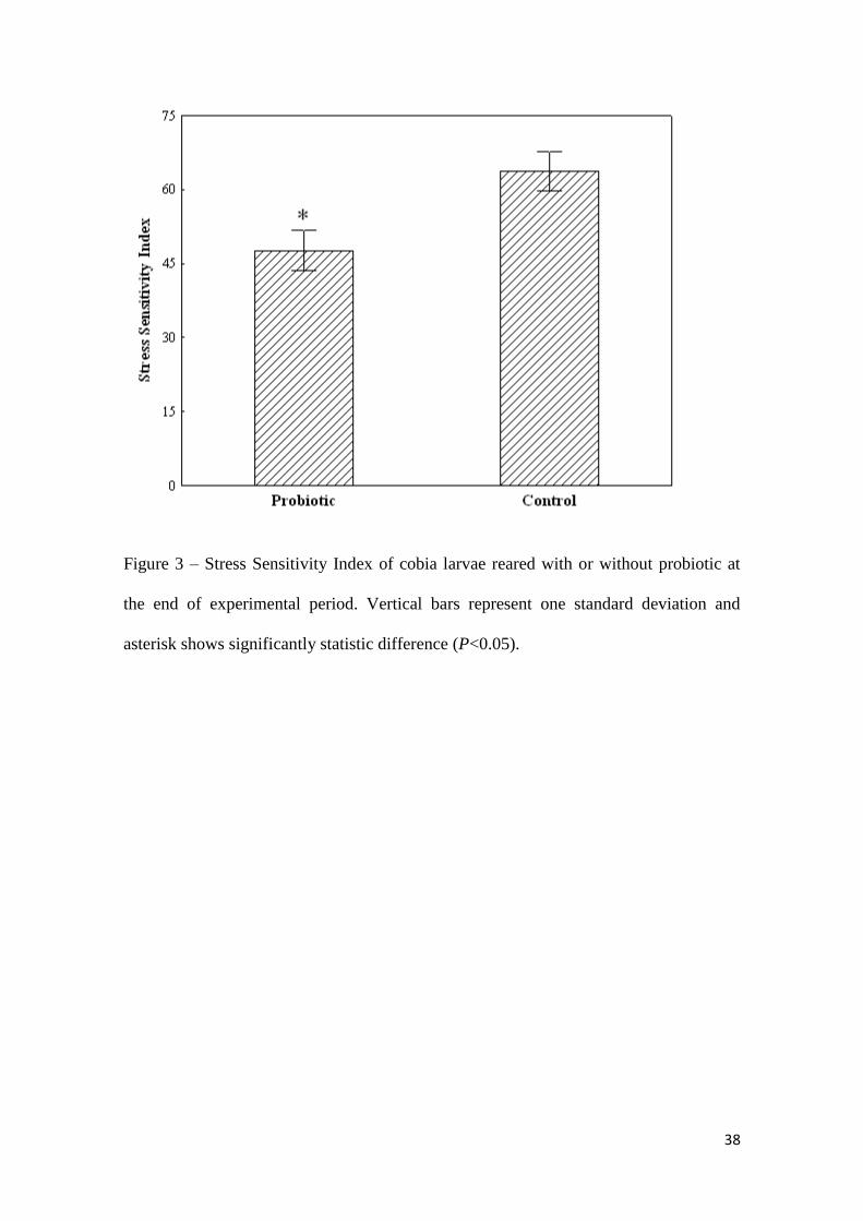
In the stress challenge, larvae of the Probiotic treatment had a better Stress
Sensitivity Index than the control (P<0.01) (Fig. 3).
INSERT FIGURE 3

22
Morphometric analysis showed that the larvae of the Probiotic treatment have
larger head kidneys than larvae of the control treatment (Probiotic: 0.97±0.33 mm;
Control: 0.69±0.22 mm) (P<0.01) (Fig. 4).
INSERT FIGURE 4
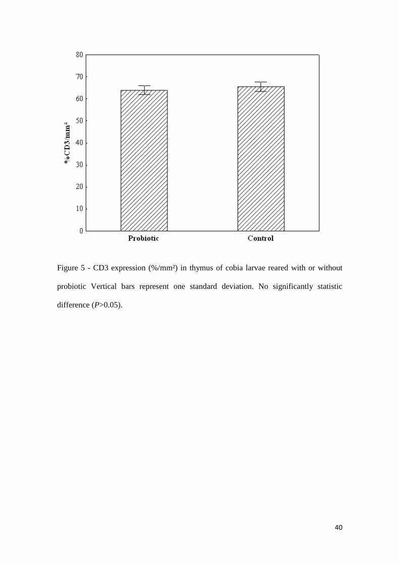
The expression of CD3 in thymic tissue showed no statistical differences
(P>0.05) between groups (Probiotic: 63.9±5.4 % CD3 cell/mm²; Control: 65.5±5.0 %
CD3 cell/mm²) (Fig. 5, Fig. 6a and Fig. 6b). However CD4 expression was significantly
higher (P<0.05) in the Probiotic treatment than in the control (Probiotic: 43.7±6.2 %
CD4 cell/mm²; Control: 25.4±3.5 % CD4 cell/mm²) (Fig. 7, Fig. 8a and Fig. 8b).
INSERT FIGURE 5
INSERT FIGURE 6a
INSERT FIGURE 6b
INSERT FIGURE 7
INSERT FIGURE 8a
INSERT FIGURE 8b
4. Discussion
The use of Bacillus species as a probiotic in aquaculture is common (Gatesoupe,
1999). For example, it has been shown that Bacillus subtilis can improve the immune
conditions, growth and/or survival of Tilapia nilotica (Oreochromis niloticus) (Aly et
al., 2008), catfish (Ictalurus punctatus) (Queiroz and Boyd, 1998) and shrimp (Penaeus
monodon) (Rengpipat et al. 2000), and can inhibit the growth of pathogenic Vibrio
strains (Decamp et al. 2008).
Throughout the experimental period all water quality parameters were
maintained within suitable ranges for cobia larviculture (Holt et al. 2007). However, the
accidental temperature increase observed 6 dph (temperature reached 29.4 °C) due to a

23
broken air conditioning system, led to an increase in bacterial concentrations in both
treatments. Marine bacteria, such as Vibrio sp. and some kinds of Bacillus have higher
growth rates at higher temperatures (Stanley and Morita 1968). Although the
temperature rise was similar for both treatments, the abundance of Vibrio in the Control
treatment was much higher than in the Probiotic treatment. This may indicate an
inhibitory effect of circulating Bacillus on Vibrio loads, as reported by Moriarty (1998),
Gatesoupe (1999), Chen and Chen (2001), Vaseeharan and Ramasamy (2003), among
others.
Probiotics have been used in organisms raised in closed or RAS (McIntosh et al.,
2000; Taoka et al., 2006). These systems utilize water more efficiently with multiple
production cycles per year (Ridha and Cruz, 2001), due to consistent and optimal
environmental conditions such as temperature. In RAS, the water may be colonized by
opportunistic pathogens due to the high load of organic matter generally associated with
minimal water exchange in RAS (Sharrer et al., 2005). In these situations, water
disinfection is beneficial (Liltved et al., 1995). Thus, the frequency of probiotic addition
in closed-culture systems equipped with UV must be determined and would likely differ
from that recommended by the manufacturers for open systems (Garrido-Pereira et al.
2010). UV radiation has been successfully used for the elimination of pathogenic
bacteria and viruses in seawater in closed recirculating water systems (Liltved et al.
1995; Sharrer et al. 2005; Sharrer and Summerfelt 2007). However, the time necessary
for this radiation to affect probiotics is unknown (McIntosh et al. 2000; Taoka et al.
2006).
Due to the addition of commercial Bacillus strains into Probiotic treatment, total
Bacillus counts were higher in this group. The abundance of Bacillus increased until 5
dph; thereafter it decreased, reaching levels similar to the Control treatment. It can be a

24
result of the higher flow rate through the mechanical filters and/or the UV sterilizer. The
tank water exchange rate with the RAS is increased as fish grow larger to maintain
homogeneous fish and live feed distribution (Holt et al. 2007). This practice makes
water recirculate fast in the system filters, which can remove probiotics mechanically,
and it also increases the flow of water into UV sterilizer. UV radiation can cause lethal
effects, like disturbance of metabolic functions or generate mutations in essential genes,
resulting in the death of the microorganism. Sublethal effects, on the other hand, do not
cause cell death but can negatively influence bacterial growth and metabolism (Moran
and Zepp 2000, Summerfelt 2003).
The probiotic showed no effects on growth and survival of fish in this trial,
probably due to the low abundance of Bacillus. In open aquaculture systems such as
ponds and tanks, probiotics are added after each water exchange. However, in RAS the
appropriate frequency of adding probiotics has not been determined, mostly because the
effect of UV radiation on probiotics in water is unclear. The addition of Bacillus subtilis
and Bacillus licheniformis to closed culture systems with UV lamps must be carried out
at least every 6 h, because after this period, the bacteria suffer lethal effects of the UV
radiation (Garrido-Pereira et al. 2010). As the use of UV radiation in RAS is an
effective way to disinfect the water, it can hamper the positive effects of added probiotic
bacteria.
Larvae reared with probiotic during this trial showed better resistance to the
stress test, suggesting that larvae supplemented with Bacillus spp. are stronger and have
better quality than those reared without probiotic. The rational for this test is that when
tested organisms are exposed to different treatments they will first undergo some
physiological changes, which will inevitably affect their resistance, before the impact on
growth and eventually survival may be noticed (Dhert et al. 1992b). As survival during
25
larval rearing is influenced by many parameters it should not be considered as the sole
indicator in determining larval quality, since survival does not reveal the actual
condition of the fish (Dhert et al. 1992a).
The immune system of teleost fish is different from mammals: bone marrow,
lymph nodes and Peyer's patches are absent (Rombout et al. 2005). Instead of bone
marrow as the primary site for hemopoiesis, teleosts use the head kidney, which has no
renal function and lacks nephrons (Zwollo et al. 2005). At the same time, the head
kidney also serves as a secondary lymphoid organ – a lymph node analogue, important
in the induction and elaboration of immune responses (Press and Evensen 1999). Next
to the thymus as primary T cell organ, head kidney is considered the primary B cell
organ (Rombout et al. 2005). However, young fish do not possess a mature specific
immune system and depend on nonspecific cellular defense mechanisms to resist
microbial infections (Raa 1996). Probiotics can be added at this stage and enhance local
and systemic immunity (Nayak 2010).
The thymus plays a pivotal role in the development of the adaptive immune
system (Picchietti et al. 2009b), and the CD3 complex is a co-receptor that serves as a
marker for general T-cell identification in fish (Øvergård et al. 2009). One of the major
subclasses of T cells is CD4-expressing cells called T helper (Buonocore et al. 2008).
These types of lymphocytes stimulate the expression and secretion of cytokines that
produce antibody responses or lead to macrophage activation (Buonocore et al. 2008).
According to Conrad et al. (2007), CD3 and CD4 co-receptors are evolutionarily
conserved from fish to mammals and can be used in immunity research in species with
less well-characterized immune systems, such as cobia. Immunohistochemestry analysis
confirmed CD3 and CD4 thymocytes at 26 dph in cobia, suggesting that critical events
of differentiation and selection of T-lymphocytes could occur before weaning in this

26
species. There was no increase in the number of T lymphocytes in general, but there
was an increase of T helper type in the Probiotic treatment. This demonstrates the
immunostimulation of T helper achieved by Bacillus spp. in the Probiotic treatment,
since the expression of CD4 was higher in this group (Fig. 7).
Irianto and Austin (2002) reported that probiotic addition in feed led to a
stimulation of cellular rather than humeral immunity, increasing the number of
lymphocytes, macrophages and erythrocytes. Nayak (2010), in turn, showed that
probiotics can enhance immune responses in head kidney. Morphometric analysis
showed that Probiotic treatment larvae had a larger head kidney. As the kidney is an
organ that plays hematopoietic and lymphoid function, it can be inferred that this
enlargement is due an increase of B lymphocytes, since immunostimulatory effect
occurred on T helper in thymus, and the T helper stimulates B cells (Romano 2010).
Further work will be carried out to elucidate which type of cells in head kidney are
stimulated by probiotics.
Olafsen (2001) describes the potential use of live feed (Artemia and rotifers)
cultured with selected bacterial strains to improve growth and survival of fish larvae.
Assuming the harmfull effects of RAS filters and sterilizers on probiotic bacteria are not
fully understood, the ingestion of live feed containing probiotics may have been crucial
to the effects on stress and immunity observed for cobia in this trial. Therefore, the
relative importance of probiotic added in the water or to the food should be evaluated.
27
5. Conclusions
The probiotic bacteria (B. subtilis, B. pumilus and B licheniformis) tested in the
present work improved stress tolerance and immunity of cobia larvae reared in RAS,
although it had no noticeable effect on survival and growth.
Acknowledgements
The authors wish to acknowledge the support of this project from the Virginia
Tech – VSAREC and the International Initiative for Sustainable and Biosecure
Aquafarming (IISBA). M. Angélica Garrido-Pereira and Ricardo V. Rodrigues are
supported by Brazilian CNPq. L.A. Sampaio is a research fellow of Brazilian CNPq (#
308013/2009-3). Partial fundings for this study were provided by MCT/CNPq/CT-
Agronegócio/MPA (Edital 036/2009, # 559741/ 2009-0) and EMBRAPA.
References
Aly, S.M., Ahmed, Y.A., Ghareeb, A.A., Mohamed, M.F., 2008. Studies on Bacillus
subtilis and Lactobacillus acidophilus, as potential probiotics, on the immune
response and resistance of Tilapia nilotica (Oreochromis niloticus) to challenge
infections. Fish Shellfish Immun. 25, 128-136.
Arnold, C.R., Kaiser, J.B., Holt, G.J., 2002. Spawning of cobia Rachycentron canadum
in captivity. J. World Aquacult. Soc. 33, 205-208.
Avella, M.A., Gioacchini, G., Decamp O., Makridis P., Bracciatelli C., Carnevali, O.,
2010. Application of multi-species of Bacillus in sea bream larviculture.
Aquaculture 305, 12–19.
Banwart, G.J., 1989. Basic Food Microbiology. New York, AVI Book, 2nd edition, 773
pp.

28
Benetti, D.D., Orhunm, R., Sardenberg, B., O‟hanlon, B., Welch, A., Hoenig, R., Zink,
I., Rivera, J.Á., Denlinger, B., Bacoat, D., Palmer, K., Cavalin, F., 2008.
Advances in hatchery and grow-out technology of Cobia Rachycentron
canadum (Linnaeus). Aquaculture Res. 39, 701-711.
Buonocore, F., Randelli, E., Casani, D., Guerra, L., Picchietti, S., Costantini S.,
Facchiano, A.M., Zouc, J., Secombes, C.J., Scapigliati, G., 2008. A CD4
homologue in sea bass (Dicentrarchus labrax): molecular characterization and
structural analysis. Mol. Immunol. 45, 3168–3177.
Chen, C.C., Chen, S.N., 2001. Water quality management with Bacillus spp. in the
high-density culture of red-parrot fish: Cichlasoma citrinellum x C. synspilum.
N. Am. J. Aquacult. 63, 66-73.
Conrad, M.L., Davis, W.C., Koop, B.F., 2007. TCR and CD3 antibody cross-reactivity
in 44 species. Citometry A, 71, 925-933.
Decamp, O., Moriarty, D.J.W., 2006. Probiotics as alternative to antimicrobials:
Limitations and potential. J. World Aquacult. Soc. 37, 60-62.
Decamp, O., Moriarty, D.J.W., Lavens, P., 2008. Probiotics for shrimp larviculture:
review of field data from Asia and Latin America. Aquacult. Res. 39, 334-338.
Dhert, P., Lavens, P., Sorgeloos, P., 1992a. A simple test for quality evaluation of
cultured fry of marine fish. Med. Fac. Landbouww Univ. Gent. 57/4B, 2135-
2142.
Dhert, P., Lavens, P., Sorgeloos, P., 1992b. Stress evaluation: a tool for quality control
of hatchery-produced shrimp and fish fry. Aquacult. Europe 17, 6-10.
Fraser, T.W.K., Davies, S.J., 2009. Nutritional requirements of cobia, Rachycentron
canadum (Linnaeus): a review. Aquacult. Res. 40, 1219-1234.
29
Garrido-Pereira, M.A.R., Rocha, A.F., Braga, A.L, Sampaio, L.A., Abreu, P.C., 2010.
Efeito da exposição à radiação ultravioleta “C” na taxa de respiração e na
abundância de probióticos adicionados em água marinha. 4th Congress of
Brazilian Society of Aquaculture and Aquatic Biology, 12-15 September 2010,
Recife/Pernambuco, Brazil. Abstract.
Gatesoupe, F.J., 1999. The use of probiotics in aquaculture. Aquaculture 180, 147–165.
Hansen, G.H., Olafsen, J.A., 1999. Bacterial Interactions in Early Life Stages of Marine
Cold Water Fish. Microb. Ecol. 38, 1–26.
Holt, G.J., Faulk, C.K., Schwarz, M.H., 2007. A review of the larviculture of cobia
Rachycentron canadum, a warm water marine fish. Aquaculture 268, 181-187.
Hsu, S.M., Raine, L., Fanger, H., 1981. Use of avidin-biotin peroxidase complex (ABC)
in immunoperoxidase techniques: a comparison between ABC and unlabelled
antibody (PAP) procedures. J. Histochem. Ccytochem. 29, 577-580.
Irianto, A., Austin, B., 2002. Probiotics in aquaculture. J. Fish Dis., 25, 633–642.
Kaiser, J.B., Holt, G.J., 2005. Species Profile – Cobia. Southern Regional Aquaculture
Center Publication nº 7202.
Kesarcodi-Watson, A., Kaspar, H., Lategan, M.J., Gibson, L., 2008. Probiotics in
aquaculture: The need, principles and mechanisms of action and screening
processes. Aquaculture 274, 1-14.
Laing, K.J., Zou, J.J., Purcell, M.K., Phillips, R., Secombes, C.J., Hansen, J.D., 2006.
Evolution of the CD4 family: Teleost fish possess two divergent forms of CD4
in Addition to Lymphocyte Activation Gene-3. J. Immunol. 177, 3939-3951.
Liao, I.C., Huang, T.S., Tsai W.S., Hsueh, C.M., Chang, S.L., Leaño. E.M., 2004. Cobia
culture in Taiwan: current status and problems. Aquaculture 237, 155-165.
30
Liltved, H., Hektoen, H., Efraimsen, H., 1995. Inactivation of bacterial and viral fish
pathogens by ozonation or UV radiation in water of different salinity.
Aquacult. Eng. 14, 107-122.
Lin, J.H.Y., Chen, T.H., Chen, M.S., Chen, H.E., Chou, R.L., Chen, T.I., Su, M.S.,
Yang, H.L., 2006. Vaccination with three inactivated pathogens of Cobia
(Rachycentron canadum) stimulates protective immunity. Aquaculture 255,
125–132.
Lopez, M., Li, N., Kataria, J., Russel, M., Neu, J., 2008. Live and ultraviolet-inactivated
Lactobacillus rhamnosus GG decrease flagellin-induced interleukin-8
production in Caco-2 Cells¹-³. J. Nutr. 138, 2264-2268.
Lyndon, A.R., 1999. Fish Growth in Marine Culture Systems: A Challenge for
Biotechnology. Mar. Biotechnol. 1, 376–379.
McIntosh, D., Samocha, T.M., Jones, E.R., Lawrence, A.L., Mckee, D.A., Horowitz, S.,
Horowitz, A., 2000. The effect of a commercial bacterial supplement on the
high-density culturing off Litopenaeus vannamei with a low-protein diet in an
outdoor tank system and no water exchange. Aquacult. Eng. 21, 215-227.
McLean, E., Salze, G., Craig, S.R., 2008. Parasites, diseases and deformities of Cobia.
Ribarstvo 66, 1-16.
McLean, E., Salze, G., Schwarz, M.H., Craig S.R., 2009. Cobia cultivation. In: Burnell,
G., Allan, G. (Eds.), New Technologies in Aquaculture: Improving production
efficiency, quality and environmental management, Woodhead Publishing
Limited, Cambridge, United Kingdom, 1191 pp.
Miskin, I., Rhodes, G., Lawlor, K., Saunded, J.R., Pickup, R.K., 1998. Bacteria in post-
glacial freshwater sediments. Microbiol. 144, 2427-2439.

31
Moran, M.A., Zepp, R.G., 2000. UV radiation effects on microbes and microbial
processes. In: Kirchman, D.L. (Ed.), Microbial ecology of the oceans. Wiley-
Liss, USA, pp. 201-228.
Moriarty, D., 1998. Control of luminous Vibrio species in penaeid aquaculture ponds.
Aquaculture 164, 351-358.
Nayak, S.K., 2010. Probiotics and immunity: a fish perspective. Fish Shellfish
Immunol. 29, 2-14.
Olafsen, J.A., 2001. Interactions between fish larvae and bacteria in marine aquaculture.
Aquaculture 200, 223-247.
Ouwehand, A.C., Tölkkö, S., Kulmala, J., Salminen, S., Salminen, E., 2000. Adhesion
of inactivated probiotic strains to intestinal mucus. Lett. Appl. Microbiol. 31,
82-86.
Øvergård, A.C., Hordvik, I., Nerland, A.H., Eikeland, G., Patel, S., 2009. Cloning and
expression analysis of Atlantic halibut (Hippoglossus hippoglossus) CD3
genes. Fish Shellfish Immunol. 27, 707–713.
Picchietti, S., Fausto, A.M., Randelli, E., Carnevali, O., Taddei, A.R., Buonocore, F.,
Scapgloati, G., Abelli, L., 2009a. Early treatment with Lactobacillus
delbrueckii strain induces an increase in intestinal T-cells and granulocytes and
modulates immune-related genes of larval Dicentrarchus labrax (L.). Fish
Shellfish Immun. 26, 368-376.
Picchietti, S., Guerra, L., Buonocore, F., Randelli, E., Fausto, A.M., Abelli, L., 2009b.
Lymphocyte differentiation in sea bass thymus: CD4 and CD8-a gene
expression studies. Fish Shellfish Immunol. 27, 50–56.
Picchietti, S., Mazzini, M., Taddei, A.R., Renna, R., Fausto, A.M., Mulero, V.,
Carnevali, O., Cresci, A., Abelli, L., 2007. Effects of administration of
32
probiotic strains on GALT of larval gilthead seabream: immunohistochemical
and ultrastructural studies. Fish Shellfish Immunol. 22, 57–67.
Press, C.Mc.L., Evensen, Ø., 1999. The morphology of the immune system in teleost
fishes. Fish Shellfish Immunol. 9, 309-318.
Prophet, E.B., Millis. B., Arrington, J.B., Sobin, L.H., 1992. Laboratory methods in
histotechnology. Washington, Armed Forces Institute of Pathology. USA.
Queiroz, F., Boyd, C., 1998. Effects of a bacterial inoculum in channel catfish ponds. J.
World Aquaculture Soc. 29, 67-73.
Raa, J., 1996. The use of immunostimulatory substances in fish and shellfish farming.
Rev. Fish Sci. 4, 229–288.
Rengpipat, S., Rukpratanporn, S., Piyatiratitivorakul, S., Menasaveta, P., 2000.
Immunity enhancement in black tiger shrimp (Penaeus monodon) by a probiont
bacterium (Bacillus S11). Aquaculture 191, 271-288.
Ridha, M.T., Cruz, E.M., 2001. Effect of biofilter media on water quality and biological
performance of the Nile tilapia Oreochromis niloticus L. reared in a simple
recirculating system. Aquacult. Eng. 24, 157-166.
Rollo, A., Sulpizio, R., Nardi, M., Silvi, S., Orpianesi, C., Caggiano, M., Cresci, A.,
Carnevali, O., 2006. Live microbial feed supplement in aquaculture for
improvement of stress tolerance. Fish Physiol. Biochem. 32, 167–177.
Romano, L.A., 2010. El sistema Imune Inespecífico de los Peces. In: Patologia e
Sanidade de organismos Aquáticos. Silva–Souza, A.T., Perez Lizama, M.A.,
Takemoto, R.M., ABRAPOA. 211-222.
Romano, L.A., Ferder, M.D., Stella, I.Y., Inserra, F., Ferder, L.I., 1996. High
correlation in renal tissue between computed image analysis and classical
morphometric analysis. J. Histotechnol. 19, 121-123.
33
Rombout, J.H.W., Huttenhuis, H.B.T., Picchietti, S., Scapigliati, G., 2005. Phylogeny
and ontogeny of fish leucocytes. Fish Shellfish Immunol. 19, 441-455.
Sharrer, M.J., Summerfelt, S.T., 2007. Ozonation followed by ultraviolet irradiation
provides effective bacteria inactivation in a freshwater recirculating system.
Aquacult. Eng. 37, 180-191.
Sharrer, M.J., Summerfelt, S.T., Bullock, G.L., Gleason, L.E., Taeuber, J., 2005.
Inactivation of bacteria using ultraviolet irradiation in a recirculating salmonid
culture system. Aquacult. Eng. 33, 135-149.
Silvi, S., Nardi, M., Sulpizio, R., Orpianesi, C., Caggiano, M., Carnevali, O., Cresci, A.,
2008. Effects of addition of Lactobacillus delbrueckii subsp delbrueckii on gut
microbiota composition and contribution to the well-being of the European sea
bass (Dicentrarchus labrax L.). Microb. Ecol. Health 20, 53–59.
Stanley, S.R.Y., Morita, R.Y., 1968. Salinity effect on the maximal growth temperature
of some bacteria isolated from marine environments. J Bacteriol. 95, 169–173.
Summerfelt, S.T., 2003. Ozonation and UV irradiation – an introduction and examples
of current applications. Aquacult. Eng. 28, 21-36.
Taoka, Y., Maeda, H., Jo, J., Jeon, M., Bai, S.C., Lee, W., Yuge, K., Koshio, S., 2006.
Growth, stress tolerance and non-specific immune response of Japanese
flounder Paralichthys olivaceus to probiotics in a closed recirculating system.
Fisheries Sci. 72, 310-321.
Tinh, N.T.N., Dierckens, K., Sorgeloos, P., Bossier, P., 2008. A review of the
functionality of probiotics in the larviculture food chain. Mar. Biotechnol. 10,
1-12.

34
Vaseeharan, B., Ramasamy, P., 2003. Control of pathogenic Vibrio spp. By Bacillus
subtilis BT23, a possible probiotic treatment for black tiger shrimp Penaeus
monodon. Lett. Appl. Microbiol. 36, 83-87.
Velmurugan, S., Rajagopal, S., 2009. Beneficial uses of probiotics in mass scale
production of marine ornamental fish. Afr. J. Microbiol. Res. 3, 185-190.
Venkat, H.K., Sahu, N.P., Jain, K.K., 2004. Effect of feeding Lactobacillus-based
probiotics on the gut microflora, growth and survival of postlarvae of
Macrobrachium rosenbergii (de Man). Aquaculture Res. 35, 501-507.
Verschuere, L, Rombaut, G., Sorgeloos, P., Verstraete, W., 2000. Probiotic Bacteria as
Biological Control Agents in Aquaculture. Microbiology and Molecular
Biology Reviews December, 655–671.
Vine, N.G., Leukes, W.D., Kaiser, H., 2006. Probiotics in marine larviculture.
Microbiol. Rev. 30, 404–427.
Wang, Y., Li, J., Lin, J., 2008. Probiotics in aquaculture: Challenges and outlook.
Aquaculture 281, 1-4.
Weibel, E.R., 1979. Stereological Methods: Practical Methods for Biological
Morphometry, Vol 1. Academic Press, London, 415 pp.
Yanong, R.P.E., Francis-Floyd, R., 2010. Streptococcal infections of fish. University of
Florida, Institute of Food and Agricultural Sciences - Fisheries and Aquatic
Sciences Department Circular, 57.
Zwollo, P., Cole, S., Bromage, E., Kaattari, S., 2005. B Cell heterogeneity in the teleost
kidney: evidence for a maturation gradient from anterior to posterior kidney. J.
Immunol. 174, 6608-6616.
35
Tables:
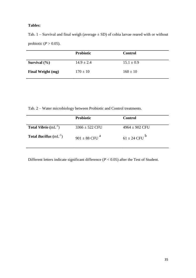
Tab. 1 – Survival and final weigh (average ± SD) of cobia larvae reared with or without
probiotic (P > 0.05).
Probiotic Control
Survival (%) 14.9 ± 2.4 15.1 ± 0.9
Final Weight (mg) 170 ± 10 160 ± 10
Tab. 2 – Water microbiology between Probiotic and Control treatments.
Probiotic Control
Total Vibrio (mL-1
) 3366 ± 522 CFU 4964 ± 902 CFU
Total Bacillus (mL-1
) 901 ± 88 CFU
a 61 ± 24 CFU
b
Different letters indicate significant difference (P < 0.05) after the Test of Student.
36
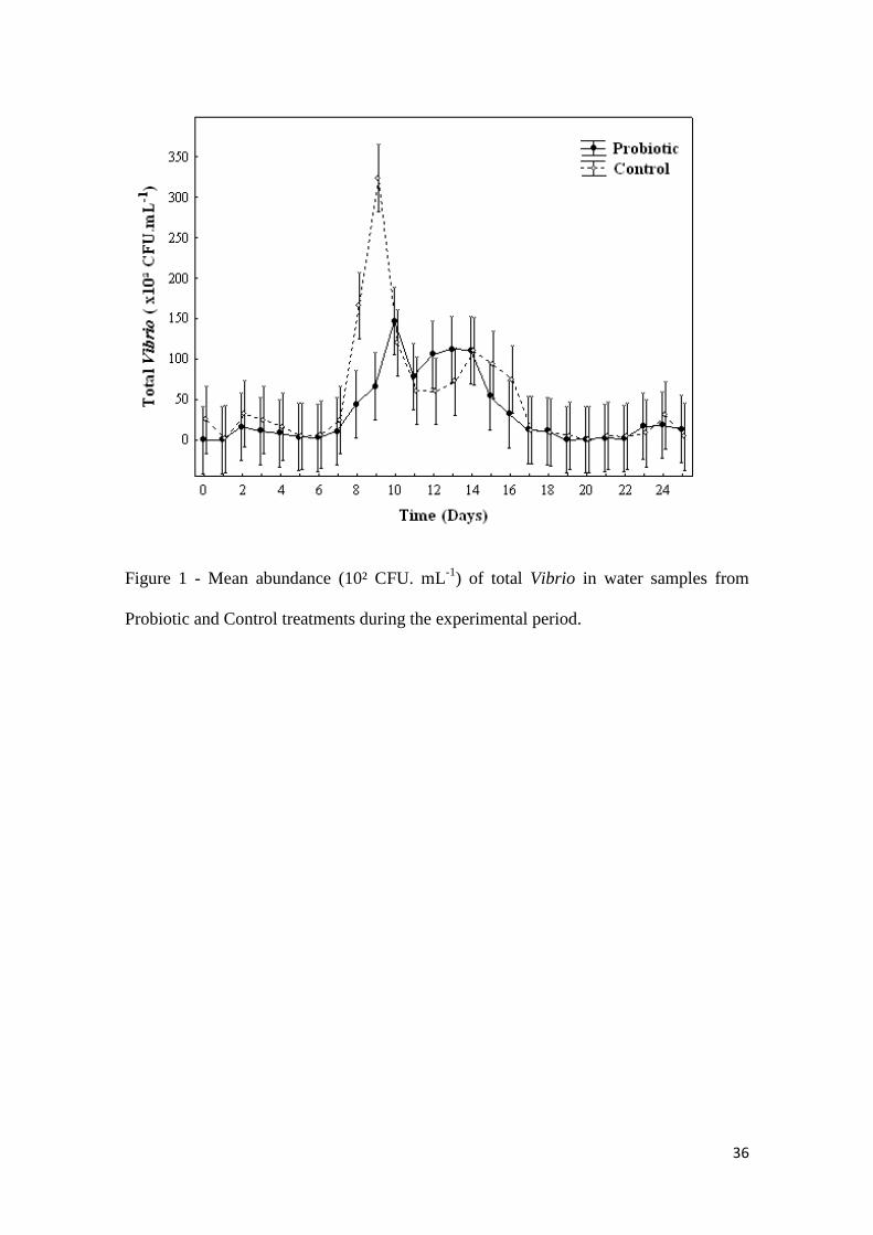
Figure 1 - Mean abundance (10² CFU. mL-1
) of total Vibrio in water samples from
Probiotic and Control treatments during the experimental period.
37
Figure 2 - Mean abundance (10² CFU. mL-1
) of total Bacillus in water samples from
Probiotic and Control treatments during the experimental period.
38
Figure 3 – Stress Sensitivity Index of cobia larvae reared with or without probiotic at
the end of experimental period. Vertical bars represent one standard deviation and
asterisk shows significantly statistic difference (P<0.05).

39
Figure 4 - Morphometric analysis of head kidney of cobia larvae reared with or without
probiotic. Vertical bars represent one standard deviation and asterisk shows
significantly statistic difference (P<0.01).
40
Figure 5 - CD3 expression (%/mm²) in thymus of cobia larvae reared with or without
probiotic Vertical bars represent one standard deviation. No significantly statistic
difference (P>0.05).
41
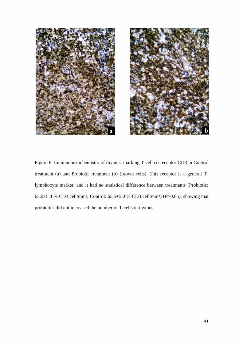
Figure 6. Immunohistochemistry of thymus, marking T-cell co-receptor CD3 in Control
treatment (a) and Probiotic treatment (b) (brown cells). This receptor is a general T-
lymphocyte marker, and it had no statistical difference between treatments (Probiotic:
63.9±5.4 % CD3 cell/mm²; Control: 65.5±5.0 % CD3 cell/mm²) (P>0.05), showing that
probiotics did not increased the number of T-cells in thymus.

42
Figure 7 - CD4 expression (%/mm²) in thymus of cobia larvae reared with or without
probiotic. Vertical bars represent one standard deviation and asterisk shows
significantly statistic difference (P<0.01).
43
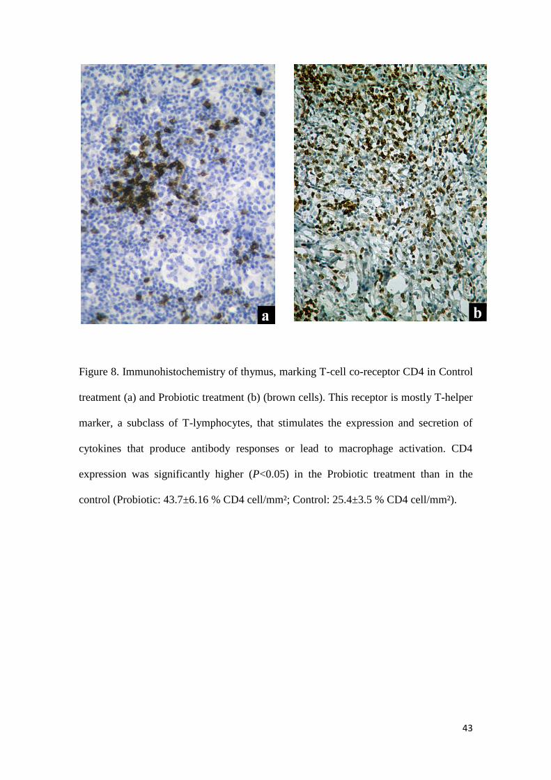
Figure 8. Immunohistochemistry of thymus, marking T-cell co-receptor CD4 in Control
treatment (a) and Probiotic treatment (b) (brown cells). This receptor is mostly T-helper
marker, a subclass of T-lymphocytes, that stimulates the expression and secretion of
cytokines that produce antibody responses or lead to macrophage activation. CD4
expression was significantly higher (P<0.05) in the Probiotic treatment than in the
control (Probiotic: 43.7±6.16 % CD4 cell/mm²; Control: 25.4±3.5 % CD4 cell/mm²).