Embed Size (px)
Citation preview

PONTIFIacuteCIA UNIVERSIDADE CATOacuteLICA DO RIO GRANDE DO SUL
INSTITUTO DE GERIATRIA E GERONTOLOGIA
POacuteS-GRADUACcedilAtildeO EM GERONTOLOGIA BIOMEacuteDICA
ALTERACcedilOtildeES ELETRONEUROMIOGRAacuteFICAS EM IDOSOS
COM ESTENOSE DE CANAL RAQUIacuteDEO LOMBAR
DEGENERATIVA ASSOCIACcedilAtildeO COM AS ALTERACcedilOtildeES DE
IMAGEM E CLIacuteNICA
Marcus Sofia Ziegler
Porto Alegre
2012
PONTIFIacuteCIA UNIVERSIDADE CATOacuteLICA DO RIO GRANDE DO SUL
INSTITUTO DE GERIATRIA E GERONTOLOGIA
POacuteS-GRADUACcedilAtildeO EM GERONTOLOGIA BIOMEacuteDICA
ALTERACcedilOtildeES ELETRONEUROMIOGRAacuteFICAS EM IDOSOS
COM ESTENOSE DE CANAL RAQUIacuteDEO LOMBAR
DEGENERATIVA ASSOCIACcedilAtildeO COM AS ALTERACcedilOtildeES DE
IMAGEM E CLIacuteNICA
Marcus Sofia Ziegler
Dissertaccedilatildeo de mestrado apresentada para obtenccedilatildeo do tiacutetulo de Mestre em Gerontologia Biomeacutedica pela Pontifiacutecia Universidade Catoacutelica do Rio Grande do Sul
Orientador Dr Irenio Gomes Filho Colaborador Dr Erasmo de Abreu Zardo
Porto Alegre
2012
DADOS INTERNACIONAIS DE CATALOGACcedilAtildeO NA PUBLICACcedilAtildeO (CIP)
Karin Lorien Menoncin
Bibliotecaacuteria CRB 102147
Z66a Ziegler Marcus Sofia
Alteraccedilotildees eletroneuromiograacuteficas em idosos com estenose de canal raquiacutedeo lombar
degenerativa associaccedilatildeo com as alteraccedilotildees de imagem e cliacutenica Marcus Sofia
Ziegler orientador ndash Porto Alegre PUCRS 2012
63 f tab Inclui artigo de perioacutedico submetido agrave publicaccedilatildeo
Orientador Prof Dr Irenio Gomes Filho
Dissertaccedilatildeo (Mestrado) ndash Pontifiacutecia Universidade Catoacutelica do Rio Grande do Sul
Instituto de Geriatria e Gerontologia Mestrado em Gerontologia Biomeacutedica
1 Estenose do canal raquiacutedeo lombar degenerativa 2 Eletroneuromiografia 3
Estudos de conduccedilatildeo nervosa 5 Gerontologia Biomeacutedica 6 Idosos I Gomes Filho
Irenio II Tiacutetulo
CDD - 6189767
11 ENSAIO CLIacuteNICO CONTROLADO
ALEATOacuteRIO I Schneider Rodolfo Herberto II Tiacutetulo
Marcus Sofia Ziegler
ALTERACcedilOtildeES ELETRONEUROMIOGRAacuteFICAS EM IDOSOS COM ESTENOSE DE
CANAL RAQUIacuteDEO LOMBAR DEGENERATIVA ASSOCIACcedilAtildeO COM AS
ALTERACcedilOtildeES DE IMAGEM E CLIacuteNICA
Dissertaccedilatildeo de Mestrado apresentada ao Curso de Poacutes-
Graduaccedilatildeo em Gerontologia Biomeacutedica da Pontifiacutecia
Universidade Catoacutelica do Rio Grande do Sul para obtenccedilatildeo de
tiacutetulo de Mestre em Gerontologia Biomeacutedica
Aprovada em______de_________________de_________
BANCA EXAMINADORA
________________________________________
Profa Dra Carla Helena Schwanke- PUCRS
________________________________________
Prof Dr Elcio Landim- UNICAMP
Dedico essa dissertaccedilatildeo a minha esposa meus pais meus irmatildeos e professores
AGRADECIMENTOS
Agradeccedilo especialmente agrave minha esposa Liacutevia Fay Ziegler e ao meu filho
Gustavo que me apoiaram e compreenderam minha ausecircncia enquanto eu me
dedicava profissionalmente
Agrave minha famiacutelia pais e irmatildeos que sempre confiaram e acreditaram em mim
Aos meus colegas de equipe que estiveram sempre dispostos a me ajudar
durante a elaboraccedilatildeo deste trabalho
Ao meu orientador Prof Dr Irenio Gomes que aceitou uma proposta de pesquisa
e o desafio de me orientar para contribuir com a ciecircncia
Agrave Profordf Dra Carla Schwanke que me incentivou e que esteve sempre disposta a
me ajudar nas minhas dificuldades
Aos meus colegas de mestrado que entenderam o meu silecircncio nas aulas e o
companheirismo fora delas
Ao meu grande professor Dr Erasmo de Abreu Zardo que tem influecircncia direta na
minha formaccedilatildeo praacutetica e acadecircmica
Ao Dr Afrane Serdeira que me incentivou desde o comeccedilo a sempre buscar um
algo mais de forma eacutetica
Agrave minha Labrador Dora que sempre esteve deitada aos meus peacutes desde as
pesquisas iniciais ateacute a conclusatildeo final desta dissertaccedilatildeo
ldquoIf you have a job to do you have to do it wellrdquo
Live and let die
Paul McCartney
RESUMO
O objetivo deste estudo eacute identificar o papel da eletroneuromiografia (ENMG) no
diagnoacutestico da estenose de canal raquideo lombar degenerativa (ECRLD) Um estudo
transversal prospectivo com coleta de dados envolvendo 31 pacientes com diagnoacutestico
de ECRLD confirmado com RNM Todos os pacientes apresentavam dor 839 dos
pacientes queixavam-se de dor moderada ou severa e 90 dos pacientes faziam uso
de medicaccedilatildeo para aliacutevio da dor ECRLD natildeo alterou a neuroconduccedilatildeo ou a
ldquoSympathetic Skin Responserdquo SSR A EMG confirmou alta frequecircncia de radiculopatia
particularmente multiradiculopatia As raiacutezes de L5 e S1 foram as mais lesadas
Identificamos tambeacutem uma alta prevalecircncia de radiculopatia em L4 Correlacionando
EMG aos achados cliacutenicos identificamos que a apresentaccedilatildeo cliacutenica o ponto chave de
iniacutecio de uma avaliaccedilatildeo eacute pobre no que tange a identificar radiculopatia frequente
consequecircncia da ECRLD Por esta razatildeo acreditamos que a EMG pode ser uma
ferramenta importante visto que possibilita identificar se os sintomas satildeo de natureza
neurogecircnicos Somado a isso pode servir para focar o tratamento em um segmento
especiacutefico da coluna vertebral
Palavras-chave Estenose do canal raquiacutedeo lombar degenerativa
Eletroneuromiografia Estudos de conduccedilatildeo nervosa Radiculopatia Dor
ABSTRACT
The aim of this study is to identify the role of EMG-NCS on LSS diagnosis A cross-
sectional study with prospective data collection was conducted involving 31 symptomatic
patients with LSS confirmed by MRI All patients reported pain 839 of patients
reported it to be moderate or severe and 90 of patients took pain medication LSS did
not affect NCS or SSR EMG confirmed high frequency of radiculopathy particularly
multiradiculopathy L5 and S1 roots were the most susceptible to injuries We also found
a higher prevalence of L4 radiculopathy Correlating EMG with clinical findings we
found that the clinical presentation the most important starting point of an evaluation is
poor in terms of identifying radiculopathy a frequent consequence of LSS For this
reason we theorize that EMG may play an important role as a diagnostic tool being
useful in determining when symptoms are neurogenic in nature In addition it may serve
to focus treatment only in the area where it is really necessary
Keywords Lumbar spinal stenosis Electromyography Nerve conduction studies
Radiculopathy Pain
LISTA DE TABELAS
Table 1 - Medical history data of 31 elderly patients with stenosis of the lumbar spinal
canalPorto Alegre 2012 46
Table 2 - Clinical and magnetic resonance exam data of 31 elderly patients with
stenosis of the lumbar spinal canal Porto Alegre 2012 47
Table 3 - Nerve conduction parameters of 31 elderly patients with stenosis of the lumbar
spinal canal as compared to a healthy elderly control group Porto Alegre
2012 48
Table 4 - Results of the needle electromyographic evaluation of 31 elderly patients with
stenosis of the lumbar spinal canal Porto Alegre 2012 48
Table 5 - Comparison of clinical and imaging variables between 20 patients with an
electromyographic diagnosis of radiculopathy and 11 patients with a normal
electromyography study in 31 elderly patients with stenosis of the lumbar
spinal canal Porto Alegre 2012 49
LISTA DE ABREVIATURAS E SIGLAS
ECRLD- Estenose do canal raquiacutedeo lombar degenerativo
ENMG- Eletroneuromiografia
RNM- Ressonacircncia Nuclear Magneacutetica
IBGE- Instituto Brasileiro de Geografia e Estatiacutestica
TC- Tomografia Computadorizada
TENS- Estimulaccedilatildeo eleacutetrica transcutacircnea
AINEs- Anti-inflamatoacuterios natildeo esteroides
SSR- ldquoSympathetic Skin Responserdquo
EMG- Eletromiografia
SNA- Sistema Nervoso Autocircnomo
SNC- Sistema Nervoso Central
SUMAacuteRIO
1 INTRODUCcedilAtildeO 13
2 REVISAtildeO DE LITERATURA 15
21 Envelhecimento populacional e transiccedilatildeo epidemioloacutegica 15
22 Estenose do canal raquiacutedeo lombar degenerativa 18
221 Definiccedilatildeo 18
222 Envelhecimento da coluna vertebral 18
223 Avaliaccedilatildeo cliacutenica 20
224 Diagnoacutestico por imagens 21
225 Tratamentos 22
23 Avaliaccedilatildeo neurofisioloacutegica 23
231 Neuroconduccedilatildeo motora 24
232 Neuroconduccedilatildeo sensitiva 25
233 Eletromiografia (EMG) 25
234 ldquoSympathetic Skin Responserdquo (SSR) 26
24 Neurofisiologia e ECRLD 27
3 OBJETIVOS 29
31 Objetivo geral 29
32 Objetivos especiacuteficos 29
321 Principal 29
322 Secundaacuterios 29
33 Hipoacutetese 30
4 JUSTIFICATIVA 31
5 MEacuteTODOS 32
6 ARTIGO EM INGLEcircS SUBMETIDO Agrave PUBLICACcedilAtildeO 34
Abstract 35
Introduction 36
Methods 37
Results 38
Discussion 40
Acknowledgments 43
References 44
7 CONSIDERACcedilOtildeES FINAIS 51
8 CONCLUSAtildeO 52
REFEREcircNCIAS 53
APEcircNDICE B - Ficha de anamnese e exame fiacutesico 60
APEcircNDICE C - Avaliaccedilatildeo eletroneuromiograacutefica 61
APEcircNDICE D - Aprovaccedilatildeo da comissatildeo cientiacutefica 62
APEcircNDICE E - Aprovaccedilatildeo do Comitecirc de Eacutetica e Pesquisa da PUCRS 63
APEcircNDICE F - Comprovaccedilatildeo da submissatildeo do artigo 64
ANEXO A - RNM ndash Classificaccedilatildeo de RNM 65
ANEXO B - Escala visual da dor (VAS) 66
ANEXO C - Questionaacuterio de Oswestry 67
13
1 INTRODUCcedilAtildeO
O envelhecimento da coluna lombar eacute um processo fisioloacutegico que acaba
resultando em alteraccedilotildees degenerativas que podem gerar estenose do canal raquiacutedeo
(ECRLD ndash ldquoEstenose do Canal Raquiacutedeo Lombar Degenerativardquo) Claudicaccedilatildeo
neurogecircnica e ciatalgia resultante de compressatildeo da raiz nervosa satildeo as manifestaccedilotildees
cliacutenicas mais frequentes e podem causar incapacidade funcional e inabilidade nas
atividades da vida diaacuteria exigindo tratamento especializado Esta condiccedilatildeo acomete
principalmente a populaccedilatildeo idosa comprometendo sua independecircncia e qualidade de
vida Haacute controveacutersia na literatura quanto ao melhor meacutetodo agrave abordagem e aos
resultados no tratamento da estenose da coluna lombar nessa populaccedilatildeo de pacientes
que apresentam com frequecircncia comorbidades resultantes da insuficiecircncia de outros
sistemas1 Muitos estudos tecircm enfatizado a morbidade associada ao tratamento
ciruacutergico da estenose lombar em idosos e outros tecircm recomendado tratamento natildeo
ciruacutergico (conservador) da estenose da coluna lombar em idosos Johnsson et al num
estudo investigando a evoluccedilatildeo natural da estenose lombar natildeo encontrou piora da
claudicaccedilatildeo espinhal (neurogecircnica) apoacutes 4 anos de observaccedilatildeo e portanto sugere o
tratamento conservador e a observaccedilatildeo como alternativa ao tratamento ciruacutergico2
A prevalecircncia da ECRLD mesmo sendo desconhecida sabe-se que vem
aumentando juntamente ao aumento da expectativa de vida O diagnoacutestico diferencial
inclui desde diferentes alteraccedilotildees biomecacircnicas da coluna vertebral do quadril e do
joelho ateacute doenccedilas vasculares e polineuropatia34 A fisiopatologia dos sintomas da
estenose do canal raquiacutedeo lombar ainda segue pouco compreendida mas o aumento
da pressatildeo venosa a diminuiccedilatildeo do fluxo sanguiacuteneo e axoplasmaacutetico jaacute foram
identificados em modelos animais de estenose de canal lombar Dentre as alteraccedilotildees
nervosas identificadas estatildeo o edema a desmielinizaccedilatildeo e a degeneraccedilatildeo valeriana
das fibras aferentes e eferentes Quando haacute compressatildeo de nervo perifeacuterico ocorre
parestesia deacuteficit motor e sensorial A dor ocorre quando haacute reaccedilatildeo inflamatoacuteria4
A ECRLD eacute definida por medida do diacircmetro ou da aacuterea do saco tecal ou canal
espinhal atraveacutes de radiografia mielografia tomografia computadorizada ou
ressonacircncia nuclear magneacutetica (RNM) A relaccedilatildeo entre as medidas radioloacutegicas e a
14
siacutendrome cliacutenica ainda natildeo eacute bem definida pela literatura3567 A eletroneuromiografia
(ENMG) eacute usada por muitos na ECRLD para auxiliar no diagnoacutestico e para fazer o
diagnoacutestico diferencial com outras patologias Apesar disso a maioria das revisotildees
bibliograacuteficas natildeo citam a ENMG38
Verificamos portanto que a ECRLD eacute uma condiccedilatildeo frequente na populaccedilatildeo
idosa que leva a importante incapacidade cujos mecanismos fisiopatoloacutegicos natildeo
estatildeo ainda esclarecidos e cujo tratamento eacute bastante controverso O entendimento
fisiopatoloacutegico dessa doenccedila nos idosos eacute de grande relevacircncia pois serve de
conhecimento inicial para esclarecer a melhor forma de diagnosticaacute-la e para propor
terapecircuticas adequadas a serem testadas futuramente O objetivo do presente trabalho
eacute estudar as alteraccedilotildees neurofisioloacutegicas encontradas em idosos com ECRLD
correlacionando-as aos achados cliacutenicos (sinais e sintomas) e de imagem (RNM)
15
2 REVISAtildeO DE LITERATURA
Com o envelhecimento da populaccedilatildeo no mundo as doenccedilas que acompanham o
envelhecimento devem ser melhor compreendidas A seguir satildeo mencionados os
toacutepicos importantes para o entendimento do tema e da pesquisa relacionada nesse
trabalho
21 Envelhecimento populacional e transiccedilatildeo epidemioloacutegica
Neste iniacutecio de seacuteculo testemunhamos no Brasil o nuacutemero de idosos aumentar
muito raacutepido A faixa etaacuteria de 60 anos ou mais eacute a que mais cresce em termos
proporcionais Segundo projeccedilotildees estatiacutesticas da Organizaccedilatildeo Mundial de Sauacutede de
1950 ateacute 2025 a populaccedilatildeo de idosos no paiacutes deve atingir um crescimento de 16 vezes
contra 5 vezes da populaccedilatildeo total o que coloca o paiacutes em termos absolutos como a
sexta populaccedilatildeo de idosos do mundo isto eacute com mais de 32 milhotildees de pessoas com
60 anos ou mais O campo da geriatria continua a ganhar atenccedilatildeo devido ao raacutepido
crescimento desse segmento da populaccedilatildeo e ao impacto socioeconocircmico previsto
neste seacuteculo9
A populaccedilatildeo brasileira atual eacute de quase 191 milhotildees de habitantes segundo
dados do Instituto Brasileiro de Geografia e Estatiacutestica (IBGE) Destes 74 satildeo
idosos totalizando 21 milhotildees 47 As regiotildees sul e sudeste comportam 81 dos idosos
do paiacutes sendo que o Rio Grande do Sul (RGS) lidera este ranking com um total de
994613 indiviacuteduos com 65 anos ou mais e com 60 anos ou mais totalizando
1459597
Associado ao envelhecimento populacional fenocircmeno conhecido como transiccedilatildeo
demograacutefica na qual as populaccedilotildees humanas passam gradualmente de um periacuteodo
com alta natalidade e mortalidade precoce para um periacuteodo de baixa natalidade e
mortalidade precoce10 ocorre tambeacutem a transiccedilatildeo epidemioloacutegica Nesse processo as
pessoas satildeo acometidas em menor proporccedilatildeo por doenccedilas infecto-parasitaacuterias e em
maior proporccedilatildeo por doenccedilas crocircnicas e degenerativas10 Como as pessoas ficam mais
16
velhas ficam mais suscetiacuteveis aos efeitos de processos degenerativos nos diferentes
oacutergatildeos e sistemas
As mudanccedilas morfoloacutegicas e funcionais que alteram a homeostase do organismo
caracterizam o envelhecimento41 Este eacute um processo natural complexo individual e
multifatorial que afeta oacutergatildeos e sistemas nem sempre de forma homogecircnea Por essa
razatildeo natildeo haacute um padratildeo nem ritmo de envelhecimento bem determinado 42 Mesmo em
indiviacuteduos idosos de mesma idade o decliacutenio das funccedilotildees orgacircnicas varia de um oacutergatildeo
para outro Dessa forma fatores extriacutensecos podem determinar o ritmo de
envelhecimento 43
Diversas hipoacuteteses e teorias tecircm sido sugeridas com o intuito de explicar o
processo de envelhecimento Alguns autores chegam a um consenso de que uma
programaccedilatildeo geneacutetica exerce papel fundamental 444546
As alteraccedilotildees na coluna lombar relacionadas agrave idade manifestam-se como um
processo patoloacutegico sintomaacutetico em alguns pacientes Os sintomas cliacutenicos satildeo
analisados com seus possiacuteveis geradores de dor As modificaccedilotildees normais associadas
ao envelhecimento ocorrem no disco intervertebral resultando em lombalgia
instabilidade e heacuternia de disco associada ou natildeo a compressatildeo nervosa11 Com o
aumento da expectativa de vida os discos intervertebrais sofrem mudanccedilas com
consequentes alteraccedilotildees funcionais no que tange agrave transferecircncia de cargas e de
estabilidade Essas alteraccedilotildees normais relacionadas agrave idade afetam por fim a
integridade do disco tornando-o menos efetivo e ficando mais suscetiacutevel agrave lesatildeo
Quando o segmento vertebral eacute analisado como um complexo ldquotriarticularrdquo pelo
fato de as facetas tambeacutem terem papel importante funcional a progressatildeo da
degeneraccedilatildeo vertebral eacute descrita em trecircs estaacutegios I- alteraccedilotildees na bioquiacutemica e
fisiologia causando sintomas cliacutenicos II- aumento do movimento dos segmentos
vertebrais causando instabilidade sintomaacutetica III- formaccedilatildeo osteofitaacuteria reduzindo a
instabilidade e causando rigidez do segmento Todas as alteraccedilotildees exercem o efeito
cumulativo de reduzir as dimensotildees do canal vertebral11
Com os estudos que examinam a relaccedilatildeo das alteraccedilotildees bioquiacutemicas relacionadas
agrave idade e agraves propriedades mecacircnicas da coluna vertebral a doenccedila sintomaacutetica
comeccedila a ser mais bem compreendida Mas ainda esperamos uma compreensatildeo mais
17
abrangente em situaccedilotildees que o envelhecimento fisioloacutegico torna-se um processo
patoloacutegico11
18
22 Estenose do canal raquiacutedeo lombar degenerativa
A estenose do canal raquideo lombar degenerativo eacute uma das consequumlecircncias
evidentes que acompanham o envelhecimento populacional
221 Definiccedilatildeo
A estenose lombar pode ser definida como qualquer tipo de estreitamento do
canal vertebral ou dos foramens Esta estenose pode ser decorrente de muacuteltiplas
causas isoladas ou associadas Contudo a sintomatologia seraacute resultante de um
processo fisiopatoloacutegico final comum de desequiliacutebrio da relaccedilatildeo conteuacutedo-continente
com consequente compressatildeo mecacircnica das estruturas nervosas por meio da
expansatildeo das estruturas que formam as paredes do canal vertebral ou por meio da
congestatildeo e isquemia decorrentes das alteraccedilotildees do fluxo sanguiacuteneo radicular312
As diversas causas de compressatildeo devem ser bem identificadas classificadas e
estar em relaccedilatildeo direta aos sintomas do paciente pois somente com a soma de todos
os fatores seraacute possiacutevel definir um prognoacutestico e adotar a melhor opccedilatildeo terapecircutica
seja ela conservadora seja ela ciruacutergica
222 Envelhecimento da coluna vertebral
A ECRL mais comum eacute a degenerativa (ECRLD) O aumento da expectativa de
vida tem uma relaccedilatildeo direta com o aumento desta patologia A estimativa eacute que a
incidecircncia seja de 1 em 1000 pacientes acima de 65 anos de idade sendo a maior
causa de cirurgia neste grupo de pacientes3
O processo degenerativo comeccedila com a perda da altura discal e a desidrataccedilatildeo
com substituiccedilatildeo das fibras de colaacutegeno tipo II e proteoglicanos por tecido fibroso
aumentando a mobilidade do segmento Isto resulta em protrusatildeo discal degeneraccedilatildeo
com hipertrofia facetaacuteria e formaccedilatildeo de osteoacutefitos podendo gerar estenose central de
recesso lateral ou foraminal
19
Cada disco intervertebral eacute constituiacutedo por um nuacutecleo pulposo gelatinoso
circundado por um anel fibroso laminado O anel fibroso forma o limite externo do disco
sendo constituiacutedo por camadas concecircntricas de tecido fibrocartilaginoso e por proteiacutenas
fibrosas que se dirigem obliquamente de uma veacutertebra agrave outra Existem 12 lamelas
concecircntricas que envolvem o nuacutecleo O nuacutecleo pulposo na embriologia surge da
notocorda Jaacute a aacuterea mesenquimal daacute origem ao acircnulo fibroso No disco embrionaacuterio
haacute uma composiccedilatildeo e estrutura distinta entre o fluido do nuacutecleo pulposo rico em ceacutelulas
de notocorda e proteoglicanos e o acircnulo fibroso rico em ceacutelula fibroblaacutestica e
colaacutegeno Na margem superior e na inferior do disco a notocorda desaparece sendo
substituiacuteda por ceacutelulas mesenquimais as quais proporcionam o crescimento do tecido
cartilaginoso formando os platocircs das veacutertebras12
A partir dos 10 anos de idade as ceacutelulas notocordais estatildeo ausentes Com o
envelhecimento as diferenccedilas na composiccedilatildeo do acircnulo fibroso e no nuacutecleo pulposo
tornam-se cada vez mais difiacuteceis de identificar Com o passar dos anos os platocircs
vertebrais vatildeo afilando devido agrave ossificaccedilatildeo endocondral A densidade celular do disco
tambeacutem diminui Na maturidade eacute um dos tecidos mais finos do corpo dificultando a
manutenccedilatildeo das ceacutelulas no decorrer da vida O que gera certa resistecircncia ao acircnulo eacute a
orientaccedilatildeo das fibras12
O disco intervertebral possui muacuteltiplos tipos de colaacutegeno incluindo os tipos I II III
V VI IX XI XII e XIV dos quais a abundacircncia muda conforme a idade O acircnulo fibroso
conteacutem mais o colaacutegeno tipo I jaacute o nuacutecleo pulposo mais o colaacutegeno tipo II O colaacutegeno
discal estaacute sujeito a accedilotildees enzimaacuteticas mais acentuadas no nuacutecleo pulposo Com o
envelhecimento haacute acuacutemulo de lipiacutedeos e de derivados de carboidratos alterando a cor
do disco para o tom marrom o que diminui tambeacutem sua elasticidade12
Existem duas proteiacutenas na matriz do disco que merecem maior atenccedilatildeo A
primeira eacute a fibronectina que aumenta com a idade e o grau de degeneraccedilatildeo
intensificando a fragmentaccedilatildeo proteoliacutetica o que pode estimular eventos cataboacutelicos A
segunda eacute a elastina que eacute a mais resistente agrave proteoacutelise A proteoacutelise eacute a maior
responsaacutevel pelo envelhecimento do disco mas ainda natildeo estaacute claro qual a proteinase
eacute responsaacutevel por essa degeneraccedilatildeo O pH abaixo de 7 da matriz extracelular do disco
favorece a accedilatildeo das proteinases12
20
Outro fator importante eacute a influecircncia nutricional O disco em crescimento se nutre
atraveacutes das cartilagens dos platocircs Com a calcificaccedilatildeo destes platocircs haacute o
comprometimento da nutriccedilatildeo diminuindo a quantidade de oxigecircnio aumentando o
metabolismo anaeroacutebico e provocando aumento de aacutecido laacutetico A mudanccedila do pH gera
maior accedilatildeo das proteinases12
As alteraccedilotildees biomecacircnicas estatildeo diretamente ligadas agrave degeneraccedilatildeo do disco
intervertebral A pressatildeo sobre o disco eacute necessaacuteria mas em curta duraccedilatildeo e natildeo de
forma contiacutenua Com a perda da altura o disco gera uma mobilidade anormal no
segmento provocando instabilidade Esta instabilidade acelera a degeneraccedilatildeo12
No que tange agrave geneacutetica efeitos cataboacutelicos aumentados por genes polimoacuterficos
predispotildeem a degeneraccedilatildeo e promovem baixa toleracircncia a mudanccedilas biomecacircnicas
Os genes polimoacuterficos satildeo encontrados em vaacuterios tipos de colaacutegenos Natildeo estaacute claro se
um ou dois alelos curtos satildeo necessaacuterios para observar o aumento da degeneraccedilatildeo
Sabe-se que o estiacutemulo externo influencia12
O disco intervertebral eacute incapaz de reparo Vaacuterias hipoacuteteses para ajudar o reparo
satildeo estudadas injeccedilatildeo de fator de crescimento (BMP7 ou BMP2) peptiacutedeos anaboacutelicos
e terapia gecircnica com ceacutelulas tronco12
223 Avaliaccedilatildeo cliacutenica
A ECRLD em geral causa dor ou desconforto lombar que se irradia para gluacuteteos
ou membros inferiores Os pacientes podem ter queixas de radiculopatia ou claudicaccedilatildeo
neurogecircnica dependendo do tipo de compressatildeo que a estenose esta causando
Raramente se encontra disfunccedilatildeo esfincteriana ou siacutendrome de cauda equina A
severidade dos sintomas deveria ser medida usando escalas como a escala visual da
dor (VAS) ou Oswestry
No exame fiacutesico os achados neuroloacutegicos natildeo satildeo comumente detectados O
teste de Lasegue em geral eacute negativo a extensatildeo lombar causa desconforto e ocorre
aliacutevio com a flexatildeo Fraqueza muscular natildeo eacute comum e pode haver diminuiccedilatildeo da
sensibilidade no dermaacutetomo correspondente Os sintomas evoluem gradativamente e
persistem por alguns meses ou anos
21
A histoacuteria e o exame fiacutesico devem ser bem coletados para que seja feito um
diagnoacutestico diferencial Dentre as patologias que podem ser confundidas estatildeo a
osteoartrite de quadril bursite trocanteacuterica neuropatia perifeacuterica e claudicaccedilatildeo vascular
aleacutem de outras patologias de coluna neoplasias e infecccedilotildees34
224 Diagnoacutestico por imagens
Alguns problemas podem ser encontrados na investigaccedilatildeo por imagem nos
pacientes com ECRLD Primeiro porque as alteraccedilotildees degenerativas satildeo altamente
prevalentes em pacientes assintomaacuteticos (20 acima de 60 anos) Segundo porque
imagens tendem a exagerar as alteraccedilotildees degenerativas A maioria dos diagnoacutesticos
devem ser feitos pelas queixas cliacutenicas e pelo exame fiacutesico Os exames de imagem
devem ser solicitados quando se estaacute planejando algum tipo de intervenccedilatildeo ciruacutergica ou
para diagnoacutestico diferencial 3
A Ressonacircncia Nuclear Magneacutetica (RNM) eacute o exame de imagem de escolha Ela
natildeo produz radiaccedilatildeo ionizante e permite perceber melhor os tecidos moles adjacentes
Em compensaccedilatildeo natildeo pode ser realizado em pacientes com marca-passo
claustrofobia ou que possuam outros implantes metaacutelicos O exame eacute realizado nas
sequecircncias ponderadas em T1 e T2 sagital e axial Pode ser adicionada sequecircncia
com supressatildeo de gordura para visualizar melhor a medula oacutessea As imagens em T2
nos fornecem um aspecto mielograacutefico com a vantagem de natildeo ser um procedimento
invasivo34
A estenose de canal eacute considerada de acordo com a medida de diacircmetro acircntero-
posterior que estaraacute em torno de 10-12 mm (valor fisioloacutegico de 22-25 mm) Quando o
valor absoluto eacute menor que 10 mm o paciente em geral eacute sintomaacutetico A estenose de
recesso lateral eacute considerada quando o diacircmetro eacute menor que 2 mm (valor fisioloacutegico eacute
3-5 mm)3567
A Tomografia Computadorizada (TC) eacute o exame de mais faacutecil acesso agrave populaccedilatildeo
em geral e permite uma avaliaccedilatildeo precisa do canal espinhal Atualmente a TC tem sido
realizada em multislice e com reconstruccedilatildeo em 3D A limitaccedilatildeo deste exame estaacute na
22
visualizaccedilatildeo das estruturas intratecais pois as estruturas nervosas apresentam a
mesma densidade do liacutequor A mielografia TC eacute um recurso que ajuda a visualizar
melhor as estruturas intratecais Pode ser indicada sua realizaccedilatildeo quando haacute
contraindicaccedilatildeo para realizar a RNM ou quando seus resultados satildeo inconclusivos34
225 Tratamentos
A discussatildeo no que tange o tratamento conservador ou ciruacutergico eacute bastante
ampla Katz et al estudaram os resultados a longo prazo da laminectomia
descompressiva para estenose lombar degenerativa e observaram que entre os fatores
associados os com resultados ruins foram aqueles com enfermidades coexistentes
como cardiopatia doenccedila pulmonar crocircnica artrite reumatoide e osteoartrite Em outro
estudo Katz et al concluiacuteram que pacientes com comorbidades severas ficam
significativamente menos satisfeitos com os resultados da cirurgia para estenose
degenerativa da coluna lombar Deyo et al encontraram um iacutendice de 18 de
complicaccedilotildees apoacutes cirurgia lombar em pacientes acima de 75 anos Esse estudo no
entanto natildeo foi restrito a pacientes idosos Outros autores tecircm notado que artrodese
em muacuteltiplos niacuteveis na presenccedila de osso osteoporoacutetico e idade avanccedilada podem
provocar uma morbidade operatoacuteria aumentada
Contrariamente muitos autores tecircm recomendado e demonstrado resultados
favoraacuteveis apoacutes cirurgia para estenose da coluna lombar na populaccedilatildeo idosa
Sanderson e Wood relataram excelentes resultados em 64 e bons resultados em 17
nos 31 pacientes estudados maiores de 65 anos Silvers et al concluiacuteram no seu estudo
que a laminectomia lombar descompressiva eacute uma operaccedilatildeo relativamente segura com
uma taxa de sucesso a meacutedio e a longo prazo para todas as idades ateacute pelo menos a
oitava deacutecada de vida O periacuteodo de segmento miacutenimo no entanto foi de apenas 4
meses
Dentre os tratamentos estipulados estatildeo o natildeo ciruacutergico (conservador) e o
ciruacutergico O tratamento conservador consiste em reabilitaccedilatildeo com fisioterapia e
fortalecimento abdominal para melhora do quadro aacutelgico Existem vaacuterias maneiras de se
obter a analgesia adequada como o calor local crioterapia faixas lombares
23
estimulaccedilatildeo eleacutetrica transcutacircnea (TENS) e o ultrassom Os recursos farmacoloacutegicos
satildeo os anti-inflamatoacuterios natildeo-esteroides (AINEs) e o paracetamol na fase inicial Na
falha destes podem ser usados miorrelaxantes e opioides Seguidamente satildeo usados
antidepressivos e anticonvulsivantes na tentativa de controle da dor embora natildeo haja
ainda evidecircncia cliacutenica34
Em pacientes com dor radicular persistente as injeccedilotildees de corticoides podem
aliviar os sintomas por semanas e ateacute por meses Estas injeccedilotildees satildeo feitas no espaccedilo
epidural ou foraminais e sua eficaacutecia eacute motivo de controveacutersia34
Na falha do tratamento conservador o tratamento ciruacutergico deve ser oferecido
tendo sempre em vista que o objetivo principal a ser alcanccedilado eacute a descompressatildeo do
canal raquiacutedeo lombar e a liberaccedilatildeo das raiacutezes nervosas Nos casos em que haacute
instabilidade ou em que a instabilidade pode ser provocada atraveacutes do procedimento
descompressivo a fusatildeo poderaacute ser necessaacuteria com ou sem instrumentaccedilatildeo34
23 Avaliaccedilatildeo neurofisioloacutegica
O exame de ENMG eacute um estudo eletrofisioloacutegico do sistema nervoso perifeacuterico
Com ele pode-se identificar lesotildees no neurocircnio motor do corno anterior da medula nas
raiacutezes nervosas nos plexos nervosos nos troncos dos nervos perifeacutericos na junccedilatildeo
neuromuscular e nas fibras musculares18 O exame de rotina eacute feito atraveacutes do estudo
das neuroconduccedilotildees sensitivas e motoras e dos potenciais musculares em repouso e
em contraccedilatildeo voluntaacuteria atraveacutes de um eletrodo de agulha introduzido nos muacutesculos
Associado a esta rotina baacutesica diversas teacutecnicas especiacuteficas satildeo utilizadas como por
exemplo a onda F para medir o tempo de conduccedilatildeo na regiatildeo proximal e a estimulaccedilatildeo
repetitiva para estudo do funcionamento da sinapse neuromuscular18 As teacutecnicas
habituais no entanto tecircm a limitaccedilatildeo de estudarem as conduccedilotildees apenas das fibras
raacutepidas mielinizadas Comprometimento exclusivo de fibras finas habitualmente natildeo satildeo
identificados nos exames de ENMG de rotina Uma teacutecnica uacutetil para o estudo das fibras
finas eacute o teste da resposta cutacircnea simpaacutetica (SSR ndash ldquosympathetic skin responserdquo)19
Este teste embora muito utilizado em pesquisa natildeo tem uma padronizaccedilatildeo definida
com boa sensibilidade e especificidade para utilizaccedilatildeo cliacutenica Na avaliaccedilatildeo da ECRLD
24
a ENMG eacute um exame que apesar de natildeo muito solicitado por alguns profissionais eacute
muito uacutetil tanto para verificar o grau de comprometimento do sistema nervoso perifeacuterico
pela doenccedila quanto para o diagnoacutestico diferencial com plexopatias e neuropatias
perifeacutericas Este exame tem um amplo espectro de possibilidades de resultados na
ECRLD desde apresentar exame normal ateacute perda axonal e dismielinizaccedilatildeo nos casos
mais graves 348
231 Neuroconduccedilatildeo motora
Para se avaliar somente as fibras motoras dos nervos perifeacutericos eacute necessaacuterio um
registro do potencial de accedilatildeo composto das fibras musculares enervadas pelo nervo
estimulado Eacute realizado um estiacutemulo eleacutetrico sobre o nervo que se quer estudar atraveacutes
de um estimulador bipolar (caacutetodo e acircnodo) A despolarizaccedilatildeo dos axocircnios ocorre sob
os caacutetodos pela sua negatividade na regiatildeo isso proporciona reduccedilatildeo da diferenccedila de
potencial o que gera um potencial de accedilatildeo nas fibras nervosas Este potencial percorre
a regiatildeo distal do nervo fazendo com que seja liberada acetilcolina na junccedilatildeo
neuromuscular Isso gera um potencial de accedilatildeo nas fibras musculares que vai levar agrave
contraccedilatildeo do muacutesculo Eletrodos de registro colocados sobre o muacutesculo captam o
potencial de accedilatildeo das fibras musculares Um eletrodo eacute colocado no ponto meacutedio do
ventre muscular e outro eletrodo de referecircncia eacute colocado na origem ou inserccedilatildeo do
muacutesculo geralmente sobre uma superfiacutecie oacutessea4850
Na neuroconduccedilatildeo motora natildeo se mede a velocidade de conduccedilatildeo pela latecircncia e
distacircncia distais devido ao tempo que a acetilcolina leva a promover a sinapse A
velocidade soacute eacute calculada no segmento proximal do nervo subtraindo a latecircncia distal
da proximal e a distacircncia entre os dois pontos de estimulaccedilatildeo Entretanto
estabelecendo-se os valores normais para as latecircncias distais pode-se avaliar alguns
distuacuterbios nervosos como os de compressatildeo distal 4850
25
232 Neuroconduccedilatildeo sensitiva
A neuroconduccedilatildeo sensitiva eacute obtida com estiacutemulo e registro sobre o nervo
Geralmente satildeo colocados eletrodos de registro sobre o trajeto de um nervo misto ou
sensitivo Este mesmo nervo eacute entatildeo estimulado proximal ou distalmente Quando o
estiacutemulo eacute proximal ao registro a teacutecnica eacute denominada antidrocircmica pois o nervo
conduz o estiacutemulo no sentido oposto ao normal de suas fibras Quando o estiacutemulo eacute
distal e o potencial percorre o nervo em seu sentido natural eacute denominada ortodrocircmica
Habitualmente satildeo obtidos 3 paracircmetros amplitude e latecircncia de pico e
velocidade de conduccedilatildeo A velocidade de conduccedilatildeo eacute obtida dividindo-se a distacircncia
entre o estiacutemulo e o registro pela latecircncia distal Diferentemente do estudo motor a
amplitude do potencial sensitivo eacute medida em microvolts em vez de milivolts tornando
a interpretaccedilatildeo mais difiacutecil em matildeos inexperientes 4850
233 Eletromiografia (EMG)
A eletromiografia (EMG) eacute obtida pela introduccedilatildeo de eletrodo (agulhas) no
muacutesculo Os muacutesculos satildeo avaliados em repouso - miacutenima atividade e maacutexima
atividade Durante o repouso procura-se atividade espontacircnea (fibrilaccedilotildees descargas
mioquiacutemicas miotocircnicas complexas e neuromiotocircnicas) que podem representar perda
axonal Durante a contraccedilatildeo se observa o padratildeo de recrutamento das fibras
musculares e os potencias de unidade motora 4850
A unidade motora consiste em uma ceacutelula do corno anterior da medula com seu
axocircnio e todas as fibras musculares inervadas por ela Apoacutes avaliar a atividade
espontacircnea estuda-se a morfologia e as caracteriacutesticas dos potenciais da unidade
motora durante atividades voluntaacuterias do muacutesculo Assim pode-se diferenciar doenccedilas
miopaacuteticas ou neurogecircnicas adicionando severidade e cronicidade O recrutamento se
refere agrave participaccedilatildeo ordenada dos potenciais de unidade motora que ocorre com uma
contraccedilatildeo muscular Alguns paracircmetros satildeo usados na avaliaccedilatildeo dos potenciais de
unidade motora a duraccedilatildeo a amplitude e o nuacutemero de fases 4850
26
234 ldquoSympathetic Skin Responserdquo (SSR)
O sistema nervoso autonocircmico (SNA) representa uma estrutura complexa com
efeitos diretos em cada oacutergatildeo e sistema O diagnoacutestico de um distuacuterbio autonocircmico eacute
difiacutecil e baseia-se na acuraacutecia da seleccedilatildeo e na interpretaccedilatildeo de testes individuais e suas
combinaccedilotildees
O termo SSR representa o potencial eleacutetrico e a resposta endosomaacutetica da pele
SSR eacute a mudanccedila registrada em potencial na superfiacutecie da pele e representa uma
atividade sudomotora (sudorese) Isso eacute resultado da ativaccedilatildeo do arco reflexo
polissinaacuteptico A parte eferente do arco reflexo consiste em fibras simpaacuteticas
mielinizadas de neurocircnios do nuacutecleo intermediolateral entre o segmento T1 e L2 As
fibras poacutes-ganglionares natildeo satildeo mielinizadas e inervam as glacircndulas eacutecrinas
sudoriacuteparas
A SSR se apresenta em forma de uma onda lenta que pode ser trifaacutesica bifaacutesica
ou raramente monofaacutesica Nos membros inferiores em geral satildeo bifaacutesicas A latecircncia
quando medida nas matildeos eacute geralmente menor que nas pernas enquanto a amplitude
eacute maior quando comparada nos mesmos locais 32
Os fatores que influenciam a SSR satildeo repeticcedilatildeo de estiacutemulos sexo idade e
altura (controverso) tipo de estiacutemulo usado (alguns autores natildeo encontraram diferenccedila
mas Shahani e Kira encontraram mudanccedila na amplitude) e temperatura corporal
(controlada pela manutenccedilatildeo da temperatura da sala de 15 a 20 minutos de
permanecircncia para o exame) A modalidade de estiacutemulo eleacutetrico determina o arco reflexo
do trato aferente 32
No que tange ao resultado esperado pelo teste a avaliaccedilatildeo qualitativa aceita que
apenas a ausecircncia de SSR se caracteriza como patoloacutegica Entretanto quanto agrave
avaliaccedilatildeo quantitativa alguns autores analisam a latecircncia e outros a amplitude A SSR
eacute usada para diagnoacutestico de lesotildees em fibras finas em neuropatia diabeacutetica ou urecircmica
Pode tambeacutem ser encontrada anormal em neuropatia amiloide familiar e lepra A
ausecircncia de SSR foi observada em neuropatia inflamatoacuteria crocircnica e aguda com
disfunccedilatildeo autonocircmica Nos estaacutegios precoces de HIV tambeacutem podem ser vistas
anormalidades da SSR Alteraccedilotildees do sistema nervoso central (SNC) podem mostrar
27
anormalidades da SSR em mais de 50 de pacientes com esclerose muacuteltipla e em 40
com esclerose lateral amiotroacutefica Uma latecircncia prolongada ou uma queda da amplitude
foram observadas em pacientes com Parkinson 333435
Outras patologias como mielopatia cervical seringomielia distrofias Huntington
disfunccedilatildeo ereacutetil esclerodermia Sjoumlgren vitiligo e hipotiroidismo autoimune psoriacutease
artrite reumatoide depressatildeo psicose e botulismo podem alterar o SSR 3637
24 Neurofisiologia e ECRLD
A eletroneuromiografia (ENMG) pode ser de grande valor pois testa as
consequecircncias fisioloacutegicas provocadas pela estenose do canal raquiacutedeo podendo
detectar numerosas outras desordens no diagnoacutestico diferencial que natildeo tenham
achados anatocircmicos bem evidentes (polineuropatia ou neuropatia focal compressiva
como por exemplo por tumor peacutelvico) Natildeo haacute ateacute o momento nenhum estudo
descrevendo a associaccedilatildeo entre achados eletroneuromiograacuteficos e severidade dos
sintomas ou medidas anatocircmicas do canal vertebral 38
A ECRLD apresenta-se como uma siacutendrome cliacutenica e eacute definida por imagem
Poreacutem a relaccedilatildeo entre as medidas radioloacutegicas e a cliacutenica natildeo eacute linear Estudos
indicam que a mediccedilatildeo isolada do canal raquiacutedeo tem pobre correlaccedilatildeo
interobservador indicando que se fazem necessaacuterias medidas mais objetivas A medida
acircntero-posterior e a aacuterea do saco tecal na RNM tecircm reprodutibilidade com o grau dos
sintomas na estenose espinhal Entretanto quando se trata da medida foraminal a
acuraacutecia desta mensuraccedilatildeo natildeo eacute tatildeo precisa A medida foraminal se faz importante por
estar relacionada diretamente a alguns sintomas da ECRLD
O eletrodiagnoacutestico tem sido usado por deacutecadas para diagnoacutestico e para localizar
lesotildees ambiacuteguas Algumas publicaccedilotildees confirmaram que a RNM natildeo correlaciona bem
siacutendrome cliacutenica da estenose enquanto a ENMG poderia correlacionar Aleacutem disso
pode-se perguntar se os achados em cada segmento na RNM tecircm relaccedilatildeo com os
paracircmetros neurofisioloacutegicos Caso haja essa relaccedilatildeo a RNM poderia orientar o
tratamento ciruacutergico de raiacutezes especiacuteficas caso natildeo haja outros achados da histoacuteria
como exame fiacutesico e ENMG podem ser mais precisos na localizaccedilatildeo da lesatildeo 39
28
Em geral a RNM eacute o primeiro exame complementar para diagnosticar
radiculopatia enquanto a ENMG fica em segundo plano A ENMG verifica a integridade
fisioloacutegica do nervo enquanto a RNM mostra detalhes das raiacutezes acompanhada de
suas estruturas adjacentes Na radiculopatia ambos exames complementares satildeo
ferramentas valiosas mas cada uma com suas limitaccedilotildees A ENMG pode ser negativa
se realizada precocemente e permanecer negativa em radiculopatias leves ou
predominantemente sensitivas Em contrapartida a RNM pode revelar estruturas
anormais irrelevantes com a cliacutenica apresentada40
29
3 OBJETIVOS
Os objetivos dessa pesquisa satildeo descritos abaixo
31 Objetivo geral
Estudar as alteraccedilotildees encontradas na eletroneuromiografia de pacientes idosos
com estenose do canal raquiacutedeo lombar degenerativa (ECRLD) e suas associaccedilotildees
com os dados cliacutenicos e de imagem
32 Objetivos especiacuteficos
Para atingir os objetivos especiacuteficos relacionam-se os objetivos principais e
secundaacuterios
321 Principal
- Descrever as alteraccedilotildees encontradas na ENMG de pacientes idosos com
ECRLD
322 Secundaacuterios
Verificar se existe em pacientes idosos com ECRLD associaccedilatildeo entre
a) os achados da ENMG e os de imagem (ressonacircncia magneacutetica)
b) os achados da ENMG e os cliacutenicos (queixas e alteraccedilotildees ao exame cliacutenico)
c) os achados de imagem e os cliacutenicos
30
33 Hipoacutetese
Existe associaccedilatildeo entre os achados do exame de ENMG os de imagem e os
cliacutenicos
31
4 JUSTIFICATIVA
Embora se conheccedila a sintomatologia relacionada agrave ECRLD a manifestaccedilatildeo
cliacutenica eacute variaacutevel e natildeo se sabe por que uns pacientes tecircm dor ou claudicaccedilatildeo e outros
natildeo tecircm Apesar de ser uma doenccedila relativamente prevalente e incapacitante para a
populaccedilatildeo idosa seu mecanismo fisiopatoloacutegico eacute muito pouco estudado O benefiacutecio
de se estudar a fisiopatologia da doenccedila eacute muito claro trazendo conhecimento que se
relaciona diretamente com um melhor diagnoacutestico e com o desenvolvimento de
terapecircuticas efetivas jaacute que a abordagem ciruacutergica eacute muitas vezes contraindicada
nessa faixa etaacuteria O estudo neurofisioloacutegico da ECRLD se propotildee portanto a trazer
informaccedilotildees importantes em relaccedilatildeo aos mecanismos fisiopatoloacutegicos aleacutem de ser uacutetil
no diagnoacutestico diferencial da doenccedila Com esse objetivo o presente projeto apresenta
as alteraccedilotildees eletrofisioloacutegicas que ocorrem nos pacientes com ECRLD relacionando-
as com as alteraccedilotildees encontradas na imagem e com a manifestaccedilatildeo cliacutenica da doenccedila
32
5 MEacuteTODOS
Um estudo transversal com coleta de dados prospectiva foi conduzido envolvendo
pacientes de 60 anos ou mais de idade tendo diagnoacutestico de ECRLD Todos os
indiviacuteduos provenientes do Grupo de Cirurgia da Coluna Vertebral do Hospital Satildeo
Lucas da Pontifiacutecia Universidade Catoacutelica do Rio Grande do Sul (PUCRS) no periacuteodo
de marccedilo a outubro de 2011 O diagnoacutestico foi feito por ressonacircncia magneacutetica
utilizando os criteacuterios de mediccedilatildeo anteroposterior e lateral Pacientes com diagnoacutestico
de outras doenccedilas do sistema nervoso central ou perifeacuterico diabetes mellitus ou histoacuteria
de alcoolismo crocircnico foram excluiacutedos
O projeto foi aprovado pela Comissatildeo Cientiacutefica e de Eacutetica do Hospital Satildeo Lucas
(HSL-PUCRS) e todos os pacientes assinaram um termo de consentimento livre
esclarecido depois de receber uma explicaccedilatildeo sobre os objetivos e meacutetodos do estudo
Todos os pacientes foram submetidos a um padratildeo de avaliaccedilatildeo cliacutenica e
neurofisioloacutegica incluindo a preenchimento de um formulaacuterio com informaccedilotildees
demograacuteficas uma histoacuteria cliacutenica e exame fiacutesico Aleacutem disso o questionaacuterio de
incapacidade Oswetry foi aplicado
A eletromiografia (EMG) foi realizada por dois neurofisiologistas especializados no
Serviccedilo de Neurofisiologia Cliacutenica da PUCRS-HSL com o mesmo equipamento
utilizado e seguindo a mesma protocolo Isto incluiu (i) estudo da conduccedilatildeo nervosa
sensorial antidrocircmica (SNCs) dos nervos fibular superficial e sural gravado na altura do
tornozelo e com a estimulaccedilatildeo perna seguindo suas respectivas vias anatocircmicas (ii)
motor estudo da conduccedilatildeo nervosa (PTM) do nervo fibular comum (gravado no muacutesculo
extensor curto dos dedos e estimulaccedilatildeo no tornozelo e cabeccedila da fiacutebula) e tibial do
nervo (gravado no muacutesculo abdutor do haacutelux e estimulaccedilatildeo no tornozelo) (iii)
eletromiografia de agulha concecircntrica do iliacuteaco (considerado L2-L3) vasto medial e
adutor magno (considerado L4) tibial anterior e gluacuteteo medius (considerado L5) e
gastrocnecircmio medial e biacuteceps femoral (cabeccedila curta) muacutesculos (considerado como S1)
(iv) a resposta da pele simpaacutetico (SSR) registrada na regiatildeo do plantar direita e
estimulaccedilatildeo na tiacutebia esquerda nervo Pelo menos trecircs SNCs trecircs multinacionais e EMG
foram realizados para todos os muacutesculos descritos Viasys Synergy foi o equipamento
33
utilizado Outros exames foram realizados conforme necessidade e as extremidades
inferiores foram aquecidos quando a temperatura caiu para abaixo da pele 32o C
Os dados de entrada foram dispostos em uma planilha e analisados utilizando
Statistical Package for Social Sciences (SPSS) versatildeo 17 para Windows As variaacuteveis
categoacutericas foram descritas por frequecircncias absolutas e relativas e as variaacuteveis
quantitativas como meacutedias e desvios padratildeo Os meios para a idade e paracircmetros
neurofisioloacutegicos estabelecidos por cada neuroconduccedilatildeo foram comparados entre os
pacientes do estudo e os grupos de controle usando um amostras independentes t-
teste tendo em conta a semelhanccedila entre variacircncias verificada pelo teste de Levene
As frequecircncias de imagem e variaacuteveis cliacutenicas (categoacutericas) foram comparados entre os
idosos com e sem diagnoacutestico de radiculopatia eletromiografica utilizando o teste qui-
quadrado Os valores de p menor ou igual a 5 foram considerados significativos
34
6 ARTIGO EM INGLEcircS SUBMETIDO Agrave PUBLICACcedilAtildeO
Electromyography and nerve conduction studies in patients with lumbar spinal stenosis is
neurophysiological examination an important tool
Marcus Sofia Ziegler1 Renata Siciliani Scalco
2 Erasmo de Abreu Zardo
1 Jefferson Becker
2
Irenio Gomes2
1 Orthopedic Services Vertebral-Spine Surgery Pontifiacutecia Universidade Catoacutelica do Rio Grande
do Sul PUCRS Porto Alegre RS Brazil
2 Neurology Service Pontifiacutecia Universidade Catoacutelica do Rio Grande do Sul PUCRS Porto
Alegre RS Brazil
Address
Irenio Gomes
Rua Andreacute Puente 441 Sala 302 - Independecircncia
CEP 90035-150 - Porto Alegre ndash RS ndash Brazil
PhoneFax (5551) 3062 0404 (5551) 3024 5404
Email ireniofilhopucrsbr
35
ABSTRACT
The aim of this study is to identify the role of EMG-NCS on LSS diagnosis A cross-sectional
study with prospective data collection was conducted involving 31 symptomatic patients with
LSS confirmed by MRI All patients reported pain 839 of patients reported it to be moderate
or severe and 90 of patients took pain medication
LSS did not affect NCS or SSR EMG confirmed high frequency of radiculopathy particularly
multiradiculopathy L5 and S1 roots were the most susceptible to injuries We also found a higher
prevalence of L4 radiculopathy Correlating EMG with clinical findings we found that the
clinical presentation the most important starting point of an evaluation is poor in terms of
identifying radiculopathy a frequent consequence of LSS For this reason we theorize that EMG
may play an important role as a diagnostic tool being useful in determining when symptoms are
neurogenic in nature In addition it may serve to focus treatment only in the area where it is
really necessary
Keywords Lumbar spinal stenosis electromyography nerve conduction studies radiculopathy
pain
36
INTRODUCTION
Lumbar spinal stenosis (LSS) refers to a spinal canal narrowing compressing the spinal
cord and its nerves at the level of the lumbar vertebra12
It was defined in 1976 as being any kind
of narrowing of the spinal canal root canal or intervertebral foramina The narrowing can be
local segmental or generalized and may be caused by bone structure soft tissue or both34
It is
classified as primary or secondary and according to anatomy as central lateral or foraminal
stenosis1
The nervous system syndrome includes leg weakness or numbness back pain radicular
pain neurogenic claudication and rarely sphincter dysfunction3689
Some patients are
asymptomatic10
The natural course is the insidious development of symptoms occasionally
exacerbated by trauma or heavy activity310
It is a prevalent disorder seen in 1 of 1000 people
over age 65 years7 and the annual incidence is reported to be 5 of 100000 individuals
1 It is the
most common indication for lumbar spine surgery in elderly111
It is commonly caused by a
progressive degeneration of the spine which is a physiological process of aging58
The
physiopathology of this is complex and comprises of a progressive disorder involving the entire
spinal motion segment110
Diagnosing LSS properly is extremely important in guiding its treatment especially in
elderly as multiple degenerative changes are found and may cause pain By defining the exact
cause of symptoms we can formulate a better treatment Medical history and physical
examination are part of the diagnosis Currently magnetic resonance imaging (MRI) is used as
the preferred imaging test for the diagnosis of lumbar stenosis13
MRI is not a screening tool and
should confirm LSS in cases of a consistent neurological complain such as radiculopathy and
neurologic claudication10
Despite advances in the clinical understanding of LSS and
improvements in imaging techniques it still remains difficult to diagnose for varying reasons
some LSS symptoms may mimic other disorders other comorbidities of the elderly may also
result in pain for instance arthritis or diabetic polyneuropathy some people with LSS may be
asymptomatic difficulties in determining whether MRI abnormalities are truly the cause of the
patient low back pain or are just a morphological variation the lumbar spine of the elderly may
present degenerative changes that are also present in asymptomatic people and may lead to false-
37
positive MRI results MRI scans may identify a larger involvement than the clinical
repercussion11012131423
Consequently there is no single test that defines properly LSS diagnosis and diagnosis of
the syndrome continues to rely on clinical judgment For this reason the aim of this study is to
identify the role of electromyography and nerve-conduction studies (EMG-NCS) on LSS
diagnosis We believe that EMG-NCS may add as a diagnostic tool We theorize that it could
help rating disease severity and that it could improves the understanding of disease
physiopathology
METHODS
A cross-sectional study with prospective data collection was conducted involving patients
of 60 years of age or older having a diagnosis of spinal canal stenosis All subjects attended the
Spinal Surgery Clinic of the Sao Lucas Hospital Pontifiacutecia Universidade Catoacutelica do Rio Grande
do Sul (HSL-PUCRS) in the period from March to October 2011 The diagnosis was made by
MRI using the measurement criteria in the anteroposterior and lateral recesses1016
Patients with a
diagnosis of other diseases of the central or peripheral nervous system diabetes mellitus or a
history of chronic alcoholism were excluded
The project was approved by the Research and Ethics Committee of HSL-PUCRS and all
patients signed an Informed Consent Form after receiving an explanation of the aims and
methods of the study All patients underwent a standard clinical and neurophysiological
evaluation including completion of a form with demographic information a medical history and
physical examination In addition the Oswetry disability questionnaire were completed 28
Electromyography (EMG) exams were performed by two specialist neurophysiologists at
the Clinical Neurophysiology Service of the HSL-PUCRS with the same equipment being used
and following the same protocol2030
This included (i) antidromic sensory nerve conduction study
(SNCS) of the superficial peroneal and sural nerves recorded at ankle height and with leg
stimulation following their respective anatomical pathways (ii) motor nerve conduction study
(MNCS) of the common peroneal nerve (recorded in the extensor digitorum brevis muscle and
stimulation at the ankle and fibular head) and tibial nerve (recorded in the abductor hallucis
muscle and stimulation at the ankle) (iii) concentric needle electromyography of the iliacus
38
(regarded as L2-L3) vastus medialis and adductor magnus (regarded as L4) tibialis anterior and
gluteus medius (regarded as L5) and medial gastrocnemius and biceps femoris (short head)
muscles (regarded as S1) (iv) sympathetic skin response (SSR) recorded in the region of the right
plantar and stimulation in the left tibial nerve At least 3 SNCS 3 MNCS and EMG were
conducted for all the muscles described Viasys Synergy equipment was used Other exams were
performed as necessary and the lower extremities were warmed when skin temperature fell to
below 32o C
The data was input onto a spreadsheet and analysed using Statistical Package for Social
Sciences (SPSS) software version 17 for Windows Categorical variables were described by
absolute and relative frequencies and the quantitative variables as means and standard deviations
The means for age and neurophysiological parameters established through each neuroconduction
were compared between the study patients and the control groups using an independent samples
t-test taking into account the similarity between variances verified by Levenes test The
frequencies of imaging and clinical variables (categorical) were compared between the elderly
with and without an electromyography diagnosis of radiculopathy using Pearsons chi-squared
test P values less than or equal to 5 were considered to be significant
RESULTS
A total of 31 patients were studied of which 9 (29) were male and 22 (71) female
Ages ranged from 60 years to 84 years with a mean and standard deviation of 71 plusmn 82 Table 1
shows the distribution by gender age range and other medical history details The presence of
pain was reported by all patients with approximately 90 complaining of back pain and 80
complaining of sciatic pain (radiating to lower limbs) Less than 15 of patients reported mild
pain The sciatic pain was bilateral for approximately half the patients suffering from this with
about a 14 more having right-sided and a 14 having left-sided pain Almost 40 of patients
were unable to walk 100 metres The use of pain medication was seen in 90 of the elderly of
which 25 used nonsteroidal anti-inflammatory drugs 32 used narcotics and 43
corticosteroids
On examination 3 patients (10) had reflex asymmetry and 2 had significant muscle
weakness whilst 9 had alterations in sensitivity in a metameric distribution with 3 (10) of
39
these being in the region of L2-L4 and 6 (19) at L5-S1 Examination of magnetic resonance
imaging showed that compression was by the vertebra in 29 of patients by the intervertebral
space in 61 and 10 by both (Table 2) Only one patient had compression by the vertebral
body (anterior) The remaining vertebral compressions were by the arch with 5 (16) at only
one level and 6 (19) at two or more levels Sixteen patients (52) had stable compression of
the intervertebral space mostly of lateral (23) or centrolateral (26) location Unstable
intervertebral involvement was seen in 16 of patients with most being centrolateral in location
The area of stenosis was 16 in the entrance zone 52 in the mid-zone and 32 in the exit
zone
The visual analogue pain scale showed values of 3 to 10 with a mean and standard
deviation of 74 plusmn 18 median of 8 and interquartile range of 6 to 9 The Oswestry scale revealed
a disability of 18 to 82 mean and standard deviation of 447 plusmn 168 median of 44 and
interquartile range of 32 to 54
Sensory nerve conduction studies of the superficial peroneal and sural nerves and motor
conduction studies of the deep peroneal and tibial nerves were compared with 4 control groups
(one for each nerve) composed of 45 men (30) and 105 women (70) balanced by age group
for the 31 patients studied These groups were taken at random from the database of standard
individuals of the Clinical Neurophysiology Service of the HSL-PUCRS No difference was
found between the 31 patients and each one of the control groups in relation to the mean and the
variance of age Table 3 shows a comparison of measurements of latency amplitude and
conduction velocity between study subjects and control groups The latency of only the sural
nerve and sensory conduction velocities were significantly different (Plt001) Latency was less
and conduction velocities were slightly greater in the patients with stenosis of the lumbar spinal
canal
No patient had absent SSR and only 4 patients had amplitude of less than 300μV with 3 of
these having multiradicular lesions The latency ranged from 13 to 35ms with a mean of 22ms
and standard deviation of 05ms The amplitude ranged from 60 to 4129 μV with a mean of 1056
μV and standard deviation of 952 μV No difference was seen between the mean amplitude for
patients with stenosis of the lumbar spinal canal and a control group of elderly people from our
service (Pgt005)
40
Radiculopathy was found in 645 of the elderly population studied Impairment of only
one root was seen in 30 of patients with radiculopathy whilst 40 had 4 or more roots with
lesions The radicular lesion was bilateral in half the cases and the frequency between the two
sides was similar The roots L5 and S1 were the most often affected with equal frequency (Table
4) The amplitude of SSR (μV) was 12239 plusmn10215 in the elderly with an electromyography
diagnosis of radiculopathy and 963 plusmn9249 for those without this diagnosis with this not being
statistically significant (P=0475)
Table 5 shows the comparison of radiculopathy frequency with different clinical and
imaging variables Radicular lesion was present in 78 of men and 59 of women It featured in
77 of the elderly as a whole between the ages of 60 to 69 years 55 between 70 to 79 years of
age and 57 in those 80 years or over In addition to gender and age range no other clinical
variable showed a statistically significant association with the electromyography diagnosis of
radiculopathy In relation to the imaging exams it was observed that the frequency of
radiculopathy was 33 74 and 100 for those with compression of the spinal canal by the
vertebra by the intervertebral space and by both respectively with this difference being
statistically significant (P=0046) The other image characteristics showed no significant
association with radiculopathy
DISCUSSION
A cross-sectional study of people with LSS was performed to correlate NCS-EMG
findings LSS symptoms and MRI abnormalities We believe that our findings are relevant
although we also consider that it is important to repeat this study with a larger population
We found LSS to be more common in women There is no consensus regarding the exact
LSS gender ratio A higher proportion of female patients with lateral stenosis (68) was
described in 1993 as opposed to central stenosis where just 46 were women19
On the other
hand Getty found an equal gender distribution21
All patients reported the presence of pain Lower back pain was the most prevalent
complaint followed by sciatic pain The presence of pain was considered a major factor as 839
of patients reported it to be moderate or severe and the vast majority of patients took pain
medication Based on our research we believe pain to be the most common symptom of LSS
41
although the exact prevalence of pain among LSS patients is not clear from the literature Joumlnsson
and Stroumlmqvist described that pain at rest and at night were more common in patients with lateral
stenosis (86 and 79 respectively) than with disc herniation (85 and 71) or central stenosis
(63 and 55)19
Amundsen et al reported that pain and sciatica were present in 95 of their
population and claudication was present in 9114
Neurologic claudication was also an important
finding of our research with only 1 patient not having it Joumlnsson and Stroumlmqvist also discussed
that symptoms of central spinal stenosis usually involved spinal claudication and reduction of
walking capacity19
Furthermore Getty found a higher prevalence of pain and neurologic
abnormalities21
Amato et al wrote that the most recognizable symptomatic expression of spinal
stenosis is neurogenic claudication18
Other authors have reported a different frequency of
symptoms concluding that LSS is a clinically heterogeneous neurological disorder15
In relation to the neurophysiology findings the nerve conduction study (NCS) was
normal in LSS patients The only difference encountered was that sensory conduction velocities
for patients were better than for the controls however this variation was small and has no
clinical significance It has been previously suggested that SSR could be important in diagnosing
spinal stenosis24
This present study is the first to analyze SSR in LSS and found that the SSR was
also normal in the LSS patients We conclude that lumbar stenosis does not affect NCS or SSR
although we do agree that NCS is an important tool for differential diagnosis among
neuromuscular diseases820222526
An interesting finding of this research was the EMG abnormalities Our study showed a
high frequency of radiculopathy particularly multiradiculopathy L5 and S1 roots were the most
susceptible to injuries and the prevalence of L5 and S1 radiculopathies were almost the same We
also found a higher prevalence of L4 radiculopathy It has been described when considering MRI
abnormalities10
that L4-5 is the level most commonly involved in LSS followed by L5-S1 and
L3-4 Similar results based on EMG were also found in this study confirming that this may be a
reliable examination tool for the root assessment in LSS According to the literature LSS is the
most common cause of polyradiculopathy In older adults spondylosis is a more common cause
of root disease18
We believe that EMG may be of assistance in grading the severity of LSS
especially where multiradiculopathy is a factor as it gives an objective indication of the presence
of root injury EMG may facilitate in treatment planning by defining which root is compromised
That is to say EMG may help in the understanding of the LSS pathophysiology because it tests
42
the physiological consequences of the stenosis It may also help grade disease severity and guide
surgery decision making particularly if a diffuse MRI abnormality is present In accordance with
Nardin we believe that EMG and MRI are complementary modalities for the evaluation of
radiculopathy27
An important analysis of this study is the comparison between each clinical variable and the
EMG diagnosis of radiculopathy This showed that medical history is not necessarily a good
indicator of the presence of the disease Three patients from this study reported having no back
pain and yet all three showed signs of radiculopathy following EMG hence 100 of patients
without pain had an abnormal EMG We conclude that it is not possible to define radiculopathy
by back pain Sciatic pain is also a poor indicator of the disease as half of those reporting an
absence of sciatic pain had a diagnostic EMG for radiculopathy All clinical variables based on
the P value were not significant
The only variable to show a significant association with EMG diagnosis for radiculopathy
was the higher frequency of intervertebral space compression though this does not define MRI as
being a single diagnostic tool for radiculopathy This further strengthens the idea of having a test
that objectively assesses the impairment of the nerve roots In terms of MRI abnormalities the
majority of patients with radiculopathy presented a compression of the intervertebral space or a
combined compression ndash type B and C according to Landim17
Campbell also confirmed a high
frequency of intervertebral space compression despite not using Landims classification to
describe this finding10
In summary there is currently no specific test that gives an accurate diagnosis of LSS in the
clinical setting The clinical presentation the most important starting point of an evaluation is
poor in terms of identifying radiculopathy a frequent consequence of LSS For this reason we
theorize that EMG may play an important role as a diagnostic tool being useful in determining
when symptoms are neurogenic in nature In addition it may serve to focus treatment only in the
area where it is really necessary
43
ACKNOWLEDGMENTS
No conflict of interest No financial support
The clinical radiologic and neurophysiological evaluations were part of routine
44
REFERENCES
1) Siebert E Pruss H Klingebiel R Failli V Einhaumlupl KM Schwab JM 2009 Lumbar
spinal stenosis syndrome diagnostics and treatment Nat Rev Neurol 5(7)392-403
2) Miyamoto M Genbum Y Ito H 2002 Diagnosis and treatment of lumbar spinal canal
stenosis J Nihon Med Sch 69(6)583-7
3) Filho TEPB Juacutenior RB Cristante AF Arauacutejo MP 2009 Coluna toracolombar siacutendromes
dolorosas In Hebert S Filho TEPB Xavier R Pardini Jr AG editors Ortopedia e
Traumatologia Priniacutecios e Praacutetica 4th ed Porto Alegre Artmed p 122-130
4) Arnoldi CC Brodsky AE Cauchoix J Crock HV Dommisse GF Edgar MA Gargano
FP Jacobson RE Kirkaldy-Willis WH Kurihara A Langenskioumlld A Macnab I McIvor
GW Newman PH Paine KW Russin LA Sheldon J Tile M Urist MR Wilson WE
Wiltse LL 1976 Lumbar spinal stenosis and nerve root entrapment syndromes
Definition and classification Clin Orthop 115 4-5
5) Theisen DM Ziegler MS Zardo EA Zardo BC 2007 Anaacutelise dos resultados do
tratamento ciruacutergico da estenose do canal raquideo lombar em pacientes com idade
avanccedilada ColunaColumna 6 4 223-225
6) Chiodo A Haig AJ Yamakawa KS Quint D Tong H Choksi VR 2008 Magnetic
resonance imaging vs electrodiagnostic root compromise in lumbar spinal stenosis a
masked controlled study Am J Phys Med Rehabil 87(10)789-97
7) Andersson GBJ 1997 The epidemiology of spinal disorders In Frymoyer JW editor
The Adult Spine Principles and Practice 2nd ed Philadelphia Lippincott- Raven p 93ndash
142
8) Joaquim AF Sansur CA Hamilton DK Shaffrey CI 2009 Degenerative Lumbar
Stenosis update Arq Neuropsiquiatr 67(2-B)553-558
9) Sheehan JM Shaffrey CI Jane JA Sr 2001 Degenerative lumbar stenosis the
neurosurgical perspective Clin Orthop Relat Res 38461-74
10) Curlee PM 2008 Spinal Stenosis In Canale ST Beaty JH editors Campbellrsquos
Operative Orthopaedics 11 ed Mosby p 2274-2288
11) Ciol MA Deyo RA Howell E Kreif S 1996 An assessment of surgery for spinal
stenosis time trends geographic variations complications and reoperations J Am
Geriatr Soc 44285-290
12) Sheehan NJ 2010 Magnetic resonance imaging for low back pain indications and
limitations Ann Rheum Dis 69 7-11
13) Boden SD Davis DO Dina TS Patronas NJ Wiesel SW 1990 Abnormal magnetic-
resonance scans of the lumbar spine in asymptomatic subjects A prospective
investigation J Bone Joint Surg Am 72 403ndash408
14) Amundsen T Weber H Lillearings F Nordal HJ Abdelnoor M Magnaes B 1995 Lumbar
spinal stenosis Clinical and radiologic features Spine 20 1178ndash1186
45
15) Goh KJ Khalifa W Anslow P Cadoux-Hudson T Donaghy M 2004 The clinical
syndrome associated with lumbar spinal stenosis Eur Neurol 52 242ndash249
16) Lurie JD Tosteson AN Tosteson TD Carragee E Carrino JA Kaiser J Sequeiros RT
Lecomte AR Grove MR Blood EA Pearson LH Weinstein JN Herzog R 2008
Reliability of readings of magnetic resonance imaging features of lumbar spinal stenosis
Spine 1533(14)1605-10
17) Landim E 2008 A new classification for lumbar stenosis ColunaColumna 7(2)97-100
18) Amato AA Russell JA 2008 Radiculopathies Plexopathies and Mononeuropathies of
the Lower Extremity In Amato AA Russell JA editors Neuromuscular Disorders 1st
ed United States of America McGraw-Hill Professional p 415-455
19) Joumlnsson B Stroumlmqvist B 1993 Symptoms and signs in degeneration of the lumbar spine
A prospective consecutive study of 300 operated patients J Bone Joint Surg Br
75(3)381-5
20) Amato AA Russell JA 2008 Neuromuscular Disorders 1st ed United States of
America McGraw-Hill Professional 775 p
21) Getty CJM 1980 Lumbar spinal stenosis the clinical spectrum and the results of
operation J Bone Joint Surg Br 62-B(4)481-5
22) Haig AJ Tong HC Yamakawa KS Quint DJ Hoff JT Chiodo A Miner JA Choksi VR
Geisser ME 2005 The Sensitivity and Specificity of Electrodiagnostic Testing for the
Clinical Syndrome of Lumbar Spinal Stenosis Spine 130(23)2667-76
23) de Graaf I Prak A Bierma-Zeinstra S Thomas S Peul W Koes B 2006 Diagnosis of
lumbar spinal stenosis a systematic review of the accuracy of diagnostic tests Spine
131(10)1168-76
24) Ogura T Kubo T Lee K Katayama Y Kira Y Aramaki S 2004 Sympathetic skin
response in patients with spinal cord injury J Orthop Surg (Hong Kong) 12(1)35-9
25) Aminoff MJ 1998 Electromyography in clinical practice 3rd ed New York Churchill
Livingstone p 630
26) Mansukhani KA Doshi BH 2008 Interpretation of electroneuromyographic studies in
diseases of neuromuscular junction and myopathies Neurol India 56(3)339-47
27) Nardin RA Patel MR Gudas TF Rutkove SB Raynor EM 1999 Electromyography and
magnetic resonance imaging in the evaluation of radiculopathy Muscle Nerve 22151-
155
28) Coelho RA Siqueira FB Ferreira PH Ferreira ML 2008 Responsiveness of the
Brazilian-Portuguese version of the Oswestry Disability Index in subjects with low back
pain Eur Spine J 17(8)1101-6
29) Fairbank JC Pynsent PB 2000 The Oswestry Disability Index Spine 252940ndash2952
30) Dumitru D Amato AA Zwarts M 2002 Electrodiagnostic Medicine 2nd ed
Philadelphia Hanley amp Belfus 1524 p
46
Table 1 - Medical history data of 31 elderly patients with stenosis of the lumbar spinal canal Porto Alegre 2012
VARIABLE N ()
GENDER
Male 9 (290)
Female 22 (710)
AGE RANGE (years)
60-69 13 (419)
70-79 11 (355)
80 + 7 (226)
LUMBAR PAIN
Yes 28 (903)
No 3 (97)
SEVERITY OF LUMBAR PAIN
Mild 7 (250)
Moderate 13 (464)
Severe 8 (286)
SCIATIC PAIN
Yes 25 (806)
No 6 (194)
SEVERITY OF SCIATIC PAIN
Mild 4 (160)
Moderate 12 (480)
Severe 9 (360)
SIDE OF SCIATIC PAIN
Right 5 (200)
Left 6 (240)
Both 14 (560)
NEUROGENIC CLAUDICATION
Yes 30 (968)
No 1 (32)
WALKING DISTANCE (metres)
0-50 4 (129)
50-100 8 (258)
100-300 4 (129)
300+ 15 (484)
PAIN DURING WALKING
Yes 31 (100)
No 0 (00)
SEVERITY OF PAIN DURING WALKING
47
Mild 5 (161)
Moderate 14 (452)
Severe 12 (387)
MEDICATION USE
Yes 28 (903)
No 3 (97)
CLASS OF MEDICATION
NSAID 7 (250)
Narcotic 9 (321)
Corticoid 12 (429)
Table 2 - Clinical and magnetic resonance exam data of 31 elderly patients with stenosis of the lumbar spinal canal Porto Alegre
2012
VARIABLE N ()
CLASSIFICATION OF MR IMAGING
Vertebra 9 (290)
Intervertebral space 19 (613)
Combined 3 (97)
INVOLVEMENT OF THE VERTEBRA
Body (1 level) 0 (00)
Body (2+ levels) 1 (32)
Arch (1 level) 5 (161)
Arch (2+ levels) 6 (194)
INVOLEMENT OF THE INTERVERTEBRAL SPACE
Stable 16 (516)
Central 1 (32)
Lateral 7 (226)
Centrolateral 8 (258)
Unstable 5 (161)
Central 1 (32)
Lateral 1 (32)
Centrolateral 3 (97)
REGION OF STENOSIS
Entrance zone 5 (161)
Mid-zone 16 (516)
Exit zone 10 (323)
48
Table 3 - Nerve conduction parameters of 31 elderly patients with stenosis of the lumbar spinal canal as compared to a healthy
elderly control group Porto Alegre 2012
NEUROPHYSIOLOGIC
PARAMETERS
PATIENTS
CONTROLS
P N m plusmn sd N m plusmn sd
SENSORY NERVE CONDUCTION
Superficial fibular
Latency (ms) 58 268 plusmn 151 141 271 plusmn 045 0877
Amplitude (μV) 58 1200 plusmn 507 141 1159 plusmn 665 0679
Conduction velocity (ms) 58 4802 plusmn 573 141 4572 plusmn 413 0007
Sural
Latency (ms) 21 293 plusmn 058 121 329 plusmn 056 0009
Amplitude (μV) 21 1241 plusmn 311 121 1191 plusmn 608 0575
Conduction velocity (ms) 21 4847 plusmn 466 121 4581 plusmn 390 0006
MOTOR NERVE CONDUCTION
Deep fibular
Distal latency (ms) 52 407 plusmn 110 150 393 plusmn 073 0402
Distal amplitude (mV) 52 405 plusmn 190 150 434 plusmn 208 0364
Proximal amplitude (mV) 52 356 plusmn 177 150 392 plusmn 194 0238
Conduction velocity (ms) 52 4787 plusmn 628 150 4640 plusmn 408 0122
Tibial
Distal latency (ms) 53 392 plusmn 080 150 396 plusmn 071 0728
Distal amplitude (mV) 53 888 plusmn 422 150 1003 plusmn 369 0063
Table 4 - Results of the needle electromyographic evaluation of 31 elderly patients with stenosis of the lumbar spinal canal Porto
Alegre 2012
RESULTS OF ELECTROMYOGRAPHY N ()
RADICULOPATHY
Yes 20 (645)
No 11 (355)
NUMBER OF ROOTS INVOLVED
1 only 6 (300)
2 or 3 6 (300)
4+ 8 (400)
SIDE OF RADICULOPATHY
Right 5 (250)
Left 4 (200)
Both 11 (550)
ROOT COMPROMISED
49
L4 R 2 (100)
L4 L 5 (250)
L5 R 12 (600)
L5 L 14 (700)
S1 R 14 (700)
S1 L 12 (600)
Table 5 - Comparison of clinical and imaging variables between 20 patients with an electromyographic diagnosis of
radiculopathy and 11 patients with a normal electromyography study in 31 elderly patients with stenosis of the lumbar
spinal canal Porto Alegre2012
VARIABLE
WITHOUT
RADICULOPATHY
N()
WITH
RADICULOPATHY
N()
P
Gender
Male 2 (222) 7 (778) 0429
Female 9 (409) 13 (591)
Age range (years)
60-69 3 (231) 10 (769)
0468 70-79 5 (455) 6 (545)
80+ 3 (429) 4 (571)
Lumbar pain
Yes 11 (393) 17 (607) 0535
No 0 (00) 3 (1000)
Sciatic pain
Yes 8 (320) 17 (680) 0638
No 3 (500) 3 (500)
Asymmetric reflex
Yes 11 (393) 17 (607) 0535
No 3 (429) 4 (571)
Significant weakness
Yes 10 (345) 19 (655) 1000
No 1 (500) 1 (500)
Alteration in sensitivity
No 9 (409) 13 (591)
0544 L2-L4 1 (333) 2 (667)
L5-S1 1 (167) 5 (833)
Classification by MR imaging
Vertebra 6 (667) 3 (333)
0046 Intervertebral space 5 (263) 14 (737)
Combined 0 (00) 3 (1000)
50
Vertebral arch involvement
1 level 4 (800) 1 (200) 0242
2+ levels 2 (333) 4 (667)
Intervertebral space involvement
Central 0 (00) 2 (1000)
0342 Lateral 1 (125) 7 (875)
Centrolateral 4 (364) 7 (636)
Spinal stability
Stable 3 (188) 13 (812) 0553
Unstable 2 (400) 3 (600)
Region of stenosis
Entrance zone 2 (400) 3 (600)
0456 Mid-zone 7 (438) 9 (562)
Exit zone 2 (200) 8 (800)
51
7 CONSIDERACcedilOtildeES FINAIS
As pesquisas em geriatria e gerontologia satildeo fundamentais nos dias de hoje
tendo em vista que a populaccedilatildeo idosa vem crescendo exponencialmente Os cuidados
com o idoso exigem equipe multidisciplinar pois as exigecircncias satildeo variadas Natildeo basta
hoje em dia apenas ter como objetivo chegar a uma idade avanccedilada deve-se
envelhecer com qualidade de vida
Os estudos e os tratamentos em relaccedilatildeo agrave coluna vertebral vecircm aumentando na
atualidade oferecendo opccedilotildees diversas para cada patologia A cadeia degenerativa da
coluna vertebral eacute bem conhecida mas suas manifestaccedilotildees cliacutenicas devem ser
analisadas com muita cautela pois eacute necessaacuterio fazer uma associaccedilatildeo das queixas
com o exame fiacutesico e com os exames complementares Aleacutem disso em se tratando do
idoso natildeo podemos esquecer de vaacuterias outras patologias que devem ser descartadas
no diagnoacutestico diferencial
O estudo realizado aleacutem do que foi demonstrado com os resultados serve como
alerta de que natildeo se pode esquecer os princiacutepios fundamentais da medicina
correlacionar anamnese exame fiacutesico e exames complementares Nenhum exame
isolado traraacute um diagnoacutestico ou iraacute definir uma conduta
No concernente ao tratamento do idoso o presente estudo pode auxiliar a quebrar
o paradigma de que o idoso natildeo pode ser submetido a procedimentos ciruacutergicos
usando como argumento a idade avanccedilada Com trabalho colocado em praacutetica usando
todos os artifiacutecios para definir o diagnoacutestico e o local exato das queixas do paciente
podemos optar por procedimentos menos invasivos e de recuperaccedilatildeo mais raacutepida
diminuindo morbi-mortalidade e melhorando a qualidade de vida
52
8 CONCLUSAtildeO
Podemos concluir com o presente estudo que a cliacutenica eacute o ponto inicial mais
importante mas pode ser falha para identificar algumas radiculopatias Natildeo haacute um teste
completo que caracterize em todos os fatores a ECRLD o somatoacuterio da anamnese
exame fiacutesico e exames complementares satildeo fundamentais Por fim a EMG pode ser
uma ferramenta importante para diagnoacutestico diferencial identificar a natureza dos
sintomas neurogecircnicos localizar dermatomos e guiar para um tratamento focado nas
raiacutezes dos segmentos comprometidos
53
REFEREcircNCIAS
1 Theisen DM Ziegler MS Zardo EA Zardo BC Anaacutelise dos resultados do tratamento
ciruacutergico da estenose do canal raquiacutedeo lombar em pacientes com idade avanccedilada
ColunaColumna 2007 6(4) 223-225
2 Johnsson K Rosen I Uden A The natural course of lumbar spinal stenosis Clin
Orthop199227982-6
3 Joaquim AF Sansur CA Hamilton D Shaffrey CI Degenerative lumbar stenosis Arq
Neuropsquiatr 2009 67(2-B)553-558
4 Siebert E Pruss H Klingebiel R Failli V Einhaumlupl KM Schwab JM Lumbar spinal
stenosis syndrome diagnostics and treatment Nature Reviews Neurol 2009 5392-
403
5 Lurie JD Tosteson AN Tosteson TD and cols Reliability of readings of magnetic
resonance imaging features of lumbar spinal stenosis Spine 200833(14)1605-
1610
6 Graaf I Prak A Bierma-Zeinstra S Thomas S Peul W Koes B Diagnosis of lumbar
spinal stenosis a systematic review of the accuracy of dianostic tests Spine 2006
31(10)1168-1176
7 Speciale AC Pietrobon R Urban CW and cols Observer variability in assessing
lumbar spinal stenosis severity on magnetic resonance imaging and its relation to
cross sectional spinal canal area Spine 2002 27(10)1082-1086
54
8 Haig AJ Tong HC Yamakawa KSJ Quint DJ and cols The sensitivity and
specificity of electrodiagnostic testing for the clinical syndrome of lumbar spinal
stenosis Spine 200530(23) 2667-2676
9 Voight C Consideraccedilotildees sobre a reabilitaccedilatildeo de pacientes geriaacutetricos In Prentice
WE Voight ML Teacutecnicas em reabilitaccedilatildeo musculoesqueleacutetica Traduzido por Oppido
T Arauacutejo MA Porto Alegre Artes Meacutedicas 2003
10 United Nations Department of Economic and Social Affairs Population division
World Population Prospects the 2008 revision highlights Key findings New York
United Nations 2009 Working paper No ESAPWP210 pIX-XIII [107 p] [Internet]
2009 [Acesso em 2010 jun 20] Disponiacutevel em
httpwwwunorgesapopulationpublicationswpp2008wpp2008_highlightspdf
11 Vaccaro AR Betz RR Zeidman SM Cirurgia da Coluna Vertebral princiacutepios e
praacutetica Rio de Janeiro DiLivros 2007 p83-96
12 Roughley PJ Biology of Intervertebral Disc Aging and Degeneration Involvment of
the extracellular matrix Spine 2004292691-2699
13 Landim E Uma nova classificaccedilatildeo para estenose vertebral lombar ColunaColumna
20087(2)97-100
14 Katz JN Lipson SJ Brick GW et al Clinical correlates of patient satisfaction after
laminectomy for degenerative lumbar spinal stenosis Spine 1995201155-60
15 Katz JN Lipson SJ Larson MG et al The outcome of decompressive laminectomy
for degenerative lumbar stenosis J Bone Joint Surg [Am] 199173809-
16 Deyo RA Cherkin DC Loeser JD et al Morbity and mortality in association with
operations on the lumbar spine The influence of age diagnosis and procedure J
Bone Joint Surg [Am] 199274536-43
55
17 Sanderson PL Wood PL Surgery for lumbar spinal stenosis in old people J Bone
Joint Surg [Br] 199375393-7
18 Silvers HR Lewis PJ Asch HL Decompressive lumbar laminectomy for spinal
stenosis J Neurosurg 199378695-701
19 American Association of Neuromuscular amp Electrodiagnostic Medicine Proper
Performance and Interpretation of Electrodiagnostic Studies Muscle Nerve
200633436-9
20 Vetrugno R Liguori R Cortelli P Montagna P Sympathetic skin response basic
mechanisms and clinical applications Clin Auton Res 200313(4)256-70
21 Videman T Battie MC Gibbons LE Maravilla K Manninen H Kaprio J Associations
between back pain history and lumbar MRI findings Spine 2003 28(6)582-588
22 Aota Y Niwa T Yoshikawa K Fujiwara A Asada T Saito T Magnetic Resonance
imaging and resonance magnetic imaging myelography in presurgical diagnosis of
lumbar foraminal stenosis Spine 2007 32(8)896-903
23 Kjaer P Leboeuf-Yde C Korsholm L Sorensen JS Bendix T Magnetic Resonance
Imaging and low back pain in adults a diagnostic imaging study of 40 year old men
and women Spine 2005 30(10)173-1180
24 Garfin SR Herkowitz HN Mirkovic S Spinal Stenosis J Bone Joint Surg [Am]
199981572-86
25 Hall S Bartleson JD Onofrio BM et al Lumbar spinal stenosis Clinical features
diagnostic procedures and results of surgical treatment in 68 patients Ann Intern
Med1985103271-5
56
26 Herkowitz HN Kurz LT Degenerative lumbar spondylolisthesis with spinal stenosis
A prospective study comparing decompression with decompression and
intertransverse process arthrodesis J Bone Joint Surg [Am] 199173802-8
27 Johnsson KE Willner S Pettersson H Analysis of operated cases with lumbar spinal
stenosis Acta Orthop Scand198152427-33
28 Chiodo A Haig AJ Yamakawa KSJ Quint D Tong H Chosksi VR Magnetic
resonance imaging vs electrodiagnostic root compromise in lumbar spinal stenosis
a masked controlled study Am J Phys Med Rehabil 2008 87 789-797
29 Park DK An HS Lurie JD Zhao W and cols Does multilevel lumbar stenosis lead to
poorer outcomes A subanalysis of the spine patient outcomes research trial
(SPORT) lumbar stenosis study Spine 2010 35 (4) 439-446
30 Abbas J Hamound K Masharawi YM May H Hay D and cols Ligamentus flavum
thickness in normal and stenotic lumbar spines Spine 2010 35(12) 1225-1230
31 Medeiros RC Cardoso IM Jaccard APB Landim E and cols Avaliaccedilatildeo intra-
observador e inter-observador do sistema de classificaccedilatildeo de Landim para estenose
vertebral lombar ColunaColumna 2009 8(1) 63-67
32 Kucera P Goldenberg Z Kurca E Bratisl Lek Listy Sympathetic skin response
review of the method and its clinical use Clinical Study 2004 105 (3) 108 ndash 116
33 Fusina S Conte S Bertolasi L Fincati E Nardelli E Bongiovan- ni LG Sympathetic
skin response asymmetry in early stage idiopathic Parkinsonrsquos disease Clin
Neurophysiol 1999 feb 110(2) 358mdash366
57
34 Haapaniemi TH Korpelainen JT Tolonen U Suominen K So- taniemi KA Myllyla
VV Suppressed sympathetic skin response in Parkinson disease Clin Auton Res
2000 dec 10(6)337mdash342
35 Zakrzewska-Pniewska B Jamrozik Z Are electrophysiologic autonomic tests useful
in the assessment of dysautonomia in Parkin- sonrsquos disease Parkinsonism Relat
Disord 2003 jan 9(3)179mdash183
36 Nair KP Taly AB Arunodaya GR Rao S Murali T Sympathe- tic skin response in
myelopathies Clin Auton Res 1998 8207mdash211
37 Ogura T Kubo T Lee K Katayama Y Kira Y Aramaki S Sympathetic skin response
in patients with spinal cord injury J Orthop Surg (Hong Kong) 2004 12(1)35-9
38 Haig AJ Tong HC Yamakawa KS Quint DJ Hoff JT Chiodo A Miner JA Choksi
VR Geisser ME 2005 The Sensitivity and Specificity of Electrodiagnostic Testing
for the Clinical Syndrome of Lumbar Spinal Stenosis Spine 130(23)2667-76
39 Chiodo A Haig AJ Yamakawa KS Quint D Tong H Choksi VR Magnetic
resonance imaging vs electrodiagnostic root compromise in lumbar spinal stenosis
a masked controlled study Am J Phys Med Rehabil 200887(10)789- 97
40 Nardin RA Patel MR Gudas TF Rutkove SB Raynor EM Electromyography and
magnetic resonance imaging in the evaluation of radiculopathy Muscle nerve
199922151-155
41 Jeckel-Neto EA Cunha GL Teorias bioloacutegicas do envelhecimento In Freitas EV et
al editores Tratado de Geriatria e Gerontologia 2ed Rio de Janeiro Guanabara
Koogan 2006 p 13-22
42 Busnello FM Aspectos Nutricionais no Processo do Envelhecimento Satildeo Paulo
Atheneu 2007
58
43 Netto MP O estudo da velhice histoacuterico definiccedilatildeo do campo e termos baacutesicos In
Freitas EV et al editores Tratado de Geriatria e Gerontologia 2ed Rio de Janeiro
Guanabara Koogan 2006 p2-12
44 Arcking R Biology of aging observation and principles 2nd ed Massachusetts
Sinauer 1998
45 Hayflick L Como e porque envelhecemos Satildeo Paulo Campus 1996 366p
46 Troen BR The biology of aging Mt Sinai J Med 200370(1)3-22
47 Instituto Brasileiro de Geografia e Estatiacutestica Dados preliminares do Censo e
nuacutemeros da ldquoSiacutentese de Indicadores Sociaisrdquo Resultados preliminares do
Recenseamento Geral do Brasilndash IBGE Diretoria de Pesquisas Coordenaccedilatildeo de
Populaccedilatildeo e Indicadores Sociais Estudos e Pesquisas Informaccedilatildeo Demograacutefica e
Socioeconocircmica Rio de Janeiro 2010
48 Amato AA Russell JA Neuromuscular Disorders New York McGraw-Hill
Professional 2008 p20-2531-35
49 Coelho RA Siqueira FB Ferreira PH Ferreira ML Responsiveness of the Brazilian-
Portuguese version of the Oswestry Disability Index in subjects with low back pain
Eur Spine J 2008 17(8)1101-6
50 Primer AS Blum SB The Clinical Neurophysiology Primer New Jersey Humana
Press 2007 p207-16 p229-250
59
APEcircNDICE A - Termo de Consentimento Livre Esclarecido (TCLE)
Termo de Consentimento Livre e Esclarecido
ALTERACcedilOtildeES ELETRONEUROMIOGRAacuteFICAS EM IDOSOS COM ESTENOSE DE CANAL
RAQUIacuteDEO LOMBAR ASSOCIACcedilAtildeO COM AS ALTERACcedilOtildeES DE IMAGEM E CLIacuteNICA
Vocecirc estaacute sendo convidado para participar de uma pesquisa Estamos convidando
todas as pessoas em investigaccedilatildeo e tratamento para estenose do canal raquiacutedeo lombar degenerativa
(aperto e desgaste da coluna) que consultarem com o Grupo de Cirurgia da Coluna Vertebral no periacuteodo
de agosto de 2010 a agosto de 2011
Caso vocecirc aceite participar responderaacute algumas perguntas sobre suas queixas e
faraacute exames que fazem parte da avaliaccedilatildeo de rotina para sua doenccedila que natildeo oferecem nenhum perigo
Com esta pesquisa poderemos verificar se a eletroneuromiografia (estudo dos nervos) apresenta
associaccedilatildeo com a cliacutenica e a Ressonacircncia Nuclear Magnetica nos pacientes com estenose do canal
raquideo lombar degenerativa
Garantimos que as respostas obtidas seratildeo utilizadas apenas para fins cientiacuteficos
Assim como a identidade dos participantes seraacute preservada
Eu declaro que
participarei desta pesquisa pois fui informado de forma clara e objetiva livre de qualquer forma de
constrangimento e coerccedilatildeo da justificativa dos procedimentos a que serei submetido e dos provaacuteveis
benefiacutecios aleacutem de saber que esta pesquisa natildeo ofereceraacute risco a minha sauacutede Tambeacutem fui informado
que receberei resposta a qualquer pergunta relacionado com a pesquisa Sei que a minha decisatildeo de
participar ou natildeo do estudo natildeo influenciaraacute no meu atendimento na instituiccedilatildeo
Fui informado que tenho liberdade de retirar meu consentimento a qualquer
momento e que posso deixar de participar do estudo E tambeacutem que a minha participaccedilatildeo natildeo me
acarretaraacute gastos adicionais
Sei que o pesquisador responsaacutevel Marcus Sofia Ziegler (fone 05199824255)
responderaacute a qualquer duacutevida referente a este estudo Declaro que recebi coacutepia deste termo Este
documento foi aprovado pelo Comitecirc de Eacutetica em Pesquisa ( 3320 3345) dessa Instituiccedilatildeo em
____ __________________
Paciente Pesquisador
60
APEcircNDICE B - Ficha de anamnese e exame fiacutesico
Nome __________________________________ Data __________
Local Avaliador ___________________ Idade Sexo
DOR LOMBAR ( ) NAO
( ) SIM( ) LEVE ( ) MODERADA ( ) SEVERA
DOR CIAacuteTICA (dor na perna) ( ) NAtildeO
( ) SIM( ) LEVE ( ) MODERADA ( ) SEVERA
LADO ( ) Dir ( ) Esq ( ) Ambos
CLAUDICACcedilAtildeO NEUROGEcircNICA ( ) NAtildeO ( ) SIM
DISTAcircNCIA DA MARCHA
0-50m ( ) 50-100m ( ) 100-300m ( ) +300 ( )
DOR NA MARCHA ( ) NAtildeO
( ) SIM ( ) LEVE ( ) MODERADA ( ) SEVERA
MEDICACcedilAtildeO ( ) NAtildeO
( ) SIM ( ) AINE ( ) NARCOacuteTICO ( ) CORTICOESTEROIDE
EXAME FIacuteSICO
REFLEXOS PATELAR ( ) NORMAL ( ) DIMINUIacuteDO ( ) ABOLIDO
AQUILEU ( ) NORMAL ( ) DIMINUIacuteDO ( ) ABOLIDO
FRAQUEZA
QUADRIL FLEXAtildeO ( ) SIM ____ ( ) NAO EXTENSAtildeO( ) SIM ____ ( ) NAtildeO
JOELHO FLEXAtildeO ( ) SIM ____ ( ) NAO EXTENSAtildeO( ) SIM ____ ( ) NAtildeO
HALUX DORSIFLEXAtildeO ( ) SIM ____ ( ) NAtildeO
TORNOZELO FLEXAtildeO PLANTAR ( ) SIM ____ ( ) NAtildeO
SENSIBILIDADE ( ) NORMAL ( ) DIMINUIacuteDA
QUAL METAMERO______ SUBJETIVA QUAL METAMERO______
61
APEcircNDICE C - Avaliaccedilatildeo eletroneuromiograacutefica
Nome ________________________________Data____________
AVALIACcedilAtildeO ELETRONEUROMIOGRAacuteFICA
1) NEUROCONDUCcedilAtildeO SENSITIVA
NERVO Latecircncia
distal Amplitude
Velocidade de
conduccedilatildeo
Fibular superficial D
Fibular superficial E
Sural D
Sural E
2) NEUROCONDUCcedilAtildeO MOTORA
NERVO Latecircncia
distal
Amplitude
distal
Amplitude
proximal
Velocidade de
conduccedilatildeo
Fibular profundo D
Fibular profundo E
Tibial D
Tibial E
3) ELETROMIOGRAFIA
MUacuteSCULO DESNERVACcedilAtildeO
(0 a 4+)
RECRUTAMENTO
(0 a 8)
REINERVACcedilAtildeO
(0 a 5)
Vasto medial D
Vasto medial E
Tibial anterior D
Tibial anterior E
Gastrocnecircmio medial D
Gastrocnecircmio medial E
Biacuteceps fem CC D
Biacuteceps fem CC E
Gluacuteteo meacutedio D
Gluacuteteo meacutedio E
3) SSR
LATEcircNCIA
AMPLITUDE
62
APEcircNDICE D - Aprovaccedilatildeo da comissatildeo cientiacutefica
63
APEcircNDICE E - Aprovaccedilatildeo do Comitecirc de Eacutetica e Pesquisa da PUCRS
64
APEcircNDICE F - Comprovaccedilatildeo da submissatildeo do artigo
65
ANEXO A - RNM ndash Classificaccedilatildeo de RNM
RNM- CLASSIFICACcedilAO DE LANDIN
A1- corpo vertebral
( ) A11 um niacutevel
( ) A12 dois ou mais niacuteveis
A2- arco vertebral
( ) A21 um niacutevel
( ) A22 dois ou mais niacuteveis
B1- estaacutevel (sem listese)
( ) B11 central
( ) B12 lateral
( ) B13 central e lateral
B2- instaacutevel (com listese)
( ) B21 central
( ) B22 lateral
( ) B23 central e lateral
C- combinada
( ) C1- corpo + espaccedilo intervertebral
( ) C2- arco + espaccedilo intervertebral
Lee and cols
( ) 1 zona de entrada (recesso lateral)
( ) 2 zona intermediaacuteria ( sob a pars e abaixo do pediacuteculo)
( ) 3 zona de saiacuteda ( intraforaminal)
Fonte Landim Lee cols
66
ANEXO B - ESCALA VISUAL DA DOR (VAS)
___________________________________________________________ Fonte SCOTT J HUSKISSON EC Graphic representation of pain Pain1976 jun 2(2) 175-184
67
ANEXO C - QUESTIONAacuteRIO DE OSWESTRY
___________________________________________________________________
Fonte Coelho RA Siqueira FB Ferreira PH Ferreira ML Responsiveness of the
Brazilian-Portuguese version of the Oswestry Disability Index in subjects with low back pain Eur Spine J 2008 17(8)1101-6

ABSTRACT
The aim of this study is to identify the role of EMG-NCS on LSS diagnosis A cross-
sectional study with prospective data collection was conducted involving 31 symptomatic
patients with LSS confirmed by MRI All patients reported pain 839 of patients
reported it to be moderate or severe and 90 of patients took pain medication LSS did
not affect NCS or SSR EMG confirmed high frequency of radiculopathy particularly
multiradiculopathy L5 and S1 roots were the most susceptible to injuries We also found
a higher prevalence of L4 radiculopathy Correlating EMG with clinical findings we
found that the clinical presentation the most important starting point of an evaluation is
poor in terms of identifying radiculopathy a frequent consequence of LSS For this
reason we theorize that EMG may play an important role as a diagnostic tool being
useful in determining when symptoms are neurogenic in nature In addition it may serve
to focus treatment only in the area where it is really necessary
Keywords Lumbar spinal stenosis Electromyography Nerve conduction studies
Radiculopathy Pain
LISTA DE TABELAS
Table 1 - Medical history data of 31 elderly patients with stenosis of the lumbar spinal
canalPorto Alegre 2012 46
Table 2 - Clinical and magnetic resonance exam data of 31 elderly patients with
stenosis of the lumbar spinal canal Porto Alegre 2012 47
Table 3 - Nerve conduction parameters of 31 elderly patients with stenosis of the lumbar
spinal canal as compared to a healthy elderly control group Porto Alegre
2012 48
Table 4 - Results of the needle electromyographic evaluation of 31 elderly patients with
stenosis of the lumbar spinal canal Porto Alegre 2012 48
Table 5 - Comparison of clinical and imaging variables between 20 patients with an
electromyographic diagnosis of radiculopathy and 11 patients with a normal
electromyography study in 31 elderly patients with stenosis of the lumbar
spinal canal Porto Alegre 2012 49
LISTA DE ABREVIATURAS E SIGLAS
ECRLD- Estenose do canal raquiacutedeo lombar degenerativo
ENMG- Eletroneuromiografia
RNM- Ressonacircncia Nuclear Magneacutetica
IBGE- Instituto Brasileiro de Geografia e Estatiacutestica
TC- Tomografia Computadorizada
TENS- Estimulaccedilatildeo eleacutetrica transcutacircnea
AINEs- Anti-inflamatoacuterios natildeo esteroides
SSR- ldquoSympathetic Skin Responserdquo
EMG- Eletromiografia
SNA- Sistema Nervoso Autocircnomo
SNC- Sistema Nervoso Central
SUMAacuteRIO
1 INTRODUCcedilAtildeO 13
2 REVISAtildeO DE LITERATURA 15
21 Envelhecimento populacional e transiccedilatildeo epidemioloacutegica 15
22 Estenose do canal raquiacutedeo lombar degenerativa 18
221 Definiccedilatildeo 18
222 Envelhecimento da coluna vertebral 18
223 Avaliaccedilatildeo cliacutenica 20
224 Diagnoacutestico por imagens 21
225 Tratamentos 22
23 Avaliaccedilatildeo neurofisioloacutegica 23
231 Neuroconduccedilatildeo motora 24
232 Neuroconduccedilatildeo sensitiva 25
233 Eletromiografia (EMG) 25
234 ldquoSympathetic Skin Responserdquo (SSR) 26
24 Neurofisiologia e ECRLD 27
3 OBJETIVOS 29
31 Objetivo geral 29
32 Objetivos especiacuteficos 29
321 Principal 29
322 Secundaacuterios 29
33 Hipoacutetese 30
4 JUSTIFICATIVA 31
5 MEacuteTODOS 32
6 ARTIGO EM INGLEcircS SUBMETIDO Agrave PUBLICACcedilAtildeO 34
Abstract 35
Introduction 36
Methods 37
Results 38
Discussion 40
Acknowledgments 43
References 44
7 CONSIDERACcedilOtildeES FINAIS 51
8 CONCLUSAtildeO 52
REFEREcircNCIAS 53
APEcircNDICE B - Ficha de anamnese e exame fiacutesico 60
APEcircNDICE C - Avaliaccedilatildeo eletroneuromiograacutefica 61
APEcircNDICE D - Aprovaccedilatildeo da comissatildeo cientiacutefica 62
APEcircNDICE E - Aprovaccedilatildeo do Comitecirc de Eacutetica e Pesquisa da PUCRS 63
APEcircNDICE F - Comprovaccedilatildeo da submissatildeo do artigo 64
ANEXO A - RNM ndash Classificaccedilatildeo de RNM 65
ANEXO B - Escala visual da dor (VAS) 66
ANEXO C - Questionaacuterio de Oswestry 67
13
1 INTRODUCcedilAtildeO
O envelhecimento da coluna lombar eacute um processo fisioloacutegico que acaba
resultando em alteraccedilotildees degenerativas que podem gerar estenose do canal raquiacutedeo
(ECRLD ndash ldquoEstenose do Canal Raquiacutedeo Lombar Degenerativardquo) Claudicaccedilatildeo
neurogecircnica e ciatalgia resultante de compressatildeo da raiz nervosa satildeo as manifestaccedilotildees
cliacutenicas mais frequentes e podem causar incapacidade funcional e inabilidade nas
atividades da vida diaacuteria exigindo tratamento especializado Esta condiccedilatildeo acomete
principalmente a populaccedilatildeo idosa comprometendo sua independecircncia e qualidade de
vida Haacute controveacutersia na literatura quanto ao melhor meacutetodo agrave abordagem e aos
resultados no tratamento da estenose da coluna lombar nessa populaccedilatildeo de pacientes
que apresentam com frequecircncia comorbidades resultantes da insuficiecircncia de outros
sistemas1 Muitos estudos tecircm enfatizado a morbidade associada ao tratamento
ciruacutergico da estenose lombar em idosos e outros tecircm recomendado tratamento natildeo
ciruacutergico (conservador) da estenose da coluna lombar em idosos Johnsson et al num
estudo investigando a evoluccedilatildeo natural da estenose lombar natildeo encontrou piora da
claudicaccedilatildeo espinhal (neurogecircnica) apoacutes 4 anos de observaccedilatildeo e portanto sugere o
tratamento conservador e a observaccedilatildeo como alternativa ao tratamento ciruacutergico2
A prevalecircncia da ECRLD mesmo sendo desconhecida sabe-se que vem
aumentando juntamente ao aumento da expectativa de vida O diagnoacutestico diferencial
inclui desde diferentes alteraccedilotildees biomecacircnicas da coluna vertebral do quadril e do
joelho ateacute doenccedilas vasculares e polineuropatia34 A fisiopatologia dos sintomas da
estenose do canal raquiacutedeo lombar ainda segue pouco compreendida mas o aumento
da pressatildeo venosa a diminuiccedilatildeo do fluxo sanguiacuteneo e axoplasmaacutetico jaacute foram
identificados em modelos animais de estenose de canal lombar Dentre as alteraccedilotildees
nervosas identificadas estatildeo o edema a desmielinizaccedilatildeo e a degeneraccedilatildeo valeriana
das fibras aferentes e eferentes Quando haacute compressatildeo de nervo perifeacuterico ocorre
parestesia deacuteficit motor e sensorial A dor ocorre quando haacute reaccedilatildeo inflamatoacuteria4
A ECRLD eacute definida por medida do diacircmetro ou da aacuterea do saco tecal ou canal
espinhal atraveacutes de radiografia mielografia tomografia computadorizada ou
ressonacircncia nuclear magneacutetica (RNM) A relaccedilatildeo entre as medidas radioloacutegicas e a
14
siacutendrome cliacutenica ainda natildeo eacute bem definida pela literatura3567 A eletroneuromiografia
(ENMG) eacute usada por muitos na ECRLD para auxiliar no diagnoacutestico e para fazer o
diagnoacutestico diferencial com outras patologias Apesar disso a maioria das revisotildees
bibliograacuteficas natildeo citam a ENMG38
Verificamos portanto que a ECRLD eacute uma condiccedilatildeo frequente na populaccedilatildeo
idosa que leva a importante incapacidade cujos mecanismos fisiopatoloacutegicos natildeo
estatildeo ainda esclarecidos e cujo tratamento eacute bastante controverso O entendimento
fisiopatoloacutegico dessa doenccedila nos idosos eacute de grande relevacircncia pois serve de
conhecimento inicial para esclarecer a melhor forma de diagnosticaacute-la e para propor
terapecircuticas adequadas a serem testadas futuramente O objetivo do presente trabalho
eacute estudar as alteraccedilotildees neurofisioloacutegicas encontradas em idosos com ECRLD
correlacionando-as aos achados cliacutenicos (sinais e sintomas) e de imagem (RNM)
15
2 REVISAtildeO DE LITERATURA
Com o envelhecimento da populaccedilatildeo no mundo as doenccedilas que acompanham o
envelhecimento devem ser melhor compreendidas A seguir satildeo mencionados os
toacutepicos importantes para o entendimento do tema e da pesquisa relacionada nesse
trabalho
21 Envelhecimento populacional e transiccedilatildeo epidemioloacutegica
Neste iniacutecio de seacuteculo testemunhamos no Brasil o nuacutemero de idosos aumentar
muito raacutepido A faixa etaacuteria de 60 anos ou mais eacute a que mais cresce em termos
proporcionais Segundo projeccedilotildees estatiacutesticas da Organizaccedilatildeo Mundial de Sauacutede de
1950 ateacute 2025 a populaccedilatildeo de idosos no paiacutes deve atingir um crescimento de 16 vezes
contra 5 vezes da populaccedilatildeo total o que coloca o paiacutes em termos absolutos como a
sexta populaccedilatildeo de idosos do mundo isto eacute com mais de 32 milhotildees de pessoas com
60 anos ou mais O campo da geriatria continua a ganhar atenccedilatildeo devido ao raacutepido
crescimento desse segmento da populaccedilatildeo e ao impacto socioeconocircmico previsto
neste seacuteculo9
A populaccedilatildeo brasileira atual eacute de quase 191 milhotildees de habitantes segundo
dados do Instituto Brasileiro de Geografia e Estatiacutestica (IBGE) Destes 74 satildeo
idosos totalizando 21 milhotildees 47 As regiotildees sul e sudeste comportam 81 dos idosos
do paiacutes sendo que o Rio Grande do Sul (RGS) lidera este ranking com um total de
994613 indiviacuteduos com 65 anos ou mais e com 60 anos ou mais totalizando
1459597
Associado ao envelhecimento populacional fenocircmeno conhecido como transiccedilatildeo
demograacutefica na qual as populaccedilotildees humanas passam gradualmente de um periacuteodo
com alta natalidade e mortalidade precoce para um periacuteodo de baixa natalidade e
mortalidade precoce10 ocorre tambeacutem a transiccedilatildeo epidemioloacutegica Nesse processo as
pessoas satildeo acometidas em menor proporccedilatildeo por doenccedilas infecto-parasitaacuterias e em
maior proporccedilatildeo por doenccedilas crocircnicas e degenerativas10 Como as pessoas ficam mais
16
velhas ficam mais suscetiacuteveis aos efeitos de processos degenerativos nos diferentes
oacutergatildeos e sistemas
As mudanccedilas morfoloacutegicas e funcionais que alteram a homeostase do organismo
caracterizam o envelhecimento41 Este eacute um processo natural complexo individual e
multifatorial que afeta oacutergatildeos e sistemas nem sempre de forma homogecircnea Por essa
razatildeo natildeo haacute um padratildeo nem ritmo de envelhecimento bem determinado 42 Mesmo em
indiviacuteduos idosos de mesma idade o decliacutenio das funccedilotildees orgacircnicas varia de um oacutergatildeo
para outro Dessa forma fatores extriacutensecos podem determinar o ritmo de
envelhecimento 43
Diversas hipoacuteteses e teorias tecircm sido sugeridas com o intuito de explicar o
processo de envelhecimento Alguns autores chegam a um consenso de que uma
programaccedilatildeo geneacutetica exerce papel fundamental 444546
As alteraccedilotildees na coluna lombar relacionadas agrave idade manifestam-se como um
processo patoloacutegico sintomaacutetico em alguns pacientes Os sintomas cliacutenicos satildeo
analisados com seus possiacuteveis geradores de dor As modificaccedilotildees normais associadas
ao envelhecimento ocorrem no disco intervertebral resultando em lombalgia
instabilidade e heacuternia de disco associada ou natildeo a compressatildeo nervosa11 Com o
aumento da expectativa de vida os discos intervertebrais sofrem mudanccedilas com
consequentes alteraccedilotildees funcionais no que tange agrave transferecircncia de cargas e de
estabilidade Essas alteraccedilotildees normais relacionadas agrave idade afetam por fim a
integridade do disco tornando-o menos efetivo e ficando mais suscetiacutevel agrave lesatildeo
Quando o segmento vertebral eacute analisado como um complexo ldquotriarticularrdquo pelo
fato de as facetas tambeacutem terem papel importante funcional a progressatildeo da
degeneraccedilatildeo vertebral eacute descrita em trecircs estaacutegios I- alteraccedilotildees na bioquiacutemica e
fisiologia causando sintomas cliacutenicos II- aumento do movimento dos segmentos
vertebrais causando instabilidade sintomaacutetica III- formaccedilatildeo osteofitaacuteria reduzindo a
instabilidade e causando rigidez do segmento Todas as alteraccedilotildees exercem o efeito
cumulativo de reduzir as dimensotildees do canal vertebral11
Com os estudos que examinam a relaccedilatildeo das alteraccedilotildees bioquiacutemicas relacionadas
agrave idade e agraves propriedades mecacircnicas da coluna vertebral a doenccedila sintomaacutetica
comeccedila a ser mais bem compreendida Mas ainda esperamos uma compreensatildeo mais
17
abrangente em situaccedilotildees que o envelhecimento fisioloacutegico torna-se um processo
patoloacutegico11
18
22 Estenose do canal raquiacutedeo lombar degenerativa
A estenose do canal raquideo lombar degenerativo eacute uma das consequumlecircncias
evidentes que acompanham o envelhecimento populacional
221 Definiccedilatildeo
A estenose lombar pode ser definida como qualquer tipo de estreitamento do
canal vertebral ou dos foramens Esta estenose pode ser decorrente de muacuteltiplas
causas isoladas ou associadas Contudo a sintomatologia seraacute resultante de um
processo fisiopatoloacutegico final comum de desequiliacutebrio da relaccedilatildeo conteuacutedo-continente
com consequente compressatildeo mecacircnica das estruturas nervosas por meio da
expansatildeo das estruturas que formam as paredes do canal vertebral ou por meio da
congestatildeo e isquemia decorrentes das alteraccedilotildees do fluxo sanguiacuteneo radicular312
As diversas causas de compressatildeo devem ser bem identificadas classificadas e
estar em relaccedilatildeo direta aos sintomas do paciente pois somente com a soma de todos
os fatores seraacute possiacutevel definir um prognoacutestico e adotar a melhor opccedilatildeo terapecircutica
seja ela conservadora seja ela ciruacutergica
222 Envelhecimento da coluna vertebral
A ECRL mais comum eacute a degenerativa (ECRLD) O aumento da expectativa de
vida tem uma relaccedilatildeo direta com o aumento desta patologia A estimativa eacute que a
incidecircncia seja de 1 em 1000 pacientes acima de 65 anos de idade sendo a maior
causa de cirurgia neste grupo de pacientes3
O processo degenerativo comeccedila com a perda da altura discal e a desidrataccedilatildeo
com substituiccedilatildeo das fibras de colaacutegeno tipo II e proteoglicanos por tecido fibroso
aumentando a mobilidade do segmento Isto resulta em protrusatildeo discal degeneraccedilatildeo
com hipertrofia facetaacuteria e formaccedilatildeo de osteoacutefitos podendo gerar estenose central de
recesso lateral ou foraminal
19
Cada disco intervertebral eacute constituiacutedo por um nuacutecleo pulposo gelatinoso
circundado por um anel fibroso laminado O anel fibroso forma o limite externo do disco
sendo constituiacutedo por camadas concecircntricas de tecido fibrocartilaginoso e por proteiacutenas
fibrosas que se dirigem obliquamente de uma veacutertebra agrave outra Existem 12 lamelas
concecircntricas que envolvem o nuacutecleo O nuacutecleo pulposo na embriologia surge da
notocorda Jaacute a aacuterea mesenquimal daacute origem ao acircnulo fibroso No disco embrionaacuterio
haacute uma composiccedilatildeo e estrutura distinta entre o fluido do nuacutecleo pulposo rico em ceacutelulas
de notocorda e proteoglicanos e o acircnulo fibroso rico em ceacutelula fibroblaacutestica e
colaacutegeno Na margem superior e na inferior do disco a notocorda desaparece sendo
substituiacuteda por ceacutelulas mesenquimais as quais proporcionam o crescimento do tecido
cartilaginoso formando os platocircs das veacutertebras12
A partir dos 10 anos de idade as ceacutelulas notocordais estatildeo ausentes Com o
envelhecimento as diferenccedilas na composiccedilatildeo do acircnulo fibroso e no nuacutecleo pulposo
tornam-se cada vez mais difiacuteceis de identificar Com o passar dos anos os platocircs
vertebrais vatildeo afilando devido agrave ossificaccedilatildeo endocondral A densidade celular do disco
tambeacutem diminui Na maturidade eacute um dos tecidos mais finos do corpo dificultando a
manutenccedilatildeo das ceacutelulas no decorrer da vida O que gera certa resistecircncia ao acircnulo eacute a
orientaccedilatildeo das fibras12
O disco intervertebral possui muacuteltiplos tipos de colaacutegeno incluindo os tipos I II III
V VI IX XI XII e XIV dos quais a abundacircncia muda conforme a idade O acircnulo fibroso
conteacutem mais o colaacutegeno tipo I jaacute o nuacutecleo pulposo mais o colaacutegeno tipo II O colaacutegeno
discal estaacute sujeito a accedilotildees enzimaacuteticas mais acentuadas no nuacutecleo pulposo Com o
envelhecimento haacute acuacutemulo de lipiacutedeos e de derivados de carboidratos alterando a cor
do disco para o tom marrom o que diminui tambeacutem sua elasticidade12
Existem duas proteiacutenas na matriz do disco que merecem maior atenccedilatildeo A
primeira eacute a fibronectina que aumenta com a idade e o grau de degeneraccedilatildeo
intensificando a fragmentaccedilatildeo proteoliacutetica o que pode estimular eventos cataboacutelicos A
segunda eacute a elastina que eacute a mais resistente agrave proteoacutelise A proteoacutelise eacute a maior
responsaacutevel pelo envelhecimento do disco mas ainda natildeo estaacute claro qual a proteinase
eacute responsaacutevel por essa degeneraccedilatildeo O pH abaixo de 7 da matriz extracelular do disco
favorece a accedilatildeo das proteinases12
20
Outro fator importante eacute a influecircncia nutricional O disco em crescimento se nutre
atraveacutes das cartilagens dos platocircs Com a calcificaccedilatildeo destes platocircs haacute o
comprometimento da nutriccedilatildeo diminuindo a quantidade de oxigecircnio aumentando o
metabolismo anaeroacutebico e provocando aumento de aacutecido laacutetico A mudanccedila do pH gera
maior accedilatildeo das proteinases12
As alteraccedilotildees biomecacircnicas estatildeo diretamente ligadas agrave degeneraccedilatildeo do disco
intervertebral A pressatildeo sobre o disco eacute necessaacuteria mas em curta duraccedilatildeo e natildeo de
forma contiacutenua Com a perda da altura o disco gera uma mobilidade anormal no
segmento provocando instabilidade Esta instabilidade acelera a degeneraccedilatildeo12
No que tange agrave geneacutetica efeitos cataboacutelicos aumentados por genes polimoacuterficos
predispotildeem a degeneraccedilatildeo e promovem baixa toleracircncia a mudanccedilas biomecacircnicas
Os genes polimoacuterficos satildeo encontrados em vaacuterios tipos de colaacutegenos Natildeo estaacute claro se
um ou dois alelos curtos satildeo necessaacuterios para observar o aumento da degeneraccedilatildeo
Sabe-se que o estiacutemulo externo influencia12
O disco intervertebral eacute incapaz de reparo Vaacuterias hipoacuteteses para ajudar o reparo
satildeo estudadas injeccedilatildeo de fator de crescimento (BMP7 ou BMP2) peptiacutedeos anaboacutelicos
e terapia gecircnica com ceacutelulas tronco12
223 Avaliaccedilatildeo cliacutenica
A ECRLD em geral causa dor ou desconforto lombar que se irradia para gluacuteteos
ou membros inferiores Os pacientes podem ter queixas de radiculopatia ou claudicaccedilatildeo
neurogecircnica dependendo do tipo de compressatildeo que a estenose esta causando
Raramente se encontra disfunccedilatildeo esfincteriana ou siacutendrome de cauda equina A
severidade dos sintomas deveria ser medida usando escalas como a escala visual da
dor (VAS) ou Oswestry
No exame fiacutesico os achados neuroloacutegicos natildeo satildeo comumente detectados O
teste de Lasegue em geral eacute negativo a extensatildeo lombar causa desconforto e ocorre
aliacutevio com a flexatildeo Fraqueza muscular natildeo eacute comum e pode haver diminuiccedilatildeo da
sensibilidade no dermaacutetomo correspondente Os sintomas evoluem gradativamente e
persistem por alguns meses ou anos
21
A histoacuteria e o exame fiacutesico devem ser bem coletados para que seja feito um
diagnoacutestico diferencial Dentre as patologias que podem ser confundidas estatildeo a
osteoartrite de quadril bursite trocanteacuterica neuropatia perifeacuterica e claudicaccedilatildeo vascular
aleacutem de outras patologias de coluna neoplasias e infecccedilotildees34
224 Diagnoacutestico por imagens
Alguns problemas podem ser encontrados na investigaccedilatildeo por imagem nos
pacientes com ECRLD Primeiro porque as alteraccedilotildees degenerativas satildeo altamente
prevalentes em pacientes assintomaacuteticos (20 acima de 60 anos) Segundo porque
imagens tendem a exagerar as alteraccedilotildees degenerativas A maioria dos diagnoacutesticos
devem ser feitos pelas queixas cliacutenicas e pelo exame fiacutesico Os exames de imagem
devem ser solicitados quando se estaacute planejando algum tipo de intervenccedilatildeo ciruacutergica ou
para diagnoacutestico diferencial 3
A Ressonacircncia Nuclear Magneacutetica (RNM) eacute o exame de imagem de escolha Ela
natildeo produz radiaccedilatildeo ionizante e permite perceber melhor os tecidos moles adjacentes
Em compensaccedilatildeo natildeo pode ser realizado em pacientes com marca-passo
claustrofobia ou que possuam outros implantes metaacutelicos O exame eacute realizado nas
sequecircncias ponderadas em T1 e T2 sagital e axial Pode ser adicionada sequecircncia
com supressatildeo de gordura para visualizar melhor a medula oacutessea As imagens em T2
nos fornecem um aspecto mielograacutefico com a vantagem de natildeo ser um procedimento
invasivo34
A estenose de canal eacute considerada de acordo com a medida de diacircmetro acircntero-
posterior que estaraacute em torno de 10-12 mm (valor fisioloacutegico de 22-25 mm) Quando o
valor absoluto eacute menor que 10 mm o paciente em geral eacute sintomaacutetico A estenose de
recesso lateral eacute considerada quando o diacircmetro eacute menor que 2 mm (valor fisioloacutegico eacute
3-5 mm)3567
A Tomografia Computadorizada (TC) eacute o exame de mais faacutecil acesso agrave populaccedilatildeo
em geral e permite uma avaliaccedilatildeo precisa do canal espinhal Atualmente a TC tem sido
realizada em multislice e com reconstruccedilatildeo em 3D A limitaccedilatildeo deste exame estaacute na
22
visualizaccedilatildeo das estruturas intratecais pois as estruturas nervosas apresentam a
mesma densidade do liacutequor A mielografia TC eacute um recurso que ajuda a visualizar
melhor as estruturas intratecais Pode ser indicada sua realizaccedilatildeo quando haacute
contraindicaccedilatildeo para realizar a RNM ou quando seus resultados satildeo inconclusivos34
225 Tratamentos
A discussatildeo no que tange o tratamento conservador ou ciruacutergico eacute bastante
ampla Katz et al estudaram os resultados a longo prazo da laminectomia
descompressiva para estenose lombar degenerativa e observaram que entre os fatores
associados os com resultados ruins foram aqueles com enfermidades coexistentes
como cardiopatia doenccedila pulmonar crocircnica artrite reumatoide e osteoartrite Em outro
estudo Katz et al concluiacuteram que pacientes com comorbidades severas ficam
significativamente menos satisfeitos com os resultados da cirurgia para estenose
degenerativa da coluna lombar Deyo et al encontraram um iacutendice de 18 de
complicaccedilotildees apoacutes cirurgia lombar em pacientes acima de 75 anos Esse estudo no
entanto natildeo foi restrito a pacientes idosos Outros autores tecircm notado que artrodese
em muacuteltiplos niacuteveis na presenccedila de osso osteoporoacutetico e idade avanccedilada podem
provocar uma morbidade operatoacuteria aumentada
Contrariamente muitos autores tecircm recomendado e demonstrado resultados
favoraacuteveis apoacutes cirurgia para estenose da coluna lombar na populaccedilatildeo idosa
Sanderson e Wood relataram excelentes resultados em 64 e bons resultados em 17
nos 31 pacientes estudados maiores de 65 anos Silvers et al concluiacuteram no seu estudo
que a laminectomia lombar descompressiva eacute uma operaccedilatildeo relativamente segura com
uma taxa de sucesso a meacutedio e a longo prazo para todas as idades ateacute pelo menos a
oitava deacutecada de vida O periacuteodo de segmento miacutenimo no entanto foi de apenas 4
meses
Dentre os tratamentos estipulados estatildeo o natildeo ciruacutergico (conservador) e o
ciruacutergico O tratamento conservador consiste em reabilitaccedilatildeo com fisioterapia e
fortalecimento abdominal para melhora do quadro aacutelgico Existem vaacuterias maneiras de se
obter a analgesia adequada como o calor local crioterapia faixas lombares
23
estimulaccedilatildeo eleacutetrica transcutacircnea (TENS) e o ultrassom Os recursos farmacoloacutegicos
satildeo os anti-inflamatoacuterios natildeo-esteroides (AINEs) e o paracetamol na fase inicial Na
falha destes podem ser usados miorrelaxantes e opioides Seguidamente satildeo usados
antidepressivos e anticonvulsivantes na tentativa de controle da dor embora natildeo haja
ainda evidecircncia cliacutenica34
Em pacientes com dor radicular persistente as injeccedilotildees de corticoides podem
aliviar os sintomas por semanas e ateacute por meses Estas injeccedilotildees satildeo feitas no espaccedilo
epidural ou foraminais e sua eficaacutecia eacute motivo de controveacutersia34
Na falha do tratamento conservador o tratamento ciruacutergico deve ser oferecido
tendo sempre em vista que o objetivo principal a ser alcanccedilado eacute a descompressatildeo do
canal raquiacutedeo lombar e a liberaccedilatildeo das raiacutezes nervosas Nos casos em que haacute
instabilidade ou em que a instabilidade pode ser provocada atraveacutes do procedimento
descompressivo a fusatildeo poderaacute ser necessaacuteria com ou sem instrumentaccedilatildeo34
23 Avaliaccedilatildeo neurofisioloacutegica
O exame de ENMG eacute um estudo eletrofisioloacutegico do sistema nervoso perifeacuterico
Com ele pode-se identificar lesotildees no neurocircnio motor do corno anterior da medula nas
raiacutezes nervosas nos plexos nervosos nos troncos dos nervos perifeacutericos na junccedilatildeo
neuromuscular e nas fibras musculares18 O exame de rotina eacute feito atraveacutes do estudo
das neuroconduccedilotildees sensitivas e motoras e dos potenciais musculares em repouso e
em contraccedilatildeo voluntaacuteria atraveacutes de um eletrodo de agulha introduzido nos muacutesculos
Associado a esta rotina baacutesica diversas teacutecnicas especiacuteficas satildeo utilizadas como por
exemplo a onda F para medir o tempo de conduccedilatildeo na regiatildeo proximal e a estimulaccedilatildeo
repetitiva para estudo do funcionamento da sinapse neuromuscular18 As teacutecnicas
habituais no entanto tecircm a limitaccedilatildeo de estudarem as conduccedilotildees apenas das fibras
raacutepidas mielinizadas Comprometimento exclusivo de fibras finas habitualmente natildeo satildeo
identificados nos exames de ENMG de rotina Uma teacutecnica uacutetil para o estudo das fibras
finas eacute o teste da resposta cutacircnea simpaacutetica (SSR ndash ldquosympathetic skin responserdquo)19
Este teste embora muito utilizado em pesquisa natildeo tem uma padronizaccedilatildeo definida
com boa sensibilidade e especificidade para utilizaccedilatildeo cliacutenica Na avaliaccedilatildeo da ECRLD
24
a ENMG eacute um exame que apesar de natildeo muito solicitado por alguns profissionais eacute
muito uacutetil tanto para verificar o grau de comprometimento do sistema nervoso perifeacuterico
pela doenccedila quanto para o diagnoacutestico diferencial com plexopatias e neuropatias
perifeacutericas Este exame tem um amplo espectro de possibilidades de resultados na
ECRLD desde apresentar exame normal ateacute perda axonal e dismielinizaccedilatildeo nos casos
mais graves 348
231 Neuroconduccedilatildeo motora
Para se avaliar somente as fibras motoras dos nervos perifeacutericos eacute necessaacuterio um
registro do potencial de accedilatildeo composto das fibras musculares enervadas pelo nervo
estimulado Eacute realizado um estiacutemulo eleacutetrico sobre o nervo que se quer estudar atraveacutes
de um estimulador bipolar (caacutetodo e acircnodo) A despolarizaccedilatildeo dos axocircnios ocorre sob
os caacutetodos pela sua negatividade na regiatildeo isso proporciona reduccedilatildeo da diferenccedila de
potencial o que gera um potencial de accedilatildeo nas fibras nervosas Este potencial percorre
a regiatildeo distal do nervo fazendo com que seja liberada acetilcolina na junccedilatildeo
neuromuscular Isso gera um potencial de accedilatildeo nas fibras musculares que vai levar agrave
contraccedilatildeo do muacutesculo Eletrodos de registro colocados sobre o muacutesculo captam o
potencial de accedilatildeo das fibras musculares Um eletrodo eacute colocado no ponto meacutedio do
ventre muscular e outro eletrodo de referecircncia eacute colocado na origem ou inserccedilatildeo do
muacutesculo geralmente sobre uma superfiacutecie oacutessea4850
Na neuroconduccedilatildeo motora natildeo se mede a velocidade de conduccedilatildeo pela latecircncia e
distacircncia distais devido ao tempo que a acetilcolina leva a promover a sinapse A
velocidade soacute eacute calculada no segmento proximal do nervo subtraindo a latecircncia distal
da proximal e a distacircncia entre os dois pontos de estimulaccedilatildeo Entretanto
estabelecendo-se os valores normais para as latecircncias distais pode-se avaliar alguns
distuacuterbios nervosos como os de compressatildeo distal 4850
25
232 Neuroconduccedilatildeo sensitiva
A neuroconduccedilatildeo sensitiva eacute obtida com estiacutemulo e registro sobre o nervo
Geralmente satildeo colocados eletrodos de registro sobre o trajeto de um nervo misto ou
sensitivo Este mesmo nervo eacute entatildeo estimulado proximal ou distalmente Quando o
estiacutemulo eacute proximal ao registro a teacutecnica eacute denominada antidrocircmica pois o nervo
conduz o estiacutemulo no sentido oposto ao normal de suas fibras Quando o estiacutemulo eacute
distal e o potencial percorre o nervo em seu sentido natural eacute denominada ortodrocircmica
Habitualmente satildeo obtidos 3 paracircmetros amplitude e latecircncia de pico e
velocidade de conduccedilatildeo A velocidade de conduccedilatildeo eacute obtida dividindo-se a distacircncia
entre o estiacutemulo e o registro pela latecircncia distal Diferentemente do estudo motor a
amplitude do potencial sensitivo eacute medida em microvolts em vez de milivolts tornando
a interpretaccedilatildeo mais difiacutecil em matildeos inexperientes 4850
233 Eletromiografia (EMG)
A eletromiografia (EMG) eacute obtida pela introduccedilatildeo de eletrodo (agulhas) no
muacutesculo Os muacutesculos satildeo avaliados em repouso - miacutenima atividade e maacutexima
atividade Durante o repouso procura-se atividade espontacircnea (fibrilaccedilotildees descargas
mioquiacutemicas miotocircnicas complexas e neuromiotocircnicas) que podem representar perda
axonal Durante a contraccedilatildeo se observa o padratildeo de recrutamento das fibras
musculares e os potencias de unidade motora 4850
A unidade motora consiste em uma ceacutelula do corno anterior da medula com seu
axocircnio e todas as fibras musculares inervadas por ela Apoacutes avaliar a atividade
espontacircnea estuda-se a morfologia e as caracteriacutesticas dos potenciais da unidade
motora durante atividades voluntaacuterias do muacutesculo Assim pode-se diferenciar doenccedilas
miopaacuteticas ou neurogecircnicas adicionando severidade e cronicidade O recrutamento se
refere agrave participaccedilatildeo ordenada dos potenciais de unidade motora que ocorre com uma
contraccedilatildeo muscular Alguns paracircmetros satildeo usados na avaliaccedilatildeo dos potenciais de
unidade motora a duraccedilatildeo a amplitude e o nuacutemero de fases 4850
26
234 ldquoSympathetic Skin Responserdquo (SSR)
O sistema nervoso autonocircmico (SNA) representa uma estrutura complexa com
efeitos diretos em cada oacutergatildeo e sistema O diagnoacutestico de um distuacuterbio autonocircmico eacute
difiacutecil e baseia-se na acuraacutecia da seleccedilatildeo e na interpretaccedilatildeo de testes individuais e suas
combinaccedilotildees
O termo SSR representa o potencial eleacutetrico e a resposta endosomaacutetica da pele
SSR eacute a mudanccedila registrada em potencial na superfiacutecie da pele e representa uma
atividade sudomotora (sudorese) Isso eacute resultado da ativaccedilatildeo do arco reflexo
polissinaacuteptico A parte eferente do arco reflexo consiste em fibras simpaacuteticas
mielinizadas de neurocircnios do nuacutecleo intermediolateral entre o segmento T1 e L2 As
fibras poacutes-ganglionares natildeo satildeo mielinizadas e inervam as glacircndulas eacutecrinas
sudoriacuteparas
A SSR se apresenta em forma de uma onda lenta que pode ser trifaacutesica bifaacutesica
ou raramente monofaacutesica Nos membros inferiores em geral satildeo bifaacutesicas A latecircncia
quando medida nas matildeos eacute geralmente menor que nas pernas enquanto a amplitude
eacute maior quando comparada nos mesmos locais 32
Os fatores que influenciam a SSR satildeo repeticcedilatildeo de estiacutemulos sexo idade e
altura (controverso) tipo de estiacutemulo usado (alguns autores natildeo encontraram diferenccedila
mas Shahani e Kira encontraram mudanccedila na amplitude) e temperatura corporal
(controlada pela manutenccedilatildeo da temperatura da sala de 15 a 20 minutos de
permanecircncia para o exame) A modalidade de estiacutemulo eleacutetrico determina o arco reflexo
do trato aferente 32
No que tange ao resultado esperado pelo teste a avaliaccedilatildeo qualitativa aceita que
apenas a ausecircncia de SSR se caracteriza como patoloacutegica Entretanto quanto agrave
avaliaccedilatildeo quantitativa alguns autores analisam a latecircncia e outros a amplitude A SSR
eacute usada para diagnoacutestico de lesotildees em fibras finas em neuropatia diabeacutetica ou urecircmica
Pode tambeacutem ser encontrada anormal em neuropatia amiloide familiar e lepra A
ausecircncia de SSR foi observada em neuropatia inflamatoacuteria crocircnica e aguda com
disfunccedilatildeo autonocircmica Nos estaacutegios precoces de HIV tambeacutem podem ser vistas
anormalidades da SSR Alteraccedilotildees do sistema nervoso central (SNC) podem mostrar
27
anormalidades da SSR em mais de 50 de pacientes com esclerose muacuteltipla e em 40
com esclerose lateral amiotroacutefica Uma latecircncia prolongada ou uma queda da amplitude
foram observadas em pacientes com Parkinson 333435
Outras patologias como mielopatia cervical seringomielia distrofias Huntington
disfunccedilatildeo ereacutetil esclerodermia Sjoumlgren vitiligo e hipotiroidismo autoimune psoriacutease
artrite reumatoide depressatildeo psicose e botulismo podem alterar o SSR 3637
24 Neurofisiologia e ECRLD
A eletroneuromiografia (ENMG) pode ser de grande valor pois testa as
consequecircncias fisioloacutegicas provocadas pela estenose do canal raquiacutedeo podendo
detectar numerosas outras desordens no diagnoacutestico diferencial que natildeo tenham
achados anatocircmicos bem evidentes (polineuropatia ou neuropatia focal compressiva
como por exemplo por tumor peacutelvico) Natildeo haacute ateacute o momento nenhum estudo
descrevendo a associaccedilatildeo entre achados eletroneuromiograacuteficos e severidade dos
sintomas ou medidas anatocircmicas do canal vertebral 38
A ECRLD apresenta-se como uma siacutendrome cliacutenica e eacute definida por imagem
Poreacutem a relaccedilatildeo entre as medidas radioloacutegicas e a cliacutenica natildeo eacute linear Estudos
indicam que a mediccedilatildeo isolada do canal raquiacutedeo tem pobre correlaccedilatildeo
interobservador indicando que se fazem necessaacuterias medidas mais objetivas A medida
acircntero-posterior e a aacuterea do saco tecal na RNM tecircm reprodutibilidade com o grau dos
sintomas na estenose espinhal Entretanto quando se trata da medida foraminal a
acuraacutecia desta mensuraccedilatildeo natildeo eacute tatildeo precisa A medida foraminal se faz importante por
estar relacionada diretamente a alguns sintomas da ECRLD
O eletrodiagnoacutestico tem sido usado por deacutecadas para diagnoacutestico e para localizar
lesotildees ambiacuteguas Algumas publicaccedilotildees confirmaram que a RNM natildeo correlaciona bem
siacutendrome cliacutenica da estenose enquanto a ENMG poderia correlacionar Aleacutem disso
pode-se perguntar se os achados em cada segmento na RNM tecircm relaccedilatildeo com os
paracircmetros neurofisioloacutegicos Caso haja essa relaccedilatildeo a RNM poderia orientar o
tratamento ciruacutergico de raiacutezes especiacuteficas caso natildeo haja outros achados da histoacuteria
como exame fiacutesico e ENMG podem ser mais precisos na localizaccedilatildeo da lesatildeo 39
28
Em geral a RNM eacute o primeiro exame complementar para diagnosticar
radiculopatia enquanto a ENMG fica em segundo plano A ENMG verifica a integridade
fisioloacutegica do nervo enquanto a RNM mostra detalhes das raiacutezes acompanhada de
suas estruturas adjacentes Na radiculopatia ambos exames complementares satildeo
ferramentas valiosas mas cada uma com suas limitaccedilotildees A ENMG pode ser negativa
se realizada precocemente e permanecer negativa em radiculopatias leves ou
predominantemente sensitivas Em contrapartida a RNM pode revelar estruturas
anormais irrelevantes com a cliacutenica apresentada40
29
3 OBJETIVOS
Os objetivos dessa pesquisa satildeo descritos abaixo
31 Objetivo geral
Estudar as alteraccedilotildees encontradas na eletroneuromiografia de pacientes idosos
com estenose do canal raquiacutedeo lombar degenerativa (ECRLD) e suas associaccedilotildees
com os dados cliacutenicos e de imagem
32 Objetivos especiacuteficos
Para atingir os objetivos especiacuteficos relacionam-se os objetivos principais e
secundaacuterios
321 Principal
- Descrever as alteraccedilotildees encontradas na ENMG de pacientes idosos com
ECRLD
322 Secundaacuterios
Verificar se existe em pacientes idosos com ECRLD associaccedilatildeo entre
a) os achados da ENMG e os de imagem (ressonacircncia magneacutetica)
b) os achados da ENMG e os cliacutenicos (queixas e alteraccedilotildees ao exame cliacutenico)
c) os achados de imagem e os cliacutenicos
30
33 Hipoacutetese
Existe associaccedilatildeo entre os achados do exame de ENMG os de imagem e os
cliacutenicos
31
4 JUSTIFICATIVA
Embora se conheccedila a sintomatologia relacionada agrave ECRLD a manifestaccedilatildeo
cliacutenica eacute variaacutevel e natildeo se sabe por que uns pacientes tecircm dor ou claudicaccedilatildeo e outros
natildeo tecircm Apesar de ser uma doenccedila relativamente prevalente e incapacitante para a
populaccedilatildeo idosa seu mecanismo fisiopatoloacutegico eacute muito pouco estudado O benefiacutecio
de se estudar a fisiopatologia da doenccedila eacute muito claro trazendo conhecimento que se
relaciona diretamente com um melhor diagnoacutestico e com o desenvolvimento de
terapecircuticas efetivas jaacute que a abordagem ciruacutergica eacute muitas vezes contraindicada
nessa faixa etaacuteria O estudo neurofisioloacutegico da ECRLD se propotildee portanto a trazer
informaccedilotildees importantes em relaccedilatildeo aos mecanismos fisiopatoloacutegicos aleacutem de ser uacutetil
no diagnoacutestico diferencial da doenccedila Com esse objetivo o presente projeto apresenta
as alteraccedilotildees eletrofisioloacutegicas que ocorrem nos pacientes com ECRLD relacionando-
as com as alteraccedilotildees encontradas na imagem e com a manifestaccedilatildeo cliacutenica da doenccedila
32
5 MEacuteTODOS
Um estudo transversal com coleta de dados prospectiva foi conduzido envolvendo
pacientes de 60 anos ou mais de idade tendo diagnoacutestico de ECRLD Todos os
indiviacuteduos provenientes do Grupo de Cirurgia da Coluna Vertebral do Hospital Satildeo
Lucas da Pontifiacutecia Universidade Catoacutelica do Rio Grande do Sul (PUCRS) no periacuteodo
de marccedilo a outubro de 2011 O diagnoacutestico foi feito por ressonacircncia magneacutetica
utilizando os criteacuterios de mediccedilatildeo anteroposterior e lateral Pacientes com diagnoacutestico
de outras doenccedilas do sistema nervoso central ou perifeacuterico diabetes mellitus ou histoacuteria
de alcoolismo crocircnico foram excluiacutedos
O projeto foi aprovado pela Comissatildeo Cientiacutefica e de Eacutetica do Hospital Satildeo Lucas
(HSL-PUCRS) e todos os pacientes assinaram um termo de consentimento livre
esclarecido depois de receber uma explicaccedilatildeo sobre os objetivos e meacutetodos do estudo
Todos os pacientes foram submetidos a um padratildeo de avaliaccedilatildeo cliacutenica e
neurofisioloacutegica incluindo a preenchimento de um formulaacuterio com informaccedilotildees
demograacuteficas uma histoacuteria cliacutenica e exame fiacutesico Aleacutem disso o questionaacuterio de
incapacidade Oswetry foi aplicado
A eletromiografia (EMG) foi realizada por dois neurofisiologistas especializados no
Serviccedilo de Neurofisiologia Cliacutenica da PUCRS-HSL com o mesmo equipamento
utilizado e seguindo a mesma protocolo Isto incluiu (i) estudo da conduccedilatildeo nervosa
sensorial antidrocircmica (SNCs) dos nervos fibular superficial e sural gravado na altura do
tornozelo e com a estimulaccedilatildeo perna seguindo suas respectivas vias anatocircmicas (ii)
motor estudo da conduccedilatildeo nervosa (PTM) do nervo fibular comum (gravado no muacutesculo
extensor curto dos dedos e estimulaccedilatildeo no tornozelo e cabeccedila da fiacutebula) e tibial do
nervo (gravado no muacutesculo abdutor do haacutelux e estimulaccedilatildeo no tornozelo) (iii)
eletromiografia de agulha concecircntrica do iliacuteaco (considerado L2-L3) vasto medial e
adutor magno (considerado L4) tibial anterior e gluacuteteo medius (considerado L5) e
gastrocnecircmio medial e biacuteceps femoral (cabeccedila curta) muacutesculos (considerado como S1)
(iv) a resposta da pele simpaacutetico (SSR) registrada na regiatildeo do plantar direita e
estimulaccedilatildeo na tiacutebia esquerda nervo Pelo menos trecircs SNCs trecircs multinacionais e EMG
foram realizados para todos os muacutesculos descritos Viasys Synergy foi o equipamento
33
utilizado Outros exames foram realizados conforme necessidade e as extremidades
inferiores foram aquecidos quando a temperatura caiu para abaixo da pele 32o C
Os dados de entrada foram dispostos em uma planilha e analisados utilizando
Statistical Package for Social Sciences (SPSS) versatildeo 17 para Windows As variaacuteveis
categoacutericas foram descritas por frequecircncias absolutas e relativas e as variaacuteveis
quantitativas como meacutedias e desvios padratildeo Os meios para a idade e paracircmetros
neurofisioloacutegicos estabelecidos por cada neuroconduccedilatildeo foram comparados entre os
pacientes do estudo e os grupos de controle usando um amostras independentes t-
teste tendo em conta a semelhanccedila entre variacircncias verificada pelo teste de Levene
As frequecircncias de imagem e variaacuteveis cliacutenicas (categoacutericas) foram comparados entre os
idosos com e sem diagnoacutestico de radiculopatia eletromiografica utilizando o teste qui-
quadrado Os valores de p menor ou igual a 5 foram considerados significativos
34
6 ARTIGO EM INGLEcircS SUBMETIDO Agrave PUBLICACcedilAtildeO
Electromyography and nerve conduction studies in patients with lumbar spinal stenosis is
neurophysiological examination an important tool
Marcus Sofia Ziegler1 Renata Siciliani Scalco
2 Erasmo de Abreu Zardo
1 Jefferson Becker
2
Irenio Gomes2
1 Orthopedic Services Vertebral-Spine Surgery Pontifiacutecia Universidade Catoacutelica do Rio Grande
do Sul PUCRS Porto Alegre RS Brazil
2 Neurology Service Pontifiacutecia Universidade Catoacutelica do Rio Grande do Sul PUCRS Porto
Alegre RS Brazil
Address
Irenio Gomes
Rua Andreacute Puente 441 Sala 302 - Independecircncia
CEP 90035-150 - Porto Alegre ndash RS ndash Brazil
PhoneFax (5551) 3062 0404 (5551) 3024 5404
Email ireniofilhopucrsbr
35
ABSTRACT
The aim of this study is to identify the role of EMG-NCS on LSS diagnosis A cross-sectional
study with prospective data collection was conducted involving 31 symptomatic patients with
LSS confirmed by MRI All patients reported pain 839 of patients reported it to be moderate
or severe and 90 of patients took pain medication
LSS did not affect NCS or SSR EMG confirmed high frequency of radiculopathy particularly
multiradiculopathy L5 and S1 roots were the most susceptible to injuries We also found a higher
prevalence of L4 radiculopathy Correlating EMG with clinical findings we found that the
clinical presentation the most important starting point of an evaluation is poor in terms of
identifying radiculopathy a frequent consequence of LSS For this reason we theorize that EMG
may play an important role as a diagnostic tool being useful in determining when symptoms are
neurogenic in nature In addition it may serve to focus treatment only in the area where it is
really necessary
Keywords Lumbar spinal stenosis electromyography nerve conduction studies radiculopathy
pain
36
INTRODUCTION
Lumbar spinal stenosis (LSS) refers to a spinal canal narrowing compressing the spinal
cord and its nerves at the level of the lumbar vertebra12
It was defined in 1976 as being any kind
of narrowing of the spinal canal root canal or intervertebral foramina The narrowing can be
local segmental or generalized and may be caused by bone structure soft tissue or both34
It is
classified as primary or secondary and according to anatomy as central lateral or foraminal
stenosis1
The nervous system syndrome includes leg weakness or numbness back pain radicular
pain neurogenic claudication and rarely sphincter dysfunction3689
Some patients are
asymptomatic10
The natural course is the insidious development of symptoms occasionally
exacerbated by trauma or heavy activity310
It is a prevalent disorder seen in 1 of 1000 people
over age 65 years7 and the annual incidence is reported to be 5 of 100000 individuals
1 It is the
most common indication for lumbar spine surgery in elderly111
It is commonly caused by a
progressive degeneration of the spine which is a physiological process of aging58
The
physiopathology of this is complex and comprises of a progressive disorder involving the entire
spinal motion segment110
Diagnosing LSS properly is extremely important in guiding its treatment especially in
elderly as multiple degenerative changes are found and may cause pain By defining the exact
cause of symptoms we can formulate a better treatment Medical history and physical
examination are part of the diagnosis Currently magnetic resonance imaging (MRI) is used as
the preferred imaging test for the diagnosis of lumbar stenosis13
MRI is not a screening tool and
should confirm LSS in cases of a consistent neurological complain such as radiculopathy and
neurologic claudication10
Despite advances in the clinical understanding of LSS and
improvements in imaging techniques it still remains difficult to diagnose for varying reasons
some LSS symptoms may mimic other disorders other comorbidities of the elderly may also
result in pain for instance arthritis or diabetic polyneuropathy some people with LSS may be
asymptomatic difficulties in determining whether MRI abnormalities are truly the cause of the
patient low back pain or are just a morphological variation the lumbar spine of the elderly may
present degenerative changes that are also present in asymptomatic people and may lead to false-
37
positive MRI results MRI scans may identify a larger involvement than the clinical
repercussion11012131423
Consequently there is no single test that defines properly LSS diagnosis and diagnosis of
the syndrome continues to rely on clinical judgment For this reason the aim of this study is to
identify the role of electromyography and nerve-conduction studies (EMG-NCS) on LSS
diagnosis We believe that EMG-NCS may add as a diagnostic tool We theorize that it could
help rating disease severity and that it could improves the understanding of disease
physiopathology
METHODS
A cross-sectional study with prospective data collection was conducted involving patients
of 60 years of age or older having a diagnosis of spinal canal stenosis All subjects attended the
Spinal Surgery Clinic of the Sao Lucas Hospital Pontifiacutecia Universidade Catoacutelica do Rio Grande
do Sul (HSL-PUCRS) in the period from March to October 2011 The diagnosis was made by
MRI using the measurement criteria in the anteroposterior and lateral recesses1016
Patients with a
diagnosis of other diseases of the central or peripheral nervous system diabetes mellitus or a
history of chronic alcoholism were excluded
The project was approved by the Research and Ethics Committee of HSL-PUCRS and all
patients signed an Informed Consent Form after receiving an explanation of the aims and
methods of the study All patients underwent a standard clinical and neurophysiological
evaluation including completion of a form with demographic information a medical history and
physical examination In addition the Oswetry disability questionnaire were completed 28
Electromyography (EMG) exams were performed by two specialist neurophysiologists at
the Clinical Neurophysiology Service of the HSL-PUCRS with the same equipment being used
and following the same protocol2030
This included (i) antidromic sensory nerve conduction study
(SNCS) of the superficial peroneal and sural nerves recorded at ankle height and with leg
stimulation following their respective anatomical pathways (ii) motor nerve conduction study
(MNCS) of the common peroneal nerve (recorded in the extensor digitorum brevis muscle and
stimulation at the ankle and fibular head) and tibial nerve (recorded in the abductor hallucis
muscle and stimulation at the ankle) (iii) concentric needle electromyography of the iliacus
38
(regarded as L2-L3) vastus medialis and adductor magnus (regarded as L4) tibialis anterior and
gluteus medius (regarded as L5) and medial gastrocnemius and biceps femoris (short head)
muscles (regarded as S1) (iv) sympathetic skin response (SSR) recorded in the region of the right
plantar and stimulation in the left tibial nerve At least 3 SNCS 3 MNCS and EMG were
conducted for all the muscles described Viasys Synergy equipment was used Other exams were
performed as necessary and the lower extremities were warmed when skin temperature fell to
below 32o C
The data was input onto a spreadsheet and analysed using Statistical Package for Social
Sciences (SPSS) software version 17 for Windows Categorical variables were described by
absolute and relative frequencies and the quantitative variables as means and standard deviations
The means for age and neurophysiological parameters established through each neuroconduction
were compared between the study patients and the control groups using an independent samples
t-test taking into account the similarity between variances verified by Levenes test The
frequencies of imaging and clinical variables (categorical) were compared between the elderly
with and without an electromyography diagnosis of radiculopathy using Pearsons chi-squared
test P values less than or equal to 5 were considered to be significant
RESULTS
A total of 31 patients were studied of which 9 (29) were male and 22 (71) female
Ages ranged from 60 years to 84 years with a mean and standard deviation of 71 plusmn 82 Table 1
shows the distribution by gender age range and other medical history details The presence of
pain was reported by all patients with approximately 90 complaining of back pain and 80
complaining of sciatic pain (radiating to lower limbs) Less than 15 of patients reported mild
pain The sciatic pain was bilateral for approximately half the patients suffering from this with
about a 14 more having right-sided and a 14 having left-sided pain Almost 40 of patients
were unable to walk 100 metres The use of pain medication was seen in 90 of the elderly of
which 25 used nonsteroidal anti-inflammatory drugs 32 used narcotics and 43
corticosteroids
On examination 3 patients (10) had reflex asymmetry and 2 had significant muscle
weakness whilst 9 had alterations in sensitivity in a metameric distribution with 3 (10) of
39
these being in the region of L2-L4 and 6 (19) at L5-S1 Examination of magnetic resonance
imaging showed that compression was by the vertebra in 29 of patients by the intervertebral
space in 61 and 10 by both (Table 2) Only one patient had compression by the vertebral
body (anterior) The remaining vertebral compressions were by the arch with 5 (16) at only
one level and 6 (19) at two or more levels Sixteen patients (52) had stable compression of
the intervertebral space mostly of lateral (23) or centrolateral (26) location Unstable
intervertebral involvement was seen in 16 of patients with most being centrolateral in location
The area of stenosis was 16 in the entrance zone 52 in the mid-zone and 32 in the exit
zone
The visual analogue pain scale showed values of 3 to 10 with a mean and standard
deviation of 74 plusmn 18 median of 8 and interquartile range of 6 to 9 The Oswestry scale revealed
a disability of 18 to 82 mean and standard deviation of 447 plusmn 168 median of 44 and
interquartile range of 32 to 54
Sensory nerve conduction studies of the superficial peroneal and sural nerves and motor
conduction studies of the deep peroneal and tibial nerves were compared with 4 control groups
(one for each nerve) composed of 45 men (30) and 105 women (70) balanced by age group
for the 31 patients studied These groups were taken at random from the database of standard
individuals of the Clinical Neurophysiology Service of the HSL-PUCRS No difference was
found between the 31 patients and each one of the control groups in relation to the mean and the
variance of age Table 3 shows a comparison of measurements of latency amplitude and
conduction velocity between study subjects and control groups The latency of only the sural
nerve and sensory conduction velocities were significantly different (Plt001) Latency was less
and conduction velocities were slightly greater in the patients with stenosis of the lumbar spinal
canal
No patient had absent SSR and only 4 patients had amplitude of less than 300μV with 3 of
these having multiradicular lesions The latency ranged from 13 to 35ms with a mean of 22ms
and standard deviation of 05ms The amplitude ranged from 60 to 4129 μV with a mean of 1056
μV and standard deviation of 952 μV No difference was seen between the mean amplitude for
patients with stenosis of the lumbar spinal canal and a control group of elderly people from our
service (Pgt005)
40
Radiculopathy was found in 645 of the elderly population studied Impairment of only
one root was seen in 30 of patients with radiculopathy whilst 40 had 4 or more roots with
lesions The radicular lesion was bilateral in half the cases and the frequency between the two
sides was similar The roots L5 and S1 were the most often affected with equal frequency (Table
4) The amplitude of SSR (μV) was 12239 plusmn10215 in the elderly with an electromyography
diagnosis of radiculopathy and 963 plusmn9249 for those without this diagnosis with this not being
statistically significant (P=0475)
Table 5 shows the comparison of radiculopathy frequency with different clinical and
imaging variables Radicular lesion was present in 78 of men and 59 of women It featured in
77 of the elderly as a whole between the ages of 60 to 69 years 55 between 70 to 79 years of
age and 57 in those 80 years or over In addition to gender and age range no other clinical
variable showed a statistically significant association with the electromyography diagnosis of
radiculopathy In relation to the imaging exams it was observed that the frequency of
radiculopathy was 33 74 and 100 for those with compression of the spinal canal by the
vertebra by the intervertebral space and by both respectively with this difference being
statistically significant (P=0046) The other image characteristics showed no significant
association with radiculopathy
DISCUSSION
A cross-sectional study of people with LSS was performed to correlate NCS-EMG
findings LSS symptoms and MRI abnormalities We believe that our findings are relevant
although we also consider that it is important to repeat this study with a larger population
We found LSS to be more common in women There is no consensus regarding the exact
LSS gender ratio A higher proportion of female patients with lateral stenosis (68) was
described in 1993 as opposed to central stenosis where just 46 were women19
On the other
hand Getty found an equal gender distribution21
All patients reported the presence of pain Lower back pain was the most prevalent
complaint followed by sciatic pain The presence of pain was considered a major factor as 839
of patients reported it to be moderate or severe and the vast majority of patients took pain
medication Based on our research we believe pain to be the most common symptom of LSS
41
although the exact prevalence of pain among LSS patients is not clear from the literature Joumlnsson
and Stroumlmqvist described that pain at rest and at night were more common in patients with lateral
stenosis (86 and 79 respectively) than with disc herniation (85 and 71) or central stenosis
(63 and 55)19
Amundsen et al reported that pain and sciatica were present in 95 of their
population and claudication was present in 9114
Neurologic claudication was also an important
finding of our research with only 1 patient not having it Joumlnsson and Stroumlmqvist also discussed
that symptoms of central spinal stenosis usually involved spinal claudication and reduction of
walking capacity19
Furthermore Getty found a higher prevalence of pain and neurologic
abnormalities21
Amato et al wrote that the most recognizable symptomatic expression of spinal
stenosis is neurogenic claudication18
Other authors have reported a different frequency of
symptoms concluding that LSS is a clinically heterogeneous neurological disorder15
In relation to the neurophysiology findings the nerve conduction study (NCS) was
normal in LSS patients The only difference encountered was that sensory conduction velocities
for patients were better than for the controls however this variation was small and has no
clinical significance It has been previously suggested that SSR could be important in diagnosing
spinal stenosis24
This present study is the first to analyze SSR in LSS and found that the SSR was
also normal in the LSS patients We conclude that lumbar stenosis does not affect NCS or SSR
although we do agree that NCS is an important tool for differential diagnosis among
neuromuscular diseases820222526
An interesting finding of this research was the EMG abnormalities Our study showed a
high frequency of radiculopathy particularly multiradiculopathy L5 and S1 roots were the most
susceptible to injuries and the prevalence of L5 and S1 radiculopathies were almost the same We
also found a higher prevalence of L4 radiculopathy It has been described when considering MRI
abnormalities10
that L4-5 is the level most commonly involved in LSS followed by L5-S1 and
L3-4 Similar results based on EMG were also found in this study confirming that this may be a
reliable examination tool for the root assessment in LSS According to the literature LSS is the
most common cause of polyradiculopathy In older adults spondylosis is a more common cause
of root disease18
We believe that EMG may be of assistance in grading the severity of LSS
especially where multiradiculopathy is a factor as it gives an objective indication of the presence
of root injury EMG may facilitate in treatment planning by defining which root is compromised
That is to say EMG may help in the understanding of the LSS pathophysiology because it tests
42
the physiological consequences of the stenosis It may also help grade disease severity and guide
surgery decision making particularly if a diffuse MRI abnormality is present In accordance with
Nardin we believe that EMG and MRI are complementary modalities for the evaluation of
radiculopathy27
An important analysis of this study is the comparison between each clinical variable and the
EMG diagnosis of radiculopathy This showed that medical history is not necessarily a good
indicator of the presence of the disease Three patients from this study reported having no back
pain and yet all three showed signs of radiculopathy following EMG hence 100 of patients
without pain had an abnormal EMG We conclude that it is not possible to define radiculopathy
by back pain Sciatic pain is also a poor indicator of the disease as half of those reporting an
absence of sciatic pain had a diagnostic EMG for radiculopathy All clinical variables based on
the P value were not significant
The only variable to show a significant association with EMG diagnosis for radiculopathy
was the higher frequency of intervertebral space compression though this does not define MRI as
being a single diagnostic tool for radiculopathy This further strengthens the idea of having a test
that objectively assesses the impairment of the nerve roots In terms of MRI abnormalities the
majority of patients with radiculopathy presented a compression of the intervertebral space or a
combined compression ndash type B and C according to Landim17
Campbell also confirmed a high
frequency of intervertebral space compression despite not using Landims classification to
describe this finding10
In summary there is currently no specific test that gives an accurate diagnosis of LSS in the
clinical setting The clinical presentation the most important starting point of an evaluation is
poor in terms of identifying radiculopathy a frequent consequence of LSS For this reason we
theorize that EMG may play an important role as a diagnostic tool being useful in determining
when symptoms are neurogenic in nature In addition it may serve to focus treatment only in the
area where it is really necessary
43
ACKNOWLEDGMENTS
No conflict of interest No financial support
The clinical radiologic and neurophysiological evaluations were part of routine
44
REFERENCES
1) Siebert E Pruss H Klingebiel R Failli V Einhaumlupl KM Schwab JM 2009 Lumbar
spinal stenosis syndrome diagnostics and treatment Nat Rev Neurol 5(7)392-403
2) Miyamoto M Genbum Y Ito H 2002 Diagnosis and treatment of lumbar spinal canal
stenosis J Nihon Med Sch 69(6)583-7
3) Filho TEPB Juacutenior RB Cristante AF Arauacutejo MP 2009 Coluna toracolombar siacutendromes
dolorosas In Hebert S Filho TEPB Xavier R Pardini Jr AG editors Ortopedia e
Traumatologia Priniacutecios e Praacutetica 4th ed Porto Alegre Artmed p 122-130
4) Arnoldi CC Brodsky AE Cauchoix J Crock HV Dommisse GF Edgar MA Gargano
FP Jacobson RE Kirkaldy-Willis WH Kurihara A Langenskioumlld A Macnab I McIvor
GW Newman PH Paine KW Russin LA Sheldon J Tile M Urist MR Wilson WE
Wiltse LL 1976 Lumbar spinal stenosis and nerve root entrapment syndromes
Definition and classification Clin Orthop 115 4-5
5) Theisen DM Ziegler MS Zardo EA Zardo BC 2007 Anaacutelise dos resultados do
tratamento ciruacutergico da estenose do canal raquideo lombar em pacientes com idade
avanccedilada ColunaColumna 6 4 223-225
6) Chiodo A Haig AJ Yamakawa KS Quint D Tong H Choksi VR 2008 Magnetic
resonance imaging vs electrodiagnostic root compromise in lumbar spinal stenosis a
masked controlled study Am J Phys Med Rehabil 87(10)789-97
7) Andersson GBJ 1997 The epidemiology of spinal disorders In Frymoyer JW editor
The Adult Spine Principles and Practice 2nd ed Philadelphia Lippincott- Raven p 93ndash
142
8) Joaquim AF Sansur CA Hamilton DK Shaffrey CI 2009 Degenerative Lumbar
Stenosis update Arq Neuropsiquiatr 67(2-B)553-558
9) Sheehan JM Shaffrey CI Jane JA Sr 2001 Degenerative lumbar stenosis the
neurosurgical perspective Clin Orthop Relat Res 38461-74
10) Curlee PM 2008 Spinal Stenosis In Canale ST Beaty JH editors Campbellrsquos
Operative Orthopaedics 11 ed Mosby p 2274-2288
11) Ciol MA Deyo RA Howell E Kreif S 1996 An assessment of surgery for spinal
stenosis time trends geographic variations complications and reoperations J Am
Geriatr Soc 44285-290
12) Sheehan NJ 2010 Magnetic resonance imaging for low back pain indications and
limitations Ann Rheum Dis 69 7-11
13) Boden SD Davis DO Dina TS Patronas NJ Wiesel SW 1990 Abnormal magnetic-
resonance scans of the lumbar spine in asymptomatic subjects A prospective
investigation J Bone Joint Surg Am 72 403ndash408
14) Amundsen T Weber H Lillearings F Nordal HJ Abdelnoor M Magnaes B 1995 Lumbar
spinal stenosis Clinical and radiologic features Spine 20 1178ndash1186
45
15) Goh KJ Khalifa W Anslow P Cadoux-Hudson T Donaghy M 2004 The clinical
syndrome associated with lumbar spinal stenosis Eur Neurol 52 242ndash249
16) Lurie JD Tosteson AN Tosteson TD Carragee E Carrino JA Kaiser J Sequeiros RT
Lecomte AR Grove MR Blood EA Pearson LH Weinstein JN Herzog R 2008
Reliability of readings of magnetic resonance imaging features of lumbar spinal stenosis
Spine 1533(14)1605-10
17) Landim E 2008 A new classification for lumbar stenosis ColunaColumna 7(2)97-100
18) Amato AA Russell JA 2008 Radiculopathies Plexopathies and Mononeuropathies of
the Lower Extremity In Amato AA Russell JA editors Neuromuscular Disorders 1st
ed United States of America McGraw-Hill Professional p 415-455
19) Joumlnsson B Stroumlmqvist B 1993 Symptoms and signs in degeneration of the lumbar spine
A prospective consecutive study of 300 operated patients J Bone Joint Surg Br
75(3)381-5
20) Amato AA Russell JA 2008 Neuromuscular Disorders 1st ed United States of
America McGraw-Hill Professional 775 p
21) Getty CJM 1980 Lumbar spinal stenosis the clinical spectrum and the results of
operation J Bone Joint Surg Br 62-B(4)481-5
22) Haig AJ Tong HC Yamakawa KS Quint DJ Hoff JT Chiodo A Miner JA Choksi VR
Geisser ME 2005 The Sensitivity and Specificity of Electrodiagnostic Testing for the
Clinical Syndrome of Lumbar Spinal Stenosis Spine 130(23)2667-76
23) de Graaf I Prak A Bierma-Zeinstra S Thomas S Peul W Koes B 2006 Diagnosis of
lumbar spinal stenosis a systematic review of the accuracy of diagnostic tests Spine
131(10)1168-76
24) Ogura T Kubo T Lee K Katayama Y Kira Y Aramaki S 2004 Sympathetic skin
response in patients with spinal cord injury J Orthop Surg (Hong Kong) 12(1)35-9
25) Aminoff MJ 1998 Electromyography in clinical practice 3rd ed New York Churchill
Livingstone p 630
26) Mansukhani KA Doshi BH 2008 Interpretation of electroneuromyographic studies in
diseases of neuromuscular junction and myopathies Neurol India 56(3)339-47
27) Nardin RA Patel MR Gudas TF Rutkove SB Raynor EM 1999 Electromyography and
magnetic resonance imaging in the evaluation of radiculopathy Muscle Nerve 22151-
155
28) Coelho RA Siqueira FB Ferreira PH Ferreira ML 2008 Responsiveness of the
Brazilian-Portuguese version of the Oswestry Disability Index in subjects with low back
pain Eur Spine J 17(8)1101-6
29) Fairbank JC Pynsent PB 2000 The Oswestry Disability Index Spine 252940ndash2952
30) Dumitru D Amato AA Zwarts M 2002 Electrodiagnostic Medicine 2nd ed
Philadelphia Hanley amp Belfus 1524 p
46
Table 1 - Medical history data of 31 elderly patients with stenosis of the lumbar spinal canal Porto Alegre 2012
VARIABLE N ()
GENDER
Male 9 (290)
Female 22 (710)
AGE RANGE (years)
60-69 13 (419)
70-79 11 (355)
80 + 7 (226)
LUMBAR PAIN
Yes 28 (903)
No 3 (97)
SEVERITY OF LUMBAR PAIN
Mild 7 (250)
Moderate 13 (464)
Severe 8 (286)
SCIATIC PAIN
Yes 25 (806)
No 6 (194)
SEVERITY OF SCIATIC PAIN
Mild 4 (160)
Moderate 12 (480)
Severe 9 (360)
SIDE OF SCIATIC PAIN
Right 5 (200)
Left 6 (240)
Both 14 (560)
NEUROGENIC CLAUDICATION
Yes 30 (968)
No 1 (32)
WALKING DISTANCE (metres)
0-50 4 (129)
50-100 8 (258)
100-300 4 (129)
300+ 15 (484)
PAIN DURING WALKING
Yes 31 (100)
No 0 (00)
SEVERITY OF PAIN DURING WALKING
47
Mild 5 (161)
Moderate 14 (452)
Severe 12 (387)
MEDICATION USE
Yes 28 (903)
No 3 (97)
CLASS OF MEDICATION
NSAID 7 (250)
Narcotic 9 (321)
Corticoid 12 (429)
Table 2 - Clinical and magnetic resonance exam data of 31 elderly patients with stenosis of the lumbar spinal canal Porto Alegre
2012
VARIABLE N ()
CLASSIFICATION OF MR IMAGING
Vertebra 9 (290)
Intervertebral space 19 (613)
Combined 3 (97)
INVOLVEMENT OF THE VERTEBRA
Body (1 level) 0 (00)
Body (2+ levels) 1 (32)
Arch (1 level) 5 (161)
Arch (2+ levels) 6 (194)
INVOLEMENT OF THE INTERVERTEBRAL SPACE
Stable 16 (516)
Central 1 (32)
Lateral 7 (226)
Centrolateral 8 (258)
Unstable 5 (161)
Central 1 (32)
Lateral 1 (32)
Centrolateral 3 (97)
REGION OF STENOSIS
Entrance zone 5 (161)
Mid-zone 16 (516)
Exit zone 10 (323)
48
Table 3 - Nerve conduction parameters of 31 elderly patients with stenosis of the lumbar spinal canal as compared to a healthy
elderly control group Porto Alegre 2012
NEUROPHYSIOLOGIC
PARAMETERS
PATIENTS
CONTROLS
P N m plusmn sd N m plusmn sd
SENSORY NERVE CONDUCTION
Superficial fibular
Latency (ms) 58 268 plusmn 151 141 271 plusmn 045 0877
Amplitude (μV) 58 1200 plusmn 507 141 1159 plusmn 665 0679
Conduction velocity (ms) 58 4802 plusmn 573 141 4572 plusmn 413 0007
Sural
Latency (ms) 21 293 plusmn 058 121 329 plusmn 056 0009
Amplitude (μV) 21 1241 plusmn 311 121 1191 plusmn 608 0575
Conduction velocity (ms) 21 4847 plusmn 466 121 4581 plusmn 390 0006
MOTOR NERVE CONDUCTION
Deep fibular
Distal latency (ms) 52 407 plusmn 110 150 393 plusmn 073 0402
Distal amplitude (mV) 52 405 plusmn 190 150 434 plusmn 208 0364
Proximal amplitude (mV) 52 356 plusmn 177 150 392 plusmn 194 0238
Conduction velocity (ms) 52 4787 plusmn 628 150 4640 plusmn 408 0122
Tibial
Distal latency (ms) 53 392 plusmn 080 150 396 plusmn 071 0728
Distal amplitude (mV) 53 888 plusmn 422 150 1003 plusmn 369 0063
Table 4 - Results of the needle electromyographic evaluation of 31 elderly patients with stenosis of the lumbar spinal canal Porto
Alegre 2012
RESULTS OF ELECTROMYOGRAPHY N ()
RADICULOPATHY
Yes 20 (645)
No 11 (355)
NUMBER OF ROOTS INVOLVED
1 only 6 (300)
2 or 3 6 (300)
4+ 8 (400)
SIDE OF RADICULOPATHY
Right 5 (250)
Left 4 (200)
Both 11 (550)
ROOT COMPROMISED
49
L4 R 2 (100)
L4 L 5 (250)
L5 R 12 (600)
L5 L 14 (700)
S1 R 14 (700)
S1 L 12 (600)
Table 5 - Comparison of clinical and imaging variables between 20 patients with an electromyographic diagnosis of
radiculopathy and 11 patients with a normal electromyography study in 31 elderly patients with stenosis of the lumbar
spinal canal Porto Alegre2012
VARIABLE
WITHOUT
RADICULOPATHY
N()
WITH
RADICULOPATHY
N()
P
Gender
Male 2 (222) 7 (778) 0429
Female 9 (409) 13 (591)
Age range (years)
60-69 3 (231) 10 (769)
0468 70-79 5 (455) 6 (545)
80+ 3 (429) 4 (571)
Lumbar pain
Yes 11 (393) 17 (607) 0535
No 0 (00) 3 (1000)
Sciatic pain
Yes 8 (320) 17 (680) 0638
No 3 (500) 3 (500)
Asymmetric reflex
Yes 11 (393) 17 (607) 0535
No 3 (429) 4 (571)
Significant weakness
Yes 10 (345) 19 (655) 1000
No 1 (500) 1 (500)
Alteration in sensitivity
No 9 (409) 13 (591)
0544 L2-L4 1 (333) 2 (667)
L5-S1 1 (167) 5 (833)
Classification by MR imaging
Vertebra 6 (667) 3 (333)
0046 Intervertebral space 5 (263) 14 (737)
Combined 0 (00) 3 (1000)
50
Vertebral arch involvement
1 level 4 (800) 1 (200) 0242
2+ levels 2 (333) 4 (667)
Intervertebral space involvement
Central 0 (00) 2 (1000)
0342 Lateral 1 (125) 7 (875)
Centrolateral 4 (364) 7 (636)
Spinal stability
Stable 3 (188) 13 (812) 0553
Unstable 2 (400) 3 (600)
Region of stenosis
Entrance zone 2 (400) 3 (600)
0456 Mid-zone 7 (438) 9 (562)
Exit zone 2 (200) 8 (800)
51
7 CONSIDERACcedilOtildeES FINAIS
As pesquisas em geriatria e gerontologia satildeo fundamentais nos dias de hoje
tendo em vista que a populaccedilatildeo idosa vem crescendo exponencialmente Os cuidados
com o idoso exigem equipe multidisciplinar pois as exigecircncias satildeo variadas Natildeo basta
hoje em dia apenas ter como objetivo chegar a uma idade avanccedilada deve-se
envelhecer com qualidade de vida
Os estudos e os tratamentos em relaccedilatildeo agrave coluna vertebral vecircm aumentando na
atualidade oferecendo opccedilotildees diversas para cada patologia A cadeia degenerativa da
coluna vertebral eacute bem conhecida mas suas manifestaccedilotildees cliacutenicas devem ser
analisadas com muita cautela pois eacute necessaacuterio fazer uma associaccedilatildeo das queixas
com o exame fiacutesico e com os exames complementares Aleacutem disso em se tratando do
idoso natildeo podemos esquecer de vaacuterias outras patologias que devem ser descartadas
no diagnoacutestico diferencial
O estudo realizado aleacutem do que foi demonstrado com os resultados serve como
alerta de que natildeo se pode esquecer os princiacutepios fundamentais da medicina
correlacionar anamnese exame fiacutesico e exames complementares Nenhum exame
isolado traraacute um diagnoacutestico ou iraacute definir uma conduta
No concernente ao tratamento do idoso o presente estudo pode auxiliar a quebrar
o paradigma de que o idoso natildeo pode ser submetido a procedimentos ciruacutergicos
usando como argumento a idade avanccedilada Com trabalho colocado em praacutetica usando
todos os artifiacutecios para definir o diagnoacutestico e o local exato das queixas do paciente
podemos optar por procedimentos menos invasivos e de recuperaccedilatildeo mais raacutepida
diminuindo morbi-mortalidade e melhorando a qualidade de vida
52
8 CONCLUSAtildeO
Podemos concluir com o presente estudo que a cliacutenica eacute o ponto inicial mais
importante mas pode ser falha para identificar algumas radiculopatias Natildeo haacute um teste
completo que caracterize em todos os fatores a ECRLD o somatoacuterio da anamnese
exame fiacutesico e exames complementares satildeo fundamentais Por fim a EMG pode ser
uma ferramenta importante para diagnoacutestico diferencial identificar a natureza dos
sintomas neurogecircnicos localizar dermatomos e guiar para um tratamento focado nas
raiacutezes dos segmentos comprometidos
53
REFEREcircNCIAS
1 Theisen DM Ziegler MS Zardo EA Zardo BC Anaacutelise dos resultados do tratamento
ciruacutergico da estenose do canal raquiacutedeo lombar em pacientes com idade avanccedilada
ColunaColumna 2007 6(4) 223-225
2 Johnsson K Rosen I Uden A The natural course of lumbar spinal stenosis Clin
Orthop199227982-6
3 Joaquim AF Sansur CA Hamilton D Shaffrey CI Degenerative lumbar stenosis Arq
Neuropsquiatr 2009 67(2-B)553-558
4 Siebert E Pruss H Klingebiel R Failli V Einhaumlupl KM Schwab JM Lumbar spinal
stenosis syndrome diagnostics and treatment Nature Reviews Neurol 2009 5392-
403
5 Lurie JD Tosteson AN Tosteson TD and cols Reliability of readings of magnetic
resonance imaging features of lumbar spinal stenosis Spine 200833(14)1605-
1610
6 Graaf I Prak A Bierma-Zeinstra S Thomas S Peul W Koes B Diagnosis of lumbar
spinal stenosis a systematic review of the accuracy of dianostic tests Spine 2006
31(10)1168-1176
7 Speciale AC Pietrobon R Urban CW and cols Observer variability in assessing
lumbar spinal stenosis severity on magnetic resonance imaging and its relation to
cross sectional spinal canal area Spine 2002 27(10)1082-1086
54
8 Haig AJ Tong HC Yamakawa KSJ Quint DJ and cols The sensitivity and
specificity of electrodiagnostic testing for the clinical syndrome of lumbar spinal
stenosis Spine 200530(23) 2667-2676
9 Voight C Consideraccedilotildees sobre a reabilitaccedilatildeo de pacientes geriaacutetricos In Prentice
WE Voight ML Teacutecnicas em reabilitaccedilatildeo musculoesqueleacutetica Traduzido por Oppido
T Arauacutejo MA Porto Alegre Artes Meacutedicas 2003
10 United Nations Department of Economic and Social Affairs Population division
World Population Prospects the 2008 revision highlights Key findings New York
United Nations 2009 Working paper No ESAPWP210 pIX-XIII [107 p] [Internet]
2009 [Acesso em 2010 jun 20] Disponiacutevel em
httpwwwunorgesapopulationpublicationswpp2008wpp2008_highlightspdf
11 Vaccaro AR Betz RR Zeidman SM Cirurgia da Coluna Vertebral princiacutepios e
praacutetica Rio de Janeiro DiLivros 2007 p83-96
12 Roughley PJ Biology of Intervertebral Disc Aging and Degeneration Involvment of
the extracellular matrix Spine 2004292691-2699
13 Landim E Uma nova classificaccedilatildeo para estenose vertebral lombar ColunaColumna
20087(2)97-100
14 Katz JN Lipson SJ Brick GW et al Clinical correlates of patient satisfaction after
laminectomy for degenerative lumbar spinal stenosis Spine 1995201155-60
15 Katz JN Lipson SJ Larson MG et al The outcome of decompressive laminectomy
for degenerative lumbar stenosis J Bone Joint Surg [Am] 199173809-
16 Deyo RA Cherkin DC Loeser JD et al Morbity and mortality in association with
operations on the lumbar spine The influence of age diagnosis and procedure J
Bone Joint Surg [Am] 199274536-43
55
17 Sanderson PL Wood PL Surgery for lumbar spinal stenosis in old people J Bone
Joint Surg [Br] 199375393-7
18 Silvers HR Lewis PJ Asch HL Decompressive lumbar laminectomy for spinal
stenosis J Neurosurg 199378695-701
19 American Association of Neuromuscular amp Electrodiagnostic Medicine Proper
Performance and Interpretation of Electrodiagnostic Studies Muscle Nerve
200633436-9
20 Vetrugno R Liguori R Cortelli P Montagna P Sympathetic skin response basic
mechanisms and clinical applications Clin Auton Res 200313(4)256-70
21 Videman T Battie MC Gibbons LE Maravilla K Manninen H Kaprio J Associations
between back pain history and lumbar MRI findings Spine 2003 28(6)582-588
22 Aota Y Niwa T Yoshikawa K Fujiwara A Asada T Saito T Magnetic Resonance
imaging and resonance magnetic imaging myelography in presurgical diagnosis of
lumbar foraminal stenosis Spine 2007 32(8)896-903
23 Kjaer P Leboeuf-Yde C Korsholm L Sorensen JS Bendix T Magnetic Resonance
Imaging and low back pain in adults a diagnostic imaging study of 40 year old men
and women Spine 2005 30(10)173-1180
24 Garfin SR Herkowitz HN Mirkovic S Spinal Stenosis J Bone Joint Surg [Am]
199981572-86
25 Hall S Bartleson JD Onofrio BM et al Lumbar spinal stenosis Clinical features
diagnostic procedures and results of surgical treatment in 68 patients Ann Intern
Med1985103271-5
56
26 Herkowitz HN Kurz LT Degenerative lumbar spondylolisthesis with spinal stenosis
A prospective study comparing decompression with decompression and
intertransverse process arthrodesis J Bone Joint Surg [Am] 199173802-8
27 Johnsson KE Willner S Pettersson H Analysis of operated cases with lumbar spinal
stenosis Acta Orthop Scand198152427-33
28 Chiodo A Haig AJ Yamakawa KSJ Quint D Tong H Chosksi VR Magnetic
resonance imaging vs electrodiagnostic root compromise in lumbar spinal stenosis
a masked controlled study Am J Phys Med Rehabil 2008 87 789-797
29 Park DK An HS Lurie JD Zhao W and cols Does multilevel lumbar stenosis lead to
poorer outcomes A subanalysis of the spine patient outcomes research trial
(SPORT) lumbar stenosis study Spine 2010 35 (4) 439-446
30 Abbas J Hamound K Masharawi YM May H Hay D and cols Ligamentus flavum
thickness in normal and stenotic lumbar spines Spine 2010 35(12) 1225-1230
31 Medeiros RC Cardoso IM Jaccard APB Landim E and cols Avaliaccedilatildeo intra-
observador e inter-observador do sistema de classificaccedilatildeo de Landim para estenose
vertebral lombar ColunaColumna 2009 8(1) 63-67
32 Kucera P Goldenberg Z Kurca E Bratisl Lek Listy Sympathetic skin response
review of the method and its clinical use Clinical Study 2004 105 (3) 108 ndash 116
33 Fusina S Conte S Bertolasi L Fincati E Nardelli E Bongiovan- ni LG Sympathetic
skin response asymmetry in early stage idiopathic Parkinsonrsquos disease Clin
Neurophysiol 1999 feb 110(2) 358mdash366
57
34 Haapaniemi TH Korpelainen JT Tolonen U Suominen K So- taniemi KA Myllyla
VV Suppressed sympathetic skin response in Parkinson disease Clin Auton Res
2000 dec 10(6)337mdash342
35 Zakrzewska-Pniewska B Jamrozik Z Are electrophysiologic autonomic tests useful
in the assessment of dysautonomia in Parkin- sonrsquos disease Parkinsonism Relat
Disord 2003 jan 9(3)179mdash183
36 Nair KP Taly AB Arunodaya GR Rao S Murali T Sympathe- tic skin response in
myelopathies Clin Auton Res 1998 8207mdash211
37 Ogura T Kubo T Lee K Katayama Y Kira Y Aramaki S Sympathetic skin response
in patients with spinal cord injury J Orthop Surg (Hong Kong) 2004 12(1)35-9
38 Haig AJ Tong HC Yamakawa KS Quint DJ Hoff JT Chiodo A Miner JA Choksi
VR Geisser ME 2005 The Sensitivity and Specificity of Electrodiagnostic Testing
for the Clinical Syndrome of Lumbar Spinal Stenosis Spine 130(23)2667-76
39 Chiodo A Haig AJ Yamakawa KS Quint D Tong H Choksi VR Magnetic
resonance imaging vs electrodiagnostic root compromise in lumbar spinal stenosis
a masked controlled study Am J Phys Med Rehabil 200887(10)789- 97
40 Nardin RA Patel MR Gudas TF Rutkove SB Raynor EM Electromyography and
magnetic resonance imaging in the evaluation of radiculopathy Muscle nerve
199922151-155
41 Jeckel-Neto EA Cunha GL Teorias bioloacutegicas do envelhecimento In Freitas EV et
al editores Tratado de Geriatria e Gerontologia 2ed Rio de Janeiro Guanabara
Koogan 2006 p 13-22
42 Busnello FM Aspectos Nutricionais no Processo do Envelhecimento Satildeo Paulo
Atheneu 2007
58
43 Netto MP O estudo da velhice histoacuterico definiccedilatildeo do campo e termos baacutesicos In
Freitas EV et al editores Tratado de Geriatria e Gerontologia 2ed Rio de Janeiro
Guanabara Koogan 2006 p2-12
44 Arcking R Biology of aging observation and principles 2nd ed Massachusetts
Sinauer 1998
45 Hayflick L Como e porque envelhecemos Satildeo Paulo Campus 1996 366p
46 Troen BR The biology of aging Mt Sinai J Med 200370(1)3-22
47 Instituto Brasileiro de Geografia e Estatiacutestica Dados preliminares do Censo e
nuacutemeros da ldquoSiacutentese de Indicadores Sociaisrdquo Resultados preliminares do
Recenseamento Geral do Brasilndash IBGE Diretoria de Pesquisas Coordenaccedilatildeo de
Populaccedilatildeo e Indicadores Sociais Estudos e Pesquisas Informaccedilatildeo Demograacutefica e
Socioeconocircmica Rio de Janeiro 2010
48 Amato AA Russell JA Neuromuscular Disorders New York McGraw-Hill
Professional 2008 p20-2531-35
49 Coelho RA Siqueira FB Ferreira PH Ferreira ML Responsiveness of the Brazilian-
Portuguese version of the Oswestry Disability Index in subjects with low back pain
Eur Spine J 2008 17(8)1101-6
50 Primer AS Blum SB The Clinical Neurophysiology Primer New Jersey Humana
Press 2007 p207-16 p229-250
59
APEcircNDICE A - Termo de Consentimento Livre Esclarecido (TCLE)
Termo de Consentimento Livre e Esclarecido
ALTERACcedilOtildeES ELETRONEUROMIOGRAacuteFICAS EM IDOSOS COM ESTENOSE DE CANAL
RAQUIacuteDEO LOMBAR ASSOCIACcedilAtildeO COM AS ALTERACcedilOtildeES DE IMAGEM E CLIacuteNICA
Vocecirc estaacute sendo convidado para participar de uma pesquisa Estamos convidando
todas as pessoas em investigaccedilatildeo e tratamento para estenose do canal raquiacutedeo lombar degenerativa
(aperto e desgaste da coluna) que consultarem com o Grupo de Cirurgia da Coluna Vertebral no periacuteodo
de agosto de 2010 a agosto de 2011
Caso vocecirc aceite participar responderaacute algumas perguntas sobre suas queixas e
faraacute exames que fazem parte da avaliaccedilatildeo de rotina para sua doenccedila que natildeo oferecem nenhum perigo
Com esta pesquisa poderemos verificar se a eletroneuromiografia (estudo dos nervos) apresenta
associaccedilatildeo com a cliacutenica e a Ressonacircncia Nuclear Magnetica nos pacientes com estenose do canal
raquideo lombar degenerativa
Garantimos que as respostas obtidas seratildeo utilizadas apenas para fins cientiacuteficos
Assim como a identidade dos participantes seraacute preservada
Eu declaro que
participarei desta pesquisa pois fui informado de forma clara e objetiva livre de qualquer forma de
constrangimento e coerccedilatildeo da justificativa dos procedimentos a que serei submetido e dos provaacuteveis
benefiacutecios aleacutem de saber que esta pesquisa natildeo ofereceraacute risco a minha sauacutede Tambeacutem fui informado
que receberei resposta a qualquer pergunta relacionado com a pesquisa Sei que a minha decisatildeo de
participar ou natildeo do estudo natildeo influenciaraacute no meu atendimento na instituiccedilatildeo
Fui informado que tenho liberdade de retirar meu consentimento a qualquer
momento e que posso deixar de participar do estudo E tambeacutem que a minha participaccedilatildeo natildeo me
acarretaraacute gastos adicionais
Sei que o pesquisador responsaacutevel Marcus Sofia Ziegler (fone 05199824255)
responderaacute a qualquer duacutevida referente a este estudo Declaro que recebi coacutepia deste termo Este
documento foi aprovado pelo Comitecirc de Eacutetica em Pesquisa ( 3320 3345) dessa Instituiccedilatildeo em
____ __________________
Paciente Pesquisador
60
APEcircNDICE B - Ficha de anamnese e exame fiacutesico
Nome __________________________________ Data __________
Local Avaliador ___________________ Idade Sexo
DOR LOMBAR ( ) NAO
( ) SIM( ) LEVE ( ) MODERADA ( ) SEVERA
DOR CIAacuteTICA (dor na perna) ( ) NAtildeO
( ) SIM( ) LEVE ( ) MODERADA ( ) SEVERA
LADO ( ) Dir ( ) Esq ( ) Ambos
CLAUDICACcedilAtildeO NEUROGEcircNICA ( ) NAtildeO ( ) SIM
DISTAcircNCIA DA MARCHA
0-50m ( ) 50-100m ( ) 100-300m ( ) +300 ( )
DOR NA MARCHA ( ) NAtildeO
( ) SIM ( ) LEVE ( ) MODERADA ( ) SEVERA
MEDICACcedilAtildeO ( ) NAtildeO
( ) SIM ( ) AINE ( ) NARCOacuteTICO ( ) CORTICOESTEROIDE
EXAME FIacuteSICO
REFLEXOS PATELAR ( ) NORMAL ( ) DIMINUIacuteDO ( ) ABOLIDO
AQUILEU ( ) NORMAL ( ) DIMINUIacuteDO ( ) ABOLIDO
FRAQUEZA
QUADRIL FLEXAtildeO ( ) SIM ____ ( ) NAO EXTENSAtildeO( ) SIM ____ ( ) NAtildeO
JOELHO FLEXAtildeO ( ) SIM ____ ( ) NAO EXTENSAtildeO( ) SIM ____ ( ) NAtildeO
HALUX DORSIFLEXAtildeO ( ) SIM ____ ( ) NAtildeO
TORNOZELO FLEXAtildeO PLANTAR ( ) SIM ____ ( ) NAtildeO
SENSIBILIDADE ( ) NORMAL ( ) DIMINUIacuteDA
QUAL METAMERO______ SUBJETIVA QUAL METAMERO______
61
APEcircNDICE C - Avaliaccedilatildeo eletroneuromiograacutefica
Nome ________________________________Data____________
AVALIACcedilAtildeO ELETRONEUROMIOGRAacuteFICA
1) NEUROCONDUCcedilAtildeO SENSITIVA
NERVO Latecircncia
distal Amplitude
Velocidade de
conduccedilatildeo
Fibular superficial D
Fibular superficial E
Sural D
Sural E
2) NEUROCONDUCcedilAtildeO MOTORA
NERVO Latecircncia
distal
Amplitude
distal
Amplitude
proximal
Velocidade de
conduccedilatildeo
Fibular profundo D
Fibular profundo E
Tibial D
Tibial E
3) ELETROMIOGRAFIA
MUacuteSCULO DESNERVACcedilAtildeO
(0 a 4+)
RECRUTAMENTO
(0 a 8)
REINERVACcedilAtildeO
(0 a 5)
Vasto medial D
Vasto medial E
Tibial anterior D
Tibial anterior E
Gastrocnecircmio medial D
Gastrocnecircmio medial E
Biacuteceps fem CC D
Biacuteceps fem CC E
Gluacuteteo meacutedio D
Gluacuteteo meacutedio E
3) SSR
LATEcircNCIA
AMPLITUDE
62
APEcircNDICE D - Aprovaccedilatildeo da comissatildeo cientiacutefica
63
APEcircNDICE E - Aprovaccedilatildeo do Comitecirc de Eacutetica e Pesquisa da PUCRS
64
APEcircNDICE F - Comprovaccedilatildeo da submissatildeo do artigo
65
ANEXO A - RNM ndash Classificaccedilatildeo de RNM
RNM- CLASSIFICACcedilAO DE LANDIN
A1- corpo vertebral
( ) A11 um niacutevel
( ) A12 dois ou mais niacuteveis
A2- arco vertebral
( ) A21 um niacutevel
( ) A22 dois ou mais niacuteveis
B1- estaacutevel (sem listese)
( ) B11 central
( ) B12 lateral
( ) B13 central e lateral
B2- instaacutevel (com listese)
( ) B21 central
( ) B22 lateral
( ) B23 central e lateral
C- combinada
( ) C1- corpo + espaccedilo intervertebral
( ) C2- arco + espaccedilo intervertebral
Lee and cols
( ) 1 zona de entrada (recesso lateral)
( ) 2 zona intermediaacuteria ( sob a pars e abaixo do pediacuteculo)
( ) 3 zona de saiacuteda ( intraforaminal)
Fonte Landim Lee cols
66
ANEXO B - ESCALA VISUAL DA DOR (VAS)
___________________________________________________________ Fonte SCOTT J HUSKISSON EC Graphic representation of pain Pain1976 jun 2(2) 175-184
67
ANEXO C - QUESTIONAacuteRIO DE OSWESTRY
___________________________________________________________________
Fonte Coelho RA Siqueira FB Ferreira PH Ferreira ML Responsiveness of the
Brazilian-Portuguese version of the Oswestry Disability Index in subjects with low back pain Eur Spine J 2008 17(8)1101-6
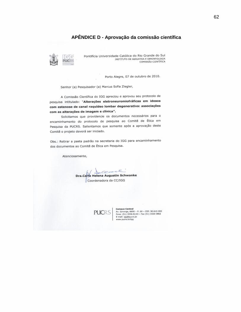