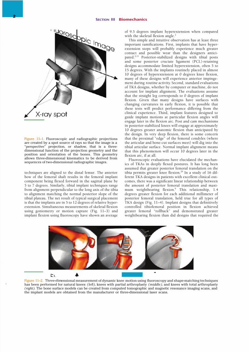
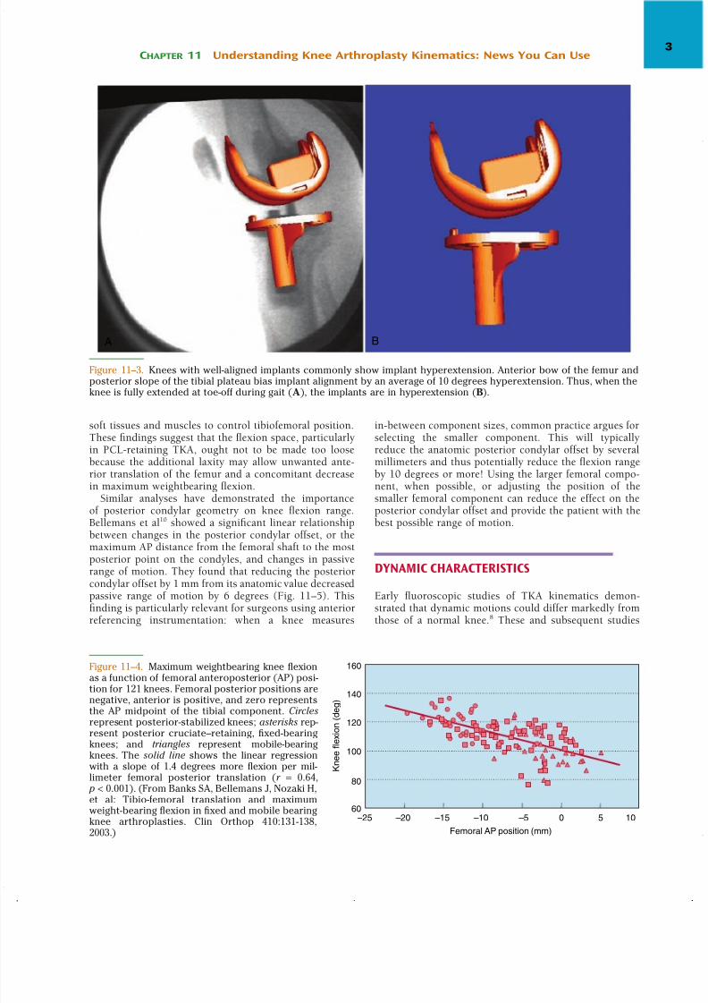
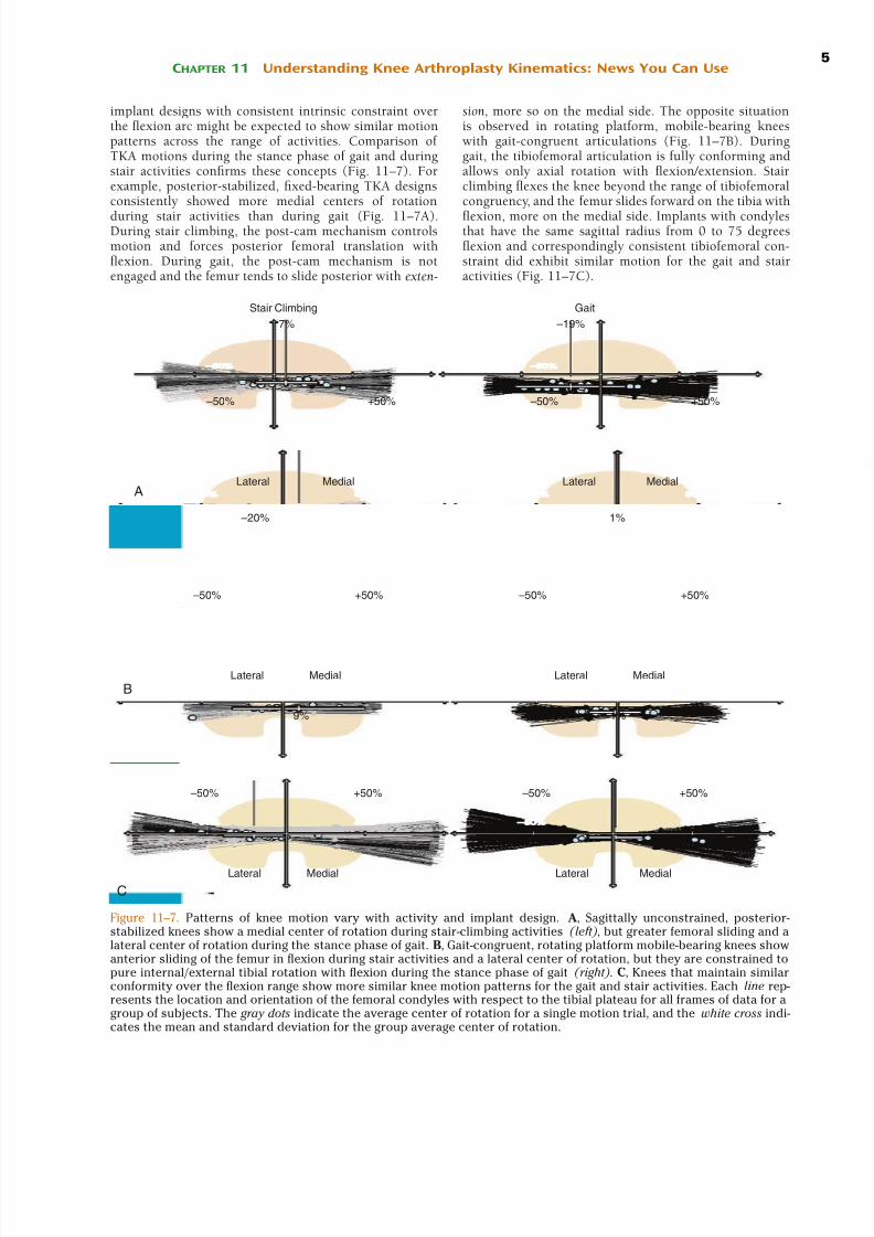
Radiographic imaging and shape-matching techniques have been used since the late 1980s to quantify the motions of knee replacements in vivo. These studies have shown how knee implants move in vivo, how implant design affects knee kinematics, and how different surgical and design factors influence knee mechanics and patient function. In general, knee implants that definitively control the anteroposterior (AP) position of the femur with respect to the tibia achieve greater weightbearing flexion and exhibit kinematics that are more likely to result in better patient function and implant longevity. THREE-DIMENSIONAL KINEMATICS FROM TWO-DIMENSIONAL IMAGES By the late 1980s, total knee arthroplasty (TKA) had become a fairly routine procedure for the treatment ofsevere knee arthritis. A wide variety of implant designs were being used with predictable success and reasonable durability. The focus of designers was shifting from basic knee function and implant fixation to improvement in knee performance and implant longevity. In part, what was needed to continue evolving knee replacements was more precise information on how knee replacements moved once implanted. Unfortunately, the gait laboratories and computed tomography (CT) scanners of the day could not provide accurate three-dimensional kinematic informa- tion of knee replacement motion during weightbearing dynamic activities. In 1988, my surgeon colleague W. Andrew Hodge and I set out to develop a better method for measuring knee arthroplasty kinematics. Having failed to use the gait lab- oratory motion capture system to accurately measure implant motion, Hodge suggested that we should directly image the joint with x-ray fluoroscopy and develop an image-based measurement technique. I developed a “shape-matching”–based measurement approach that worked well, 1,3 and this technique and evolved forms have been used since to provide a better understanding of knee replacement function. The details of shape-matching–based motion measure- ment are beyond the scope of th is volume, but the p rocess follows logically: radiographic images are produced when x-rays pass through space and are attenuated by the patient’s anatomy before striking a sensitive medium and causing a chemical or electrical reaction. The x-ray beam emanates from a single point in space with rays diverging in all directions to create a perspective projection of the object—in essence a shadow (Fig. 11–1). The location ofthe x-ray source with respect to the image plane can be measured so that the same projection can be reproduced on a computer. Computer-aided design information is available for knee implant components, and bone surfaces can be reconstructed from CT or magnetic resonance imaging (Fig. 11–2), thus making it a simple process to synthesize on the computer images of implants at any pos- sible position. These synthetic views can be iteratively modified until they match the views obtained from patients. Once matched, the positions and orientations ofthe models represent the physical position and orientation of the patients’ implants that created the radiographic projection. Many groups the world over have used shape-matching techniques for determining implant motion from single- plane radiographic views and have studied a range ofactivities, including gait, 12 stair climbing, 13 and deep knee bends. 19 Although details of the methods vary, measure- ment precision for each moving segment is typically 0.5 to 1.0 mm for implant motions parallel to the image plane and 0.5 to 1.0 degree for rotations. Importantly, this is monocular vision, not stereo or binocular, and all these techniques have much reduced accuracy for determining translation perpendicular to the image plane, where pre- cision is typicall y 3.0 to 6.0 mm. Propagating these me as- urement errors to the articular surfaces, one can typically expect measure ment uncertainties of greater than 1.2 mm for single observations of condylar contact or separation. POSITIONAL FINDINGS Findings from image-based TKA studies can be organized into positional and dynamic observations. Positional observations relate closely to how implant design and sur- gical alignment influence articular contact and knee func- tion at the extreme ranges of motion. Knee implants are typically designed to maximize the tibiofemoral contact area with the knee in extension and to accommodate 10 to 15 degrees of hyperextension. Implant wear testing is performed such that the implants reach 0 degrees relative flexion at simulated early stance. Yet neither context takes into account the fact that surgi- cal alignment may place the implants in positions that differ from 0 degrees relative flexion. Femoral components implanted with intramedullary rods or extramedullary CHAPTER 11 Understanding Knee Arthroplasty Kinematics: News You Can Use Scott A. Banks 1


![ARTROPLASTIA TOTAL DO JOELHO [Modo de Compatibilidade]](https://img.document.onl/doc/110x75/5517f23b497959a47a8b460c/artroplastia-total-do-joelho-modo-de-compatibilidade.jpg)