Embed Size (px)
Citation preview

FACULDADE DE MEDICINA DA UNIVERSIDADE DE COIMBRA
TRABALHO FINAL DO 6º ANO MÉDICO COM VISTA À ATRIBUIÇÃO DO
GRAU DE MESTRE NO ÂMBITO DO CICLO DE ESTUDOS DE MESTRADO
INTEGRADO EM MEDICINA
SARA LOPES PETRONILHO
ANTITUMOR ACTIVITY OF SPLICING INHIBITOR
PLADIENOLIDE B IN ERYTHROLEUKEMIA – A
STUDY IN CELL LINES
ARTIGO CIENTÍFICO
ÁREA CIENTÍFICA DE HEMATOLOGIA/BIOLOGIA MOLECULAR
APLICADA
TRABALHO REALIZADO SOB A ORIENTAÇÃO DE:
PROF. DOUTORA ANA BELA SARMENTO ANTUNES CRUZ RIBEIRO
DRA. ANA CRISTINA PEREIRA GONÇALVES
MARÇO/2015

Antitumor activity of splicing inhibitor Pladienolide B in erythroleukemia – a study in cell lines
1
Antitumor activity of splicing inhibitor Pladienolide B in
erythroleukemia – a study in cell lines
Sara Petronilho1, Raquel Alves
2,3,4,, Ana Cristina Gonçalves
2,3,4, Ana Bela Sarmento-
Ribeiro2,3,4,5
1- Medical Student, Faculty of Medicine of University of Coimbra (FMUC), Portugal;
2- Applied Molecular Biology and University Clinic of Hematology, FMUC, Portugal;
3- Center for Neuroscience and Cell Biology (CNC.IBILI), University of Coimbra, Portugal;
4- Center of Investigation in Environment, Genetics and Oncobiology (CIMAGO), FMUC,
Portugal;
5- Hematology Department, Centro Hospitalar Universitário de Coimbra (CHUC), Portugal

Antitumor activity of splicing inhibitor Pladienolide B in erythroleukemia – a study in cell lines
2
Table of Contents
Abstract/Resumo 3
Abbreviations 7
Introduction 9
Materials and Methods 13
Cell culture 13
Cell density and viability analysis 13
Assessment of cell death 14
Cell cycle analysis 15
Spliceosome mutation detection 16
Statistical analysis 17
Results 18
Antitumoral activity of Pladienolide B 18
Cell death analysis 20
Cell cycle analysis 23
SF3B1 mutation analysis 24
Discussion and Conclusion 25
Acknowledgements 29
References 30

Antitumor activity of splicing inhibitor Pladienolide B in erythroleukemia – a study in cell lines
3
Abstract
The splicing of pre-mRNA into functional mRNA, carried out by the spliceosome,
represents a crucial step for the cell's genetic expression. Mutations in some of the
spliceosome’s components have been identified in several hematological malignancies,
including myelodysplastic syndromes and acute myeloid leukemia (AML), which could
constitute a potential therapeutic target to be explored. In this context, we evaluated the
therapeutic potential of a splicing inhibitor, Pladienolide B (Pla-B), in two erythroleukemia
cell-lines.
K562 and HEL cells were incubated in the absence or presence of increasing
concentrations of Pla-B in single dose (from 0.25 to 100 nM) and in daily administration (for
0.5 nM). Cell viability and density were evaluated using the trypan blue method. Cell death
was determined by optical microscopy (May-Grunwald Giemsa staining) and flow cytometry
(FC). Cell cycle analysis was evaluated by FC, using a PI/RNAse solution. DNA sequencing
was performed to assess the presence of SF3B1 mutations in exons 14 and 15.
Treatment with Pla-B significantly decreased the viability and proliferation of the
K562 and HEL cells in a time, concentration and administration schedule dependent manner.
HEL cells were more sensible to Pla-B than K562 cells (after 72 hours of incubation the IC50
was 1.5 nM and 25 nM, respectively), which may be due to different cell genetic
backgrounds. In fact, K562 cells present the BCR-ABL fusion gene and HEL cells the JAK2
V617F mutation. However, SF3B1 mutations in exons 14 or 15 were not detected in any cell
model used, suggesting that the observed cytotoxic effect is not dependent on this
spliceosome mutation. Pla-B induced cell death preferentially by apoptosis and induced also
an accumulation of cells in the G0/G1 phase of the cell cycle.

Antitumor activity of splicing inhibitor Pladienolide B in erythroleukemia – a study in cell lines
4
Our results show that Pla-B induces a cytostatic and cytotoxic effect in K562 and HEL
cells, suggesting that Pla-B could represent a new therapeutic approach in the treatment of
erythroleukemia.
Keywords: Pladienolide B, Splicing inhibitor, SF3B1, AML, Erythroleukemia

Antitumor activity of splicing inhibitor Pladienolide B in erythroleukemia – a study in cell lines
5
Resumo
O splicing de pre-mRNA em mRNA funcional, mediado pelo spliceossoma, representa
uma etapa fundamental na expressão genética da célula. Nos últimos anos, mutações em
alguns dos componentes do spliceossoma foram identificadas em várias neoplasias
hematológicas, incluindo síndromes mielodisplásicos e leucemia mielóide aguda,
representando alvos terapêuticos por explorar. Neste contexto, avaliámos o potencial
terapêutico de um inibidor do splicing, Pladienolide B (Pla-B), em duas linhas celulares de
eritroleucemia.
As células K562 e HEL foram incubadas na presença ou ausência de concentrações
crescentes de Pla-B, em dose única (0.25 nM a 100 nM) e dose diária (0.5 nM). A viabilidade
e a densidade celulares foram avaliadas pelo método de azul tripano. A morte celular foi
determinada por microscopia óptica (coloração de May-Grunwald Giemsa) e citometria de
fluxo (FC). A análise do ciclo celular foi realizada por FC, usando uma solução de PI/RNAse.
Mutações nos exões 14 ou 15 do gene SF3B1 foram pesquisadas através de sequenciação do
DNA.
O tratamento das células K562 e HEL com Pla-B reduziu significativamente a
viabilidade e proliferação celulares, de um modo dependente de tempo, concentração e modo
de administração do fármaco. As células HEL mostraram-se mais sensíveis ao fármaco do que
as células K562 (após 72 horas de incubação, o IC50 foi de 1.5 nM e 25 nM, respectivamente),
o que pode ser devido a diferenças genéticas. De facto, as células K562 apresentam o gene de
fusão BCR-ABL, enquanto as HEL apresentam a mutação do JAK2 V617F. No entanto, não
foram encontradas mutações, em nenhum dos modelos, nos exões 14 ou 15 do gene SF3B1, o
que sugere que o efeito citotóxico observado não é dependente desta mutação. Verificámos
que o Pla-B induz morte celular preferencialmente por apoptose, bem como induz uma

Antitumor activity of splicing inhibitor Pladienolide B in erythroleukemia – a study in cell lines
6
acumulação das células na fase G0/G1 do ciclo celular. Mutações nos exões 14 e 15 do gene
SF3B1 foram excluídas.
Os nossos resultados sugerem que o Pla-B apresenta um efeito anti-proliferativo e
citotóxico em ambas as linhas celulares, e que poderá representar uma nova abordagem
terapêutica no tratamento da eritroleucemia.
Palavras-Chaves: Pladienolide B, inibidor do splicing, SF3B1, LMA, eritroleucemia

Antitumor activity of splicing inhibitor Pladienolide B in erythroleukemia – a study in cell lines
7
Abbreviations
AEL : acute eryrholeukemia
AML: acute myeloid leukemia
AML-MRC: acute myeloid leukemia with myelodysplasia-related changes
APC: allophycocyanin
ATCC: american type culture collection
ATP: adenosine triphosphate
AV: annexin V
CML: chronic myelogenous leukemia
ddNTP: dideoxy nucleoside triphosphates
DMSO: Dimethyl sulfoxide
FAB: french-american-british
FBS: fetal bovine serum
FC: flow cytometry
IC50: half maximal inhibitory concentration
MDS: myelodysplastic syndrome
MDS/MPN: myelodysplastic syndrome/myeloproliferative syndrome
MIF: mean intensity fluorescence

Antitumor activity of splicing inhibitor Pladienolide B in erythroleukemia – a study in cell lines
8
mRNA: messenger ribonucleic acid
PBS: phosphate buffer solution
PI: propidium iodide
Pla-B: pladienolide B
PRPF40B: Pre-mRNA processing factor 40 homolog B
RNA: ribonucleic acid
RPMI: roswell park memorial institute
RUNX1: runt related transcription factor 1
PCR: polymerase chain reaction
SEM: standard error of the mean
SF1: splicing factor 1
SF3B: splicing factor subunit 3
SRSF2: serine/arginine-rich splicing factor 2
snRNA: small nuclear ribonucleic acids
TET2: ten eleven translocation 2 gene
U2AF: U2-associated factor
WHO: world health organization
ZRSR2: zinc finger, RNA-binding motif and serine/arginine rich 2

Antitumor activity of splicing inhibitor Pladienolide B in erythroleukemia – a study in cell lines
9
Introduction
Splicing is a fundamental cellular process by which the non-coding sequences known
as introns are removed from pre-mRNA and the flanking exons are ligated to form functional
mRNA. This complex multistep process is carried out by the spliceosome, a macromolecule
composed of five small nuclear ribonucleic acids (snRNA) and numerous associated proteins
(Figure 1) [1]. It starts with the spliceosome assembly phase, with formation of the E (Early)
complex, which results from the ligation of the U1 snRNP to the 5’ splice site and from the
U2AF (U2-Associated Factor) to the 3’ splice site and adjacent polypyrimidine tract
(mediated by the subunits U2AF1 and U2AF2, respectively). This step is followed by an ATP
dependent alteration in the transcript positioning which brings together the two exons and
consequently facilitates the ligation of the U2 snRNP to the branch point sequencing,
generating the A complex. The SF3B1 is the part of this U2 snRNP which mediates the
binding to the intronic branch point sequence. The B complex is later formed by addition of
the U4/U5/U6, leading to an ATP dependent conformational rearrangement which constitutes
a catalytically active C complex, with the resultant release of U2AF, U1 and U4 snRNPs.
Finally, two transesterifications occur and the intronic sequence is removed, with consequent
connection of the contiguous exons [2].
Somatic mutations interfering with the splicing process have been identified in several
malignancies, mostly in hematological neoplasms [2]. They were found to be especially
prevalent in patients presenting with myelodysplastic syndromes (MDS), a heterogeneous
group of diseases characterized by cell line dysplasia, ineffective hematopoiesis, peripheral
cytopenias and higher predisposition to acute myeloid leukemia (AML) [2]. The most
common mutations were found in U2AF1, SRSF2 and SF3B1, the latter being associated with
the specific phenotype of ringed sideroblasts. These mutations were heterozygous, mutually

Antitumor activity of splicing inhibitor Pladienolide B in erythroleukemia – a study in cell lines
10
exclusive and often missense, occurring in specific hotspots for SF3B1 (in exons 14 and 15)
and SRSF2. U2AF1 mutations affected exons 2 and 6, which correspond to the 2 zinc finger
domains of the protein. Interestingly, evidence shows that the mutations did not result in a
widespread splicing dysfunction, but rather in alterations of the splicing pattern of specific
genes. For instance, U2AF1 mutations were associated with defective splicing of intron 5 of
TET2 gene, whereas SF3B1 and SRSF2 mutations affected splicing of RUNX1 gene [3].
Figure 1. Schematic representation of the pre-mRNA splicing process, from J. Boultwood et al. in [1]. The
E complex is formed by binding of the U1 snRNP to the 5’ splice site and SF1, SRSF2, ZRSRS2, U2AF35 and
U2AF65 to the 3’ splice site. The U2 snRNP replaces SF1 and binds to the branch site, forming complex A, and
aids the binding of U4/U5/U6 snRNP, to form complex B. This brings the two adjacent splice sites together and
forms complex C, with subsequent intron removal and ligation of the contiguous exons.

Antitumor activity of splicing inhibitor Pladienolide B in erythroleukemia – a study in cell lines
11
Given this new genetic insight, over the past few years several substances that target
the splicing process have been tested as antineoplastic agents in different malignancies,
including breast, brain, colon, lung, ovarian, renal, gastric and prostate neoplasias in vitro
[4,5]. Some in vivo studies have also been conducted, both in animal models [4,5] and in
patients with advanced solid tumors [6], in the context of a phase I study. Between these new
drugs is Pladienolide B (Pla-B), a twelve-membered macrolide ring, which inhibits the
splicing process through direct targeting of the SF3B complex [7]. Various studies have
shown its strong antitumor activity against the previously stated human cancer cell lines, as
well as in primary cultures, with an IC50 value in a low nanomolar range. Moreover, it has
been proven to inhibit tumor growth and even induce complete regression in colon and gastric
human cancer xenograph models in mice [4,5]. Furthermore, since Pla-B has shown a potent
effect in cell lines resistant to conventional drugs [4] it could be a viable option in the
treatment of refractory disease. However, to our knowledge, no data are available regarding
the antineoplastic effect of this drug against hematological malignancies. The fact that
spliceosome mutations are present in a variety of myeloid malignancies suggests that these
could play an important role in defining a malignant phenotype.
Acute erythroleukemia (AEL) is a rare type of AML which consists of a clonal
proliferation of erythroid precursors and usually also other myeloid precursors in bone
marrow . Until recently, it was classified as an M6 AML, according to the French-American-
British (FAB) classification system [8]. In 2008, the WHO proposed a new classification,
where the category of AML with myelodysplasia-related changes (AML-MRC) was
proposed. This category includes all cases of AML with presence of either morphologic
evidence of significant multilineage dysplasia, specific MDS–related cytogenetic
abnormalities, or a history of MDS or a MDS/MPN. Therefore, some AEL cases are now
classified as AML-MRC. Cases with blasts comprising less than 20% of all cells but more

Antitumor activity of splicing inhibitor Pladienolide B in erythroleukemia – a study in cell lines
12
than 50% of erythroid precursors are classified as AEL, if blasts constitute 20% or more of the
nonerythroid cells. . If blasts constitute less than 20% of nonerythroid cells, the cases are
classified into various categories of myelodysplastic syndromes (MDS). In a retrospective,
multi-institutional study of patients with AEL conducted in 2010 [9], the majority of patients
presented a secondary leukemia, whether with a previous history of MDS, chronic cytopenia
or therapy-related disease. Furthermore, AEL is frequently accompanied by unfavorable
cytogenetics. Another study [10] indicated that the outcome is better for patients treated with
hypomethylating agents when compared to standard cytarabin based chemotherapy. However,
the prognosis was still poor, with a median overall survival time of 15.4 months in the
patients who received azacytidine. Thus, the development of novel drugs for the better
management of this disease is imperative.
In this context, we studied the antineoplastic effect of Pladienolide B in two
erythroleukemia cell lines, the K562 and HEL cells. We evaluated the effect of this new drug
in cell growth and viability, and performed a cell death and cell cycle analysis, in order to
better elucidate the mechanism of action of this drug. In addition, both cell lines were
screened for the presence of mutations in the SF3B1 gene, to correlate mutation status with
Pla-B efficacy.

Antitumor activity of splicing inhibitor Pladienolide B in erythroleukemia – a study in cell lines
13
Materials and Methods
Cell culture: In this study we used two different cell lines, K562 and HEL, which were
purchased from the American Type Culture Collection (ATCC). The K562 cells were
originally obtained from the pleural effusion of a 53-year-old female with chronic
myelogenous leukemia (CML) in blast crisis (characterized as erythroleukemia) and bear the
translocation (9; 22), with consequent presence of the Philadelphia Chromosome [11]. The
HEL cells were originally obtained from the bone marrow of a 30–year-old caucasian male
with erythroleukemia and are JAK2-V617F mutated [12]. All cell lines were routinely grown
in an advanced RPMI-1640 medium (L-glutamine 2 mM, HEPES-Na 25 mM, penicillin 100
U/mL and Streptomycin 100 µg/mL), supplemented with 5% heat-inactivated fetal bovine
serum at 37ºC in a humidified atmosphere containing 5% CO2. Cells were cultured at initial
density of 0.5 x 10⁶ cells/mL for the K562 and 0.4 x 10
6 cells/mL for the HEL cell line.
Pladienolide B (Pla-B) was purchased from Santa Cruz Biotechnology (UK) and dissolved in
DMSO. Both the HEL and K652 cell lines were incubated for 72 hours in the absence and in
the presence of increasing concentrations of Pla-B, ranging from 0.25 to 100 nM. The drug
was administered in single dose and for 0.5 nM of Pla-B was also tested a daily administration
scheme. For flow cytometry and morphological studies was used 2.5 nM of Pla-B.
Cell viability and density analysis: The K652 and HEL cells viability and density was
accessed every 24 hours during 72 hours by the trypan blue exclusion method. This method is
based on the principle that live/viable cells possess intact cell membranes that exclude the
dye, whereas dead/non-viable cells do not. Briefly, equal volumes of cell suspension and
trypan blue (Sigma Aldrich) were added, loaded in a hemacytometer and counted under a
microscope. Cell viability was calculated as the percentage of viable cells whereas cell
Antitumor activity of splicing inhibitor Pladienolide B in erythroleukemia – a study in cell lines
14
density was determined by the number of viable cells. The IC50 (drug concentration that
reduces the viability in 50%) was evaluated by a dose response curve.
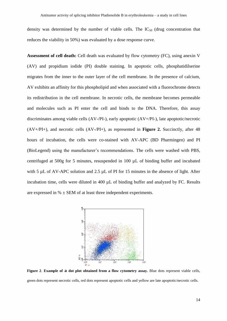
Assessment of cell death: Cell death was evaluated by flow cytometry (FC), using anexin V
(AV) and propidium iodide (PI) double staining. In apoptotic cells, phosphatidilserine
migrates from the inner to the outer layer of the cell membrane. In the presence of calcium,
AV exhibits an affinity for this phospholipid and when associated with a fluorochrome detects
its redistribution in the cell membrane. In necrotic cells, the membrane becomes permeable
and molecules such as PI enter the cell and binds to the DNA. Therefore, this assay
discriminates among viable cells (AV-/PI-), early apoptotic (AV+/PI-), late apoptotic/necrotic
(AV+/PI+), and necrotic cells (AV-/PI+), as represented in Figure 2. Succinctly, after 48
hours of incubation, the cells were co-stained with AV-APC (BD Pharmingen) and PI
(BioLegend) using the manufacturer’s recommendations. The cells were washed with PBS,
centrifuged at 500g for 5 minutes, resuspended in 100 µL of binding buffer and incubated
with 5 µL of AV-APC solution and 2.5 µL of PI for 15 minutes in the absence of light. After
incubation time, cells were diluted in 400 µL of binding buffer and analyzed by FC. Results
are expressed in % ± SEM of at least three independent experiments.
Figure 2. Example of a dot plot obtained from a flow cytometry assay. Blue dots represent viable cells,
green dots represent necrotic cells, red dots represent apoptotic cells and yellow are late apoptotic/necrotic cells.
Antitumor activity of splicing inhibitor Pladienolide B in erythroleukemia – a study in cell lines
15
The ApoStat Probe is designed to identify and quantify caspase activity in cells by FC.
About 1x10⁶ cells were resuspended in 1000 µL of PBS and incubated with 1µg of ApopStat.
After a 15 min incubation period at 37°C, the cells were washed and resuspended in 400 µL
of PBS for analyzed by FC. Results are represented in % ± SEM and in MIF (mean
fluorescence intensity) ± SEM, which represents the mean fluorescence intensity detected in
cells and is proportional to the number of molecules labeled with the antibody.
Cell death essays were conducted using a FACSCalibur flow cytometer (Becton
Dickinson, San Jose, CA) and at least 10.000 events were collected by acquisition using
CellQuest software (Becton Dickinson, San Jose, CA) and analyzed using Paint-a-Gate
(Becton Dickinson, San Jose, CA).
Morphological studies were conducted to evaluate the alterations induce by exposure to Pla-
B. After 48 hours of incubation, the cells were centrifuged at 1000xg for 5 minutes, being the
supernatant excluded. The pellet was resuspended in 10 µL of FBS. Smears were stained for 3
minutes with May-Grünwald solution, and then for 15 minutes with Giemsa solution. After
rinsing with distilled water, cell morphology was analyzed by light microscopy (Nikon
Eclipse 80i microscope equipped with a Nikon digital camera DXm 1200F).
Cell cycle analysis: Cell cycle analysis was performed by FC, using PI/RNAse solution
(Immunostep). As previously said, PI is a fluorescent dye that stains DNA in permeable cells.
The fluorescence intensity, read by FC, is proportional to the DNA quantity of each cell,
allowing us to determine the relative proportion of cells in the G0/G1 phase (fewer amount of
DNA), S phase (coincident with DNA replication) and G2/M phase (double DNA of the
G0/G1 phase). Given that apoptotic cells undergo the process of DNA fragmentation, these
cells are represented as an apoptotic peak pre-G1, with the fewest DNA quantity. Briefly,
after 48 hours of incubation, cells were collected and washed with PBS for 5 min at 1000 xg.

Antitumor activity of splicing inhibitor Pladienolide B in erythroleukemia – a study in cell lines
16
The pellet was resuspended in 200 µL of ice cold 70% ethanol solution, during vortex
agitation, being incubated during 30 min on cold. Then, cells were washed with PBS,
resuspended in 400 µL of PI/RNase solution and analyzed by FC. A FACSCalibur flow
cytometer (Becton Dickinson, San Jose, CA) was used and at least 10.000 events were
collected by acquisition using CellQuest software (Becton Dickinson, San Jose, CA). Results
were analyzed using Modfit software (Becton Dickinson, San Jose, CA) and are expressed in
% ± SEM of at least 3 independent experiments.
Spliceosome mutation detection: The presence or exclusion of mutations on exons 14 and
15 of the SF3B1 gene was analyzed using the Sanger sequencing method. This method allows
DNA sequencing by selective incorporation of chain terminating ddNTP by a DNA
polymerase during in vitro DNA replication. Briefly, the SF3B1 fragment was amplified using
PCR primers flanking exons 14 and 15 (Table 1).
Table 1. Sequences of primers used in the present study
PCR was performed in a reaction volume of 24 µl containing 2 µl of genomic DNA, 0.2µl of
Taq polymerase 5U/µl, 2.5 µl of Taq PCR Buffer, 0.5 µl of dNTP (10 mM each) and 0.5 µl of
each of the primers. The PCR program was as follows: initial denaturation at 95 °C for 3
minutes and 34 cycles of amplification at 95 °C for 45 seconds, 60°C or 54ºC (for exon 14 or
Primer Sequence (5’-3’) Fragment Size (bp)
SF3B1 exon 14F CCAACTCATGACTGTCCTTTCTT 362
SF3B1 exon 14R GGGCAACATAGTAAGACCCTGT
SF3B1 exon 15F TTGGGGCATAGTTAAAACCTG 261
SF3B1 exon 15R AATCAAAAGGTAATTGGTGGA

Antitumor activity of splicing inhibitor Pladienolide B in erythroleukemia – a study in cell lines
17
15, respectively) for 45 seconds and 72 °C for 45 seconds. A final extension step at 72 °C for
10 minutes was performed. After purification of the amplicons using the ExoSAP-IT enzyme,
according to the manufacturer’s protocol, cycle sequencing was performed using a Big Dye
Terminator v3.1 Cycle Sequencing kit, following the manufacturer’s instructions. The thermal
cycling profile was as follows: initial denaturation at 94 °C for 3 minutes and 24 rounds of
amplification at 96 °C for 10 seconds, 50°C for 5 seconds and 60 °C for 1 minute and 45
seconds. The products of the sequencing reactions were purified using DyeEx 2.0 columns
and read in an optic microplate. Sequencing data were analyzed using Chromas software.
4.6 Statistical analysis: Statistical analysis was performed using the GraphPad Prism
software. Student’s t test, analysis of variance, Dunnett’s test and Tuckey test were used to
compare the different groups. A significance level of p < 0.05 was considered statistically
significant. Results are expressed in mean ± SEM of the number of independent experiments
indicated in the figure legend.
Antitumor activity of splicing inhibitor Pladienolide B in erythroleukemia – a study in cell lines
18
Results
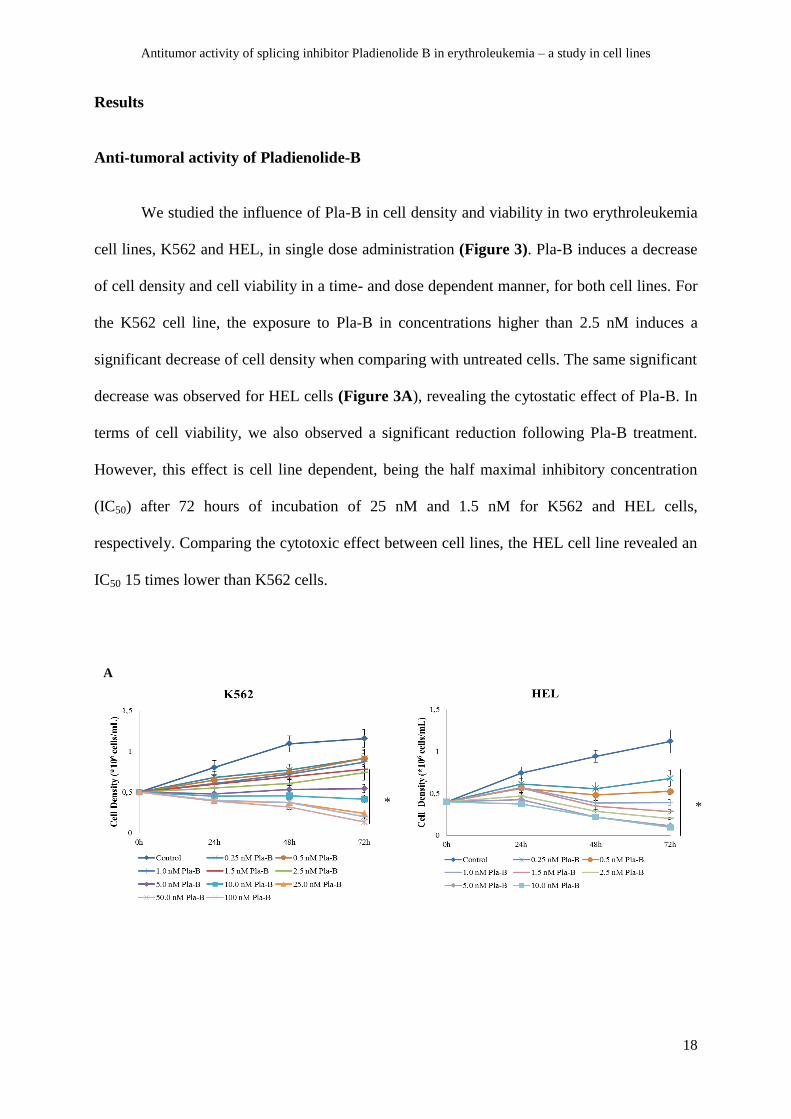
Anti-tumoral activity of Pladienolide-B
We studied the influence of Pla-B in cell density and viability in two erythroleukemia
cell lines, K562 and HEL, in single dose administration (Figure 3). Pla-B induces a decrease
of cell density and cell viability in a time- and dose dependent manner, for both cell lines. For
the K562 cell line, the exposure to Pla-B in concentrations higher than 2.5 nM induces a
significant decrease of cell density when comparing with untreated cells. The same significant
decrease was observed for HEL cells (Figure 3A), revealing the cytostatic effect of Pla-B. In
terms of cell viability, we also observed a significant reduction following Pla-B treatment.
However, this effect is cell line dependent, being the half maximal inhibitory concentration
(IC50) after 72 hours of incubation of 25 nM and 1.5 nM for K562 and HEL cells,
respectively. Comparing the cytotoxic effect between cell lines, the HEL cell line revealed an
IC50 15 times lower than K562 cells.
A
* *

Antitumor activity of splicing inhibitor Pladienolide B in erythroleukemia – a study in cell lines
19
Figure 3. Dose-response curves of Pla-B in K562 and HEL cell lines. Cells were incubated in a density of 0.5
and 0.4 x 10⁶ cells/mL, respectively, for 72 hours, in the absence and presence of increasing concentrations of
Pla-B. Cell density (A) and viability (B) were established by the trypan blue method, as previously described in
material and methods. Cell viability is expressed in cell percentage, normalized to control. Data are expressed as
mean ± SEM obtained from at least 5 independent experiments. * p<0.05
To assess if the frequency of administration influences the cytostatic and cytotoxic
effect of Pla-B, a daily administration scheme was tested using the concentration of 0.5 nM
(Figure 4). A daily administration of 0.5 nM was compared with 1.5 nM of Pla-B at single
dose, since this corresponds to the cumulative dose at the end of 72 hours. The daily
administration of Pla-B reveals to be more effective in the reduction of cell density and cell
viability than the administration of 1.5 nM at single dose, especially in the HEL cells. After
72 hours of exposure, the daily dose induced an additional reduction of 15% and 45% in K562
and HEL cells viability, respectively, when compared to the single dose. The differences in
cell viability induced by the diferent schemes of drug adminitration were statistical
significant.
*
B
*

Antitumor activity of splicing inhibitor Pladienolide B in erythroleukemia – a study in cell lines
20
Figure 4. Dose response curves for daily administration of Pla-B in K562 and HEL cell lines. Cells were
incubated in a density of 0.5 and 0.4 x 10⁶ cells/mL, respectively, for 72 hours, in the absence (control) and
presence Pla-B, in single or daily administration (#). Cell density (A) and viability (B) were established by the
trypan blue method, as previously described. Cell viability is expressed in cell percentage, normalized to control.
Data are expressed as mean ± SEM obtained from at least 5 independent experiments. * p<0.05 comparing with
1.5nM of Pla-B at single dose.
Cell death analysis
We analyzed the cell death induce by Pla-B using Annexin V/PI double staining and
by optical microscopy. As represented in figure 4, the exposure to 2.5 nM of Pla-B results in a
significant reduction of viable cells with a significant increase of cells in early apoptosis. The
cell death induced was mainly mediated by apoptosis, but in case of HEL cells, we also
observed significant differences in percentage of cells in necrosis.
A
B
*
*

Antitumor activity of splicing inhibitor Pladienolide B in erythroleukemia – a study in cell lines
21
Figure 5. Cell death analysis induced by Pla-B in K562 and HEL cell lines using flow cytometry. Cells were
incubated in a density of 0.5 and 0.4 x 10⁶ cells/mL, respectively, for 48 hours, in the absence (control) or
presence of 2.5nM of Pla-B. Cell death was detected by annexin V/ propidium iodide staining, analyzed by flow
cytometry. Data are expressed in percentage (%) and represent mean ± SEM of at least 3 independent
determinations. * p<0.05, ** p<0.01, *** p<0.001.
In agreement with flow cytometry analysis, the morphological evaluation showed
typical alterations of cell death mediated by apoptosis in both cell lines. After treatment with
Pla-B it was possible to observe blebbing, cell shrinkage, nuclear fragmentation and
chromatin condensation, confirming apoptosis (Figure 6). In the HEL cell line, we also
observed cells with permeable membrane and intact nucleus characteristic of necrosis,
revealing the activation of both cell death mechanisms.
HEL
K562
***
***
** **
**
*
A
B
Control 2.5 nM Pla-B
Control 2.5 nM Pla-B

Antitumor activity of splicing inhibitor Pladienolide B in erythroleukemia – a study in cell lines
22
Figure 6. Cell morphological analysis by optical microscopy . K562 and HEL cells were incubated in a
density of 0.5 and 0.4 x 10⁶ cells/mL, respectively for the K562 (A) and HEL (B) cells, for 48 hours, in the
absence (control) or presence of 2.5 nM of Pla-B and stained using a May-Grünwald-Giemsa method. The cells
were analyzed by light microscopy (amplification 100x).
We also analyzed if cell death induced by Pla-B was mediated by caspases activation
(Figure 7). Treatment with Pla-B induced an increase in percentage of cells with activated
caspases (of 13% for the K562 cells and 30% for the HEL cells, represented in Figure 7A)
and in the expression level of caspases (of 5% in the HEL cells, represented in Figure 7B),
being more pronounced in HEL cells. The differences in percentage of cells were statistical
significant for HEL cell line.
Figure 7. Evaluation of caspases expression levels in K562 and HEL cells treated with Pla-B by flow
cytometry. Cells were incubated in a density of 0.5 and 0.4 x 10⁶ cells/mL, respectively, for 48 hours, in the
absence (control) or presence of 2.5nM of Pla-B. Caspases expression levels were analyzed using the ApoStat
probe. Results are expressed in percentage of cells positive (A) and in MIF (B) and represent the mean ± SEM
of at least 3 independent experiments. ** p<0.01
K562 K562 HEL
HEL
A B
**

Antitumor activity of splicing inhibitor Pladienolide B in erythroleukemia – a study in cell lines
23
Cell cycle analysis
Besides the confirmed cytotoxic effect induced by Pla-B, to confirm the cytostatic effect,
we analyzed the effect on cell cycle progression by FC using a PI/RNAse. As represented in
Figure 8, Pla-B induced a significant arrest in the G0/G1 phase in both cell lines, when
compared with untreated cells (respectively, 44±1.9% vs 37±1% in K562 cells and 63±0.9%
vs 35±2.1% in HEL cells). Additionally, by the same technique it was detected the presence
of an apoptotic peak (pre-G1), corresponding to DNA fragmentation typical of apoptotic cells.
Figure 8. Cell cycle distribution of K562 and HEL cells after Pla-B treatment. Cells were incubated in a
density of 0.5 and 0.4 x 10⁶ cells/mL, respectively, for 48 hours, in the absence (control) or presence of 2.5 nM
of Pla-B. Cell cycle distribution was detected using PI/RNAse by flow cytometry. Data are expressed as
percentage of cells in G0/G1 phase, S phase, G2/M phase and apoptotic peak and represent mean ± SEM
obtained from at least 3 independent experiments. * p<0.05, ** p<0.01, *** p<0.001.
K562 HEL
***
*
**
***
**

Antitumor activity of splicing inhibitor Pladienolide B in erythroleukemia – a study in cell lines
24
SF3B1 mutation analysis:
The mutational status of SF3B1 could influence the response levels of cells to Pla-B.
Thus, using the previously described DNA sequencing method, we searched for mutations in
exons 14 and exon 15 of the SF3B1 gene in both cell lines. For our in vitro models, we did not
find any mutations in these two exons. Particularly, no substitutions were revealed in
positions H662D or K700E, respectively.
Antitumor activity of splicing inhibitor Pladienolide B in erythroleukemia – a study in cell lines
25
Discussion and Conclusion
In this study, we evaluated the therapeutic efficacy of Pladienolide B (Pla-B), a
splicing inhibitor that specifically targets the ribonucleoprotein complex SF3B. By impairing
the binding of the spliceosome to the branch sequence, it leads to a splicing dysfunction that
results in an accumulation of un-spliced mRNA in the cell and consequently in a reduction of
cell viability [7, 13]. Nevertheless, its mechanism of action is still not fully understood. As
mutations in spliceosome components are extremely prevalent in myeloid neoplasias [3], this
drug could represent a new therapeutic target in the treatment of such diseases.
Our study shows that Pla-B presents a cytotoxic and a cytostatic effect against
erythroleukemia cell lines in a time, dose and administration scheme dependent manner.
Induction of cell death was mainly mediated by apoptosis, with activation of caspases. These
results were in accordance with the experimental study conducted by Sato et al. in gastric
cancer cell lines, which reveal the activation of apoptotic cell death [5]. We verified that Pla-
B induces an accumulation of cells in the G0/G1 phase, reflecting the anti-proliferative action
of this drug. A previous study conducted by Mizui et al. in WiDr cells had shown a
preferential arrest in G1 phase after treatment with Pla-B, but also in the G2/M-phase, in a
time dependent manner [4].
The administration scheme has been explored with attempt to minimize the side
effects of anti-cancer agents. We analyzed two different administration schemes, namely the
single dose administration and the small daily dose administration. Our results show that the
daily dose administration of 0.5 nM of Pla-B appears to have more efficacy in both cell lines
when compared with 1.5 nM at single dose, since it induces a higher cytotoxicity. Daily
administration scheme could constitute a valid option to reduce the potential side effects of
Pla-B.
Antitumor activity of splicing inhibitor Pladienolide B in erythroleukemia – a study in cell lines
26
Interestingly, Pladienolide B also presented a cell type dependent effect. In fact, HEL
cells were much more sensitive to Pla-B than K562 cells, with an IC50 at 72 hours of exposure
of 1.5 nM vs 25 nM, respectively. Further studies are needed to disclose the exact reason by
which this phenomenon occurs; however, several differences between these two cell lines can
potentially contribute to the differential response.
Firstly, although both K562 and HEL are erythroleukemia cell lines, they have a
different origin and genetic background. As previously described, K562 cells were first
obtained from a patient with a CML in blast crisis, and present the translocation between
chromosomes 9 and 21, that originates Philadelphia chromosome and the fusion gene BCR-
ABL [11]. The HEL cell line, on the other hand, was obtained from a patient with an
erythroleukemia, who had previously received treatment for a Hodgkin Lymphoma, and
present the JAK2-V617F mutation [12]. These genetic differences may contribute to the
different drug-responses. In fact, the presence of the BCR-ABL fusion gene has been
associated with changes in the expression of proteins involved in alternative pre-mRNA
splicing, a process which greatly diversifies the transcriptome and which is thought to
contribute to the oncogenic processes. [14] Additionally, the BRC-ABL p210
oncoprotein has
been shown to increase the expression of multiple genes involved in pre-mRNA splicing [14].
These aberrances in the splicing process can lead to important cell dysfunction, with a
considerable effect in the regulation of cell proliferation and apoptosis [15]. Thus, it would be
necessary to study the splicing function of the K562 cells in order to better understand the
lower drug-sensitivity and if response is correlated with previous splicing dysfunction.
Several authors have also hypothesized that cells may react differently to Pla-B
according to the presence or absence of different splicing factor mutations [1,2,16,18]. In the
present study, only exons 14 and 15 of gene SF3B1 were screened for mutations. As stated in

Antitumor activity of splicing inhibitor Pladienolide B in erythroleukemia – a study in cell lines
27
the results section, no mutations were found in the K562 or the HEL cell lines in neither exon.
However, we did not exclude other mutations in the same or in a different gene. The possible
mutation in other point may also contribute to the different response levels. H662D (C>G)
and K700E (A>G) mutations in exons 14 and 15, respectively, have been described as
hotspots in SF3B1 gene. Even though these mutations are between the most frequents in
hematological neoplasias [16], numerous other mutations have been identified, both in SF3B1
and in other splicing related genes. Particularly, the mutation R1074H in SF3B1 gene has
been associated with resistance to Pladienolide B in colorectal carcinoma cells in vitro [7].
The mutation confers resistance since it impairs the binding of Pla-B to the target [7].
However, a mutation in this residue has so far not been detected in patients [2].
Other mutated genes which affect splicing that have been identified in myeloid
neoplasias may also influence the efficacy of drugs that target this process. These include
mutations in genes SRSF2, U2AF1, ZRSR2, SF3A1, U2AF2, SF1 and PRPF40B [16].
Therefore, and given the high occurrence of mutations affecting these genes in myeloid
neoplasias [1,17], we cannot exclude their existence in our models. Nevertheless, it is
important to realize that even though the presence of a mutation may explain a different
response to the drug, the cause-effect mechanism is not linear or well understood. In fact, it is
not yet well established whether splicing mutations represent a gain or loss of function,
although the presence of hot spots and the absence of nonsense or frameshift changes suggest
that the vast majority are gain of function/neomorphic mutations [1]. Hypothetically, in the
presence of a gain of function/neomorphic mutation, the use of specific spliceosome inhibitors
may allow a preferential targeting of the mutant phenotype. Moreover, since splicing factor
mutations appear to be mostly heterozygous [3], disruption of the remaining allele using
splicing inhibitors may induce preferential killing of the mutant cells and, eventually, spare
normal cells. Additionally, it has been proposed that these mutations may result in aberrant

Antitumor activity of splicing inhibitor Pladienolide B in erythroleukemia – a study in cell lines
28
alternative splicing, in which case the splicing inhibitor may inhibit the gain of function effect
of the mutation and restore a normal phenotype [18]. On the other hand, in loss of function
mutations/dominant-negative effect, the use of splicing inhibitors may result in dysfunction of
the normal allele, with worsening of the phenotype [2]. Therefore, not only is it important to
identify the mutations, but also to better characterize their effect, in order to improve our
understanding and prediction of drug response.
Moreover, a study conducted in gastric cell lines using a Pla-B derivative associated a
higher expression of cell cycle proteins, such as cyclin E and p16, with higher sensitivity to
this drug [5]. The authors suggest that these molecules could be used as a biological
biomarkers in the future, although a more profound analysis in needed [5]. Therefore,
differences in the expression of these molecules in our cell lines should be tested in a
posterior study to clarify the different sensitivity to Pla-B.
Undoubtedly, further studies are needed to clarify the mechanism of action of Pla-B,
as well as its potential role in the treatment of hematological malignancies. In particular,
understanding the reason why these two cell lines respond differently in the presence of the
drug may help to understand the mechanism of action of Pla-B, as well as the profile of
patients which may benefit from this treatment.
In conclusion, Pladienolide-B shows a high antitumor activity against HEL and K562
cell lines, inducing cell death preferentially by apoptosis and a G0/G1 cell cycle arrest. Even
though further elucidations are needed, Pla-B most certainly represents a promising new
approach in the treatment of erythroleukemia.

Antitumor activity of splicing inhibitor Pladienolide B in erythroleukemia – a study in cell lines
29
Acknowledgements
This project was supported by Center of Investigation in Environment, Genetics and
Oncobiology (CIMAGO).
I thank Doutora Letícia Ribeiro and Dra. Margarida Coucelo for their support in DNA
analysis (Hemato Oncology Laboratory of the Pediatric Hospital, Coimbra).
I thank Professor Ana Bela Sarmento, as well as the whole laboratory team, for their
support in numerous occasions, without which this work would not have been possible.

Antitumor activity of splicing inhibitor Pladienolide B in erythroleukemia – a study in cell lines
30
References
1. Boultwood J, Dolatshad H, Varanasi SS, Yip BH, Pellagatti A. The role of splicing factor
mutations in the pathogenesis of the myelodysplastic syndromes. Advances in biological
regulation. 2014;54:153-61.
2. Scott LM, Rebel VI. Acquired mutations that affect pre-mRNA splicing in hematologic
malignancies and solid tumors. Journal of the National Cancer Institute.
2013;105(20):1540-9.
3. Makishima H, Visconte V, Sakaguchi H, Jankowska AM, Abu Kar S, Jerez A, et al.
Mutations in the spliceosome machinery, a novel and ubiquitous pathway in
leukemogenesis. Blood. 2012;119(14):3203-10.
4. Mizui Y, Sakai T, Iwata M, Uenaka T, Okamoto K, Shimizu H, et al. Pladienolides, new
substances from culture of Streptomyces platensis Mer-11107. III. In vitro and in vivo
antitumor activities. The Journal of antibiotics. 2004;57(3):188-96.
5. Sato M, Muguruma N, Nakagawa T, Okamoto K, Kimura T, Kitamura S, et al. High
antitumor activity of pladienolide B and its derivative in gastric cancer. Cancer science.
2014;105(1):110-6.
6. Eskens FA, Ramos FJ, Burger H, O'Brien JP, Piera A, de Jonge MJ, et al. Phase I
pharmacokinetic and pharmacodynamic study of the first-in-class spliceosome inhibitor
E7107 in patients with advanced solid tumors. Clinical cancer research : an official
journal of the American Association for Cancer Research. 2013;19(22):6296-304.
7. Yokoi A, Kotake Y, Takahashi K, Kadowaki T, Matsumoto Y, Minoshima Y, et al.
Biological validation that SF3b is a target of the antitumor macrolide pladienolide. The
FEBS journal. 2011;278(24):4870-80.
Antitumor activity of splicing inhibitor Pladienolide B in erythroleukemia – a study in cell lines
31
8. Santos FP, Faderl S, Garcia-Manero G, Koller C, Beran M, O'Brien S, et al. Adult acute
erythroleukemia: an analysis of 91 patients treated at a single institution. Leukemia.
2009;23(12):2275-80.
9. Hasserjian RP, Zuo Z, Garcia C, Tang G, Kasyan A, Luthra R, et al. Acute erythroid
leukemia: a reassessment using criteria refined in the 2008 WHO classification. Blood.
2010;115(10):1985-92.
10. Vigil CE, Tan W, Wilding GE, Garcia-Manero G, Wang ES, Wetzler M, List AF.
Comparison of outcome in erythroleukemia patients treated with standard chemotherapy
regimens or hypomethylating agents : ASCO Meeting Abstracts Part 1. Journal of clinical
oncology 2011; 29(15 Suppl.):6630.
11. Druker BJ, Tamura S, Buchdunger E, Ohno S, Segal GM, Fanning S, et al. Effects of a
selective inhibitor of the Abl tyrosine kinase on the growth of Bcr-Abl positive cells.
Nature medicine. 1996;2(5):561-6.
12. Quentmeier H, MacLeod RA, Zaborski M, Drexler HG. JAK2 V617F tyrosine kinase
mutation in cell lines derived from myeloproliferative disorders. Leukemia.
2006;20(3):471-6.
13. Effenberger KA, Anderson DD, Bray WM, Prichard BE, Ma N, Adams MS, et al.
Coherence between cellular responses and in vitro splicing inhibition for the anti-tumor
drug pladienolide B and its analogs. The Journal of biological chemistry.
2014;289(4):1938-47.
14. Salesse S, Dylla SJ, Verfaillie CM. p210
BCR/ABL-induced alteration of pre-mRNA
splicing in primary human CD34+ hematopoietic progenitor cells. Leukemia.
2004;18(4):727-33.

Antitumor activity of splicing inhibitor Pladienolide B in erythroleukemia – a study in cell lines
32
15. Adamia S, Pilarski PM, Bar-Natan M, Stone RM, Griffin JD. Alternative splicing in
chronic myeloid leukemia (CML): a novel therapeutic target? Current cancer drug targets.
2013;13(7):735-48.
16. Cazzola M, Rossi M, Malcovati L, Associazione Italiana per la Ricerca sul Cancro
Gruppo Italiano Malattie M. Biologic and clinical significance of somatic mutations of
SF3B1 in myeloid and lymphoid neoplasms. Blood. 2013;121(2):260-9.
17. Papaemmanuil E, Cazzola M, Boultwood J, Malcovati L, Vyas P, Bowen D, et al. Somatic
SF3B1 mutation in myelodysplasia with ring sideroblasts. The New England journal of
medicine. 2011;365(15):1384-95.
18. Visconte V, Makishima H, Maciejewski JP, Tiu RV. Emerging roles of the spliceosomal
machinery in myelodysplastic syndromes and other hematological disorders. Leukemia.
2012;26(12):2447-54.