1
UNIVERSIDADE DO EXTREMO SUL CATARINENSE – UNESC PROGRAMA DE PÓS-GRADUAÇÃO EM CIÊNCIAS DA SAÚDE
– PPGCS
VÂNIA KÁTIA MENEGALLI MOOJEN
EFEITOS DO PRÉ E DO PÓS-CONDICIONAMENTO COM N-METIL-D-ASPARTATO SOBRE O COMPORTAMENTO E A VIABILIDADE CELULAR EM CAMUNDONGOS EXPOSTOS
AO TRAUMATISMO CRÂNIO- ENCEFÁLICO
Tese apresentada ao Programa de Pós-Graduação em Ciências da Saúde da Universidade do Extremo Sul Catarinense, para obtenção do título de Doutora em Ciências da Saúde. Orientador: Profa. Dra Carina R. Boeck
CRICIÚMA, ABRIL DE 2012.
2
Dados Internacionais de Catalogação na Publicação
Bibliotecária Eliziane de Lucca Alosilla – CRB 14/1101 Biblioteca Central Prof. Eurico Back - UNESC
M817e Moojen, Vânia Kátia Menegalli. Efeitos do pré e do pós-condicionamento com N-metil- D-aspartato sobre o comportamento e a viabilidade celular em camundongos expostos ao traumatismo crânio-encefálico / Vânia Kátia Menegalli Moojen ; Orientadora : Carina R. Boeck. – Criciúma : Ed. do Autor, 2012. 92 f. : il. ; 21 cm.
Tese (Doutorado) - Universidade do Extremo Sul Catarinense, Programa de Pós-Graduação em Ciências da Saúde,Criciúma, 2012. 1. Traumatismo cranioencefálico. 2. Lesão cerebral. 3. Memória. 4. Ansiedade. 5. Depressão. 6. N-metil-D- aspartato.I. Título. CDD. 22. ed. 616.81
3

4

5
A minha família.

6
7
AGRADECIMENTOS
A minha orientadora professora Doutora Carina Rodrigues Boeck, que me oportunizou buscar cada vez mais a excelência pessoal e profissional, mostrando que a dedicação e empenho são fundamentais para o êxito.
Ao professor João Luciano Quevedo do Laboratório de Neurociências da Unesc, que não tem medido esforços para tornar o Programa de Pós-Graduaçao em Ciências da Saúde da UNESC, uma referência na área da Pesquisa, bem como ter me incentivado a desenvolver o Doutorado neste Programa de Pós-Graduação.
Aos professores da Pós-Graduação que contribuíram para ampliar a capacidade de buscar os objetivos com maior eficácia.
Aos bolsistas, Bruna Bardini Pescador, Daniela Bavaresco, Douglas Nuernberg de Matos, Michele de Lima Garcez do Laboratório de Biologia Celular e Molecular-LABIM que foram grandes parceiros na busca incansável de conhecimento e agilidade.
A bolsista Gislaine Zilli Réus, que sempre contribuiu incansavelmente na ampliação de conhecimento e aprimoramento.
Ao meu esposo pelo estímulo constante e pela compreensão da ausência em várias atividades familiares e sociais.
Aos meus pais e familiares, que sempre se dedicaram em me repassar os melhores ensinamentos e em demonstrar admiração pelas minhas buscas.
Aos meus sobrinhos Marcel (in memorian) e Willian, que foram fontes de inspiração, aos meus colegas e amigos pela sabedoria partilhada e aos alunos que demonstravam orgulho pela trajetória, que muito me encorajava a buscar cada vez mais conhecimento.
A coordenação do curso de Psicologia da Unesc pelo apoio. Aos colegas e amigos que demonstravam orgulho pelo estudo
realizado.

8

9
A Deus, por estar sempre ao meu lado, principalmente nos momentos de grandes perdas e de conquistas.

10

11
“Somos o que repetidamente fazemos. Eexcelência,
portanto, não é um feito, mas um hábito”. (Aristóteles)

12
13
RESUMO
Estudos mostram um importante papel do N-metil-D-aspartato (NMDA) em lesão cerebral. O presente estudo teve como objetivo verificar parâmetros de memória, ansiedade, depressão e viabilidade celular em cérebro de camundongos pré ou pós-condicionados com NMDA e expostos ao modelo animal de traumatismo crânio-encefálico (TCE) leve. Os camundongos albino CF-1 machos foram pré tratados com NMDA (75 mg/kg) 24 h antes ou, 15 min ou 1 h após o TCE. Após 24 horas do TCE os camundongos foram submetidos a testes comportamentais de memória, ansiedade e depressão. Os testes de memória foram realizados 1,5 h e 24 h e 7 dias após o tratamento. A viabilidade celular foi avaliada no córtex cerebral e hipocampo 96 h após o trauma. No teste de habituação, todos os animais aprenderam a tarefa. No teste da esquiva inibitória, apenas os camundongos pré tratados com NMDA mostraram comprometimento da memória de longo prazo (7 dias após a sessão de treino). No teste de reconhecimento de objetos, camundongos tratados com NMDA foram protegidos contra os déficits causados pelo TCE, em ambos os regimes de tratamento com NMDA. Após o TCE não foi observado comportamento do tipo depressivo ou ansioso. Foi observado diminuição da viabilidade celular no hipocampo após o TCE, porém o pré e o pós-condicionamento com NMDA foram eficazes em aumentar a viabilidade celular. Em conclusão, tanto o pré quanto o pós-condicionamento com NMDA protegeu os déficits na memória e aumentou a viabilidade celular no hipocampo de camundongos expostos ao TCE.
Palavras-chave: Pré-condicionamento; Pós-condicionamento; Receptor NMDA; Memória; Depressão; Ansiedade; Viabilidade Celular; Traumatismo Crânio-Encefálico.

14
15
ABSTRACT
Recently, studies have appointed for a role of N-methyl-D-aspartate (NMDA) in the brain injury. This study aimed to verify memory, anxiety/depression parameters and cellular viability in the brain of mice pre and postconditioned with NMDA and subjected to the model of mild traumatic brain injury. Male albino CF-1 mice were pre-treated with NMDA (75 mg/kg) and subjected to brain trauma, and then 24 h after mice were submitted to memory tasks, anxiety and depression-like behavioral tests. The memory tests were evaluated at 1.5h and 24 h and 7 days after the training. The cellular viability was evaluated in the cerebral cortex and hippocampus 96h after trauma. In another part, it was evaluate the effects of postconditioned with NMDA (75 mg/kg) on habituation and novel object recognition. The cellular viability was assessed 96h after TBI. In the habituation test, all mice learn the task. In the step-down inhibitory avoidance test, only the mice treated only with NMDA shown impairment long-term memory (7 days after training session). In the object recognition task mice preconditioned with NMDA were protected against impairment induced by TBI in both short and long-term memory. The evaluation of anxiety/depression behavior showed any changes after TBI. In the hippocampus of the mice subjected to trauma the cellular viability was reduced and both pre and postconditioning with NMDA was able to protect this damage. In conclusion, NMDA pre and postconditioning induced impairment of long-term memory, but it was able to protect to the novel recognition memory impairment and increase the cellular survival in hippocampus of mice exposed to traumatic brain injury.
Keywords: Precondiotining; Postconditioning; N-methyl-D-aspartate Receptor; Memory; Depression; Anxiety; Cellular Viability; Traumatic Brain Injury.

16
17
LISTA DE ABREVIATURAS E SIGLAS AMPA - Ácido α-amino-3-hidróxi-5-metil-4-isoxazol-
propiônico (sigla do Inglês). AMPc – Monofosfato de Adenosina Cíclico (sigla do Inglês). AVC - Acidente Vascular Cerebral. BDNF – Fator Neurotrófico-derivado do Cérebro (sigla do
Inglês). CREB – Proteína ligante ao elemento responsivo ao AMPc
(sigla do Inglês). ERO – Espécie Reativa de Oxigênio. EAAT – Transportador de Aminoácidos Excitatórios (sigla do
Inglês). iGluR – Receptor Glutamatérgico Ionotrópico (sigla do Inglês). IL-6 - Interleucina-6. mGluR – Receptor Glutamatérgico Metabotrópico (sigla do
Inglês). NF-KB - Fator Nuclear Kappa B (sigla do Inglês) GDNF - Fator Neurotrófico-derivado da Glia (sigla do Inglês). NMDA - N-metil-D-aspartato. SNC – Sistema Nervoso Central. TCE - Traumatismo Crânio-Encefálico

18
19
SUMÁRIO 1 INTRODUÇÃO ................................................................................. 21 1.1 TRAUMATISMO CRÂNIO-ENCEFÁLICO (TCE) ...................... 21 1.2 SISTEMA GLUTAMATÉRGICO ............................................... ...22 1.3 MECANISMOS DE NEUROPROTEÇÃO NO TRAUMATISMO CRÂNIO-ENCEFÁLICO (TCE).......................................................25 1.4 NEUROPROTEÇÃO POR PRÉ E PÓS-CONDICIONAMENTO....................................................................28 2 JUSTIFICATIVA E PROBLEMA ..................................................... 31 2.1 OBJETIVOS ................................................................................... 31 2.1.1 Objetivo Geral .............................................................................. 31 2.1.2Objetivos Específicos .................................................................... 31 2 ARTIGO I......................................................................................32 NMDA Preconditioning Prevents Object Recognition Memory Impairment and Increases Brain Viability in Mice Exposed to Traumatic Brain Injury ........................................................................................... 32 1. Introduction ....................................................................................... 34 2. Results.........................................................................................35 3. Discussion ......................................................................................... 36 4. Experimental Procedures ................................................................... 39 References........................................................................................45 Legends............................................................................................50 Artigo II...........................................................................................58 NMDA postconditioning protecting object recognition memory impairment and cellular death after traumatic brain injury in mice ...... 58 INTRODUCTION ................................................................................. 59 MATERIALS AND METHODS .......................................................... 60 REFERENCES ...................................................................................... 69 LEGENDS.......................................................................................75 PARTE III.......................................................................................79 DISCUSSÃO ......................................................................................... 79 REFERÊNCIAS .................................................................................... 81

20
21
PARTE I
1 INTRODUÇÃO A cada ano 235 milhões de americanos são hospitalizados por
traumatismo crânio-encefálico (TCE) e a estimativa é de que 43,3% destes apresentem incapacidades residuais um ano após o trauma (Corrigan et al., 2010). Além disso, o TCE é uma importante causa de morte e de deficiência física e mental, superado apenas pelo acidente vascular cerebral (AVC) como doença neurológica com maior impacto na qualidade de vida. Lesões cerebrais ocorrem em todas as faixas etárias, sendo mais comuns em adultos jovens, na faixa entre 15 e 24 anos. A incidência é de três a quatro vezes maior nos homens do que nas mulheres.
Na Europa, baseado em estudo de diferentes países, Tagliaferri e colaboradores (2006) calcularam uma incidência anual de 235 casos para cada 100.000 habitantes. Além disso, 6,3 milhões de pessoas vivem com algum nível de incapacidade ou prejuízo relacionados ao TCE.
No Brasil, um estudo realizado no Distrito Federal mostrou que a incidência de TCE é estimada em 341 para cada 100.000 habitantes, atribuindo-se a isto o elevado número de acidentes no trânsito (Massini, 1994). Conforme dados que constam na Rede SARHA de Hospitais de Reabilitação do Brasil nos últimos 10 anos a Rede atendeu 5.133 pacientes com TCE e 20.422 pacientes com AVC. A idade média dos pacientes com TCE foi 30,9 anos e 77,3 % foram do sexo masculino. A idade média dos pacientes com AVC foi 60,3 anos e a proporção homem/mulher foi igual a 1:1 (Rede Sarah, 2010).
1.1 TRAUMATISMO CRÂNIO-ENCEFÁLICO (TCE)
O TCE é causado por uma força física externa à cabeça que
pode produzir um estado diminuído ou alterado de consciência, resultando em comprometimento das habilidades cognitivas e/ou motoras. Sendo a maior causa de morbidade, mortalidade e incapacidade neurológica entre adultos jovens. Após o TCE muitos pacientes mostram uma larga gama de problemas que podem incluir dor de cabeça, fadiga, danos na memória, déficits de concentração e atenção, alterações na personalidade, depressão, irritabilidade, distúrbios do sono e disfunção sexual (Lewine et al., 2007), diversos déficits motores, déficits no equilíbrio (Brink et al., 1970), além de processos
22
desencadeados por danos que acometem o tecido cerebral (Xiong et al., 2007). Os TCEs podem ocasionar diferentes padrões de prejuízo, dependendo de dois critérios: gravidade (podendo ser leve, moderado ou severo) e ao tipo de lesão (que pode ser do tipo difusa ou focal). As lesões difusas podem acarretar ao paciente lentidão de pensamento e do processamento de informações, dificuldades de atenção, fadiga. Se as lesões forem associadas ao TCE do tipo grave podem acarretar alterações de linguagem e visual-espaciais, além de outras alterações cerebrais que podem desencadear deficiências física e mental e até mesmo a morte do paciente (Cheng et al., 2004).
As consequências do TCE ao encéfalo são bem definidas, mas sua fisiopatologia não está completamente elucidada. Sugere-se que o dano celular decorrente do trauma seja um conjunto de fatores citotóxicos mediados basicamente por espécies reativas de oxigênio (EROs), citocinas pró-inflamatórias e excessiva estimulação de receptores glutamatérgicos (Sanganahalli et al., 2006).
1.2 SISTEMA GLUTAMATÉRGICO
O glutamato é o principal neurotransmissor excitatório do
Sistema Nervoso Central (SNC) de mamíferos e participam de sinalizações celulares excitatórias incluídas na plasticidade celular, memória e aprendizagem (Izquierdo, 2002). O glutamato também está envolvido no desenvolvimento neural, como nas fases de proliferação e migração celular (McDonald & Johnston, 1990), e em vários processos bioquímicos, a exemplo do metabolismo energético, da síntese de ácidos graxos, da regulação dos níveis de amônia e da composição de proteínas e peptídeos (Carobrez, 2003).
Para exercer suas funções como neurotransmissor, o glutamato age em receptores específicos localizados na superfície da membrana celular, classificados de acordo com as suas propriedades farmacológicas e funcionais (Sanacora et al., 2008). Os receptores do glutamato são encontrados principalmente no SNC, mas sua expressão já foi descrita em outras células, como nas ilhotas pancreáticas, nos osteoclastos e osteoblastos, nas terminações de nervos sensoriais da pele, na cavidade peritonial, nas papilas gustativas, nos gânglios cardíacos, nos testículos, e nas glândulas adrenal, pituitária e pineal (Dingledine et al., 1999; Hinoi et al., 2004). Sua função como neurotransmissor é exercida após a sua liberação para o espaço
23
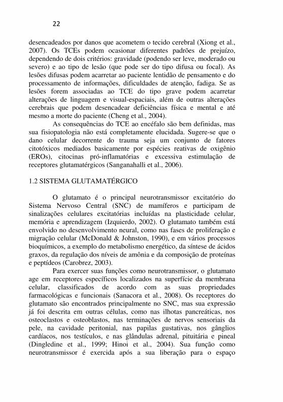
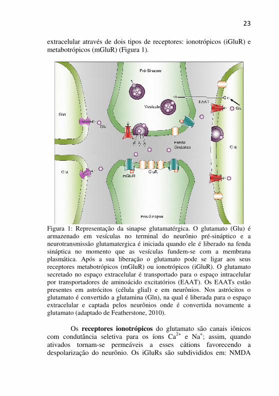
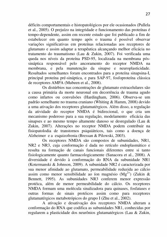
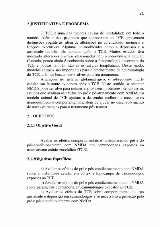
extracelular através de dois tipos de receptores: ionotrópicos (iGluR) e metabotrópicos (mGluR) (Figura 1).
Figura 1: Representação da sinapse glutamatérgica. O glutamato (Glu) é armazenado em vesículas no terminal do neurônio pré-sináptico e a neurotransmissão glutamatergica é iniciada quando ele é liberado na fenda sináptica no momento que as vesículas fundem-se com a membrana plasmática. Após a sua liberação o glutamato pode se ligar aos seus receptores metabotrópicos (mGluR) ou ionotrópicos (iGluR). O glutamato secretado no espaço extracelular é transportado para o espaço intracelular por transportadores de aminoácido excitatórios (EAAT). Os EAATs estão presentes em astrócitos (célula glial) e em neurônios. Nos astrócitos o glutamato é convertido a glutamina (Gln), na qual é liberada para o espaço extracelular e captada pelos neurônios onde é convertida novamente a glutamato (adaptado de Featherstone, 2010).
Os receptores ionotrópicos do glutamato são canais iônicos
com condutância seletiva para os íons Ca2+ e Na+; assim, quando ativados tornam-se permeáveis a esses cátions favorecendo a despolarização do neurônio. Os iGluRs são subdivididos em: NMDA
24
(N-metil-D-aspartato), AMPA (ácido α-amino-3-hidróxi-5-metil-4-isoxazol-propiônico) e cainato (Dingledine et al., 1999).
Os receptores metabotrópicos modificam a excitabilidade neuronal e glial através da ativação da proteína G, que quando ativada leva a formação de segundos mensageiros como o diacilglicerol e AMPc (adenosina monofosfato cíclico) que modulam a atividade de canais iônicos presentes na membrana plasmática dos neurônios (Meldrum, 2000). Os mGluRs estão presentes em todas as regiões do encéfalo e são considerados um dos mais importantes receptores que modulam os níveis de segundos mensageiros no SNC de mamíferos (Bressan & Pilowsky, 2003).
Os transportadores do glutamato estão presentes na membrana celular de neurônios e em astrócitos, sendo os transportadores gliais considerados os mais importantes (Schousboe, 1981). O sistema de transporte neuronal do glutamato possibilita que o neurotransmissor seja armazenado em vesículas citoplasmáticas no neurônio pré-sináptico e que exerça suas funções nos receptores dos neurônios pré e pós-sinápticos após a exocitose (Rosenberg et al., 1992). A atividade dos transportadores de glutamato das células gliais é responsável por manter a baixos níveis (~ 1-2 µmol/L) a concentração de glutamato no espaço extracelular do SNC (Ni et al., 2007).
Entretanto, a homeostase do glutamato em fluidos extracelulares do cérebro não é regulada somente por seus transportadores, mas também por processos que acontecem na superfície de vasos capilares do cérebro, que permitem o efluxo do excesso de glutamato presente nos fluidos extracelulares do cérebro para o sangue (Teichberg et al., 2009).
Os receptores ionotrópicos do sub-tipo NMDA são considerados extremamente importantes para a funcionalidade do sistema glutamatérgico, sendo então muito estudados, pois fisiologicamente a sua ativação é fundamental para a ocorrência de processos como a aprendizagem e a memória (Bliss & Collingridge, 1993). Contudo, a excessiva estimulação deste receptor é relacionada com a etiologia da epilepsia e com danos cerebrais associados à isquemia/hipoglicemia devido ao aumento do influxo de íons para o meio intracelular, principalmente do Ca2+ que fluem através do receptor (Olney, 1994). O acúmulo intracelular de Ca2+ leva à ativação de mecanismos de lesão celular e subsequente morte por necrose e/ou apoptose. Esta neurotoxicidade induzida pelo receptor NMDA pode ser bloqueada por antagonistas do receptor, por altas concentrações dos íons
25
magnésio (Mg2+) ou por remoção do Ca2+ extracelular pela presença de agentes quelantes (Choi, 1990; Köhr, 2007; Ginsberg, 2008).
1.3 MECANISMOS DE NEUROPROTEÇÃO NO TRAUMATISMO
CRÂNIO-ENCEFÁLICO (TCE) A neuroproteção por moduladores glutamatérgicos foi mostrada
por diversos pesquisadores (Gong et al., 1995; Dempsey et al., 2000). De fato, o antagonista do receptor NMDA, ifenprodil, na dose de 10 mg/kg exerceu efeitos neuroprotetores na região cortical de ratos Sprage-Dawley após o TCE (Dempsey et al., 2000). A memantina, a qual também é antagonista do receptor NMDA, administrada nas doses de 10 e 20 mg/kg imediatamente após o TCE foi capaz de prevenir o dano neural nas regiões CA2 e CA3 do hipocampo (Rao et al., 2001). Além disso, antagonistas dos receptores metabotrópicos do grupo I foram neuroprotetores em vários estudos (Gong et al., 1995; Allen; Knoblack & Faden, 2000; Faden et al., 2001).
No TCE o cérebro depende de mecanismos internos de defesa para a sua proteção, dentre eles pode-se destacar, a ativação de moléculas da via anti-apoptótica em neurônios, da liberação de fatores neurotróficos e da produção de citocinas (revisado em Shpargel et al., 2008).
Os processos apoptóticos são controlados por ativadores e inibidores de caspases que podem representar alvos potenciais para a neuroproteção. Após isquemia e reperfusão cerebral a apoptose é uma das principais vias que levam a morte celular (Mattson et al., 2001). Danos mitocondriais e liberação de citocromo c ocasionados por danos ao DNA e carência de fatores de crescimento podem ser um dos fatores que levam aos processos apoptóticos. Alguns estudos tem demonstrado que a apoptose contribui para o desenvolvimento de infarto isquêmico com fragmentação do DNA (Chopp & Jiag, 1995). Em contraste, um estudo realizado por Xing e colaboradores (2008) investigando a expressão de moléculas chaves relacionadas a apoptose em ratos Sprague-Dawley após pós-condicionamento isquêmico demonstrou um aumento nos níveis da proteína anti-apoptótica Bcl-2, inibição da translocação da proteína anti-apoptótica Bax e inibição da liberação de citocromo c, sugerindo que os efeitos neuroprotetores estão ligados a mecanismos anti-apoptóticos. Além disso, em diferentes modelos animais de TCE tem sido mostrado um aumento na expressão de Bcl-2 em animais tratados com estrogênio, sugerindo-se que este aumento
26
possa ser em parte devido a neuroproteção mediada por estrogênios (Soustiel & Larisch, 2010).
Em um estudo utilizando camundongos nocautes para o fator neurotrófico- derivado do cérebro (BDNF), foi demonstrado aumento de morte celular no giro denteado do hipocampo após TCE moderado (Gao & Chen, 2009), sugerindo que o BDNF está envolvido na regulação da sobrevivência de neurônios hipocampais e potencialmente pode ser um alvo para prevenção de morte celular após o TCE. Além disso, recentemente um estudo realizado por Minnich e colaboradores (2010), mostrou que o fator neurotrófico derivado da glia (GDNF, sigla em inglês) protegeu contra morte celular em neurônios corticais após TCE em roedores.
Estudos prévios têm mostrado que citocinas pró e anti-inflamatórias apresentam alterações resultantes de TCE (Kadhim et al., 2008), a interleucina-6 (IL-6) tem sido relacionada ao momento de admissão e sobrevivência no TCE (Minambres et al., 2003), em análises de marcadores biológicos identificou-se a IL-6 como um marcador após TCE em pacientes (Berger et al., 2008; Hergenroeder et al., 2010).
O dano celular do TCE pode ser divido em dois estágios, o primeiro é inevitável e é decorrente do próprio trauma, e o segundo é considerado o dano celular que ocorre num período de tempo variável e pode ser evitado, esse processo é iniciado com ativação dos receptores NMDA, AMPA e cainato, resultando no influxo de íons Ca2+ para o meio intracelular (Obrenovitch & Urenjak, 1997). O mecanismo fisiopatológico do dano secundário é composto de inflamação, produção de radicais livres, aumento do cálcio intracelular, peroxidação lipídica e produção de oxido nítrico via oxido nítrico sintase (Acarin et al., 2005). O excesso de Ca2+ age ativando cascatas protéicas que atacam diversos componentes estruturais e funcionais do neurônio com a subsequente formação de EROs e morte celular (Sanganahalli et al., 2006). A inflamação neuronal reflete na quebra da barreira hematoencefálica e edema cerebral surgindo uma cascata de eventos que podem culminar em morte celular (Lescot et al., 2006).
As EROs formadas após excessiva ativação dos receptores NMDA, agem exercendo o papel de destaque na patologia do TCE, pois são responsáveis por comandar a peroxidação lipídica, oxidação e nitrificação protéica, além de realizar danos no DNA neuronal (Shanganahalli et al., 2006; Mubeen et al., 2008). O estresse oxidativo também contribui com as sequelas secundárias ao TCE e medeia os
27
déficits comportamentais e histopatológicos por ele ocasionados (Pullela et al., 2005). O prejuízo na integridade e funcionamento das proteínas é tempo-dependente, assim em recente estudo que foi publicado a fim de estabelecer em quanto tempo após o trauma é possível observar variações significativas em proteínas relacionadas aos receptores de glutamato e assim adaptar a terapêutica alcançando melhor eficácia no tratamento do traumatismo (Lau & Zukin, 2007). Foi verificada uma queda nos níveis da proteína PSD-95, localizada na membrana pós-sináptica responsável pelo ancoramento do receptor NMDA na membrana, e pela manutenção da sinapse e neuroplasticidade. Resultados semelhantes foram encontrados para a proteína sinapsina-I, principal proteína pré-sináptica, e para SAP-97, fosfoproteína clássica de receptores AMPA (Mubeen et al., 2008).
Os distúrbios nas concentrações de glutamato extracelulares são a causa primária da morte neuronal em decorrência de trauma agudo como infartos ou convulsões (Hardingham, 2006). Observa-se um padrão semelhante no trauma craniano (Whiting & Hamm, 2008) devido a uma ativação dos receptores glutamatérgicos. Além disso, a regulação da atividade do receptor NMDA é bidirecional, o que cria um mecanismo poderoso para a sua regulação, modelamento eficácia das sinapses e ao mesmo tempo altamente danoso se desregulado (Lau & Zukin, 2007). Alterações no receptor NMDA podem contribuir na fisiopatolodia de transtornos psiquiátricos, tais como a doença de Alzheimer e a esquizofrenia (Bressan & Pilowski, 2003).
Os receptores NMDA são compostos de subunidades, NR1, NR2 e NR3, cuja conformação é dada no retículo endoplasmático e resulta na formação de canais funcionais diferentes entre si tanto fisiologicamente quanto farmacologicamente (Sanacora et al., 2008). A diversidade é devido à conformação do RNA da subunidade NR1 (Kotermanski & Johnson, 2009). A subunidade NR2 é caracterizada por sua menor afinidade ao glutamato, permeabilidade reduzida ao cálcio assim como menor sensibilidade ao íon magnésio (Mg2+) (Zukin & Bennett, 1995). As subunidades NR3 conferem menor expressão protéica, além de menor permeabilidade do cálcio. Os receptores NMDA formam uma molécula sinalizadora para quinases, fosfatases e outras formas de sinais protéicos assim como para receptores glutamatérgicos metabotrópicos do grupo I (Zhu et al., 2002).
A ativação e desativação dos receptores NMDA alteram a conformação do RNA que codifica as subunidades NR1, conhecidas por regularem a plasticidade dos neurônios glutamatérgicos (Lau & Zukin,
28
2007), o que sugere que a modulação do receptor NMDA possa ser uma alvo importante na redução de danos citotóxicos, incluindo os decorrentes do TCE (Youssef et al., 2006; Shein et al., 2007; Schumann et al., 2008). De fato, Kreutz e colaboradores (1998) estudando os efeitos do trauma axonal do nervo óptico na expressão de variantes da subunidade NR1 do receptor NMDA em células da retina, mostraram que um aumento na expressão das subunidades NR1-4b é crucial para a sobrevivência neuronal após trauma axonal parcial. Além disso, tem sido mostrado o papel das distintas subunidades de NR2 do receptor NMDA na neurotoxicidade em camundongos mutantes. Camundongos deficientes da subunidade NR1A tiveram uma redução de infarto (Morikawa et al., 1998) e camundongos mutantes para a subunidade NR2C submetidos ao modelo animal de isquemia cerebral tiveram uma redução no dano neuronal (Kodotani et al., 1998).
1.4 NEUROPROTEÇÃO POR PRÉ E PÓS-CONDICIONAMENTO
A neuroproteção pode ser induzida por baixas doses de NMDA,
que ativam os receptores sinápticos e extrasinápticos de NMDA (Francesc et al., 2006). A atividade sináptica do receptor de NMDA promove a neuroproteção pela ativação de cascatas intracelulares que aumentam a sobrevivência celular frente a sinais patológicos, incluindo a ativação do fator nuclear CREB (proteína de ligação ao elemento de resposta ao AMPc) e expressão de proteínas envolvidas com sobrevivência celular (Lee et al., 2005; Papadia et al., 2005).
O pré-condicionamento diminui a prevalência de morte celular no miocárdio decorrente de oclusão em artéria coronária (Murry et al., 1990). Estudos estratégicos de pré-condicionamento como isquemia (Chen & Simon, 1997), hipóxia (Pugliese et al., 2003) e hipotermia (Nischo et al., 2000) resultaram em proteção contra o dano isquêmico subsequente. O NMDA e o glutamato podem atuar como agentes de pré-condicionamento químicos para melhorar significativamente o dano isquêmico (Schurr et al., 2001) em cortes histológicos de hipocampo como também em cultura de células (Xu et al., 2002) e in vivo (Boeck et al., 2004). Assim a administração de doses subconvulsivas de NMDA via intraperitoneal têm sido utilizada como modelo de pré-condicionamento químico in vivo frente a diversos insultos letais posteriores (Ogita et al., 2003; Boeck et al., 2004). Sabe-se que pacientes que são submetidos a exames de cateterismo, que sofrem uma pequena obstrução de uma determinada artéria por um curto intervalo de
29
tempo, apresentam menos morbidade após um AVC; caracterizando este evento como uma tolerância a um possível dano subsequente (Raphael, 2010). Vários estudos de pré-condicionamento mostram sua eficiência na prevenção de danos acometidos ao cérebro ou ao outro órgão que venha sofrer isquemia (Murry et al., 1990; Chen & Simon, 1997). Contudo, por razões clínicas e éticas, a aplicação de estratégias terapêuticas do tipo pré-condicionamento é bastante limitada. Ao contrário, em 2003, Zhao e colaboradores apresentaram o conceito do pós-condicionamento isquêmico, que consiste na realização de um ou mais ciclos curtos de isquemia seguidos por um ou mais ciclos curtos de reperfusão. Estes autores demonstraram que o pós-condicionamento isquêmico foi tão eficaz quanto o pré-condicionamento isquêmico na prevenção das lesões de reperfusão. Estes bons resultados foram observados também por outros autores na isquemia miocárdica, renal, cerebral e da medula espinhal (Cabrera et al., 2012; Liang et al., 2012; Wever et al., 2012).
Além da relação do NMDA com a neuroproteção, outras vias são conhecidas. De fato, recentes estudos tem mostrado a importância das vias de sinalização intracelulares. Recentemente foi demonstrado que pré-condicionamento em ratos potencialmente ativou a via de sinalização PI-3 quinase, a qual está envolvida com sobrevivência celular; em contraste a inibição da PI-3 quinase pelo inibidor LY294002 aboliu os efeitos protetores do pré-condicionamento por estimular as vias apoptóticas e fragamentação do DNA (Uchiyama et al., 2004). Neste estudo os autores sugerem que a PI-3 quinase desempenha uma importante função nos efeitos anti-apoptóticos do pré-condicionamento. Outro estudo demonstrou que dano cerebral por isquemia/reperfusão em ratos levou a um aumento nos níveis de malondialdeído, o qual é um produto da peroxidação lipídica, contrariamente a isto pré-condicionamento isquêmico regulou a translocação de Bcl-2 e Bax, regulou a atividade da caspase-3 e também reduziu os níveis de estresse oxidativo (Abas et at., 2010).
O óxido nítrico também pode ter um importante papel como mediador em resposta ao pré-condicionamento neuronal em conjunto com a ativação do receptor NMDA ou independentemente dele. A inibição da enzima óxido nítrico sintase durante o pré-condicionamento atenua a tolerância neuronal induzida (Atochin et al., 2003). Além disso, infusão de baixas doses de adenosina reduziu a carga isquêmica e melhorou a disfunção sistólica ventricular em pacientes com isquemia
30
miocárdica induzida por exercícios (Sadigh et al., 2009), mostrando um efeito positivo do pré-condicionamento com adenosina em humanos.
O BDNF, uma neurotrofina muito importante na plasticidade celular, tem recentemente se mostrado importante em estudos de pré-condicionamento. De fato, pré-condicionamento promove plasticidade em neurônios sensoriais ascendentes e aumenta a expressão de BDNF e de seus receptores, TrkB e P75 (Li et al., 2009). Outro grupo utilizando pré-condicionamento por hipoxia mostrou um aumento nas concentrações de BDNF em soro de homens jovens saudáveis (Hubold et al., 2009), sugerindo que o BDNF pode ser de relevância clínica como opções terapêuticas para doenças vasculares isquêmicas.
31
2 JUSTIFICATIVA E PROBLEMA
O TCE é uma das maiores causas de mortalidade em todo o mundo. Além disso, pacientes que sobrevivem ao TCE apresentam disfunções cognitivas, além de alterações no aprendizado, memória e funções executivas. Algumas co-morbidades como a depressão e a ansiedade também são comuns após o TCE. Muitos estudos têm mostrado alterações em vias relacionadas com a sobrevivência celular. Contudo, pouco ainda é conhecido sobre a fisiopatologia decorrente do TCE e poucas também são as estratégias terapêuticas. Desse modo, modelos animais são importantes para o entendimento da neurobiologia do TCE, além de buscar novos alvos para seu tratamento.
Alterações no sistema glutamatérgico, e subsequente morte celular são bastante evidentes após o TCE. Neste sentido, o receptor NMDA pode ser alvo para induzir efeitos neuroprotetores. Sendo assim, estudos que avaliam os efeitos do pré e pós-tratamento com NMDA em modelo animal de TCE ajudam a desvendar melhor os mecanismos neuroquímicos e comportamentais, além de ajudar no desenvolvimento de novas estratégias para o tratamento pós-trauma.
2.1 OBJETIVOS
2.1.1 Objetivo Geral
Avaliar os efeitos comportamentais e moleculares do pré e do pós-condicionamento com NMDA em camundongos expostos ao traumatismo crânio-encefálico (TCE).
2.1.2Objetivos Específicos
a) Avaliar os efeitos do pré e pós-condicionamento com NMDA sobre a viabilidade celular em córtex e hipocampo de camundongos expostos ao TCE;
b) Avaliar os efeitos do pré e pós-condicionamento com NMDA sobre parâmetros de memória em camundongos expostos ao TCE;
c) Avaliar os efeitos do TCE sobre comportamento do tipo ansiedade e depressão em camundongos e se necessário a proteção pelo pré e pós-condicionamento com NMDA.
32
PARTE II 2 ARTIGO I Artigo aceito para publicação no periódico Brain Research, 2012.
NMDA Preconditioning Prevents Object Recognition Memory Impairment and Increases Brain Viability in Mice Exposed to
Traumatic Brain Injury Vânia K. M. Moojena, Marcela Damiani-Nevesa, Daniela V.
Bavarescoa, Bruna B. Pescadora, Clarissa M. Comimb, João Quevedob, Carina R. Boecka*
aLaboratório de Biologia Celular e Molecular, Instituto Nacional de Ciência e Tecnologia Translacional em Medicina (INCT-TM), Programa de Pós-Graduação em Ciências da Saúde, Unidade Acadêmica de Ciências da Saúde, Universidade do Extremo Sul Catarinense-UNESC, Criciúma, SC, Brazil;
bLaboratório de Neurociências, Instituto Nacional de Ciência e
Tecnologia Translacional em Medicina (INCT-TM), Programa de Pós-Graduação em Ciências da Saúde, Unidade Acadêmica de Ciências da Saúde, Universidade do Extremo Sul Catarinense-UNESC, Criciúma, SC, Brazil.
Corresponding Authors: Carina Rodrigues Boeck, M.SC, Ph.D. Laboratório de Neurociências – PPGCS - UNASAU Universidade do Extremo Sul Catarinense 88806-000, Criciúma, SC, Brazil Phone: + 55 48 3431 2757 E-mail: [email protected] Abstract Recent studies have focused on the role of N-methyl-D-
aspartate (NMDA) in brain injury. The present study is aimed at verifying memory, anxiety/depression parameters, and cellular viability in the brain of mice preconditioned with NMDA and subjected to the
33
model of mild traumatic brain injury. For this purpose, male albino CF-1 mice were pre-treated with NMDA (75 mg/kg) and subjected to brain trauma, and after 24 h submitted to memory tasks, and anxiety and depression-like behavioral tests. The memory tests were evaluated at 1.5 h, 24 h, and 7 days after the training. In addition, the cellular viability was evaluated in the cerebral cortex and hippocampus 96 h after the trauma. It was observed that the cellular viability was reduced in the hippocampus of the mice subjected to trauma and the preconditioning with NMDA was able to protect this damage. All mice learnt the task in the habituation test, but in the object recognition task the mice preconditioned with NMDA were protected against impairment induced by TBI in both short and long-term memory. On the other hand, in the step-down inhibitory avoidance test, only the mice treated with NMDA showed impairment of long-term memory (7 days after training session). The evaluation of anxiety/depression behavior showed no changes after TBI. In conclusion, NMDA preconditioning induced impairment of long-term memory; however, it was able to protect against the novel recognition memory impairment and increase the cellular survival in the hippocampus of mice exposed to traumatic brain injury.
Keywords: NMDA preconditioning; memory; cellular viability;
traumatic brain injury. Abbreviations: NMDA, N-methyl-D-aspartate; TBI, traumatic
brain injury.
34
1. Introduction Traumatic Brain Injury (TBI) is a leading cause of mortality and
morbidity with harmful socioeconomic consequences (Max et al., 1991). Patients who survive after TBI might show severe cognitive dysfunctions and impairments in learning, memory, and executive functions (Murray et al., 1992; Levin et al., 1998).
According to the clinical knowledge, TBI can be divided into two components: (1) primary damage, which is characterized by mechanical injury caused by the traumatic event itself, and (2) secondary damage, which occurs as changes at the cellular and molecular levels at areas distant to the impact and appears until several hours or days post injury (Levine et al., 2006). The cause of the secondary damage includes neuroinflammation, glutamatergic excitotoxicity, oxidative stress, DNA damage, neuronal cell death, amongst others (Bernert and Turski, 1996; Fan et al., 1996; Love, 1999; Sullivan et al., 1999; Leinhase et al., 2006).
The animal models of TBI have proved useful for gaining a better understanding of the pathophysiology of brain damage after trauma, as well as investigating the effective treatment (Laurer and McIntosh, 1999). The glutamatergic system appears as a main factor for the development of damage. This is because the system induces excitotoxicity at least in part due to the increase at the influx of Ca2+ through N-methyl-D-aspartate (NMDA) receptors (Choi et al., 1988). The neuroprotective strategy in humans involves blocking of excitotoxicity using antagonists of NMDA receptors (see review: Willis et al., 2004) and in the animal model, the protection by antagonists has been largely studied (Kuo et al., 2004).
A subtoxic dose of NMDA was found to induce preconditioning as it evokes a cellular tolerance state to a subsequent lethal event (Chuang et al., 1992; Soriano et al., 2006). In our group, it was demonstrated that NMDA preconditioning is effective against brain damage induced by glutamatergic system in vivo and in vitro (Boeck et al., 2004; Boeck et al., 2005). Further, the motor coordination and gaiting deficits induced by the TBI were 0070revented by NMDA preconditioning in mice (Costa et al., 2010). As there is no treatment available for reversing the pathogenic cellular cascade underlying the progression of cell death, a better understanding of the alterations in memory and cognitive and mood impairments remains an important goal of studies of glutamatergic system on experimental TBI research.
35
Thus, the present study is mainly aimed at investigating the effects of NMDA preconditioning as a neuroprotective strategy against memory deficits and cell death in mice subjected to TBI.
2. Results
The macroscopic analysis of the brain samples of mice that
underwent TBI revealed no signs of external damage to the cerebral cortex, no hemorrhages in the periventricular white matter below the cortex, and no apparent distortion of the inner structures. All mice survived following the induction of TBI.
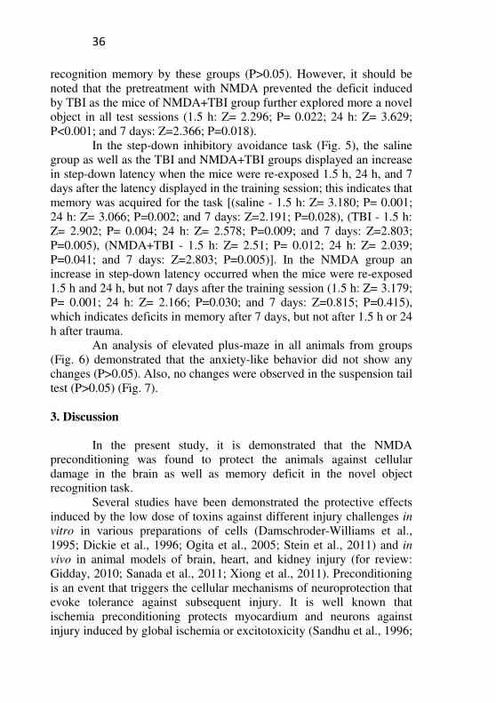
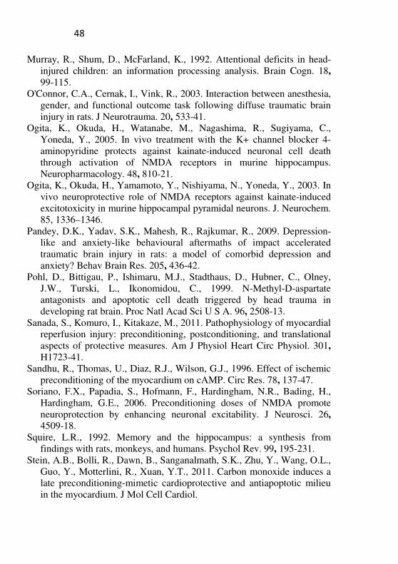
The TBI group presented a decrease of the cellular viability in the hippocampus as measured by the MTT assay 96 h after trauma (Fig. 1A), when compared with the control group. It is interesting to note that the cellular viability in the NMDA+TBI group was preserved against the damage induced by TBI, an effect statistically dependent on the interaction of NMDA and TBI (F(1, 17)=49,601; P<0.001). In addition, pretreatment with NMDA or TBI did not alter the cellular viability in the cerebral cortex when applied per se or as associated (Fig. 1B).
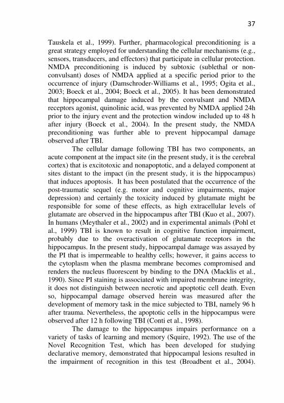
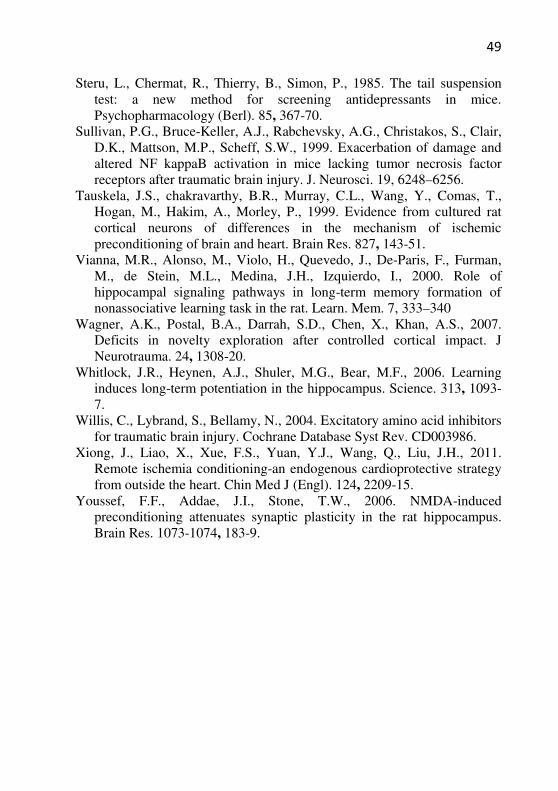
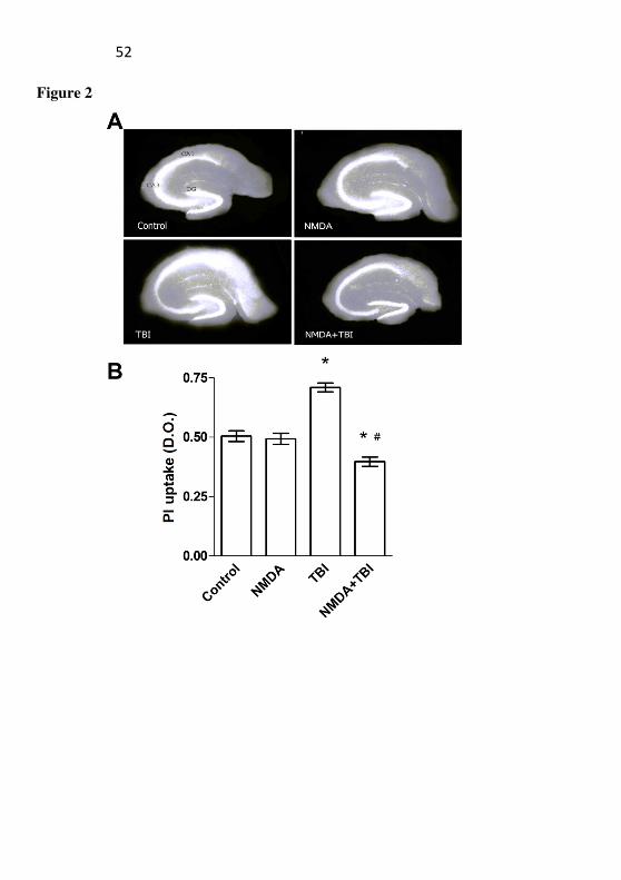
During the assessment of the cellular viability by the uptake of PI, it was observed that the staining in the hippocampus showed striking cellular damage in the TBI group when compared to the saline group (Fig. 2). In addition, it was observed that the NMDA did not induce damage per se, and that it prevented the damage induced by TBI in mice, an effect statistically dependent on the interaction of NMDA and TBI (F(1, 20)=8,6522, P=,00807).
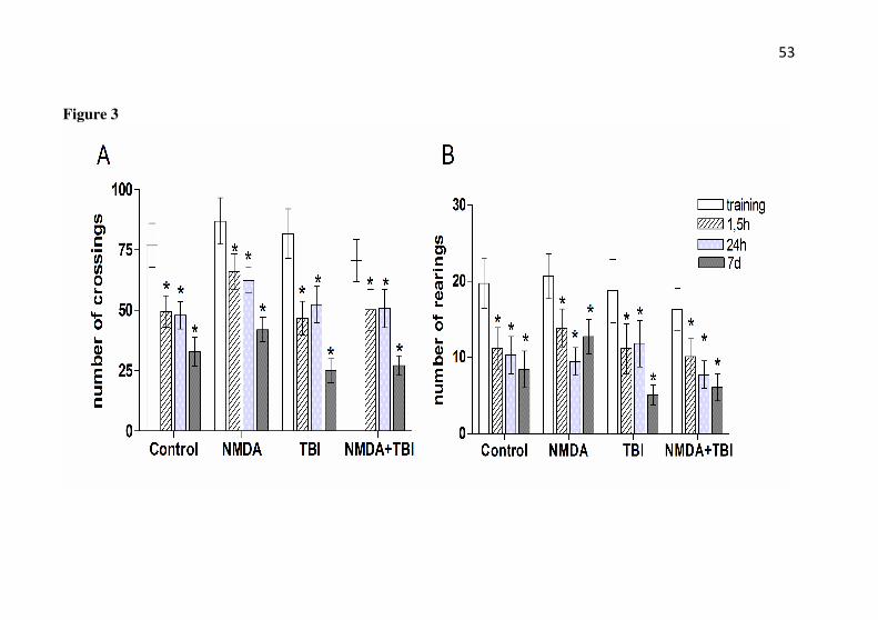
It should be mentioned that in the habituation to the open-field apparatus, the number of crossings (Fig. 3A) and rearings (Fig. 3B) were reduced when the mice were re-exposed to the apparatus 1.5 h, 24 h, and 7 days after the test session, thus indicating habituation to the environment by all groups (saline, NMDA, TBI, and NMDA+TBI). There were not statistical differences among the groups, neither NMDA nor TBI affect the locomotor activity of mice (P>0.05).
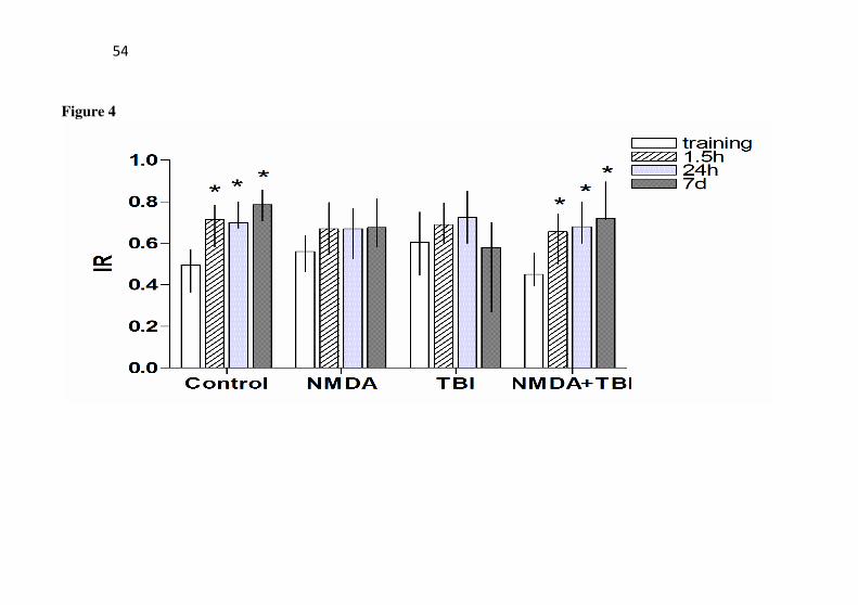
In the object recognition task (Fig. 4), statistical differences were observed in the object exploration index in saline as the mice further explored more a novel object in all test sessions (1.5 h: Z= 2.334; P= 0.019; 24 h: Z= 3.113; P=0.002; and 7 days: Z=2.31; P=0.021). Nevertheless, in the NMDA or TBI groups no statistical differences were observed in the object exploration index in the test sessions when compared with the training session, which suggests a deficit in the
36
recognition memory by these groups (P>0.05). However, it should be noted that the pretreatment with NMDA prevented the deficit induced by TBI as the mice of NMDA+TBI group further explored more a novel object in all test sessions (1.5 h: Z= 2.296; P= 0.022; 24 h: Z= 3.629; P<0.001; and 7 days: Z=2.366; P=0.018).
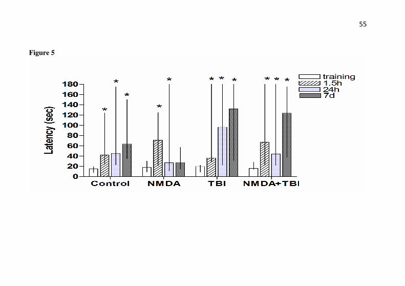
In the step-down inhibitory avoidance task (Fig. 5), the saline group as well as the TBI and NMDA+TBI groups displayed an increase in step-down latency when the mice were re-exposed 1.5 h, 24 h, and 7 days after the latency displayed in the training session; this indicates that memory was acquired for the task [(saline - 1.5 h: Z= 3.180; P= 0.001; 24 h: Z= 3.066; P=0.002; and 7 days: Z=2.191; P=0.028), (TBI - 1.5 h: Z= 2.902; P= 0.004; 24 h: Z= 2.578; P=0.009; and 7 days: Z=2.803; P=0.005), (NMDA+TBI - 1.5 h: Z= 2.51; P= 0.012; 24 h: Z= 2.039; P=0.041; and 7 days: Z=2.803; P=0.005)]. In the NMDA group an increase in step-down latency occurred when the mice were re-exposed 1.5 h and 24 h, but not 7 days after the training session (1.5 h: Z= 3.179; P= 0.001; 24 h: Z= 2.166; P=0.030; and 7 days: Z=0.815; P=0.415), which indicates deficits in memory after 7 days, but not after 1.5 h or 24 h after trauma.
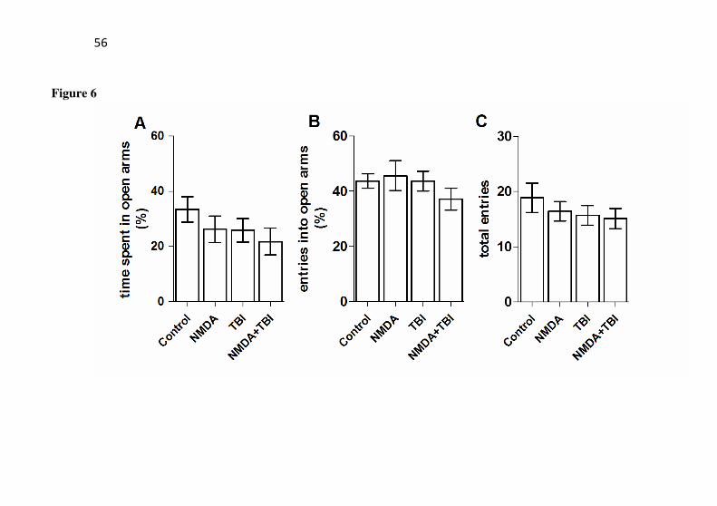
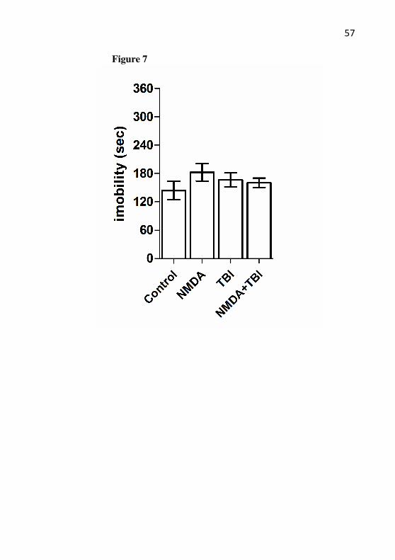
An analysis of elevated plus-maze in all animals from groups (Fig. 6) demonstrated that the anxiety-like behavior did not show any changes (P>0.05). Also, no changes were observed in the suspension tail test (P>0.05) (Fig. 7).
3. Discussion
In the present study, it is demonstrated that the NMDA
preconditioning was found to protect the animals against cellular damage in the brain as well as memory deficit in the novel object recognition task.
Several studies have been demonstrated the protective effects induced by the low dose of toxins against different injury challenges in
vitro in various preparations of cells (Damschroder-Williams et al., 1995; Dickie et al., 1996; Ogita et al., 2005; Stein et al., 2011) and in
vivo in animal models of brain, heart, and kidney injury (for review: Gidday, 2010; Sanada et al., 2011; Xiong et al., 2011). Preconditioning is an event that triggers the cellular mechanisms of neuroprotection that evoke tolerance against subsequent injury. It is well known that ischemia preconditioning protects myocardium and neurons against injury induced by global ischemia or excitotoxicity (Sandhu et al., 1996;
37
Tauskela et al., 1999). Further, pharmacological preconditioning is a great strategy employed for understanding the cellular mechanisms (e.g., sensors, transducers, and effectors) that participate in cellular protection. NMDA preconditioning is induced by subtoxic (sublethal or non-convulsant) doses of NMDA applied at a specific period prior to the occurrence of injury (Damschroder-Williams et al., 1995; Ogita et al., 2003; Boeck et al., 2004; Boeck et al., 2005). It has been demonstrated that hippocampal damage induced by the convulsant and NMDA receptors agonist, quinolinic acid, was prevented by NMDA applied 24h prior to the injury event and the protection window included up to 48 h after injury (Boeck et al., 2004). In the present study, the NMDA preconditioning was further able to prevent hippocampal damage observed after TBI.
The cellular damage following TBI has two components, an acute component at the impact site (in the present study, it is the cerebral cortex) that is excitotoxic and nonapoptotic, and a delayed component at sites distant to the impact (in the present study, it is the hippocampus) that induces apoptosis. It has been postulated that the occurrence of the post-traumatic sequel (e.g. motor and cognitive impairments, major depression) and certainly the toxicity induced by glutamate might be responsible for some of these effects, as high extracellular levels of glutamate are observed in the hippocampus after TBI (Kuo et al., 2007). In humans (Meythaler et al., 2002) and in experimental animals (Pohl et al., 1999) TBI is known to result in cognitive function impairment, probably due to the overactivation of glutamate receptors in the hippocampus. In the present study, hippocampal damage was assayed by the PI that is impermeable to healthy cells; however, it gains access to the cytoplasm when the plasma membrane becomes compromised and renders the nucleus fluorescent by binding to the DNA (Macklis et al., 1990). Since PI staining is associated with impaired membrane integrity, it does not distinguish between necrotic and apoptotic cell death. Even so, hippocampal damage observed herein was measured after the development of memory task in the mice subjected to TBI, namely 96 h after trauma. Nevertheless, the apoptotic cells in the hippocampus were observed after 12 h following TBI (Conti et al., 1998).
The damage to the hippocampus impairs performance on a variety of tasks of learning and memory (Squire, 1992). The use of the Novel Recognition Test, which has been developed for studying declarative memory, demonstrated that hippocampal lesions resulted in the impairment of recognition in this test (Broadbent et al., 2004).
38
Recognition memory involves temporal lobe areas, such as perirhinal and parahippocampal/postrhinal cortices as well as the entorhinal cortex. The hippocampus is the final stage of convergence within the medial temporal lobe, receiving projections from these areas (Lavenex and Amaral, 2000). Habituation memory is a nonassociative memory that is acquired when repeated or continuous exposure of an animal to a novel environment changes the behavioral responses to it. The long-term memory of habituation depends on the functionality of hippocampal NMDA and AMPA/kainate receptors (Vianna et al., 2000). The inhibitory avoidance task is a behavioral test that is widely used for studying the effects of drugs on aversive memory. It is easy to use and it displays relatively good pharmacological prediction to amnesic and enhancer memory drugs (Gold, 1986; Izquierdo e Medina, 1997). In the present study, TBI was found to induce impairment in the anterograde novel object recognition memory without affecting habituation and aversive memory. Baratz and colls (2011) also observed deficit on novel object recognition memory in mice 7 days after the closed head weight-drop model of TBI.
However, it is not possible to exclude the hypothesis that the habituation and aversive memory involving hippocampal mechanisms could be impaired at 96 h after TBI, as the studies demonstrated the deficit after trauma induced by the controlled cortical impact (O’Connor et al., 2003; Wagner et al., 2007). On the other hand, NMDA preconditioning impaired the long-term retention of memory for inhibitory avoidance. It is possible that a low dose of NMDA had a specific effect that requires further study. The inhibitory avoidance training induces LTP in the CA1 of hippocampus dependent of NMDA receptors (Whitlock et al., 2006); however, NMDA preconditioning attenuates the long-term potentiation (Youssef et al. 2006) that could alter synaptic plasticity. Further, NMDA preconditioning confers protection via PKA, PI3K and MAPK/ERK pathways that are important to memory and neuronal survival (Herculano et al., 2011). Thus, NMDA preconditioning offers protection against cellular damage, but could have a specific effect in cognitive outcome after injury.
The incidence rates of major depression in human survivors to the TBI are 15.3% to 33% and the prevalence rates are 18.5% to 61% (Kim et al., 2007). In addition, anxiety is a common comorbidity observed in 41.2% of TBI patients with depression (Jorge et al., 1993). The low volume hippocampal observed after TBI could be associated with the incidence of depression in TBI patients, because similarly,
39
changes in hippocampal functioning and morphology are observed in major depressive patients (Campbell and MacQuen, 2004). In a well-designed study, was observed in rats high level of anxiety and depression-like behaviors after TBI induced by impact acceleration using the weight-drop model (Pandey et al., 2009). Further, the treatment with magnesium (which is an ion blocker of NMDA receptors in neurons during resting) ameliorated the anxiety/depression-like behaviors in 61% of animals exposed to TBI (From et al., 2004). It was observed that the weight drop of 25 g at the skull caused short-term motor incoordination (but not locomotion) (Costa et al., 2010), and probably the present data reinforced the characterization of the TBI model in mice as it also revealed hippocampal damage by PI; although anxiety/depression-like behaviors were not observed in our mice with TBI. Also, the deficit on novel object recognition memory observed herein corroborate with the study of Baratz and colls. (2011), in which mice showed deficit on the memory 7 days after mild traumatic brain injury.
The preconditioning method has its limitations when used as treatment for TBI; however it is useful in evaluating the protective effect of a low dose of NMDA. Further, it can be used for directing future studies that are focused in pharmacological interventions saved in therapies for TBI.
4. Experimental Procedures
Animals
Male CF-1 adult mice (2–3 months old, weighing 30–35 g) were obtained from the breeding colony (UNESC). A total of four to six animals were housed per cage with food and water freely available and were maintained on a 12-h light/dark cycle (lights on at 7:00 a.m.). All experimental procedures involving the animals were performed in accordance with the National Institute of Health Guide for the Animal Care and Use of Laboratory Animals (NIH) and the recommendations of the Sociedade Brasileira de Ciências em Animais de Laboratório (SBCAL) for animal care, and were designed to minimize suffering and limit the number of animals used. The animals were used only once to avoid circadian variations; all experiments were carried out between 8:00 a.m. and 4:00 p.m. The apparatus was cleaned with ethanol 10% after the evaluation of each mouse in the tasks. A trained observer who
40
was unaware of the treatment performed all the behavioral experiments. This study was approved by the local ethics committee (Comitê de Ética em Pesquisa da Universidade do Extremo Sul Catarinense, n°. 77/2007 and 60/2009).
Pretreatment with NMDA
NMDA (Sigma-Aldrich; St. Louis, MO, USA) was dissolved in saline solution and the pH adjusted to 7.4 with NaOH 1mEq/mol. The animals were injected intraperitoneally (i.p.) with a low, nonconvulsant dose of NMDA (75 mg/kg) or vehicle (saline, 0.9% NaCl, w/v) 24 h prior to performing the cortical trauma injury induction (Giménez-Llort et al., 1995). The mice were housed in acrylic boxes (25 x 25 x 25 cm) and observed for 30 min immediately after NMDA administration for the occurrence of behavioral alterations (Boeck et al., 2004). All the solutions were administered i.p. at 10 mL/kg of the animal.
Diffuse TBI
A diffuse TBI was produced using the closed-head weight-drop method as previously described (Adelson et al., 1996), with minor modifications. This apparatus consisted of a metal tube, 1 m in length and 10 mm in inner diameter, attached to a ring stand. The mice, anesthetized by inhalation with a mixture of O2/N2O (33%:66%), were placed on a loosely fixed foam bed with their head slightly elevated to avoid spinal cord damage. A brass weight of 25g with concentric grooves on its face fell downward freely from a height of 80 cm. Just after the impact, the foam bed with the animal was slid out from under the tube to prevent a second impact to it on recoil. After trauma, the mice received supporting oxygenation with 100% O2 until they were fully awake (approximately 1 min) and were then brought back to their cages. The training sessions in the behavioral tests were assessed 24 h after trauma and the testing sessions were executed 1.5 h (short-term memory), 24 h, and 7 days (long-term memory) after the training test. At 96 h after TBI induction, the mice were euthanized for the subsequent analysis of cerebral cortex and hippocampal viability.
41
Habituation to an open field
In the habituation to an open-field apparatus, the animals were exposed twice, with a 24-h interval. The test was carried out in a 40×40 cm (length and width) open-field surrounded by 20 cm high walls made of brown plywood with a frontal glass wall. The floor of the open field was divided by black lines into 16 equal squares. In both sessions (training and testing-sessions), the animals were gently placed on the left rear square of the apparatus and allowed to explore freely for 5 min (Vianna et al., 2000). The training was executed 24 h after trauma. The testing sessions were executed 1.5 h (short-term memory), 24 h, and 7 days (long-term memory) after the training test. The number of black line crossings and rearings were recorded in both sessions for evaluating the effects of NMDA preconditioning on the habituation to the novel environment.
Object recognition task
Novel Object Recognition Memory was evaluated as originally developed by Ennaceur and Delacour (1988), which is based on the natural tendency of rodents to explore a novel object more than a familiar one. The object recognition task was carried out in a 40×40 cm open field surrounded by 20 cm high walls made of plywood with a frontal glass wall. The floor of the open field was divided into 16 equal rectangles by black lines. All animals were submitted to a habituation session where they were allowed to freely explore the open field for 5 min. No objects were placed in the box during the habituation trial. Twenty-four hours after habituation, training was conducted by placing individual rats for 5 min in the open field, in which two identical objects (objects A1 and A2) were positioned in two adjacent corners at 10 cm from the walls. In a short-term recognition memory test given at 1.5 h after the training, the rats explored the open field for 5 min in the presence of one familiar (A1) and one novel (B1) object.
In a long-term recognition memory test given at 24 h or 7 days after training, the rats explored the open field for 5 min in the presence of one familiar (A1) and one another novel object (B2; different from B1). All objects had similar textures, colors, and sizes but had distinctive shapes. A recognition index was calculated for each animal in the test session, which reports the ratio of time spent in exploring a novel object [TN/(TA + TN); TA = time spent in exploring the familiar
42
object A; TN = time spent in exploring the novel object B1 (24 h) or B2 (7 days)]. Between trials, the objects were washed with 10% ethanol solution. Exploration was defined as touching the object with the nose and/or forepaws.
Step-down inhibitory avoidance task
The step-down inhibitory avoidance apparatus consisted of a 50 × 25 × 25 cm plastic box with a front glass wall, where the floor had parallel 10-mm bronze bars. The left end of the grid was occupied by a 7 cm wide, 2.5 cm high Formica platform. The rats were gently placed on the platform facing the rear wall, and their latency to step down with all four paws on the grid was recorded. In the training session, after stepping down, the animals received a 0.4-mA, 2-s scrambled footshock and were withdrawn immediately from the cage. In the test session, 1.5h and 24 h or 7 days later, the procedure was repeated, but footshock was not given. The test session step-down latency was used as a measure of retention. A ceiling of 180 s was imposed on this measure, i.e., animals whose test latency was more than 180 s were considered to have a latency of 180 s (Gold, 1986; Izquierdo and Medina, 1997).
Elevated Plus-Maze Test
As previously described by Lister (1987), the plus-maze consisted of two open (30 x 5 cm) and two wall-enclosed arms (30 x 5 x 15 cm) connected by a central platform (5 x 5 cm). The apparatus was elevated 50 cm above the floor. The floor and the walls of the enclosed arms of the maze were made of brown wood. Each mouse was placed on the central platform, facing a closed arm, and observed for a 5-min period. The frequencies of entry into either open or enclosed arms, as well as the time spent in each arm type, were recorded (in seconds). An entry was scored as such only when the animal placed all four limbs into any given arm. Further, drugs or events with anxiolytic-like effect usually increase the time spent in and/or frequency of entries into open arms, whereas the reverse holds true for the anxiogenic-like effect. Furthermore, the number of entries into closed arms was used as an index of general activity. The elevated plus-maze apparatus was placed in a small closed room lit by a red light with an intensity of 100 lx in the open arms.

43
Tail Suspension Test
As previously described by Steru and colls. (1985), the tail suspension test consisted of suspending mice individually by the tail to a horizontal ring stand bar (30 cm distant from the floor) using an adhesive tape (at 2 cm distance from the tip of the tail). The mouse was 15 cm away from the nearest object. The method is based on the observation that a mouse suspended by the tail shows alternate periods of agitation and immobility. A 6-min test session was used and the parameter of immobility was scored as the number of seconds that the mouse spent in being immobile.
MTT reduction
The mice were killed by decapitation for the removal of
cerebral structures 96 hours after TBI (48h after the novel object recognition memory test). The cell survival following TBI was measured by dehydrogenase activities to reduce MTT [3-(4,5-dimethylthiazol-2-yl)-2,5-diphenyltetrazolium bromide] (Mosmann, 1983). The tetrazolium ring of MTT was cleaved by various dehydrogenase enzymes into viable cells and then precipitated as a blue formazan product. The cerebral cortex and hippocampus were cut into 400-mm-thick transversal slices using a McIlwain tissue chopper, followed by transfer of the sections to prewarmed phosphate-buffered saline (PBS) supplemented with 0.6% glucose (pH 7.4), and separated into individual slices. Two slices from each brain structure were incubated with MTT (0.5 mg/mL) in PBS buffer for 20min at 37°C. The medium was aspirated, the precipitated formazan was solubilized with dimethyl sulfoxide, and the viable cells were quantified spectrophotometrically at a wavelength of 550nm. The variability resulting from differences in slice size was found to be minimal according to the protein content measures.
Membrane Permeability
The hippocampus dissected at 96h after TBI in mice was sliced transversely as 400-µm-thick slices on a McIIwain tissue chopper and were subsequently separated into individual slices, as cited above. On an average, six slices were obtained from the middle of the hippocampus that were placed in a 96-well multiwell plate (Nunc) containing
44
prewarmed phosphate buffer supplemented with 0.6% glucose (pH 7,4)]. The slices were incubated for 10 min prior to cell viability assay. Cell death was assessed by uptake of the fluorescent exclusion dye propidium iodide (PI), which is a polar compound that enters only dead or dying cells with damaged membranes. Once inside the cell, PI complexes with DNA and induces intense red fluorescence (630 nm) when excited by green light (495 nm). The slices were incubated with 7.5 µg/mL PI for 1 h and then imaged on a standard inverted microscope (Nikon Eclipse TE 300 Nikon Corporation, Tokyo, Japan) by means of a rhodamine filter set. The PI uptake was quantified by densitometric analysis with Scion Image software (Scion Corporations) (Boeck et al, 2004).
Statistical analysis
The results were analyzed using the Statistica version 7.0 software (StatSoft, Inc.). The normality parameter was verified by the Shapiro-Wilk test. Each value reflects the mean of seven to fifteen animals/group for behavioral parameters or the mean of five to six animals/group for cellular viability analysis. Cellular viability was statistically analyzed by employing two-way ANOVA with NMDA pretreatment and TBI as the main factors, followed by Tukey HSD test and was reported as mean ± S.E.M. The performance of the mice on habituation to the open-field was analyzed by the Student’s t test for comparison between the training and test sessions. The performance on novel recognition objects and step-down inhibitory avoidance tasks was reported as median ± interquartile ranges. The performance of the mice was analyzed by employing Wilcoxon for comparisons between the training and test for each group. P values less than 0.05 were considered to be statistically significant.
Acknowledgements
This work was supported by funds from the Brazilian National Council Research: CNPq grants n° 484193/2010-4 and UNESC.
45
References
Adelson, P.D., Robichaud, P., Hamilton, R.L., Kochanek, P.M., 1996. A model of diffuse traumatic brain injury in the immature rat. J. Neurosurg. 85, 877–884.
Baratz, R., Tweedie, D., Rubovitch, V., Luo, W., Yoon, J.S., Hoffer, B.J., Greig, N.H., Pick, C.G., 2011. Tumor necrosis factor-α synthesis inhibitor, 3,6'-dithiothalidomide, reverses behavioral impairments induced by minimal traumatic brain injury in mice. J. Neurochem. 118(6):1032-1042.
Bernert, H., Turski, L,. 1996. Traumatic brain damage prevented by the non-N-methyl-D-aspartate antagonist 2,3-dihydroxy-6-nitro-7-sulfamoylbenzo[f] quinoxaline. Proc. Natl. Acad. Sci. U S A 93, 5235–5240.
Boeck, C.R., Ganzella, M., Lottermann, A., Vendite, D., 2004. NMDA preconditioning protects against seizures and hippocampal neurotoxicity induced by quinolinic acid in mice. Epilepsia 45, 745–750.
Boeck, C.R., Kroth, E.H., Bronzatto, M.J., Vendite, D., 2005. Adenosine receptors co-operate with NMDA preconditioning to protect cerebellar granule cells against glutamate neurotoxicity. Neuropharmacology 49, 17–24.
Broadbent, N.J., Squire, L.R., Clark, R.E., 2004. Spatial memory, recognition memory, and the hippocampus. Proc Natl Acad Sci U S A. 101, 14515-20.
Choi, D.W., 1988. Glutamate neurotoxicity and diseases of the nervous system. Neuron 1, 623–634.
Chuang, D.M., Gao, X.M., Paul, S.M., 1992. N-methyl-D-aspartate exposure blocks glutamate toxicity in cultured cerebellar granule cells. Mol Pharmacol. 42, 210-6.
Conti, A.C., Raghupathi, R., Trojanowski, J.Q., McIntosh, T.K., 1998. Experimental brain injury induces regionally distinct apoptosis during the acute and delayed post-traumatic period. J Neurosci. 18, 5663-72.
Costa, T., Constantino, L.C., Mendonca, B.P., Pereira, J.G., Herculano, B., Tasca, C.I., Boeck, C.R., 2010. N-methyl-D-aspartate preconditioning improves short-term motor deficits outcome after mild traumatic brain injury in mice. J Neurosci Res. 88, 1329-37.
Damschroder-Williams, P., Irwin, R.P., Lin, S.Z., Paul, S.M., 1995. Characterization of the excitoprotective actions of N-methyl-D-aspartate in cultured cerebellar granule neurons. J Neurochem. 65, 1069-76.
46
Dickie, B.G., Holmes, C., Greenfield, S.A., 1996. Neurotoxic and neurotrophic effects of chronic N-methyl-D-aspartate exposure upon mesencephalic dopaminergic neurons in organotypic culture. Neuroscience. 72, 731-41.
Ennaceur, A., Delacour, J., 1988. A new one-trial test for neurobiological studies of memory in rats. 1: Behavioral data. Behav Brain Res. 31, 47-59.
Fan, L., Young, P.R., Barone, F.C., Feuerstein, G.Z., Smith, D.H., McIntosh, T.K., 1996. Experimental brain injury induces differential expression of tumor necrosis factor-alpha mRNA in the CNS. Brain Res. Mol. Brain Res. 36, 287–291.
Fromm, L., Heath, D.L., Vink, R., Nimmo, A.J., 2004. Magnesium attenuates post-traumatic depression/anxiety following diffuse traumatic brain injury in rats. J Am Coll Nutr. 23, 529S-533S.
Gidday, J.M., 2010. Pharmacologic Preconditioning: Translating the Promise. Transl Stroke Res. 1, 19-30.
Gimenez-Llort, L., Martinez, E., Ferre, S., 1995. Dopamine-independent and adenosine-dependent mechanisms involved in the effects of N-methyl-D-aspartate on motor activity in mice. Eur J Pharmacol. 275, 171-7.
Gold, P.E., 1986. The use of avoidance training in studies of modulation of memory storage. Behav. Neural Biol. 46, 87–98.
Herculano, B.A., Vandresen-Filho, S., Martins, W.C., Boeck, C.R., Tasca, C.I., 2011. NMDA preconditioning protects against quinolinic acid-induced seizures via PKA, PI3K and MAPK/ERK signaling pathways. Behav. Brain Res. 219, 92–97.
Izquierdo, I., Medina, J.H., 1997. Memory Formation: the sequence of biochemical events in the hippocampus and its connection to activity in other brain structures. Neurobiol. Learn. Mem. 68, 285–316.
Jennings, J.S., Gerber, A.M., Vallano, M.L., 2008. Pharmacological strategies for neuroprotection in traumatic brain injury. Mini Rev Med Chem. 8, 689-701.
Jorge, R.E., Robinson, R.G., Starkstein, S.E., Arndt, S.V., 1993. Depression and anxiety following traumatic brain injury. J Neuropsychiatry Clin Neurosci. 5, 369-74.
Kim, E., Lauterbach, E.C., Reeve, A., Arciniegas, D.B., Coburn, K.L., Mendez, M.F., Rummans, T.A., Coffey, E.C., 2007. Neuropsychiatric complications of traumatic brain injury: a critical review of the literature (a report by the ANPA Committee on Research). J Neuropsychiatry Clin Neurosci. 19, 106-27.
47
Kuo, J.R., Lo, C.J., Chio, C.C., Chang, C.P., Lin, M.T., 2007. Resuscitation from experimental traumatic brain injury by agmatine therapy. Resuscitation 75, 506–514.
Laurer, H.L., McIntosh, T.K., 1999. Experimental models of brain trauma. Curr Opin Neurol 12, 715–721.
Lavenex, P., Amaral, D.G., 2000. Hippocampal-neocortical interaction: a hierarchy of associativity. Hippocampus. 10, 420-30.
Leinhase, I., Holers, V.M., Thurman, J.M., Harhausen, D., Schmidt, O.I., Pietzcker, M., Taha, M.E., Rittirsch, D., Huber-Lang, M., Smith, W.R., Ward, P.A., Stahel, P.F., 2006. Reduced neuronal cell death after experimental brain injury in mice lacking a functional alternative pathway of complement activation. BMC Neurosci. 7, 55.
Levin, E.D., Brady, T.C., Hochrein, E.C., Oury, T.D., Jonsson, L.M., Marklund, S.L., Crapo, J.D., 1998. Molecular manipulations of extracellular superoxide dismutase: functional importance for learning. Behav Genet. 28, 381-90.
Levine, B., Fujiwara, E., O'Connor, C., Richard, N., Kovacevic, N., Mandic, M., Restagno, A., Easdon, C., Robertson, I.H., Graham, S.J., Cheung, G., Gao, F., Schwartz, M.L., Black, S.E., 2006. In vivo characterization of traumatic brain injury neuropathology with structural and functional neuroimaging. J Neurotrauma. 23, 1396-411.
Lister, R.G., 1987. The use of a plus-maze to measure anxiety in the mouse. Psychopharmacology (Berl). 92, 180-5.
Love, S., 1999. Oxidative stress in brain ischemia. Brain Pathol. 9, 119-131.
Macklis, J.D., Madison, R.D., 1990. Progressive incorporation of propidium iodide in cultured mouse neurons correlates with declining electrophysiological status: a fluorescence scale of membrane integrity. J Neurosci Methods. 31, 43-6.
Max, W., Mackenzie, E.J., Rice, D.P., 1991. Head injuries: costs and consequences. J. Head Trauma Rehabil. 6, 76–91.
Meythaler, J.M., Brunner, R.C., Johnson, A., Novack, T.A., 2002. Amantadine to improve neurorecovery in traumatic brain injury-associated diffuse axonal injury: a pilot double-blind randomized trial. J Head Trauma Rehabil. 17, 300-13.
Mosmann, T., 1983. Rapid colorimetric assay for cellular growth and survival: application to proliferation and cytotoxicity. J. Immunol. Methods 65,55–63.
48
Murray, R., Shum, D., McFarland, K., 1992. Attentional deficits in head-injured children: an information processing analysis. Brain Cogn. 18, 99-115.
O'Connor, C.A., Cernak, I., Vink, R., 2003. Interaction between anesthesia, gender, and functional outcome task following diffuse traumatic brain injury in rats. J Neurotrauma. 20, 533-41.
Ogita, K., Okuda, H., Watanabe, M., Nagashima, R., Sugiyama, C., Yoneda, Y., 2005. In vivo treatment with the K+ channel blocker 4-aminopyridine protects against kainate-induced neuronal cell death through activation of NMDA receptors in murine hippocampus. Neuropharmacology. 48, 810-21.
Ogita, K., Okuda, H., Yamamoto, Y., Nishiyama, N., Yoneda, Y., 2003. In vivo neuroprotective role of NMDA receptors against kainate-induced excitotoxicity in murine hippocampal pyramidal neurons. J. Neurochem. 85, 1336–1346.
Pandey, D.K., Yadav, S.K., Mahesh, R., Rajkumar, R., 2009. Depression-like and anxiety-like behavioural aftermaths of impact accelerated traumatic brain injury in rats: a model of comorbid depression and anxiety? Behav Brain Res. 205, 436-42.
Pohl, D., Bittigau, P., Ishimaru, M.J., Stadthaus, D., Hubner, C., Olney, J.W., Turski, L., Ikonomidou, C., 1999. N-Methyl-D-aspartate antagonists and apoptotic cell death triggered by head trauma in developing rat brain. Proc Natl Acad Sci U S A. 96, 2508-13.
Sanada, S., Komuro, I., Kitakaze, M., 2011. Pathophysiology of myocardial reperfusion injury: preconditioning, postconditioning, and translational aspects of protective measures. Am J Physiol Heart Circ Physiol. 301, H1723-41.
Sandhu, R., Thomas, U., Diaz, R.J., Wilson, G.J., 1996. Effect of ischemic preconditioning of the myocardium on cAMP. Circ Res. 78, 137-47.
Soriano, F.X., Papadia, S., Hofmann, F., Hardingham, N.R., Bading, H., Hardingham, G.E., 2006. Preconditioning doses of NMDA promote neuroprotection by enhancing neuronal excitability. J Neurosci. 26, 4509-18.
Squire, L.R., 1992. Memory and the hippocampus: a synthesis from findings with rats, monkeys, and humans. Psychol Rev. 99, 195-231.
Stein, A.B., Bolli, R., Dawn, B., Sanganalmath, S.K., Zhu, Y., Wang, O.L., Guo, Y., Motterlini, R., Xuan, Y.T., 2011. Carbon monoxide induces a late preconditioning-mimetic cardioprotective and antiapoptotic milieu in the myocardium. J Mol Cell Cardiol.
49
Steru, L., Chermat, R., Thierry, B., Simon, P., 1985. The tail suspension test: a new method for screening antidepressants in mice. Psychopharmacology (Berl). 85, 367-70.
Sullivan, P.G., Bruce-Keller, A.J., Rabchevsky, A.G., Christakos, S., Clair, D.K., Mattson, M.P., Scheff, S.W., 1999. Exacerbation of damage and altered NF kappaB activation in mice lacking tumor necrosis factor receptors after traumatic brain injury. J. Neurosci. 19, 6248–6256.
Tauskela, J.S., chakravarthy, B.R., Murray, C.L., Wang, Y., Comas, T., Hogan, M., Hakim, A., Morley, P., 1999. Evidence from cultured rat cortical neurons of differences in the mechanism of ischemic preconditioning of brain and heart. Brain Res. 827, 143-51.
Vianna, M.R., Alonso, M., Violo, H., Quevedo, J., De-Paris, F., Furman, M., de Stein, M.L., Medina, J.H., Izquierdo, I., 2000. Role of hippocampal signaling pathways in long-term memory formation of nonassociative learning task in the rat. Learn. Mem. 7, 333–340
Wagner, A.K., Postal, B.A., Darrah, S.D., Chen, X., Khan, A.S., 2007. Deficits in novelty exploration after controlled cortical impact. J Neurotrauma. 24, 1308-20.
Whitlock, J.R., Heynen, A.J., Shuler, M.G., Bear, M.F., 2006. Learning induces long-term potentiation in the hippocampus. Science. 313, 1093-7.
Willis, C., Lybrand, S., Bellamy, N., 2004. Excitatory amino acid inhibitors for traumatic brain injury. Cochrane Database Syst Rev. CD003986.
Xiong, J., Liao, X., Xue, F.S., Yuan, Y.J., Wang, Q., Liu, J.H., 2011. Remote ischemia conditioning-an endogenous cardioprotective strategy from outside the heart. Chin Med J (Engl). 124, 2209-15.
Youssef, F.F., Addae, J.I., Stone, T.W., 2006. NMDA-induced preconditioning attenuates synaptic plasticity in the rat hippocampus. Brain Res. 1073-1074, 183-9.
50
Legends Figure 1: Cellular viability after TBI in mice with or without
preconditioning with NMDA. Mice received NMDA (75 mg/kg) 24 h prior to TBI, and cellular viability by MTT reduction was assessed 96 h after trauma in the hippocampus (A) and cerebral cortex (B) and is represented as a percentage of control (saline; dashed line). Data are shown as mean of percentage of control ± S.E.M. (n = 6 mice/group). * P < 0.05 compared with the saline group.
Figure 2: Effect of TBI and NMDA preconditioning on
propidium iodite (PI) uptake. The cell viability was measured 96h after TBI in the hippocampus. Mice received NMDA (75 mg/kg) 24 h prior to TBI. (A) Representative photomicrographs of the hippocampal slices for each group. The fluorescence area corresponds to the cellular damage. (B) Quantification of the PI uptake. Images were captured and then analyzed using the Scion Image software. Data are expressed as mean of PI uptake ± SEM (n = 4-6 mice/group). *P<0.05 difference compared to the control group; #P<0.05 difference compared to the TBI group.
Figure 3: Effect of TBI and NMDA preconditioning on
habituation memory. Number of crossings (A) and rearings (B) on the habituation to the open-field test measured after TBI in mice with or without preconditioning with NMDA. Mice received NMDA (75 mg/kg) 24 h prior to TBI. The training session was assessed 24 h after trauma and the test sessions were assessed 1.5 h and 24 h or 7 days after training. Data are shown as mean ± S.E.M. (n= 20 mice/group). * P < 0.05 compared with the training session for each group.
Figure 4: Effect of TBI and NMDA preconditioning on novel
object recognition memory. The object recognition training was performed 24h after TBI in mice with or without preconditioning with NMDA. The mice received NMDA (75 mg/kg) 24 h prior to TBI. The training session was assessed 24 h after trauma and the recognition index was assessed 1.5 h and 24 h or 7 days after the training session. Data are shown as median ± interquartile ranges (n = 20 mice/group). * P < 0.05 compared with the training session for each group.
Figure 5: Effect of TBI and NMDA preconditioning on
avoidance inhibitory memory. The step-down inhibitory avoidance task
51
was performed 24h after TBI in mice with or without preconditioning with NMDA. The mice received NMDA (75 mg/kg) 24 h prior to TBI. The latency to step-down in the training session was assessed 24 h after trauma and in the test sessions 1.5 h and 24 h or 7 days after training. Data are shown as median ± interquartile ranges (n = 10-15 mice/group). * P < 0.05 compared with the training session.
Figure 6: Effect of TBI and NMDA preconditioning on
anxiety-like behaviors. The plus-maze task was performed 24h after TBI in mice with or without preconditioning with NMDA. The mice received NMDA (75 mg/kg) 24 h prior to TBI. Data are shown as mean ± S.E.M. (n = 14-15 mice/group).
Figure 7: Effect of TBI and NMDA preconditioning on
depression-like behavior. The tail suspension test task was performed 24h after TBI in mice with or without preconditioning with NMDA. The mice received NMDA (75 mg/kg) 24 h prior to TBI. Data are shown as mean of immobility ± S.E.M. (12 mice/group).
Figure 1
52
Figure 2
53
Figure 3
54
Figure 4
55
Figure 5
56
Figure 6
57
Figure 7
58
Artigo II
Artigo submetido para publicação no periódico Journal of
Neurotrauma, 2012.
NMDA postconditioning protecting object recognition memory impairment and cellular death after traumatic brain injury in mice
Daniela Vicente Bavaresco (B.Sc), Bruna Bardini Pescador
(B.Sc), Lívia Toth Dias (undergraduate), Leandro Nunes (B.Sc), Douglas Nuernberg de Matos (B.Sc), Vânia Kátia Menegalli Moojen (M.Sc), Carina Rodrigues Boeck (Ph.D)
1Laboratório de Biologia Celular e Molecular, Instituto Nacional de Ciência e Tecnologia Translacional em Medicina (INCT-TM), Programa de Pós-Graduação em Ciências da Saúde, Unidade Acadêmica de Ciências da Saúde, Universidade do Extremo Sul Catarinense-UNESC, Criciúma, 88806-000, SC, Brazil
Running title: NMDA postconditioning protects brain Corresponding Author: Carina Rodrigues Boeck, M.SC, Ph.D.;
Laboratório de Biologia Celular e Molecular – PPGCS – UNASAU, Universidade do Extremo Sul Catarinense-UNESC, 88806-000, Criciúma, SC, Brazil; Phone: + 55 48 3431 2757; E-mail: [email protected].
ABSTRACT Traumatic brain injury (TBI) is the leading cause of morbidity
and mortality in human. Excitotoxicity models in which glutamate levels are elevated, the antagonists of NMDA receptors are used as neuroprotective agents. Postconditioning represents another promising strategy of neuroprotection. In the present study, an examination is done to determine whether in vivo postconditioning with a subtoxic dose of NMDA could be a strategy against memory deficit and cellular viability in mice subjected to TBI. For this purpose, male adult mice CF-1 were postconditioned with NMDA (75 mg/kg) for 15 min or 1 h after the TBI induction. Subsequently, under anesthesia with O2/N2O (33%:66%) inhalation, the animals were subjected to the experimental model of trauma caused by the impact of a 50g weight on the skull. The mice were then subjected to the habituation and novel object recognition
59
memory tasks at 24h after the TBI. The cellular viability was assessed at 96h after TBI. It was observed that NMDA postconditioning protected the observed deficit on object recognition memory evoked by TBI when it was applied 15min after trauma. Further, it was observed that TBI induced cellular damage in the hippocampus that was protected by NMDA applied 15min or 1h after trauma. In the present study, the postconditioning with NMDA was evidenced as being neuroprotective after TBI in mice. The postconditioning would solve the problem associated with the application of preconditioning as a therapeutic method; however, additional studies must be performed to detail its mechanism of neuroprotection. Key-words: Traumatic brain injury; Excitotoxicity; postconditioning; neuroprotection.
INTRODUCTION
Traumatic brain injury (TBI) is the leading cause of morbidity
and mortality in children, young adults, and geriatric people (Marik et al, 2002). The cellular damage that follows TBI constitutes a dynamic pathophysiologic process of the brain because of the combination of primary (unavoidable damage occurring at the time of injury) and secondary damage (avoidable damage occurring at variable times after the injury). The severity of the secondary injury level over time depends on the degree of mechanical impact (Katz et al., 2004; Ucar et al., 2006). Further, the apoptotic cell death following TBI affects areas distant from the impact (Pohl et al., 1999). The glutamatergic excitotoxicity is an event involved in the acute brain injury (Bernert and Turski, 1996), which leads to the impairment of synaptic plasticity and the modulation of N-methyl-D-aspartate (NMDA) receptor activity and contents (Miller et al., 1990; Schumann et al., 2008). There is extensive evidence indicating that the NMDA receptor is crucial for the induction of activity-dependent synaptic plasticity (Castellano et al., 2001; Riedel et al., 2003). In the long-term, the TBI causes persistent cognitive damage such as memory impairment, sensory and motor dysfunctions (Fujimoto et al., 2004).
Among the processes triggered by TBI is the derangement of glutamatergic neurotransmission, including impaired synaptic plasticity and excitotoxicity (Sanders et al., 2000; Bernert and Turski, 1996). The survival of several neuronal cell types is dependent on normal patterns of synaptic NMDA receptor activity (Ikonomidou and
60
Turski, 2002; Hardingham and Bading, 2003). Excitotoxicity induced by glutamate involves excessive stimulation of the NMDA receptor, which causes overloading of the regulatory mechanisms and leads to excess calcium influx with resultant cell death by necrotic or apoptotic mechanisms (Muir, 2006). In excitotoxicity models, antagonists of NMDA receptors are used as neuroprotective agents (Hartnett et al., 1997; Adamchik and Baskys, 2000), including after TBI in rats (Pohl et al., 1999; Dempsey et al., 2000).
Nevertheless, other studies have shown that subtoxic doses of NMDA protect neurons against excitotoxicity induced by high levels of glutamate (Chuang et al., 1992; Soriano et al., 2006). In addition, preconditioning with NMDA also prevents cellular damage following seizures induced by quinolinic acid or TBI in mice (Boeck et al., 2004; Costa et al., 2010).
In this direction, postconditioning represents another promising strategy of neuroprotection (Leconte et al., 2009); recent studies have shown that ischemic postconditioning too induces powerful neuroprotective effects (Pérez-Pinzón et al., 2004; Zhao et al., 2006) when compared to preconditioning strategies. In postconditioning, cellular tolerance is evoked after a damage event (Pignataro et al., 2009). In a chemical postconditioning, the group I mGlu receptor agonist 3,5-dihydroxyphenylglycine (DHPG) protects hippocampal slices from damage induced by oxygen–glucose deprivation (Scartabelli et al., 2008).
The present study is aimed at examining whether in vivo postconditioning with a subtoxic dose of NMDA could work as a strategy against memory impairment and cellular viability in mice subjected to TBI.
MATERIALS AND METHODS
Animals Male CF-1 mice (2–3 months, 30–35g) were maintained
on a 12h light/dark cycle (lights on at 7:00 AM) with food and water made available. All experimental procedures involving the animals were performed in accordance with the National Institute of Health's Guide for the Care and Use of Laboratory Animals (National Institutes of Health Publication) and the recommendations of the Sociedade Brasileira de Ciências em Animais de Laboratório (SBCAL) designed for minimizing the suffering of and limiting the number of animals used.
61
To avoid circadian variations, all experiments were carried out between 1:00 p.m. and 5:00 p.m. This study was approved by the local ethics committee (Comissão de Ética no Uso de Animais-CEUA da Universidade do Extremo Sul Catarinense, nº 60/2009).
Traumatic Brain Injury (TBI)
A diffuse TBI was produced using the closed-head weight-drop method as previously described (Adelson et al., 1996) with minor modifications as published by Costa and coll. (2010). The apparatus consisted of a metal tube, with dimensions 1 m in length and 10 mm in inner diameter, attached to a ring stand. The mouse was anesthetized by inhalation of a mixture of O2/N2O (33%:66%) and was placed on a fixed foam bed with its head slightly elevated to avoid spinal cord damage. A brass weighing 50g with concentric grooves on its face was made to fall downward freely from a height of 80 cm. Immediately after the impact the foam bed with the animal was slipped out from under the tube to prevent a second impact to it on recoil. Following the trauma, the mouse received supporting oxygenation with 100% (O2) until it became fully awake (approximately 1min).
Post-treatment with NMDA
NMDA (Sigma-Aldrich, St. Louis, MO) was dissolved in saline solution and the pH was adjusted to 7.4 with NaOH 1mEq/mol. The animals were intraperitoneally injected (i.p.) with a subtoxic dose of NMDA (75 mg/kg) (Giménez-Llort et al., 1995) or saline (NaCl 0,9 g%) 15min or 1h after TBI induction. The mice were then observed in acrylic boxes for 30min immediately after NMDA administration to check for the occurrence of behavioral alterations (Boeck et al., 2004). All the solutions administered i.p. were prepared at 10mL/kg of the animal. See scheme of protocol in Figure 1.
The animals were divided into five groups in accordance with the treatment regimen (14-16 mice/group, total of 81 animals): Saline (anesthesia and saline i.p.); NMDA (anesthesia and NMDA i.p.); TBI (anesthesia + trauma and saline i.p.); TBI+NMDA 15min (anesthesia + trauma and NMDA i.p. 15min after TBI); and TBI+NMDA 1h (anesthesia + trauma and NMDA i.p. 1h after TBI).
62
Behavioral procedures After twenty four hours of the TBI, the mice were
subjected to the following memory training: open-field and object recognition. The object recognition test was performed in the Open Field apparatus. The animals were tested at 24h after the training in each test with the aim of evaluating the long-term memory (LTM). See scheme of protocol in Figure 1.
Open-field habituation
The open-field apparatus consisted of a 40 x 40 cm wide arena in which the black landline floor was divided into 16 equal squares by white lines. In the first exploration session, the animals were placed in the rear left square and allowed to explore the arena freely for a 5min period during which the number of line crossings and rearings were counted and used for measuring locomotion and exploratory activity. After 24h, the animals were left to explore the apparatus again for another 5min and the same measures were recorded for evaluating habituation to the task, which corresponded to the test session (Barros et al., 2006). The decrease in the number of crossings and rearings between the two sessions was taken as a measure of the retention of habituation.
Object Recognition Test
Memory was evaluated by the novel object recognition task as originally developed by Ennaceur and Delacour (1988), which is based on the natural tendency of rodents to explore a novel object more than a familiar one. On the first day, mice were left for 5min without any object in the open field arena. On the same day, after 1.5h of habituation the animals were returned to the apparatus where two objects were equal in shape, size, and color (object A1 and object A2). Subsequently, the time spent by the mouse to explore each object was registered (A1 and A2) for later analysis. The animal was then replaced in the arena 24h after the training session, for 5 min, to explore two objects, one familiar object (A1) and another novel object (B1) in color, shape, and size (Rossato et al., 2007). A recognition index was calculated for each animal by expressing the ratio of the time spent in exploring the familiar object (TA) and the time spent in exploring the novel object (TB) [TB/(TA + TB)]. Between trials, the objects were washed with 10% ethanol solution. Exploration was defined as the mouse sniffing or touching the object with the nose and/or forepaws (Lima et al., 2004).
63
MTT reduction The mice were killed by decapitation for the removal of
cerebral structures 96 hours after TBI (48h after the memory test). The cell survival following TBI was measured by dehydrogenase activities to reduce MTT [3-(4,5-dimethylthiazol-2-yl)-2,5-diphenyltetrazolium bromide] (Mosmann, 1983; Liu et al., 1997). The tetrazolium ring of MTT was cleaved by various dehydrogenase enzymes into viable cells and then precipitated as a blue formazan product. The cerebral cortex and hippocampus were cut into 400-mm-thick transversal slices using a McIlwain tissue chopper, followed by transfer of the sections to prewarmed phosphate-buffered saline (PBS) supplemented with 0.6% glucose (pH 7.4), and were separated into individual slices. Two slices from each brain structure were incubated with MTT (0.5 mg/ml) in PBS buffer for 20min at 37°C. The medium was aspirated, the precipitated formazan was solubilized with dimethyl sulfoxide, and the viable cells were quantified spectrophotometrically at a wavelength of 550nm. The variability resulting from differences in slice size was found to be minimal according to the protein content measures.
Membrane Permeability
The hippocampus dissected at 96h after TBI in mice were sliced transversely as 400-µm-thick slices on a McIIwain tissue chopper and were subsequently separated into individual slices, as cited above. On an average, six slices were obtained from the middle of the hippocampus that were placed in a 96-well multiwell plate (Nunc) containing prewarmed phosphate buffer supplemented with 0.6% glucose (pH 7,4)]. The slices were incubated for 10 min prior to cell viability assay. Cell death was assessed using uptake of the fluorescent exclusion dye propidium iodide (PI), which is a polar compound that enters only dead or dying cells with damaged membranes; once inside the cell, PI complexes with DNA and induces intense red fluorescence (630 nm) when excited by green light (495 nm). The slices were incubated with 7.5 µg/mL PI for 1 h and then imaged on a standard inverted microscope (Nikon Eclipse TE 300 Nikon Corporation, Tokyo, Japan) by means of a rhodamine filter set. The PI uptake was quantified by densitometric analysis with Scion Image software (Scion Corporations) (Boeck et al, 2004).
64
Statistical Analysis The results were analyzed with the SPSS 18.0 program.
The normality parameter was verified by the Shapiro-Wilk test. The data of Open-field Habituation test were analyzed by the Student’s t-test for dependent variables to compare the performance between training and test for each group. On the Object Recognition test, the index recognition was analyzed by employing Wilcoxon to compare between training and test for each group. The Cellular Viability data obtained were analyzed using one-way ANOVA followed by Tukey HSD multiple range test as post hoc analysis. A comparison of the number of dead animals between groups was performed by Fisher's exact test. P<0.05 was considered to indicate statistical significance.
RESULTS
IT was found that the macroscopic analysis of the brain samples of mice that underwent TBI did not reveal signs of external damage to the cerebral cortex, no hemorrhages in the periventricular white matter below the cortex, and no apparent distortion of the inner structures. All mice survived after the induction of TBI. However, 6.25% for NMDA+TBI 15 min group (p=1 compared to TBI, 1 animal of 16 in total) and 5.7% for NMDA+TBI 1h group (p=0.24 compared to TBI, 3 animals of 19 in total) of animals died after displayed convulsion behaviors when the NMDA was administrated at 15min or 1h after TBI, respectively.
In the habituation to the open-field apparatus, all groups displayed a reduced number of crossings [Figure 2 – Saline (p=0008; t(14)=3.069), NMDA (p=0.001; t(14)=3.969), TBI (p<0.001; t(14)=5.319), TBI+NMDA 15min (p=0.046; t(13)= 2.206) and TBI+NMDA 1h (p=0.021; t(15)= 2.602) and rearing - Saline (p<0.001; t(14)=4.895), NMDA (p<0.001; t(14)=7.109), TBI (p=0.001; t(14)=4.197), TBI+NMDA 15min (p=0.007; t(13)= 3.211) and TBI+NMDA 1h (p=0.001; t(14)= 3.880)] when re-exposed 24h later (test session) to the apparatus, thus indicating habituation to the environment. Further, if animals had any motor deficit, it did not influence the performance in the apparatus, because the results in the training section were not statistically different between the saline group and the others when compared by the Student’s t-test for independent variables.
In the object recognition task (Figure 3), the Wilcoxon test indicated that the saline group (p=0.033; Z=2.13) spent more time in
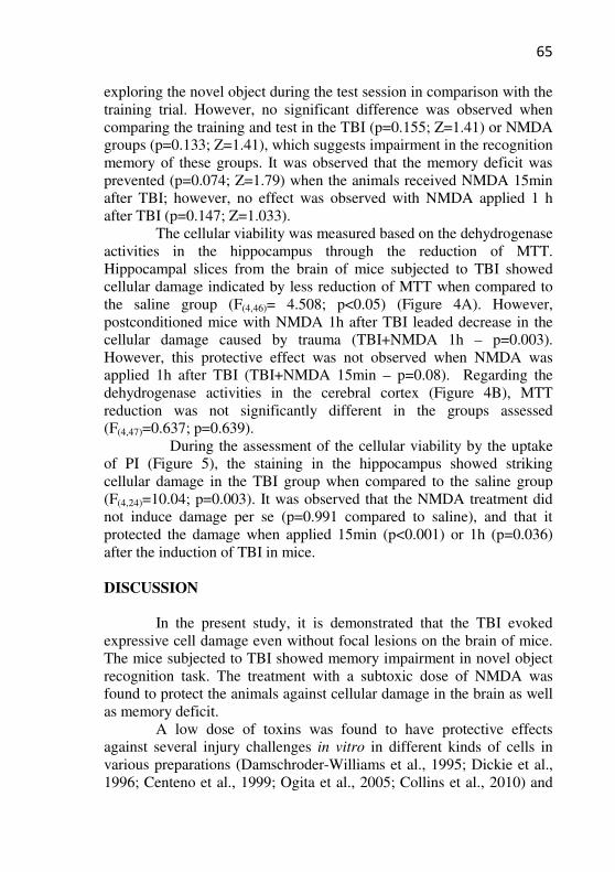
65
exploring the novel object during the test session in comparison with the training trial. However, no significant difference was observed when comparing the training and test in the TBI (p=0.155; Z=1.41) or NMDA groups (p=0.133; Z=1.41), which suggests impairment in the recognition memory of these groups. It was observed that the memory deficit was prevented (p=0.074; Z=1.79) when the animals received NMDA 15min after TBI; however, no effect was observed with NMDA applied 1 h after TBI (p=0.147; Z=1.033).
The cellular viability was measured based on the dehydrogenase activities in the hippocampus through the reduction of MTT. Hippocampal slices from the brain of mice subjected to TBI showed cellular damage indicated by less reduction of MTT when compared to the saline group (F(4,46)= 4.508; p<0.05) (Figure 4A). However, postconditioned mice with NMDA 1h after TBI leaded decrease in the cellular damage caused by trauma (TBI+NMDA 1h – p=0.003). However, this protective effect was not observed when NMDA was applied 1h after TBI (TBI+NMDA 15min – p=0.08). Regarding the dehydrogenase activities in the cerebral cortex (Figure 4B), MTT reduction was not significantly different in the groups assessed (F(4,47)=0.637; p=0.639).
During the assessment of the cellular viability by the uptake of PI (Figure 5), the staining in the hippocampus showed striking cellular damage in the TBI group when compared to the saline group (F(4,24)=10.04; p=0.003). It was observed that the NMDA treatment did not induce damage per se (p=0.991 compared to saline), and that it protected the damage when applied 15min (p<0.001) or 1h (p=0.036) after the induction of TBI in mice.
DISCUSSION
In the present study, it is demonstrated that the TBI evoked
expressive cell damage even without focal lesions on the brain of mice. The mice subjected to TBI showed memory impairment in novel object recognition task. The treatment with a subtoxic dose of NMDA was found to protect the animals against cellular damage in the brain as well as memory deficit.
A low dose of toxins was found to have protective effects against several injury challenges in vitro in different kinds of cells in various preparations (Damschroder-Williams et al., 1995; Dickie et al., 1996; Centeno et al., 1999; Ogita et al., 2005; Collins et al., 2010) and
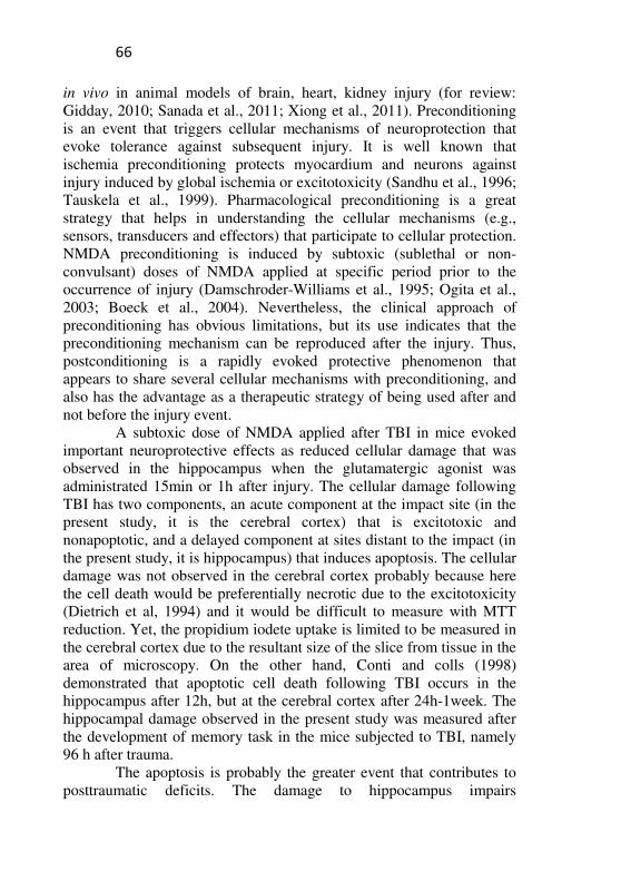
66
in vivo in animal models of brain, heart, kidney injury (for review: Gidday, 2010; Sanada et al., 2011; Xiong et al., 2011). Preconditioning is an event that triggers cellular mechanisms of neuroprotection that evoke tolerance against subsequent injury. It is well known that ischemia preconditioning protects myocardium and neurons against injury induced by global ischemia or excitotoxicity (Sandhu et al., 1996; Tauskela et al., 1999). Pharmacological preconditioning is a great strategy that helps in understanding the cellular mechanisms (e.g., sensors, transducers and effectors) that participate to cellular protection. NMDA preconditioning is induced by subtoxic (sublethal or non-convulsant) doses of NMDA applied at specific period prior to the occurrence of injury (Damschroder-Williams et al., 1995; Ogita et al., 2003; Boeck et al., 2004). Nevertheless, the clinical approach of preconditioning has obvious limitations, but its use indicates that the preconditioning mechanism can be reproduced after the injury. Thus, postconditioning is a rapidly evoked protective phenomenon that appears to share several cellular mechanisms with preconditioning, and also has the advantage as a therapeutic strategy of being used after and not before the injury event.
A subtoxic dose of NMDA applied after TBI in mice evoked important neuroprotective effects as reduced cellular damage that was observed in the hippocampus when the glutamatergic agonist was administrated 15min or 1h after injury. The cellular damage following TBI has two components, an acute component at the impact site (in the present study, it is the cerebral cortex) that is excitotoxic and nonapoptotic, and a delayed component at sites distant to the impact (in the present study, it is hippocampus) that induces apoptosis. The cellular damage was not observed in the cerebral cortex probably because here the cell death would be preferentially necrotic due to the excitotoxicity (Dietrich et al, 1994) and it would be difficult to measure with MTT reduction. Yet, the propidium iodete uptake is limited to be measured in the cerebral cortex due to the resultant size of the slice from tissue in the area of microscopy. On the other hand, Conti and colls (1998) demonstrated that apoptotic cell death following TBI occurs in the hippocampus after 12h, but at the cerebral cortex after 24h-1week. The hippocampal damage observed in the present study was measured after the development of memory task in the mice subjected to TBI, namely 96 h after trauma.
The apoptosis is probably the greater event that contributes to posttraumatic deficits. The damage to hippocampus impairs
67
performance on a variety of tasks of learning and memory (Squire, 1992). There is no consensus about the cerebral areas associated with the performance in the novel object recognition task; however, there is probably the involvement of hippocampus in this task, which indicates that the damage to the hippocampus produces memory impairment (Broadbent et al., 2004). Recognition memory is a declarative memory that involves temporal lobe areas, such as perirhinal and parahippocampal/postrhinal cortices as well as the entorhinal cortex. The hippocampus is the final stage of convergence within the medial temporal lobe, receiving projections from these areas (Lavenex and Amaral, 2000). It is important to note that the object recognition task taxes memory after only one trial (of 5min); this makes the assay more sensitive to amnesic experimental interventions when compared to other tasks in which incremental learning across multiple trials is induced such as in the radial maze and water maze tasks (Dere et al. 2007). In the present study, TBI was found to induce impairment in the anterograde novel object recognition memory without affecting habituation memory. Habituation memory is a nonassociative memory that is acquired when repeated or continuous exposure of an animal to a novel environment changes the behavioral responses to it. The long-term memory of habituation depends on the functionality of hippocampal NMDA and AMPA/kainate receptors (Vianna et al., 2000).
There are some limitations to the present study. Firstly, the memory evaluations were initiated between 4 and 15 days post-injury, but the animals were tested for memory tasks at 48h after TBI. Thus, a delay in the development of memory dysfunctions is possible; however, the study presents results that strongly support the notion that NMDA postconditioning indeed protects the brain against TBI. Second, the time of viability cell measure occurs after the mice were tested for memory performance. It is not possible to exclude the hypothesis that the habituation memory involving hippocampal mechanisms could be impaired at 96h after TBI, because the studies demonstrated the deficit after trauma induced by the controlled cortical impact (O’Connor et al., 2003; Wagner et al., 2007).
The use of the NMDA antagonists in humans is very limited as they protect against excitotoxicity but increase the apoptotic cell death induced by TBI (Pohl et al., 1999) or stroke (Lee et al., 1999) in animal model. The death evoked by NMDA antagonists is probably due to the fact that the receptor activity induces mechanisms prosurvival in neurons in development (Pohl et al., 1999) and this effect could be
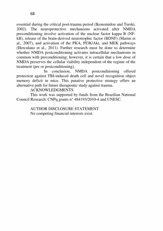
68
essential during the critical post-trauma period (Ikonomidou and Turski, 2002). The neuroprotective mechanisms activated after NMDA preconditioning involve activation of the nuclear factor kappa B (NF-kB), release of the brain-derived neurotrophic factor (BDNF) (Marini et al., 2007), and activation of the PKA, PI3K/Akt, and MEK pathways (Herculano et al., 2011). Further research must be done to determine whether NMDA postconditioning activates intracellular mechanisms in common with preconditioning; however, it is certain that a low dose of NMDA preserves the cellular viability independent of the regime of the treatment (pre or postconditioning).
In conclusion, NMDA postconditioning offered protection against TBI-induced death cell and novel recognition object memory deficit in mice. This putative protective strategy offers an alternative path for future therapeutic study against trauma.
ACKNOWLEDGMENTS This work was supported by funds from the Brazilian National
Council Research: CNPq grants n° 484193/2010-4 and UNESC. AUTHOR DISCLOSURE STATEMENT No competing financial interests exist.
69
REFERENCES
Adamchik, Y., Baskys, A. (2000) Glutamate-mediated neuroprotection against N-methyl-D-aspartate toxicity: a role for metabotropic glutamate receptors. J. Neurosci. 99(4):731-736.
Adelson, P.D., Robichaud, P., Hamilton, R.L., Patrick, M.K. (1996) A model of diffuse traumatic brain injury in the immature rat. J. Neurosurg. 85(5):877- 884.
Barros, D., Amaral, O.B., Izquierdo, I., Geracitano, L., Raseira, M.C.B., Henriques, A.T., Ramirez, M.R. (2006) Behavioral and genoprotective effects of Vaccinium berries intake in mice. Pharmacol. Biochem. Behav. 84:229–234.
Berliocchi, L., Bano, D., Nicotera, P. (2005) Ca2+ signals and death programmed in neurons. Philos. Trans. R. Soc. Lond. B. Biol. Sci. 360:2255–2258.
Bernert, H., Turski, L. (1996) Traumatic brain damage prevented by the non-N-methyl-D-aspartate antagonist 2,3-dihydroxy-6-nitro-7-sulfamoylbenzolfquinoxaline. Proc. Natl. Acad. Sci. USA 93:5235-5240.
Boeck, C.R., Ganzella, M., Lottermann, A., Vendite, D. (2004) NMDA preconditioning protects against seizures and hippocampal neurotoxicity induced by quinolinic acid in mice. Epilepsia 45(7):745-750.
Broadbent, N.J., Squire, L.R., Clark, R.E. (2004) Spatial memory, recognition memory and the hippocampus. Proc. Natl. Acad. Sci. USA 101:14515-14520.
Castellano, C., Cestari, V., Ciamei, A. (2001) NMDA receptors and learning and memory processes. Curr. Drug Targets 2:273-283.
Centeno, J.M., Orti, M., Salom, J.B., Sick, T.J., Pérez-Pinzón, M.A. (1999) Nitric oxide is involved in anoxic preconditioning neuroprotection in rat hippocampal slices. Brain Res. 836(1-2):62-9.
Chuang, D.M., Gao, X.M., Paul, S.M. (1992) N-methyl-D-aspartate exposure blocks glutamate toxicity in cultured cerebellar granule cells. Mol. Pharmacol. 42:210-216.
Collins, M.A., Neafsey, E.J., Wang, K., Achille, N.J., Mitchell, R.M., Sivaswamy, S. (2010) Moderate ethanol preconditioning of rat brain cultures engenders neuroprotection against dementia-inducing neuroinflammatory proteins: possible signaling mechanisms. Mol. Neurobiol. 41(2-3):420-425.
70
Conti, A.C., Raghupathi, R., Trojanowski, J.Q., McIntosh, T.K. (1998) Experimental brain injury induces regionally distinct apoptosis during the acute and delayed post-traumatic period. J. Neurosci. 18(15):5663–5672.
Costa, T., Constantino, L.C., Mendonça, B.P., Pereira, J.G., Herculano, B., Tasca, C.I., Boeck, C.R. (2010) N-Methyl-D-Aspartate preconditioning improves short-term motor deficits outcome after mild traumatic brain injury in mice. J. Neurosci. 88:1329–1337.
Damschroder-Williams, P., Irwin, R.P., Lin, S.Z., Paul, S.M. (1995) Characterization of the excitoprotective actions of N-methyl-D-aspartate in cultured cerebellar granule neurons. J. Neurochem. 65:1069-1076.
Dempsey, R.J., Baskaya, M.K., Dogan, A. (2000) Attenuation of brain edema, blood-brain barrier breakdown, and injury volume by ifenprodil, a polyamine-site N-methyl-D-aspartate receptor antagonist, after experimental traumatic brain injury in rats. Neurosurgery 47(2):399-404.
Dere, E., Huston, J.P., De Souza, S.M.A. (2007) The pharmacology neuroanatomy and neurogenetics of one-trial object recognition in rodents. Neurosci. Biobehav. Rev. 31:673–704.
Dickie, B.G.M., Holmes, C., Greenfield, S.A. (1996) Neurotoxic and neurotrophic effects of chronic N-methyl-D-aspartate exposure upon mesencephalic dopaminergic neurons in organotypic culture. J. Neurosci. 72(3):731-741.
Dietrich, W.D., Alonso, O., Halley, M. (1994) Early microvascular and neuronal consequences of traumatic brain injury: a light and electron microscopic study in rats. J. Neurotrauma 11:289 –301.
Ennaceur, A., Delacour, J. (1988) A new one-trial test for neurobiological studies of memory in rats. 1: Behavioral data. Behav Brain Res 31:47-59.
Fujimoto, S., Katsuki, H., Kume, T., Kaneko, S., Akaike, A. (2004) Mechanisms of oxygen glucose deprivation-induced glutamate release from cerebrocortical slice cultures. J. Neurosci. Res. 50:179–187.
Gagliardi, R.J. (2000) Neuroprotection, excitotoxicity and NMDA antagonists. Arq. Neuropsi. 58(2B):583-588.
Gidday, J.M. (2010) Pharmacologic preconditioning: translating the promise. Transl. Stroke Res. 1(1):19-30.
71
Giménez-Llort, L., Martínez, E., Ferré, S. (1995) Dopamine-independent and adenosine-dependent mechanisms involved in effects of N-methyl-D-aspartate on motor activity in mice. Eur. J. Pharmacol. 275:171-177.
Grabb, M.C., Choi, D.W. (1999) Ischemic tolerance in murine cortical cell culture: critical role for NMDA receptors. J. Neurosci. 19(5):1657–1662.
Hardingham, G.E., Bading, H. (2003) The Yin and Yang of NMDA receptor signaling. Trends Neurosci. 26(2): 81-89.
Hartnett, K.A., Stout, A.K., Rajdev, S., Rosenberh, P.A., Reymolds, I.J., Aizenman, E. (1997) NMDA receptor-mediated neurotoxicity: a paradoxical requirement for extracellular Mg2+ in Na+/ Ca2+ - free solutions in rat cortical neurons in vitro. J. Neurochem. 68:1836-1845.
Herculano, B.A., Vandresen-Filho, S., Martins, W.C., Boeck, C.R., Tasca, C.I. (2011) NMDA preconditioning protects against quinolinic acid-induced seizures via PKA, PI3K and MAPK/ERK signaling pathways. Behav. Brain Res. 219(1):92-7.
Ikonomidou, C., Turski, L. (2002) Why did NMDA receptor antagonists fail clinical trials for stroke and traumatic brain injury? Lancet Neurol. 1(6):383-386.
Katz, D.I., White, D.K., Alexander, M.P., Klein, R.B. (2004) Recovery of ambulation after traumatic brain injury. Arch. Phys. Med. Rehabil. 86:865-869.
Lavenex, P., Amaral, D.G. (2000) Hippocampal-neocortical interaction: a hierarchy of associativity. Hippocampus 10:420-430.
Leconte, C., Tixier, E., Freret, T., Toutain, J., Saulnier, R., Boulouard, M., Roussel, S., Schumann-Bard, P., Bernaudin, M. (2009) Delayed hypoxic postconditioning protects against cerebral ischemia in the mouse. Stroke 40:3349-3355.
Lee, J.M., Zipfel, G.J., Choi, D.W. (1999) The changing landscape of ischaemic brain injury mechanisms. Nature 399: A7–A14.
Lima, M.N.M., Laranja, D.C., Bromberg, E., Roesler, R., Schröder, N. (2004) Pre- or post-training administration of the NMDA receptor blocker MK-801 impairs object recognition memory in rats. Beha.v Brain Res. 156:139-143.
Liu, Y., Peterson, D.A., Kimura, H., Schubert, D. (1997) Mechanism of cellular 3-(4,5-dimethylthiazol-2-yl)-2,5-diphenyltetrazolium bromide (MTT) reduction. J. Neurochem. 69:581–593.
72
Marik, P.E., Varon, J., Trask, T. (2002) Management of head trauma. Chest 122(4):1116-1118.
Marini, A.M., Jiang, X., Wu, X., Pan, H., Guo, Z., Mattson, M.P., Blondeau, N. (2007) Preconditioning and neurotrophins: a model for brain adaptation to seizures, ischemia and other stressful stimuli. Amino Acids 32:299-304.
Miller, L.P., Lyeth, B.G., Jenkins, L.W., Oleniak, L., Panchision, D., Hamm, R.J., Phillips, L.L., Dixon, C.E., Clifton, G.L., Hayes, R.L. (1990) Excitatory amino acid receptor subtype binding following traumatic brain injury. Brain Res. 526:103-107.
Mosmann, T. (1983) Rapid colorimetric assay for cellular growth and survival: application to proliferation and cytotoxicity assay. J. Immunol. 65: 55–63.
Muir, K.W. (2006) Glutamate-based therapeutic approaches: clinical trials with NMDA antagonists. Curr. Opin. Pharmacol. 6(1):53-60.
Ogita, K., Okuda, H., Watanabe, M., Nagashima, R., Sugiyama, C., Yoneda, Y. (2005) In vivo treatment with the K+ channel blocker 4-aminopyridine protects against kainate-induced neuronal cell death through activation of NMDA receptors in murine hippocampus. Neuropharmacology 48:810-821.
Ogita, K., Okuda, H., Yamamoto, Y., Nishiyama, N., Yoneda, Y. (2003) In vivo neuroprotective role of NMDA receptors against kainate-induced excitotoxicity in murine hippocampal pyramidal neurons. J. Neurochem. 85:1336–1346.
O’Connor, C.A., Cernak, I., Vink, R. (2003) Interaction between anesthesia, gender, and functional outcome task following diffuse traumatic brain injury in rats. J. Neurotrauma 20:533–541.
Pérez-Pinzón, M.A. (2004) Neuroprotective effects of ischemic preconditioning in brain mitochondria following cerebral ischemia. J. Bioenerg. Biomembr. 36(4):323-327.
Pignataro, G., Scorziello, A., Di Renzo, G., Annunziato, L. (2009) Post-ischemic brain damage: effect of ischemic preconditioning and postconditioning and identification of potential candidates for stroke therapy. FEBS J. 276(1):46-57.
Pohl, D., Bittigau, P., Ishimaru, M.J., Stadthaus, D., Hübner, C., Olney, J.W., Turski, L., Ikonomidou, C. (1999) N-Methyl-D-aspartate antagonists and apoptotic cell death triggered by head trauma in developing rat brain. J. Pharmacol. 96:2508–2513.
73
Riedel, G., Platt, B., Micheau, J. (2003) Glutamate receptor function in learning and memory. Behav. Brain. Res. 140(1-2):1-47.
Rossato, J.I., Bevilaqua, L.R.M., Myskiw, J.C., Medina, J.H., Izquierdo, I., Cammarota, M. (2007) On the role of hippocampal protein synthesis in the consolidation and reconsolidation of object recognition memory. Learn Mem. 14: 36–46.
Sanada, S., Komuro, I., Kitakaze, M. (2011) Pathophysiology of myocardial reperfusion injury: preconditioning, postconditioning, and translational aspects of protective measures. Am. J. Physiol. Heart Circ. Physiol. 301(5):H1723-1741.
Sanders, M.J., Sick, T.J., Perez-Pinzon, M.A., Dietrich, W.D., Green, E.J. (2000) Chronic failure in the maintenance of long term potentiation following fluid percussion injury in the rat. Brain Res. 861:69–76.
Sandhu, R., Thomas, U., Diaz, R.J., Wilson, G.J. (1996) Effect of ischemic preconditioning of the myocardium on cAMP. Circulation Res. 78:137-147.
Scartabelli, T., Gerace. E., Landucci, E., Moroni, F., Pellegrini-Giampietro, D.E. (2008) Neuroprotection by group I mGlu receptors in a rat hippocampal slice model of cerebral ischemia is associated with the PI3K–Akt signaling pathway: A novel postconditioning strategy? Neuropharmacology 55:509-516.
Schumann, J., Alexandrovich, G.A., Biegon, A., Yaka, R. (2008) Inhibition of NR2B phosphorylation restores alterations in NMDA receptor expression and improves functional recovery following traumatic brain injury in mice. J. Neurotrauma 25:945–957.
Squire, L. R. (1992) Memory and the hippocampus: a synthesis from findings with rats, monkeys, and humans. Psychol. Rev. 99(2):195–231.
Soriano, F.X., Papadia, S., Hofmann, F., Hardingham, N.R., Bading, H., Hardingham, G.E. (2006) Preconditioning doses of NMDA promote neuroprotection by enhancing neuronal excitability. J. Neurosci. 26(17):4509–4518.
Tauskela, J.S., Chakravarthy, B.R., Murray, C.L., Wang, Y., Comas, T., Hogan, M., Hakim, A., Morley, P. (1999) Evidence from culture rat cortical neurons of differences in the mechanism of ischemic preconditioning of brain and heart. Brain Res. 827:143-151.
74
Ucar, T., Tanriover, G., Gurer, I., Onal, M.Z., Kazan, S. (2006) Modified experimental mild traumatic brain injury model. J. Trauma 60(3):558-65.
Vianna, M.R., Alonso, M., Viola, H., Quevedo, J., Paris, F., Furman, M., Stein, M.L., Medina, J.H., Izquierdo, I. (2000) Role of hippocampal signaling pathways in long-term memory formation of a nonassociative learning task in the rat. Learn. Mem. 7:333–340.
Wagner, A.K., Postal, B.A., Darrah, S.D., Chen, X., Khan, A.S. (2007) Deficits in novelty exploration after controlled cortical impact. J. Neurotrauma 24(8):1308-1320.
Xiong, J., Liao, X., Xue, F.S., Yuan, Y.J., Wang, Q., Liu, J.H. (2011) Remote ischemia conditioning-an endogenous cardioprotective strategy from outside the heart. Chin. Med. J. (Engl) 124(14):2209-2215.
Zhao, H., Sapolsky, R.M., Steinberg, G.K. (2006) Interrupting reperfusion as a stroke therapy: ischemic postconditioning reduces infarct size after focal ischemia in rats. J. Cereb. Blood. Flow. Metab. 26:1114–1121.
75
LEGENDS Figure 1: Scheme of the experimental protocol. Figure 2: Effect of post-treatment with a non-convulsant dose of
NMDA (75 mg/kg) or saline (NaCl 0.9 g%) in habituation memory after TBI. Training session was performed 24h after TBI and NMDA administration. Data are expressed as mean ± SEM of crossing and rearing of training and test session performance in open field with a 24h interval between sessions. *P<0.05 compared between training and test sessions in the same group.
Figure 3: Effect of post-treatment with a non-convulsant dose of
NMDA (75 mg/kg) or saline (NaCl 0.9 g%) in object recognition memory after TBI. Training session was performed 24h after TBI. Data are expressed as median ± interquartile ranges in recognition indexes of animals of each group. *P<0.05 compared between training and test sessions in the same group.
Figure 4: Effect of post-treatment with a nonconvulsant dose of
NMDA (75 mg/kg) or saline (NaCl 0.9 g%) in deshydrogenase activity based MTT reduction after TBI. The cell viability was measured at 96h after TBI in the (A) hippocampus and (B) cerebral cortex of mice and was represented by the control percentage (saline - dashed line). Data are expressed as mean ± SEM of MTT reduction. *P<0.05 difference compared to control group; #P<0.05 difference compared to TBI group.
Figure 5: Effect of post-treatment with a nonconvulsant dose of
NMDA (75 mg/kg) or saline (NaCl 0.9 g%) in propidium iodite (PI) activity after TBI. The cell viability was measured 96h after TBI in the hippocampus. (A) Representative photomicrographs of the hippocampal slices for each group. The fluorescence area corresponds to the cellular damage. (B) Quantification of the PI uptake. Images were captured and then analyzed using the Scion Image software. Data are expressed as mean ± SEM of PI uptake. *P<0.05 difference compared to control group; #P<0.05 difference compared to TBI group.
76
Figure 1
Figure 2
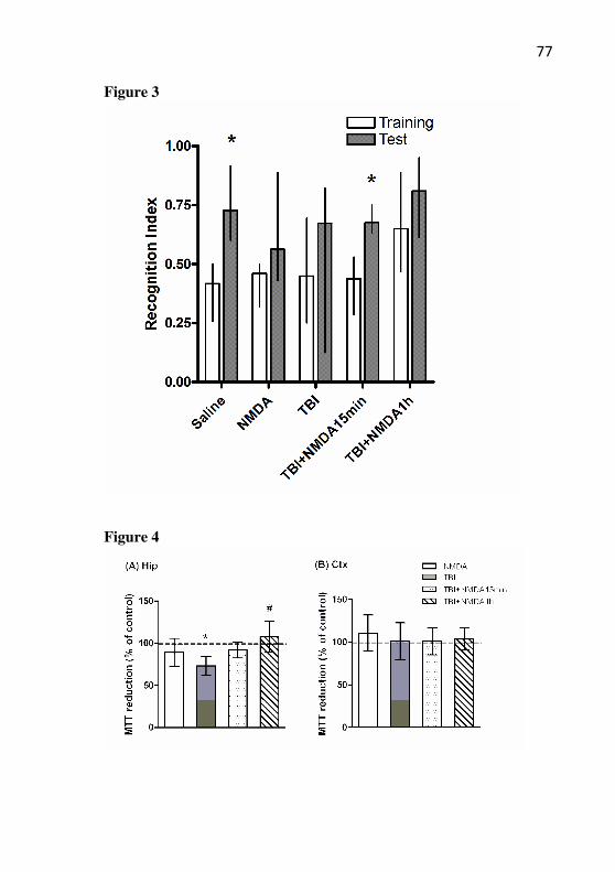
77
Figure 3
Figure 4
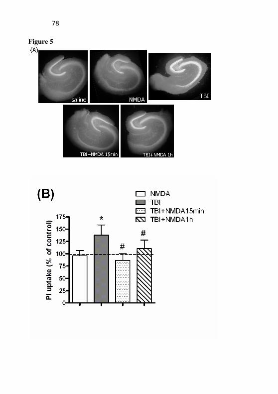
78
Figure 5
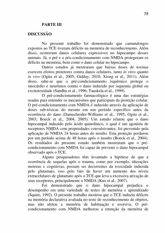
79
PARTE III
DISCUSSÃO
No presente trabalho foi demonstrado que camundongos expostos ao TCE tiveram déficits na memória de reconhecimento. Além disso, ocorreram danos celulares expressivos no hipocampo desses animais. Já, o pré e o pós-condicionamento com NMDA protegeram os déficits na memória, bem como o dano celular no hipocampo.
Outros estudos já mostraram que baixas doses de toxinas exercem efeitos protetores contra danos celulares, tanto in vitro quanto in vivo (Ogita et al., 2005; Gidday, 2010; Xiong et al., 2011). Além disso, sabe-se que o pré-condicionamento isquêmico protege o miocárdio e neurônios contra o dano induzido por isquemia global ou excitotoxidade (Sandhu et al., 1996; Tauskela et al., 1999).
O pré-condicionamento farmacológico é uma das estratégias usadas para entender os mecanismos que participam da proteção celular. O pré-condicionamento com NMDA é induzido através da aplicação de doses sub-tóxicas do mesmo em um período específico antes da ocorrência do dano (Damschroder-Williams et al., 1995; Ogita et al., 2003; Boeck et al., 2004, 2005). Um estudo relatou que o dano hipocampal induzido pelo ácido quinolínico, o qual é um agonista de receptores NMDA com propriedades convulsivantes, foi prevenido pela aplicação de NMDA 24 horas antes do insulto. Esta proteção perdurou por um período acima de 48 horas após o insulto (Boeck et al., 2004). Os resultados do presente estudo também mostraram que o pré-condicionamento com NMDA foi capaz de prevenir o dano hipocampal observado após o TCE.
Alguns pesquisadores têm levantado a hipótese de que a ocorrência de sequelas após o trauma, como por exemplo, alterações motoras e cognitivas, possam ser decorrentes da toxicidade induzida pelo glutamato, isso pelo fato de haver um aumento dos níveis extracelulares de glutamato após o TCE que leva a excessiva ativação de seus receptores, principalmente o NMDA (Kuo et al., 2007).
Foi demonstrado que o dano hipocampal prejudica o desempenho em uma variedade de testes de memória e aprendizado (Squire, 1992). O presente trabalho mostrou que o TCE induziu déficits na memória declarativa avaliada no teste de reconhecimento de objetos, mas não afetou a memória de habituação e aversiva. O pré-condicionamento com NMDA melhorou a retenção da memória de
80
longa duração no teste da esquiva inibitória. Tais efeitos, podem ser relacionados, pelo menos em parte, ao aumento da viabilidade celular no hipocampo, uma estrutura responsável pelos processos de formação da memória.
Além dos prejuízos cognitivos também têm sido relatado uma prevalência de 15,3 a 33% de depressão em humanos sobreviventes ao TCE (Kim et al., 1997). Além disso, a ansiedade é uma comorbidade comum encontrada nesses pacientes (Jorge et al., 1993). A incidência de depressão após o TCE pode ser relacionada a uma atrofia hipocampal, as quais são também observadas em pacientes com depressão (Campbell & MacQuen, 2004). Em um estudo realizado com ratos também foi observado aumento no comportamento do tipo ansioso e depressivo após exposição ao TCE (Pandey et al., 2009). From e colaboradores (2004) mostraram que o tratamento com magnésio, que é um bloqueador do receptor NMDA, melhorou comportamentos do tipo depressivo e ansioso em 61% dos animais expostos ao TCE. Entretanto, no presente estudo não foi observado comportamento do tipo depressivo ou ansioso em camundongos com TCE, o que sugere-se que o TCE não tenha alterado vias cerebrais envolvidas com a memória aversiva e com o medo, como por exemplo a amígdala, propondo-se estudos que avaliem viabilidade celular nessa região cerebral. No presente trabalho foi demonstrado também que o pós-condicionamento com NMDA protegeu contra o dano celular cerebral e os déficits na memória em camundongos expostos ao TCE. O pós-condicionamento é um fenômeno protetor que induz a ativação de diversos mecanismos celulares, assim como ocorre no pré-condicionamento. Porém, há uma vantagem importante do pós-condicionamento, pois diferente do pré-condicionamento esse pode ser considerado uma boa estratégia terapêutica já que pode ser usado após o dano.
O dano hipocampal observado no presente estudo nos camundongos após o TCE, onde avaliou-se os efeitos do pós-condicionamento com NMDA, foi verificado após os testes de memória em camundongos expostos ao TCE, ou seja, 96 horas após o trauma. Também foi mostrado que o TCE induziu déficits na memória de reconhecimento, mas não alterou a memória de habituação. Os mecanismos neuroprotetores ativados após o pré-condicionamento com NMDA envolvem a ativação do fator nuclear kappa B (NF-KB), liberação do fator de BDNF e ativação de vias de sinalização celular, PKA, PI3K/AKT e MEK, as quais são envolvidas com a sobrevivência celular (Herculano et al., 2011; Marini et al., 2007). Assim, sugere-se no
81
presente estudo que sejam realizadas mais pesquisas com o intuito de verificar se o pós-condicionamento ativa vias intracelulares, assim como ocorre com o pré-condicionamento.
Em conclusão, o presente estudo mostrou que tanto o pré quanto o pós-condicionamento com NMDA apresentaram proteção contra dano celular induzido pelo TCE, assim como os déficits na memória de reconhecimento de objetos. Além disso, sugere-se mais estudos para avaliar os efeitos protetores de baixas doses de NMDA focando em intervenções farmacológicas como terapia para o TCE.
REFERÊNCIAS
ABAS F; ALKAN T; GOREN B; TASKAPILIOGLU O; SARANDOL
E; TOLUNAY S. Neuroprotective effects of postconditioning on lipid peroxidation and apoptosis after focal cerebral ischemia/reperfusion injury in rats. Turkish Neurosurgery 20: 1-8. 2010.
ACARIN L; PELUFFO H; BARBEITO L; CASTELLANO B; GONZALEZ B. Astroglial nitration after postnatal excitotoxic damage: correlation with nitric oxide sources, cytoskeletal, apoptotic and antioxidant proteins. Journal of Neurotrauma 22: 189–200. 2005.
ACEVEDO HR; ROJAS MD; ARCEO SDB; HERNANDEZ MS; VÁSQUEZ MM; TERRAZAS T; DEL TORO GV. Effect of nonadecyl salicyclic acid and its methyl ester on the induction of micronuclei in polychromatic erythrocytes in mouse peripheral blood. Mutation Research 609: 43-46. 2006.
ADELSON PD; ROBICHAUD P; HAMILTON RL; KOCHANEK PM. A model of diffuse traumatic brain injury in the immature rat. Journal Neurosurgery 85: 877-884. 1996.
ALLEN JW; KNOBLACH SM; FADEN AI. Activation of group I metabotropic glutamate receptors reduces neuronal apoptosis but increases necrotic cell death in vitro. Cell Death and Differentiation 7: 470–476. 2000.
ALKAYED NJ; GOTO S; SUGO N; JONH HD; CRAIN BJ; BERNARD O; TRAYSTMAN RJ; HURN PD. Estrogen and Bcl-2: gene induction and effect of transgene in experimental stroke. Journal Neuroscience 21: 7543-7550. 2001.
ATOCHIN DN; CLARK J; DEMCHENKO IT; MOSKOWITZ MA; HUANG PL. Rapid cerebral ischemic preconditioning in mice
82
deficient in endothelial and neuronal nitric oxide synthases. Stroke 34: 1299–1303. 2003.
BARICHELLO T; MARTINS R; REINKE A; FEIER G; RITTER C; QUEVEDO J; DAL-PIZZOL F. Cognitive Impairment in sepsis survivors from cecal ligation and perforation. Critical Care Medicine 33: 221-223. 2005.
BERGER RP; TAASAN S; RAND A; LOKSHIN A; KOCHANEK P. Multiplex assessment of serum biomarker concentrations in well-appearing children with inflicted traumatic brain injury. Pediatric Research 65: 97-102. 2008.
BEVILAQUA LR; KERR DS; MEDINA JH; IZQUIERDO I; CAMMAROTTA M. Inhibition of hippocampal Jun N-terminal kinase enhances short-term memory but blocks long-term memory formation and retrieval of an inhibitory avoidance task. European Journal of Neuroscience 17: 897- 902. 2003.
BIANCHIN M; DA SILVA RC; SCHMITZ PK; MEDINA JH; IZQUIERDO I. Memory of inhibitory avoidance in the rat is regulated by glutamate metabotropic receptors in the hippocampus. Behavioural Pharmacology 5: 356-359. 1994.
BLISS TV; COLLINGRIDGE GL. A synaptic model of memory: long-term potentiation in the hippocampus. Nature 36: 31-39. 1993.
BOECK CR; GANZELLA M; LOTTERMANN A; VENDITE D. NMDA preconditioning protects against seizures and hippocampal neurotoxicity induced by quinolinic acid in mice. Epilepsia 45: 745-750. 2004.
BOLT MW; MAHONEY PA. High-efficiency blotting of proteins of diverse sizes following sodium dodecyl sulfate-polyacrylamide gel electrophoresis. Analitical Biochemistry 247: 185-192. 1997.
BRESSAN RA; PILOWSKY L. Hipótese Glutamatérgica da Esquizofrenia. Revista Brasileira de Psiquiatria 25: 177-183. 2003.
BRESSAN RA; PILOWSKY L. Hipótese Glutamatérgica da Esquizofrenia. Revista Brasileira de Psiquiatria 25: 177-183. 2003.
BRINK JD; GARRETT AL; HALE WR; NICKEL VL; WOO–SAM J. Recovery of motor and intellectual function in children sustaining severe head injuries. Developmental Medicine & Child Neurology 12: 565–571. 1970.
83
CABRERA JA; ZIEMBA EA; COLBERT R; ANDERSON L; SLUITER W; DUNCKER DJ; BUTTERICK TA; SIKORA J; WARD HB; KELLY RF; MCFALLS EO. Altered expression of mitochondrial electron transport proteins and improved myocardial energetic state during late ischemic preconditioning. American Journal of Physiology Heart and Circulatory Physiology 302: 1974-1982. 2012.
CAMMAROTA M; BEVILAQUA LRM; IZQUIERDO I. Aprendizado e Memória. In: LENT R. Neurociências da Mente e do Comportamento. Rio de Janeiro: Guanabara Koogan. 2008.
CAROBREZ AP. Transmissão pelo glutamato como alvo molecular na ansiedade. Revista Brasileira de Psiquiatria 25: 52-58. 2003.
CHEN JK; JOHNSTON KM; FREY S; PEDRITES M; WORSLEY K; PTITO A. Functional abnormalities in symptomatic concussed athletes: an FMRI study. Neuroimage 22: 68–82. 2004.
CHEN J; SIMON R. Ischemic tolerance in the brain. Neurology 48: 306-311. 1997.
CHOI DW. Possible mechanisms limiting N-methyl-D-aspartate receptor overactivation and the therapeutic efficacy of N-methyl-D-aspartate antagonists. Stroke 11: 20-22. 1990.
COLLINS A; DUSINSKA M; FRANKLIN M; SOMOROSYSKA M; PETROVSKA H; DUTHIE S; FILLION L; PANAYIUTIDIS M; RASLOVA K; VAUGHAN N. Comet assay in human biomonitoring studies: reability, validation and applications. Environmental Molecular Mutagenesis 30: 139-146. 1997.
CORRIGAN JD; SELASSIE AW; ORMAN JA. The epidemiology of traumatic brain injury. The Journal of Head Trauma Rehabilitation 25: 72-80. 2010.
COSTA T; CONSTANTINO LC; MENDONÇA BP; HERCULANO B; TASCA CI; BOECK CR. NMDA preconditioning improves short and long-term motor deficits outcome after diffuse traumatic brain injury. Journal of Neuroscience Research 88: 1329-1337. 2010.
DAMSCHRODER-WILLIAMS P; IRWIN RP; LIN SZ; PAUL SM. Characterization of the excitoprotective actions of N-methyl-D-aspartate in cultured cerebellar granule neurons. Journal of Neurochemistry 65: 1069-1076. 1995.
DEMPSEY RJ; BAŞKAYA MK; DOĞAN A. Attenuation of brain edema, blood-brain barrier breakdown, and injury volume by ifenprodil, a polyamine-site N-methyl-D-aspartate receptor
84
antagonist, after experimental traumatic brain injury in rats. Neurosurgery 47: 399-404. 2000.
DINGLEDINE R; BORGES K; BOWIE D; TRAYNELIS SF. The glutamate receptor ion channels. Pharmacological Reviews 51: 7-61. 1999.
DUBAL DB; SHUGHRUE PJ; WILSON ME; MERCHENTHALER I; WISE PM. Estradiol modulates bcl-2 in cerebral ischemia: a potential role for estrogen receptors. Journal of Neuroscience 19: 6385–6393. 1999.
FADEN AI; O'LEARY DM; FAN L; BAO W; MULLINS PG; MOVSESYAN VA. Selective blockade of the mGluR1 receptor reduces traumatic neuronal injury in vitro and improves outcome after brain trauma. Experimental Neurology 167: 435-444. 2001.
FEATHERSTONE DE. Intercellular glutamate signaling in the nervous system and beyond. ACS Chemical Neuroscience 1: 4-12. 2010.
FRANCESC X; PAPADIA SS; HOFMANN F; NEIL R; BADING HH; HARDINGHAM GE. Preconditioning doses of NMDA promote neuroprotection by enhancing neuronal excitability. The Journal of Neuroscience 26: 4509–4518. 2006.
GAO X; CHEN J. Conditional knockout of brain-derived neurotrophic factor in the hippocampus increases death of adult-born immature neurons following traumatic brain injury. Journal of Neurotrauma 26: 1325-1335. 2009.
GAVIOLI EC; RIZZI A; MARZOLA G; ZUCCHINI S; REGOLI D; CALO’ G. Altered anxiety-related behavior in nociceptin/orphanin FQ receptor gene knockout mice. Peptides 28: 1229–1239. 2007.
GIDDAY JM. Pharmacologic preconditioning: translating the promise. Translational Stroke Research 1: 19-30. 2010.
GINSBERG MD. Neuroprotection for ischemic stroke: past, present and future. Neuropharmacology 55: 363-389. 2008.
Gong QZ; DELAHUNTY TM; HAMM RJ; LYETH BG. Metabotropic glutamate antagonist, MCPG, treatment of traumatic brain injury in rats. Brain Research 700: 299–302. 1995.
HANDLEY SL; MITHANI S. Effects of alpha-adrenoceptor agonists and antagonists in a maze-exploration model of “fear”-motivated behaviour. Naunyn-Schmiedeberg’s Archives of Pharmacology 327: 1-5. 1984.
85
HARDINGHAM GE; FUKUNAGA Y; BADINGH. Extrasynaptic NMDARs oppose synaptic NMDARs by triggering CREB shut-off and cell death pathways. Nature Neuroscience 5: 405-414. 2002.
HERCULANO BA; VANDRESEN-FILHO S; MARTINS WC; BOECK CR; TASCA CI. NMDA preconditioning protects against quinolinic acid-induced seizures via PKA, PI3K and MAPK/ERK signaling pathways. Behavior Brain Research 219: 92–97. 2011.
HERGENROEDER GW; MOORE AN; MCCOY JP JR; SAMSEL L; WARD NH; CLIFTON GL; DASH PK. Serum IL-6: a candidate biomarker for intracranial pressure elevation following isolated traumatic brain injury. Journal of Neuroinflamation 7:19. 2010.
HINOI E; TAKARADA T; UESHIMA T; TSUCHIHASHI Y; YONEDA Y. Glutamate signaling in the peripheral tissues. European Journal of Biochemistry 271: 1-13. 2004.
HUBOLD C; LANG UE; GEHRING H; SCHULTES B; SCHWEIGER U; PETERS A; HELLWEG R; OLTMANNS KM. Increased serum brain-derived neurotrophic factor protein upon hypoxia in healthy young men. Journal of Neural Transmission. 116: 1221-1225. 2009.
IZQUIERDO I. Memória. Porto Alegre: Artmed, 2002. IZQUIERDO I; BARROS DM; MELLO; SOUZA T; DE SOUZA MM;
IZQUIERDO LA; MEDINA JH. Mechanisms for memory types differ. Nature 393: 635-636. 1998.
KADHIM HJ; DUCHATEAU J; SEBIRE G. Cytokines and brain injury: invited review. Journal of Intensive Care Medicine 23: 236-249. 2008.
KADOTANI H; NAMURA S; KATSUURA G; TERASHIMA T; KIKUCHI H. Attenuation of focal cerebral infarct in mice lacking NMDA receptor subunit NR2C. Neuroreport 9: 471–475. 1998.
KÖHR G. NMDA receptor antagonists: tools in neuroscience with promise for treating CNS pathologies. Journal of Physiology 581: 1-2. 2007.
KORMANSKI SE; JONHSON JW. Mg2+ Imparts NMDA receptor subtype selectiviry to the Alzheimer’s drug memantine. The Journal of Neuroscience 29: 2774-2779. 2009.
KREUTZ MR; BOCKERS TM BOCKMANN J; SEIDENBECHER CI; KRACHT B; VORWERK CK; WEISE J; SABEL BA. Axonal
86
injury alters alternative splicing of the retinal NR1 receptor: the preferential expression of the NR1b isoforms is crucial for retinal ganglion cell survival. Journal of Neuroscience 18: 8278–8291. 1998.
KRISHNA G; HAYASHI M. In vivo rodent micronucleus assay protocol: conduct and data interpretation. Mutation Research 5: 155-166. 2000.
KUO JR; LO CJ; CHIO CC; CHANG CP; LIN MT. Resuscitation from experimental traumatic brain injury by agmatine therapy. Resuscitation 75: 506–514. 2007.
LAEMMLI UK. Cleavage of structural proteins during the assembly of the head of bacteriophage T4. Nature 227: 680-685. 1970.
LAU CG; ZUKIN RS. NMDA receptor trafficking in synaptic plasticity and neuropsychiatric disorders. Nature Review Neuroscience 8: 413–426. 2007.
LEE B; BUTCHER GQ; HOYT KR; IMPEY S; OBRIETAN K; Activitydependent neuroprotection and cAMP response element-binding protein (CREB): kinase coupling, stimulus intensity, and temporal regulation of CREB phosphorylation at serine 133. Journal of Neuroscience 25: 1137–1148. 2005.
LEINHASE I; HOLERS VM; THURMAN JM. Reduced neuronal cell death after experimental brain injury in mice lacking a functional alternative pathway of complement activation. BMC Neuroscience 7: 55. 2006.
LENT R. Cem bilhões de neurônios: conceitos fundamentais de neurociência. São Paulo: Atheneu, 2005.
LESCOT T; MARCHAND-VERRECCHIA C; PUYBASSET L. Anti-inflammatory modulators in traumatic brain injury. Annales Francaises d’Anesthesie et de Reanimation 25: 755–760. 2006.
LEWINE JD; DAVIS JT; BIGLER ED; THOMA R; HILL D; FUNKE M; SLOAN JH; HALL S; ORRISON WW. Objective documentation of traumatic brain injury subsequent to mild head with MEG. SPECT and MRI. The Journal of Head Trauma Rehabilitation 3: 141-155. 2007.
LI F; LI L; SONG XY; ZHONG JH; LUO XG; XIAN CJ; ZHOU XF. Preconditioning selective ventral root injury promotes plasticity of ascending sensory neurons in the injured spinal cord of adult rats--possible roles of brain-derived neurotrophic factor, TrkB
87
and p75 neurotrophin receptor. The European Journal of Neuroscience 30: 1280-1296. 2009.
LI Y; CHOPP M; JIANG N. Temporal profile of in situ DNA fragmentation after transient middle cerebral artery occlusion in the rat. Journal of Cerebral Blood Flow and Metabolism 15: 389 –397. 1995.
LIANG CL; LU K; LILIANG PC; CHEN TB; CHAN SH; CHEN HJ. Ischemic preconditioning ameliorates spinal cord ischemia-reperfusion injury by triggering autoregulation. Journal of Vascular Surgery 55: 1116-1123. 2012.
LIMA MNM; LARANJA DC; BROMBERGA E; ROESLER R; SCHRÖDER N. Pre- or post-training administration of the NMDA receptor blocker MK-801 impairs object recognition memory in rats. Behavioural Brain Research 156: 139–143. 2005.
LISTER RG. The use of a plus-maze to measure anxiety in the mouse. Psychopharmacology 92: 180-185. 1987.
MARINI AM, JIANG X, WU X, PAN H, GUO Z, MATTSON MP, BLONDEAU N. Preconditioning and neurotrophins: a model for brain adaptation to seizures, ischemia and other stressful stimuli. Amino Acids 32: 299-304. 2007.
MATTSON MP; DUAN W; PEDERSEN WA. Neurodegenerative disorders and ischemic brain diseases. Apoptosis 6: 69–81. 2001.
McDONALD JW; JOHNSTON MV. Physiological and pathophysiological roles of excitatory amino acids during central nervous system development. Brain Research 15: 41-70. 1990.
MELDRUM BS. Glutamate as a Neurotransmitter in the Brain: Review of Physiology and Pathology. Journal of Nutrition 130: 1007-1015. 2000.
MINAMBRES E; CEMBORAIN A; SANCHEZ-VELASCO P; GANDARILLAS M; DÍAZ-REGANON G; SANCHEZ-GONZALEZ U; LEYVA-COBIÁN F. Correlation between transcranial interleukin-6 gradient and outcome in patients with acute brain injury. Critical Care Medicine 31: 933-938. 2003.
MINNICH JE; MANN SL; STOCK M; STOLZENBACH KA; MORTELL BM; SODERSTROM KE; BOHN MC; KOZLOWSKI DA. Glial cell line-derived neurotrophic factor (GDNF) gene delivery protects cortical neurons from dying following a traumatic brain injury. Restorative Neurology and Neuroscience 28: 293-309. 2010.
88
MOOJEN VKM. Avaliação dos efeitos do Milnalciprano sobre a Ansiedade e Memória em Ratos Wistar. Dissertação de mestrado. Porto Alegre: UFRGS. 83. 2005.
MORIKAWA E; MORI H; KIYAMA Y; MISHINA M; ASANO T; KIRINO T. Attenuation of focal ischemic brain injury in mice deficient in the epsilon1 (NR2A) subunit of NMDA receptor. Journal of Neuroscience 18: 9727–9732. 1998.
NETTO CA; IZQUIERDO I. Amnesia as a major side effect of electroconvulsive shock: the possible involvement of hypothalamic opioid systems. Brazilian Journal of Medical and Biological Research 17: 349-351. 1984.
YINGCHUN NI; MALARKEY EB; PARPURA V. Vesicular of glutamate mediates bidirectional signaling between astrocytes and neurons. Journal of Neurochemistry 103: 1273-1284. 2007.
NISCHO S; YUNOKI Z-F; CHEN MJ; ANZIVINO MJ; LEE KS. Ischemic tolerance in the rat neocortex following hypotermic preconditioning. Journal of Neurosurgery 93: 845-851. 2000.
OBRENOVITCH TP; URENJAK J. Is high extracellular glutamate the key to excitotoxicity in traumatic brain injury? Journal of Neurotrauma 14: 677-698. 1997.
OGITA K; OKUDA H; YAMAMOTO Y; NISHIYAMA N; YONEDA Y. In vivo neuroprotective role of NMDA receptors against kaimate- induced excitotoxicity in murine hippocampal pyramidal neurons. Journal of Neurochemistry 85: 1336-1346. 2003.
OLNEY JW. New mechanisms of excitatory transmitter neurotoxicity. Journal of Neural Transmission Supplementum 43: 47-51. 1994.
PAPADIA S; STEVENSON P; HARDINGHAM NR; BADING H; HARDINGHAM GE; Nuclear Ca2- and the cAMP response element-binding protein family mediate a late phase of activity-dependent neuroprotection. Journal of Neuroscience 25: 4279–4287. 2005.
PELLOW S; CHOPIN P; FILE SE. Are the anxiogenic effects of yohimbine mediated by its action at benzodiazepine receptors? Neuroscience Letter 55: 5-9. 1985.
PUGLIESE AM; LATINI S; CORRADETTI R; PEDATA F. Brief, repeated, oxygen-glucose deprivation episodes protect neurotransmission from a longer ischemic episode in the in vitro
89
hippocampus: role of adenosine receptors. Brazilian Journal Pharmacology 140: 305–314. 2003.
PULLELA R; RABER J; PFANKUCH T; FERRIERO DM; CLAUS CP; KOH SE; YAMAUCHI T; ROLA R; FIKE JR; NOBLE-HAEUSSLEIN LJ. Traumatic injury to the immature brain results in progressive neuronal loss, hyperactivity and delayed cognitive impairments. Developmental Neuroscience 28: 396-409. 2006.
QUEVEDO J; VIANNA M; ZANATTA MS; ROESLER R; IZQUIERDO I; JERUSALINSKY D; QUILLFELDT JA. Involvement of mechanisms dependent on NMDA receptors, nitric oxide and protein kinase A in the hippocampus but not in the caudate nucleus in memory. Behavioural Pharmacology 8: 713-717. 1997.
RAO VL; DOGAN A; TODD KG; BOWEN KK; DEMPSEY RJ. Neuroprotection by memantine, a non-competitive NMDA receptor antagonist after traumatic brain injury in rats. Brain Research 911: 96-100. 2001.
RAPHAEL J. Physiology and pharmacology of myocardial preconditioning. Seminars in Cardiothoracic and Vascular Anesthesia 14: 54-59. 2010.
ROESLER R; LESSA D; VENTURELLA R; VIANNA MR; LUFT T; HENRIQUES JA; IZQUIERDO I; SCHWARTSMANN G. Bombesin/gastrin-releasing peptide receptors in the basolateral amygdala regulate memory consolidation. European Journal Neuroscience 19: 1041-1045. 2004.
ROSENBERG PA; AMIN S; LEITNER M. Glutamate uptake disguises neurotoxic potency of glutamate agonists in cerebral cortex in dissociated cell culture. The Journal of Neuroscience 12: 56-61. 1992.
SADIGH B; QUINTANA M; SYLVÉN C; BERGLUND M; BRODIN LA. The ischemic preconditioning effect of adenosine in patients withischemic heart disease. Cardiovascular Ultrasound 7: 52. 2009.
SANDHU R; THOMAS U; DIAZ RJ; WILSON GJ. Effect of ischemic preconditining of the myocardium on cAMP. Circulation Research 78: 137-147. 1996.
SANGANAHALLI BG; JOSHI PG; JOSHI NB. NMDA and non-NMDA receptors stimulation causes differential oxidative stress
90
in rat cortical slices. Neurochemistry International 49: 475–480. 2006.
SCHOUSBOE A. Transport and metabolism of glutamate and GABA in neurons are glial cells. International Review of Neurobiology 22: 1-45. 1981.
SCHUMANN J; ALEXANDROVICH GA; BIEGON A; YAKA R. Inhibition of NR2Bphosphorylation restores alterations in NMDA receptor expression and improves functional recovery following traumatic brain injury in mice. Journal of Neurotrauma 25: 945-957. 2008.
SCHURR A; PAYNE RS; TSENG MT; GOZAL E; GOZAL D. Excitotoxic preconditioning elicited by both glutamate and hypoxia and abolished by lactate transport inhibition in rat hippocampal slices. Neuroscience 307: 151–154. 2001.
SHEIN NA; HOROWITZ M; SHOHAMI E. Heat acclimation: a unique model of physiologically mediated global preconditioning against traumatic brain injury. Progress in Brain Research 161: 353-363. 2007.
SHPARGEL KB; JALABI W; JIN Y; DADABAYEV A; PENN MS; TRAPP BD. Preconditioning paradigms and pathways in the brain. Cleve Clinical Journal Medicine 75: 77-82. 2008.
SOUSTIEL JF; LARISCH S. Mitochondrial Damage: A Target for New Therapeutic Horizons. The American Society for Experimental NeuroTherapeutics 7: 13-21. 2010.
SQUIRE LR. Memory and the hippocampus: a synthesis from findings with rats, monkeys, and humans. Psychology Review 99: 195–231. 1992.
TAGLIAFERRI F; COMPAGNONE C; KORSIC M; SERVADEI F; KRAUS J. A systematic review of brain injury epidemiology in Europe. Acta Neurochirurgica 148: 255-268.
TAUSKELA JS; CHAKRAVARTHY BR; MURRAY CL; WANG Y; COMAS T; HOGAN M; HAKIM A; MORLEY P. Evidence from culture rat cortical neurons of differences in the mechanism of ischemic preconditioning of brain and heart. Brain Research 827: 143-151. 1999.
TEICHBERG VI; COHEN-KASHI-MALINA K; COOPER I; ZLOTNIK A. Homeostasis of glutamate in brain fluids: an accelerated brain-to-blood efflux of excess glutamate is produced by blood glutamate scavenging and offers protection from neuropathologies. Neuroscience 158: 301-308. 2009.
91
TICE RR; AGURELL E; ANDERSON D; BURLINSON B; HARTMANN A; KOBAYASHI H; MIYAMAE Y; ROJAS E; RYU JC; SASKI YF. Single cell gel/comet assay: guidelines for in vitro and in vivo genetic toxicology testing. Environmental Molecular Mutagenesis 35: 206-221. 2000.
UCHIYAMA T; ENGELMAN RM; MAULIK N; DAS DK. Role of Akt signaling in mitochondrial survival pathway triggered by hypoxic preconditioning. Circulation 109: 3042-3049. 2004.
VIANNA MR; ALONSO M; VIOLA H; QUEVEDO J; DE PARIS F; FURMAN M; DE STEIN ML; MEDINA JH; IZQUIERDO I. Role of hippocampal signaling pathways in long-term memory formation of a nonassociative learning task in the rat. Learning and Memory 7: 333-340. 2000.
VILLELA IV; OLIVEIRA IM; SILVA J; HENRIQUES JAP. DNA damage and repair in haemolymph cells of golden mussel (Limnoperna fortunei) exposed to environmental contaminants. Mutation Research 605: 78-86. 2006.
WEVER KE; MENTING TP; ROVERS M; VAN DER VLIET JA; RONGEN GA; MASEREEUW R; RITSKES-HOITINGA M; HOOIJMANS CR; WARLÉ M. Ischemic preconditioning in the animal kidney, a systematic review and meta-analysis. PLoS One 7: 32296. 2012.
WHITING MD; HAMM RJ. Mechanisms of anterograde and retrograde memory impairment following experimental traumatic brain injury. Brain Research 1213: 69-77. 2008.
XING B; CHEN H; ZHANG M; ZHAO D; JIANG R; LIU X; ZHANG S. Ischemic postconditioning inhibits apoptosis after focal cerebral ischemia/reperfusion injury in the rat. Stroke 39: 2362-2369. 2008.
XIONG J; LIAO X; XUE FS; YUAN YJ; WANG Q; LIU JH. Remote ischemia conditioning-an endogenous cardioprotective strategy from outside the heart. Chinese Medicine Journal 124: 2209-2215. 2011.
XIONG Y; MAHMOOD A; LU D; QU C; GOUSSEV A; SCHALLERT T; CHOPP M. Role of gender in outcome after traumatic brain injury and therapeutic effect of erythropoietin in mice. Brain Research 1185: 301-312. 2007.
Xu GP; DAVE KR; VIVERO R; SCHIMIDT KR; SICK TJ; PEREZ-PINZON MA. Improvement in neuronal survival after ischemic
92
preconditioningin hippocampal slice cultures. Brain Research 952: 153–158. 2002.
YANG GM; PAUL SM. Cultured cerebellar granule neurons as a model of neuronal apoptosis. In: Neuromethods. (Poirier J) (Ed.). Humana Press Inc 29: 47–66. 1997.
YOUSSEF FF; ADDAE JI; STONE TW. NMDA-induced preconditioning attenuates synaptic plasticity in the rat hippocampus. Brain Research 1074: 183 -189. 2006.
ZHAO ZQ; CORVERA JS; HALKOS ME; KERENDI F; WANG NP; GUYTON RA; VINTEN-JOHANSEN J. Inhibition of myocardial injury by ischemic postconditioning during reperfusion: comparison with ischemic preconditioning. American Journal of Physiology Heart and Circulation Physiology 285: 579-588. 2003.
ZHU JJ; QIN Y; ZHAO M; VAN AELST L; MALINOW R. Ras and Rap control AMPA receptor trafficking during synaptic plasticity. Cell 110: 443–455. 2002.
ZUGNO AL; STEFANELLO FM; SCHERER EBS; MATTOS C; PEDERZOLLI CD; ANDRADE VM; WANNMACHER CMD; WAJNER M; DUTRA-FILHO CS; WYSE ATS. Guanidinoacetate decreases antioxidant defenses and total protein sulphydryl content in striatum of rats. Neurochemical Research 33:1804-1810. 2008.
ZUKIN RS; BENNETT MVL. Alternatively spliced isoforms of the NMDARI receptor subunit. Trends Neuroscience 18: 306– 313. 1995.
Recommended