Embed Size (px)
Citation preview

Rev. Inst. Med. trop. S. Paulo50(3):177-185, May-June, 2008
(1) Enfermidades Infecciosas dos Animais, Departamento de Higiene Veterinária e Saúde Pública, Faculdade de Medicina Veterinária e Zootecnia/UNESP, Botucatu, SP, Brasil.(2) Saúde Animal, Saúde Pública Veterinária e Segurança Alimentar, FMVZ/UNESP/Botucatu, SP, Brasil(3) Faculdade de Ciências Médicas, Universidade do Estado do Rio de Janeiro, RJ, Brasil.(4) Núcleo de Pesquisa em Zoonoses, Departamento de Higiene Veterinária e Saúde Pública, FMVZ/UNESP, Botucatu, SP, Brasil.Correspondence to: Márcio Garcia Ribeiro, FMVZ/Universidade Estadual Paulista/UNESP. CP 560, 18618-000 Botucatu, SP, Brasil. * Email: [email protected]
NOCARDIOSIS: AN OVERVIEW AND ADDITIONAL REPORT OF 28 CASES IN CATTLE AND DOGS
Márcio Garcia RIBEIRO(1), Tatiana SALERNO(2), Ana Luiza de MATTOS-GUARALDI(3), Thereza Cristina Ferreira CAMELLO(3), Hélio LANGONI(4), Amanda KellerSIQUEIRA(2), Antonio Carlos PAES(1), Marta Catarina FERNANDES(1) & Gustavo Henrique Batista LARA(2)
SUMMARY
Phenotypic characteristics, antimicrobial susceptibility profile, and clinical-epidemiological features of 28 Nocardia strainsisolated from 19 cases of bovine mastitis, eight cutaneous-subcutaneous lesions and one case of pneumonia in dogs were evaluated.Microbiological, biochemical, cytological and scanning electron microscopy methods were used in diagnosis. Nocardia asteroidestype IV, Nocardia otitidiscaviarum, Nocardia nova (type III) and Nocardia farcinica (type V) were isolated from bovine milk,bronchial lavage and/or cutaneous-subcutaneous abscesses in dogs. Nocardial bovine mastitis was diagnosed predominantly inclinical cases, in dairy herds with poor environmental hygienic conditions between milking and inappropriate intramammarytherapy. Canine nocardiosis was observed commonly in animals co-infected with distemper virus. Sulphamethoxazole-trimethoprim(92.8%), amikacin (92.8%) and ceftiofur (92.8%) were the most effective drugs in 28 isolates. Multiple drug resistance to three ormore and five or more antimicrobials was observed in ten (35.7%) and three (10.7%) strains, respectively, predominantly with useof cloxaxillin, cefoperazone and ampicillin. The species (type) classification, clinical-epidemiological characteristics, diagnosis,multiple drug resistance and public health considerations in Nocardia strains isolated from cattle and dogs in Brazil are discussed,with special reference to report of bovine mastitis by N. otitidiscaviarum by first time in Brazil and the similarity betweenNocardia species isolated from human and animal origin.
KEYWORDS: Nocardiosis; Nocardia asteroides; Nocardia otitidiscaviarum; Bovine; Dog; Milk; Mastitis; Brazil.
INTRODUCTION
Nocardia are microorganisms ubiquitous, soil saprophytes, foundin organic material, water and plants. Pyogranulomatous suppurativeprocesses with chronic evolution and refractory to the conventionalantimicrobial therapy, characterize infections caused by actinomycetes,including Nocardia spp26,37.
Pathogenicity of Nocardia species is influenced by bacterialintracellular localization, induction of pyogranulomatous inflammatoryresponse, host susceptibility, via of transmission and co-infection withimmunosuppressive diseases. These facultative intracellular pathogens,are able to inhibit phagosome-lizosome fusion, and resist to acid,oxidative and other enzyme mechanisms into neutrophils andmacrophages, due to presence of mycolic acids in bacterial cell wall,resulting in severe tissue inflammatory processes6,19.
The human and animal nocardiosis generally are refractory toconventional antimicrobial therapy. Trimethoprim-sulfonamidescombinations, cephalosporins, amikacin, ampicillin, imipenem,minocycline and linezolid are drugs recommended for nocardialtreatment that involves antimicrobial prolonged course combined with
support therapy and surgical procedures. However, the prognosis isuncertain, especially in intramammary infections in domestic ruminantsand disseminated infections in companion animals26,37.
Bovine mastitis is the most common clinical manifestation ofnocardiosis among domestic ruminants. Nocardial mastitis is usuallyencountered in dairy herds with poor hygienic conditions betweenmilking, caused by soil contamination in udder washing, teatdips orintramammary infusions, especially in dry period37. In earlierinvestigations done in Brazil, 24 N. asteroides and 2 N. brasiliensisstrains were identified in cattle presenting mastitis20. Later, a total of20,310 quarters from 5.216 cattle were submitted to microbiologicalculture for diagnosis of mastitis and, from these, Nocardia sp.represented 6.6% of the isolates22. Similar study evolving etiology ofbovine mastitis evaluated 91 lactating and 47 dry cows, and revealedNocardia spp. in 4.55% and 2.15% from lactating and dry cows,respectively23.
Cutaneous-subcutaneous abscess and pneumonia are usual clinicalsigns of nocardiosis in companion animals. Canine nocardiosisfrequently occur by inhalation of organism from soil or secondary toskin inoculation through a puncture wounds27.

178
RIBEIRO, M.G.; SALERNO, T.; MATTOS-GUARALDI, A.L.; CAMELLO, T.C.F.; LANGONI, H.; SIQUEIRA, A.K.; PAES, A.C.; FERNANDES, M.C. & LARA, G.H.B. - Nocardiosis: anoverview and additional report of 28 cases in cattle and dogs. Rev. Inst. Med. trop. S. Paulo, 50(3): 177-185, 2008.
Human nocardiosis is recognized as opportunistic disease,presenting commonly in cutaneous-subcutaneous, pulmonary and/orneurological signs. Severe clinical symptoms are described in specificgroups of risk, including transplanted patients, alcoholics, chronicdiseases (lupus, leukaemia, neoplasms and tuberculosis) and acquiredimmunodeficiency syndrome - aids1,15,19.
In recent years, according the biochemical characteristics,antimicrobial susceptibility and molecular methods17,40, theclassification of genera was re-defined. The more important pathogenicspecies for disease in humans and animals were classified as: Nocardiaasteroides (N. asteroides) complex, Nocardia brasiliensis (N.brasiliensis), Nocardia pseudobrasiliensis, Nocardia transvalensis andNocardia otitidiscaviarum (N. otitidiscaviarum). N. asteroides complexhave been recognized as type I, II, III, IV, V and VI. The type III iscalled as N. nova and type V as N. farcinica10,26,28,48. The present studyaimed to characterize 28 nocardiform strains isolated from clinicalsamples obtained from cattle and dogs in Brazil, using microbiological,biochemical, cytological and/or scanning electron microscopy methods.The antimicrobial susceptibility profiles of microorganism, additionto some clinical-epidemiological and Public Health aspects were alsosubjects of interest.
MATERIAL AND METHODS
Clinical and epidemiological features
Dogs: In the period between the years 2000 and 2006, eight dogswith cutaneous-subcutaneous lesions and one dog presenting pneumoniacaused by Nocardia were admitted in the Clinical Ambulatory ofInfectious Diseases of Domestic Animals at Faculdade de MedicinaVeterinária e Zootecnia-FMVZ-UNESP, Botucatu, SP, Brazil. All theanimals were submitted to complete clinical exam. Tracheo-bronchiallavage was performed in animals with signs of pneumonia and thematerial submitted to cytological and microbiological examination.Biopsy fragments (fine needle aspiration)39 were aseptically obtainedfrom cutaneous-subcutaneous lesions, and also submitted to cytologicaland microbiological procedures.
Diagnosis of debilitate or immunosuppressive infectious diseasesin dogs, with special interest in canine distemper, was performed basedon epidemiological data from animals, such as age, gender, vaccinationstatus, access to street, contact with others animals and clinical signs.Distemper virus inclusions bodies were diagnosed in peripheral bloodsmear staining by Giemsa and Leishmann18. Additionally, the dogswere submitted to complete hematological exams. The treatment ofdogs was attempted using predominantly sulphamethoxazole-trimethoprim (50 mg/kg, SC, once a day for 20 days), aggressive fluidtherapy and support therapy procedures. Post-mortem examinationincluded brain and lung impression smears stained by Giemsa fordetection of distemper inclusion bodies presence18,26,33.
Cattle: A total of 19 Holstein cows from 17 different dairy farmsmanagement in manual or machine milking, raised in São Paulo State,Brazil, with a history of clinical or subclinical mastitis were diagnosedwith nocardiosis in the Laboratory of Infectious Diseases of DomesticAnimals and Research Nucleus on Mastitis-NUPEMAS, at FMVZ-UNESP, Botucatu, SP, Brazil, between 2000 and 2006. Milk from all
quarters of 19 animals was examined by the strip cup test and CaliforniaMastitis Test-CMT46 for the diagnosis of clinical and subclinicalmastitis, respectively. Complete clinical exam of udder and mammaryglands was also performed37. Before milking procedure, teats from 19animals were washed and disinfected. Milk from all quarters fromanimals (either presenting or not mastitis) were aseptically collectedprior to milking, kept at a temperature of 0 - 4 oC, and submitted tomicrobiological exams.
Cytological techniques
Fine needle aspiration using Valeri TM, cytoaspirator (MPJEquipamentos Médicos, Ribeirão Preto, SP, Brazil), hypodermicneedles (30 x 7 mm) and individual disposable syringes (10 mL) wasperformed in eight cutaneous-subcutaneous lesions (abscesses) indogs39. Aspired material was submitted to cytological examination bymeans of Gram and Giemsa staining as described elsewhere36.
Microbiological procedures
Material obtained from bronchial lavage and cutaneous-subcutaneous lesions of the dogs, and milk from cattle were submittedto microbiological culture aerobically at a temperature of 37 oC usingthe following media: 5% defibrinated sheep blood agar and MacConkeyagar during four days and, Sabouraud-dextrose agar during 15 days.Nocardia-like colonies were submitted to Gram and acid-fast stain(modified Ziehl-Neelsen - MZN)29,36. Conventional biochemicalcharacterization of Nocardia spp. was based on use of differentsubstrates (casein, xanthine, hipoxanthine and tyrosine). Additionally,microorganisms were also submitted to the commercial API 20C AUXstrips TM, (BioMeriex, Hazelwood, MO), based on the evaluation ofdifferent carbohydrates assimilation (glucose, glycerol, galactose,glucosamine, inositol, adonitol, trehalose) and use of other substrates(arylsulfatase, acetamide, gelatin liquefaction)28,44.
Antimicrobial susceptibility test
All the Nocardia strains were submitted to antimicrobialsusceptibility test by means of diffusion disk method5,16 using amikacin(30 µg), ampicillin (10 µg), amoxicillin-clavulanic acid (20/10 µg),ceftiofur (30 µg), cefoperazone (75 µg), ceftriaxone (30 µg), cloxacillin(5 µg), gentamicin (10 µg), sulphamethoxazole-trimethoprim (25 µg).Multiple drug resistance was considered when the isolates presentresistance to three or more antimicrobials simultaneously.
Electron microscopy technique
Three strains identified as Nocardia asteroides type IV isolatedfrom bovine mastitis were submitted to scanning electron microscopytechnique24. Briefly, three or four pure colonies of microorganismsobtained in sheep blood agar (5%) were re-suspended in 5 mL of NaClsolution (0.85%) in glass tubes. Bacterial cells were fixed in 2.5% ofglutaraldeyde in 0.1M phosphate buffer (pH 7.3), post-fixed in 1.0%osmium tetroxide solution in same buffer solution. Finally, weredehydrated in graded ethanol solution, critical point dried with liquidCO
2, and coated with gold layer (10 nm). The material was
photographed and analyzed using a Philips SEM 515 Scanning ElectronMicroscope.

RIBEIRO, M.G.; SALERNO, T.; MATTOS-GUARALDI, A.L.; CAMELLO, T.C.F.; LANGONI, H.; SIQUEIRA, A.K.; PAES, A.C.; FERNANDES, M.C. & LARA, G.H.B. - Nocardiosis: anoverview and additional report of 28 cases in cattle and dogs. Rev. Inst. Med. trop. S. Paulo, 50(3): 177-185, 2008.
179
RESULTS
Table 1 summarizes the most relevant clinical-epidemiologicalfeatures of Nocardia strains isolated from cattle and dogs highlightedin the present study.
In the great majority of dogs, vaccination status was incomplete orunknown and the animals had access to street. There was no significantdifference in occurrence of canine nocardiosis when evaluated genderand age (Table 1). The cutaneous-subcutaneous lesions (abscesses) ineight dogs were characterized by firm to fluctuant masses, with multipledraining sinuses, and exudation of serosanguinous to purulent secretioncontaining whitish granules (“sulfur granules”), predominantly incervical and inguinal regions of skin (Fig. 1). In some animals, regionallymph nodes were committed. Dyspnea, fever, apathy, mucopurulentnasal discharge, weight loss and anorexia were the most-commonclinical signs in dog with pneumonia. Five (55.5%) from nine dogswith nocardiosis were found co-infected with distemper virus, and fromthese, four showed worsened corporal conditions and died, includingthe animal with pneumonia (Table1).
Exams of the udder revealed that 16 (84.2%) and three (15.8%)cows present clinical and subclinical mastitis, respectively. Affectedglands in clinical cases showed edema, fibrosis and masses to palpation,with draining sinuses. Viscous, seropurulent, yellow to grayish secretioncontaining white particles (“sulfur granules”) were observed aftermilking of affected quarters. Subclinical mastitis displayed 2 to 3 + inCMT. In only three dairy farms were found two animals with nocardialmastitis. In two herds, were observed one animal with clinical andother with subclinical mastitis (Table 1, animals 04, 05 and 17, 18)while in the third herd two animals presented clinical mastitis (Table1, animals 10, 11). Intramammary therapy was attempted in only fivefrom 19 cattle with clinical mastitis, without efficacy.
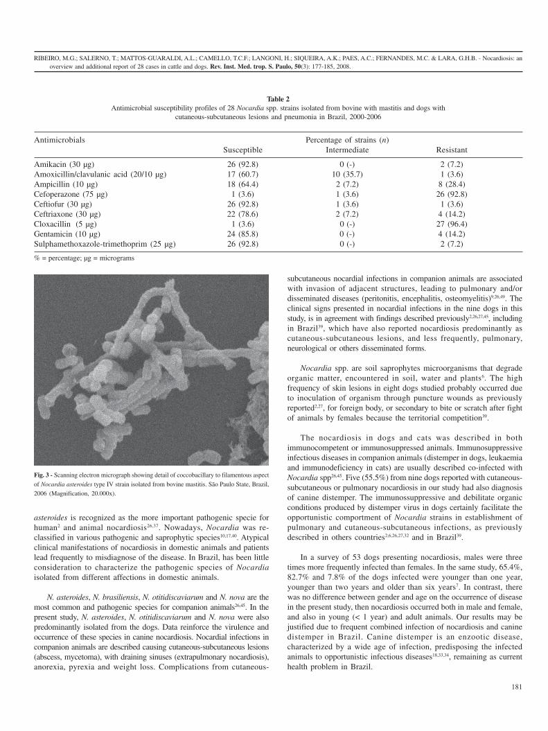
Whitish to orange, circular, dry, convex colonies, with powderysurface firmly adherent to agar surface, were observed in sheep bloodagar after 72-96 hours of microbiological culture, obtained from bovinemilk, bronchial lavage and aspired secretion from cutaneous-subcutaneous lesions in dogs. Likewise, dry, whitish to orange, irregularcolonies were isolated in Sabouraud dextrose agar, after 96 hours ofincubation. Gram-positive, partially acid-fast (MZN-positive),filamentous to cocobacillary organisms were identified in both media.Growth of microorganisms was not observed in MacConkey agar.Aspired material from cutaneous-subcutaneous lesions from dogsrevealed long filamentous organisms with tendency to fragmentationin cocobacillary forms, with poor dense mats of microorganisms,suggestive of Nocardia spp. (Fig. 2). The scanning electron microscopyrevealed detail of filamentous to cocobacillary organisms of Nocardiaasteroides type IV isolated from bovine mastitis (Fig. 3).
Sulphamethoxazole-trimethoprim (92.8%), amikacin (92.8%),ceftiofur (92.8%) and gentamicin (85.8%) were the most effective drugsfor 28 Nocardia clinical isolates. The strains showed high levels ofresistance to cloxacillin (96.4%), cefoperazone (92.8%) and ampicillin(28.4%) (Table 2). Multiple drug resistance to three or more and fiveor more antimicrobials was observed in ten (35.7%) and three (10.7%)strains, respectively, predominantly associated to the use of cloxaxillin,cefoperazone and ampicillin.
DISCUSSION
Currently, nocardiosis has been considered an emerging diseaseamong humans and domestic animals worldwide. Classically, N.
Fig. 1 - Fluctuant mass containing purulent secretion in a dog with cutaneous-subcutaneous
lesion, caused by Nocardia otitidiscaviarum. São Paulo State, Brazil, 2005.
Fig. 2 - Gram-positive, filamentous organisms with tendency to fragmentation in
coccobacillary forms of Nocardia otitidiscaviarum obtained by biopsy of cutaneous-
subcutaneous lesion in a dog. São Paulo State, Brazil, 2005 (Gram, Magnification 1000x).

180
RIBEIRO, M.G.; SALERNO, T.; MATTOS-GUARALDI, A.L.; CAMELLO, T.C.F.; LANGONI, H.; SIQUEIRA, A.K.; PAES, A.C.; FERNANDES, M.C. & LARA, G.H.B. - Nocardiosis: anoverview and additional report of 28 cases in cattle and dogs. Rev. Inst. Med. trop. S. Paulo, 50(3): 177-185, 2008.
Table 1 Clinico-epidemiological aspects and species (type) characterization of 28 Nocardia strains identified in cattle and dogs. Brazil, 2000 - 2006
Number Age Year Animal/Gender Clinical Combined Outcome Type classification(months) manifestation infection
01 > 48 2000 Bovine/Female Clinical mastitis No Unknown Nocardia otitidiscaviarum02 > 48 2001 Bovine/Female Clinical mastitis No Unknown Nocardia asteroides type III
(N. nova)03 > 48 2001 Bovine/Female Clinical mastitis No Unknown Nocardia asteroides type III
(N. nova)04 > 48 2001 Bovine/Female Subclinical mastitis No Unknown Nocardia asteroides type IV05 > 48 2001 Bovine/Female Clinical mastitis No Unknown Nocardia asteroides type IV06 > 48 2003 Bovine/Female Clinical mastitis No Unknown Nocardia asteroides type V
(N. farcinica)07 > 48 2003 Bovine/Female Clinical mastitis No Unknown Nocardia asteroides type V
(N. farcinica)08 > 48 2003 Bovine/Female Subclinical mastitis No Unknown Nocardia asteroides type III
(N. nova)09 > 48 2003 Bovine/Female Clinical mastitis No Unknown Nocardia asteroides type III
(N. nova)10 > 48 2003 Bovine/Female Clinical mastitis No Unknown Nocardia asteroides type III
(N. nova)11 > 48 2003 Bovine/Female Clinical mastitis No Unknown Nocardia asteroides type III
(N. nova)12 > 48 2003 Bovine/Female Clinical mastitis No Unknown Nocardia asteroides type III
(N. nova)13 > 48 2003 Bovine/Female Clinical mastitis No Unknown Nocardia otitidiscaviarum14 > 48 2004 Bovine/Female Clinical mastitis No Unknown Nocardia otitidiscaviarum15 > 48 2005 Bovine/Female Clinical mastitis No Unknown Nocardia asteroides type IV16 > 48 2006 Bovine/Female Clinical mastitis No Unknown Nocardia asteroides type IV17 > 48 2006 Bovine/Female Subclinical mastitis No Unknown Nocardia asteroides type IV18 > 48 2006 Bovine/Female Clinical mastitis No Unknown Nocardia asteroides type IV19 > 48 2006 Bovine/Female Clinical mastitis No Unknown Nocardia asteroides type IV20 48 2000 Canine/Female Cutaneous- Canine Dead Nocardia otitidiscaviarum
subcutaneous distemperabscesses
21 48 2000 Canine/Female Cutaneous- No Dead Nocardia otitidiscaviarumsubcutaneous
abscesses22 48 2000 Canine/Female Cutaneous- No Unknown Nocardia otitidiscaviarum
subcutaneousabscesses
23 24 2001 Canine/Male Cutaneous- No Unknown Nocardia otitidiscaviarumsubcutaneous
abscesses24 > 48 2001 Canine/Female Pneumonia Canine Dead Nocardia asteroides type III
distemper (N. nova)25 06 2001 Canine/Male Cutaneous- Canine Alive Nocardia otitidiscaviarum
subcutaneous distemperabscesses
26 03 2001 Canine/Female Cutaneous- Canine Dead Nocardia otitidiscaviarumsubcutaneous distemper
abscesses27 > 48 2001 Canine/Male Cutaneous- Canine Dead Nocardia asteroides type IV
subcutaneous distemperabscesses
28 06 2001 Canine/Male Cutaneous- No Unknown Nocardia otitidiscaviarumsubcutaneous
abscesses
RIBEIRO, M.G.; SALERNO, T.; MATTOS-GUARALDI, A.L.; CAMELLO, T.C.F.; LANGONI, H.; SIQUEIRA, A.K.; PAES, A.C.; FERNANDES, M.C. & LARA, G.H.B. - Nocardiosis: anoverview and additional report of 28 cases in cattle and dogs. Rev. Inst. Med. trop. S. Paulo, 50(3): 177-185, 2008.
181
asteroides is recognized as the more important pathogenic specie forhuman2 and animal nocardiosis26,37. Nowadays, Nocardia was re-classified in various pathogenic and saprophytic species10,17,40. Atypicalclinical manifestations of nocardiosis in domestic animals and patientslead frequently to misdiagnose of the disease. In Brazil, has been littleconsideration to characterize the pathogenic species of Nocardiaisolated from different affections in domestic animals.
N. asteroides, N. brasiliensis, N. otitidiscaviarum and N. nova are themost common and pathogenic species for companion animals26,45. In thepresent study, N. asteroides, N. otitidiscaviarum and N. nova were alsopredominantly isolated from the dogs. Data reinforce the virulence andoccurrence of these species in canine nocardiosis. Nocardial infections incompanion animals are described causing cutaneous-subcutaneous lesions(abscess, mycetoma), with draining sinuses (extrapulmonary nocardiosis),anorexia, pyrexia and weight loss. Complications from cutaneous-
subcutaneous nocardial infections in companion animals are associatedwith invasion of adjacent structures, leading to pulmonary and/ordisseminated diseases (peritonitis, encephalitis, osteomyelitis)9,26,49. Theclinical signs presented in nocardial infections in the nine dogs in thisstudy, is in agreement with findings described previously2,26,27,45, includingin Brazil39, which have also reported nocardiosis predominantly ascutaneous-subcutaneous lesions, and less frequently, pulmonary,neurological or others disseminated forms.
Nocardia spp. are soil saprophytes microorganisms that degradeorganic matter, encountered in soil, water and plants6. The highfrequency of skin lesions in eight dogs studied probably occurred dueto inoculation of organism through puncture wounds as previouslyreported2,27, for foreign body, or secondary to bite or scratch after fightof animals by females because the territorial competition39.
The nocardiosis in dogs and cats was described in bothimmunocompetent or immunosuppressed animals. Immunosuppressiveinfectious diseases in companion animals (distemper in dogs, leukaemiaand immunodeficiency in cats) are usually described co-infected withNocardia spp26,45. Five (55.5%) from nine dogs reported with cutaneous-subcutaneous or pulmonary nocardiosis in our study had also diagnosisof canine distemper. The immunossuppressive and debilitate organicconditions produced by distemper virus in dogs certainly facilitate theopportunistic comportment of Nocardia strains in establishment ofpulmonary and cutaneous-subcutaneous infections, as previouslydescribed in others countries2,6,26,27,32 and in Brazil39.
In a survey of 53 dogs presenting nocardiosis, males were threetimes more frequently infected than females. In the same study, 65.4%,82.7% and 7.8% of the dogs infected were younger than one year,younger than two years and older than six years7. In contrast, therewas no difference between gender and age on the occurrence of diseasein the present study, then nocardiosis occurred both in male and female,and also in young (< 1 year) and adult animals. Our results may bejustified due to frequent combined infection of nocardiosis and caninedistemper in Brazil. Canine distemper is an enzootic disease,characterized by a wide age of infection, predisposing the infectedanimals to opportunistic infectious diseases18,33,34, remaining as currenthealth problem in Brazil.
Table 2Antimicrobial susceptibility profiles of 28 Nocardia spp. strains isolated from bovine with mastitis and dogs with
cutaneous-subcutaneous lesions and pneumonia in Brazil, 2000-2006
Antimicrobials Percentage of strains (n)Susceptible Intermediate Resistant
Amikacin (30 µg) 26 (92.8) 0 (-) 2 (7.2)Amoxicillin/clavulanic acid (20/10 µg) 17 (60.7) 10 (35.7) 1 (3.6)Ampicillin (10 µg) 18 (64.4) 2 (7.2) 8 (28.4)Cefoperazone (75 µg) 1 (3.6) 1 (3.6) 26 (92.8)Ceftiofur (30 µg) 26 (92.8) 1 (3.6) 1 (3.6)Ceftriaxone (30 µg) 22 (78.6) 2 (7.2) 4 (14.2)Cloxacillin (5 µg) 1 (3.6) 0 (-) 27 (96.4)Gentamicin (10 µg) 24 (85.8) 0 (-) 4 (14.2)Sulphamethoxazole-trimethoprim (25 µg) 26 (92.8) 0 (-) 2 (7.2)
% = percentage; µg = micrograms
Fig. 3 - Scanning electron micrograph showing detail of coccobacillary to filamentous aspect
of Nocardia asteroides type IV strain isolated from bovine mastitis. São Paulo State, Brazil,
2006 (Magnification, 20.000x).

182
RIBEIRO, M.G.; SALERNO, T.; MATTOS-GUARALDI, A.L.; CAMELLO, T.C.F.; LANGONI, H.; SIQUEIRA, A.K.; PAES, A.C.; FERNANDES, M.C. & LARA, G.H.B. - Nocardiosis: anoverview and additional report of 28 cases in cattle and dogs. Rev. Inst. Med. trop. S. Paulo, 50(3): 177-185, 2008.
N. asteroides, N. farcinica, N. nova and N. otitidiscaviarum werespecies of microorganism identified in cattle with mastitis in this trial.This result is in agreement with authors who also have described N.asteroides, N. farcinica and N. nova as most common causal agents ofnocardial mastitis in domestic ruminants37, including in Brazil20,42.However, there was no report of bovine mammary infections by N.otitidiscaviarum in the literature reviewed. N. otitidiscaviarum usuallyis described in cats26,31 and humans12. Thus, bovine mammary infectionby N. otitidiscaviarum is reported by the first time in Brazil.
Classically, etiological agents of mastitis in domestic ruminantsare classified in contagious or environmental microorganisms, relatedfrequently in subclinical and clinical presentations, respectively4,37,42.Intramammary infection in domestic ruminants is recognized as themost frequent clinical presentation of nocardiosis, usually affectingone or two cattle in the herd37. In the present study, nocardial infectionsin cows also were identified in one or two animals in dairy herds,predominantly causing clinical mastitis, presenting edema, fibrosis,enlargement to udder, draining sinuses, and milk containing serous topurulent secretion and white particles (“sulfur granules”).
Commonly clinical cases of nocardial mastitis are characterizedby history of chronic evolution, and refractory to conventional therapy.Mammary infections are associated with inadequate management ofmilking, especially by soil contamination of udder washing, teat-dipprocedure or intramammary infusions in dry period20,37,42. Cattlediagnosed with nocardiosis in the present study were also raised indairy herds with serious hygienic problems in the management of milk,including excess of organic matter in the environment between milking,inadequate concentrations of anti-septic on teat dips, inappropriatehygienic conditions of water used for udder washing and inadequateaseptic procedure for antimicrobial udder infusions, especially in drycow therapy. Therefore, poor hygiene conditions in milking in dairyherds certainly contribute on the transmission of Nocardia bycontamination of water and equipments, or accidental inoculation oforganism in udder infusions. The efficacy of therapy in nocardialmastitis is uncertain. The microorganism present intracellularlocalization, is refractory to conventional antimicrobials, and induceto pyogranulomatous process, extensive fibrosis and glandular damage,that difficult the resolution of mammary inflammatory process6,37.
Routine microbiological diagnosis of the genus Nocardia isrelatively simple. The organism is Gram-positive, filamentous withtendency to fragmentation, partial acid-fast, isolated in ovine or bovineblood agar and Sabouraud agar, aerobically, after 48 to 72 hours.Microorganisms are usually recovered in pure culture and colonies aresmooth or rough, pigmented (white, cream, orange, red tonalities),with characteristic powdery surface, like fungal organisms. However,confirmation of diagnosis and species characterization depend ofbiochemical characterization, based on hydrolysis of variouscarbohydrates and other substrates10,28,36, and more recently, by use ofmolecular techniques17,40.
Different authors have indicated skin biopsy procedure in diagnosisof companion animals suspect of nocardiosis26,27,45, including in Brazil39.The aspiration material from cutaneous-subcutaneous lesions in eightdogs allow either cytological identification of Nocardia using differentstaining methods (Gram, Giemsa), simultaneously to the
microbiological culture of organism. Fine needle aspiration of dogswith cutaneous-subcutaneous lesions is characterized by technique ofsimple execution, low costs, reduced tecidual damage, and enable fastand accurate previous diagnosis of cutaneous-subcutaneous nocardialinfection.
In companion animals, differential diagnosis is recommended forActinomyces species because the microbiological appearance andclinical similarities of this agent with Nocardia spp.26,27,45. However,Actinomyces spp. is characterized by facultative anaerobic, non-acid-fast, frequently encountered in dense mats of long filamentousorganisms in microbiologic and cytologic exams29,36. Thus, the presenceof Gram-positive, aerobic, partial acid-fast, long filamentous organismswith tendency to fragmentation in cocobacillary forms, identified incutaneous-subcutaneous or disseminated infections in companionanimals is suggestive of nocardiosis26,27,39. In contrast, Actinomycesspp. do not represent a problem as a causal agent of mastitis in domesticruminants37. Scanning electron microscopy enabled to observe detailsof filamentous organisms with tendency to fragmentation, characteristicfrom the genus Nocardia, in strain isolated from bovine mastitis (Fig.2), confirming the diagnosis of nocardiosis. However, this method rarelyhas been used on routine diagnosis of animal nocardiosis6, due torepresent expensive technique and/or by difficulties in accesses tolaboratories that development routine of electron microscopy.
Usually nocardial therapy involves prolonged use of antimicrobialscombined with surgical debridation/drainage, extirpation of foreignbodies and lavage of infected lesions using anti-septicsolutions2,10,19,25,26,30. Long-term antimicrobial therapy is recommendeddue to intracellular localization of agent, development ofpyogranulomatous inflammatory reaction and resistant pattern strainsthat lead to poor response to the conventional antimicrobialtreatment10,19,32. In companion animals, surgical care procedures arespecially indicated in cutaneous-subcutaneous, peritonitis andosteomyelitis manifestations9,27,45,49. Prognosis is uncertain, consideredusually good in localized lesions (cutaneous-subcutaneous), but poorto guard in disseminated (systemic), pulmonary and neurologicalinvolvement in dogs and cats2,26,32,45,49 and mastitis in domesticruminants37,42.
Trimethoprim-sulfonamides combinations, aminoglycosides(amikacin), linezolid and late generation beta-lactam compounds(cefotaxime, ceftriaxone, imipenem) used isolated or combined aredrugs of choice for nocardial therapy in humans1,10 and domesticanimals26,45. The duration of therapy is uncertain in virtue of relapsesafter use of shorter course protocols. In companion animals, the therapyin cutaneous lesions is recommended generally from one to threemonths2,26,27, while in pulmonary, neurological or other systemicinfections in animals and human (especially immunocompromisedpatients), for six to 12 months3,26. Additionally, prolonged use of certainantimicrobials (sulfonamides, aminoglycosides) may induce adverseor toxic reactions in nocardial therapy 26,32 .
Efficacy of antimicrobial therapy in companion animals iscontroversial. Successful management of nocardial empyema in a dogand cat was reported11. In contrast, unsuccessful therapy of seven dogswith nocardiosis was described in USA2. Likewise, poor success ontherapy of nine from ten dogs with nocardiosis, using trimethoprim-

RIBEIRO, M.G.; SALERNO, T.; MATTOS-GUARALDI, A.L.; CAMELLO, T.C.F.; LANGONI, H.; SIQUEIRA, A.K.; PAES, A.C.; FERNANDES, M.C. & LARA, G.H.B. - Nocardiosis: anoverview and additional report of 28 cases in cattle and dogs. Rev. Inst. Med. trop. S. Paulo, 50(3): 177-185, 2008.
183
sulfonamides or cephalosporins was reported in Brazil39. In despite ofaggressive fluid therapy, surgical drainage and antimicrobial treatmentbased on in vitro antimicrobial susceptibility tests, five from nine dogsstudied dead reinforced the uncertain prognosis and the difficulties onestablishment of therapy in canine nocardiosis.
Nocardia spp. organisms show large variation in susceptibility invitro to different antimicrobials. Sulfonamides-trimethoprim, amikacinand cephalosporins commonly show efficacy in human3,25 and animalstrains26,45. Antimicrobial susceptibility tested in 28 Nocardia strainsin this trial showed that amikacin, ceftiophur, sulphamethoxazole-trimethoprim and gentamicin were the most-effective drugs. Theseresults are in agreement with other authors that have indicatedsulfonamides combined with trimethoprim, aminoglycosides andcephalosporins as drugs of choice in nocardial therapy in domesticanimals2,26,27,32,37,39,45. However, these results should be interpreted withcare because in vitro susceptibility do not predict necessarily in vivosuccess in nocardial therapy 25,39 .
Multiple drug resistance in Nocardia strains was observed mainlyfor cloxacillin, cefoperazone and ampicillin. Interestingly, these drugsfrom beta-lactam group and derivates that displayed poor efficacy inthe Nocardia strains, represent the principal antimicrobialscommercialized for bovine intramammary therapy in lactation and dryperiod in Brazil4. Thus, the limitation of use of these antimicrobialsdifficult the establishment of intramammary therapy procedures fornocardial mastitis in domestic ruminants, reinforcing the eliminationof quarters or animals infected by Nocardia as control measure ofmammary infections in dairy herds.
Since last taxonomic changes, few studies were conducted ininvestigation of in vitro susceptibility profile and multiple drug resistanceof pathogenic Nocardia strains isolated from domestic animals. Nocardiaspp. strains resistant to beta-lactam group and derivates, fluoroquinolones,macrolides and aminoglycosides have been reported25,26. The 28 Nocardiastrains isolated from cattle and dogs in this trial show multiple drugresistance especially using beta-lactam group and derivates. These resultsconfirm the importance to perform susceptibility antimicrobial testsbefore initiate nocardial therapy, to increase the efficacy of treatment39.Therefore, success on therapy of nocardiosis depends on several factorsincluding virulence of strain, precocious diagnosis, severity andlocalization of the infection, host immune status, efficacy and potentialinteractions of antimicrobials used in therapy3,26.
Human nocardiosis is recognized as an opportunistic disease thatmay occur in either immunocompetent or immunocompromisedpeoples1. In the United States, human nocardiosis was recognized atleast in 1,000 cases each year6. Clinical cases are intimately related toconcurrent immunosuppressive diseases, especially patients committedby aids, organ transplantation, malignancy illness (lymphoma,lymphosarcoma), cirrhosis, diabetes, alcoholism, or prolonged use ofcorticosteroids1,3,6,19. However, 384 from 1,000 cases of nocardiosisdescribed since 1950, occurred in patients without combined illness oridentifiable predisposing factors, representing therefore, primaryinfections in health human6. Clinically, human nocardiosis is manifestedoften by pulmonary, cutaneous-subcutaneous (mycetoma) andneurological signs1,15,30. N. asteroides, N. brasiliensis, N. farcinica andN. nova are species frequently associated to human disease2,19,30,44.
The environment is the natural reservoir for both human and animalnocardiosis and suggests that majority transmission of Nocardia spp.for people occur by inhalation of the organism in dry warm climateregions that facilitate the aerosolization and dispersal of pathogen,enhancing the infection by respiratory route. A smaller number ofinfections are caused by traumatic skin inoculation of organism fromsoil. Human nocardiosis probably are not thought to be transmittedfrom person-to-person form, or acquired nosocomially6,44. Furthermore,remains unclear the impact of domestic animals as primary reservoirof pathogenic Nocardia spp. strains for human1,6. However, severalcases of cutaneous-subcutaneous infections transmitted to people bybite or scratch from clinically health dogs and cats have beendescribed1,8,26,41. Nocardia spp. are soil saprophytes, opportunisticmicroorganisms, characterized by infections in several domesticanimals. Thus, precautions should be attempted in groups committedby immune dysfunction (patients with aids, organ transplantation,cancer or receiving immunosuppressive drug therapy), with specialattention to contact with soil or organic matter contaminated bydomestic animals, and management of domestic animals suspected ofnocardiosis.
In Brazil, human nocardiosis is recognized as an infrequent disease.Mycetoma and pulmonary infections appears to be predominant clinicalmanifestations of nocardiosis in this country, followed by neurologicsigns10,15,35. Severe mycetoma produced by N. asteroides was reportedin a 9-year-old child that in virtue of severity and evolution of theinflammatory process was submitted to amputation member forresolution of infectious process43. Clinical and epidemiological aspectsof 41 cases of mycetoma in the city of São Paulo revealed that 68%were caused by actinomycetes, and from these, N. brasiliensis wasresponsible by 13 cases13. Among 71 cases of death in renal transplantedpatients, Nocardia sp. was isolated in 10.6%38. Recently, twenty-twocases of nocardial infections diagnosed in Porto Alegre city between1977-1998 were retrospectively reviewed. This study revealed that sixcases were caused by N. asteroides complex, one N. asteroides sensustrictu, one N. brasiliensis and fourteen Nocardia spp., predominantlyin immunosuppressed patients (15 cases) and presenting lung infection(17 cases)15. Interestingly, in the present study was observed greatsimilarity between Nocardia species (type) isolated from cattle anddogs, and more–common species described by others authors in humannocardiosis1,19,28,30,44.
Besides infrequent mammary infections, study attempted todetermine the temperature resistance of N. asteroides strains isolatedfrom bovine mastitis in Brazil, and revealed that microorganismsupported experimental time/temperature conditions usually employedin pasteurization (65 oC/30 minutes and 75 oC/20 seconds). All eightstrains showed resistance to 65 oC/30 minutes and seven to 75 oC/20seconds, alerting for thermo-tolerance of N. asteroides to pasteurizationprocedures, and potential risk of transmission of microorganism bymilk and derivates21.
In conclusion, nocardiosis is an unusual opportunistic disease inhuman and domestic animals, transmitted predominantly fromenvironment. None specific control measures are recommended fornocardiosis in companion animals. However, hygienic environmentalconditions, early diagnosis, appropriate initial therapy and control ofpredisposing factors (immunosuppressive diseases) may decrease the

184
RIBEIRO, M.G.; SALERNO, T.; MATTOS-GUARALDI, A.L.; CAMELLO, T.C.F.; LANGONI, H.; SIQUEIRA, A.K.; PAES, A.C.; FERNANDES, M.C. & LARA, G.H.B. - Nocardiosis: anoverview and additional report of 28 cases in cattle and dogs. Rev. Inst. Med. trop. S. Paulo, 50(3): 177-185, 2008.
mortality rates in disease. From a similar way, no specific controlprocedures are indicated to nocardial mastitis. However, properlyhygiene conditions at milking, appropriate clean for intramammaryinfusions, early mastitis diagnosis, chemical drying of affected quarteror elimination of the infected animals, remains as the best methods ofthe control of nocardial mastitis in domestic ruminants. Due to lowprevalence and atypical clinical manifestations, the disease is usuallymisdiagnosed. The prognosis and efficacy of therapy is uncertain, andantimicrobials are used by long time. Great similarity was observedbetween Nocardia species identified in the dogs and cattle studied,with isolates described previously in human nocardiosis. Bovineintramammary infections by N. otitidiscaviarum are reported by firsttime in Brazil. Studies of type characterization, virulence, clinical-epidemiological features and predisposing factors from Nocardia strainsisolated from human, animal and environmental origin may elucidaterisk factors related to nocardial infections.
RESUMO
Nocardiose: visão geral e relato de 28 casos em vacas e cães
A caracterização fenotípica, perfil de sensibilidade aosantimicrobianos e aspectos clínico-epidemiológicos foram avaliadosem 28 linhagens de Nocardia isoladas de 19 casos de mastite, oitolesões tegumentares e um caso de pneumonia em cão. Foram utilizadosno diagnóstico métodos microbiológicos, bioquímicos, citológicos emicroscopia eletrônica de varredura. Nocardia asteroides tipo IV, N.otitidiscaviarum, N. nova (tipo III) e N. farcinica (tipo V) foram isoladasdo leite de vacas com mastite, de material de lavado transtraqueal e delesões cutâneas de cães. Nocardiose mamária bovina foi diagnosticadapredominantemente sob a forma clínica, em propriedades com precáriascondições de higiene na pré e pós-ordenha, e inadequado procedimentode terapia intramamária. Nocardiose canina foi diagnosticadacomumente em animais co-infectados com o vírus da cinomose.Sulfametoxazole/trimetoprim (92,8%), amicacina (92,8%) e ceftiofur(92,8%) foram os antimicrobianos mais efetivos frente às linhagensde Nocardia. Resistência múltipla a três ou mais e cinco ou maisantimicrobianos foram observadas, respectivamente, em dez (35,7%)e três (10,7%) linhagens, notadamente frente à cloxacilina,cefoperazona e ampicilina. A caracterização de espécies (tipo), aspectosclínico-epidemiológicos, diagnóstico, resistência múltipla aosantimicrobianos e reflexos em saúde pública de linhagens de Nocardiaisoladas de bovinos e cães no Brasil foram discutidos. Foi destacada asimilaridade entre as espécies de Nocardia isoladas de animais e dohomem, e a primeira descrição no Brasil de N. otitidiscaviarum naetiologia da mastite bovina.
REFERENCES
1. ACHA, P.N. & SZYFRES, B. - Nocardiosis. In:___ Zoonosis y enfermedadestransmisibles communes al hombre y a los animals. 3. ed. Washington,Organización Panamericana de la Salud, 2003. v.1, p.212-216.
2. ACKERMAN, N.; GRAIN Jr., E. & CASTLEMAN, W. - Canine nocardiosis. J. Amer.Anim. Hosp. Ass., 18: 147-153, 1982.
3. AGTEROF, M.J.; VAN DER BRUGGEN, T.; TERSMETTE, M. et al. - Nocardiosis: acase series and a mini review of clinical and microbiological features. Neth. J. Med.,65: 199-202, 2007.
4. ANDRADE, S.F.; GIUFFRIDA, R. & RIBEIRO, M.G. - Quimioterápicos, antimicrobianose antibióticos. In: ANDRADE, S.F. Manual de terapêutica veterinária. 2. ed. SãoPaulo, Roca, 2002. p.13-58.
5. BAUER, A.W.; KIRBY, W.M.; SHERRIS, J.C. & TURCK, M. - Antibiotic susceptibilitytesting by a standardized single disk method. Amer. J. clin. Path., 45: 493-496,1966.
6. BEAMAN, B.L. & BEAMAN, L. - Nocardia species: host-parasite relationships. Clin.Microbiol. Rev., 7: 213-264, 1994.
7. BEAMAN, B.L. & SUGAR, A.M. - Nocardia in naturally acquired and experimentalinfections in animals. J. Hyg. (Lond.), 91: 393-419, 1983.
8. BOTTEI, E.; FLAHERTY, J.P.; KAPLAN, L.J. & DUFFEE–KERR, L. -Lymphocutaneous Nocardia brasiliensis infection via cat scratch: a second case.Clin. infect. Dis., 18: 649-650, 1994.
9. BRADNEY, I.W. - Vertebral osteomyelitis due to Nocardia in a dog. Aust. vet. J., 62:315-316, 1985.
10. BROWN, J.M. & McNEIL, M.M. - Nocardia, Rhodococcus, Gordonia, Actinomadura,Streptomyces, and other aerobic actinomycetes In: MURRAY, P.R.; BARON, E.J.;JORGENSEN, J.H.; PFALLER, M.A. & YOLKEN, R.H. Manual of clinicalMicrobiology. 8. ed. Washington, ASM Press, 2003. p. 502-531.
11. CAMPBEL, B. & SCOTT, D.W. - Successful management of nocardial empyema in adog and cat. J. Amer. Anim. Hosp. Ass., 11: 769-773, 1975.
12. CASTELLI, L.; ZLOTNIK, H.; PONTI, R. & VIDOTTO, V. - First reported Nocardiaotitidiscaviarum infection in an AIDS patient in Italy. Mycopathologia (Den Haag),126: 131-136, 1994.
13. CASTRO, L.G.; BELDA JÚNIOR, W.; SALEBIAN, A. & CUCÉ, L.C. - Mycetoma: aretrospective study of 41 cases seen in São Paulo, Brazil, from 1978 to 1989. Mycoses,36: 89-95, 1993.
14. CASTRO, L.G.M.; VALENTE, N.Y.S.; GERMANO, J.A.M.; VACCARI, E.M. & daSILVA LACAZ, C. - Mycetoma in an HIV-infected patient. Rev. Hosp. Clín. Fac.Med. S. Paulo, 54: 169-171, 1999.
15. CHEDID, M.B.; CHEDID, M.F.; PORTO, N.S.; SEVERO, C.B. & SEVERO, L.C. -Nocardial infections: report of 22 cases. Rev. Inst. Med. trop. S. Paulo, 49: 239-246, 2007.
16. CLINICAL AND LABORATORY STANDARDS INSTITUTE - Performancestandards for antimicrobial disk susceptibility test. 8. ed. Pennsylvania, CLSI-NCCLS, 2003.
17. CONVILLE, P.S. & WITEBSKY, F.G. - Current issues pertaining to the Nocardia species.Clin. Microbiol. Newsl., 26: 57-62, 2004.
18. CORRÊA, W.M. & CORRÊA, C.N.M. - Nocardioses. In: ___. Enfermidades infecciosasdos mamíferos domésticos. Rio de Janeiro, Medsi, 1992. p. 355-360.
19. CORTI, M.E. & VILLAFAÑE–FIOTI, M.F. - Nocardiosis: a review. Int. J. infect. Dis.,7: 243-250, 2003.
20. COSTA, E.O.; MACEDO, M.M.; COUTINHO, S.D. et al. - Isolamento de Actinomicetalesaeróbios do gênero Nocardia de processos infecciosos dos animais domésticos. Rev.Fac. Med. vet. Univ. S. Paulo, 24: 17-21, 1987.
21. COSTA, E.O.; RIBEIRO, A.R.; RIBEIRO, M.G. et al. - Nocardia sp. strains isolatedfrom clinical and subclinical bovine mastitis: evaluation of the thermic resistance onthe milk pasteurization (temperatura/time). In: WORLD BUIATRIC CONGRESS,19., Edinburgh, 1996. Proceedings. p. 206-207.
22. COSTA, E.O.; RIBEIRO, A.R.; WATANABE, E.T. & MELVILLE, P.A. - Infectious bovinemastitis caused by environment organisms. Zbl. Veterinarmed. B, 45: 65-71, 1998.

RIBEIRO, M.G.; SALERNO, T.; MATTOS-GUARALDI, A.L.; CAMELLO, T.C.F.; LANGONI, H.; SIQUEIRA, A.K.; PAES, A.C.; FERNANDES, M.C. & LARA, G.H.B. - Nocardiosis: anoverview and additional report of 28 cases in cattle and dogs. Rev. Inst. Med. trop. S. Paulo, 50(3): 177-185, 2008.
185
23. COSTA, E.O.; RIBEIRO, A.R.; WATANABE, E.T. et al. - An increased incidence ofmastitis caused by Prototheca species and Nocardia species on a farm in São Paulo,Brazil. Vet. Res. Comun., 20: 237-241, 1996.
24. COSTA, E.O.; RIBEIRO, M.G.; RIBEIRO, A.R. et al. - Diagnosis of clinical bovinemastitis by fine needle aspiration followed by staining and scanning electronmicroscopy in a Prototheca zopfii outbreak. Mycopathologia, 158: 81-85, 2004.
25. GLUPCZYNSKI, Y.; BERHIN, C.; JANSSENS, M. & WAUTERS, G. - Determinationof antimicrobial susceptibility patterns of Nocardia spp. from clinical specimens byEtest. Clin. Microbiol. Infect., 12: 905-912, 2006.
26. GREENE, C.E. - Actinomycosis and nocardiosis. In:___. Infectious diseases of the dogand cat. 3. ed. Philadelphia, Saunders; Elsevier, 2006. p. 451-461.
27. KIRPENSTEIJN, J. & FINGLAND, R.B. - Cutaneous actinomycosis and nocardiosis indogs: 48 cases (1980-1990). J. Amer. vet. med. Ass., 201: 917-920, 1992.
28. KISKA, D.L.; HICKS, K. & PETTIT, D.J. - Identification of medically relevant Nocardiaspecies with an abbreviated battery of test. J. clin. Microbiol., 40: 1346-1351, 2002.
29. KONEMAN, E.W. - Nocardioforms and aerobic actinomycetes. In: __.Color atlas andtextbook of diagnostic microbiology. 4. ed. Philadelphia, Lippincott, 1997. p. 691-695.
30. LERNER, P.I. - Nocardiosis. Clin. infect. Dis., 22: 891-905, 1996.
31. LUQUE, R.; ASTORGA, C. & TARRADAS, B. - Nocardia otitidiscaviarum infection ina cat. Vet. Rec., 151: 488, 2002.
32. MARINO, D.J. & JAGGY, A. - Nocardiosis. A literature review with selected case reportsin two dogs. J. Vet. intern. Med., 7: 4-11, 1993.
33. MORETTI, L.D.; DA SILVA, A.V.; RIBEIRO, M.G.; PAES, A.C. & LANGONI, H. -Toxoplasma gondii genotyping in a dog coinfected with distemper virus andehrlichiosis rickettsia. Rev. Inst. Med. trop. S. Paulo, 48: 359-363, 2006.
34. MORETTI, L.D.; UENO, T.E.; RIBEIRO, M.G. et al. - Toxoplasmose em cães co-infectados com o vírus da cinomose. Semina, 23: 85-92, 2002.
35. PETRILLO, V.F.; SEVERO, L.C.; LONDERO, A.T. & PORTO, N.S. - Pulmonarynocardiosis report of the first two Brazilian cases. Mycopathologia (Den Haag),66: 17-20, 1978.
36. QUINN, P.J.; CARTER, M.E.; MARKEY, B.K. & CARTER, G.R. - The Actinomycetes.In:__. Clinical veterinary Microbiology. London, Wolfe, 1994. p. 144-155.
37. RADOSTITS, O.M.; GAY, C.C.; HINCHCLIFF, K.W. & CONSTABLE, P.D. - Diseasesof the mammary gland. In:__.Veterinary Medicine: a textbook of the diseases ofcattle, horses, sheep, pigs, and goats. 10. ed. Philadelphia, Saunders; Elsevier, 2007.p. 673-762.
38. REIS, M.A.; COSTA, R.S. & FERRAZ, A.S. - Causes of death in renal transplantrecipients: a study of 102 autopsies from 1968 to 1991. J. roy. Soc. Med., 88: 24-27,1995.
39. RIBEIRO, M.G.; AGUIAR, D.M.; PAES, A.C. et al. - Nocardiose cutânea associada àcinomose em cães. Relato de dez casos. Rev. Clín. vet., 39: 34-42, 2002.
40. ROTH, A.; ANDRESS, S.; KROPPENSTEDT, R.M.; HARMSEN, D. & MAUCH, H. -Phylogeny of the genus Nocardia based on reassessed 16S rDNA gene sequencesreveals underspeciation and division of strains classified as Nocardia asteroides intothree established species and to unnamed taxons. J. clin. Microbiol., 41: 851-856,2003.
41. SACHS, M.K. - Lymphocutaneous Nocardia brasiliensis infection acquired from a catscratch: case report and review. Clin. infect. Dis., 15: 710-711, 1992.
42. SANTOS, M.V. & FONSECA, L.F.L. - Principais agentes causadores de mastite. In:___. Estratégias para controle de mastite e melhoria da qualidade do leite. SãoPaulo, Manole, 2007. p.24–37.
43. SARAÇA, G.D.; TOWERSEY, L.; HAY, R.J. et al. - Mycetoma by Nocardia asteroides:a 9 year follow–up. Rev. Inst. Med. trop. S. Paulo, 35: 199-204, 1993.
44. SAUBOLLE, M.A. & SUSSLAND, D. - Nocardiodis: review of clinical and laboratoryexperience. J. clin. Microbiol., 41: 4497-4501, 2003.
45. SCOTT, W.; MILLER, W.H. & GRIFFIN, C.E. - Bacterial skin diseases. In: __. SmallAnimal Dermatology, 6.ed. Philadelphia, W.B. Saunders, 2001. p.323-324.
46. SCHALM, O.W. & NOORLANDER, D.O. - Experimental and observation leading todevelopment of California Mastitis Test. J. Amer. Vet. med. Ass., 139: 199-204,1957.
47. SILVA, A.G., MARTINS, E.M.L.; MARCHIORI, E. et al. - Nocardiose pulmonar empacientes com síndrome da imunodeficiência adquirida - Relato de caso. Radiol.bras., 35: 1-7, 2000.
48. STEINGRUBE, V.A.; BROWN, B.A.; GIBSON, J.L. et al. - DNA amplification andrestriction endonuclease analysis for differentiation of 12 species and taxa ofNocardia, including recognition of four new taxa within the Nocardia asteroidescomplex. J. clin. Microbiol., 33: 3096-3101, 1995
49. TILGNER, S.L. & ANSTEY, S.I. - Nocardial peritonitis in a cat. Aust. Vet. J., 74: 430-432,1996.
Received: 26 October 2007Accepted: 21 February 2008